Article

The Role of ClpV in the Physiology and Pathogenicity of Xanthomonas citri subsp. citri Strain zlm1908

College of Coastal Agricultural Science, Guangdong Ocean University, Zhanjiang 524088, China
*
Author to whom correspondence should be addressed.
Microorganisms 2024, 12(12), 2536; https://doi.org/10.3390/microorganisms12122536
Submission received: 24 October 2024 / Revised: 29 November 2024 / Accepted: 7 December 2024 / Published: 9 December 2024
(This article belongs to the Section Plant Microbe Interactions)

Abstract

:
Xanthomonas citri subsp. citri (Xcc) is a Gram-negative bacterium responsible for citrus canker, a significant threat to citrus crops. ClpV is a critical protein in the type VI secretion system (T6SS) as an ATPase involved in bacterial motility, adhesion, and pathogenesis to the host for some pathogenic bacteria. In order to investigate the function of clpV gene in Xcc, the clpV-deletion strain ΔclpV was constructed, its biological properties were evaluated, and the differences in gene expression levels between the wild-type strain and ΔclpV were analyzed by transcriptomics. The results exhibited significantly reduced biofilm formation, extracellular polysaccharide synthesis, and swarming motility in ΔclpV compared to the wild-type strain. Although the clpV-deletion did not significantly affect bacterial growth or pathogenicity in terms of disease symptoms on citrus leaves, the mutant showed increased sensitivity to environmental stresses (NaCl, SDS, and H2O2) and antibiotics (β-lactams and aminoglycosides). Transcriptome analysis revealed that clpV-deletion altered the expression of motility-related genes and the efflux pump gene mexH. Our findings underscore the importance of ClpV in maintaining biofilm integrity and suggest a multifaceted role in adaptive strategies of Xcc, positioning ClpV as a potential target for mitigating citrus canker disease.

1. Introduction

Xanthomonas citri subsp. citri (Xcc) is a Gram-negative bacterial pathogen that causes citrus canker disease, one of the most serious diseases affecting citrus crops, resulting in significant economic losses for the citrus industry [1]. This pathogen infects host plants through stomata and wounds, invading the intercellular spaces in the apoplast [2]. It produces erumpent, corky necrotic lesions often surrounded by a chlorotic halo on leaves, young stems, and fruits. As the lesions develop, they lead to defoliation, rupture of the leaf epidermis, and premature fruit drop in citrus plants [3].
Currently, Xcc has been extensively studied to elucidate the molecular mechanisms underlying its pathogenesis and interactions with host plants. The type VI secretion system (T6SS) is recognized as a contact-dependent secretion machinery capable of directly injecting toxins into both other bacteria and eukaryotic cells. Several studies have indicated that T6SS serves as a virulence factor in many plant-pathogenic bacteria, including Burkholderia glumae, Xanthomonas oryzae pv. oryzae, and Ralstonia solanacearum [4,5,6]. T6SS delivers antimicrobial or cytotoxic effector proteins to recipient cells through direct contact, exerting toxic effects and serving as an important weapon for most Gram-negative pathogenic bacteria in both environmental competition and host infection [7]. Despite its significance, information about the functions of T6SS in plant pathogens remains limited. T6SS consists of a trans-envelope complex that docks a contractile tail composed of an inner tube made of stacked Hcp (hemolysin co-regulated protein) hexamers, topped by the VgrG (valine-glycine repeat protein G) syringe, and surrounded by a sheath formed from polymerized TssB and TssC subunits, along with Hcp, IcmF (intracellular multiplication protein), VgrG, and ClpV. ClpV is a critical protein in this machinery as an ATPase that forms a hexameric complex, providing the energy required for the secretion of T6SS substrates [8]. Li et al. [9] demonstrated that the release of Hcp1 depends on ATP hydrolysis facilitated by the ClpV domain in Pseudomonas aeruginosa, indicating that the role of ClpV in Hcp1 secretion is energy-dependent.
The clpV (also named tssH) deletion mutation in Xanthomonas phaseoli pv. manihotis has confirmed that the disruption of clpV reduces the resistance to H2O2-induced oxidative stress [10]. In Agrobacterium tumefaciens, clpV deletion resulted in the inability to secrete Hcp into the supernatant, although Hcp secretion was partially restored in the complementary strain [11]. ClpV has been identified as a virulence factor in Pseudomonas plecoglossicida [12,13]. ClpV3 of the H3-type T6SS (H3-T6SS) has been recognized as a multiple virulence factor in P. aeruginosa, where the intracellular concentration of the secondary messenger cAMP was reduced in the PA01 (ΔclpV3) strain [9].
As the core components and effector functions of T6SS, the function of ClpV has still been unknown in Xcc. In this study, we constructed a clpV-deletion strain and studied its biological characteristics, then analyzed the differences in gene expression levels between the wild-type and clpV-deletion strain by transcriptomics.

2. Materials and Methods

2.1. Bacterial Strains, Plasmids, and Experimental Plants

The bacterial strains and plasmids utilized in this study are detailed in Table 1. Xcc zlm1908 was isolated in August 2019 from the leaf of Citrus sinensis ‘hongjiang’ suffering from citrus canker and stored in our laboratory. The strain was cultured at 30 °C in Luria–Bertani (LB) medium. Escherichia coli DH5α and β2163 strains were cultured at 37 °C in LB medium. Healthy 3-year-old citrus trees (C. sinensis ‘hongjiang’) were obtained from Zhanjiang Huacheng Agriculture Development Company (Chikan district, Zhanjiang City, China) and cultivated in 20 cm × 30 cm pots in a greenhouse. The leaves of C. sinensis ‘hongjiang’, known to be susceptible to citrus canker, were used for Xcc infection experiments.

2.2. Cloning and Sequencing of clpV Gene from Xcc zlm1908

Genomic DNA from Xcc zlm1908 (WT) was extracted using the Steadypure Universal Genomic DNA Extraction Kit (AG Accurate Biotechnology, Changsha, China). The primers clpV-CF and clpV-CR (Table 2) were employed to amplify the clpV gene. PCR was conducted under the following conditions: an initial denaturation at 96 °C for 4 min, followed by 30 cycles of 95 °C for 60 s, 55 °C for 45 s, and 72 °C for 45 s, with a final extension at 72 °C for 7 min. The PCR products were purified using the Takara MiniBEST DNA Fragment Purification Kit Ver. 4.0 (Takara, Dalian, China). The purified PCR product was then ligated into the pMD18-T plasmid and sequenced by Sangon Biological Engineering Technology & Services Co., Ltd. (Sangon, Guangzhou, China).

2.3. Construction of the clpV Deletion Mutant

For the construction of ΔclpV, upstream fragments A and downstream fragments B of the clpV gene were amplified using primers clpV-UF/clpV-UR and clpV-DF/clpV-DR, respectively. The fragments containing the upstream region and the downstream region of clpV were obtained, respectively. An overlap sequence of 15 bp allowed the amplification of a fragment containing a deletion from nucleotides 51 to 1432 of the clpV ORF through a second PCR with the primer pairs clpV-UF/clpV-DR. The two fragments with overlapping sequences were then used as templates for subsequent PCR amplification.
The PCR product was ligated into the suicide vector pLP12, and the recombinant vector pLP12-clpV was extracted and transformed into Escherichia coli β2163. Single crossover mutant strains were generated by conjugal transfer of the recombinant vector into the WT genome. Deletion mutant strains were screened on 0.3% D-glucose LB plates. Primer pair clpV-UF/clpV-DR was employed for PCR identification to confirm the successful construction of ΔclpV. For the construction of the complementing plasmid for the clpV-deletion, the clpV fragment was amplified using specific primers and ligated into the pBAD33CM-rp4 vector. The recombinant vector clpV-pBAD33CM-rp4 was transformed into E. coli DH5α. The plasmid DNA was isolated and transformed into ΔclpV to produce the complementary strain. Positive clones were selected and sequenced to confirm the successful construction of C-clpV.

2.4. Characteristic Analysis of ΔclpV

2.4.1. Genetic Stability Analysis of ΔclpV

The ΔclpV and C-clpV were continuously cultured for 50 generations in LB plates at 30 °C for 12 h. PCR identification using primers clpV-UF/clpV-DR was performed to confirm the stability of each generation of ΔclpV and C-clpV.

2.4.2. Observation of Morphological Characteristics

Strains WT, ΔclpV, and C-clpV were cultured in LB plates at 30 °C for 12 h. The colonies were suspended in sterile water to prepare bacterial suspension for scanning electron microscope. The samples were viewed and photographed using a Hitachi XA-650 scanning electron microscope (SEM, Tokyo, Japan).

2.4.3. Growth Curve in Medium and on Leaves

Strains WT, ΔclpV, and C-clpV were cultured in LB media. Bacterial solutions (OD600 = 0.2) were inoculated into 20 mL LB media at a 1:100 ratio and incubated with shaking at 180 rpm and 30 °C for 60 h. The OD600 value was measured every 4 h to generate the growth curve. For the growth curve on leaves, 40 μL of each bacterial solution (OD600 = 0.2) was infiltrated into C. sinensis ‘hongjiang’ leaves using a needleless syringe [14]. Leaf samples were collected at different time points (0–6 days post inoculation). Leaf disks (1 cm2 in diameter) near the infection site were excised with a cork borer and rinsed with 75% alcohol and then with ddH2O three times. The disks were ground in 1 mL ddH2O using a sterilized mortar. A 100 μL aliquot of the sample homogenate was serially diluted and cultured on LB plates at 30 °C for 72 h. The bacterial colonies were counted to evaluate the growth of each strain. The experiments were repeated independently three times.

2.4.4. Biofilm Formation and Adhesion Assay

Biofilm formation was assessed in glass tubes using the method of Yan and Wang [15]. A 5 mL adjusted bacterial solution (OD600 = 0.2) of WT, ΔclpV, and C-clpV was, respectively, transferred to glass tubes and incubated statically at 30 °C for 24, 48, and 72 h. After incubation, the culture medium was removed, and the tubes were washed three times with ddH2O and air-dried. Then, 5 mL of 0.1% crystal violet was added to each tube for 15 min. The tubes were rinsed with ddH2O and dried, and biofilm formation was visualized by the crystal violet stain. The dye was dissolved in 95% alcohol, and absorbance was measured at 570 nm for quantification.
On the leaf surface, 20 μL of each bacterial suspension (OD600 = 0.2) was incubated on C. sinensis ‘hongjiang’ leaves at 30 °C for 24 h in a moist environment. Bacterial adhesion to the leaves was measured using the same crystal violet method. Each assay was performed independently three times with nine inoculation sites for each strain.

2.4.5. Swarming Motility Analysis

For swarming motility analysis, 2 μL of bacterial suspension (OD600 = 0.2) was added to LB media supplemented with 0.3% agar in Petri dishes, following the method of Gottig et al. [16]. The inoculated plates were incubated at 30 °C for 72 h. After incubation, the motility zone was measured by calculating the diameter of the swarming area. Three spots were tested for each strain in every experiment, and the experiments were independently repeated three times.

2.4.6. Sensitivity Analysis to Stress and Antibiotics

The stress factors used included 0.15 M NaCl, 0.01% sodium dextran sulfate (SDS), and 0.01 mM H2O2. Antibiotics β-lactams (Cefotaxime and Ceftriaxone), aminoglycosides (kanamycin and neomycin sulfate), and sulfonamides (Trimethoprim and sulfafurazole) were prepared at 10 mg/mL, filtered (0.22 μm), and stored in 4 °C. Serial 2-fold dilutions were made to concentrations of 1024 to 0.25 μg/mL. WT, ΔclpV, and C-clpV strains were cultured in LB media at 30 °C for 12 h, adjusting OD600 to 0.2. The bacterial solutions were inoculated into the stress factors and diluted antibiotics solutions (1:1 ratio) and cultured at 180 rpm and 30 °C for 24 h. The OD600 values were measured using a spectrophotometer, with LB media serving as a control.

2.4.7. Extracellular Polysaccharide (EPS) Formation Ability

According to the method of Montenegro et al. [10], 2 mL of bacterial suspension (OD600 = 0.2) from WT, ΔclpV, and C-clpV strains was incubated in a 50 mL centrifuge tube at 30 °C for 48 h. After incubation, three volumes of anhydrous ethanol were added, and the mixture was centrifuged at −4 °C for 10 min. The supernatant was discarded, and the precipitate was washed with 70% anhydrous ethanol to extract soluble polysaccharides. The soluble polysaccharide was then dissolved in 1 mL of 5 M NaOH. Following this, 1 mL of 5% phenol and 2 mL of concentrated sulfuric acid were sequentially added. The samples were treated in darkness for 10 min, vortexed for 30 s, and then bathed in water at 25 °C for 20 min. Absorbance was measured at 490 nm to determine the content of soluble exopolysaccharides. Each experiment was repeated three times to ensure reproducibility.

2.4.8. Extracellular Enzyme Activity

In this experiment, 2 μL of bacterial suspension (OD600 = 0.2) from WT, ΔclpV, and C-clpV strains were inoculated onto LB plates supplemented with 1% soluble starch, 2% β-mannose, and 1% carboxymethyl cellulose, respectively. After culturing at 30 °C for 72 h, extracellular enzyme activities, including amylase, β-mannanase, and cellulase, were assessed. For amylase activity, 15 mL of Lugol’s iodine solution was added to the plates and stained for 15 min. The diameter of the transparent hydrolysis zone (D) and the colony diameter (d) were measured, and the ratio D/d was calculated. To evaluate β-mannanase activity, 20 mL of 0.1% Congo red staining solution was added, followed by rinsing with 1 M NaCl twice for 10 min each. The diameter of the yellow hydrolysis zone was recorded. The cellulase activity was assessed using the same method as for β-mannanase. Three colonies were used for each stain in every experiment, with each experiment repeated three times for accuracy.

2.4.9. Pathogenicity Assay

Pathogenicity assays were conducted according to the method described by Guo et al. [17]. The 3-year-old C. sinensis ‘hongjiang’ trees were grown in a quarantine greenhouse facility at Guangdong Ocean University. Fully expanded leaves were rinsed sequentially with 75% alcohol and distilled water (ddH2O) and then air-dried in preparation for the pathogenicity assay. The strains WT, ΔclpV, and C-clpV were incubated in a shaking incubator at 180 rpm and 30 °C. Samples were collected at 24 h, and the different bacterial concentrations (OD600 = 0.1, 0.2, 0.5, 0.7, and 0.9) were adjusted.
For the pathogenicity assay assessing different bacterial concentrations, a 40 μL aliquot of bacterial suspension was inoculated onto leaf surfaces by either puncturing the leaves along both sides of the main vein with sterilized insect needles (pinprick) or by infiltrating the suspension directly with a sterile syringe (infiltration). All inoculated leaves were maintained in a moist environment using wet, degreased cotton for 3 d. Symptoms and disease progression were monitored and recorded phenotypically when a pinhole-sized lesion appeared. Each strain was inoculated on at least five leaves per experiment, and the experiments were repeated three times.

2.5. Transcriptome Sequencing

2.5.1. Transcriptome

WT and ΔclpV strains were cultured in LB liquid medium at 30 °C for 12 h. Samples were concentrated by centrifugation at 4 °C, then immediately frozen in liquid nitrogen and stored at −80 °C. Total RNA was extracted from the frozen samples using the RNA Prep Pure Cell/Bacteria Kit (Tiangen Biotech, Beijing, China) for transcriptome sequencing. Strand-specific RNA-seq libraries were prepared using the NEBNext® Ultra™ Directional RNA Library Prep Kit for Illumina® (NEB, Ipswich, MA, USA). Following cluster generation, the library preparations were sequenced on an Illumina HiSeq platform (Illumina, San Diego, CA, USA) at Beijing Novogene Bioinformatics Technology Co., Ltd. (Novogene, Beijing, China).

2.5.2. qRT-PCR Validation

The cDNA was synthesized using the EasyScript One-Step gDNA Removal and cDNA Synthesis SuperMix (Takara) for reverse transcription of total RNA. Quantitative real-time PCR (qRT-PCR) was conducted with SYBR Green Pro Taq HS Premix II (Takara) on the CFX Connect™ Optics Module Real-Time System (Bio-Rad, Hercules, CA, USA). All data were analyzed using 16S rDNA as an internal reference for qRT-PCR, and relative expression levels were calculated as fold change values using the 2−ΔΔCt method [18]. A total of four genes identified from the RNA-seq data were assessed by qRT-PCR. Specific primer pairs for each gene were designed and are listed in Table 2. Experiments were performed in triplicates. The Pearson correlation coefficient between the qRT-PCR and RNA-seq results was calculated using IBM SPSS 22.0 software (SPSS, New York, NY, USA).

2.6. Statistical Analysis

All data are expressed as the mean ± SEM. Statistical analysis was performed using Student’s t-test or one-way analysis of variance (ANOVA), followed by the post hoc test (least significant difference test and Duncan’s multiple range test). Differences were considered to be significant if p < 0.05.

3. Results

3.1. Cloning of clpV Gene and Construction of ΔclpV

A 1569 bp fragment was obtained by PCR using the primer pair clpV-CF/CR. Sequence analysis in GenBank confirmed that clpV was cloned correctly. The upstream fragment A and downstream fragment B of clpV gene, measuring 408 bp and 435 bp, respectively, were obtained. The AB fragment, which is 828 bp in length, was amplified through overlapping PCR using fragments A and B as templates. The deletion mutant strain ΔclpV was constructed by overlapping amplification, removing base pairs 51 to 1432 of the clpV gene using forward and reverse screening methods. The deletion mutant ΔclpV was confirmed by PCR amplification, yielding the expected band size (Figure 1A). The complementation of ΔclpV was achieved using forward screening methods, resulting in the successful construction of C-clpV, as confirmed by the screening of positive clones and PCR (Figure 1B).

3.2. Characterization of ΔclpV

3.2.1. Genetic Stability of ΔclpV

The stable inheritance of WT, ΔclpV, and C-clpV were confirmed by continuous 50 generations. The results show that 2209 bp and 828 bp fragments were, respectively, obtained by PCR for WT and ΔclpV for all of colonies of 1 to 50 generations, indicating all the tested strains exhibited stable inheritance (Figure 1B).

3.2.2. Morphological Characteristics

The morphology of WT, ΔclpV, and C-clpV was observed using scanning electron microscopy. All strains displayed a short rod shape, rounded at both ends, with a single polar flagellum and encapsulation. These observations indicate that there are no significant morphological differences among the three strains (Figure 2).

3.2.3. Growth Curve

Strains WT, ΔclpV, and C-clpV exhibited similar growth curves in LB media and on leaves (Figure 3A). In LB media, the exponential growth phase ranged from 8 to 30 h, with the stationary phase beginning at 32 h for all strains. The in-frame deletion of clpV did not affect the growth of Xcc in the medium. Regarding the growth curves on leaves, changes of the Xcc population near the inoculated dot were monitored from 0 to 6 d post inoculation (dpi) for WT, ΔclpV, and C-clpV. The results indicate that all strains had colonized the leaves. From 2 dpi to 4 dpi, the population of ΔclpV was maximal, having significant difference compared to those of WT and C-clpV. These results indicate that the deletion of the clpV gene had reduced colonization on the surface of C. sinensis ‘hongjiang’ leaves.

3.2.4. Biofilm Formation and Adhesion Ability

Biofilm formation was measured at OD570 in glass tubes and on leaf surfaces using the crystal violet ammonium oxalate assay. In the glass tubes, the results indicate that the OD570 values for WT, C-clpV, and ΔclpV gradually increased with the incubation times. The ΔclpV strain exhibited a significantly reduced biofilm formation at 24, 48, and 72 h (Figure 3B). On the leaf surface, the adherence ability of ΔclpV significantly decreased compared to both WT and C-clpV (p < 0.05).

3.2.5. Swarming Motility

The cell motility of WT, ΔclpV, and C-clpV was evaluated on 0.3% agar plates at 30 °C for 72 h. The results show that the swarming circle diameters were 37.5 ± 1.5 mm for WT, 15.4 ± 0.75 mm for ΔclpV, and 35.8 ± 0.9 mm for C-clpV (Figure 3C). A significant difference in swarming diameter was observed between ΔclpV versus both WT and C-clpV (p < 0.01).

3.2.6. Sensitivity to Stress and Antibiotics

The sensitivity of WT, ΔclpV, and C-clpV was tested under stress conditions involving 0.3 M NaCl, 0.01% SDS, 0.01 mM H2O2, and diluted antibiotics. The OD600 value for ΔclpV was the lowest among the three strains when subjected to 0.3 M NaCl, 0.01% SDS, and 0.01 mM H2O2 stress, with significant differences observed between ΔclpV versus both WT and C-clpV (p < 0.05). The deletion of clpV exhibited lower resistance to these stresses. The minimal inhibitory concentration (MIC) of ΔclpV for β-lactam antibiotics (cefotaxime and ceftriaxone) and aminoglycosides (kanamycin and neomycin sulfate) was two times higher than that of WT and C-clpV. However, the MIC of ΔclpV for sulfonamides (trimethoprim and sulfafurazole) was similar to that of WT and C-clpV (Figure 3D). These results indicate that the deletion of clpV leads to a reduction in sensitivity to some antibiotics.

3.2.7. Extracellular Polysaccharides Formation

Extracellular polysaccharides (EPS), recognized as a key virulence factor, are closely associated with the infection and pathogenicity of bacteria. In this study, the EPS values were measured as 0.068 ± 0.02 for ΔclpV, 0.119 ± 0.01 for WT, and 0.124 ± 0.05 for C-clpV (Figure 3E). A significant difference was observed between ΔclpV versus both WT and C-clpV (p < 0.01). These results indicate that the deletion of clpV leads to a decrease in EPS synthesis.

3.2.8. Extracellular Enzyme

The enzyme activities of extracellular amylase, β-mannanase, and cellulase in ΔclpV, WT, and C-clpV were evaluated by measuring the diameter of the hydrolysis circle. The results indicate that the ability to hydrolyze β-mannan and cellulose was absent in all three strains (Figure 3F). No significant differences in extracellular amylase activity were found between ΔclpV versus both WT and C-clpV.

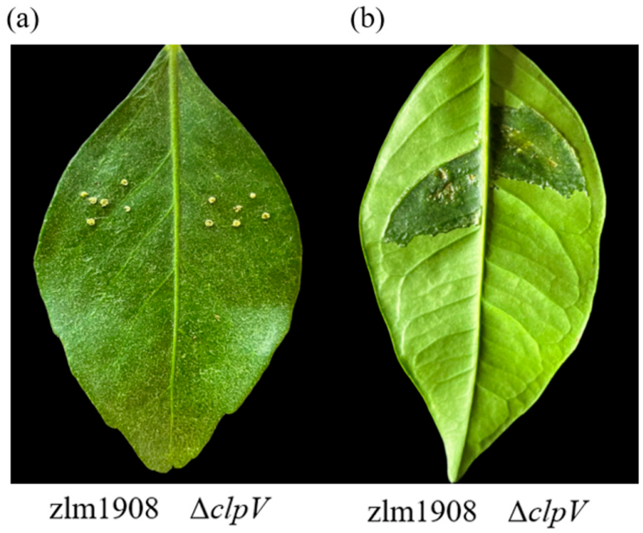
3.2.9. Pathogenicity

The pathogenicity of ΔclpV, WT, and C-clpV was assessed on 3-year-old C. sinensis ‘hongjiang’. The leaves were inoculated by the pinprick or infiltration using different bacterial concentrations, and the most severe disease symptoms were observed at 15 dpi. When leaves were inoculated with the different concentrations of WT, ΔclpV, and C-clpV (OD600 = 0.1, 0.2, 0.5, 0.7, and 0.9) after 24 h of incubation, there were no significant differences observed in the disease symptoms on leaves inoculated with the same method and concentration among the ΔclpV, WT, and C-clpV strains (Figure 4 and Table 3).

3.3. Transcriptome Analysis

3.3.1. Identification and Analysis of Differentially Expresses Genes (DEGS)

The constructed library was sequenced using the Illumina platform, yielding 7,596,458 uniquely mapped reads for WT and 7,433,469 for ΔclpV. A significance threshold of p ≤ 0.05 and |log2(Fold change)| ≥ 1.0 were applied to identify differentially expressed genes (DEGs) between WT and ΔclpV. The analysis revealed a total of 76 unigenes, comprising 34 upregulated and 42 downregulated genes. The DEGs were primarily associated with metabolism, iron receptor protein transport, bacterial motility, and stress resistance (Supplementary Table S1).

3.3.2. Gene Ontology Functional Classification of DEGS

Gene ontology (GO) classification and enrichment analysis were performed based on clusters. We successfully annotated 39 DEGs and categorized them into these three classes: biological processes, cellular components, and molecular functions (Figure 5). Genes associated with functions such as cilium- or flagellum-dependent cell motility (GO:0001539), membrane (GO:0016020), methyltransferase activity (GO:0008168), ion binding (GO:0005506), and metal ion binding (GO:0046914) exhibited substantial changes in their expression levels. This suggests that the absence of clpV gene leads to alterations in motility, abnormal metal ion transport, and changes in hydrolase activity within the strain. Additionally, four DEGs related to motility, namely flgG, fliM, flgB, and flgH were identified, all of which exhibited downregulation in ΔclpV strain. These imply that ClpV may positively regulate the motility of Xcc zlm1908 by potentially influencing the transcription of motility-related genes.
Further analysis using KEGG pathway enrichment identified the most significant metabolic and transduction pathways for DEGs. The most representative pathways include flagellar assembly (KEGG-ID: xcm02040), two-component system (KEGG-ID: xcm02020), bacterial chemotaxis (KEGG-ID: xcm02030), selenocompound metabolism (KEGG-ID: xcm00450), fructose and mannose metabolism (KEGG-ID: xcm00051), and cysteine and methionine metabolism (KEGG-ID: xcm00270). These enriched KEGG pathways indicate that ClpV may regulate relevant phenotypic changes through these pathways. According to the RNA-seq results, the top 10 significantly upregulated genes were hslV, AMD14_RS15995, AMD14_RS09150, AMD14_RS13175, bfr, metH, msrB, AMD14_RS13675, AMD14_RS14885, and AMD14_RS02230. Conversely, the top 10 significantly downregulated genes were AMD14_RS24660, AMD14_RS04820, mreD, AMD14_RS14990, fliN, flgH, AMD14_RS13515, AMD14_RS00135, AMD14_RS12895, and flgB.

3.3.3. The Validation of Transcriptome Data

The selected genes were subjected to RT-qPCR to validate the RNA-seq data. The genes chosen for validation included flgH, fliM, mcp, and mexH. As shown in Figure 6, the results demonstrated that the expression trends of these genes were consistent with the RNA-seq data, affirming the reliability of the RNA-seq findings.

4. Discussion

A variety of virulence factors are present and extensively investigated in X. campestris pv. campestris, including commonly encoded factors such as the EPS [19], lipopolysaccharides [20], chemotaxis, flagella-driven motility, twitching motility, biofilm formation, type II secretion system (T2SS), and type III secretion system (T3SS) [21,22]. The type VI secretion system (T6SS) has been also shown to play a role in bacterial interactions, biofilm formation, interactions with eukaryotic cells, acquisition of metal ions, and responses to environmental stress in X. campestris pv. campestris [23]. Additionally, it has a significant role in the pathogenicity of Citrobacter freundii [24] and X. phaseoli pv. manihotis [10]. As an ATPase, the T6SS component protein ClpV (TssH) is a protein that plays a crucial role in the functioning of the T6SS, which is a complex apparatus used by various Gram-negative bacteria to transport effector proteins into target cells [25]. ClpV may utilize ATP hydrolysis to promote the unfolding of substrate proteins and serves several important roles associated with biofilm formation, chaperone activity, motility, response to stress, and aiding the secretion of effectors in Vibrio cholerae, Pseudomonas aeruginosa, and Escherichia coli [25,26,27]. Currently, there is limited information regarding the role of the T6SS component protein ClpV in the virulence of Xanthomonas.
Bacterial growth leads to increased population density, which allows bacteria to coordinate the expression of virulence factors based on their population size and enhances their ability to infect or cause disease. In our study, the results revealed no significant differences in growth between the deletion of clpV and wild strains during the lag phase, logarithmic phase, stationary phase, or decline phase. The deletion of clpV did not influence growth kinetics, indicating that the T6SS may not be essential for growth of Xcc zlm1908, at least in nutrient-rich environments. Biofilm formation and motility are crucial virulence factors that enable microorganisms to survive in harsh environments and infect host plants. Biofilm formation enhances bacterial resistance to both host defenses and antimicrobial agents [28]. It has been suggested that during biofilm formation, flagella serve dual functions: acting as surface adhesins and facilitating motility. In many Gram-negative bacteria, flagellar-mediated motility is essential for both initial attachment and subsequent biofilm development [29,30]. Biofilm formation assays of this study have demonstrated that the ΔclpV strain exhibits a reduced ability to form biofilms, aligning with previous studies on X. phaseoli pv. manihotis, Aliivibrio avenae subsp. avenae, and Enterobacter cloacae, where the deletion of clpV resulted in diminished biofilm formation [10,27,31]. Additionally, the migration patterns of Xcc zlm1908 and ΔclpV strains on swarming agar plates indicate that ClpV positively regulates the motility of Xcc. This finding is consistent with observations in P. aeruginosa, where deletion of clpV led to the decreased collective motility [9]. Moreover, RNA sequencing analysis revealed that the clpV deletion resulted in the decreased expression of four motility-related genes (fliN, flgH, flgB, and fliM) in comparison to Xcc zlm1908. These results underscore the critical role of ClpV in motility and flagella synthesis in Xcc. Moreover, the motility-related genes have a variety of further functions beyond their essential role as motility structures. They seem to contribute to biofilm formation, which seems the common mode of bacterial growth in the environment [32].
As the primary component of the biofilm matrix, EPS protect bacteria from host immune responses and antibiotic attacks, serving as a critical virulence factor for Gram-negative bacteria. By forming a protective barrier, EPS enhance bacterial survival in hostile environments, promote adhesion to surfaces, and contribute to the structural integrity of biofilms. This resilience against host defenses and antimicrobial agents underscores the importance of EPS in the pathogenicity of Gram-negative bacteria [33]. Our research shows that the ΔclpV produces significantly less EPS compared to Xcc zlm1908, suggesting that ClpV could play a vital role in the generation of extracellular polymeric substances. Some studies have indicated that when the related genes of EPS were depressed or downregulated, bacterial susceptibility to harsh environments increased [34]. There are few studies on plant-associated bacteria using the T6SS to resist abiotic stresses. The expression of T6SS-related genes has been shown to be altered under abiotic stress conditions. Although it has been speculated that this is due to the upregulation and downregulation of genes triggered by plant bacteria to resist environmental stress, the specific regulatory mechanisms remain unclear [35]. In this study, ΔclpV is significantly reduced in tolerance of stress (NaCl) and some antibiotics (β-lactam antibiotics and aminoglycosides), implying that ClpV could be an important factor for these processes through regulating the related proteins of EPS. Moreover, transcriptional analysis indicates that deletion of clpV reduces expression levels of genes associated with bacterial chemotaxis (fliM and fliN) and the efflux pump gene mexH limiting transport of β-lactam antibiotics to the periplasmic space, suggesting ClpV could regulate tolerance of antibiotics through multiple pathways.
In this study, the deletion of clpV significantly decreased the amounts of EPS, the ability of biofilm formation, adhesion on leaf surface, swarming motility, and sensitivity to stress and antibiotics in Xcc but did not significantly affect the pathogenicity infecting leaves of citrus hosts. This result is in agreement with findings in Campylobacter jejuni [7], P. syringae pv. actinidiae [36], and Acidovorax citrulli [37]. Some studies have shown ClpV is a crucial virulence factor, and clpV-deletion resulted in a significant decrease in pathogenicity in P. plecoglossicida, Salmonella enterica serovar Typhimurium, and E. coli infecting animals [38,39,40]. Those results suggest ClpV could play versatile roles in different bacterial species.
Interestingly, RNA-seq analysis revealed that the deletion of clpV did not affect the expression of other core genes in the T6SS. Furthermore, there was no impact on the expression of core genes in the T1SS, T2SS, and T3SS. This suggests that the ClpV in Xcc may primarily be involved in bacterial competition rather than in pathogenic effects on hosts. The σ54 factor serves as a multifunctional regulatory element in various vital biological processes and plays a crucial role in governing a range of phenotypic traits. It has been shown to regulate important characteristics, including motility, biofilm formation, and antibiotic resistance [41]. RNA-seq sequencing revealed a significant downregulation of the σ54 transcription regulator (AMD14_RS04820) in clpV-deletion strain. The abnormal expression of σ54 may lead to decreased virulence factors, such as motility, biofilm formation, EPS production, and resistance to various stresses [42]. Transcriptomic data also indicate that the absence of clpV impacts several metabolic processes, including phosphate transport, lactose metabolism, starch and sucrose metabolism, and selenium compound metabolism.

5. Conclusions

In conclusion, the results of this study suggest that ClpV negatively impacts motility, biofilm formation, extracellular polymeric substance (EPS) production, and stress resistance. The RNA-seq data indicate that ClpV is associated with bacterial chemotaxis and flagellum assembly.

Supplementary Materials

The following supporting information can be downloaded at: https://www.mdpi.com/article/10.3390/microorganisms12122536/s1, Table S1: RNA-sequencing analysis of differentially expressed genes in ΔclpV vs Xcc zlm1908.

Author Contributions

Conceptualization, Y.L.; methodology, Z.W. and D.L.; software, W.X.; validation, K.C. and J.D.; data curation, Y.K.; writing—original draft preparation, Y.L.; writing—review and editing, X.H.; supervision, Y.L. All authors have read and agreed to the published version of the manuscript.

Funding

This research received no external funding.

Data Availability Statement

The raw data supporting the conclusions of this article will be made available by the authors on request.

Conflicts of Interest

The authors declare no conflicts of interest.

References

  1. Kobayashi, A.K.; Vieira, L.G.E.; Bespalhok Filho, J.C.; Leite, R.P.; Pereira, L.F.P.; Molinari, H.B.C.; Marques, V.V. Enhanced resistance to citrus canker in transgenic sweet orange expressing the sarcotoxin IA gene. Eur. J. Plant Pathol. 2017, 149, 865–873. [Google Scholar] [CrossRef]
  2. Li, R.F.; Cui, P.; Wei, P.Z.; Liu, X.Y.; Tang, J.L.; Lu, G.T. HprKXcc is a serine kinase that regulates virulence in the Gram-negative phytopathogen Xanthomonas campestris. Environ. Microbiol. 2019, 21, 4504–4520. [Google Scholar] [CrossRef] [PubMed]
  3. Andrade, M.O.; da Silva, J.C.; Soprano, A.S.; Shimo, H.M.; Leme, A.F.P.; Benedetti, C.E. Suppression of citrus canker disease mediated by flagellin perception. Mol. Plant Pathol. 2023, 24, 331–345. [Google Scholar] [CrossRef] [PubMed]
  4. Asolkar, T.; Ramesh, R. The involvement of the Type Six Secretion System (T6SS) in the virulence of Ralstonia solanacearum on brinjal. 3 Biotech 2020, 10, 324. [Google Scholar] [CrossRef]
  5. Choi, Y.; Kim, N.; Mannaa, M.; Kim, H.; Park, J.; Jung, H.; Han, G.; Lee, H.H.; Seo, Y.S. Characterization of Type VI Secretion System in Xanthomonas oryzae pv. oryzae and Its Role in Virulence to Rice. Plant Pathol. J. 2020, 36, 289–296. [Google Scholar]
  6. Kim, N.; Kim, J.J.; Kim, I.; Mannaa, M.; Park, J.; Kim, J.; Lee, H.; Lee, S.; Park, D.; Sul, J.; et al. Type VI secretion systems of plant-pathogenic Burkholderia glumae BGR1 play a functionally distinct role in interspecies interactions and virulence. Mol. Plant Pathol. 2020, 21, 1055–1069. [Google Scholar] [CrossRef]
  7. Bleumink-Pluym, N.M.; van Alphen, L.B.; Bouwman, L.I.; Wösten, M.M.; van Putten, J.P. Identification of a functional type VI secretion system in Campylobacter jejuni conferring capsule polysaccharide sensitive cytotoxicity. PLoS Pathog. 2013, 9, e1003393. [Google Scholar] [CrossRef]
  8. Zheng, J.; Leung, K. Dissection of a type VI secretion system in Edwardsiella tarda. Mol. Microbiol. 2007, 66, 1192–1206. [Google Scholar] [CrossRef]
  9. Li, Y.; Chen, L.; Zhang, P.; Bhagirath, A.Y.; Duan, K. ClpV3 of the H3-Type VI Secretion System (H3-T6SS) Affects Multiple Virulence Factors in Pseudomonas aeruginosa. Front. Microbiol. 2020, 11, 1096. [Google Scholar] [CrossRef]
  10. Montenegro, B.N.A.; Alvarez, B.A.; Arrieta-Ortiz, M.L.; Rodriguez, R.L.M.; Botero, D.; Tabima, J.F.; Luisa, C.; Cesar, T.; Silvia, R.; Adriana, B. The type VI secretion system of Xanthomonas phaseoli pv. manihotis is involved in virulence and in vitro motility. BMC Microbiol. 2021, 21, 14. [Google Scholar]
  11. Wu, C.F.; Lin, J.S.; Shaw, G.C.; Lai, E.M. Acid-induced type VI secretion system is regulated by ExoR-ChvG/ChvI signaling cascade in Agrobacterium tumefaciens. PLoS Pathog. 2012, 8, e1002938. [Google Scholar] [CrossRef] [PubMed]
  12. Sun, Y.; Zhuang, Z.; Wang, X.; Huang, H.; Fu, Q.; Yan, Q. Dual RNA-seq reveals the effect of the flgM gene of Pseudomonas plecoglossicida on the immune response of Epinephelus coioides. Fish Shellfish Immunol. 2019, 287, 515–523. [Google Scholar] [CrossRef] [PubMed]
  13. Zhang, B.; Zhuang, Z.; Wang, X.; Huang, H.; Fu, Q.; Yan, Q. Dual RNA-Seq reveals the role of a transcriptional regulator gene in pathogen-host interactions between Pseudomonas plecoglossicida and Epinephelus coioides. Fish Shellfish Immunol. 2019, 87, 778–787. [Google Scholar] [CrossRef]
  14. Chincinska, I.A. Leaf infiltration in plant science: Old method, new possibilities. Plant Methods 2021, 17, 83. [Google Scholar] [CrossRef]
  15. Yan, Q.; Wang, N. The ColR/ColS two-component system plays multiple roles in the pathogenicity of the citrus canker pathogen Xanthomonas citri subsp. citri. J. Bacteriol. 2011, 193, 1590–1599. [Google Scholar] [CrossRef] [PubMed]
  16. Gottig, N.; Garavaglia, B.S.; Garofalo, C.G.; Orellano, E.G.; Ottado, J. A filamentous hemagglutinin-like protein of Xanthomonas axonopodis pv. citri, the phytopathogen responsible for citrus canker, is involved in bacterial virulence. PLoS ONE 2009, 4, e4358. [Google Scholar] [CrossRef]
  17. Guo, Y.; Sagaram, U.S.; Kim, J.S.; Wang, N. Requirement of the galU gene for polysaccharide production by and pathogenicity and growth In Planta of Xanthomonas citri subsp. citri. Appl. Environ. Microbiol. 2010, 76, 2234–2242. [Google Scholar] [CrossRef]
  18. Livak, K.J.; Schmittgen, T.D. Analysis of relative gene expression data using realtime quantitative PCR and 2−ΔΔCt method. Method 2001, 25, 402–408. [Google Scholar] [CrossRef]
  19. Li, J.; Wang, N. The wxacO gene of Xanthomonas citri ssp. citri encodes a protein with a role in lipopolysaccharide biosynthesis, biofilm formation, stress tolerance and virulence. Mol. Plant Pathol. 2011, 12, 381–396. [Google Scholar] [CrossRef]
  20. Rigano, L.A.; Siciliano, F.; Enrique, R.; Sendín, L.; Filippone, P.; Torres, P.S.; Qüest, J.; Dow, J.M.; Castagnaro, A.P.; Vojnow, A.A.; et al. Biofilm formation, epiphytic fitness, and canker development in Xanthomonas axonopodis pv. citri. Mol. Plant Microbe Interact. 2007, 20, 1222–1230. [Google Scholar] [CrossRef]
  21. Büttner, D.; Bonas, U. Regulation and secretion of Xanthomonas virulence factors. FEMS Microbiol. Rev. 2010, 34, 107–133. [Google Scholar] [CrossRef] [PubMed]
  22. Zhou, X.; Hu, X.; Li, J.; Wang, N. A Novel Periplasmic Protein, VrpA, Contributes to Efficient Protein Secretion by the Type III Secretion System in Xanthomonas spp. Mol. Plant Microbe Interact. 2015, 28, 143–153. [Google Scholar] [CrossRef] [PubMed]
  23. Si, M.R.; Wang, Y.; Zhang, B.; Zhao, C.; Kang, Y.W.; Bai, H.N.; Wei, D.W.; Zhu, L.F.; Zhang, L.; Dong, T.G. The Type VI Secretion System Engages a Redox-Regulated Dual-Functional Heme Transporter for Zinc Acquisition. Cell Rep. 2017, 20, 949–959. [Google Scholar] [CrossRef]
  24. Liu, L.; Hao, S.; Lan, R.; Wang, G.; Xiao, D.; Sun, H.; Xu, J. The Type VI Secretion System Modulates Flagellar Gene Expression and Secretion in Citrobacter freundii and Contributes to Adhesion and Cytotoxicity to Host Cells. Infect. Immun. 2015, 83, 2596–2604. [Google Scholar] [CrossRef] [PubMed]
  25. Joshi, A.; Kostiuk, B.; Rogers, A.; Teschler, J.; Pukatzki, S.; Yildiz, F.H. Rules of Engagement: The Type VI Secretion System in Vibrio cholerae. Trends Microbiol. 2017, 25, 267–279. [Google Scholar] [CrossRef]
  26. Förster, A.; Planamente, S.; Manoli, E.; Lossi, N.S.; Freemont, P.S.; Filloux, A. Coevolution of the ATPase ClpV, the sheath proteins TssB and TssC, and the accessory protein TagJ/HsiE1 distinguishes type VI secretion classes. J. Biol. Chem. 2014, 289, 33032–33043. [Google Scholar] [CrossRef]
  27. Douzi, B.; Brunet, Y.R.; Spinelli, S.; Lensi, V.; Legrand, P.; Blangy, S.; Kumar, A.; Journet, L.; Cascales, E.; Cambillau, C. Structure and specificity of the Type VI secretion system ClpV-TssC interaction in enteroaggregative Escherichia coli. Sci. Rep. 2016, 6, 34405. [Google Scholar] [CrossRef] [PubMed]
  28. Masum, M.M.I.; Yang, Y.; Li, B.; Olaitan, O.S.; Chen, J.; Zhang, Y.; Fang, Y.; Qiu, W.; Wang, Y.; Sun, G. Role of the Genes of Type VI Secretion System in Virulence of Rice Bacterial Brown Stripe Pathogen Acidovorax avenae subsp. avenae Strain RS-2. Int. J. Mol. Sci. 2017, 18, 2024. [Google Scholar] [CrossRef]
  29. O’Toole, G.; Kaplan, H.B.; Kolter, R. Biofilm formation as microbial development. Annu. Rev. Microbiol. 2000, 54, 49–79. [Google Scholar] [CrossRef]
  30. Schell, M.A.; Ulrich, R.L.; Ribot, W.J.; Brueggemann, E.E.; Hines, H.B. Type VI secretion is a major virulence determinant in Burkholderia mallei. Mol. Microbiol. 2007, 64, 1466–1485. [Google Scholar] [CrossRef]
  31. Soria-Bustos, J.; Ares, M.A.; Gómez-Aldapa, C.A.; González-Y-Merchand, J.A.; Girón, J.A.; De la Cruz, M.A. Two Type VI Secretion Systems of Enterobacter cloacae Are Required for Bacterial Competition, Cell Adherence, and Intestinal Colonization. Front. Microbiol. 2020, 11, 560488. [Google Scholar] [CrossRef] [PubMed]
  32. Altegoer, F.; Schuhmacher, J.; Pausch, P.; Bange, G. From molecular evolution to biobricks and synthetic modules: A lesson by the bacterial flagellum. Biotechnol. Genet. Eng. Rev. 2014, 30, 49–64. [Google Scholar] [CrossRef] [PubMed]
  33. Limoli, D.H.; Jones, C.J.; Wozniak, D.J. Bacterial Extracellular Polysaccharides in Biofilm Formation and Function. Microbiol. Spectr. 2015, 3, 223–247. [Google Scholar] [CrossRef] [PubMed]
  34. Caggianiello, G.; Kleerebezem, M.; Spano, G. Exopolysaccharides produced by lactic acid bacteria: From health-promoting benefits to stress tolerance mechanisms. Appl. Microbiol. Biotechnol. 2016, 100, 3877–3886. [Google Scholar] [CrossRef]
  35. Yin, R.; Cheng, J.; Lin, J. The role of the type VI secretion system in the stress resistance of plant-associated bacteria. Stress Biol. 2024, 4, 16. [Google Scholar] [CrossRef]
  36. Wang, N.; Han, N.; Tian, R.; Chen, J.; Gao, X.; Wu, Z.; Liu, Y.; Huang, L. Role of the Type VI Secretion System in the Pathogenicity of Pseudomonas syringae pv. actinidiae, the Causative Agent of Kiwifruit Bacterial Canker. Front. Microbiol. 2021, 12, 627785. [Google Scholar]
  37. Tian, Y.; Zhao, Y.; Wu, X.; Liu, F.; Hu, B.; Walcott, R.R. The type VI protein secretion system contributes to biofilm formation and seed-to-seedling transmission of Acidovorax citrulli on melon. Mol. Plant Pathol. 2015, 16, 38–47. [Google Scholar] [CrossRef]
  38. Luo, G.; Xu, X.; Zhao, L.; Qin, Y.; Huang, L.; Su, Y.; Yan, Q. clpV is a key virulence gene during in vivo Pseudomonas plecoglossicida infection. J. Fish Dis. 2019, 42, 991–1000. [Google Scholar] [CrossRef]
  39. Zhong, H.; Wang, P.; Chen, Y.; Wang, H.; Li, J.; Li, J.; Zhu, G.; Cui, L.; Meng, X. ClpV1 in avian pathogenic Escherichia coli is a crucial virulence factor contributing to meningitis in a mouse model in vivo. Vet. Microbiol. 2021, 263, 109273. [Google Scholar] [CrossRef]
  40. Chen, S.; Du, F.; Shang, K.; Chen, H.; Guo, R.; Liao, C.; Jia, Y.; Yu, Z.; Li, J.; Zhang, C.; et al. Colonization Mediated by T6SS-ClpV Disrupts Host Gut Microbiota and Enhances Virulence of Salmonella enterica serovar Typhimurium. J. Agric. Food Chem. 2024, 72, 19155–19166. [Google Scholar] [CrossRef]
  41. Lundgren, B.R.; Shoytush, J.M.; Scheel, R.A.; Sain, S.; Sarwar, Z.; Nomura, C.T. Utilization of L-glutamate as a preferred or sole nutrient in Pseudomonas aeruginosa PAO1 depends on genes encoding for the enhancer-binding protein AauR, the sigma factor RpoN and the transporter complex AatJQMP. BMC Microbiol. 2021, 21, 83. [Google Scholar] [CrossRef] [PubMed]
  42. Yan, J.; Guo, X.; Li, J.; Li, Y.; Sun, H.; Li, A.; Cao, B. RpoN is required for the motility and contributes to the killing ability of Plesiomonas shigelloides. BMC Microbiol. 2022, 22, 299. [Google Scholar] [CrossRef] [PubMed]
Figure 1. Construction of ΔclpV mutant strain of X. citri. subsp. citri strain zlm1908. (A) The strategy of clpV gene was knocked out by allele exchange in Xcc. (B) The construction of the deletion strain was confirmed by PCR amplification. M: DL2000 marker; lane 1: Xcc wild type; lane 2: ΔclpV mutant strain; lane 3: 10 generations of ΔclpV; lane 4: 20 generations of ΔclpV; lane 5: 30 generations of ΔclpV; lane 6: 40 generations of ΔclpV; lane 7: 50 generations of ΔclpV.
Microorganisms 12 02536 g001
Figure 2. Morphological characteristics of X. citri subsp. citri strain zlm1908 (A), ΔclpV (B), and C-clpV (C) (SEM, 25,000× magnification).
Microorganisms 12 02536 g002
Figure 3. The biological characteristics of ΔclpV mutant compared with Xcc wild type. (A) The growth curve of Xcc zlm1908 and ΔclpV in LB culture medium for 60 h; (B) biofilm formation; (C) swarming motilities; (D) susceptibility to various stress conditions; (E) the production of EPS; (F) extracellular enzyme activity: (F-a) extracellular amylase, (F-b) extracellular amylase hydrolysis circle diameter/colony diameter, (F-c) β-mannanase, and (F-d) cellulase. ns: no significant difference (p > 0.05) The experimental results were averaged over the three replicates, and the error bars represent standard deviations (SD). * p < 0.05; ** p < 0.001; ns, not significant (Student’s t-test) (n = 3 independent experiments).
Microorganisms 12 02536 g003
Figure 4. Disease symptoms of the leaves at 15 days after inoculated with an OD600 of 0.2 in C. sinensis ‘hongjiang’. (a) pinprick inoculation (b) infiltration inoculation.
Microorganisms 12 02536 g004
Figure 5. Comparison of the global transcriptomes of Xcc zlm1908 and ΔclpV by RNA-Seq. (A) A total of 76 genes were significantly differentially expressed in ΔclpV compared to Xcc zlm1908, with absolute fold change >1 and FDR < 0.05. GO Classification: A bar chart depicting the gene ontology (GO) classification of the 39 DEGs, categorized into three classes: “Cellular Component” (CC), “Molecular Function” (MF), and “Biological Process” (BP). (B) KEGG enrichment upregulated pathways. (C) KEGG enrichment downregulated pathways.
Microorganisms 12 02536 g005
Figure 6. Transcription verification of RNA-seq in the Xcc zlm1908 and ΔclpV by qRT-PCR. The results are representative of three independent experiments. * p < 0.05; ** p < 0.001.
Microorganisms 12 02536 g006
Table 1. Strains and plasmids used in this study.
Strains and PlasmidsRelevant CharacteristicSource or Reference
Xcc strains
zlm1908Wild type, isolated from the leaf of diseased Citrus sinensis ‘hongjiang’Stored in our lab
ΔclpVzlm1908, in-frame deletion of clpV51-1432This study
C-clpVΔclpV with clpV-pBAD33CM-rp4
Recombinant vector
This study
E. coli strains
DH5αCompetent cellTakara
β2163Competent cellTakara
Plasmids
pLP12Suicide vector, Cm+This study
pBAD33Suicide vector, Cm+This study
pMD18-TCloning vector, Amp+Takara
Table 2. The primers used in this study.
Primer NamePrimer Sequence (5′-3′)
Primers for construction of the clpV deletion mutant
clpV-CF/CRTGTGGAATTGGTGCATTGGC/TGCATCGCGAGATCACTAGC
clpV-F/RACCGCATGCTGAGCCTGTTC/CAAGGGTTCATTAACTCGGG
clpV-UFGGAATCTAGACCTTGAGTCGCGAGCGACCAGTTCCGTGCG
clpV-URCATAGGTTGCCATTCTTAGTGAACAGGGAGCTGCGGGAGA
clpV-DFTCTCCCGCAGCTCCCTGTTCACTAAGAATGGCAACCTATG
clpV-DRACAGCTAGCGACGATATGTCTCAAAGACCAGACCCCAGAC
pLP-UF/UR GACACAGTTGTAACTGGTCCA/CAGGAACACTTAACGGCTGAC
pLP-UTF/UTRCAGGAACACTTAACGGCTGAC/CAGGAATCTAGACCTTGAGTCG
Primers for qRT-PCR
flgH-F/RTGAACCTGGTCGAAAGCACC/TTCTCAAGCACGTTCAAGCC
fliM-F/RTGATCTGCTTTCCCAGGACG/TCCTGGCTGGACAGATCGTA
mcp-F/RAGTCGCTCATCACGTCCATC/CAGGAACACTTAACGGCTGAC
mexH-F/RCAGCACCTACGTCAACGACT/AATGGTCGATGTCCTGTGGG
Table 3. The lesion area of the leaves at 15 days post inoculation (mm2).
OD600PinprickInfiltration
ΔclpVWTC-clpVΔclpVWTC-clpV
0.15.24 ± 0.015.26 ± 0.015.23 ± 0.0175.47 ± 2.1576.19 ± 3.3278.32 ± 3.65
0.25.56 ± 0.025.62 ± 0.035.57 ± 0.0279.32 ± 6.1283.19 ± 4.6584.33 ± 4.76
0.55.44 ± 0.025.65 ± 0.035.54 ± 0.0380.32 ± 6.4383.16 ± 7.2182.76 ± 4.68
0.75.97 ± 0.035.92 ± 0.045.88 ± 0.0283.43 ± 7.5382.65 ± 8.3289.54 ± 8.32
0.96.09 ± 0.046.24 ± 0.046.13 ± 0.0585.43 ± 9.0282.14 ± 10.3286.45 ± 9.46
Disclaimer/Publisher’s Note: The statements, opinions and data contained in all publications are solely those of the individual author(s) and contributor(s) and not of MDPI and/or the editor(s). MDPI and/or the editor(s) disclaim responsibility for any injury to people or property resulting from any ideas, methods, instructions or products referred to in the content.

