Article

Characterization and Transcriptional Regulation of the 2-Ketogluconate Utilization Operon in Pseudomonas plecoglossicida

School of Food and Biological Engineering, Jiangsu University, Zhenjiang 212013, China
*
Author to whom correspondence should be addressed.
Microorganisms 2024, 12(12), 2530; https://doi.org/10.3390/microorganisms12122530
Submission received: 14 November 2024 / Revised: 2 December 2024 / Accepted: 5 December 2024 / Published: 8 December 2024
(This article belongs to the Section Microbial Biotechnology)

Abstract

:
Pseudomonas plecoglossicida JUIM01 is an industrial 2-keto-d-gluconate (2KGA)-producing strain. However, its regulation mechanism of 2KGA metabolism remains to be clarified. Among other reported Pseudomonas species, the 2-ketogluconate utilization operon (kgu operon) plays key roles in 2KGA catabolism. In this study, the structural genes of the kgu operon and its promoter in P. plecoglossicida JUIM01 were identified using reverse transcription PCR and lacZ reporter gene fusion. The results showed the kgu operon in P. plecoglossicida was composed of four structural genes: kguE, kguK, kguT, and kguD. The ptxS gene located upstream of kguE was excluded from the kgu operon. Then, the knockout and corresponding complementation strains of kguE, kguK, kguT, and kguD were constructed, respectively, to prove the kgu operon was involved in 2KGA catabolism of P. plecoglossicida. The knockout stains, especially JUIM01ΔkguE, showed potential as industrial production strains for 2KGA. Moreover, the transcriptional regulation mechanism of PtxS on the kgu operon was elucidated using multiple methods. In P. plecoglossicida, the LacI-family transcription regulator PtxS could recognize a 14 bp palindrome (5′-TGAAACCGGTTTCA-3′) within the promoter region of the kgu operon and specifically bind to a 26 bp region where the palindrome was located. As the binding sites overlapped with the transcription start site of the kgu operon, the binding of PtxS possibly hindered the binding of RNA polymerase, thus repressing the transcription of the kgu operon and further regulating 2KGA catabolism. 2KGA bound to PtxS as an effector to dissociate it from the kgu operon promoter region, so as to relieve the transcription repression. The results will provide strategies for improving the product accumulation in 2KGA industrial production and theoretical bases for the construction of a Pseudomonas chassis.

1. Introduction

2-Keto-d-gluconate (2KGA), an intermediate of glucose metabolism in several oxidative bacteria, can be used as a renewable raw material for regioselective or stereoselective chemical reactions in the synthesis of heterocyclic and other functional compounds [1]. The major application of 2KGA is as the precursor to synthesize d-erythorbate (d-isoascorbate, isovitamin C) and its salts, which are widely used food antioxidants [2,3,4]. Currently, 2KGA is produced industrially by fermentation, and Pseudomonas plecoglossicida JUIM01 is an industrial 2KGA-producing strain in China [5,6,7,8].
The genes directly related to 2KGA metabolism in Pseudomonas mainly include gcd (encoding glucose dehydrogenase); the glucose acid dehydrogenase operon composed of gndS, gndL, and gndC (encoding three subunits of gluconate dehydrogenase); kguE (encoding a putative epimerase); kguK (encoding 2-ketogluconate kinase); kguT (encoding 2-ketogluconate transporter); and kguD (encoding 2-keto-6-phosphogluconate reductase) [5,9,10,11,12,13]. Among them, gcd, gndS, gndL, and gndC play roles in 2KGA synthesis [14,15], while kguE, kguK, kguT, and kguD are related to its catabolism (Figure 1A). In Pseudomonas putida KT2440, the latter four genes constitute the 2-ketogluconate utilization operon (kgu operon) [11]. However, there is debate about the composition of the kgu operon in Pseudomonas aeruginosa PAO1: Swanson et al. (2000) suggest that these four genes form the kgu operon along with the upstream ptxS [16], while Daddaoua et al. (2012) maintain that ptxS is independent of the kgu operon [12]. We have cloned a DNA fragment containing the kgu operon from the P. plecoglossicida JUIM01 genome and conducted bioinformatics analysis. The results indicate that ptxS is unlikely a part of the kgu operon in P. plecoglossicida [17].
PtxS is a LacI-family transcription factor ubiquitous in Pseudomonas species [12]. In P. putida and P. aeruginosa, PtxS functions as a transcriptional repressor that specifically recognizes and binds to a 14 bp palindrome (5′-TGAAACCGGTTTCA-3′), thereby globally regulating the metabolism of 2KGA, and 2KGA generates feedback control on PtxS as an effector [11,12,16,18,19,20,21]. However, the metabolic regulation mechanism of 2KGA in other Pseudomonas species, especially in industrial 2KGA-producing strains, remains to be elucidated. We previously demonstrated that the PtxS of P. plecoglossicida can also specifically recognize and bind to the 14 bp palindrome (5′-TGAAACCGGTTTCA-3′) and 2KGA is the effector of PtxS [22]. Further analysis of the P. plecoglossicida JUIM01 genome revealed that the upstream promoter region of kguE contains the aforementioned 14 bp palindrome [17].
Based on the above studies, we made a prediction that the kgu operon in P. plecoglossicida JUIM01 was composed of kguE, kguK, kguT, and kguD, directly related to 2KGA catabolism, and was regulated by the LacI-family transcription factor PtxS. Therefore, in the present study, the kgu operon in P. plecoglossicida JUIM01 was identified using reverse transcription PCR and promoter fusion. Whereafter, the role of the kgu operon in the 2KGA degradation metabolism of P. plecoglossicida was verified by the knockout and complementation of each structure gene. The transcriptional regulation mechanism of PtxS on the kgu operon was further analyzed using multiple methods. The results would provide strategies for improving the product accumulation in 2KGA industrial production and theoretical bases for the construction of a Pseudomonas chassis.

2. Materials and Methods

2.1. Strains, Plasmids, and Primers

The strains and plasmids used are shown in Table 1.
The knockout of kguE, kguD, and ptxS, as well as the complementation of kguE and kguD in P. plecoglossicida JUIM01, were carried out using reported methods [5,23], with the corresponding primers changed (Table S1). The knockout and complemented strains of kguT and kguK have been previously constructed [5,23].
Using the JUIM01 genome as the template and K1/K2 as the primers (Table S2 and Figure 1B), a ptxS-kguE transgene fragment with 20 bp vector homologous arms at both ends was amplified. The fragment was seamlessly linked with a linearized pME6522 vector using the ClonExpress® II One Step Cloning Kit (Nanjing Vazyme Biotech Co., Ltd., Nanjing, China) to construct the recombinant plasmid pME6522-kgu. pME6522-kgu was then electroporated into JUIM01 and JUIM01ΔptxS competent cells to construct recombinants JUIM01/pME6522-kgu and JUIM01ΔptxS/pME6522-kgu, respectively. JUIM01/pME6522 was also constructed as the negative control.
The primers used are shown in Tables S1 and S2 and Figure 1B and Figure S1, which were synthesized by Sangon Biotech Co., Ltd. (Shanghai, China).

2.2. lacZ Reporter Gene Fusions

The recombinants JUIM01/pME6522, JUIM01/pME6522-kgu, and JUIM01ΔptxS/pME6522-kgu were separately spread on LB plates (containing 10 g/L NaCl, 10 g/L tryptone, 5 g/L yeast extract, and 15 g/L agar) with 10 μg/mL tetracycline and incubated overnight at 30 °C. Single colonies were picked from the plates and inoculated into 5 mL of LB liquid media (containing 10 g/L NaCl, 10 g/L tryptone, 5 g/L yeast extract, and 20 g/L glucose) with 10 μg/mL tetracycline and incubated at 30 °C and 180 rpm for 12 and 24 h. The OD650 and β-galactosidase activities of the culture were then measured using a spectrophotometer and the β-Galactosidase Enzyme Assay System with Reporter Lysis Buffer (Promega, Madison, WI, USA).

2.3. Comparison of 2KGA Utilization Abilities

The activated P. plecoglossicida JUIM01 and its derived gene-deletion strains were resuspended in 1 mL of sterile water, and then inoculated into 500 mL shake flasks containing 50 mL of seed media (20.0 g/L calcium 2-ketogluconate, 10.0 g/L corn steep liquor, 2.0 g/L urea, 2.0 g/L KH2PO4 and 0.5 g/L MgSO4·7H2O, pH 7.0) at 30 °C and 265 rpm on a rotary shaker for 24 h. Cell growth and 2KGA concentration were measured using reported methods [6,23].

2.4. 2KGA Fermentation

The activated cells were inoculated into the 50 mL of seed media (20.0 g/L glucose, 5.0 g/L corn syrup powder, 2.0 g/L urea, 2.0 g/L KH2PO4, 0.5 g/L MgSO4·7H2O and 5.0 g/L CaCO3, pH 7.0) in 500 mL shake flasks and cultivated at 33 °C and 265 rpm for at least 28 h.

2.5. Primer Extension

The total RNA of P. plecoglossicida JUIM01 was extracted using the RNAPrep Pure Cell/Bacteria Kit (TIANGEN Biotech Co., Ltd., Beijing, China). The primer kguE-PE(FAM) was designed and synthesized for the kgu operon and its upstream sequence, with a FAM label at the 5′ end (Table S2 and Figure S1). Primer extension was performed based on a reported protocol [25].

2.6. Electrophoretic Mobility Shift Assay (EMSA)

The DNA probe JUIM01-kgu(FAM) (labeled with FAM at its 5′ end) containing the kgu operon promoter region sequence was synthesized by Sangon Biotech Co., Ltd. (Shanghai, China). The EMSA was performed based on a reported protocol [26]. First, the amount of the probe JUIM01-kgu(FAM) remained 50 ng in the reaction system, and the amount of PtxS protein was 0 for the control lane and 0.5 μg for the other lanes to investigate the specific binding of PtxS to the kgu operon promoter region. The amount of 2KGA then gradually increased from 0 to 50 mM, while the amounts of both JUIM01-kgu(FAM) and PtxS remained constant, to investigate the effect of the effector 2KGA on the binding of PtxS to the kgu operon promoter region. The heterologous expression and purification of PtxS were previously reported [22].

2.7. DNase I Footprinting

The 400 ng JUIM01-kgu(FAM) probe was used to bind to different amounts (0, 0.9, and 2.2 μg) of PtxS at 25 °C for 30 min, and then was digested with 0.015 U DNase I (Promega, Madison, WI, USA) at 37 °C for 1 min. The digested sample was extracted with a phenol–chloroform–isoamyl alcohol mixture (25:24:1) to remove protein, and the supernatant was added to 2.5-fold the volume of absolute ethanol and 1/10 volume of 3 M sodium acetate. The sample was placed at −20 °C for 1 h to precipitate DNA and centrifuged to remove the supernatant. The precipitate was washed with 1 mL of 75% ethanol, centrifuged, and dried. The resulting DNA was dissolved in Mili-Q ultrapure water and detected by an ABI 3130xl Genetic Analyzer (Foster City, CA, USA), using GeneScan™ 500 LIZ® Size Standard (ABI, Foster City, CA, USA) as the ladder. The Peak Scanner software v1.0 (Applied Biosystems) was used to output the fluorescence peak graphs and base sequences based on the sequencing results.

2.8. Real-Time Fluorescence Quantitative PCR (RT-qPCR)

The exponential-phase cells of P. plecoglossicida JUIM01 and JUIM01ΔptxS were collected to extract their RNAs using the UNIQ-10 column Trizol total RNA extraction kit (Sangon Biotech Co., Ltd., Shanghai, China). The RNAs were reversely transcribed using the FastQuant RT Kit (With gDNase) (TIANGEN Biotech Co., Ltd., Beijing, China) to prepare corresponding cDNAs. Taking the cDNAs as the templates, the upstream and downstream primers were designed according to the conserved regions of each target gene (Table S2 and Figure S1), and PCR amplifications were performed. SuperReal PreMix Plus (SYBR Green) (TIANGEN Biotech Co., Ltd., Beijing, China) was used to detect the expression levels of the target genes (kguE, kguK, kguT, and kguD). The relative expression levels of the genes were calculated using the 2−ΔΔCTmethod and rpoD as an internal reference [27].

2.9. Statistical Analysis

Three replicates were set for data analysis. The results were presented as mean ± standard deviation. Statistical comparisons were performed using one-way analysis of variance and Duncan’s multiple comparison test. The results were considered significant at p < 0.01.

3. Results and Discussion

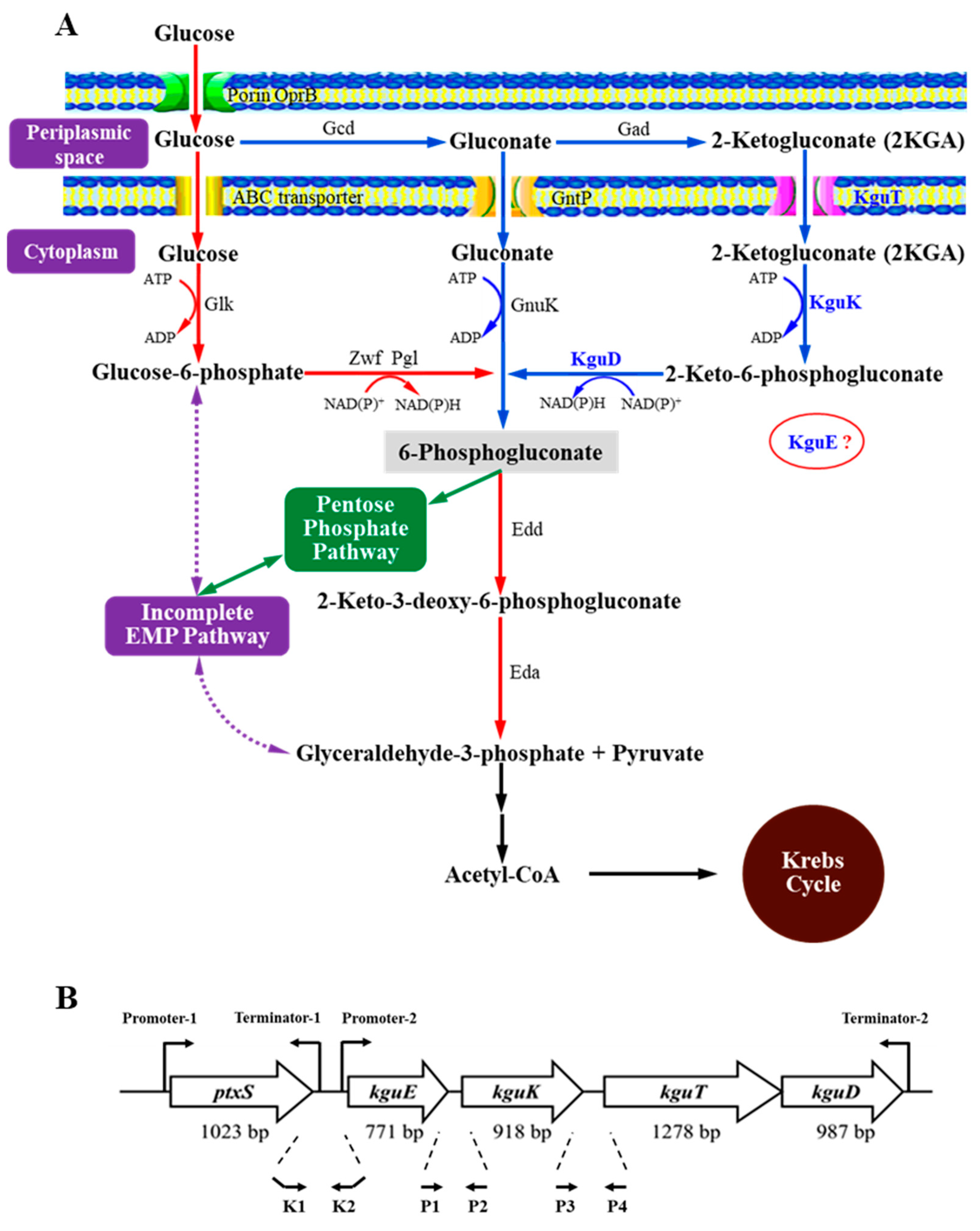
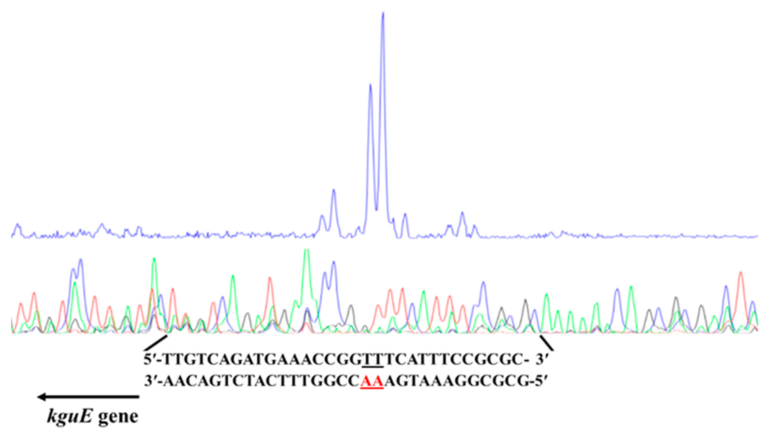
3.1. Identification of the Structural Genes of the kgu Operon in Pseudomonas plecoglossicida

A DNA fragment containing ptxS, kguE, kguK, kguT, and kguD was cloned from the P. plecoglossicida JUIM01 genome (Figure 1B). Bioinformatics analysis shows that the kgu operon is supposed to be composed of kguE, kguK, kguT, and kguD [17]. To confirm this prediction, co-transcription analysis was performed on these genes. The total RNA of P. plecoglossicida JUIM01 was extracted to prepare cDNA by reverse transcription PCR. Using P1/P2 and P3/P4 as the primers (Table S2 and Figure 1B) and the cDNA as the template, the kguE-kguK and kguK-kguT target DNA fragments’ transgene regions were amplified, respectively. Meanwhile, the genome DNA and ddH2O were chosen as positive and negative control templates. Agarose gel electrophoresis analysis showed that the sizes of the two target fragments (311 and 400 bp) were consistent with their positive controls (Figure S2). Further sequencing confirmed the results, indicating kguE, kguK, and kguT were co-transcribed. Due to the fact that there is only 9 bp between kguT and kguD (impossible to contain a promoter), and they have same transcription directions, we concluded kguT and kguD also transcribed jointly. In summary, kguE, kguK, kguT, and kguD were located within the same operon and underwent co-transcription in P. plecoglossicida.
We have found a promoter in the upstream of ptxS and a terminator in the ptxS-kguE transgene region in P. plecoglossicida [17]. In view of the prediction that ptxS is independent of the kgu operon [17], there should be a promoter in the ptxS-kguE transgene region. The lacZ reporter gene fusions were conducted to prove it. Taking K1/K2 as the primers (Table S2 and Figure 1), the ptxS-kguE transgene region fragment was amplified from the JUIM01 genome and integrated into the pME6522 vector to obtain the recombinant promoter probe pME6522-kgu. The pME6522-kgu and empty vector pME6522 were transformed into P. plecoglossicida JUIM01 to construct the recombinant strain JUIM01/pME6522-kgu and its control JUIM01/pME6522, respectively. Then, the growth and β-galactosidase activities of the two strains were compared during cultivation. No significant difference in growth was observed (Figure S3A). However, there was a significant difference in their β-galactosidase activities (p < 0.01). After 12 and 24 h cultivation, the β-galactosidase activities of JUIM01/pME6522-kgu were 1.21- and 2.16-fold that of JUIM01/pME6522, respectively (Figure S3B). The results indicated that there was a promoter between ptxS and kguE, confirming the prediction that ptxS transcribes independently. In conclusion, the kgu operon consisted of four structural genes, kguE, kguK, kguT, and kguD, and it is supposed to play important roles in the 2KGA catabolism of P. plecoglossicida based on our previous studies [5,17,23].

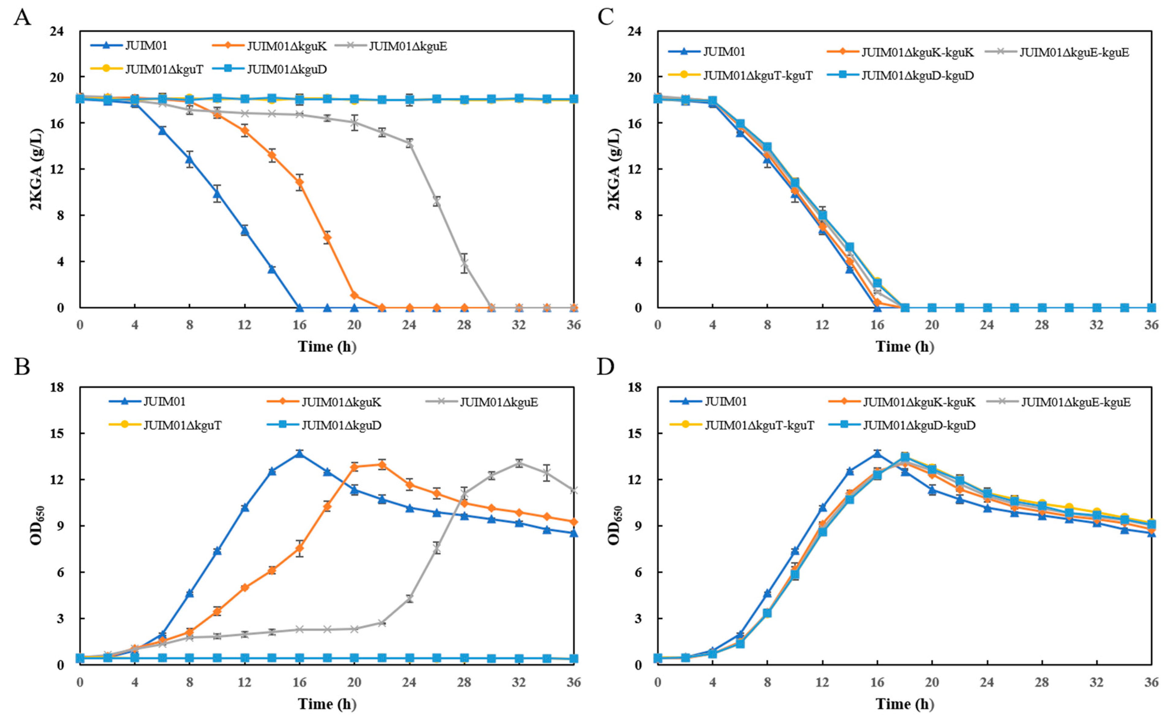
3.2. Effects of the Structural Genes of the kgu Operon on 2KGA Catabolism in P. plecoglossicida

We further constructed four knockout strains (named JUIM01Δx, where x represents any gene of the kgu operon) and their corresponding complementation strains (named JUIM01Δx-x) from P. plecoglossicida JUIM01 and comprehensively investigated the 2KGA utilization abilities of the recombinant strains to verify the roles of these genes in 2KGA catabolism. The strains (with JUIM01 as a control) were cultured in seed media containing 2KGA as the main carbon source to observe 2KGA consumption and cell growth. The results showed that the knockout of the genes had apparent impacts on the 2KGA utilization ability of JUIM01. The deletion of kguK and kguE reduced the average consumption rate of 2KGA (the 2KGA consumed per hour in the period from 0 h to the time 2KGA was used up), delaying the time for 2KGA depletion in the media from 16 to 22 and 30 h, respectively, and the time for the recombinant strains to reach their highest OD650 values was also delayed accordingly. After the knockout of kguT and kguD, the recombinants almost completely lost their abilities to utilize 2KGA (Figure 2A,B). Then, the four structural genes were integrated into the expression vectors and complemented back into the corresponding gene knockout strains, respectively. Afterwards, the 2KGA utilization abilities in the obtained complementation strains were restored (Figure 2C,D). The results indicated that all four structural genes of the kgu operon were involved in the 2KGA catabolism of P. plecoglossicida and played important roles.
According to existing studies, 2KGA is synthesized in the periplasmic space and then secreted into the extracellular space or enters the cytoplasm for metabolism in Pseudomonas. The intracellular metabolic pathway mainly includes the following aspects: 2KGA in the periplasmic space is transported into the cell by the 2KGA transporter KguT and phosphorylated into 2-keto-6-phosphogluconate by the 2-keto-6-phosphogluconate kinase KguK, which is then reduced to 6-phosphogluconate by the 2-keto-6-phosphogluconate reductase KguD, and further enters the Entner–Doudoroff pathway for metabolism (Figure 1A). Interestingly, the importance of KguE, KguK, KguT, and KguD in 2KGA utilization seems to be neither consistent with their order in the 2KGA catabolic pathway nor their physical sequence in the kgu operon. The inactivation of KguT possibly stopped the transportation of 2KGA into cell, leading to JUIM01 losing its capability to utilize 2KGA. KguK is supposed to catalyze the first reaction of 2KGA catabolism after 2KGA enters the cytoplasm. However, the 2KGA utilization was not completely inhibited in JUIM01ΔkguK, indicating there might be an alternative pathway in the cytoplasm. Although the reaction catalyzed by KguD is after that by KguK, the inactivation of KguD led to greater inhibition in 2KGA utilization, suggesting KguD might have an unknown extra function. KguE is predicted to be an epimerase with an unknown function and has never been annotated in the central metabolic pathway of Pseudomonas [11,16]. This study proved for the first time that KguE did participate in 2KGA metabolism in Pseudomonas.
During 2KGA fermentation, when the substrate glucose is completely consumed, the produced 2KGA is then utilized by Pseudomonas as an alternative carbon source, resulting in reduced production [5]. The knockout of kguE, kguK, kguT, or kguD could effectively alleviate or even inhibit the reutilization of 2KGA, indicating that the kgu operon would be an ideal target for the modification of 2KGA-producing strains.

3.3. 2KGA Fermentation Using the kguK/kguE/kguT/kguD-Knockout and Complementation Recombinants Derived from P. plecoglossicida JUIM01

In industrial production, the reuse of 2KGA by production strains is a problem that is detrimental to the final yield. In the 2KGA utilization experiments, the utilization of 2KGA by JUIM01ΔkguK/kguE/kguT/kguD was delayed or even completely blocked. These strains showed potential as industrial production strains for 2KGA. Therefore, we further investigated the 2KGA fermentation capacity of the kguK/kguE/kguT/kguD-knockout and complementation recombinants in shake flasks. JUIM01ΔkguK/kguE/kguT/kguD exhibited glucose utilization ability similar to JUIM01 (Figure 3A), while the accumulation ability of 2KGA underwent significant changes, as shown in the results of the 2KGA utilization experiments (Figure 3B). Due to the inability of JUIM01ΔkguT and JUIM01ΔkguD to reuse 2KGA as a growth carbon source, their maximum OD650 of 10.4 was significantly lower than the JUIM01 of 16.2. However, JUIM01ΔkguK and JUIM01ΔkguE took a longer time to reach the maximum OD650 of above 15 (Figure 3C). Among them, JUIM01ΔkguE might be the best candidate for 2KGA industrial production, as it left a longer time to recover 2KGA from the broth before it was rapidly reused without affecting its cell growth. In the complementation strains, the glucose utilization, synthesis and utilization of 2KGA, and cell growth were similar to those of JUIM01 (Figure 3D–F).

3.4. Transcriptional Start Site of the kgu Operon in P. plecoglossicida

The transcription start site of the kgu operon in P. plecoglossicida was determined by primer extension. First, a primer kguE-PE(FAM) (Table S2 and Figure S1) was designed and synthesized for the kgu operon and its upstream sequence, and then hybridized with the RNA of JUIM01. The result showed two sharp peaks at around 120 bp from the 5′ end of the specific reverse primer, corresponding to the base pair TT/AA, which was identified as the transcription start site of the kgu operon (Figure 4). This site was located 34 bp upstream of the initiation codon of kguE (the first structural gene of the kgu operon). It is worth noting that the transcription start site was located within a 14 bp palindrome (5′-TGAAACCGGTTTCA-3′, the letters in bold represent the transcription start site).

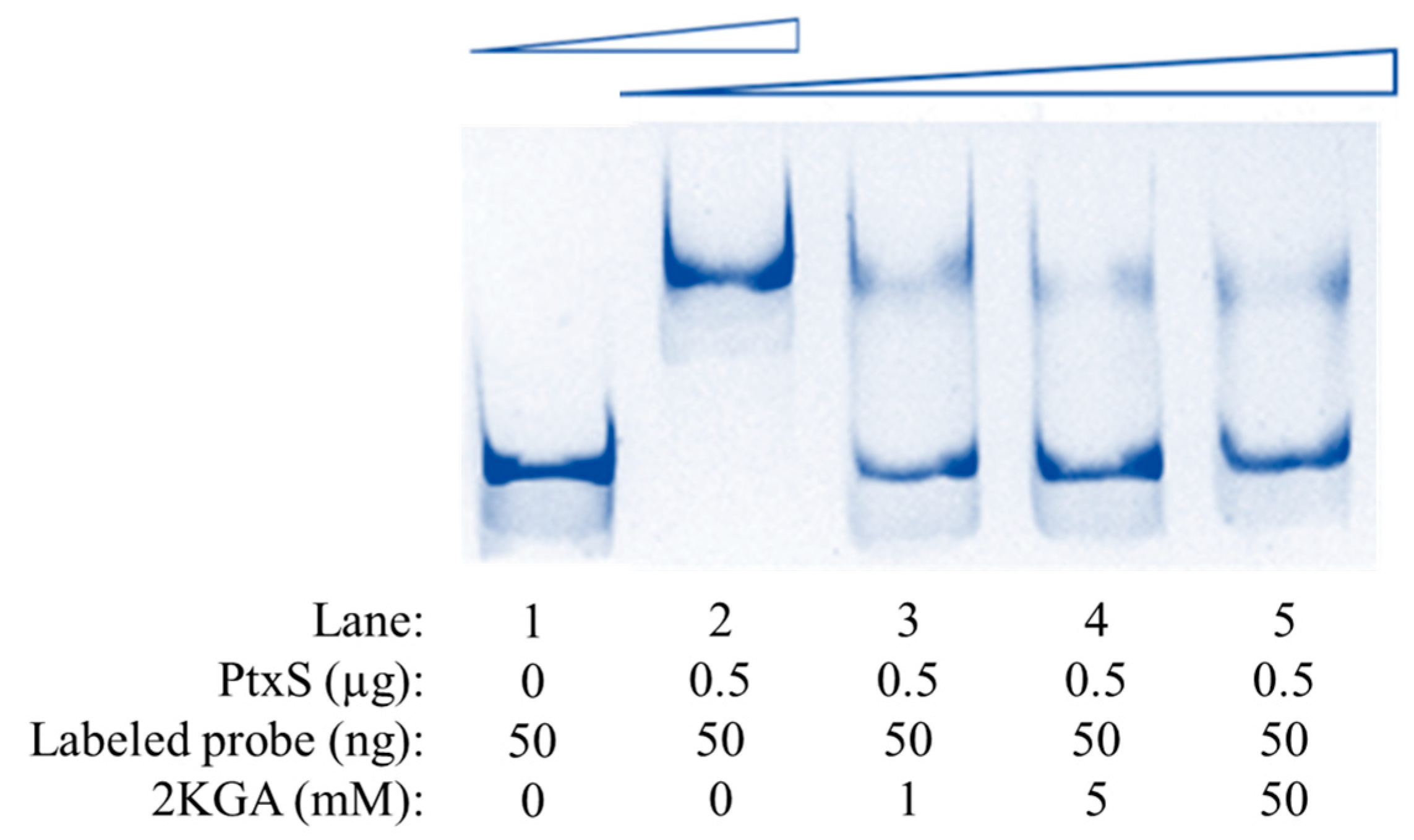
3.5. Specific Binding Between PtxS and the kgu Operon Promoter Region in P. plecoglossicida

In P. aeruginosa and P. putida, the transcription regulator PtxS specifically recognizes and binds to the 14 bp palindrome (5′-TGAAACCGGTTTCA-3′) in the kgu operon promoter region, thereby regulating the transcription of the kgu operon [11,16]. A previous study has shown that PtxS from P. plecoglossicida can also specifically bind to the 14 bp palindrome [22]. Therefore, PtxS is likely to regulate the transcription of the kgu operon and 2KGA metabolism by specifically binding to the region where the 14 bp palindrome is located in P. plecoglossicida. To verify this hypothesis, based on the kgu operon and its upstream sequence, a specific probe JUIM01-kgu(FAM) labeled with FAM was synthesized. PtxS from JUIM01 was then heterologously expressed and purified. Finally, the interaction between PtxS and the kgu operon promoter region was investigated by EMSA. Reactions were implemented using a 50-ng labeled probe, and without or with 0.5 μg of PtxS. As shown in Figure 5, in the absence of PtxS, the band of the free DNA probe is displayed at the bottom of the gel (Lane 1). As PtxS was added, the molecular weight of the protein–probe complex became larger, and its mobility was restrained correspondingly (Lane 2). The results confirmed that PtxS could specifically bind to the kgu operon promoter region of P. plecoglossicida.
In P. aeruginosa and P. putida, the regulator PtxS regulates the metabolism of 2KGA, which acts as an effector of PtxS. 2KGA can bind to PtxS to release the binding between PtxS and the regulated DNA, thereby relieving the repression of the corresponding genes at the transcriptional level [11,16]. In P. plecoglossicida, 2KGA can also bind to PtxS [22]. To clarify the effect of 2KGA on the specific binding of PtxS to the kgu operon promoter region in P. plecoglossicida, different concentrations of 2KGA were added to the EMSA systems (Figure 5). When 2KGA was not present, PtxS bound to JUIM01-kgu(FAM), and the band was located at the top of the gel (Lane 2). With the addition of 2KGA, the bands showed accelerated mobility and shifted to lower positions (Lane 3–5), indicating that the retardation of mobility caused by the binding of PtxS–probe was relieved. In summary, similar to P. aeruginosa and P. putida, in P. plecoglossicida, PtxS could also specifically bind to the kgu operon promoter region, while 2KGA acted as an effector binding to PtxS, thereby releasing PtxS from the kgu operon promoter region.
DNaseI footprint analysis further revealed the binding site of PtxS in the kgu operon promoter region. Compared with the control group without PtxS, a distinct protective region appeared when 0.9 and 2.2 μg PtxS was added to the systems with 400 ng of JUIM01-kgu(FAM). The protective region covered a sequence of about 26 bp (5′-AATGAAACCGGTTTCATCTGACAAGA-3′) (Figure 6). Interestingly, the 14 bp palindrome (the underlined bases) was found in this region, located 25–39 bp upstream of kguE.

3.6. Transcriptional Regulation of PtxS on the kgu Operon in P. plecoglossicida

To explore the transcriptional regulation mechanism of PtxS on the kgu operon in P. plecoglossicida, a ptxS gene-knockout strain JUIM01ΔptxS was constructed. The expression levels of each gene within the kgu operon before and after ptxS knockout were compared by RT-qPCR. Under the same culture conditions, the expression levels of the structural genes in the kgu operon in JUIM01ΔptxS were significantly up-regulated to 1.75–2.39-fold compared to JUIM01 (p < 0.01) (Figure S4), indicating PtxS negatively regulated the expression of the kgu operon.
The negative regulation of PtxS was further verified by lacZ reporter gene fusions. The recombinant plasmid pME6522-kgu was transformed into JUIM01 and JUIM01ΔptxS to obtain the recombinant strains JUIM01/pME6522-kgu and JUIM01ΔptxS/pME6522-kgu, respectively. The results showed no significant difference in the growth between the two strains (Figure S5A), but a significant difference in their β-galactosidase activities (p < 0.01): after 12 and 24 h cultivation, JUIM01ΔptxS/pME6522-kgu was 1.87- and 1.37-fold of JUIM01/pME6522-kgu, respectively (Figure S5B), indicating the presence of ptxS had a repressive effect on the fusion expression of kgu-lacZ, thereby demonstrating that PtxS negatively regulated the expression of the kgu operon at transcriptional level.
In natural edatope, 2KGA is one of the most commonly secreted organic acids by various rhizosphere bacteria including Pseudomonas, which is beneficial to plant growth and root colonization [28,29,30,31,32]. The repression of PtxS on the kgu operon transcription provides a mechanism for 2KGA secretion in Pseudomonas. Additionally, during fermentation, 2KGA is both the target product and an intermediate metabolite that can be utilized by Pseudomonas [9,33,34,35,36,37]. The negative regulation of PtxS on the kgu operon is essential to 2KGA accumulation.

4. Conclusions

In this study, the structural genes of the kgu operon in P. plecoglossicida were first identified. Unlike P. aeruginosa reported by Swanson et al. (2000) [16], the kgu operon in P. plecoglossicida consisted of four structural genes, kguE, kguK, kguT, and kguD. The expression of ptxS located upstream of kguE is independent of the operon. Subsequently, the role of each structural gene in 2KGA metabolism was determined via gene knockout and complementation, demonstrating that the kgu operon was involved in and essential to 2KGA metabolism in P. plecoglossicida. The results indicated the deletion of gene(s) in the kgu operon would be an ideal strategy to improve 2KGA accumulation. The knockout stains, especially JUIM01ΔkguE, showed potential as industrial production strains for 2KGA. In this study, we proved that KguE participated in glucose/2KGA metabolism in Pseudomonas, which was barely mentioned in related studies. However, the reaction KguE exactly catalyzes remains to be studied. The 2KGA utilization was not completely inhibited in the kguK-knockout mutant, indicating there might be an alternative pathway for 2KGA catabolism in the cytoplasm. At last, the regulation of PtxS on the kgu operon was analyzed using multiple methods. In P. plecoglossicida, the LacI-family transcriptional regulator PtxS recognized the 14 bp palindrome located in the kgu operon promoter region and specifically bound to a 26 bp region containing the palindrome (Figure 7A). Since the binding site overlapped with the transcription start site, the binding of PtxS possibly hindered the binding of RNA polymerase, thereby inhibiting the transcription of the kgu operon and further regulating 2KGA metabolism. Meanwhile, 2KGA could bind to PtxS as an effector, dissociating from the kgu operon promoter region, thereby releasing the transcriptional repression (Figure 7B). The above transcriptional regulation mechanism was similar to that reported in P. aeruginosa and P. putida and might be a common mechanism in the genus Pseudomonas.

Supplementary Materials

The following supporting information can be downloaded at https://www.mdpi.com/article/10.3390/microorganisms12122530/s1: Figure S1: Graphical representation of some primers used in this study; Figure S2: Electrophoretic analysis of the kguE-kguK and kguK-kguT transgene region amplification with different templates. M: DL 2000 DNA Marker; Lane 1: the kguE-kguK transgene region amplification with cDNA as template; Lane 2: the kguE-kguK transgene region amplification with genome DNA as template (positive control); Lane 3: the kguE-kguK transgene region amplification with ddH2O as template (negative control); Lane 4: the kguK-kguT transgene region amplification with cDNA as template; Lane 5: the kguK-kguT transgene region amplification with genome DNA as template (positive control); Lane 6: the kguK-kguT transgene region amplification with ddH2O as template (negative control); Figure S3: Comparison of (A) the growth and (B) the β-galactosidase activities between the recombinants JUIM01/pME6522 and JUIM01/pME6522-kgu after 12/24 h cultivation. The different superscript letters indicate significant differences (p < 0.01) between the corresponding values; Figure S4: Comparison of the relative expression levels of the kgu operon in P. plecoglossicida JUIM01 and JUIM01ΔptxS. The different superscript letters indicate significant differences (p < 0.01) between the corresponding values; Figure S5: Comparison of (A) the growth and (B) the β-galactosidase activities between the recombinants JUIM01/pME6522-kgu and JUIM01ΔptxS/pME6522-kgu after 12/24 h cultivation. The different superscript letters indicate significant differences (p < 0.01) between the corresponding values; Table S1: The primers for construction of the knockout and complemented strains; Table S2: Main primers used in this study.

Author Contributions

Conceptualization, L.S. (Lei Sun), D.W. and W.S.; methodology, L.S. (Lei Sun), D.W. and W.S.; validation, X.Z. and D.W.; formal analysis, W.S.; investigation, L.S. (Lei Sun), W.Y. and L.L.; resources, L.S. (Lei Sun) and W.S.; data curation, L.S. (Lei Sun), L.L. and D.W.; writing—original draft preparation, L.S. (Lei Sun), D.W. and W.S.; writing—review and editing, F.C., X.Q. and L.S. (Ling Sun); supervision, L.S. (Ling Sun) and W.S.; funding acquisition, L.S. (Lei Sun) and W.S. All authors have read and agreed to the published version of the manuscript.

Funding

This research was financially supported by the National Natural Science Foundation of China (32272286, 32201995) and the Natural Science Foundation of Jiangsu Province (BK20220526).

Data Availability Statement

The original contributions presented in the study are included in the article/Supplementary Material, further inquiries can be directed to the corresponding author.

Acknowledgments

Thank Yuquan Xu and his research team from Shanghai Jiao Tong University for generously providing the plasmid pME6522.

Conflicts of Interest

The authors declare no conflicts of interest.

References

  1. Stottmeister, U.; Aurich, A.; Wilde, H.; Andersch, J.; Schmidt, S.; Sicker, D. White biotechnology for green chemistry: Fermentative 2-oxocarboxylic acids as novel building blocks for subsequent chemical syntheses. J. Ind. Microbiol. Biot. 2005, 32, 651–664. [Google Scholar] [CrossRef] [PubMed]
  2. Pappenberger, G.; Hohmann, H.P. Industrial production of l-ascorbic acid (vitamin C) and d-isoascorbic acid. In Biotechnology of Food and Feed Additives; Advances in Biochemical Engineering/Biotechnology; Zorn, H., Czermak, P., Eds.; Springer: Berlin/Heidelberg, Germany, 2014; Volume 143, pp. 143–188. [Google Scholar] [CrossRef]
  3. EFSA Panel on Food Additives and Nutrient Sources Added to Food (ANS). Scientific Opinion on the re-evaluation of erythorbic acid (E 315) and sodium erythorbate (E 316) as food additives. EFSA J. 2016, 14, 4360. [Google Scholar] [CrossRef]
  4. Sun, L.; Zhang, X.J.; Zhou, Y.Z.; Peng, Z.; Cui, F.J.; Zhou, Q.; Man, Z.W.; Guo, J.; Sun, W.J. Can cadmium-contaminated rice be used to produce food additive sodium erythorbate? Food Chem. 2025, 462, 140923. [Google Scholar] [CrossRef] [PubMed]
  5. Sun, W.J.; Wang, Q.H.; Luan, F.; Man, Z.W.; Cui, F.J.; Qi, X.H. The role of kguT gene in 2-ketogluconate-producing Pseudomonas plecoglossicida JUIM01. Appl. Biochem. Biotech. 2019, 187, 965–974. [Google Scholar] [CrossRef] [PubMed]
  6. Sun, L.; Wang, D.M.; Sun, W.J.; Cui, F.J.; Gong, J.S.; Zhang, X.M.; Shi, J.S.; Xu, Z.H. Two-stage semi-continuous 2-keto-gluconic acid (2KGA) production by Pseudomonas plecoglossicida JUIM01 from rice starch hydrolyzate. Front. Bioeng. Biotech. 2020, 8, 120. [Google Scholar] [CrossRef]
  7. Wang, D.M.; Chen, X.; Guo, H.; Wang, Q.H.; Sun, L.; Sun, W.J. Exploring the response mechanism of Pseudomonas plecoglossicida to high-temperature stress by transcriptomic analyses for 2-keto gluconic acid production. Food Biosci. 2024, 62, 105063. [Google Scholar] [CrossRef]
  8. Sun, W.J.; Zhang, Q.N.; Li, L.L.; Qu, M.X.; Zan, X.Y.; Cui, F.J.; Zhou, Q.; Wang, D.M.; Sun, L. The functional characterization of the 6-phosphogluconate dehydratase operon in 2-ketogluconic acid industrial producing strain Pseudomonas plecoglossicida JUIM01. Foods 2024, 13, 3444. [Google Scholar] [CrossRef]
  9. del Castillo, T.; Ramos, J.L.; Rodríguez-Herva, J.J.; Fuhrer, T.; Sauer, U.; Duque, E. Convergent peripheral pathways catalyze initial glucose catabolism in Pseudomonas putida: Genomic and flux analysis. J. Bacteriol. 2007, 189, 5142–5152. [Google Scholar] [CrossRef]
  10. del Castillo, T.; Duque, E.; Ramos, J.L. A set of activators and repressors control peripheral glucose pathways in Pseudomonas putida to yield a common central intermediate. J. Bacteriol. 2008, 190, 2331–2339. [Google Scholar] [CrossRef]
  11. Daddaoua, A.; Krell, T.; Alfonso, C.; Morel, B.; Ramos, J.L. Compartmentalized glucose metabolism in Pseudomonas putida is controlled by the PtxS repressor. J. Bacteriol. 2010, 192, 4357–4366. [Google Scholar] [CrossRef]
  12. Daddaoua, A.; Fillet, S.; Fernández, M.; Udaondo, Z.; Krell, T.; Ramos, J.L. Genes for carbon metabolism and the ToxA virulence factor in Pseudomonas aeruginosa are regulated through molecular interactions of PtxR and PtxS. PLoS ONE 2012, 7, e39390. [Google Scholar] [CrossRef] [PubMed]
  13. Udaondo, Z.; Ramos, J.L.; Segura, A.; Krell, T.; Daddaoua, A. Regulation of carbohydrate degradation pathways in Pseudomonas involves a versatile set of transcriptional regulators. Microb. Biotechnol. 2018, 11, 442–454. [Google Scholar] [CrossRef] [PubMed]
  14. Wang, D.M.; Sun, L.; Sun, W.J.; Cui, F.J.; Gong, J.S.; Zhang, X.M.; Shi, J.S.; Xu, Z.H. Purification, characterization and gene identification of a membrane-bound glucose dehydrogenase from 2-keto-d-gluconic acid industrial producing strain Pseudomonas plecoglossicida JUIM01. Int. J. Biol. Macromol. 2018, 118, 534–541. [Google Scholar] [CrossRef] [PubMed]
  15. Wang, D.M.; Sun, L.; Sun, W.J.; Cui, F.J.; Gong, J.S.; Zhang, X.M.; Shi, J.S.; Xu, Z.H. A membrane-bound gluconate dehydrogenase from 2-keto-d-gluconic acid industrial producing strain Pseudomonas plecoglossicida JUIM01: Purification, characterization, and gene identification. Appl. Biochem. Biotech. 2019, 188, 897–913. [Google Scholar] [CrossRef] [PubMed]
  16. Swanson, B.L.; Hager, P.; Phibbs, P., Jr.; Ochsner, U.; Vasil, M.L.; Hamood, A.N. Characterization of the 2-ketogluconate utilization operon in Pseudomonas aeruginosa PAO1. Mol. Microbiol. 2000, 37, 561–573. [Google Scholar] [CrossRef]
  17. Luan, F.; Wang, D.M.; Sun, W.J.; Cui, F.J.; Qi, X.H.; Guo, Q.; Zhang, X.F.; Li, Y.Z. Cloning and bioinformatics analysis of 2-ketogluconate utilization operon from Pseudomonas plecoglossicida. J. Chin. Inst. Food Sci. Technol. 2018, 18, 292–298. (In Chinese) [Google Scholar] [CrossRef]
  18. Colmer, J.A.; Hamood, A.N. Characterization of ptxS, a Pseudomonas aeruginosa gene which interferes with the effect of the exotoxin A positive regulatory gene, ptxR. Mol. Gen. Genet. MGG 1998, 258, 250–259. [Google Scholar] [CrossRef]
  19. Swanson, B.L.; Colmer, J.A.; Hamood, A.N. The Pseudomonas aeruginosa exotoxin A regulatory gene, ptxS: Evidence for negative autoregulation. J. Bacteriol. 1999, 181, 4890–4895. [Google Scholar] [CrossRef]
  20. Swanson, B.L.; Hamood, A.N. Autoregulation of the Pseudomonas aeruginosa protein PtxS occurs through a specific operator site within the ptxS upstream region. J. Bacteriol. 2000, 182, 4366–4371. [Google Scholar] [CrossRef]
  21. Daddaoua, A.; Krell, T.; Ramos, J.L. Transcriptional control by two interacting regulatory proteins: Identification of the PtxS binding site at PtxR. Nucleic Acids Res. 2013, 41, 10150–10156. [Google Scholar] [CrossRef]
  22. Sun, L.; Wang, D.M.; Sun, W.J.; Zhang, X.F.; Cui, F.J.; Su, C.; Zhang, X.M.; Xu, G.Q.; Shi, J.S.; Xu, Z.H. Characterization of a transcriptional regulator PtxS from Pseudomonas plecoglossicida for regulating 2-ketogluconic acid metabolism. Int. J. Biol. Macromol. 2021, 174, 330–338. [Google Scholar] [CrossRef] [PubMed]
  23. Sun, L.; Wang, D.M.; Sun, W.J.; He, X.Y.; Cui, F.J.; Zhang, X.M.; Gong, J.S.; Shi, J.S.; Xu, Z.H. A 2-ketogluconate kinase KguK in Pseudomonas plecoglossicida JUIM01: Enzymatic characterization and its role in 2-keto-d-gluconic acid metabolism. Int. J. Biol. Macromol. 2020, 165 Pt B, 2640–2648. [Google Scholar] [CrossRef]
  24. Lu, J.S.; Huang, X.Q.; Li, K.; Li, S.N.; Zhang, M.Y.; Wang, Y.; Jiang, H.X.; Xu, Y.Q. LysR family transcriptional regulator PqsR as repressor of pyoluteorin biosynthesis and activator of phenazine-1-carboxylic acid biosynthesis in Pseudomonas sp. M18. J. Biotechnol. 2009, 143, 1–9. [Google Scholar] [CrossRef] [PubMed]
  25. Fekete, R.A.; Miller, M.J.; Chattoraj, D.K. Fluorescently labeled oligonucleotide extension: A rapid and quantitative protocol for primer extension. Biotechniques 2003, 35, 90–98. [Google Scholar] [CrossRef] [PubMed]
  26. Hellman, L.M.; Fried, M.G. Electrophoretic mobility shift assay (EMSA) for detecting protein-nucleic acid interactions. Nat. Protoc. 2007, 2, 1849–1861. [Google Scholar] [CrossRef]
  27. de Bruijn, I.; de Kock, M.J.D.; de Waard, P.; van Beek, T.A.; Raaijmakers, J.M. Massetolide A biosynthesis in Pseudomonas fluorescens. J. Bacteriol. 2008, 190, 2777–2789. [Google Scholar] [CrossRef]
  28. Hwangbo, H.; Park, R.D.; Kim, Y.W.; Rim, Y.S.; Park, K.H.; Kim, T.H.; Suh, J.S.; Kim, K.Y. 2-Ketogluconic acid production and phosphate solubilization by Enterobacter intermedium. Curr. Microbiol. 2003, 47, 0087–0092. [Google Scholar] [CrossRef]
  29. Archana, G.; Buch, A.; Kumar, G.N. Pivotal role of organic acid secretion by rhizobacteria in plant growth promotion. In Microorganisms in Sustainable Agriculture and Biotechnology; Satyanarayana, T., Johri, B., Eds.; Springer: Dordrecht, The Netherlands, 2012; pp. 35–53. [Google Scholar] [CrossRef]
  30. Kumar, C.; Yadav, K.; Archana, G.; Naresh Kumar, G. 2-Ketogluconic acid secretion by incorporation of Pseudomonas putida KT 2440 gluconate dehydrogenase (gad) operon in Enterobacter asburiae PSI3 improves mineral phosphate solubilization. Curr. Microbiol. 2013, 67, 388–394. [Google Scholar] [CrossRef]
  31. Wagh, J.; Chanchal, K.; Sonal, S.; Praveena, B.; Archana, G.; Kumar, G.N. Inoculation of genetically modified endophytic Herbaspirillum seropedicae Z67 endowed with gluconic and 2-ketogluconic acid secretion, confers beneficial effects on rice (Oriza sativa) plants. Plant Soil 2016, 409, 51–64. [Google Scholar] [CrossRef]
  32. Yu, K.; Liu, Y.; Tichelaar, R.; Savant, N.; Lagendijk, E.; van Kuijk, S.J.L.; Stringlis, I.A.; van Dijken, A.J.H.; Pieterse, C.M.J.; Bakker, P.A.H.M.; et al. Rhizosphere-associated Pseudomonas suppress local root immune responses by gluconic acid-mediated lowering of environmental pH. Curr. Biol. 2019, 29, 3913–3920. [Google Scholar] [CrossRef]
  33. Choi, W.J.; Lee, E.Y.; Choi, C.Y. Effect of dissolved oxygen concentration on the metabolism of glucose in Pseudomonas putida BM014. Biotechnol. Bioprocess Eng. 1998, 3, 109–111. [Google Scholar] [CrossRef]
  34. Latrach Tlemçani, L.; Corroler, D.; Barillier, D.; Mosrati, R. Physiological states and energetic adaptation during growth of Pseudomonas putida mt-2 on glucose. Arch. Microbiol. 2008, 190, 141–150. [Google Scholar] [CrossRef] [PubMed]
  35. Nikel, P.I.; Chavarría, M.; Fuhrer, T.; Sauer, U.; de Lorenzo, V. Pseudomonas putida KT2440 strain metabolizes glucose through a cycle formed by enzymes of the Entner-Doudoroff, Embden-Meyerhof-Parnas, and pentose phosphate pathways. J. Biol. Chem. 2015, 290, 25920–25932. [Google Scholar] [CrossRef] [PubMed]
  36. Kohlstedt, M.; Wittmann, C. GC-MS-based 13C metabolic flux analysis resolves the parallel and cyclic glucose metabolism of Pseudomonas putida KT2440 and Pseudomonas aeruginosa PAO1. Metab. Eng. 2019, 54, 35–53. [Google Scholar] [CrossRef]
  37. Volke, D.C.; Gurdo, N.; Milanesi, R.; Nikel, P.I. Time-resolved, deuterium-based fluxomics uncovers the hierarchy and dynamics of sugar processing by Pseudomonas putida. Metab. Eng. 2023, 79, 159–172. [Google Scholar] [CrossRef]
Figure 1. (A) The deduced glucose metabolism in Pseudomonas based on published studies, and (B) physical map of ptxS and the putative kgu operon (kguE, kguK, kguT, and kguD) in P. plecoglossicida JUIM01. Gcd, glucose dehydrogenase; Gad, gluconate dehydrogenase (encoded by 3 genes: gndS, gndL, and gndC); Glk, glucokinase; Zwf, glucose-6-phosphate dehydrogenase; Pgl, 6-phosphogluconolactonase; GntP, gluconate permease; GnuK, gluconokinase; KguT, 2-ketogluconate transporter; KguK, 2-ketogluconate kinase; KguD, 2-keto-6-phosphogluconate reductase; KguE, a putative isomerase with unknown function; Edd, 6-phosphogluconate dehydratase; Eda, 2-keto-3-deoxy-6-phosphogluconate aldolase. K1, K2, P1, P2, P3, and P4 are primers used for the PCR amplification of the intergenic regions.
Microorganisms 12 02530 g001
Figure 2. 2KGA utilization experiments of the (A,B) kguK/kguE/kguT/kguD-knockout and (C,D) complementation recombinants.
Microorganisms 12 02530 g002
Figure 3. 2KGA fermentation of the (AC) kguK/kguE/kguT/kguD-knockout and (DF) complementation recombinants.
Microorganisms 12 02530 g003
Figure 4. Determination of the transcriptional start site of the kgu operon from P. plecoglossicida by primer extension analysis. The AA letters in red represent the transcription start site.
Microorganisms 12 02530 g004
Figure 5. EMSA analysis of the binding between PtxS and the kgu operon from P. plecoglossicida (Lane 1 and 2) and the effect of 2KGA on the PtxS-kgu operon binding (Lane 2–5).
Microorganisms 12 02530 g005
Figure 6. Determination of the binding sites of the kgu operon promoter region from P. plecoglossicida with PtxS by DNase I footprinting analysis. The letters in blue represent a 14 bp palindrome within the 26 bp binding site.
Microorganisms 12 02530 g006
Figure 7. (A) Analysis of the kgu operon promoter region in P. plecoglossicida and (B) the transcriptional regulation mechanism of PtxS on the kgu operon in P. plecoglossicida. The AA with larger font size indicates the transcription start site, the sequence in the dotted box indicates the PtxS binding sites, the underlined sequence is the 14 bp palindrome, and the ATG in the blue box is the start codon of the kguE gene (the first ORF in the kgu operon).
Microorganisms 12 02530 g007
Table 1. Strains and plasmids.
Strains and PlasmidsDescriptionSources
Strains
P. plecoglossicida JUIM01Industrial 2KGA-producing strainOur lab [4,5,6,7,8]
JUIM01ΔkguEDerived from JUIM01 with kguE knockoutThis work
JUIM01ΔkguKDerived from JUIM01 with kguK knockoutOur lab [23]
JUIM01ΔkguTDerived from JUIM01 with kguT knockoutOur lab [5]
JUIM01ΔkguDDerived from JUIM01 with kguD knockoutThis work
JUIM01ΔptxSDerived from JUIM01 with ptxS knockoutThis work
JUIM01ΔkguE-kguEDerived from JUIM01ΔkguE with kguE complementedThis work
JUIM01ΔkguK-kguKDerived from JUIM01ΔkguK with kguK complementedOur lab [23]
JUIM01ΔkguT-kguTDerived from JUIM01ΔkguT with kguT complementedOur lab [5]
JUIM01ΔkguD-kguDDerived from JUIM01ΔkguD with kguD complementedThis work
JUIM01/pME6522Negative control for lacZ fusion analysis, TcRThis work
JUIM01/pME6522-kguRecombinant strain for lacZ fusion analysis, TcRThis work
JUIM01ΔptxS/pME6522-kguRecombinant strain for lacZ fusion analysis, TcRThis work
Escherichia coli BL21(DE3)/pET-28a-ptxSPtxS heterologous-expression strain, KanROur lab [22]
Plasmids
pME6522E. coli-Pseudomonas shuttle vector for transcriptional lacZ fusions and promoter probing, TcR[24]
pME6522-kguDerived from pME6522, containing a 132 bp ptxS-kguE intergenic sequence, TcRThis work
pK18mobSacBSuicide vector for in-frame deletions, KanROur lab [23]
pK18mobSacBkguEDerived from pK18mobSacB, containing an incomplete kguE gene, for in-frame deletion of kguE in JUIM01, KanRThis work
pK18mobSacBkguDDerived from pK18mobSacB, containing an incomplete kguD gene, for in-frame deletion of kguD in JUIM01, KanRThis work
pK18mobSacBptxSDerived from pK18mobSacB, containing an incomplete ptxS gene, for in-frame deletion of ptxS in JUIM01, KanRThis work
pBBR1MCS-2E. coli-Pseudomonas shuttle vector for expression, KanROur lab [23]
pBBRkguEDerived from pBBR1MCS-2, for constitutive expression of kguE, KanRThis work
pBBRkguDDerived from pBBR1MCS-2, for constitutive expression of kguD, KanRThis work
Disclaimer/Publisher’s Note: The statements, opinions and data contained in all publications are solely those of the individual author(s) and contributor(s) and not of MDPI and/or the editor(s). MDPI and/or the editor(s) disclaim responsibility for any injury to people or property resulting from any ideas, methods, instructions or products referred to in the content.

