Article

Antibiotic Resistance in Vibrio Bacteria Associated with Red Spotting Disease in Sea Urchin Tripneustes gratilla (Echinodermata)

by
Mayan Ben Natan
1,2,
Matan Masasa
2,
Nadav Shashar
1,3 and
Lior Guttman
2,3,*
1
Marine Biology and Biotechnology Program, Department of Life Sciences, Ben-Gurion University of the Negev, Eilat Campus, Eilat 8855630, Israel
2
Israel Oceanographic and Limnological Research, The National Center for Mariculture, P.O. Box 1212, Eilat 8811201, Israel
3
Department of Life Sciences, Ben-Gurion University of the Negev, Be’er Sheva 8410501, Israel
*
Author to whom correspondence should be addressed.
Microorganisms 2024, 12(12), 2460; https://doi.org/10.3390/microorganisms12122460
Submission received: 1 November 2024 / Revised: 25 November 2024 / Accepted: 28 November 2024 / Published: 29 November 2024
(This article belongs to the Special Issue Pathogens in Aquaculture Environments)

Abstract

:
The red spotting disease harms sea urchins to the extent of mass mortality in the ocean and echinocultures, accompanied by environmental damage and economic losses. The current study emphasizes the antimicrobial resistance of three isolated bacteria, closely related to Vibrio harveyi, Vibrio owensii, and Vibrio fortis, associated with red spotting in the cultured sea urchin Tripneustes gratilla. In vitro trials examined the susceptibility of these bacterial isolates to various antibiotics. In addition, using an in silico examination, we revealed the arsenal of antimicrobial resistance genes in available genomes of various pathogenic Vibrio associated with diseases in sea urchins, fish, shellfish, and corals. These two approaches enabled the discussion of the similarities and differences between aquatic pathogenic Vibrio and their antibiotic resistance. Among them, we revealed a core resistance to tetracyclines and penams by the in vitro examined strains. At the same time, the in silico study also supported this core resistance by the presence of the adeF and CRP genes in the bacterial genomes. Nevertheless, variability and specific resistance were evident at the species and strain levels in the Vibrio bacteria and genomes. The in vitro trials highlighted the diverse resistance of the Vibrio harveyi-like isolate to all examined antibiotics, while the other two isolates were found susceptible to nitrofurantoin and sulfamethoxazole. The resistance of the Vibrio harveyi-like isolate could not have been obtained in the genome of the proposed relative of Vibrio harveyi VHJR7 that lacks the oqxA and oqxB genes, which enables such a resistance. A unique sensitivity of the Vibrio fortis-like isolate to erythromycin is proposed when compared to other isolated Vibrio and Vibrio genomes that seem capable of resisting this drug. According to the results, we propose nitrofurantoin or sulfamethoxazole for treating two of the red-spotting-associated isolates (Vibrio fortis and Vibrio owensii-like), but not Vibrio harveyi-like. We assume that a shared resistance to some antibiotics by Vibrios is gained by a horizontal gene transfer while previous exposures of a bacterial strain to a specific drug may induce the development of a unique resistance. Finally, we discuss the novel knowledge on antibiotic resistance in Vibrio from the current research in light of the potential risks when using drugs for disease control in aquaculture.

1. Introduction

Red spotting is a common disease that causes the mass mortality of sea urchin populations in their natural habitats or echinocultures worldwide [1,2]. The disease is characterized by the loss of spines followed by the occurrence of red–purple dots on the outer skeleton where spines have been detached. The spine loss continues, accompanied by the further development of the red–purple dots into larger patches and plaques. As the disease further develops, these patches and lesions are further spread to other parts on the outer skeleton and cause similar damages while leading to a severe loss of the outer organs, holes in the skeleton, and finally to death [1,2,3] (Figure 1). Such events involve heavy environmental damages and economic losses, while some of the damages are considered irreversible. Until now, studies on red spotting are relatively negligible and seem to focus on the economically valuable purple sea urchin Strongylocentrotus intermedius, mainly from farms in China [1,3]. The collector sea urchin, Tripneustes gratilla, is an important player in the marine environment, while it also has a high economic value, which encourages its cultivation worldwide. Nevertheless, a knowledge gap exists concerning pathogens that infect this sea urchin with red spotting and the potential efficacy of antibiotics in mitigating such pathogens in echinocultures.
Among aquatic pathogens, the bacterial genus Vibrio is infamous since many members of the taxon present a broad virulence and are responsible for various diseases of sea urchins, fish, shellfish, and corals [4,5]. Diseases include vibriosis, lesions, spotting, gastro-enteritis, muscle necrosis, tail rot, white tails, and a black shell, and have been documented in various aquaculture setups and conditions. This makes the mitigation of pathogenic Vibrio from the culture challenging while using antimicrobials as the primary strategy toward this goal. Such a strategy comes with several costs that are associated with the development of antimicrobial-resistant bacteria (ARBs), which are a major concern in public health on a global scale. Antimicrobial resistance (AMR) is often carried by a virulent bacterium, but can be spread forward to other bacteria through water, air, soil, or the host animal or plant. The mechanisms by which AMR is distributed include cell conjugation, transduction, or the horizontal gene transfer (HGT) of mobile genetic elements (MGEs) in the form of transposons or plasmids [6,7]. The aquatic environment presents an extensive reservoir of antimicrobial-resistant bacteria (ARBs) that carry antimicrobial-resistant genes (ARGs), which can be transferred through the food chain to humans [8]. Over recent decades, aquaculture has intensified significantly and is currently the primary supplier of animal protein for human consumption. The rapid growth is accompanied by an expanded use of antibiotics to control disease outbreaks [9,10]. The standard antibiotic treatments involve the therapeutic administration of a specific compound following the identification of the disease-causing agent or prophylactic administration of the compound at a sub-therapeutic level to prevent the succession of such an agent. However, the inappropriate use of antibiotics may involve administering several antibiotics simultaneously to reduce the risk of pathogen outbreaks or using unsuitable doses [11]. Moreover, the permanent presence of antibiotic residues is familiar in routine use [12]. According to a recent study, much of the administrated dosage of antibiotics, between 70 and 80%, is unretained by cultured animals and released into the water [10]. It is why prophylactic treatment with antibiotics has come under criticism, as it may reduce the probability of disease outbreaks, but it increases the probability of ARB development and human exposure to the antibiotic residues in the edible seafood produced [13,14]. Moreover, the permanent background levels of the unretained antibiotics increase the probability for ARB development in the culture water and cultivated organism and, with that, a greater chance for the occurrence of new virulent ARBs and their further spread [15,16].
In Vibrio, AMR is broad and ranges from bacteria with resistance to a sole drug to such with multiple-drug resistance (MDR). Recently, the reviewed data on Vibrio bacteria from the fish and shellfish mariculture revealed that a general susceptibility is uncommon, meaning that many of the isolated bacteria are resistant to either a single or several bioactive compounds [17,18,19,20]. Among the tested antibiotics in the different studies, resistance to ampicillin and streptomycin is common in Vibrio isolates from seabass and turbot cultures [17,21], while resistance to tetracycline, quinolones, and streptomycin is frequent in Vibrio from salmon cultures [22,23]. In shrimp cultures, Vibrio resistance to ampicillin, cefoxitin, chloramphenicol, erythromycin, oxacillin, streptomycin, sulphonamide, and tetracycline is common [18,20,24,25]. Various strains of V. harveyi have shown MDR, but an interspecific dissimilarity was evident when utilizing the type of compounds to which different strains are resistant [19]. A survey of different strains of V. harveyi from disease outbreaks in marine fish cultures proposed a shared MDR against ampicillin, amoxicillin, and cefoxitin. At the same time, such shared immunity was also evident in different strains of Vibrio parahaemolyticus and Vibrio vulnificus [26]. Vibrio splendidus and Vibrio tasmaniensis revealed MDR to chloramphenicol, oxytetracycline, and ampicillin [27], while many of the strains in each of the bacterial taxa of Vibrio alginolyticus, V. harveyi, V. vulnificus, or V. parahaemolyticus were resistant to penicillin, carbenicillin, ampicillin, cefalotin, and kanamycin [28,29]. Other strains of V. alginolyticus and V. harveyi were resistant to sulfamethoxazole and cefalotin [28,29].
Concerning the above, much of the existing data on antibiotic resistance in mariculture-associated Vibrio have been generated from studies of diseases of valuable fishes like salmon, rainbow trout, turbot, and yellowtail, and valuable shrimps like Penaeus monodon, Penaeus japonicus, and Sicyonia ingentis [19,20]. Data concerning the phenomenon in bacterial pathogens of other cultured invertebrates are relatively negligible. They mainly arrive from surveys on the infected organisms after arriving at the seafood market rather than in the culture environment [30,31,32]. Although Vibrio bacteria have been associated extensively with sea urchin's diseases (Table 1), knowledge gap concerning Vibrio pathogens and their antibiotic resistance has driven the current research. That said, the current research aimed at identifying bacterial pathogens associated with red spotting disease in T. gratilla and characterizing their resistance to various drugs that are commonly used in aquaculture. In addition, we aimed to compare results on antibiotic resistance between the red-spotting-associated pathogens and other pathogenic Vibrio that harm aquatic organisms. The hypothesis of the current research was that pathogens associated with red spotting disease in an echinoculture can resist diverse drugs.

2. Materials and Methods

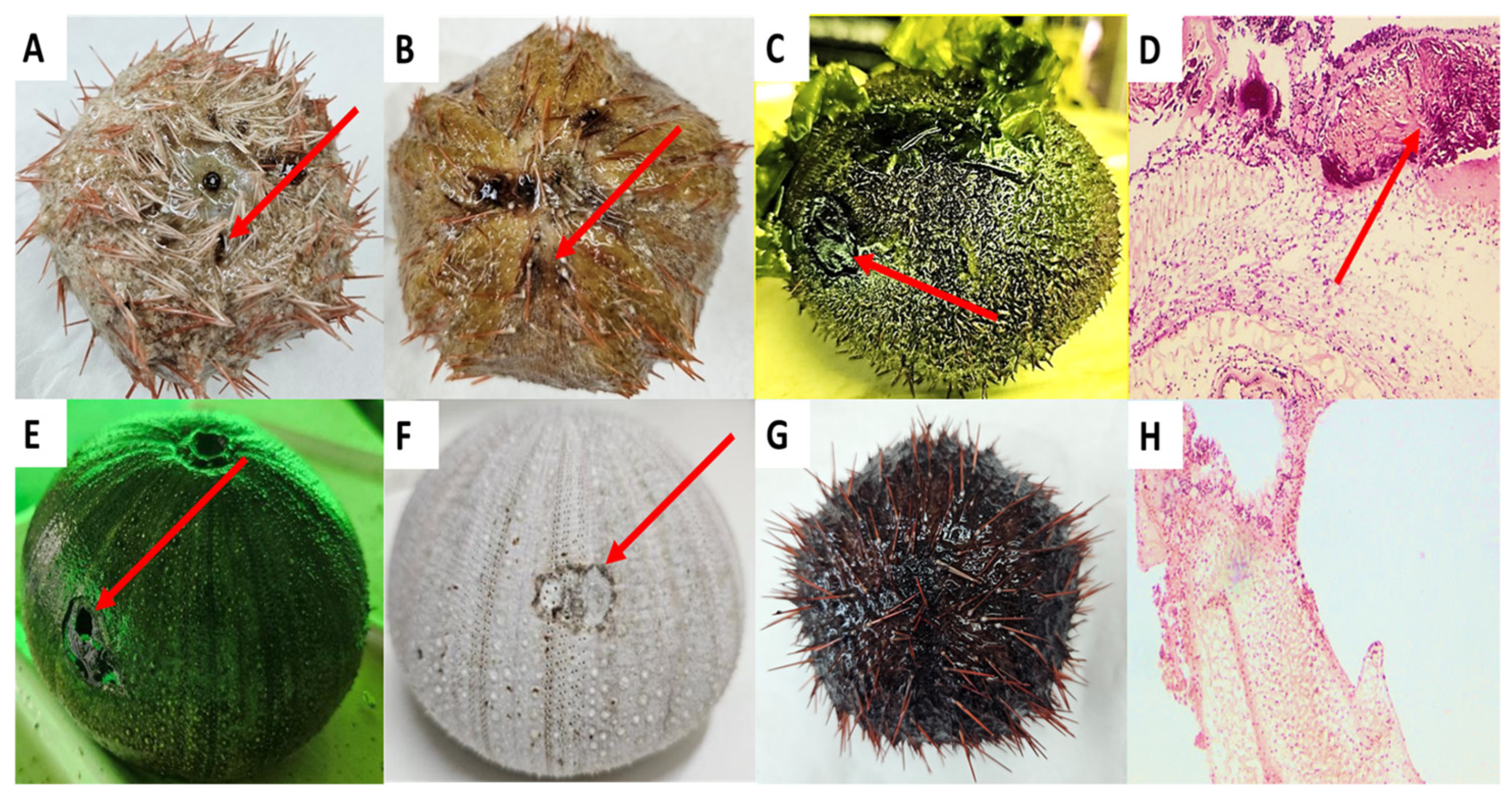
2.1. Identification of Red-Spotting-Infected Sea Urchins

Red spotting disease in the echinoculture facility at the National Center for Mariculture in the Gulf of Aqaba, Eilat, Israel (NCM), was first observed in August 2021. The echinoculture facility is located in a greenhouse and consists of ~600 individuals of Tripneustes gratilla elatensis spread over 6 U-shaped polyvinyl chloride tanks (5.3 × 0.60 × 0.3 m) [42]. The echinoculture facility at the NCM is part of an integrated multi-trophic aquaculture system (IMTA) consisting of sea urchin tanks, three fishponds for the culture of sea bream (Sparus aurata), and one culture pond for seaweed Ulva fasciata that also functions as a biofilter for the discharged effluent, as described elsewhere [42]. The echinoculture receives fresh seawater from the Gulf of Aqaba, pumped from about 300 m off-shore at a depth of 13 m (320°29′ N and 580°34′ E), which is collected into a 10 m3 header tank and then routed to the urchin culture tanks. The seawater flow rate to the sea urchin culture tanks is set at 5 m3 h−1, recommended for maintaining high-quality water [43]. Red spotting was diagnosed independently by several experts. The first was the expert aquatic pathobiologist at the NCM (Dr. Galit Sharon, DVM), the second was the pathobiology research team at the NCM (led by Dr. Rosa Strem), and the third was the local research assistant at the NCM, Mr. David Ben-Ezra, all of whom havin over 30 years of experience in sea urchins culture and research. In addition, at the same time of appearance in the echinoculture, the occurrence of red spotting was identified also in the wild population of T. gratilla in the coastal area of the Gulf of Eilat by staff members of the National Monitoring Program of The Gulf of Eilat and the Israeli National Parks Authority. The clinical symptoms of red spotting are unique compared to other diseases. Foremost, the appearance of red–purple spots on the skeleton, which is identified right after the detachment of spines from the infected area. The red–purple dots develop into red–purple patches and plaques which are further spread to new parts on the skeleton until the tissue is totally damaged and a hole appears in the skeleton, leading to the animal’s death (Figure 1). Since the discovery of red spotting in the echinoculture, the rapid monitoring of the animals and their culture environment has taken place, accompanied by the sampling and examining of the obtained infected animals and their tissues, as compared to healthy individuals and tissues.

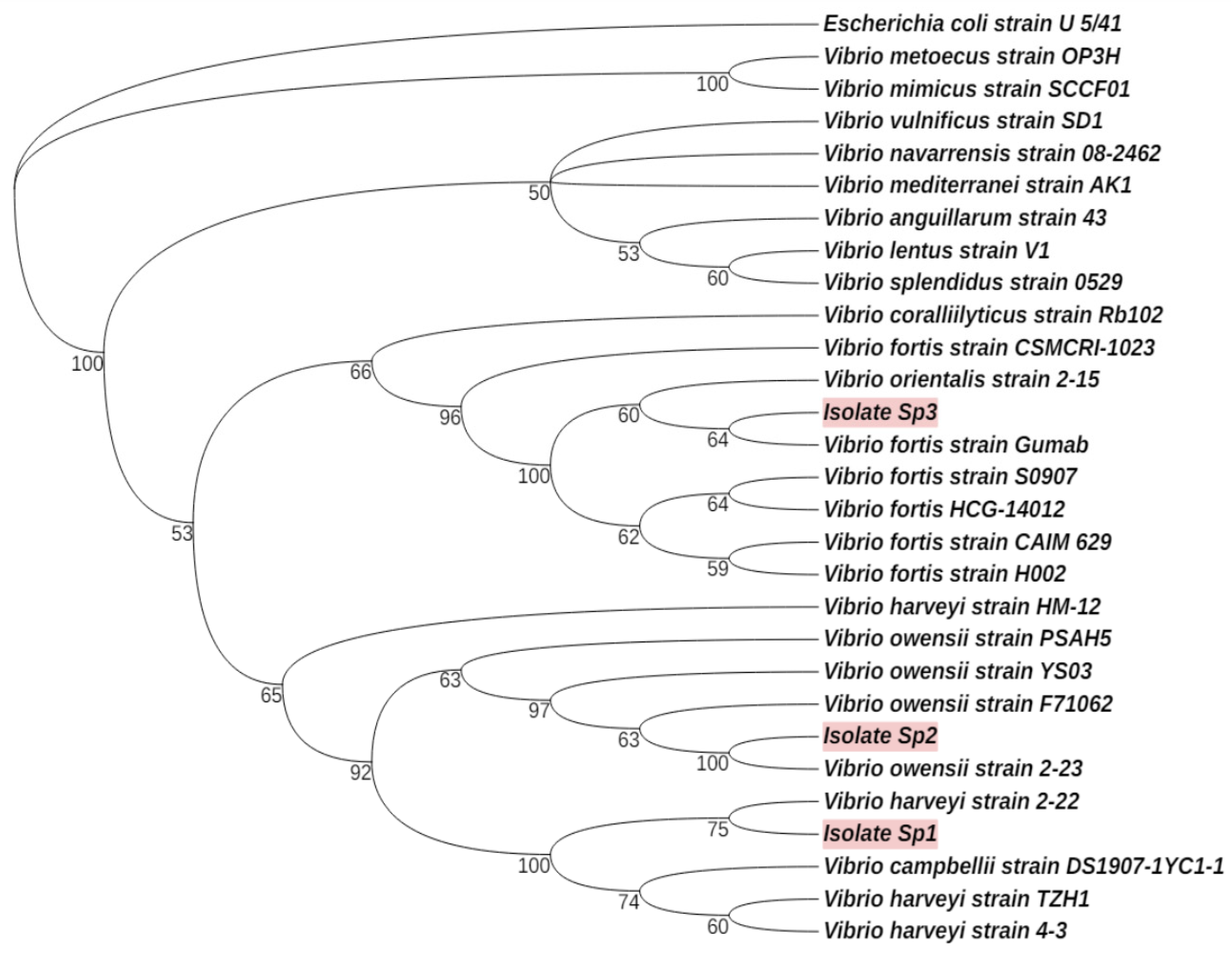
2.2. Bacterial Isolation and Identification

Following the diagnosis of red-spotting-infected sea urchins, infected and healthy tissues were sampled from infected and healthy individuals. Infected tissues were sampled from 10 infected individuals while for each individual, an additional tissue sample was taken from a distant, non-infected area on the skeleton. In addition, 6 healthy sea urchins were randomly sampled for healthy tissues. While aiming to identify the primary pathogen associated with red spotting, infected samples were such that exhibited the early signs of the disease, i.e., a small red–purple spot. All tissue samples were collected by a gentle scrubbing of the sampled area using sterile swab sticks. The sampled tissues were sown immediately on agar plates with a medium of thiosulfate citrate bile salts (TCBS). All developed colonies were transferred to new, similar agar plates until an isolated colony was observed. Following this procedure, isolated colonies were sampled for DNA extraction using the PureLinkTM Microbiome DNA Purification Kit (Thermo Fisher Scientific, Waltham, MA, USA) according to the manufacturer’s protocol. The DNA extracts were further transferred to amplify a targeted region in the 16SrRNA gene using a polymerase chain reaction (PCR) and the recommended primers set of 515F (GTGYCAGCMGCCGCGGTAA) and 926R (CCGYCAATTYMTTTRAGTTT). The amplification process included denaturation at 95 °C for 5 min, 28 cycles at 94 °C for 45 s, 50 °C for 60 s, 72 °C for 90 s, and a final elongation at 72 °C for 10 min. PCR products were run on an agarose gel to verify the length of the amplified sequence and sent for Sanger sequencing (sequencing with reverse and forward primers) at a certified sequencing lab (Hy-Labs Ltd., Rehovot, Israel). The sequencing results of both the reverse and forward primers were analyzed using BLAST software (https://blast.ncbi.nlm.nih.gov/Blast.cgi, accessed on 28 November 2024). Forward and reverse sequences were aligned while confirming their matching and further analyzed to identify their closest relatives. A similar bacteria isolation and identification procedure was performed using tissue scrubs from non-infected regions on the skeleton. Bacterial isolates that were found in both the infected and healthy tissues were sorted out to continue the research on the bacterial isolates that were present only in the infected tissue while not present in any of the samples of the healthy tissue. The 16S rRNA gene sequences of three bacterial isolates that were assumed to be pathogens in red spotting disease were deposited in the GenBank database under the respective accession numbers HQ449976.1, MT510177.1, and MT510178.1.

2.3. In Vitro Antibiotics Susceptibility Assay

The susceptibility of the isolated Vibrio strains to antibiotics was measured in vitro using the Kirby–Bauer disc diffusion method [44]. The following are the examined antibiotics and their doses (amount is per disc): amoxicillin (30 µg), ampicillin (10 µg), tetracycline (30 µg), erythromycin (15 µg), nitrofurantoin (300 µg), and sulfamethoxazole (25 µg). These antibiotics were selected because aquaculturists commonly use them for disease control and prevention [45]. Moreover, their target sites and activity mechanisms are also different [46]. The assay allows us to assess the extent to which a given bacterium resists antibiotic treatment and each compound’s feasibility and relative efficacy for controlling different Vibrio strains. The assay was performed separately for each bacterium and drug in triplicates. This means that in each trial, one of the bacteria was examined in three different agar plates (as triplicates) that consisted of a specific drug. In each plate, a bacterial colony was sown uniformly on the TSA agar surface and exposed to a Kirby–Bauer disc that was submerged in advance in a ready solution with the tested antibiotic compound. This allowed the diffusion of the harmful compound into the agar surface where bacteria grow. Following this procedure, the plate was transferred to incubation at 24 °C for 24–48 h. As a control, the same procedure was conducted using a similar set of plates (in triplicates) that consisted of the examined bacterium, but with an antibiotic-free disc. Following incubation, the diameter of the zone of inhibition was measured to evaluate the efficacy of each antibiotic in inhibiting bacterial growth and the sensitivity of the isolate to the antibiotic. The sensitivity rate of the bacterium to the antibiotic was classified according to the growth inhibition zone and rated as either sensitive (S), intermediate (I), or resistant (R). The intermediate susceptibility values in the diameter of the inhibition zone for the antibiotics ampicillin, tetracycline, erythromycin, nitrofurantoin, and sulfamethoxazole were defined both under the interpretive criteria standards by the Clinical and Laboratory Standards Institute (CLSI, 2012, [47]) and following the recommended values [48] for the antibiotic amoxicillin as follows: Amoxicillin: 19–20 mm; Ampicillin: 16–17 mm; Tetracycline: 15–18 mm; Erythromycin: 14–22 mm; Nitrofurantoin: 15–16 mm; and Sulfamethoxazole: 11–15mm. A bacterium was defined as either sensitive or resistant to the examined antibiotic if the measured diameter of the inhibition zone was extended or shortened, respectively, compared to the defined value for intermediate susceptibility.

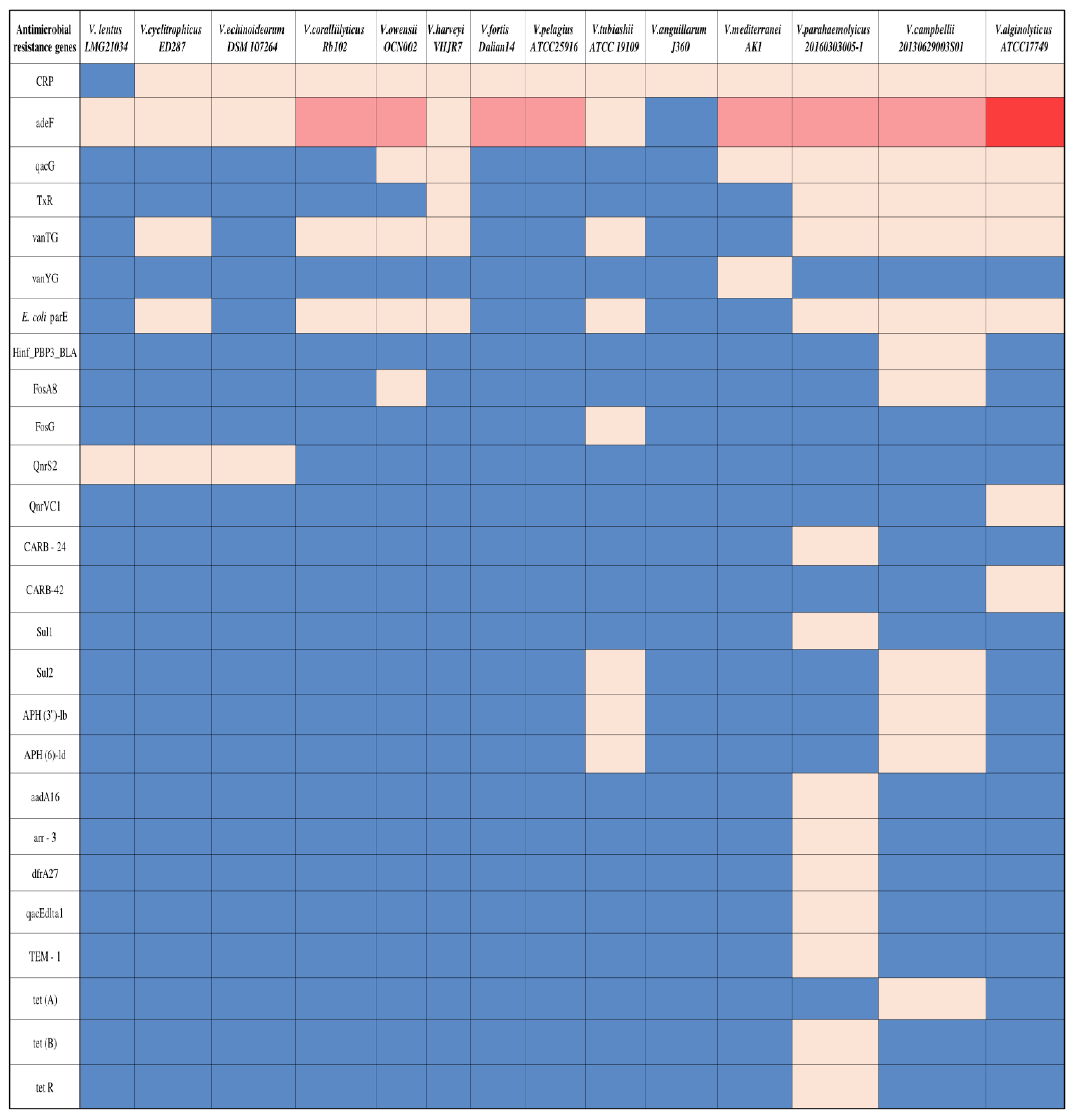
2.4. In Silico Survey of Antibiotics Resistance in Vibrio spp.

A genomic survey of antibiotic resistance genes was performed on 14 Vibrio bacteria of different species that have been identified as aquatic pathogens and have had their genome deposited in the Genbank database, as detailed in Table 2. Among the analyzed genomes, five are pathogenic Vibrio spp. that harm sea urchins, while the other nine are pathogenic Vibrio spp. that harm fish, crustaceans, or corals (Table 2). Concerning the here-isolated Vibrio bacteria, a genomic analysis was performed using genomes of pathogenic Vibrio with an identical 16S rRNA gene sequence, as confirmed by Basic Local Alignment Search Tool analysis (BLAST). All genomes were analyzed for their content of antibiotic resistance genes using the web platform of the comprehensive antibiotics resistance database (CARD) and the resistance genes identifier (RGI) tool kit.

3. Results

3.1. Potential Vibrio Pathogens Associated with Red Spotting Disease in T. gratilla

Bacterial enrichment resulted in the isolation of three potential pathogens in red spotting. The isolated bacteria were considered as such because they were present only in the infected tissues and not in healthy tissue samples. The BLAST analysis of the sequenced region in the 16S rRNA gene of these bacteria revealed the first isolate (here referred to as Vibrio sp. 1 to be closely related to the V. harveyi strain 2–22 (99.61% similarity in the analyzed sequence of the 16S rRNA gene), the second isolate (here referred to as Vibrio sp. 2 to be closely related to the Vibrio owensii strain 2–23 (95.49%), and the third isolate (here referred to as Vibrio sp. 3 to be closely related to the V. fortis strain Gumab (96.81%). Then, 16S rRNA sequences were further analyzed against other available bacterial 16S rRNA sequences in the NCBI database to construct a phylogenetic tree (Figure 2). The analysis confirmed the results by BLAST and proposed bacterial isolate Vibrio sp. 1 as close to other strains of V. harveyi including the pathogenic V. harveyi S0908 [40], Vibrio sp. 2 close to other strains of V. owensii including the pathogenic V. owensii OCN002 [41], and Vibrio sp. 3 close to other strains of V. fortis including the virulent V. fortis Gumab [40].

3.2. In Vitro Antibiotic Resistance Assay

The susceptibility test confirmed the resistance of the three bacterial isolates to various antibiotics (Figure 3). Among the bacteria, Vibrio sp. 1. of the taxon V. harveyi revealed a high resistance to all six examined antibiotics. At the same time, none of these substances inhibited its growth, even slightly. All examined bacteria are defined as resistant to tetracycline, amoxicillin, and ampicillin. Nitrofurantoin and sulfamethoxazole significantly inhibited the growth of Vibrio sp. 2 and 3 (Figure 3). According to the measured diameter of the inhibition zone in the cultures of these two bacteria, Vibrio sp. 3 of the taxon V. fortis is considered more sensitive to the different antibiotics than Vibrio sp. 2 of the taxon V. owensii.

3.3. Antibiotic-Resistant Genes of Vibrio spp.

The in silico analysis of 14 genomes of Vibrio spp. revealed a total of 26 ARGs (Table 2). The different ARGs target various drugs, including macrolides, penams, fluoroquinolones, tetracycline, glycopeptides, phosphonic acids, cephalosporin, cephamycin, sulfonamides, aminoglycosides, rifamycin, diaminopyrimidine, and cephalosporin, via various mechanisms, as described in Table 3. Among the examined bacteria, V. parahaemolyticus 20160303005-1 is considered the richest in ARG content with 15 different ARGs, 9 of them present only in the genome of this bacterium (Figure 4). In contrast, Vibrio anguillarum J360 contained only one copy of the CRP (C-reactive protein) gene in its genome and was considered the poorest among the examined bacteria. V. lentus LMG21034 is also relatively poor in ARGs, having only the adeF and QnrS2 genes that may provide immunity against fluoroquinolones. Among the detected genes, CRP, which is responsible for resistance to macrolides (e.g., erythromycin), penams (e.g., ampicillin and amoxicillin), and fluroquinolones, is the most common among the examined Vibrio genomes, being present in 13 of the 14 bacteria (V. lentus LMG21034 the exception). The adeF gene that is responsible for resistance to fluoroquinolone and tetracycline is also shared by 13 strains, with V. anguillarum J360 being the sole exception. Some detected antibiotic-resistant genes provide immunity against the same drug but via different mechanisms (Table 3). For example, CRP and E. coli parE genes are associated with resistance to fluoroquinolone and tetracycline, but the immune mechanism against these drugs differs. Furthermore, 50% of the examined genomes (7 out of 14 strains) included both genes in their genome. Six of the other seven genomes had only the CRP gene, while V. lentus LMG21034 contained neither CRP nor E. coli parE (Figure 4).

4. Discussion

Aquatic antibiotic-resistant pathogens threaten the aquaculture industry by limiting the list of efficient bioactive compounds for disease control and spreading immunity against such compounds. The current research provides a first prognosis on the potential association of three isolated strains of Vibrio with a red spotting infection in the sea urchin T. gratilla elatensis. Furthermore, the in vitro susceptibility trials and in silico genomic analyses provide innovative insight into the diverse arsenal of the ARGs of these and other pathogenic Vibrio, emphasizing strain variability at both the genus and species level. The research underlies the complexity of controlling pathogenic Vibrio in mariculture facilities, including in T. gratilla echinocultures.
Strain variability in pathogenic Vibrio bacteria was evident from the in silico screening of ARGs in the bacterial genomes and the in vitro susceptibility assays. Out of 14 bacterial genomes, only 2 bacteria, V. harveyi VHJR7 and V. coralliilyticus RB102, revealed a similar arsenal of ARGs, but with differences in the gene copy numbers. Consistent with the results at the genomic level, the in vitro trials also identified dissimilarity between the bacterial isolates concerning their immunity against the various antibiotics. Furthermore, we also report an inter-specific variability in antibiotic resistance. For example, the resistance to nitrofurantoin and sulfamethoxazole that was measured in the isolated V. harveyi was not evident in its closely related strain V. harveyi VHJR7, which lack genes like oqxA and oqxB for nitrofurantoin resistance or Sul1 and Sul2 for sulfamethoxazole resistance [62,63]. Another example is the here-isolated V. fortis-like bacterium, which resisted ampicillin, erythromycin, amoxicillin, and tetracycline, as compared to its close relative strain of V. fortis dalian14 that consists of only two ARGs of the adeF and CRP that may not be sufficient for resisting aminopenicillins like ampicillin or amoxicillin. The variation in susceptibility to antibiotics at the strain level, which is reported here for V. owensii, V. fortis, and V. harveyi, has been supported by other studies that challenged different isolates with different drugs [64,65,66]. Another important aspect of studying strain variability is identifying novel resistance or susceptibility. Such can be defined when challenging a known strain with compounds that have yet to be examined or refuting a reported resistance or susceptibility. Concerning the current isolated strains, the immunity of V. harveyi to the different antibiotics is not novel and agrees with other studies that reported immunity against these compounds in different strains [67,68]. While results of previous studies support the here-measured resistance of the isolated V. fortis-like bacterium to tetracycline and ampicillin [69,70], the sensitivity to erythromycin seems to be unique to the current isolated strain as compared to other V. fortis bacteria that were found to be resistant to this drug [64,71]. As for the V. owensii-like isolate, the here-measured immunity against tetracycline, erythromycin, amoxicillin, and ampicillin has been reported in other strains of this taxon [72,73]. Still, the identified sensitivity to sulfamethoxazole seems unique to the here-isolated strain since other studies reported resistance to this drug by various strains of V. owensii [69,73,74]. Moreover, susceptibility to sulfamethoxazole, which was found here in both the V. fortis and V. owensii-like isolates, can be considered rare in the Vibrio taxon, as demonstrated by the collection of at least 315 strains of Vibrio from various wastewater plants in South Africa and Nigeria that were found to be resistant to this drug [75,76].
Even though we have evident strain variability, our in vitro and in silico data also propose a core resistance to tetracyclines and penams like amoxicillin and ampicillin. The resistance to these compounds is widespread in Vibrio bacteria [26,77] and may be associated with the presence of the adeF and CRP genes in the genome [73]. The presence of adeF and CRP as core genes can induce resistance to other bioactive compounds of the fluoroquinolones, such as ciprofloxacin and gemifloxacin, but such bioactive chemicals are not as common as tetracyclines for disease-control in aquaculture [73]. While the three bacterial isolates share resistance to other antibiotics like ampicillin, erythromycin, and amoxicillin, their close relatives may only share resistance to erythromycin derived by the presence of the CRP gene, which induces immunity to macrolides [78]. Interestingly, the current research highlights a more remarkable similarity in antibiotic resistance between the different isolated strains of Vibrio than between the different species for which genomes were analyzed. We assume that the shared immunity against multiple drugs by the isolated strains is due to their shared origin of the red-spotting-infected tissue of sea urchins in the echinoculture. Notably, various studies confirmed that aquaculture systems are a great source of bacteria with a shared resistance to multi-drugs. One example is a shared resistance to ampicillin, erythromycin, sulfamethoxazole, tetracyclines, oxytetracycline, and chloramphenicol that was identified in isolates of Vibrio fluvialis, V. parahaemolyticus, and V. vulnificus during four independent samplings in four fishpond effluents in Benin City, Nigeria [79]. A similar phenomenon has been evidenced recently in a seafood market in Italy where Vibrio isolates from purchasable mussels shared immunity against various drugs, although their host mussels reached the market from different farms in the area [80]. These phenomena highlight the role of HGT in the aquaculture environment in providing immunity against various drugs to the resident Vibrio bacteria, including the aquaculture pathogens within this taxon. Although bacterial genomes are preserved over a short scale of time, i.e., from one generation to the following [81], plasticity, such as the acquired immunity against drugs, can be achieved over the longer run, i.e., throughout several generations, through horizontal gene transfer, mutations, and the transfer of mobile genetic elements [82,83]. One reason for this is the fact that the set conditions in the aquaculture facility, like the temperature, pH, and aerobic environment, favor not only the cultured species, but also support the succession of pathogenic and non-pathogenic Vibrio [84], which can further distribute antibiotic resistance in this environment through HGT. Diseases and the administration of antibiotics stimulate pathogen succession, expanding antibiotic resistance within the local microbial community, including between pathogens in the community [85,86]. That said, our study supports the claim that the aquaculture environment accelerates genomic modifications toward immunity against multiple drugs shared by various local bacteria. We assume that the development of immunity against any particular antibiotic is associated with the exposure of the bacterium to this drug or a bacterium that carries and spreads the required genetic elements for resistance. This can also explain the core- and strain-unique resistance, or susceptibility, within members of the Vibrio taxon.
The current study seems to be the first to report red spotting in T. gratilla sea urchins, the potential causative agents, and their immunity against various antibiotics. This knowledge is essential for decision making concerning the efficient treatment for eliminating such bacteria in echinocultures if needed. T. gratilla possesses a high commercial value due to its rapid growth and high nutritional value [87]. Despite its importance in seafood markets, reports on diseases that harm this species are relatively rare and seem to include only the lesions syndrome [51,88] and the bald sea urchin disease [89]. While not documented previously in T. gratilla, red spotting disease has been reported in another valuable sea urchin, S. intermedius, in nature and echinoculture [1,2], causing severe damage with a mass mortality of 90% of the culture [2].
Vibrio bacteria’s reputation for causing disease encompasses various invertebrates and vertebrates, including sea urchins. Among the diseases associated with the taxon in sea urchins are vibriosis, black peristomial, bald sea urchins, lesion syndrome, spotting, and red spotting. V. splendidus is probably the most infamous taxon, associated with five diseases, followed by V. harveyi and V. fortis, which were associated with both the lesion syndrome and spotting disease [35,51] in S. intermedius and now in red spotting in T. gratilla in the current study. An interesting phenomenon is the co-occurrence of these two bacteria in samples from the infected tissue of the sea urchins that were examined in these four independent studies, while none of the sea urchin diseases surveyed here reported on the isolation of only one of those two taxa and not the other. When examined for virulence, V. fortis or V. harveyi negatively affected naïve animals [35,40]. We assume that the co-occurrence of V. fortis and V. harveyi is not by chance and may hint that both harm the sea urchin while moribund, including in the red spotting of T. gratilla in the current research. Identifying these two bacteria, as well as V. owensii, as potential pathogens of the red spotting in T. gratilla is novel, as this disease has received less attention in research on sea urchins in general and on T. gratilla specifically. The fact that none of the three isolates were detected in non-infected individuals or in healthy tissues strengthens our assumption regarding their association with this disease in the echinoculture. The here-reported data on the resistance to multiple drugs by these isolates are essential for developing protocols for their elimination from red-spotting-infected echinocultures and their prevention via prophylactic treatments. Concerning the in vitro and genomics analyses, we propose that only nitrofurantoin and sulfamethoxazole may be helpful against V. fortis and V. owensii-like pathogens, but not against the V. harveyi-like one. The immunity of the isolated V. harveyi to sulfamethoxazole should be taken seriously into consideration since inheriting resistance to this antibiotic is likely through the horizontal transfer of integrating conjugative elements such as SXT, which carries multiple resistance genes against sulfamethoxazole, streptomycin, and trimethoprim [90]. On the other hand, the development and conferring of resistance to nitrofurantoin has been proposed to be relatively slow through mutations in genes like nfsA, nfsB, and ribE [91]. Therefore, using nitrofurantoin in echinocultures with red spotting outbreaks seems reasonable in parallel to studies of other drugs against these potential pathogens.

5. Conclusions

To conclude, the current research proposes three strains of Vibrio as associated with the red spotting infection in the sea urchin T. gratilla elatensis. Unfortunately, yet not surprising, all three strains exhibit multi-drug resistance which may enable them to withstand the common treatments with antibiotics in echinocultures. In agreement with the knowledge that was gained here from the genomic survey of harmful pathogens of the Vibrio, the here-reported strains also show a different profile of antibiotic resistance for each. Thus emphasizing the variability at both the genus and species level and the difficulty in disease controlling when caused by Vibrio.

Author Contributions

M.B.N.: Investigation, Methodology, Data curation, Writing—original draft. M.M.: Investigation, Software. N.S.: Validation. L.G.: Conceptualization, Methodology, Validation, Writing—review and editing, Funding acquisition, Supervision. All authors have read and agreed to the published version of the manuscript.

Funding

This research was funded by The Israeli Ministry of Health, Grant No. 3-0000-17701.

Institutional Review Board Statement

This study was approved by the Animal Experimentation Ethics Committee, Agricultural Research Organization, The Volcani Center. Experiments were conducted following the local legislation and institutional requirements.

Data Availability Statement

The authors declare that all of the data that were used in this research are available and will be provided upon request due to privacy or ethical restrictions.

Acknowledgments

The authors thank the local technical and research assisting staff at The NCM. We also thank Galit Sharon, Rosa Sterm, and Roberto Erlich from the pathobiology department at the NCM for their assistance in disease diagnosis and helpful technical advice.

Conflicts of Interest

The authors declare no conflicts of interest.

References

  1. Li, R.; Dang, H.; Huang, Y.; Quan, Z.; Jiang, H.; Zhang, W.; Ding, J. Vibrio coralliilyticus as an agent of red spotting disease in the sea urchin Strongylocentrotus intermedius. Aquac. Rep. 2020, 16, 100244. [Google Scholar] [CrossRef]
  2. Wang, B.; Li, Y.; Li, X.; Qu, J.Y.; Zhao, X.M. Pathogenic mechanism of causative Vibrio found in “red spotting” diseased sea urchin Strongylocentrotus intermedius. J. Dalian Fish. Univ. 2005, 20, 11–15. (In Chinese) [Google Scholar]
  3. Wang, L.; He, B.; Chang, Y.; Ding, J. Characterization of the bacterial community associated with red spotting disease of the echinoid Strongylocentroyus intermedius. Aquaculture 2020, 529, 735606. [Google Scholar] [CrossRef]
  4. Zhang, X.H.; He, X.; Austin, B. Vibrio harveyi: A serious pathogen of fish and invertebrates in mariculture. Mar. Life Sci. Technol. 2020, 2, 231–245. [Google Scholar] [CrossRef]
  5. Zhou, J.; Fang, W.; Yang, X.; Zhou, S.; Hu, L.; Li, X.; Qi, X.; Su, H.; Xie, L. A nonluminescent and highly virulent Vibrio harveyi strain is associated with “bacterial white tail disease” of Litopenaeus vannamei shrimp. PLoS ONE 2012, 7, e29961. [Google Scholar] [CrossRef]
  6. Marti, E.; Variatza, E.; Balcazar, J.L. The role of aquatic ecosystems as reservoirs of antibiotic resistance. Trends Microbiol. 2014, 22, 36–41. [Google Scholar] [CrossRef]
  7. Stalder, T.; Barraud, O.; Casellas, M.; Dagot, C.; Ploy, M.C. Integron involvement in environmental spread of antibiotic resistance. Front. Microbiol. 2012, 3, 119. [Google Scholar] [CrossRef]
  8. Kumarage, P.M.; De Silva, L.A.D.S.; Heo, G.J. Aquatic environments: A potential source of antimicrobial-resistant Vibrio spp. J. Appl. Microbiol. 2022, 133, 2267–2279. [Google Scholar] [CrossRef]
  9. Kalantzi, I.; Rico, A.; Mylona, K.; Pergantis, S.A.; Tsapakis, M. Fish farming, metals and antibiotics in the eastern Mediterranean Sea: Is there a threat to sediment wildlife? Sci. Total Environ. 2021, 764, 142843. [Google Scholar] [CrossRef]
  10. Turk, J. Meeting projected food demands by 2050: Understanding and enhancing the role of grazing ruminants. J. Anim. Sci. 2016, 94 (Suppl. S6), 53–62. [Google Scholar] [CrossRef]
  11. Pepi, M.; Focardi, S. Antibiotic-resistant bacteria in aquaculture and climate change: A challenge for health in the Mediterranean area. Int. J. Environ. Res. Public Health 2021, 18, 5723. [Google Scholar] [CrossRef]
  12. Manyi-Loh, C.; Mamphweli, S.; Meyer, E.; Okoh, A. Antibiotic use in agriculture and its consequential resistance in environmental sources: Potential public health implications. Molecules 2018, 23, 795. [Google Scholar] [CrossRef] [PubMed]
  13. Heuer, O.E.; Kruse, H.; Grave, K.; Collignon, P.; Karunasagar, I.; Angulo, F.J. Human health consequences of use of antimicrobial agents in aquaculture. Clin. Infect. Dis. 2009, 49, 1248–1253. [Google Scholar] [CrossRef] [PubMed]
  14. Rico, A.; Phu, T.M.; Satapornvanit, K.; Min, J.; Shahabuddin, A.M.; Henriksson, P.J.; Murray, F.J.; Little, D.C.; Dalsgaard, A.; Brink, P.J.V.D. Use of veterinary medicines, feed additives and probiotics in four major internationally traded aquaculture species farmed in Asia. Aquaculture 2013, 412, 231–243. [Google Scholar] [CrossRef]
  15. Amarasiri, M.; Sano, D.; Suzuki, S. Understanding human health risks caused by antibiotic resistant bacteria (ARB) and antibiotic resistance genes (ARG) in water environments: Current knowledge and questions to be answered. Crit. Rev. Environ. Sci. Technol. 2020, 50, 2016–2059. [Google Scholar] [CrossRef]
  16. Hossain, A.; Habibullah-Al-Mamun, M.; Nagano, I.; Masunaga, S.; Kitazawa, D.; Matsuda, H. Antibiotics, antibiotic-resistant bacteria, and resistance genes in aquaculture: Risks, current concern, and future thinking. Environ. Sci. Pollut. Res. 2022, 29, 11054–11075. [Google Scholar] [CrossRef]
  17. Dewi, R.R.; Hassan, L.; Daud, H.M.; Matori, M.F.; Nordin, F.; Ahmad, N.I.; Zakaria, Z. Prevalence and antimicrobial resistance of Escherichia coli, Salmonella and Vibrio derived from farm-raised Red Hybrid Tilapia (Oreochromis spp.) and Asian Sea Bass (Lates calcarifer, Bloch 1970) on the west coast of Peninsular Malaysia. Antibiotics 2022, 11, 136. [Google Scholar] [CrossRef]
  18. Hirshfeld, B.; Lavelle, K.; Lee, K.Y.; Atwill, E.R.; Kiang, D.; Bolkenov, B.; Gaa, M.; Li, Z.; Yu, A.; Li, X.; et al. Prevalence and antimicrobial resistance profiles of Vibrio spp. and Enterococcus spp. in retail shrimp in Northern California. Front. Microbiol. 2023, 14, 1192769. [Google Scholar] [CrossRef]
  19. Preena, P.G.; Swaminathan, T.R.; Kumar, V.J.R.; Singh, I.S.B. Antimicrobial resistance in aquaculture: A crisis for concern. Biologia 2020, 75, 1497–1517. [Google Scholar] [CrossRef]
  20. Vaiyapuri, M.; Pailla, S.; Rao Badireddy, M.; Pillai, D.; Chandragiri Nagarajarao, R.; Prasad Mothadaka, M. Antimicrobial resistance in Vibrios of shrimp aquaculture: Incidence, identification schemes, drivers and mitigation measures. Aquac. Res. 2021, 52, 2923–2941. [Google Scholar] [CrossRef]
  21. Dang, H.; Zhang, X.; Song, L.; Chang, Y.; Yang, G. Molecular determination of oxytetracycline-resistant bacteria and their resistance genes from mariculture environments of China. J. Appl. Microbiol. 2007, 103, 2580–2592. [Google Scholar] [CrossRef] [PubMed]
  22. Buschmann, A.H.; Tomova, A.; López, A.; Maldonado, M.A.; Henríquez, L.A.; Ivanova, L. Salmon aquaculture and antimicrobial resistance in the marine environment. PLoS ONE 2012, 7, e42724. [Google Scholar] [CrossRef] [PubMed]
  23. Shah, S.Q.; Cabello, F.C.; L’Abée-Lund, T.M.; Tomova, A.; Godfrey, H.P.; Buschmann, A.H.; Sørum, H. Antimicrobial resistance and antimicrobial resistance genes in marine bacteria from salmon aquaculture and non-aquaculture sites. Environ. Microbiol. 2014, 16, 1310–1320. [Google Scholar] [CrossRef] [PubMed]
  24. Akinbowale, O.L.; Peng, H.; Barton, M.D. Antimicrobial resistance in bacteria isolated from aquaculture sources in Australia. J. Appl. Microbiol. 2006, 100, 1103–1113. [Google Scholar] [CrossRef] [PubMed]
  25. Tendencia, E.A.; de la Peña, L.D. Antibiotic resistance of bacteria from shrimp ponds. Aquaculture 2001, 195, 193–204. [Google Scholar] [CrossRef]
  26. Sony, M.; Sumithra, T.G.; Anusree, V.N.; Amala, P.V.; Reshma, K.J.; Alex, S.; Sanil, N. Antimicrobial resistance and virulence characteristics of Vibrio vulnificus, Vibrio parahaemolyticus and Vibrio harveyi from natural disease outbreaks of marine/estuarine fishes. Aquaculture 2021, 539, 736608. [Google Scholar] [CrossRef]
  27. Dang, H.; Zhao, J.; Song, L.; Chen, M.; Chang, Y. Molecular characterizations of chloramphenicol-and oxytetracycline-resistant bacteria and resistance genes in mariculture waters of China. Mar. Pollut. Bull. 2009, 58, 987–994. [Google Scholar] [CrossRef]
  28. Ferrini, A.M.; Mannoni, V.; Suffredini, E.; Cozzi, L.; Croci, L. Evaluation of antibacterial resistance in Vibrio strains isolated from imported seafood and Italian aquaculture settings. Food Anal. Methods 2008, 1, 164–170. [Google Scholar] [CrossRef]
  29. Ottaviani, D.; Bacchiocchi, I.; Masini, L.; Leoni, F.; Carraturo, A.; Giammarioli, M.; Sbaraglia, G. Antimicrobial susceptibility of potentially pathogenic halophilic Vibrios isolated from seafood. Int. J. Antimicrob. Agents 2001, 18, 135–140. [Google Scholar] [CrossRef]
  30. Fukuda, A.; Tsunashima, R.; Usui, M. Antimicrobial Resistant Bacteria Monitoring in Raw Seafood Retailed: A Pilot Study Focused on Vibrio and Aeromonas. Food Saf. 2023, 11, 65–77. [Google Scholar] [CrossRef]
  31. Narayanan, S.V.; Joseph, T.C.; Peeralil, S.; Mothadaka, M.P.; Lalitha, K.V. Prevalence, virulence characterization, AMR pattern and genetic relatedness of Vibrio parahaemolyticus isolates from retail seafood of Kerala, India. Front. Microbiol. 2020, 11, 525327. [Google Scholar] [CrossRef] [PubMed]
  32. Neetoo, H.; Reega, K.; Manoga, Z.S.; Nazurally, N.; Bhoyroo, V.; Allam, M.; Jaufeerally-Fakim, Y.; Ghoorah, A.W.; Jaumdally, W.; Hossen, A.M.; et al. Prevalence, genomic characterization, and risk assessment of human pathogenic Vibrio species in seafood. J. Food Prot. 2022, 85, 1553–1565. [Google Scholar] [CrossRef] [PubMed]
  33. Zhao, T.; Liu, L.; Li, C.; Zou, Y.; Yan, H.; Zhan, Y.; Chang, Y. Isolation and characterization of four pathogenic strains of Vibrio spp. from farmed sea urchins, Strongylocentrotus intermedius, in China. Microb. Pathog. 2022, 169, 105651. [Google Scholar] [CrossRef] [PubMed]
  34. Hira, J.; Stensvåg, K. Evidence for association of Vibrio echinoideorum with tissue necrosis on test of the green sea urchin Strongylocentrotus droebachiensis. Sci. Rep. 2022, 12, 4859. [Google Scholar] [CrossRef] [PubMed]
  35. Wang, Y.N.; Liu, Y.P.; Chang, Y.Q. Comparative analysis of bacterial community composition in coelomic fluid between sick and healthy Strongylocentrotus intermedius by PCR-DGGE. J. Agric. Sci. Technol. 2011, 13, 111–116. [Google Scholar]
  36. Gilles, K.W.; Pearse, J.S. Disease in sea urchins Strogylocentrotus purpuratus: Experimental infection and bacterial virulence. Dis. Aquat. Org. 1986, 1, 105–114. [Google Scholar] [CrossRef]
  37. Grech, D.; Mandas, D.; Farina, S.; Guala, I.; Brundu, R.; Cristo, B.; Panzalis, P.A.; Salati, F.; Carella, F. Vibrio splendidus clade associated with a disease affecting Paracentrotus lividus (Lamarck, 1816) in Sardinia (Western Mediterranean). J. Invertebr. Pathol. 2022, 192, 107783. [Google Scholar] [CrossRef]
  38. Sweet, M.; Bulling, M.; Williamson, J.E. New disease outbreak affects two dominant sea urchin species associated with Australian temperate reefs. Mar. Ecol. Prog. Ser. 2016, 551, 171–183. [Google Scholar] [CrossRef]
  39. Bauer, J.C.; Young, C.M. Epidermal lesions and mortality caused by vibriosis in deep-sea Bahamian echinoids: A laboratory study. Dis. Aquat. Org. 2000, 39, 193–199. [Google Scholar] [CrossRef]
  40. Wang, Y.; Feng, N.; Li, Q.; Ding, J.; Zhan, Y.; Chang, Y. Isolation and characterization of bacteria associated with a syndrome disease of sea urchin Strongylocentrotus intermedius in North China. Aquac. Res. 2013, 44, 691–700. [Google Scholar] [CrossRef]
  41. Sun, J.; Liu, L.; Song, J.; Zhan, Y.; Zhang, W.; Wang, B.; Zhang, B.; Chang, Y. Characterization of two strains of Vibrio sp. from cultured sea urchin, Strongylocentrotus intermedius, in China. Aquac. Rep. 2021, 20, 100667. [Google Scholar] [CrossRef]
  42. Shpigel, M.; Shauli, L.; Odintsov, V.; Ben-Ezra, D.; Neori, A.; Guttman, L. The sea urchin, Paracentrotus lividus, in an Integrated Multi-Trophic Aquaculture (IMTA) system with fish (Sparus aurata) and seaweed (Ulva lactuca): Nitrogen partitioning and proportional configurations. Aquaculture 2018, 490, 260–269. [Google Scholar] [CrossRef]
  43. Neori, A.; Shpigel, M.; Ben-Ezra, D. A sustainable integrated system for culture of fish, seaweed and abalone. Aquaculture 2000, 186, 279–291. [Google Scholar] [CrossRef]
  44. Biemer, J.J. Antimicrobial susceptibility testing by the Kirby-Bauer disc diffusion method. Ann. Clin. Lab. Sci. 1973, 3, 135–140. [Google Scholar] [PubMed]
  45. Schlossberg, D.L.; Samuel, R. Antibiotics Manual: A Guide to Commonly Used Antimicrobials, 2nd ed.; John Wiley & Sons: Hoboken, NJ, USA, 2017. [Google Scholar]
  46. Etebu, E.; Arikekpar, I. Antibiotics: Classification and mechanisms of action with emphasis on molecular perspectives. Int. J. Appl. Microbiol. Biotechnol. Res. 2016, 4, 90–101. [Google Scholar]
  47. M 100-S22; Performance Standards for Antimicrobial Susceptibility Testing; Twenty-Second Informational Supplement. Clinical and Laboratory Standards Institute: Malvern, PA, USA, 2012.
  48. Aljahani, A.H.; Alarjani, K.M.; Hassan, Z.K.; Elkhadragy, M.F.; Ismail, E.A.; Al-Masoud, A.H.; Yehia, H.M. Molecular detection of methicillin heat-resistant Staphylococcus aureus strains in pasteurized camel milk in Saudi Arabia. Biosci. Rep. 2020, 40, BSR20193470. [Google Scholar] [CrossRef]
  49. Ding, J.; Dou, Y.; Wang, Y.; Chang, Y. Draft genome sequence of Vibrio fortis Dalian14 isolated from diseased sea urchin (Strongylocentrotus intermedius). Genome Announc. 2014, 2, 10–1128. [Google Scholar] [CrossRef]
  50. Hira, J.; Bentdal, S.; Devold, H.; Stensvåg, K.; Landfald, B. Vibrio echinoideorum sp. nov., isolated from an epidermal lesion on the test of a green sea urchin (Strongylocentrotus droebachiensis). Int. J. Syst. Evol. Microbiol. 2019, 69, 2277–2282. [Google Scholar] [CrossRef]
  51. Wang, Y.N.; Chang, Y.Q.; Lawrence, J.M. Disease in sea urchins. In Developments in Aquaculture and Fisheries Science; Lawrence, J.M., Ed.; Elsevier: Amsterdam, The Netherlands, 2013; Volume 38, pp. 179–186‏. [Google Scholar] [CrossRef]
  52. Schuh, N.W.; Carrier, T.J.; Schrankel, C.S.; Reitzel, A.M.; Heyland, A.; Rast, J.P. Bacterial exposure mediates developmental plasticity and resistance to lethal Vibrio lentus infection in purple sea urchin (Strongylocentrotus purpuratus) larvae. Front. Immunol. 2020, 10, 491364. [Google Scholar] [CrossRef]
  53. Ransangan, J.; Lal, T.M.; Al-Harbi, A.H. Characterization and experimental infection of Vibrio harveyi isolated from diseased Asian seabass (Lates calcarifer). Malays. J. Microbiol. 2012, 8, 104–115. [Google Scholar] [CrossRef]
  54. Ushijima, B.; Smith, A.; Aeby, G.S.; Callahan, S.M. Vibrio owensii induces the tissue loss disease Montipora white syndrome in the Hawaiian reef coral Montipora capitata. PLoS ONE 2012, 7, e46717. [Google Scholar] [CrossRef] [PubMed]
  55. Fahmy, N.M. Isolation and characterization of Vibrio alginolyticus strain HAT3 causing skin ulceration disease in cultured sea cucumber Holothuria atra (Jaeger, 1833). Egypt. J. Aquat. Res. 2022, 48, 75–81. [Google Scholar] [CrossRef]
  56. Tarazona, E.; Lucena, T.; Arahal, D.R.; Macián, M.C.; Ruvira, M.A.; Pujalte, M.J. Multilocus sequence analysis of putative Vibrio mediterranei strains and description of Vibrio thalassae sp. nov. Syst. Appl. Microbiol. 2014, 37, 320–328. [Google Scholar] [CrossRef]
  57. Yang, F.; Xu, L.; Huang, W.; Li, F. Highly lethal Vibrio parahaemolyticus strains cause acute mortality in Penaeus vannamei post-larvae. Aquaculture 2022, 548, 737605. [Google Scholar] [CrossRef]
  58. Dong, X.; Wang, H.; Zou, P.; Chen, J.; Liu, Z.; Wang, X.; Huang, J. Complete genome sequence of Vibrio campbellii strain 20130629003S01 isolated from shrimp with acute hepatopancreatic necrosis disease. Gut Pathog. 2017, 9, 31. [Google Scholar] [CrossRef]
  59. Vasquez, I.; Cao, T.; Chakraborty, S.; Gnanagobal, H.; O’Brien, N.; Monk, J.; Boyce, D.; Westcott, J.D.; Santander, J. Comparative genomics analysis of vibrio anguillarum isolated from lumpfish (Cyclopterus lumpus) in newfoundland reveal novel chromosomal organizations. Microorganisms 2020, 8, 1666. [Google Scholar] [CrossRef]
  60. Richards, G.P.; Watson, M.A.; Needleman, D.S.; Church, K.M.; Häse, C.C. Mortalities of Eastern and Pacific oyster larvae caused by the pathogens Vibrio coralliilyticus and Vibrio tubiashii. Appl. Environ. Microbiol. 2015, 81, 292–297. [Google Scholar] [CrossRef]
  61. Villamil, L.; Figueras, A.; Toranzo, A.E.; Planas, M.; Novoa, B. Isolation of a highly pathogenic Vibrio pelagius strain associated with mass mortalities of turbot, Scophthalmus maximus (L.), larvae. J. Fish Dis. 2003, 26, 293–303. [Google Scholar] [CrossRef]
  62. Poey, M.E.; Azpiroz, M.F.; Laviña, M. On sulfonamide resistance, sul genes, class 1 integrons and their horizontal transfer in Escherichia coli. Microb. Pathog. 2019, 135, 103611. [Google Scholar] [CrossRef]
  63. Wang, Y.; Batra, A.; Schulenburg, H.; Dagan, T. Gene sharing among plasmids and chromosomes reveals barriers for antibiotic resistance gene transfer. Philos. Trans. R. Soc. B 2022, 377, 20200467. [Google Scholar] [CrossRef]
  64. Agboola, T.D.; Nmema, E.E.; Odetoyin, B.W. Distribution and antibiogram of Vibrio species from hospital wastewater in Southwest Nigeria. Pan Afr. Med. J. 2023, 45, 80. [Google Scholar] [CrossRef]
  65. Isnansetyo, A.; Istiqomah, I.; Anshary, H.; Sriwulan, S.; Yudiati, E.; Subagiyo, S.; Arif, A.; Kartikasari, D.W. Identification and antibiotic-resistant properties of Vibrio owensii and V. alginolyticus isolated from the Spermonde Islands, Indonesia. Biodiversitas J. Biol. Divers. 2022, 23. [Google Scholar] [CrossRef]
  66. Mohamad, N.; Amal, M.N.A.; Saad, M.Z.; Yasin, I.S.M.; Zulkiply, N.A.; Mustafa, M.; Nasruddin, N.S. Virulence-associated genes and antibiotic resistance patterns of Vibrio spp. isolated from cultured marine fishes in Malaysia. BMC Vet. Res. 2019, 15, 176. [Google Scholar] [CrossRef]
  67. Adeyemi, A.; Enyinnia, V.; Nwanze, R.; Smith, S.; Omonigbehin, E. Antimicrobial susceptibility of potentially pathogenic halophilic Vibrio species isolated from seafoods in Lagos, Nigeria. Afr. J. Biotechnol. 2008, 7, 3791–3794. [Google Scholar]
  68. Kang, H.; Yu, Y.; Liao, M.; Wang, Y.; Yang, G.; Zhang, Z.; Li, B.; Rong, X.; Wang, C. Physiology, metabolism, antibiotic resistance, and genetic diversity of Harveyi clade bacteria isolated from coastal mariculture system in China in the last two decades. Front. Mar. Sci. 2022, 9, 932255. [Google Scholar] [CrossRef]
  69. Badhai, J.; Kumari, P.; Krishnan, P.; Ramamurthy, T.; Das, S.K. Presence of SXT integrating conjugative element in marine bacteria isolated from the mucus of the coral Fungia echinata from Andaman Sea. FEMS Microbiol. Lett. 2013, 338, 118–123. [Google Scholar] [CrossRef]
  70. Gao, Q.; Ma, X.; Wang, Z.; Chen, H.; Luo, Y.; Wu, B.; Qi, S.; Lin, M.; Tian, J.; Qiao, Y.; et al. Seasonal variation, virulence gene and antibiotic resistance of Vibrio in a semi-enclosed bay with mariculture (Dongshan Bay, Southern China). Mar. Pollut. Bull. 2022, 184, 114112. [Google Scholar] [CrossRef]
  71. You, K.G.; Bong, C.W.; Lee, C.W. Antibiotic resistance and plasmid profiling of Vibrio spp. in tropical waters of Peninsular Malaysia. Environ. Monit. Assess. 2016, 188, 171. [Google Scholar] [CrossRef]
  72. Thillaichidambaram, M.; Narayanan, K.; Selvaraj, S.; Sundararaju, S.; Muthiah, R.C.; Figge, M.J. Isolation and characterization of Vibrio owensii from Palk Bay and its infection study against post larvae of Litopenaeus vannamei. Microb. Pathog. 2022, 172, 105751. [Google Scholar] [CrossRef]
  73. Wang, Z.; Wang, H.; Chen, D.; Li, Y. Genomic characterization and comparative genomic analysis of pathogenic Vibrio isolated from aquaculture-grown white-leg shrimp (Penaeus vannamei) in Guangdong and Jiangsu, China. Aquaculture 2024, 580, 740302. [Google Scholar] [CrossRef]
  74. Misol Jr, G.N.; Kokkari, C.; Katharios, P. Biological and genomic characterization of a novel jumbo bacteriophage, vB_VhaM_pir03 with broad host lytic activity against Vibrio harveyi. Pathogens 2020, 9, 1051. [Google Scholar] [CrossRef] [PubMed]
  75. Adesiyan, I.M.; Bisi-Johnson, M.A.; Okoh, A.I. Incidence of antibiotic resistance genotypes of Vibrio species recovered from selected freshwaters in Southwest Nigeria. Sci. Rep. 2022, 12, 18912. [Google Scholar] [CrossRef] [PubMed]
  76. Okoh, A.I.; Igbinosa, E.O. Antibiotic susceptibility profiles of some Vibrio strains isolated from wastewater final effluents in a rural community of the Eastern Cape Province of South Africa. BMC Microbiol. 2010, 10, 143. [Google Scholar] [CrossRef] [PubMed]
  77. Dubert, J.; Osorio, C.R.; Prado, S.; Barja, J.L. Persistence of antibiotic resistant Vibrio spp. in shellfish hatchery environment. Microb. Ecol. 2016, 72, 851–860. [Google Scholar] [CrossRef]
  78. Zupičić, I.G.; Oraić, D.; Križanović, K.; Zrnčić, S. Whole genome sequencing of Vibrio harveyi from different sites in the Mediterranean Sea providing data on virulence and antimicrobial resistance genes. Aquaculture 2024, 581, 740439. [Google Scholar] [CrossRef]
  79. Igbinosa, E.O. Detection and antimicrobial resistance of Vibrio isolates in aquaculture environments: Implications for public health. Microb. Drug Resist. 2016, 22, 238–245. [Google Scholar] [CrossRef]
  80. Ferri, G.; Olivieri, V.; Olivastri, A.; Pennisi, L.; Vergara, A. Multi-drug resistant Vibrio spp. identified from mussels farmed for human consumption in Central Italy. J. Appl. Microbiol. 2024, 135, lxae098. [Google Scholar] [CrossRef]
  81. Banerjee, R.; Das, B.; Nair, G.B.; Basak, S. Dynamics in genome evolution of Vibrio cholerae. Infect. Genet. Evol. 2014, 23, 32–41. [Google Scholar] [CrossRef]
  82. Bian, S.; Zeng, W.; Li, Q.; Li, Y.; Wong, N.K.; Jiang, M.; Zuo, L.; Hu, Q.; Li, L. Genetic structure, function, and evolution of capsule biosynthesis loci in Vibrio parahaemolyticus. Front. Microbiol. 2021, 11, 546150. [Google Scholar] [CrossRef]
  83. Darmon, E.; Leach, D.R. Bacterial genome instability. Microbiol. Mol. Biol. Rev. 2014, 78, 1–39. [Google Scholar] [CrossRef]
  84. Deng, Y.; Mao, C.; Lin, Z.; Su, W.; Cheng, C.; Li, Y.; Gu, Q.; Gao, R.; Su, Y.; Feng, J. Nutrients, temperature, and oxygen mediate microbial antibiotic resistance in sea bass (Lateolabrax maculatus) ponds. Sci. Total Environ. 2022, 819, 153120. [Google Scholar] [CrossRef] [PubMed]
  85. Jiang, X.; Ellabaan, M.M.H.; Charusanti, P.; Munck, C.; Blin, K.; Tong, Y.; Weber, T.; Sommer, M.O.A.; Lee, S.Y. Dissemination of antibiotic resistance genes from antibiotic producers to pathogens. Nat. Commun. 2017, 8, 15784. [Google Scholar] [CrossRef] [PubMed]
  86. Zhao, Q.; Guo, W.; Luo, H.; Xing, C.; Wang, H.; Liu, B.; Si, Q.; Ren, N. Deciphering the transfers of antibiotic resistance genes under antibiotic exposure conditions: Driven by functional modules and bacterial community. Water Res. 2021, 205, 117672. [Google Scholar] [CrossRef] [PubMed]
  87. Shpigel, M.; Erez, J. Effect of diets and light regimes on calcification and somatic growth of the sea urchin Tripneustes gratilla elatensis. Aquaculture 2020, 529, 735547. [Google Scholar] [CrossRef]
  88. Becker, P.; Gillan, D.C.; Eeckhaut, I. Microbiological study of the body wall lesions of the echinoid Tripneustes gratilla. Dis. Aquat. Org. 2007, 77, 73–82. [Google Scholar] [CrossRef]
  89. Brink, M.; Rhode, C.; Macey, B.M.; Christison, K.W.; Roodt-Wilding, R. Metagenomic assessment of body surface bacterial communities of the sea urchin, Tripneustes gratilla. Mar. Genom. 2019, 47, 100675. [Google Scholar] [CrossRef]
  90. Das, B.; Verma, J.; Kumar, P.; Ghosh, A.; Ramamurthy, T. Antibiotic resistance in Vibrio cholerae: Understanding the ecology of resistance genes and mechanisms. Vaccine 2020, 38, A83–A92. [Google Scholar] [CrossRef]
  91. Osei Sekyere, J. Genomic insights into nitrofurantoin resistance mechanisms and epidemiology in clinical Enterobacteriaceae. Future Sci. OA 2018, 4, FSO293. [Google Scholar] [CrossRef]
Figure 1. Clinical symptoms of the red spotting in T. gratilla at NCM, demonstrating disease development in infected sea urchins, as compared to healthy individuals. (A) Loss of spines and occurrence of small red dots; (B) red dots expand to patches as spines continue to fall; (C) local lesion damaging the skeleton; (D) histological section of the damaged skeleton; (E) lesion creates hole in the skeleton in dying individual; (F) dead individual with the harmed skeleton; (G) healthy T. gratilla; and (H) healthy tissue of a healthy sea urchin. Red arrows indicate red dots and the infected tissue.
Microorganisms 12 02460 g001
Figure 2. A phylogenetic tree of the relative closeness of the potential Vibrio pathogens that were isolated from infected sea urchins (marked in red) to other members of Vibrio, based on results from the 16SrRNA gene sequencing. The phylogenetic tree was generated using the online MEGA11 and iTOL tools. Relative closeness was determined following UPGMA and bootstrap analyses over 1000 trials. E. coli U 5/41 was set as an outer group. The value on each branch represents the percentage of correspondence to the phylogenetic branch kinship.
Microorganisms 12 02460 g002
Figure 3. Susceptibility level of the bacterial isolates to different antibiotics. A heatmap diagram presents the susceptibility level of each of the isolated bacteria to six antibiotics common in aquaculture. Diagram colors represent the level of susceptibility as sensitive (blue), intermediate (white), or resistant (red). The value in each frame is the mean diameter (in mm) of the growth inhibition zone of the examined bacterium due to exposure to the antibiotic compound, as measured by the Kirby–Bauer disc diffusion method.
Microorganisms 12 02460 g003
Figure 4. A heatmap illustrates the capacity of antibiotic resistance genes in the various examined genomes of Vibrio sp. Each column represents a specific examined bacterial genome. The number of copies of each of the listed ARGs in the genome is identified by color with a blue-colored square for no copies, cream for one copy, pink for two copies, or red for three copies.
Microorganisms 12 02460 g004
Table 1. Vibrio bacteria associated with sea urchin diseases.
Disease NameInfected Sea UrchinProposed PathogenReferences
Black peristomial diseaseStrongylocentrotus intermediusVibrio splendidus
Vibrio lentus
Vibrio atlanticus
Vibrio echinoideorum
[33]
Lesion syndrome/Tissue necrosisStrongylocentrotus droebachiensisVibrio echinoideorum[34]
Strongylocentrotus intermediusVibrio splendidus
Vibrio fortis
Vibrio shilonii
Vibrio harveyi
[35]
Bald sea urchin diseaseStrongylocentrotus purpuratusVibrio anguillarum[36]
Paracentrotus lividusVibrio splendidus[37]
VibriosisHolopneustes purpurascensVibrio anguillarum[38]
Heliocidaris erythrogramma
Archaeopneustes hystrixVibrio alginolyticus[39]
Paleopneustes cristatus
Spotting diseaseStrongylocentrotus intermediusVibrio shilonii
Vibrio splendidus
Vibrio harveyi
Vibrio fortis
[40]
Vibrio owensii
Vibrio aquaticus
[41]
Red spotting diseaseStrongylocentrotus intermediusVibrio coralliilyticus[1]
Tripneustes gratillaVibrio harveyi
Vibrio fortis
Vibrio owensii
Current study
Table 2. Vibrio spp. examined in the in silico survey. The table provides data on the examined strains’ identity, origin, and associated diseases.
Bacterial Strain and Sequence Accession Number in the Genbank DatabaseInfected AnimalAssociated Disease References
Vibrio fortis Dalian 14
Accession no. GCF_000695685.1
Sea urchin
Strongylocentrotus intermedius
Lesion syndrome[41,49]
Vibrio coralliilyticus Rb102
Accession no. GCF_029541605.1
Sea urchin
Strongylocentrotus intermedius
Red spotting disease[1]
Vibrio echinoideorum DSM 107264
Accession no. GCF_024347455.1
Sea urchin
Strongylocentrotus droebachiensis
Lesion infection[50]
Vibrio cyclitrophicus ED287
Accession no. GCF_023206055.1
Sea urchin
Strongylocentrotus intermedius
Red spotting disease[51]
Vibrio lentus LMG21034
Accession no. GCF_024347555.1
Sea urchin
Strongylocentrotus purpuraus
Vibriosis[52]
Vibrio harveyi VHJR7
Accession no. GCF_000442925.1
Fishes
Seabass Lates calcarifer
Humpback grouper Cromileptis altivelis
Black tiger shrimp Penaeus monodon
Vibriosis [53]
Vibrio owensii OCN002
Accession no. GCF_000818275.1
Coral
Montipora capitat
White Syndrome[54]
Vibrio alginolyticus ATCC17749
Accession no. GCF_000354175.2
Sea cucumber
Holothuria atra
Skin ulceration disease[55]
Vibrio mediterranei AK1
Accession no. GCF_000181535.1
Coral
Oculina patagonica
Bleaching [56]
Vibrio parahaemolyticus 20160303005-1
Accession no. GCF_009883875.1
Shrimp (larvae)
Penaeus vannamei
Glass post-larval disease [57]
Vibrio campbellii 20130629003S01
Accession no. GCF_002140055.1
Shrimp
Litopenaeus vannamei
Acute hepatopancreatic necrosis disease (AHPND)[58]
Vibrio anguillarum strain J360
Accession no. GCF_003399575.2
Lumpfish
Cyclopterus lumpus
Vibriosis[59]
Vibrio tubiashii ATCC 19109
Accession no. GCF_000772105.1
Larval shellfish
Crassostrea virginica and
Crassostrea gigas
Bacillary necrosis[60]
Vibrio pelagius ATCC25916
Accession no. GCF_024347575.1
Turbot fish (larvae)
Scophthalmus maximus
Swelling and necrosis of gill secondary lamellae, intestinal mucosa, and tissue necrosis [61]
Table 3. Antimicrobial resistance genes, targeted drugs, and resistance mechanisms. The list consists of genes identified in the Vibrio pathogens’ examined genomes.
Antimicrobial Resistance GenesDrug ClassResistance Mechanism
CRPMacrolides, fluoroquinolones, penamsAntibiotic efflux-Intracellular pump expels antibiotics.
adeFFluoroquinolones, tetracyclines
qacGDisinfecting agents and antiseptics
TxRTetracyclines
FosA8Phosphonic acids
qacEdlta1Disinfecting agents and antiseptics
tet (A)Tetracyclines
tet (B)Tetracyclines
tet RTetracyclines
tet RTetracyclinesAntibiotic target alteration-Genetic modification disrupts antibiotic target sites.
vanTGGlycopeptides
vanYGGlycopeptides
E. coli parEFluoroquinolones
Hinf_PBP3_BLACephalosporin, cephamycin, penams
dfrA27DiaminopyrimidinesAntibiotic target replacement-Substitution of antibiotic targets occurs.
Sul1Sulfonamides
Sul2SulfonamidesAntibiotic target protection-Molecular shielding safeguards antibiotic target sites.
QnrVC1Fluoroquinolones
CARB-24PenamsAntibiotic inactivation-Chemical alteration disabling antibiotics’ effectiveness.
CARB-42Penams
APH (6)-ldAminoglycosides
aadA16Aminoglycosides
arr-3Rifamycins
TEM-1Monobactams, cephalosporins, penams
FosGPhosphonic acids
Disclaimer/Publisher’s Note: The statements, opinions and data contained in all publications are solely those of the individual author(s) and contributor(s) and not of MDPI and/or the editor(s). MDPI and/or the editor(s) disclaim responsibility for any injury to people or property resulting from any ideas, methods, instructions or products referred to in the content.

