Article

Phenotypic Characterization and Draft Genome Sequence Analyses of Two Novel Endospore-Forming Sporosarcina spp. Isolated from Canada Goose (Branta canadensis) Feces

1
College of Veterinary Medicine, Western University of Health Sciences, Pomona, CA 91766, USA
2
Biology Program, Oregon State University Cascades, Bend, OR 97702, USA
3
Poultry Microbiological Safety & Processing Research Unit, U.S. National Poultry Research Center, Athens, GA 30605, USA
*
Authors to whom correspondence should be addressed.
Microorganisms 2024, 12(1), 70; https://doi.org/10.3390/microorganisms12010070
Submission received: 22 October 2023 / Revised: 15 December 2023 / Accepted: 22 December 2023 / Published: 29 December 2023
(This article belongs to the Special Issue State-of-the-Art Veterinary Microbiology in USA (2023, 2024))

Abstract

:
In an attempt to isolate new probiotic bacteria, two Gram-variable, spore-forming, rod-shaped aerobic bacteria designated as strain A4 and A15 were isolated from the feces of Canada geese (Branta canadensis). Strain A4 was able to grow in high salt levels and exhibited lipase activity, while A15 did not propagate under these conditions. Both were positive for starch hydrolysis, and they inhibited the growth of Staphylococcus aureus. The strains of the 16S rRNA sequence shared only 94% similarity to previously identified Sporosarcina spp. The ANI (78.08%) and AAI (82.35%) between the two strains were less than the species threshold. Searches for the most similar genomes using the Mash/Minhash algorithm showed the nearest genome to strain A4 and A15 as Sporosarcina sp. P13 (distance of 21%) and S. newyorkensis (distance of 17%), respectively. Sporosarcina spp. strains A4 and A15 contain urease genes, and a fibronectin-binding protein gene indicates that these bacteria may bind to eukaryotic cells in host gastrointestinal tracts. Phenotypic and phylogenetic data, along with low dDDH, ANI, and AAI values for strains A4 and A15, indicate these bacteria are two novel isolates of the Sporosarcina genus: Sporosarcina sp. A4 sp. nov., type strain as Sporosarcina cascadiensis and Sporosarcina sp. A15 sp. nov., type strain Sporosarcina obsidiansis.

1. Introduction

The genus Sporosarcina was first proposed by Kluyver and van Niel (1936) [1] in the family Bacillaceae. Currently, this genus includes at least 17 species with valid published names (www.bacterio.net/sporosarcina.html (accessed on 6 April 2020), isolated from diverse environments such as industrial clean room floors and human blood samples [2], soils [3,4,5,6], sea sediment [7], surface water and sediments of a brackish lake [8], seawater [9], clinical specimens and cow’s milk [10], cyanobacterial pond mat [11], and equipment for soy sauce production [12]. Interestingly, during a ground-based simulated microgravity (SMG), a mouse model, Sporosarcina spp., was used as a member of the microbiome as a biomarker during SMG [13], and members of the genus were isolated from the environmental surfaces of the International Space Station [14].
Industrial uses for members of the genus Sporosarcina include microbially induced calcium carbonate precipitation during construction and geotechnical applications [15], ureolytic biomineralization [16], and as potential microorganisms for soil improvement [17]. The predominant use for these bacteria is for microbially induced calcite precipitation by ureolysis during constructional engineering and material applications [18]. Phenotypically, the species belonging to this genus are either Gram-positive or Gram-variable and morphologically rod or spherical in shape. The Sporosarcina spp. members form endospores and are heterotrophic [19]. The major isoprenoid quinone and cellular fatty acid are MK-7 and anteiso-C15:0, respectively [4]. The G+C content of the genomic DNA varies from 39 to 46.5 mol% [7].
Although most uses for Sporosarcina spp. are industrial, three members of the genus, S. pasteurii, S. globispora, and S. psychrophila, reportedly possess properties that make them potential poultry probiotics [20]. Many spore-forming, non-toxin-producing bacteria reportedly promote anti-inflammatory immune responses and promote the healthy development of the monogastric animal gut. Free-ranging as well as domestic bird species harbor diverse communities of microorganisms in their GI tract that play crucial roles in providing the host with nutrition and protection from pathogens similar to other animals such as the mouse [13]. Consequently, our hypothesis was that potentially beneficial bacterial cultures could be identified from free-ranging geese’s gastrointestinal material using simple benchtop procedures to select spore-forming bacteria. Based on previous results [20], we hypothesized that isolating chloroform-resistant bacterial strains from Canada goose (Branta canadensis) feces would be selected for spore-forming bacteria of avian species.

2. Materials and Methods

2.1. Isolation and Media Culture of Bacteria

Fresh feces from free-ranging geese were aseptically collected from resident Canada goose (Branta canadensis) populations in Bend, OR, USA (44.0582° N, 121.3153° W), utilizing sterile plastic bags during autumn of 2018. Fecal material was stored in an ultra-cold freezer (−80 °C) until processed. Goose fecal material was thawed and suspended in phosphate-buffered saline (PBS) using organic solvent-resistant polypropylene 15 or 50 mL conical centrifuge tubes followed by vortex mixing for five minutes [21]. Subsequently, low-speed centrifugation was conducted at 1000× g for five minutes to eliminate solids. Following centrifugation, chloroform was added to a concentration of approximately 3 percent, e.g., 0.3 mL per 10 mL of fecal suspension or 1.5 mL chloroform per 50 mL fecal suspension and placed on a laboratory shaker for 30 min to eliminate vegetative bacterial cells and select for bacterial spores [22,23]. Aliquots of spore suspensions (150 µL) were cultured aerobically for two days utilizing brucella agar with blood and vitamin K-hemin (BBHK) [24]. This procedure resulted in fifteen axenic aerobic bacterial isolates, of which two (designated A4 and A15) were selected for further analyses. Subsequently, the isolates were passaged on brain heart infusion (BHI) agar media [25]. The isolates were also plated on eosin-methylene blue (EMB) agar [26] and mannitol salt agar [27] media. Gram stains were completed utilizing basic bacteriological procedures [28], and the isolates were assayed by disc diffusion to streptomycin, erythromycin, chloramphenicol, penicillin, and tetracycline to assay for antibiotic resistances [29]. The isolates were also assayed for lipase activity using spirit blue agar plates [30] and for starch hydrolysis [31].

2.2. Growth Inhibition Assays for Antibacterial Analyses of Isolates

Growth inhibition assays were completed to determine antibacterial activity against Staphylococcus aureus [32]. Target bacteria and the putative spore-forming bacterial isolates were streaked from stocks on BHI agar (Becton Dickinson, Franklin Lakes, NJ, USA). Overnight cultures of both target bacteria S. aureus and the goose isolates A4 and A15 (2.5 mL) were propagated in liquid BHI media. Twenty-five µL of target bacteria (~106 cells) were inoculated into 15 mL of sterile BHI agar that had been cooled to 55 °C. The inoculated agar was poured onto a sterile Petri dish and allowed to solidify under sterile conditions. The goose test bacterium was pelleted and suspended in 200 µL of BHI media, into which sterile filter discs were saturated with the test bacterium and then placed on the target bacterial agar plates. Inoculated plates with discs were incubated at 37 °C, and the formation of a zone of clearance (ZOC) was visually assessed after 24–36 h.

2.3. Phenotype Analyses of Isolates

Additional biochemical features were determined with the use of Phenotype Microarray plates (Biolog plates 9 and 10) [33,34,35], according to the manufacturer’s instructions. All the materials and reagents used were purchased from Biolog (Hayward, CA, USA). Average responses were calculated for PM9 and 10 MicroPlates, with results recorded after 72 h at 37 °C in an Omnilog incubator. For each well, the means of the two highest values were used for reported data. Values from wells that were deemed false positives according to growth observed in negative control plates (incubated without any inoculation) were not considered for analyses. To identify positive growth responses, we combined two analytical approaches. First, Tukey honest significant difference (HSD) tests were performed by comparing 72 h optical density (OD) values for each well against values of the negative control well. Second, 72 h OD values from each well were corrected by the values in the negative control and then normalized by the maximum mean value of the respective plate. Wells with significantly higher OD (p < 0.05) according to Tukey’s HSD tests and normalized values > 0.5 were considered as a positive (+) phenotypic response for the particular carbon source, and wells with normalized values > 0.7 were considered strong positives (++) for utilization of a carbon source [34,35].

2.4. Bacterial DNA Purification and Draft Whole Genome Sequencing

DNA was extracted from bacterial colonies with the QuickExtract™ Bacterial DNA Extraction Kit (Waltham, MA, USA) or the mBio UltraClean® Microbial DNA Isolation Kit. Genomic DNA (Carlsbad, CA, USA) was used to amplify 16S rRNA genes using the primers 27F (5’-AGAGTTTGATCMTGGCTCAG-3’) and 1492R (5’-GGTTACCTTGTTACGACTT-3’) [36] with the SapphireAmp FAST PCR master mix (Clontech/Takara, Mountain View, CA, USA) for PCR. Two Gram-variable bacilli designated A4 and A15 had the most unique 16S rRNA sequences that were <94% similar to previously identified Sporosarcina spp. Genomic DNA was sent to the Bioinformatics Center at the Oregon State University, Center for Genome Research and Biocomputing (https://cgrb.oregonstate.edu/ (accessed on 18 April 2019), Corvallis campus, for whole genome sequencing using Illumina’s MiSeq platform 150 bp paired-end sequencing chemistry. Raw fastq files were used for de novo genome assembly with SPAdes v 3.10.1 [37]. Assembled scaffolds of strains A4 and A15 genome sequences were submitted to NCBI for BLAST analyses using Microbial genome Blast analysis tools [38]. Genome annotation was completed using the IMG/MER Pipeline [39]. Conserved domain analyses [40] and clusters of orthologous genes (COG) profiles [41] were also completed for both the A4 and A15 genomes.

2.5. Genome Sequence Analyses and Phylogenetic Analyses of Isolates’ Genomes

A search for similar genomes using the Mash/Minhash algorithm [42] showed the nearest genome to strain A4 and A15 as Sporosarcina sp. P13 (distance 21%) and S. newyorkensis (distance 17%), respectively. The Phyloflash pipeline [43] was also used for identification of genomes based on reconstructed 16S rRNA sequences from both the A4 and A15 genomes, revealing closest identity to an unknown species of the genus Sporosarcina. All pairwise comparisons among the set of genomes were conducted using Genome BLAST Distance Phylogeny approach (GBDP), while intergenomic distances and digital DDH values were calculated using the recommended settings of the GGDC 2.1 [44]. Average nucleotide identity (ANI) [45] and average amino acid identity (AAI) [46] were determined for 22 and 18 genomes, respectively, of the Sporosarcina spp. FastME 2.0 was used to infer phylogenies of A4 and A15 among the Sporosarcina spp., using a distance approach to produce a minimum evolution tree [47].

3. Results and Discussion

3.1. Phenotypic Characterization of Strains A4 and A15

Gram stains were completed, demonstrating that both isolates were Gram-variable in their staining (Figure 1). Although most Sporosarcina spp. are considered Gram-positive [19], there are reports of two other members of the genus, S. luteola [12] and S. aquimarina [48], that are reportedly Gram-variable. So, it is not unusual that isolates A4 and A15 stain are Gram-variable, although A4 and A15 were isolated from avian species rather than plant material [12,48]. Additionally, the isolates were exposed to streptomycin, erythromycin, chloramphenicol, penicillin, and tetracycline with a resultant antibiotic inhibition of the growth of both bacteria (Figure S1).
The isolates were both catalase and oxidase positive, indicating that, like most aerobic and facultatively anaerobic bacteria, these bacteria neutralize the bactericidal effects of hydrogen peroxide [49]. Oxidase + (Ox+) detects that the bacterium has cytochrome c oxidase as a terminal enzyme in the respiratory chain [50]. The different carbon source utilization characteristics of strains A4 and A15 were assessed by plating on eosin-methylene blue (EMB) agar and mannitol salt agar (MSA) media. A4 was able to replicate on EMB and MSA, while A15 did not propagate on either media. Although bacterium A4 was able to be propagated on both EMB and MSA media, A15 would not grow on these media. Consequently, the A4 isolate can use lactose and sucrose as a carbon source present in the EMB agar media as well as mannitol present in the MSA media. Additionally, A4 can tolerate growth in the 7.5% concentration of sodium chloride, considered high salt, which is in MSA media (Figure S2). Both A4 and A15 replicated with starch agar, resulting in a positive reaction indicating probable amylase activity [31]. Interestingly, A4 was positive on spirit blue plates, indicating lipase activity, whereas A15 was not positive for lipase (Figure S2). Consequently, A4 is potentially capable of producing the lipase enzyme, while A15 most likely does not express lipase [30]. Although Bacillus spp. and Geobacillus spp. are important sources of lipases, it was recently reported that an isolate closely related to S. ginsengisoli expressed lipase activity and the ability to digest starch [51]. Most metabolic investigations involve the use of Sporosarcina spp. for microbially induced calcium carbonate precipitation [15], and transcriptomics was utilized to determine the utilization of nitrogen sources for S. pasteurii that does not reportedly utilize glucose as a carbon source [52]. Although the utilization of glucose as a carbon source was detected phenotypically and has been reported by another investigative group along with supplementing media with maltose, lactose, fructose, and sucrose for increased bacterial growth [53].
PM9 and PM10 were used for testing ionic/osmotic and pH effects, respectively (Table S1). The average response for each well was calculated as the mean of the two highest values from at least three replicates per plate. The means of each well were normalized by the maximum mean value of the plate, values higher than 0.5 were considered positive (+), and values above 0.7 were considered strong positive (++) for PM9 and 10 (Table S1). Strain A4 and A15 could tolerate 10% and 4% of NaCl, respectively. However, both of them showed optimum growth at 3% NaCl. A4 and A15 could tolerate potassium chloride up to 4% and 6%, respectively, while both isolates could tolerate 5% sodium sulfate. A4 did not grow in the presence of urea, ethylene glycol, sodium lactate, or sodium nitrite, but A15 grew with 1% of sodium lactate, 10% of ethylene glycol, 2% of urea, or 60 mM of sodium nitrite. Strain A4 and A15 could tolerate 200 mM and 100 mM sodium phosphate (pH 7), respectively, and 20 mM and 100 mM of ammonium sulfate (pH 8), respectively. Strain A4 grew in the presence of 12 other osmolytes with 6% NaCl, and A15 could grow with 2 osmolytes and 6% NaCl (Table S1). Strain A4 growth was positive in a pH range of 8–10, while A15 grew in the pH range of 6–10. Both strains utilized 26 different amino acids, 7 amino acid-derived organic compounds, amine oxide, and urea, under the stress of pH 9.5 (Table S1). Strain A4 could grow with three glycoside stressors, while strain A15 grew with eight different glycosides. A4 showed growth with stressors X-SO4, while A15 showed growth with both X-SO4 and X-PO4. Interestingly, S. sp. B5 reportedly increased in cell numbers and biomass in seawater at −5 °C [54].

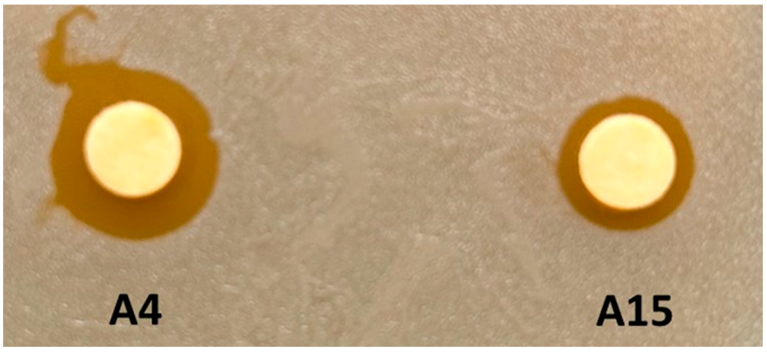
3.2. Growth Inhibition Assays of Target Bacteria for Strains A4 and A15

Growth inhibition assays were completed to determine if either A4 or A15 could inhibit the replication of Staphylococcus aureus (Figure 2). Both isolates produced a zone of clearance (ZOC) when using S. aureus as a target organism, with isolate A4 producing a zone that was double the ZOC diameter produced by A15. The ability to inhibit the growth of competing bacteria is a characteristic of probiotic microorganisms, and mining microbial sources such as bacteria or yeast to identify new antimicrobial compounds is a common path to the discovery of new feed additives [55], as reported herein.
Currently, only one previous report for members of the Sporosarcina spp. to be utilized as a potential probiotic has been proposed for use in monogastric animals, specifically for poultry [20]. S. pasteurii, S. globispora, and S. psychrophila adhered to the intestinal lumen, tolerated a pH between 2 and 4, tolerated bile to 0.5%, and inhibited the growth of several bacteria, including S. aureus and Salmonella typhi [20]. The only other report of a member of the genus as a potential nutritional supplement is S. aquimarina MS4 that is used as an overwintering agent for the sea cucumber Apostichopus japonicus [56].

3.3. Genome Analyses of Strains A4 and A15

The contigs of the A4 strain had a total length of 3,718,715 nucleotides with 85.95% coding DNA bases and GC content of 44.02%. A total of 4077 genes were predicted, including 95.93% of protein-coding genes, 1.18% of regulatory and miscellaneous features, and 2.89% of RNA genes. The functions of 74.76% of protein-coding genes were predicted. The contigs of the A15 strain had a total length of 3,642,744 nucleotides with 86.07% of coding DNA bases with a GC content of 40.97%. A total of 3942 genes were predicted, including 95.92% of protein-coding genes, 1.32% of regulatory and miscellaneous features, and 2.77% of RNA genes. The functions of 74.2% of protein-coding genes were predicted, and conserved domain analyses [40] resulted in the identification of sporulation, urea oxidase, urease, and fibronectin-binding protein genes present in the genomes of the two isolates (Tables S2 and S3).
Genes identified included those encoding a related Bacillus/Clostridium GerA spore germination protein wherein spores recognize germinants through receptor proteins encoded by gerA family of operons, which includes gerA, gerB, and gerK as well as a related Bacillus subtilis stage V sporulation protein R gene (spoVR) involved in spore cortex formation. These are gene products necessary for sporulation and germination [57,58]. Also, a gene encoding the urease accessory protein UreG was identified and was one of the four accessory proteins of urease responsible for the decomposition of urea to form ammonia and carbon dioxide [59,60]. The fibronectin-binding protein A gene (FbpA) was also present in the genomes of both isolates. This family consists of the N-terminal region of the prokaryotic fibronectin-binding protein. While fibronectin has critical roles in eukaryotic cellular processes, such as adhesion, migration, and differentiation, it is also a substrate for the attachment of bacteria. Although often a virulence factor among pathogenic bacteria, probiotic attributes of Lactobacillus fermentum include efficient binding to mucin and fibronectin [61].
Comparisons of COG abundance between the genomes of A4 and A15 with closely related genomes identified differences in the abundance of some important COG categories and pathways (Tables S2 and S3). For example, genes involved in carbohydrate transport and metabolism, amino acid transport and metabolism, and transcription regulations were 1.5–2 times more abundant in A4 than other closely related Sporosarcina genomes. For genome A15, genes involved in carbohydrate transport and metabolism and amino acid transport and metabolism were approximately 1.5 times more abundant in its genome relative to other Sporosarcina spp. genomes. In the A4 genome, the MFS family permease (COG0477) was more abundant than reference genomes (Table S3). A methyl-accepting chemotaxis protein was twice as abundant in A4 versus A15. A total of 169 COGs and 48 uncharacterized proteins were exclusive to the A4 strain compared to its closest relatives S. newyorkensis 2681 [GCA_000220335.1], S. newyorkensis DSM 23,543 [SRX2126154], S. ureae DSM 2281 [GCA_000425545.1], S. ureae P17a [GCA_002082015.1], S. ureae P37 [CP015349.1], and S. ureae S204 [GCA_002081995.1]. Fifty-five enzymes, including hydrolases, phosphoglycerate dehydrogenase, and urease, were also more abundant in the A4 genome (Table S3). The abundance of COGs representing the MFS family permease and Methyl-accepting chemotaxis protein in A15 genomes showed similar profiles to the A4 strain compared to reference genomes (Table S2). A total of 145 COGs excluding 45 uncharacterized proteins were exclusive to the A15 strain and its closest relatives S. newyorkensis 2681 [GCA_000220335.1], S. newyorkensis DSM 23,543 [SRX2126154], S. ureae DSM 2281 [GCA_000425545.1], S. ureae P17a, S. ureae P37 [GCA_002082015.1], and S. ureae S204 [GCA_002081995.1]. Compared to reference Sporosarcina spp. genomes, 59 enzymes, including hydrolases, pyruvate dehydrogenase (acetyl-transferring), and acetyl-CoA C-acetyltransferase were more abundant in the A15 genome (Table S3).
The major facilitator superfamily (MFS) family permease COG (COG0477) for strain A4 consisted of 48 genes, including the DHA1 family (bicyclomycin/chloramphenicol resistance-like; multidrug resistance protein-like) MFS transporter, DHA2 family lincomycin resistance protein-like MFS transporter, EmrB/QacA subfamily drug resistance transporter, FSR family fosmidomycin resistance protein-like MFS transporter, multidrug resistance protein, OFA family oxalate/formate antiporter-like MFS transporter, putative MFS transporter, and the YNFM family putative membrane transporter. Strain A15 had 49 genes belonging to COG0477, consisting of all genes mentioned for A4 with an additional DHA2 family metal-tetracycline-proton antiporter-like MFS transporter that also includes the includes anhydromuropeptide permease AmpG (Tables S2 and S3). These systems are a family of secondary active membrane transporters involved in the exchange of solutes and energy metabolism, such as carbohydrate transport and metabolism, amino acid transport and metabolism, and inorganic ion transport and metabolism [62]. Importantly, genes in this group have been identified as molecular signatures for bacterial phylogenomics, including the Sporosarcina spp. [63].
Because Sporosarcina spp. has been proposed to be potential probiotics [20], and the isolates reported herein have growth inhibitory properties against S. aureus (Figure 2), the genomes were searched for genes that may encode potential antimicrobials (Tables S2 and S3). An ABC-type bacteriocin/lantibiotic exporter that contains an N-terminal double-glycine peptidase domain (COG2274) was located in the A4 genome but not in other genomes of closely related Sporosarcina spp. Seven ABC-type antimicrobial peptide transport systems and permease components (COG0577) were detected in the A4 and A15 genomes but not in the Sporosarcina spp. genomes searched that are closely related to A4 or A15. Also, a peptidoglycan hydrolase (amidase) enhancer domain (COG2385) was detected in the A4 and A15 genomes. Nine and seven metal-dependent amidase/aminoacylase/carboxypeptidase (COG1473) genes were found in the A4 and A15 genomes, respectively. One phage-related protein, the tail component (COG4723), was detected in A4 and A15 genomes, and a phage tail tape-measure protein (COG3941) was in A15 but not A4. However, no phage-related lytic proteins were detected in the genomes, although many of these genes encode what could be considered potential alternatives to standard antibiotics [55].

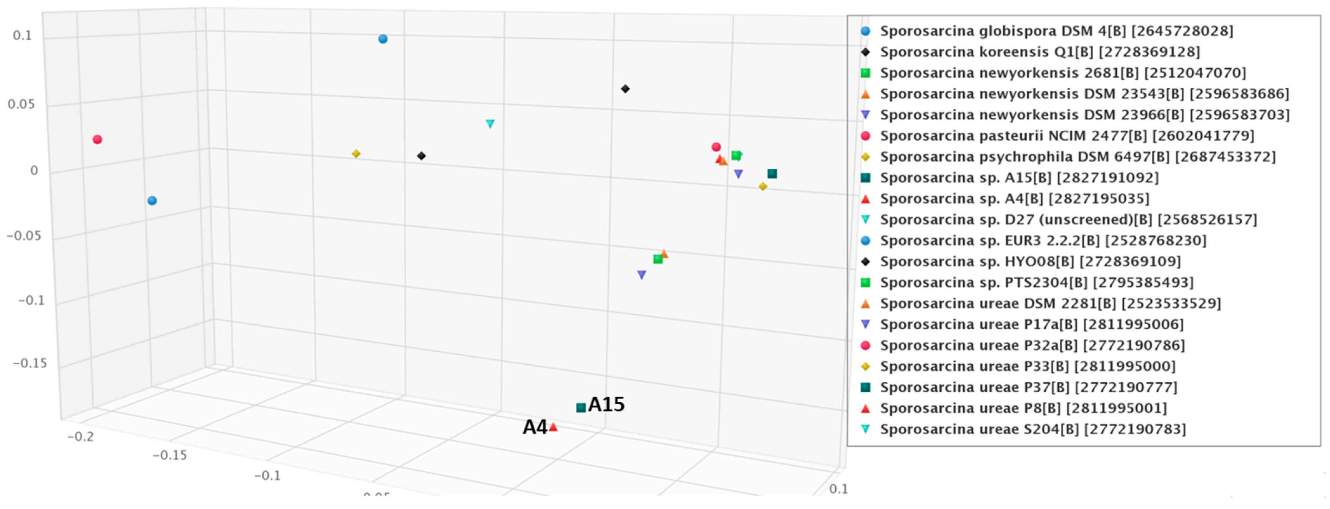
3.4. Phylogenetic Relationships among the Sporosarcina spp. with Isolates A4 and A15

The average nucleotide identity analyses of both draft genomes were compared with twenty-two genomes of Sporosarcina spp. and three other closely related genomes. Strain A4 and A15 have maximum ANI with S. newyorkensis (SRX2126154 and SRX059760) of 78.6% and 81%, respectively (Table 1). NCBI utilizes ANI to evaluate the taxonomic identity of prokaryotic genome assemblies in GenBank. The results of the ANI analysis revealed that both the A4 and A15 isolates could be potentially novel species based on accepted ANI species cutoffs below 95% [45]. All the other Sporosarcina spp. had ANI less than the type species S. newyorkensis, which is less than 78.6% and 81%, respectively. For example, both A4 and A15 are only 76.23% to 77.38% similar to Sporosarcina urea strains within the genus.
Digital DNA-DNA hybridization was performed with 18 closely related genomes [44], and no genomes were significantly similar to any other Sporosarcina spp. (Table 2). In particular, both A4 and A15 strains had the highest dDDH% with S. newyorkensis (GCF_000220335.1) of 23.5% and 27.4%, respectively. A dDDH% of 79–80% is considered the threshold for delineating subspecies [64,65], further supporting A4 and A15 as novel isolates of the genus Sporosarcina. For reference, dDDH values between S. newyorkensis 2681 (SRX059760) and S. ureae DSM 2281 (GCA_002081995.1) was 20.7%; between S. pasteurii strain BNCC 337,394 (GCA_004379295.1) and S. ureae DSM 2281 was 13.2%, S. newyorkensis 2681 and S. pasteurii strain BNCC 337,394 was 13.5%.
The obtained intergenomic distances using formula d5 [64] were used to infer a balanced minimum evolution tree [47], which demonstrated that strains A4 and A15 could not be assigned to any existing species cluster (Figure 3). Additionally, the average amino-acid identity (AAI) of the genomes was performed using protein fasta files with the AA profiler [46]. Isolate A4 was most similar to S. newyorkensis 2681 with AAI 85.2% and matched fractions of 65.2%, while A15 also had the nearest similarity to S. newyorkensis 2681 with AAI 87.1% and matched fractions of 66.3% (Figure 4). Annotated nucleotide and protein fasta files were analyzed for species identification on the EMBL webserver specI (http://vm-lux.embl.de/~mende/specI// (accessed on 19 September 2019) [46], based on 40 universal single-copy marker genes. This further supports the designation of strains A4 and A15 as novel isolates, with the closest relative S. newyorkensis 2681 having 87% and 88.8% average identity, respectively. Consistent with this and with the other analyses performed here, a whole-genome phylogeny as shown in Figure 3 and Figure 4 showed the A4 and A15 isolates were most similar to S. newyorkensis isolates, although bootstrap support in many cases was weak. The second group is composed of Sporosarcina spp., including S. urea, but also includes the strictly anaerobic and chemolithoautotrophic acetogenic bacterium Clostridium scatologenes along with a Gram-negative Flavobacterium reichenbachii strain.
Principal coordinate clustering based on COG profiles demonstrated that the A4 and A15 isolates clustered together with each other and separated from other members of the genus (Figure 5). The nearest-neighbor genomes with A4 and A15 were from S. newyorkensis isolates. The reference S. newyorkensis strains are Gram-positive; endospore-forming rods originally isolated from veterinary clinical specimens in New York State, USA, and from raw milk in Flanders, Belgium, that were considered as new species of the genus [10]. S. ureae is a cocci-shaped [66] bacterium with a reported genome of 3.4 Mb and 41.5% GC content that has been isolated from soil contaminated with chicken waste [67]. S. globispora is reportedly a “round” Gram-positive, spore-forming, psychrophilic bacterium with a 5.67 Mb genome [68] that was originally classified as an endospore containing Bacillus spp. [9]. Other phylogenetically related species include the urease-producing bacterium S. koreensis with a 44.5% GC and 4.3 Mb genome [69] described as a Gram-positive, aerobic, spore-forming rod isolated from soil [6]. S. psychrophila, also a Gram-positive, spore-forming psychrophilic bacterial strain that is widely distributed in both terrestrial and aquatic environments had a reported circular chromosome of 4,674,191 bp with a GC content of 40.3%, also encoding urea hydrolysis genes [70]. A newly identified group of 28 cocci Sporosarcina spp. isolates had an average genome size of 3.3 Mb with two clades, one with 85.9% AAI and clade 2 with only 81.5% AAI to the other cocci-shaped Sporosarcina spp. indicating potentially new bacterial species [71]. S. pasteurii (~3.3 Mb genome, 39.2% GC; NZ_CP038012.1).
Other Sporosarcina spp. members have been proposed to be potential probiotic bacteria for poultry during production [20]. This includes the reduction of phytate by Sporosarcina spp. [72], and useful phosphatase genes (e.g., COG2194, COG0494, COG0561) were detected in the genomes of the isolates reported herein.

4. Summary and Conclusions

The results reported herein support the hypothesis that the A4 and A15 isolates are unique among the Sporosarcina spp. S. cascadensis A4 (cascadiensis. L. masc. adj. cascadensis referring to the area from which the isolates were obtained in the Cascade mountain range). The cells are Gram-variable, rod-shaped, occurring alone or in pairs. Colonies grown on BBHK or BHI agar are morphologically rods. Cells are aerobic, catalase-positive, and positive for oxidase test reaction with tetramethyl-p-phenylenediamine. The optimum growth temperature is 37 °C, optimum NaCl concentration for growth is 3% with a 10% maximum. Alkaliphilic with a pH growth range of 8–10. Acetic acid, acetoacetic acid, N-acetyl-d-glucosaminitol, Sec-butylamine, butyric acid, α-keto-butyric acid, d-fucose, maltotriose, mannan, oxalic acid, pectin, and l-sorbose are utilized individually as sole carbon sources for growth. The isolate A4 was isolated from the feces of the Canada goose (Branta canadensis). The DNA G+C content of the type strain is 44.02%. The type strain of Sporosarcina cascadiensis sp. nov. (A4) is held by the U.S. Department of Agriculture Agricultural Research Service (ARS) Culture Collection under the number: NRRL Y-65557.
Sporosarcina obsidiansis A15 (obsidiansis L. masc. adj. obsidiansis) refers to the geographic area from where the isolates were recovered that is high in obsidian rock. Cells are Gram-stain-variable, rod-shaped, occurring singly or in pairs. Colonies grown on BBHK or BHI agar are morphologically rods, aerobic, catalase-positive, and oxidase test reaction with tetramethyl-p-phenylenediamine positive. The optimum growth temperature is 37 °C, the optimum NaCl concentration for growth is 3%, the maximum NaCl concentration of 4%, pH growth ranges from 6 to 10. Carbon sources: d-alanine, l-alanine, l-asparagine, α-hydroxy butyric acid, α-keto-butyric acid, dulcitol, d-fructose, d-fructose-6-phosphate, fumaric acid, d-galactonic acid γ-lactone, l-galactonic acid γ-lactone, d-galactose, d-galacturonic acid, α-d-glucose, d-gluconic acid, d-glucosaminic acid, d-glucuronic acid, glucuronamide, α-hydroxy glutaric acid γ-lactone, glycolic acid, m-inositol, l-lactic acid, d,l-malic acid, maltotriose, d-mannitol, d-mannose, maltose, d-melibiose, mucic acid, p-hydroxy phenyl acetic acid, 1,2-propanediol, d-psicose, d-sorbitol, m-tartaric acid, d-threonine, l-threonine, tricarballylic acid, tyramine are utilized individually as sole carbon sources for growth. The strain, A15, was isolated from the feces of the Canada goose (Branta canadensis). The DNA G+C content of the type strain is 40.97%. The type strain of Sporosarcina obsidiansis sp. nov. (A15) is held by the U.S. Department of Agriculture Agricultural Research Service (ARS) Culture Collection under the number: NRRL Y-65558. Sporosarcina obsidiansis A15 (obsidiansis L. masc. adj. obsidiansis referring to the geographic area from where the isolates were recovered that is high in obsidian rock).

Supplementary Materials

The following supporting information can be downloaded at: https://www.mdpi.com/article/10.3390/microorganisms12010070/s1, Figure S1: Representative antibiotic disc diffusion assay for isolates A4 and A15; Figure S2: Mannitol salt, spirit blue and starch agar plate analyses for isolates A4 and A15; Table S1: Metabolic profiling of strains A4 and A15 on Biolog PM9 and PM10 MicroPlate; Table S2: COG categories for A4 and A15 genomes; Table S3: Enzyme designations for A4 and A15 genomes.

Author Contributions

Conceptualization, P.N.B., B.B.O. and B.S.S.; methodology, P.N.B., B.B.O., J.E.L. and B.S.S.; software, J.K., J.E.L. and K.M.S.; validation, P.N.B., B.B.O., J.K., J.E.L., K.M.S. and B.S.S.; formal analysis, J.K., B.B.O., K.M.S. and B.S.S.; investigation, J.K., K.M.S., M.K.S., H.R.K., M.L.M., H.J.J., A.M.L., J.K.G., J.E.L., P.N.B., B.B.O. and B.S.S.; resources, P.N.B. and B.B.O.; data curation, J.K., J.E.L., K.M.S., B.B.O. and B.S.S.; writing—original draft preparation, J.K., B.B.O. and B.S.S.; writing—review and editing, B.S.S.; supervision, P.N.B., B.B.O. and B.S.S.; project administration, P.N.B., B.B.O. and B.S.S.; funding acquisition, P.N.B., J.E.L. and B.B.O. All authors have read and agreed to the published version of the manuscript.

Funding

This research was initially funded by PNB as a start-up grant from the OSU Faculty Innovation Committee and the Gaskins Fund to PNB; the Layman Fellowship Program for HJJ and AML; and the Undergraduate Research and Scholarship Engage Program at OSU Cascades for MKS, HRK and MLM. Partial funding was provided by USDA NIFA grants (Accession numbers 1015210 and 1011327) to Western University of Health Sciences College of Veterinary Medicine and BBO; ARS funding number: Project Number: 6040-32000-071-00-D to JKG and JEL.

Institutional Review Board Statement

The Oregon State University Institutional Biosafety Committee has approved the research reported herein as IBC Proposal #3923.

Data Availability Statement

The Whole Genome Shotgun project and 16S rRNA gene sequences have been deposited at DDBJ/ENA/GenBank. The accession numbers of strain A4 are WHVV01000000 (genome) and MN582949 (16S rRNA gene) and strain A15 are WHVW00000000 (genome) and MN582950 (16S rRNA gene).

Acknowledgments

Appreciation is extended to the Oregon State University, Center for Genome Research and Biocomputing (https://cgrb.oregonstate.edu/ (accessed on 18 April 2019) for contract sequencing of genomes. The results reported are part of ongoing undergraduate research at Oregon State University Cascades and portions of the data were presented at the Cascades Research and Scholarship Symposium: https://osucascades.edu/research/students/cascades-research-and-scholarship-symposium (accessed on 7 April 2023).

Conflicts of Interest

The authors declare no conflict of interest.

References

  1. Kluyver, A.J.; van Niel, C.B. Prospects for a natural system of classification of bacteria. Zentralbl. Bakteriol. Parasitenkd. Infektionskr. Hyg. 1936, 94, 369–403. [Google Scholar]
  2. Kämpfer, P.; Falsen, E.; Lodders, N.; Schumann, P. Sporosarcina contaminans sp. nov. and Sporosarcina thermotolerans sp. nov., two endospore-forming species. Int. J. Syst. Evol. Microbiol. 2010, 60, 1353–1357. [Google Scholar] [CrossRef] [PubMed]
  3. Yu, Y.; Xin, Y.H.; Liu, H.C.; Chen, B.; Sheng, J.; Chi, Z.M.; Zhou, P.J.; Zhang, D.C. Sporosarcina antarctica sp. nov., a psychrophilic bacterium isolated from the Antarctic. Int. J. Syst. Evol. Microbiol. 2008, 58, 2114–2117. [Google Scholar] [CrossRef] [PubMed]
  4. Sun, Y.; Zhao, Q.; Zhi, D.; Wang, Z.; Wang, Y.; Xie, Q.; Wu, Z.; Wang, X.; Li, Y.; Yu, L.; et al. Sporosarcina terrae sp. nov., isolated from orchard soil. Int. J. Syst. Evol. Microbiol. 2017, 67, 2104–2108. [Google Scholar] [CrossRef] [PubMed]
  5. Claus, D.; Fahmy, F.; Rolf, H.J.; Tosunoglu, N. Sporosarcina halophila sp. nov., an obligate, slightly halophilic bacterium from salt marsh soils. Syst. Appl. Microbiol. 1983, 4, 496–506. [Google Scholar] [CrossRef] [PubMed]
  6. Kwon, S.W.; Kim, B.Y.; Song, J.; Weon, H.Y.; Schumann, P.; Tindall, B.J.; Stackebrandt, E.; Fritze, D. Sporosarcina koreensis sp. nov. and Sporosarcina soli sp. nov., isolated from soil in Korea. Int. J. Syst. Evol. Microbiol. 2007, 57, 1694–1698. [Google Scholar] [CrossRef] [PubMed]
  7. Zhang, G.; Ren, H.; Chen, X.; Zhang, Y.; Yang, Y.; Wang, S.; Jiang, Y. Sporosarcina siberiensis sp. nov., isolated from the East Siberian Sea. Antonie Van Leeuwenhoek. 2014, 106, 489–495. [Google Scholar] [CrossRef] [PubMed]
  8. An, S.Y.; Haga, T.; Kasai, H.; Goto, K.; Yokota, A. Sporosarcina saromensis sp. nov., an aerobic endospore-forming bacterium. Int. J. Syst. Evol. Microbiol. 2007, 57 Pt 8, 1868–1871. [Google Scholar] [CrossRef]
  9. Yoon, J.H.; Lee, K.C.; Weiss, N.; Kho, Y.H.; Kang, K.H.; Park, Y.H. Sporosarcina aquimarina sp. nov., a bacterium isolated from seawater in Korea, and transfer of Bacillus globisporus (Larkin and Stokes 1967), Bacillus psychrophilus (Nakamura 1984) and Bacillus pasteurii (Chester 1898) to the genus Sporosarcina as Sporosarcina globispora comb. nov., Sporosarcina psychrophila comb. nov. and Sporosarcina pasteurii comb. nov. and emended description of th. Int. J. Syst. Evol. Microbiol. 2001, 51, 1079–1086. [Google Scholar]
  10. Wolfgang, W.J.; Coorevits, A.; Cole, J.A.; De Vos, P.; Dickinson, M.C.; Hennett, G.E.; Jose, R.; Nazarian, E.J.; Schumann, P.; Van Landschoot, A.; et al. Sporosarcina newyorkensis sp. nov. from clinical specimens and raw cow’s milk. Int. J. Syst. Evol. Microbiol. 2012, 62, 322–329. [Google Scholar] [CrossRef]
  11. Reddy, G.S.; Matsumoto, G.I.; Shivaji, S. Sporosarcina macmurdoensis sp. nov., from a cyanobacterial mat sample from a pond in the McMurdo Dry Valleys, Antarctica. Int. J. Syst. Evol. Microbiol. 2003, 53, 1363–1367. [Google Scholar] [CrossRef] [PubMed]
  12. Tominaga, T.; An, S.Y.; Oyaizu, H.; Yokota, A. Sporosarcina luteola sp. nov. isolated from soy sauce production equipment in Japan. J. Gen. Appl. Microbiol. 2009, 55, 217–223. [Google Scholar] [CrossRef] [PubMed]
  13. Yuan, L.; Zhang, R.; Li, X.; Gao, C.; Hu, X.; Hussain, S.; Zhang, L.; Wang, M.; Ma, X.; Pan, Q.; et al. Long-term simulated microgravity alters gut microbiota and metabolome in mice. Front. Microbiol. 2023, 14, 1100747. [Google Scholar] [CrossRef]
  14. Simpson, A.C.; Sengupta, P.; Zhang, F.; Hameed, A.; Parker, C.W.; Singh, N.K.; Miliotis, G.; Rekha, P.D.; Raman, K.; Mason, C.E.; et al. Phylogenomics, phenotypic, and functional traits of five novel (Earth-derived) bacterial species isolated from the International Space Station and their prevalence in metagenomes. Sci. Rep. 2023, 13, 19207. [Google Scholar] [CrossRef] [PubMed]
  15. Chuo, S.C.; Mohamed, S.F.; Mohd Setapar, S.H.; Ahmad, A.; Jawaid, M.; Wani, W.A.; Yaqoob, A.A.; Mohamad Ibrahim, M.N. Insights into the current trends in the utilization of bacteria for microbially induced calcium carbonate precipitation. Materials 2020, 13, 4993. [Google Scholar] [CrossRef] [PubMed]
  16. Wu, Y.; Li, H.; Li, Y. Biomineralization induced by cells of Sporosarcina pasteurii: Mechanisms, applications and challenges. Microorganisms 2021, 9, 2396. [Google Scholar] [CrossRef]
  17. Saif, A.; Cuccurullo, A.; Gallipoli, D.; Perlot, C.; Bruno, A.W. Advances in enzyme induced carbonate precipitation and application to soil improvement: A review. Materials 2022, 15, 950. [Google Scholar] [CrossRef]
  18. Ma, L.; Pang, A.P.; Luo, Y.; Lu, X.; Lin, F. Beneficial factors for biomineralization by ureolytic bacterium Sporosarcina pasteurii. Microb. Cell Fact. 2020, 19, 12. [Google Scholar] [CrossRef]
  19. Garrity, G.M. Bergey’s Manual of Systematic Bacteriology: The Firmicutes. In Bergey’s Manual of Systematic Bacteriology; Vos, P., Garrity, G.M., Jones, D., Krieg, N.R., Ludwig, W., Rainey, F.A., Schleifer, K.-H., Whitman, W.B., Eds.; Springer: Berlin/Heidelberg, Germany, 2009; Volume 3. [Google Scholar]
  20. Priyodip, P.; Balaji, S. A preliminary study on probiotic characteristics of Sporosarcina spp. for poultry applications. Curr. Microbiol. 2019, 76, 448–461. [Google Scholar] [CrossRef]
  21. Wise, M.G.; Siragusa, G.R. Quantitative analysis of the intestinal bacterial community in one- to three-week-old commercially reared broiler chickens fed conventional or antibiotic-free vegetable-based diets. J. Appl. Microbiol. 2007, 102, 1138–1149. [Google Scholar] [CrossRef]
  22. Atarashi, K.; Tanoue, T.; Shima, T.; Imaoka, A.; Kuwahara, T.; Momose, Y.; Cheng, G.; Yamasaki, S.; Saito, T.; Ohba, Y.; et al. Induction of colonic regulatory T cells by indigenous Clostridium species. Science 2011, 331, 337–341. [Google Scholar] [CrossRef] [PubMed]
  23. Itoh, K.; Mitsuoka, T. Production of gnotobiotic mice with normal physiological functions. I. Selection of useful bacteria from feces of conventional mice. Z. Versuchstierkd. 1980, 22, 173–178. [Google Scholar] [PubMed]
  24. Summanen, P.; Baron, E.J.; Citron, D.M.; Strong, C.A.; Wexler, H.M.; Finegold, S.M. Wadsworth Anaerobic Bacteriology Manual, 5th ed.; Star Publishing: Belmont, CA, USA, 1993. [Google Scholar]
  25. Hayden, R.L. Elective localization in the eye of bacteria from infected teeth. Arch. Int. Med. 1923, 32, 828–849. [Google Scholar] [CrossRef]
  26. Levine, M. Differentiation of B. coli and B. aerogenes on a simplified eosin-methylene blue agar. J. Infect. Dis. 1918, 23, 43–47. [Google Scholar] [CrossRef]
  27. Chapman, G.H. The significance of sodium chloride in studies of staphylococci. J. Bacteriol. 1945, 50, 201–203. [Google Scholar] [CrossRef] [PubMed]
  28. Beveridge, T.J. Mechanism of Gram Variability in Select Bacteria J. Bacteriol. 1990, 172, 1609–1620. [Google Scholar] [CrossRef]
  29. Bauer, A.W.; Kirby, W.M.M.; Sherris, J.C.; Turck, M. Antibiotic susceptibility testing by a standardized single disk method. Am. J. Clin. Pathol. 1966, 36, 493–496. [Google Scholar] [CrossRef]
  30. Star, M.P. Spirit blue agar: A medium for the detection of lipolytic microorganisms. Science 1941, 93, 333–334. [Google Scholar] [CrossRef]
  31. Flint, S.; Hartley, N. A modified selective medium for the detection of Pseudomonas species that cause spoilage of milk and dairy products. Int. Dairy J. 1996, 2, 223–230. [Google Scholar] [CrossRef]
  32. Hardy, B.L.; Bansal, G.; Hewlett, K.H.; Arora, A.; Schaffer, S.D.; Kamau, E.; Bennett, J.W.; Merrell, D.S. Antimicrobial activity of clinically isolated bacterial species against Staphylococcus aureus. Front. Microbiol. 2020, 10, 2977. [Google Scholar] [CrossRef]
  33. Bochner, B.R.; Gadzinski, P.; Panomitros, E. Phenotype microarrays for high-throughput phenotypic testing and assay of gene function. Genome Res. 2001, 11, 1246–1255. [Google Scholar] [CrossRef] [PubMed]
  34. Shea, A.; Wolcott, M.; Daefler, S.; Rozak, D.A. Biolog phenotype microarrays. Methods Mol. Biol. 2012, 881, 331–373. [Google Scholar] [PubMed]
  35. Mackie, A.M.; Hassan, K.A.; Paulsen, I.T.; Tetu, S.G. Biolog Phenotype Microarrays for phenotypic characterization of microbial cells. Methods Mol. Biol. 2014, 1096, 123–130. [Google Scholar] [PubMed]
  36. Stackebrandt, E.; Frederiksen, W.; Garrity, G.M.; Grimont, P.A.; Kämpfer, P.; Maiden, M.C.J.; Nesme, X.; Rosselló-Mora, R.; Swings, J.; Trüper, H.G.; et al. Report of the ad hoc committee for the re-evaluation of the species definition in bacteriology. Int. J. Syst. Evol. Microbiol. 2002, 52, 1043–1047. [Google Scholar] [PubMed]
  37. Bankevich, A.; Nurk, S.; Antipov, D.; Gurevich, A.A.; Dvorkin, M.; Kulikov, A.S.; Lesin, V.M.; Nikolenko, S.I.; Pham, S.; Prjibelski, A.D.; et al. SPAdes: A new genome assembly algorithm and its applications to single-cell sequencing. J. Comput. Biol. 2012, 19, 455–477. [Google Scholar] [CrossRef] [PubMed]
  38. Altschul, S.F.; Madden, T.L.; Schäffer, A.A.; Zhang, J.; Zhang, Z.; Miller, W.; Lipman, D.J. Gapped BLAST and PSI-BLAST: A new generation of protein database search programs. Nucleic Acids Res. 1997, 25, 3389–3402. [Google Scholar] [CrossRef] [PubMed]
  39. Chen, I.A.; Chu, K.; Palaniappan, K.; Ratner, A.; Huang, J.; Huntemann, M.; Hajek, P.; Ritter, S.; Varghese, N.; Seshadri, R.; et al. The IMG/M data management and analysis system v.6.0: New tools and advanced capabilities. Nucleic Acids Res. 2021, 47, D666–D677. [Google Scholar] [CrossRef] [PubMed]
  40. Marchler-Bauer, A.; Derbyshire, M.K.; Gonzales, N.R.; Lu, S.; Chitsaz, F.; Geer, L.Y.; Geer, R.C.; He, J.; Gwadz, M.; Hurwitz, D.I.; et al. CDD: NCBI’s conserved domain database. Nucleic Acids Res. 2015, 43, D222–D226. [Google Scholar] [CrossRef]
  41. Tatusov, R.L.; Galperin, M.Y.; Natale, D.A.; Koonin, E.V. The COG database: A tool for genome-scale analysis of protein functions and evolution. Nucleic Acids Res. 2000, 28, 33–36. [Google Scholar] [CrossRef]
  42. Ondov, B.D.; Treangen, T.J.; Melsted, P.; Mallonee, A.B.; Bergman, N.H.; Koren, S.; Phillippy, A.M. Mash: Fast genome and metagenome distance estimation using MinHash. Genome Biol. 2016, 17, 132. [Google Scholar] [CrossRef]
  43. Gruber-Vodicka, H.R.; Seah, B.K.B.; Pruesse, E. PhyloFlash: Rapid small-subunit rRNA profiling and targeted assembly from metagenomes. mSystems 2020, 5, e00920-20. [Google Scholar] [CrossRef] [PubMed]
  44. Meier-Kolthoff, J.P.; Göker, M. TYGS is an automated high-throughput platform for state-of-the-art genome-based taxonomy. Nat. Commun. 2019, 10, 2182. [Google Scholar] [CrossRef]
  45. Jain, C.; Rodriguez-R, L.M.; Phillippy, A.M.; Konstantinidis, K.T.; Aluru, S. High throughput ANI analysis of 90K prokaryotic genomes reveals clear species boundaries. Nat. Commun. 2018, 9, 5114. [Google Scholar] [CrossRef] [PubMed]
  46. Medlar, A.J.; Törönen, P.; Holm, L. AAI-profiler: Fast proteome-wide exploratory analysis reveals taxonomic identity, misclassification, and contamination. Nucleic Acids Res. 2018, 46, W479–W485. [Google Scholar] [CrossRef] [PubMed]
  47. Lefort, V.; Desper, R.; Gascuel, O. FastME 2.0: A Comprehensive, Accurate, and Fast Distance-Based Phylogeny Inference Program. Mol. Biol. Evol. 2015, 32, 2798–2800. [Google Scholar] [CrossRef] [PubMed]
  48. Janarthine, S.R.S.; Eganathan, P. Plant Growth Promoting of Endophytic Sporosarcina aquimarina SjAM16103 Isolated from the Pneumatophores of Avicennia marina L. Int. J. Microbiol. 2012, 2012, 532060. [Google Scholar] [CrossRef] [PubMed]
  49. Taylor, W.I.; Achanzar, D. Catalase test as an aid to the identification of Enterobacteriaceae. J. Appl. Microbiol. 1972, 24, 58–61. [Google Scholar] [CrossRef] [PubMed]
  50. Jurtshuk, P.; McQuitty, D.N. Use of a quantitative oxidase test for characterizing oxidative metabolism in bacteria. Appl. Environ. Microbiol. 1976, 3, 668–679. [Google Scholar] [CrossRef]
  51. Abdulhamid, M.B.; Costas, L.; Del Valle Loto, F.; Baigorí, M.D.; Pera, L.M. Industrial biotransformations catalyzed by microbial lipases: Screening platform and commercial aspects. Folia Microbiol. 2021, 66, 1009–1022. [Google Scholar] [CrossRef]
  52. Lapierre, F.M.; Schmid, J.; Ederer, B.; Ihling, N.; Büchs, J.; Huber, R. Revealing nutritional requirements of MICP-relevant Sporosarcina pasteurii DSM33 for growth improvement in chemically defined and complex media. Sci. Rep. 2020, 10, 22448. [Google Scholar] [CrossRef]
  53. Pei, D.; Liu, Z.; Wu, W.; Hu, B. Transcriptome analyses reveal the utilization of nitrogen sources and related metabolic mechanisms of Sporosarcina pasteurii. PLoS ONE 2021, 16, e0246818. [Google Scholar] [CrossRef] [PubMed]
  54. Bakermans, C.; Skidmore, M.L. Microbial metabolism in ice and brine at −5 °C. Environ. Microbiol. 2011, 13, 2269–2278. [Google Scholar] [CrossRef] [PubMed]
  55. Seal, B.S.; Drider, D.; Oakley, B.B.; Brüssow, H.; Bikard, D.; Rich, J.O.; Miller, S.; Devillard, E.; Kwan, J.; Bertin, G.; et al. Microbial-derived products as potential new antimicrobials. Vet. Res. 2018, 49, 66. [Google Scholar] [CrossRef] [PubMed]
  56. Yi, H.; Bai, Q.; Li, Y.; Zhan, H.; Liu, Y.; Liu, B.; Wang, J. Sporosarcina aquimarina MS4 Regulates the Digestive Enzyme Activities, Body Wall Nutrients, Gut Microbiota, and Metabolites of Apostichopus japonicus. Fishes 2022, 7, 134. [Google Scholar] [CrossRef]
  57. Galperin, M.Y.; Yutin, N.; Wolf, Y.I.; Vera Alvarez, R.; Koonin, E.V. Conservation and Evolution of the Sporulation Gene Set in Diverse Members of the Firmicutes. J. Bacteriol. 2022, 204, e0007922. [Google Scholar] [CrossRef]
  58. Yu, B.; Kanaan, J.; Shames, H.; Wicander, J.; Aryal, M.; Li, Y.; Korza, G.; Brul, S.; Kramer, G.; Li, Y.Q.; et al. Identification and characterization of new proteins crucial for bacterial spore resistance and germination. Front. Microbiol. 2023, 14, 1161604. [Google Scholar] [CrossRef]
  59. D’Urzo, A.; Santambrogio, C.; Grandori, R.; Ciurli, S.; Zambelli, B. The conformational response to Zn(II) and Ni(II) binding of Sporosarcina pasteurii UreG, an intrinsically disordered GTPase. J. Biol. Inorg. Chem. 2014, 19, 1341–1354. [Google Scholar] [CrossRef]
  60. Fong, Y.H.; Wong, H.C.; Yuen, M.H.; Lau, P.H.; Chen, Y.W.; Wong, K.B. Structure of UreG/UreF/UreH complex reveals how urease accessory proteins facilitate maturation of Helicobacter pylori urease. PLoS Biol. 2013, 11, e1001678. [Google Scholar] [CrossRef]
  61. Lehri, B.; Seddon, A.M.; Karlyshev, A.V. Lactobacillus fermentum 3872 genome sequencing reveals plasmid and chromosomal genes potentially involved in a probiotic activity. FEMS Microbiol. Lett. 2015, 362, fnv068. [Google Scholar] [CrossRef]
  62. Drew, D.; North, R.A.; Nagarathinam, K.; Tanabe, M. Structures and general transport mechanisms by the major facilitator superfamily (MFS). Chem. Rev. 2021, 121, 5289–5335. [Google Scholar] [CrossRef]
  63. Gupta, R.S.; Patel, S. Robust Demarcation of the Family Caryophanaceae (Planococcaceae) and Its Different Genera Including Three Novel Genera Based on Phylogenomics and Highly Specific Molecular Signatures. Front. Microbiol. 2020, 10, 2821. [Google Scholar] [CrossRef] [PubMed]
  64. Meier-Kolthoff, J.P.; Auch, A.F.; Klenk, H.P.; Göker, M. Genome sequence-based species delimitation with confidence intervals and improved distance functions. BMC Bioinform. 2013, 14, 60. [Google Scholar] [CrossRef] [PubMed]
  65. Meier-Kolthoff, J.P.; Hahnke, R.L.; Petersen, J.; Scheuner, C.; Michael, V.; Fiebig, A.; Rohde, C.; Rohde, M.; Fartmann, B.; Goodwin, L.A.; et al. Complete genome sequence of DSM 30083T, the type strain (U5/41T) of Escherichia coli, and a proposal for delineating subspecies in microbial taxonomy. Stand. Genom. Sci. 2014, 9, 2. [Google Scholar] [CrossRef]
  66. Thompson, R.S.; Leadbetter, E.R. On the isolation of dipicolinic acid from endospores of Sarcina ureae. Arch. Mikrobiol. 1963, 45, 27–32. [Google Scholar] [CrossRef] [PubMed]
  67. You, Y.; Hilpert, M.; Ward, M.J. Detection of a common and persistent tet(L)-carrying plasmid in chicken-waste-impacted farm soil. Appl. Environ. Microbiol. 2012, 78, 3203–3213. [Google Scholar] [CrossRef]
  68. Wang, J.P.; Liu, B.; Liu, G.H.; Xiao, R.F.; Zheng, X.F.; Shi, H.; Ge, C.B. Draft Genome Sequence of Sporosarcina globispora W 25T (DSM 4), a Psychrophilic Bacterium Isolated from Soil and River Water. Genome Announc. 2015, 3, 5. [Google Scholar] [CrossRef]
  69. Abdul Majid, S.; Graw, M.F.; Nguyen, H.; Hay, A.G. Draft Whole-Genome Sequence of Urease-Producing Sporosarcina koreensis. Genome Announc. 2016, 4, e00096-16. [Google Scholar] [CrossRef]
  70. Yan, W.; Xiao, X.; Zhang, Y. Complete genome sequence of the Sporosarcina psychrophila DSM 6497, a psychrophilic Bacillus strain that mediates the calcium carbonate precipitation. J. Biotechnol. 2016, 226, 14–15. [Google Scholar] [CrossRef]
  71. Oliver, A.; Kay, M.; Cooper, K.K. Comparative genomics of cocci-shaped Sporosarcina strains with diverse spatial isolation. BMC Genom. 2018, 19, 310. [Google Scholar] [CrossRef]
  72. Priyodip, P.; Balaji, S. An in vitro chicken gut model for the assessment of phytase producing bacteria. 3 Biotech 2019, 9, 294. [Google Scholar] [CrossRef]
Figure 1. Gram staining of A4 (a) and A15 (b) bacteria showing both as gram variables. The images were photographed with an Olympus BX-40 light microscope. A 100× oil immersion objective was used with a WH10X/22 eyepiece.
Microorganisms 12 00070 g001
Figure 2. Growth inhibition spot assay with Staphylococcus aureus target on BHI media. The A4 and A15 isolates’ saturated discs are as designated in the figure.
Microorganisms 12 00070 g002
Figure 3. Minimum evolution tree inferred with FastME 2.1.6.1 [31] from Genome BLAST Distance Phylogeny (GBDP) distances calculated from genome sequences of strains A4, A15, and closest homologous genomes. The branch lengths are scaled in terms of GBDP distance formula d5 [47]. The numbers above branches are GBDP pseudo-bootstrap support values from 100 replications.
Microorganisms 12 00070 g003
Figure 4. Average amino acid identity (AAI) of Uniprot species with strain A4 (a) and A15 (b). Species are grouped and colored by genus. Fraction of AAI vs. aligned matches of different reference species with query strains (A4 and A15) are plotted, showing S. newyorkensis as the closest species with A4 and A15 strains based on AAI (<88%). Genomes with AAI less than 0.5.
Microorganisms 12 00070 g004
Figure 5. Principal coordinate analysis (PCoA) plot of COG profiles of Sporosarcina genomes based on Bray–Curtis distance matrix. Compositional dissimilarities among genomes were measured using Bray–Curtis dissimilarity coefficient of COG abundances.
Microorganisms 12 00070 g005
Table 1. Average nucleotide identity (ANI) comparisons between A4, A15, and reference Sporosarcina spp. genomes.
Genome1Genome2 ANI1->2ANI2->1AF1->2AF2->1
A4Sporosarcina newyorkensis DSM 2354378.7178.7166.9668.03
A4Sporosarcina newyorkensis 268178.6778.6767.0567.49
A4Sporosarcina newyorkensis DSM 2396678.6578.6570.1471.90
A4Sporosarcina ureae P3777.3877.3858.7765.46
A4Sporosarcina ureae P3377.3577.3558.6467.62
A4Sporosarcina ureae S20476.5276.5257.9364.57
A4Sporosarcina ureae DSM 228176.4976.4958.8565.85
A4Sporosarcina ureae P17a76.3676.3759.1664.84
A4Sporosarcina ureae P876.3276.3258.1865.07
A4Sporosarcina ureae P32a76.2376.2359.5865.83
A4Sporosarcina sp. PTS230476.1476.1357.3962.48
A4Sporosarcina pasteurii ATCC 1185972.3272.320.0573.76
A4Sporosarcina koreensis Q171.2771.2639.8134.46
A4Sporosarcina psychrophila DSM 649770.8070.7940.9834.54
A4Sporosarcina sp. HYO0870.7970.7736.4543.88
A4Sporosarcina sp. ZBG7A70.6570.6536.4438.27
A4Sporosarcina koreensis S-K1270.5670.5534.0842.44
A4Sporosarcina sp. D27 (unscreened)70.5370.5336.9438.24
A4Sporosarcina sp. EUR3 2.2.269.0369.0424.3526.61
A4Planococcus plakortidis DSM 2399768.8468.8423.7326.88
A4Sporosarcina globispora DSM 468.2068.2020.4813.96
A4Sporosarcina pasteurii NCIM 247767.6267.6215.4813.72
A4Bacillus thuringiensis YBT-151867.2067.2116.299.72
A4Clostridium scatologenes ATCC 2577565.3865.384.982.54
A4Flavobacterium reichenbachii LMG 2551263.9463.940.480.31
A4A1578.0878.0868.3969.72
A15Sporosarcina newyorkensis 268181.0881.0867.8967.04
A15Sporosarcina newyorkensis DSM 2396681.0681.0669.6569.99
A15Sporosarcina newyorkensis DSM 2354381.0681.0668.1867.92
A15Sporosarcina sp. PTS230477.2777.2760.9064.83
A15Sporosarcina ureae S20477.0977.1059.4364.98
A15Sporosarcina ureae P877.0877.0860.3466.20
A15Sporosarcina ureae DSM 228177.0777.0760.2866.16
A15Sporosarcina ureae P17a76.9676.9659.8764.34
A15Sporosarcina ureae P32a76.9576.9560.7265.85
A15Sporosarcina ureae P3376.8976.9159.6467.56
A15Sporosarcina ureae P3776.8876.8860.1465.70
A15Sporosarcina pasteurii ATCC 1185972.8872.880.0574.11
A15Sporosarcina sp. HYO0871.2371.2338.1945.08
A15Sporosarcina psychrophila DSM 649771.0771.0742.0634.78
A15Sporosarcina koreensis Q171.0671.0540.4834.37
A15Sporosarcina sp. ZBG7A70.7570.7537.7838.91
A15Sporosarcina sp. D27 (unscreened)70.6870.6837.8438.42
A15Sporosarcina sp. EUR3 2.2.269.6469.6426.6328.59
A15Sporosarcina koreensis S-K1269.0269.0232.9340.24
A15Bacillus thuringiensis YBT-151867.8867.8917.4710.25
A15Planococcus plakortidis DSM 2399767.8767.8723.3925.99
A15Sporosarcina globispora DSM 467.6567.6520.8513.93
A15Sporosarcina pasteurii NCIM 247767.2267.2215.6613.61
A15Clostridium scatologenes ATCC 2577565.9965.995.912.96
A15Flavobacterium reichenbachii LMG 2551264.4564.450.470.30
Table 2. Pairwise comparisons of A4 and A15 genomes vs. closest Sporosarcina spp. genomes for Digital DNA/DNA hybridization and difference in G+C content.
Query StrainSubject StraindDDH
(d4, in %)
G+C Content Difference
(in %)
A4Sporosarcina newyorkensis 2681a 23.55.85
A4Sporosarcina sp. P3320.51.64
A4Sporosarcina ureae P17a204.67
A4Sporosarcina ureae S20419.44.66
A4Sporosarcina ureae DSM 2281a 19.25.6
A4Sporosarcina sp. P719.23.68
A4Sporosarcina newyorkensis DSM 23966153.99
A4Sporosarcina luteola NBRC 10537813.52.77
A4Sporosarcina koreensis Q1a 13.42.95
A4Sporosarcina koreensis S-K1213.47.81
A4Sporosarcina psychrophila DSM 649713.35.84
A4Sporosarcina pasteurii BNCC 33739413.37.01
A4Bacillus thuringiensis YBT-1518 529122.413.110.76
A4Planococcus plakortidis DSM 2399713.13.8
A4Paenisporosarcina aarctica CGMCC 1.650313.19.18
A4Sporosarcina globispora DSM413.16.62
A4Clostridium scatologenes ATCC 257751316.57
A4Flavobacterium reichenbachii LMG 25512a 12.98.8
A4A1524.20.36
A15Sporosarcina newyorkensis 2681a 27.45.49
A15Sporosarcina sp. P3320.71.28
A15Sporosarcina ureae S20420.14.3
A15Sporoscarcina ureae DSM 2281a 19.95.25
A15Sporosarcina sp. P719.83.32
A15Sporosarcina ureae P17a19.54.31
A15Sporosarcina newyorkensis DSM 2396616.23.64
A15Sporosarcina luteola NBRC 10537813.62.41
A15Sporosarcina psychrophila DSM 649713.55.48
A15Sporosarcina koreensis Q1a 13.53.31
A15Sporosarcina pasteurii BNCC 33739413.36.65
A15Sporosarcina koreensis S-K1213.38.17
A15Bacillus thuringiensis YBT-1518 529122.413.210.4
A15Paenisporosarcina aarctica CGMCC 1.650313.28.82
A15Planococcus plakortidis DSM 2399713.24.16
A15Sporosarcina globispora DSM413.16.26
A15Clostridium scatologenes ATCC 257751316.21
A15Flavobacterium reichenbachii LMG 25512a 12.98.45
Disclaimer/Publisher’s Note: The statements, opinions and data contained in all publications are solely those of the individual author(s) and contributor(s) and not of MDPI and/or the editor(s). MDPI and/or the editor(s) disclaim responsibility for any injury to people or property resulting from any ideas, methods, instructions or products referred to in the content.

