Mass Spectral Imaging to Map Plant–Microbe Interactions
Abstract
1. Introduction
2. Microbial Biosphere and Instrument Considerations
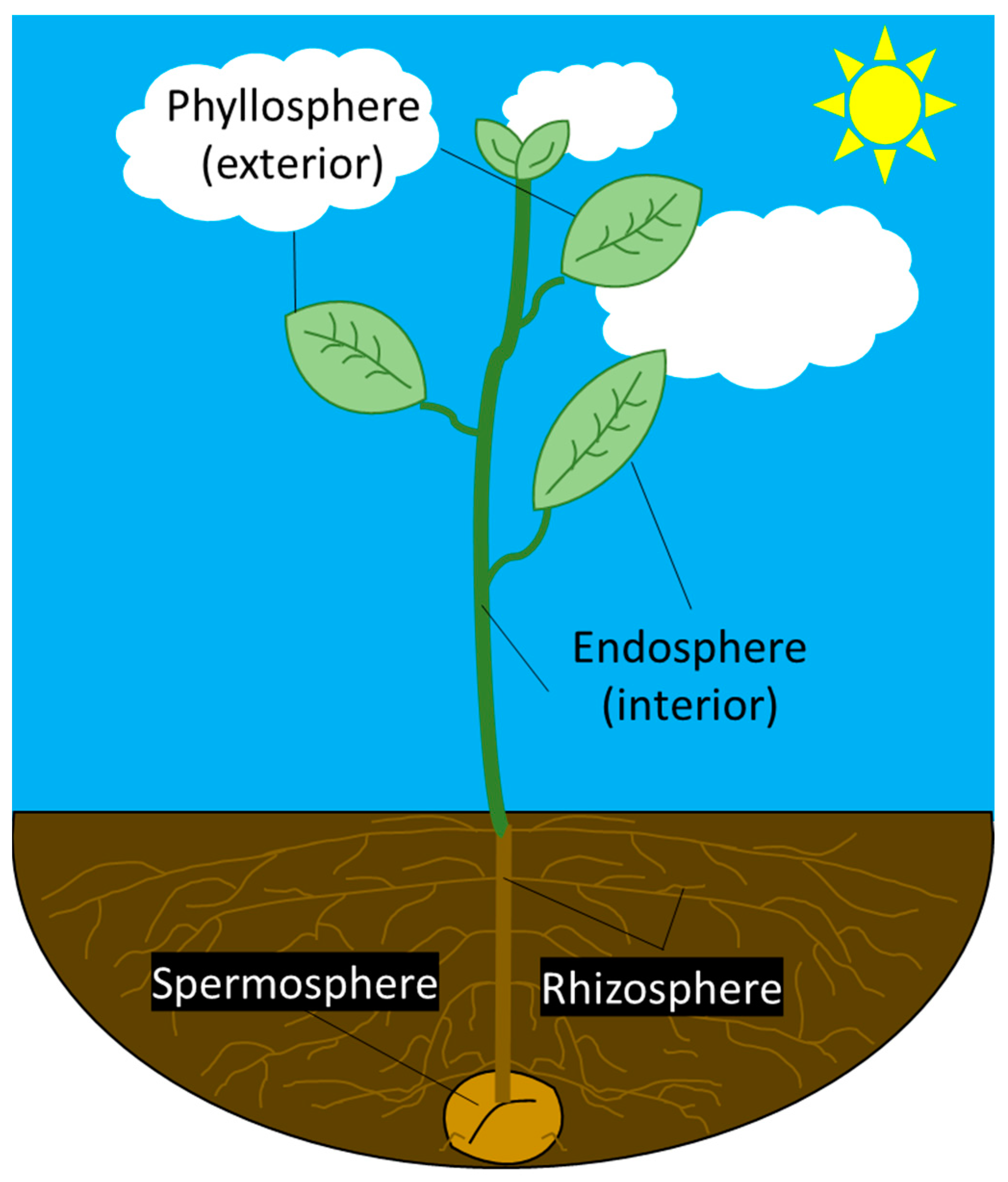
2.1. Microbial Interactions in the Biosphere
2.2. MSI Instrumentation for Imaging Bacterial–Plant Interactions
| Analysis Technique | Lateral/Spatial Resolution | Mass Analyzer | Mass Accuracy | Mass Resolution | Home-Built or Commercial | Field of Study | References | |
|---|---|---|---|---|---|---|---|---|
| SIMS | SIMS, nanoSIMS, liquid SIMS | 0.12–0.5 µm | ToF and Orbitrap | <0.2–1 ppm | unit mass 240,000 | Commercial, home-built | Plant, biological, bacterial, and plant–microbe interaction research | [35,38,39,40,41,42,43,44,45,46] |
| LDI/LDPI | fs-LDI, ns-LDPI, fs-LDPI | 2–100 µm | ToF | 330–340 ppm | 500–30,000 | Home-built | Geological, biological, and bacterial research | [4,33,47,48,49,50] |
| MALDI | MALDI, MALDI-FTICR, AP MALDI, MALDI 2 | 0.6–150 µm | ToF, Orbitrap, FT-ICR, QToF | 0.2–2 ppm | 9000–160,000 | Home-built, commercial | Plant, biological, bacterial, and plant–microbe interaction research | [23,51,52,53,54,55,56] |
| ESI/DESI | DESI and LAESI | 40–200 µm | Orbitrap, Qtrap, and microToF | ≥5 ppm | 10,000–70,000 | Home-built, commercial | Plant, bacterial, and plant–microbe interaction research | [10,57,58] |
| Other | Liquid microjunction surface sampling probe, VUV gas discharge lamp, laser ablation, and solvent capture by aspiration/ESI, and LAAPPI | 70–260 µm | Qtrap, FT-ICR, Q ToF | ≥15 ppm | 10,000–400,000 | Home-built | Plant, biological, bacterial, and plant–microbe interaction research | [24,57,59,60,61] |
2.3. Data Analysis Considerations for MSI
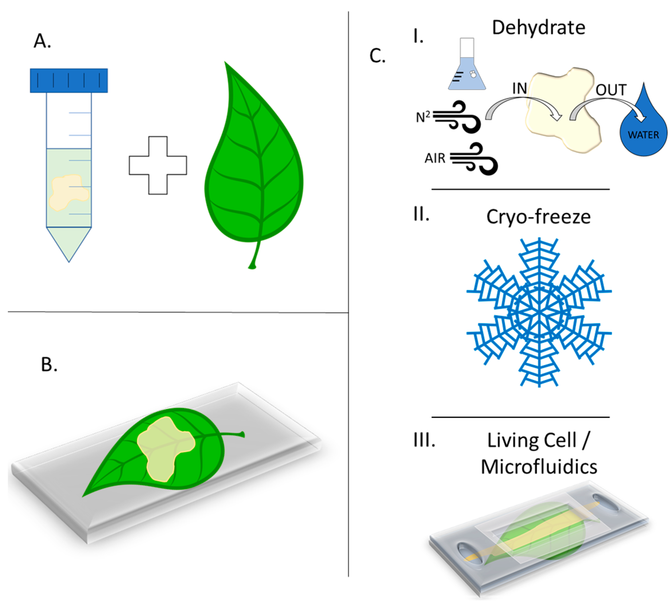
3. Sample Preparation Techniques for Plants and Microbes
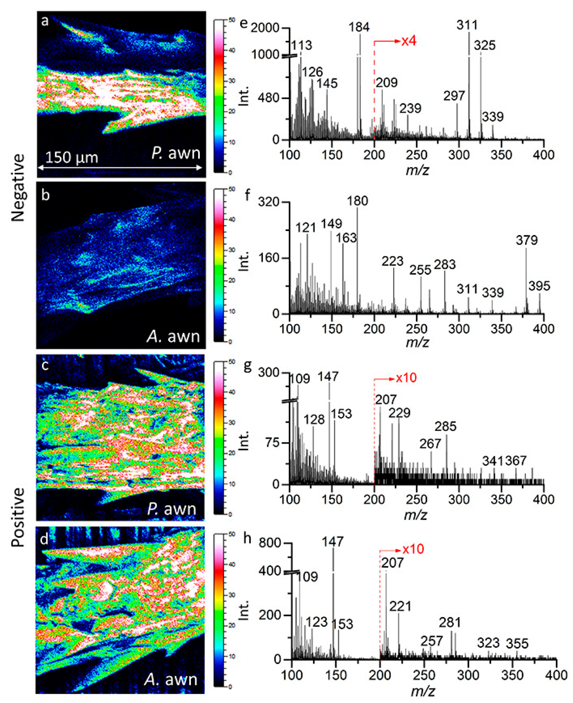
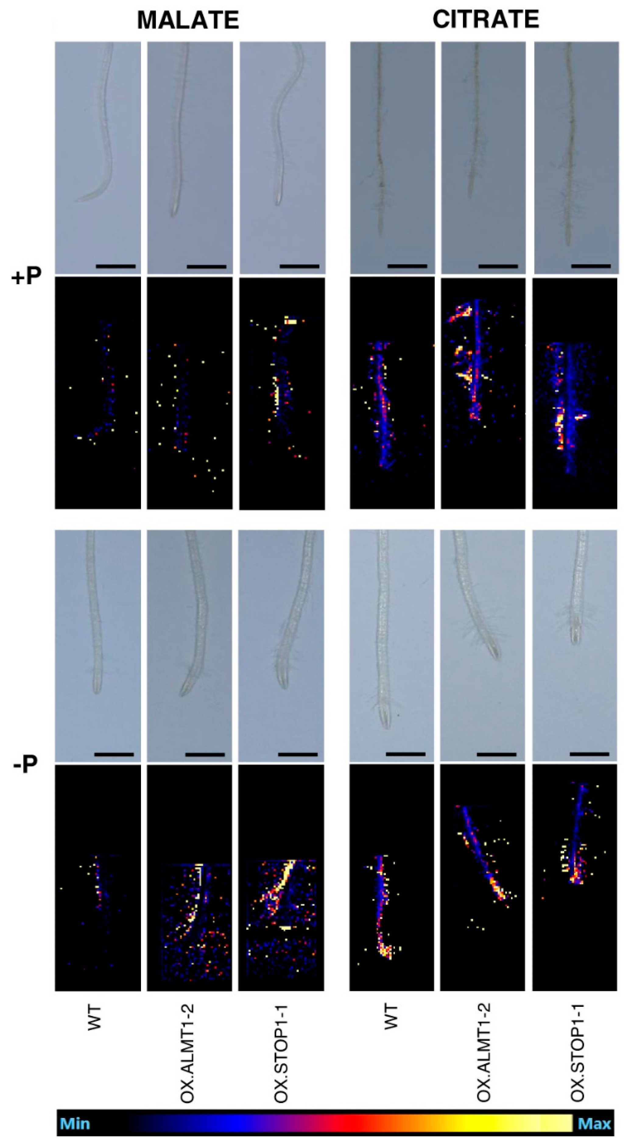
4. MSI Ionization Techniques to Investigate Plant–Microbe Interactions
4.1. Secondary Ion Mass Spectrometry
4.2. Matrix-Assisted Laser Desorption/Ionization Mass Spectrometry
4.3. Laser Desorption/Ionization Mass Spectrometry and Related Methods
4.4. Electrospray Ionization, Desorption Electrospray Ionization, and Laser Ablation Electrospray Ionization Mass Spectrometry
5. Machine Learning for ToF-SIMS and MALDI Data Analysis
6. Conclusions and Outlook
Author Contributions
Funding
Data Availability Statement
Acknowledgments
Conflicts of Interest
Glossary
| AP | Atmospheric Pressure |
| CHCA | Cyano Hydroxycinnamic Acid |
| CLSM | Confocal Laser Scanning Microscopy |
| DESI | Desorption Electrospray Ionization |
| DHB | Dihydroxybenzoic Acid |
| ESI | Electrospray Ionization |
| fs | Femtosecond |
| FT-ICR | Fourier Transform Ion Cyclotron Resonance |
| GC-MS | Gas Chromatography Mass Spectrometry |
| HPLC | High-Pressure Liquid Chromatography |
| IR | Infrared |
| LAAPPI | Laser Ablation Atmospheric Pressure Photoionization |
| LAESI | Laser Ablation Electrospray Ionization |
| LAPPI-MS | Laser Ablation Atmospheric Pressure Photoionization Mass Spectrometry |
| LDI | Laser Desorption Ionization |
| LDPI | Laser Desorption Postionization |
| m/z | Mass-to-Charge Ratio |
| MAF | Maximum Autocorrelation Factors |
| MALDI | Matrix-Assisted Laser Desorption Ionization |
| MCR | Multivariate Curve Resolution |
| ML | Machine Learning |
| MS | Mass Spectrometry |
| MSI | Mass Spectrometry Imaging |
| NMF | Non-Negative Matrix Factorization |
| ns | Nanosecond |
| PCA | Principal Component Analysis |
| QToF | Quadrupole Time-of-Flight |
| SA | Sinapinic Acid |
| SALVI | System for Analysis at the Liquid–Vacuum Interface |
| SIMS | Secondary Ion Mass Spectrometry |
| SNMS | Secondary Neutral Mass Spectrometry |
| SOM | Self-Organizing Map |
| TEM | Transmission Electron Microscopy |
| ToF | Time-of-Flight |
| ToF-SIMS | Time-of-Flight Secondary Ion Mass Spectrometry |
| t-SNE | t-Distributed Stochastic Neighbor Embedding |
| UHV | Ultra-High Vacuum |
| UMAP | Uniform Manifold Approximation Projection |
| UV | Ultraviolet |
| VUV | Vacuum Ultraviolet |
References
- Koppenaal, D.W.; Barinaga, C.J.; Denton, M.B.; Sperline, R.P.; Hieftje, G.M.; Schilling, G.D.; Andrade, F.J.; Barnes, J.H.T. MS detectors. Anal. Chem. 2005, 77, 418a–427a. [Google Scholar] [CrossRef] [PubMed][Green Version]
- Graham, A.W.G.; Ray, S.J.; Enke, C.G.; Felton, J.A.; Carado, A.J.; Barinaga, C.J.; Koppenaal, D.W.; Hieftje, G.M. Resolution and mass range performance in distance-of-flight mass spectrometry with a multichannel focal-plane camera detector. Anal. Chem. 2011, 83, 8552–8559. [Google Scholar] [CrossRef] [PubMed]
- Boughton, B.A.; Thinagaran, D.; Sarabia, D.; Bacic, A.; Roessner, U. Mass spectrometry imaging for plant biology: A review. Phytochem. Rev. 2016, 15, 445–488. [Google Scholar] [CrossRef]
- Wickramasinghe, R.C.; Pasterski, M.J.; Kenig, F.; Ievlev, A.V.; Lorenz, M.; Gross, J.M.; Hanley, L. Femtosecond laser desorption postionization ms vs tof-sims imaging for uncovering biomarkers buried in geological samples. Anal. Chem. 2021, 93, 15949–15957. [Google Scholar] [CrossRef] [PubMed]
- Hanley, L.; Wickramasinghe, R.; Yung, Y.P. Laser desorption combined with laser postionization for mass spectrometry. Annu. Rev. Anal. Chem. 2019, 12, 225–245. [Google Scholar] [CrossRef]
- Buchberger, A.R.; DeLaney, K.; Johnson, J.; Li, L. Mass spectrometry imaging: A review of emerging advancements and future insights. Anal. Chem. 2018, 90, 240–265. [Google Scholar] [CrossRef]
- Gilmore, I.S.; Heiles, S.; Pieterse, C.L. Metabolic imaging at the single-cell scale: Recent advances in mass spectrometry imaging. Annu. Rev. Anal. Chem. 2019, 12, 201–224. [Google Scholar] [CrossRef]
- Zagorac, T. Laser desorption postionization mass spectrometry. Curr. Trends Mass. Spectrom. 2021, 19, 15–19. [Google Scholar]
- Wittig, A.; Arlinghaus, H.F.; Kriegeskotte, C.; Moss, R.L.; Appelman, K.; Schmid, K.W.; Sauerwein, W.A. Laser postionization secondary neutral mass spectrometry in tissue: A powerful tool for elemental and molecular imaging in the development of targeted drugs. Mol. Cancer Ther. 2008, 7, 1763–1771. [Google Scholar] [CrossRef][Green Version]
- Taylor, M.J.; Liyu, A.; Vertes, A.; Anderton, C.R. Ambient single-cell analysis and native tissue imaging using laser-ablation electrospray ionization mass spectrometry with increased spatial resolution. J. Am. Soc. Mass. Spectrom. 2021, 32, 2490–2494. [Google Scholar] [CrossRef]
- Sgobba, E.; Daguerre, Y.; Giampa, M. Unravel the local complexity of biological environments by maldi mass spectrometry imaging. Int. J. Mol. Sci. 2021, 22, 12393. [Google Scholar] [CrossRef]
- Wu, C.; Dill, A.L.; Eberlin, L.S.; Cooks, R.G.; Ifa, D.R. Mass spectrometry imaging under ambient conditions. Mass. Spectrom. Rev. 2013, 32, 218–243. [Google Scholar] [CrossRef]
- Parrot, D.; Papazian, S.; Foil, D.; Tasdemir, D. Imaging the unimaginable: Desorption electrospray ionization–imaging mass spectrometry (desi-ims) in natural product research. Planta Med. 2018, 84, 584–593. [Google Scholar] [CrossRef] [PubMed]
- Gamalero, E.; Bona, E.; Glick, B.R. Current techniques to study beneficial plant-microbe interactions. Microorganisms 2022, 10, 1380. [Google Scholar] [CrossRef] [PubMed]
- Gupta, R.; Anand, G.; Gaur, R.; Yadav, D. Plant-microbiome interactions for sustainable agriculture: A review. Physiol. Mol. Biol. Plants 2021, 27, 165–179. [Google Scholar] [CrossRef] [PubMed]
- Semchenko, M.; Barry, K.E.; de Vries, F.T.; Mommer, L.; Moora, M.; Macia-Vicente, J.G. Deciphering the role of specialist and generalist plant-microbial interactions as drivers of plant-soil feedback. New Phytol. 2022, 234, 1929–1944. [Google Scholar] [CrossRef]
- Rompp, A.; Spengler, B. Mass spectrometry imaging with high resolution in mass and space. Histochem. Cell. Biol. 2013, 139, 759–783. [Google Scholar] [CrossRef]
- Cardinale, M. Scanning a microhabitat: Plant-microbe interactions revealed by confocal laser microscopy. Front. Microbiol. 2014, 5, 94. [Google Scholar] [CrossRef]
- Musat, N.; Musat, F.; Weber, P.K.; Pett-Ridge, J. Tracking microbial interactions with nanosims. Curr. Opin. Biotechnol. 2016, 41, 114–121. [Google Scholar] [CrossRef]
- Ho, Y.N.; Shu, L.J.; Yang, Y.L. Imaging mass spectrometry for metabolites: Technical progress, multimodal imaging, and biological interactions. Wiley Interdiscip. Rev. Syst. Biol. Med. 2017, 9, e1387. [Google Scholar] [CrossRef]
- Acuña, J.J.; Jorquera, M.A. Diversity, interaction, and bioprospecting of plant-associated microbiomes. Diversity 2020, 12, 390. [Google Scholar] [CrossRef]
- Veličković, D.; Anderton, C.R. Mass spectrometry imaging: Towards mapping the elemental and molecular composition of the rhizosphere. Rhizosphere 2017, 3, 254–258. [Google Scholar] [CrossRef]
- Debois, D.; Jourdan, E.; Smargiasso, N.; Thonart, P.; De Pauw, E.; Ongena, M. Spatiotemporal monitoring of the antibiome secreted by bacillus biofilms on plant roots using maldi mass spectrometry imaging. Anal. Chem. 2014, 86, 4431–4438. [Google Scholar] [CrossRef] [PubMed]
- Walton, C.L.; Khalid, M.; Bible, A.N.; Kertesz, V.; Retterer, S.T.; Morrell-Falvey, J.; Cahill, J.F. In situ detection of amino acids from bacterial biofilms and plant root exudates by liquid microjunction surface-sampling probe mass spectrometry. J. Am. Soc. Mass. Spectrom. 2022, 33, 1615–1625. [Google Scholar] [CrossRef] [PubMed]
- Girin, T.; David, L.C.; Chardin, C.; Sibout, R.; Krapp, A.; Ferrario-Mery, S.; Daniel-Vedele, F. Brachypodium: A promising hub between model species and cereals. J. Exp. Bot. 2014, 65, 5683–5696. [Google Scholar] [CrossRef] [PubMed]
- Mhlongo, M.I.; Piater, L.A.; Madala, N.E.; Labuschagne, N.; Dubery, I.A. The chemistry of plant-microbe interactions in the rhizosphere and the potential for metabolomics to reveal signaling related to defense priming and induced systemic resistance. Front. Plant Sci. 2018, 9, 112. [Google Scholar] [CrossRef]
- Brader, G.; Compant, S.; Mitter, B.; Trognitz, F.; Sessitsch, A. Metabolic potential of endophytic bacteria. Curr. Opin. Biotechnol. 2014, 27, 30–37. [Google Scholar] [CrossRef]
- Andreote, F.D.; Gumiere, T.; Durrer, A. Exploring interactions of plant microbiomes. Sci. Agric. 2014, 71, 528–539. [Google Scholar] [CrossRef]
- Forero-Junco, L.M.; Alanin, K.W.S.; Djurhuus, A.M.; Kot, W.; Gobbi, A.; Hansen, L.H. Bacteriophages roam the wheat phyllosphere. Viruses 2022, 14, 244. [Google Scholar] [CrossRef]
- Bashir, I.; War, A.F.; Rafiq, I.; Reshi, Z.A.; Rashid, I.; Shouche, Y.S. Phyllosphere microbiome: Diversity and functions. Microbiol. Res. 2022, 254, 126888. [Google Scholar] [CrossRef]
- Schiltz, S.; Gaillard, I.; Pawlicki-Jullian, N.; Thiombiano, B.; Mesnard, F.; Gontier, E. A review: What is the spermosphere and how can it be studied? J. Appl. Microbiol. 2015, 119, 1467–1481. [Google Scholar] [CrossRef] [PubMed]
- Lee, Y.J.; Perdian, D.C.; Song, Z.; Yeung, E.S.; Nikolau, B.J. Use of mass spectrometry for imaging metabolites in plants. Plant J. 2012, 70, 81–95. [Google Scholar] [CrossRef] [PubMed]
- Walker, A.V.; Gelb, L.D.; Barry, G.E.; Subanajouy, P.; Poudel, A.; Hara, M.; Veryovkin, I.V.; Bell, G.I.; Hanley, L. Femtosecond laser desorption ionization mass spectrometry imaging and multivariate analysis of lipids in pancreatic tissue. Biointerphases 2018, 13, 03B416. [Google Scholar] [CrossRef] [PubMed]
- Unger, W.E.S.; Stockmann, J.M.; Senoner, M.; Weimann, T.; Bütefisch, S.; Passiu, C.; Spencer, N.D.; Rossi, A. Introduction to lateral resolution and analysis area measurements in xps. J. Vac. Sci. Technol. A 2020, 38, 053206. [Google Scholar] [CrossRef]
- Zhang, Y.; Komorek, R.; Son, J.; Riechers, S.; Zhu, Z.; Jansson, J.; Jansson, C.; Yu, X.Y. Molecular imaging of plant-microbe interactions on the brachypodium seed surface. Analyst 2021, 146, 5855–5865. [Google Scholar] [CrossRef]
- Haag, A.M. Mass analyzers and mass spectrometers. Adv. Exp. Med. Biol. 2016, 919, 157–169. [Google Scholar] [CrossRef]
- Xian, F.; Hendrickson, C.L.; Marshall, A.G. High resolution mass spectrometry. Anal. Chem. 2012, 84, 708–719. [Google Scholar] [CrossRef]
- Zhang, S.; Li, H.; Hu, Q.; Wang, Z.; Chen, X. Discrimination of thermal treated bovine milk using maldi-tof ms coupled with machine learning. Food Control. 2022, 142, 109224. [Google Scholar] [CrossRef]
- Bandara, C.D.; Schmidt, M.; Davoudpour, Y.; Stryhanyuk, H.; Richnow, H.H.; Musat, N. Microbial identification, high-resolution microscopy and spectrometry of the rhizosphere in its native spatial context. Front. Plant Sci. 2021, 12, 668929. [Google Scholar] [CrossRef]
- Liu, W.; Huang, L.; Komorek, R.; Handakumbura, P.P.; Zhou, Y.; Hu, D.; Engelhard, M.H.; Jiang, H.; Yu, X.Y.; Jansson, C.; et al. Correlative surface imaging reveals chemical signatures for bacterial hotspots on plant roots. Analyst 2020, 145, 393–401. [Google Scholar] [CrossRef]
- Yu, J.; Zhou, Y.; Engelhard, M.; Zhang, Y.; Son, J.; Liu, S.; Zhu, Z.; Yu, X.Y. In situ molecular imaging of adsorbed protein films in water indicating hydrophobicity and hydrophilicity. Sci. Rep. 2020, 10, 3695. [Google Scholar] [CrossRef] [PubMed]
- Ding, Y.; Zhou, Y.; Yao, J.; Xiong, Y.; Zhu, Z.; Yu, X.Y. Molecular evidence of a toxic effect on a biofilm and its matrix. Analyst 2019, 144, 2498–2503. [Google Scholar] [CrossRef] [PubMed]
- Passarelli, M.K.; Pirkl, A.; Moellers, R.; Grinfeld, D.; Kollmer, F.; Havelund, R.; Newman, C.F.; Marshall, P.S.; Arlinghaus, H.; Alexander, M.R.; et al. The 3D orbisims—Label-free metabolic imaging with subcellular lateral resolution and high mass-resolving power. Nat. Methods 2017, 14, 1175–1183. [Google Scholar] [CrossRef] [PubMed]
- Kaiser, C.; Kilburn, M.R.; Clode, P.L.; Fuchslueger, L.; Koranda, M.; Cliff, J.B.; Solaiman, Z.M.; Murphy, D.V. Exploring the transfer of recent plant photosynthates to soil microbes: Mycorrhizal pathway vs direct root exudation. New Phytol. 2015, 205, 1537–1551. [Google Scholar] [CrossRef]
- Hua, X.; Marshall, M.J.; Xiong, Y.; Ma, X.; Zhou, Y.; Tucker, A.E.; Zhu, Z.; Liu, S.; Yu, X.-Y. Two-dimensional and three-dimensional dynamic imaging of live biofilms in a microchannel by time-of-flight secondary ion mass spectrometry. Biomicrofluidics 2015, 9, 031101. [Google Scholar] [CrossRef]
- Ding, Y.; Zhou, Y.; Yao, J.; Szymanski, C.; Fredrickson, J.; Shi, L.; Cao, B.; Zhu, Z.; Yu, X.Y. In situ molecular imaging of the biofilm and its matrix. Anal. Chem. 2016, 88, 11244–11252. [Google Scholar] [CrossRef]
- Chen, J.; Hu, Y.; Lu, Q.; Wang, P.; Zhan, H. Determination of proflavine in rat whole blood without sample pretreatment by laser desorption postionization mass spectrometry. Anal. Bioanal. Chem. 2017, 409, 2813–2819. [Google Scholar] [CrossRef]
- Cui, Y.; Veryovkin, I.V.; Majeski, M.W.; Cavazos, D.R.; Hanley, L. High lateral resolution vs molecular preservation in near-ir fs-laser desorption postionization mass spectrometry. Anal. Chem. 2015, 87, 367–371. [Google Scholar] [CrossRef]
- Blaze, M.T.M.; Akhmetov, A.; Aydin, B.; Edirisinghe, P.D.; Uygur, G.; Hanley, L. Quantification of antibiotic in biofilm-inhibiting multilayers by 7.87 ev laser desorption postionization ms imaging. Anal. Chem. 2012, 84, 9410–9415. [Google Scholar] [CrossRef][Green Version]
- Bhardwaj, C.; Moore, J.F.; Cui, Y.; Gasper, G.L.; Bernstein, H.C.; Carlson, R.P.; Hanley, L. Laser desorption vuv postionization ms imaging of a cocultured biofilm. Anal. Bioanal. Chem. 2013, 405, 6969–6977. [Google Scholar] [CrossRef]
- Gomez-Zepeda, D.; Frausto, M.; Najera-Gonzalez, H.R.; Herrera-Estrella, L.; Ordaz-Ortiz, J.J. Mass spectrometry-based quantification and spatial localization of small organic acid exudates in plant roots under phosphorus deficiency and aluminum toxicity. Plant J. 2021, 106, 1791–1806. [Google Scholar] [CrossRef] [PubMed]
- Niehaus, M.; Soltwisch, J.; Belov, M.E.; Dreisewerd, K. Transmission-mode maldi-2 mass spectrometry imaging of cells and tissues at subcellular resolution. Nat. Methods 2019, 16, 925–931. [Google Scholar] [CrossRef] [PubMed]
- Ellis, S.R.; Soltwisch, J.; Paine, M.R.L.; Dreisewerd, K.; Heeren, R.M.A. Laser post-ionisation combined with a high resolving power orbitrap mass spectrometer for enhanced maldi-ms imaging of lipids. Chem. Commun. 2017, 53, 7246–7249. [Google Scholar] [CrossRef]
- Kompauer, M.; Heiles, S.; Spengler, B. Atmospheric pressure maldi mass spectrometry imaging of tissues and cells at 1.4-mum lateral resolution. Nat. Methods 2017, 14, 90–96. [Google Scholar] [CrossRef] [PubMed]
- Soltwisch, J.; Kettling, H.; Vens-Cappell, S.; Wiegelmann, M.; Müthing, J.; Dreisewerd, K. Mass spectrometry imaging with laser-induced postionization. Science 2015, 348, 211–215. [Google Scholar] [CrossRef]
- Debois, D.; Ongena, M.; Cawoy, H.; De Pauw, E. Maldi-fticr ms imaging as a powerful tool to identify paenibacillus antibiotics involved in the inhibition of plant pathogens. J. Am. Soc. Mass. Spectrom. 2013, 24, 1202–1213. [Google Scholar] [CrossRef] [PubMed]
- Hieta, J.P.; Kopra, J.; Raikkonen, H.; Kauppila, T.J.; Kostiainen, R. Sub-100 mum spatial resolution ambient mass spectrometry imaging of rodent brain with laser ablation atmospheric pressure photoionization (laappi) and laser ablation electrospray ionization (laesi). Anal. Chem. 2020, 92, 13734–13741. [Google Scholar] [CrossRef]
- Araujo, F.; Santos, D.S.; Pagotto, C.C.; de Araujo, W.L.; Eberlin, M.N. Mass spectrometry characterization of endophytic bacterium curtobacterium sp. Strain er1/6 isolated from citrus sinensis. J. Mass. Spectrom. 2018, 53, 91–97. [Google Scholar] [CrossRef]
- Hieta, J.P.; Sipari, N.; Raikkonen, H.; Keinanen, M.; Kostiainen, R. Mass spectrometry imaging of arabidopsis thaliana leaves at the single-cell level by infrared laser ablation atmospheric pressure photoionization (laappi). J. Am. Soc. Mass. Spectrom. 2021, 32, 2895–2903. [Google Scholar] [CrossRef]
- Franchi, E.; Agazzi, G.; Rolli, E.; Borin, S.; Marasco, R.; Chiaberge, S.; Conte, A.; Filtri, P.; Pedron, F.; Rosellini, I.; et al. Exploiting hydrocarbon-degrading indigenous bacteria for bioremediation and phytoremediation of a multicontaminated soil. Chem. Eng. Technol. 2016, 39, 1676–1684. [Google Scholar] [CrossRef]
- Brauer, J.I.; Beech, I.B.; Sunner, J. Mass spectrometric imaging using laser ablation and solvent capture by aspiration (lasca). J. Am. Soc. Mass. Spectrom. 2015, 26, 1538–1547. [Google Scholar] [CrossRef]
- Fisher, G.L.; Bruinen, A.L.; Ogrinc Potočnik, N.; Hammond, J.S.; Bryan, S.R.; Larson, P.E.; Heeren, R.M.A. A new method and mass spectrometer design for tof-sims parallel imaging ms/ms. Anal. Chem. 2016, 88, 6433–6440. [Google Scholar] [CrossRef] [PubMed]
- Jones, E.A.; Deininger, S.O.; Hogendoorn, P.C.W.; Deelder, A.M.; McDonnell, L.A. Imaging mass spectrometry statistical analysis. J. Proteom. 2012, 75, 4962–4989. [Google Scholar] [CrossRef] [PubMed]
- Weiskirchen, R.; Weiskirchen, S.; Kim, P.; Winkler, R. Software solutions for evaluation and visualization of laser ablation inductively coupled plasma mass spectrometry imaging (la-icp-msi) data: A short overview. J. Cheminform. 2019, 11, 16. [Google Scholar] [CrossRef] [PubMed]
- Bokhart, M.T.; Nazari, M.; Garrard, K.P.; Muddiman, D.C. Msireader v1.0: Evolving open-source mass spectrometry imaging software for targeted and untargeted analyses. J. Am. Soc. Mass. Spectrom. 2018, 29, 8–16. [Google Scholar] [CrossRef] [PubMed]
- Graham, D.J.; Castner, D.G. Multivariate analysis of tof-sims data from multicomponent systems: The why, when, and how. Biointerphases 2012, 7, 49. [Google Scholar] [CrossRef]
- Ringner, M. What is principal component analysis? Nat. Biotechnol. 2008, 26, 303–304. [Google Scholar] [CrossRef]
- Zhang, Y.; Komorek, R.; Zhu, Z.; Huang, Q.; Chen, W.; Jansson, J.; Jansson, C.; Yu, X.Y. Mass spectral imaging showing the plant growth-promoting rhizobacteria’s effect on the brachypodium awn. Biointerphases 2022, 17, 031006. [Google Scholar] [CrossRef] [PubMed]
- Dong, Y.; Li, B.; Malitsky, S.; Rogachev, I.; Aharoni, A.; Kaftan, F.; Svatos, A.; Franceschi, P. Sample preparation for mass spectrometry imaging of plant tissues: A review. Front. Plant Sci. 2016, 7, 60. [Google Scholar] [CrossRef]
- Wei, W.; Zhang, Y.; Komorek, R.; Plymale, A.; Yu, R.; Wang, B.; Zhu, Z.; Liu, F.; Yu, X.-Y. Characterization of syntrophic geobacter communities using tof-sims. Biointerphases 2017, 12, 05G601. [Google Scholar] [CrossRef] [PubMed]
- Yang, L.; Yu, X.Y.; Zhu, Z.; Iedema, M.J.; Cowin, J.P. Probing liquid surfaces under vacuum using sem and tof-sims. Lab Chip 2011, 11, 2481–2484. [Google Scholar] [CrossRef] [PubMed]
- Wei, W.; Plymale, A.; Zhu, Z.; Ma, X.; Liu, F.; Yu, X.Y. In vivo molecular insights into syntrophic geobacter aggregates. Anal. Chem. 2020, 92, 10402–10411. [Google Scholar] [CrossRef]
- Seah, M.P.; Shard, A.G. The matrix effect in secondary ion mass spectrometry. Appl. Surf. Sci. 2018, 439, 605–611. [Google Scholar] [CrossRef]
- Shard, A.G.; Spencer, S.J.; Smith, S.A.; Havelund, R.; Gilmore, I.S. The matrix effect in organic secondary ion mass spectrometry. Int. J. Mass. Spectrom. 2015, 377, 599–609. [Google Scholar] [CrossRef]
- Perry, W.J.; Patterson, N.H.; Prentice, B.M.; Neumann, E.K.; Caprioli, R.M.; Spraggins, J.M. Uncovering matrix effects on lipid analyses in maldi imaging mass spectrometry experiments. J. Mass. Spectrom. 2020, 55, e4491. [Google Scholar] [CrossRef]
- Lanekoff, I.; Stevens, S.L.; Stenzel-Poore, M.P.; Laskin, J. Matrix effects in biological mass spectrometry imaging: Identification and compensation. Analyst 2014, 139, 3528–3532. [Google Scholar] [CrossRef]
- McMillen, J.C.; Fincher, J.A.; Klein, D.R.; Spraggins, J.M.; Caprioli, R.M. Effect of maldi matrices on lipid analyses of biological tissues using maldi-2 postionization mass spectrometry. J. Mass. Spectrom. 2020, 55, e4663. [Google Scholar] [PubMed]
- Tsuchida, S.; Umemura, H.; Murata, S.; Miyabe, A.; Satoh, M.; Matsushita, K.; Nakayama, T.; Nomura, F. Effect of humidity during sample preparation on bacterial identification using matrix-assisted laser desorption/ionization time-of-flight mass spectrometry. J. Chromatogr. B Analyt. Technol. Biomed. Life Sci. 2021, 1176, 122780. [Google Scholar] [CrossRef]
- Kummer, K.M.; Taylor, E.N.; Durmas, N.G.; Tarquinio, K.M.; Ercan, B.; Webster, T.J. Effects of different sterilization techniques and varying anodized TiO2 nanotube dimensions on bacteria growth. J. Biomed. Mater. Res. B Appl. Biomater. 2013, 101, 677–688. [Google Scholar] [CrossRef]
- Mogodiniyai Kasmaei, K.; Spörndly, R.; Udén, P. A sterilization technique with applications to silage research and inoculant evaluation. Grass Forage Sci. 2014, 69, 724–728. [Google Scholar] [CrossRef]
- Chansoria, P.; Narayanan, L.K.; Wood, M.; Alvarado, C.; Lin, A.; Shirwaiker, R.A. Effects of autoclaving, etoh, and uv sterilization on the chemical, mechanical, printability, and biocompatibility characteristics of alginate. ACS Biomater. Sci. Eng. 2020, 6, 5191–5201. [Google Scholar] [CrossRef] [PubMed]
- Odom, R.W. Secondary ion mass spectrometry imaging. Appl. Spectrosc. Rev. 2006, 29, 67–116. [Google Scholar] [CrossRef]
- Bills, D.G. Ion desorption from metal surfaces. Phys. Rev. 1957, 107, 994–995. [Google Scholar] [CrossRef]
- Liebl, H.J.; Herzog, R.F.K. Sputtering ion source for solids. J. Appl. Phys. 1963, 34, 2893–2896. [Google Scholar] [CrossRef]
- Huber, W.K.; Selhofer, H.; Benninghoven, A. An analytical system for secondary ion mass spectrometry in ultra high vacuum. J. Vac. Sci. Technol. 1972, 9, 482–486. [Google Scholar] [CrossRef]
- Fassett, J.D.; Morrison, G.H. Digital image processing in ion microscope analysis: Study of crystal structure effects in secondary ion mass spectrometry. Anal. Chem. 1978, 50, 1861–1866. [Google Scholar] [CrossRef]
- Kingham, D.R. Three dimensional secondary ion mass spectrometry imaging and retrospective depth profiling. Scanning Microsc. 1987, 1, 463–469. [Google Scholar]
- Lorquin, J. Nod factors from sinorhizobium saheli and s. Teranga bv. Sesbaniae are both arabinosylated and fucosylated, a structural feature specific to sesbania rostrata symbionts. Mol. Plant Microbe Interact. 1997, 10, 879–890. [Google Scholar]
- Cliff, J.B.; Gaspar, D.J.; Bottomley, P.J.; Myrold, D.D. Exploration of inorganic c and n assimilation by soil microbes with time-of-flight secondary ion mass spectrometry. Appl. Environ. Microbiol. 2002, 68, 4067–4073. [Google Scholar] [CrossRef]
- Clode, P.L.; Kilburn, M.R.; Jones, D.L.; Stockdale, E.A.; Cliff, J.B., 3rd; Herrmann, A.M.; Murphy, D.V. In situ mapping of nutrient uptake in the rhizosphere using nanoscale secondary ion mass spectrometry. Plant Physiol. 2009, 151, 1751–1757. [Google Scholar] [CrossRef]
- Gonzalez, D.J.; Haste, N.M.; Hollands, A.; Fleming, T.C.; Hamby, M.; Pogliano, K.; Nizet, V.; Dorrestein, P.C. Microbial competition between bacillus subtilis and staphylococcus aureus monitored by imaging mass spectrometry. Microbiology 2011, 157, 2485–2492. [Google Scholar] [CrossRef]
- Ahmad, F.; Babalola, O.O.; Tak, H.I. Potential of maldi-tof mass spectrometry as a rapid detection technique in plant pathology: Identification of plant-associated microorganisms. Anal. Bioanal. Chem. 2012, 404, 1247–1255. [Google Scholar] [CrossRef]
- Stoeckli, M. Automated mass spectrometry imaging with a matrix-assisted laser desorption ionization time-of-flight instrument. Am. Soc. Mass. Spectrom. 1998, 10, 67–71. [Google Scholar] [CrossRef]
- Dunham, S.J.; Ellis, J.F.; Li, B.; Sweedler, J.V. Mass spectrometry imaging of complex microbial communities. Acc. Chem. Res. 2017, 50, 96–104. [Google Scholar] [CrossRef]
- Watrous, J.D.; Alexandrov, T.; Dorrestein, P.C. The evolving field of imaging mass spectrometry and its impact on future biological research. J. Mass. Spectrom. 2011, 46, 209–222. [Google Scholar] [CrossRef] [PubMed]
- Watrous, J.D.; Dorrestein, P.C. Imaging mass spectrometry in microbiology. Nat. Rev. Microbiol. 2011, 9, 683–694. [Google Scholar] [CrossRef] [PubMed]
- Hu, Q.; Noll, R.J.; Li, H.; Makarov, A.; Hardman, M.; Graham Cooks, R. The orbitrap: A new mass spectrometer. J. Mass. Spectrom. 2005, 40, 430–443. [Google Scholar] [CrossRef] [PubMed]
- Honig, R.E.; Woolston, J.R. Laser-induced emission of electrons, ions, and neutral atoms from solid surfaces. Appl. Phys. Lett. 1963, 2, 138–139. [Google Scholar] [CrossRef]
- Ready, J.F. Development of plume of material vaporized by giant-pulse laser. Appl. Phys. Lett. 1963, 3, 11–13. [Google Scholar] [CrossRef]
- Linlor, W.I. Ion energies produced by laser giant pulse. Appl. Phys. Lett. 1963, 3, 210–211. [Google Scholar] [CrossRef]
- Bernal, G.E. Absorbed ion emission from laser-irradiated tungsten. Phys. Lett. 1966, 19, 645–647. [Google Scholar] [CrossRef]
- Bernal, G.E.; Levine, L.P.; Ready, J.F. Time-of-flight spectrometer for laser surface interaction studies. Rev. Sci. Instrum. 1966, 37, 938–941. [Google Scholar] [CrossRef]
- Levine, L.P.; Ready, J.F.; Bernal, G.E. Gas desorption produced by a giant pulse laser. J. Appl. Phys. 1967, 38, 331–336. [Google Scholar] [CrossRef]
- Hillenkamp, F. Laser microprobe mass analysis of organic materials. Nature 1975, 256, 119–120. [Google Scholar] [CrossRef] [PubMed]
- Akhmetov, A.; Moore, J.F.; Gasper, G.L.; Koin, P.J.; Hanley, L. Laser desorption postionization for imaging ms of biological material. J. Mass. Spectrom. 2010, 45, 137–145. [Google Scholar] [CrossRef]
- Pulukkody, A.C.; Yung, Y.P.; Donnarumma, F.; Murray, K.K.; Carlson, R.P.; Hanley, L. Spatially resolved analysis of pseudomonas aeruginosa biofilm proteomes measured by laser ablation sample transfer. PLoS ONE 2021, 16, e0250911. [Google Scholar] [CrossRef]
- Herdering, C.; Wehe, C.A.; Reifschneider, O.; Raj, I.; Ciarimboli, G.; Diebold, K.; Becker, C.; Sperling, M.; Karst, U. Laser ablation based bioimaging with simultaneous elemental and molecular mass spectrometry: Towards spatially resolved speciation analysis. Rapid Commun. Mass. Spectrom. 2013, 27, 2588–2594. [Google Scholar] [CrossRef]
- Ren, L.; Robertson, W.D.; Reimer, R.; Heinze, C.; Schneider, C.; Eggert, D.; Truschow, P.; Hansen, N.O.; Kroetz, P.; Zou, J.; et al. Towards instantaneous cellular level bio diagnosis: Laser extraction and imaging of biological entities with conserved integrity and activity. Nanotechnology 2015, 26, 284001. [Google Scholar] [CrossRef]
- Li, X.; Hang, L.; Wang, T.; Leng, Y.; Zhang, H.; Meng, Y.; Yin, Z.; Hang, W. Nanoscale three-dimensional imaging of drug distributions in single cells via laser desorption post-ionization mass spectrometry. J. Am. Chem. Soc. 2021, 143, 21648–21656. [Google Scholar] [CrossRef]
- Neuland, M.B.; Grimaudo, V.; Mezger, K.; Moreno-García, P.; Riedo, A.; Tulej, M.; Wurz, P. Quantitative measurement of the chemical composition of geological standards with a miniature laser ablation/ionization mass spectrometer designed forin situapplication in space research. Meas. Sci. Technol. 2016, 27, 035904. [Google Scholar] [CrossRef]
- Wang, J.; Wang, Z.; Liu, F.; Cai, L.; Pan, J.B.; Li, Z.; Zhang, S.; Chen, H.Y.; Zhang, X.; Mo, Y. Vacuum ultraviolet laser desorption/ionization mass spectrometry imaging of single cells with submicron craters. Anal. Chem. 2018, 90, 10009–10015. [Google Scholar] [CrossRef] [PubMed]
- Donnarumma, F.; Murray, K.K.; Hanley, L. Fundamentals of laser desorption ionization. In Photoionization and Photo-Induced Processes in Mass Spectrometry; Wiley: Hoboken, NJ, USA, 2021; pp. 305–325. [Google Scholar]
- Yung, Y.P.; Wickramasinghe, R.; Vaikkinen, A.; Kauppila, T.J.; Veryovkin, I.V.; Hanley, L. Solid sampling with a diode laser for portable ambient mass spectrometry. Anal. Chem. 2017, 89, 7297–7301. [Google Scholar] [CrossRef] [PubMed]
- Fenn, J.B. Electrospray ionization for mass spectrometry of lare biomolecules. Science 1989, 246, 64–71. [Google Scholar] [CrossRef] [PubMed]
- Nemes, P. Laser ablation electrospray ionization for atmospheric pressure, in vivo, and imaging mass spectrometry. Anal. Chem. 2007, 79, 8098–8106. [Google Scholar] [CrossRef] [PubMed]
- Gardner, W.; Hook, A.L.; Alexander, M.R.; Ballabio, D.; Cutts, S.M.; Muir, B.W.; Pigram, P.J. Tof-sims and machine learning for single-pixel molecular discrimination of an acrylate polymer microarray. Anal. Chem. 2020, 92, 6587–6597. [Google Scholar] [CrossRef]
- Gardner, W.; Winkler, D.A.; Muir, B.W.; Pigram, P.J. Applications of multivariate analysis and unsupervised machine learning to tof-sims images of organic, bioorganic, and biological systems. Biointerphases 2022, 17, 020802. [Google Scholar] [CrossRef]
- Verbeeck, N.; Caprioli, R.M.; Van de Plas, R. Unsupervised machine learning for exploratory data analysis in imaging mass spectrometry. Mass. Spectrom. Rev. 2020, 39, 245–291. [Google Scholar] [CrossRef]
- Madiona, R.M.T.; Winkler, D.A.; Muir, B.W.; Pigram, P.J. Optimal machine learning models for robust materials classification using tof-sims data. Appl. Surf. Sci. 2019, 487, 773–783. [Google Scholar] [CrossRef]
- Abbasi, K.; Smith, H.; Hoffman, M.; Farghadany, E.; Bruckman, L.S.; Sehirlioglu, A. Dimensional stacking for machine learning in tof-sims analysis of heterostructures. Adv. Mater. Interfaces 2020, 8, 2001648. [Google Scholar] [CrossRef]
- Feucherolles, M.; Nennig, M.; Becker, S.L.; Martiny, D.; Losch, S.; Penny, C.; Cauchie, H.M.; Ragimbeau, C. Combination of maldi-tof mass spectrometry and machine learning for rapid antimicrobial resistance screening: The case of campylobacter spp. Front. Microbiol. 2021, 12, 804484. [Google Scholar] [CrossRef]
- Jeon, K.; Kim, J.M.; Rho, K.; Jung, S.H.; Park, H.S.; Kim, J.S. Performance of a machine learning-based methicillin resistance of staphylococcus aureus identification system using maldi-tof ms and comparison of the accuracy according to sccmec types. Microorganisms 2022, 10, 1903. [Google Scholar] [CrossRef] [PubMed]


Disclaimer/Publisher’s Note: The statements, opinions and data contained in all publications are solely those of the individual author(s) and contributor(s) and not of MDPI and/or the editor(s). MDPI and/or the editor(s) disclaim responsibility for any injury to people or property resulting from any ideas, methods, instructions or products referred to in the content. |
© 2023 by the authors. Licensee MDPI, Basel, Switzerland. This article is an open access article distributed under the terms and conditions of the Creative Commons Attribution (CC BY) license (https://creativecommons.org/licenses/by/4.0/).
Share and Cite
Parker, G.D.; Hanley, L.; Yu, X.-Y. Mass Spectral Imaging to Map Plant–Microbe Interactions. Microorganisms 2023, 11, 2045. https://doi.org/10.3390/microorganisms11082045
Parker GD, Hanley L, Yu X-Y. Mass Spectral Imaging to Map Plant–Microbe Interactions. Microorganisms. 2023; 11(8):2045. https://doi.org/10.3390/microorganisms11082045
Chicago/Turabian StyleParker, Gabriel D., Luke Hanley, and Xiao-Ying Yu. 2023. "Mass Spectral Imaging to Map Plant–Microbe Interactions" Microorganisms 11, no. 8: 2045. https://doi.org/10.3390/microorganisms11082045
APA StyleParker, G. D., Hanley, L., & Yu, X.-Y. (2023). Mass Spectral Imaging to Map Plant–Microbe Interactions. Microorganisms, 11(8), 2045. https://doi.org/10.3390/microorganisms11082045


