Article

Epidemiology of Zoonotic Coxiella burnetii in The Republic of Guinea

by
Olesia V. Ohlopkova
1,*,
Sergey A. Yakovlev
2,
Kabwe Emmanuel
3,
Alexey A. Kabanov
1,
Dmitry A. Odnoshevsky
1,
Mikhail Yu. Kartashov
1,
Alexey D. Moshkin
1,
Igor V. Tuchkov
2,
Nikita Yu. Nosov
2,4,
Andrey A. Kritsky
2,5,
Milana A. Agalakova
6,7,
Yuriy N. Davidyuk
3,
Svetlana F. Khaiboullina
3,
Sergey P. Morzunov
8,*,
Magasuba N’Fally
9,
Sanaba Bumbali
10,
Mamadou Fode Camara
11,
Mamadou Yero Boiro
10,
Alexander P. Agafonov
1,
Elena V. Gavrilova
1 and
Rinat A. Maksyutov
1
add Show full author list remove Hide full author list
1
State Research Center of Virology and Biotechnology «Vector» of Rospotrebnadzor, Koltsovo 630559, Russia
2
Russian Research Anti-Plague Institute «Microbe» of Rospotrebnadzor, Saratov 410005, Russia
3
OpenLab “Gene and Cell Technologies”, Institute of Fundamental Medicine and Biology, Kazan (Volga Region) Federal University, Kazan 420008, Russia
4
State Research Center of Dermatovenerology and Cosmetology of Russian Ministry of Health, Moscow 107076, Russia
5
Limited Liability Company, «Biotech Campus», Moscow 117437, Russia
6
Faculty of Preventive Medicine, Ural State Medical University, Yekaterinburg 620014, Russia
7
Limited Liability Company, «Quality Med», Yekaterinburg 105318, Russia
8
Department of Pathology, University of Nevada, Reno, NV 89557, USA
9
Faculty of Medicine, Pharmacy and Dentistry, University Gamal Abdel Nasser, Conakry 001, Guinea
10
Research Institute of Applied Biology of Guinea, Kindia 100, Guinea
11
Higher Institute of Science and Veterinary Medicine, Dalaba 280, Guinea
*
Authors to whom correspondence should be addressed.
Microorganisms 2023, 11(6), 1433; https://doi.org/10.3390/microorganisms11061433
Submission received: 28 April 2023 / Revised: 25 May 2023 / Accepted: 26 May 2023 / Published: 29 May 2023
(This article belongs to the Special Issue Intracellular Bacteria: From Basic Research to Clinics 2.0)

Abstract

:
Background: Q fever is a zoonotic infectious disease characterized by fever, malaise, chills, significant weakness, and muscle pain. In some cases, the disease can become chronic and affect the inner membranes of the heart, such as the valves, leading to endocarditis and a high risk of death. Coxiella burnetii (C. burnetii) is the primary causative agent of Q fever in humans. This study aims to monitor the presence of C. burnetii in ticks collected from small mammals and cattle in the Republic of Guinea (RG). Methods: Rodents were trapped in the Kindia region of RG during 2019–2020, and ticks were collected from cattle in six regions of RG. Total DNA was extracted using a commercial kit (RIBO-prep, InterLabService, Russia) following the manufacturer’s instructions. Real-time PCR amplification was conducted using the kit (AmpliSens Coxiella burnetii-FL, InterLabService, Russia) to detect C. burnetii DNA. Results and Conclusions: Bacterial DNA was detected in 11 out of 750 (1.4%) small mammals and 695 out of 9620 (7.2%) tick samples. The high number of infected ticks (7.2%) suggests that they are the main transmitters of C. burnetii in RG. The DNA was detected in the liver and spleen of a Guinea multimammate mouse, Mastomys erythroleucus. These findings demonstrate that C. burnetii is zoonotic in RG, and measures should be taken to monitor the bacteria’s dynamics and tick prevalence in the rodent population.

1. Introduction

Coxiella burnetii is an obligate intracellular parasite and a member of the Coxiellaceae family [1]. Although this bacterium shares morphological similarities with Rickettsia, phylogenetic analysis has demonstrated that it belongs to a separate gamma subdivision of Proteobacteria [2,3]. Coxiella burnetii is a gram-negative, rod-shaped bacterium that replicates within eukaryotic cell vacuoles [4]. It exhibits high resistance to physical and chemical environmental factors [5], which contributes to its ability to cause human infection. Studies have shown that even after exposure to 1% phenol solution or 0.5% chloramine solution for more than three days, C. burnetii can remain viable [6]. Additionally, it can survive for up to 18 months in the dry feces of infected Dermacentor andersoni ticks [7]. However, boiling can kill the bacterium within 10 min in milk, whereas it can persist for up to a month in raw meat [8,9]. Furthermore, C. burnetii displays resistance to ultraviolet radiation and can persist at low temperatures. These findings suggest that C. burnetii can circulate in the environment for an extended period while maintaining a high level of virulence.
Acute Q fever typically presents as a mild or localized disease with a low risk of death. However, if left untreated, chronic Q fever can lead to mortality, primarily due to endocarditis or vascular infection. Patients treated for endocarditis caused by chronic Q fever have a 10-year mortality rate of 19%. Q fever shares clinical symptoms similar to those of various bacterial and viral infectious diseases [10]. The incubation period can last up to 60 days. The most common manifestation of the infection is an abrupt onset of fever, with body temperature rising to 39–40 °C, accompanied by chills, headache, dry cough, weakness, decreased appetite, and sleep disturbances [11]. Patients may also experience muscle and joint pain, dizziness, nausea, vomiting (less frequently), and nosebleeds. As the disease progresses, signs of organ involvement may emerge, and pneumonia may occur in 8–32% of cases [12]. Post-infectious asthenia is the most common complication of acute Q fever. Chronic coxiellosis affects 5–10% of patients. Pregnant women infected with C. burnetii, the causative agent of Q fever, may experience unfavorable pregnancy outcomes. The mortality rate for coxiellosis is less than 2.8% [13].
Humans can contract Q fever by inhaling contaminated aerosols or consuming contaminated animal products. Contaminated aerosols, which can contain C. burnetii, are generated during the processing of parturient fluids, placenta, or wool from infected animals [14]. While C. burnetii has been found in ticks, arthropod-borne transmission to humans is believed to play only a minor role in outbreaks [15]. Q fever is a global concern, often associated with cattle and sheep farming [16]. One of the largest documented outbreaks occurred in the Netherlands between 2007 and 2010, where over 4000 individuals became ill and 14 died [17,18].
Despite efforts by the World Health Organization (WHO) to reduce its spread and control morbidity and mortality, C. burnetii remains a public health problem in many countries [19]. Factors such as globalization, migration, and tourism can contribute to the spread of the disease [20]. In the Republic of Guinea (RG), low life expectancy is linked to various viral and bacterial diseases, including Lassa fever, West Nile virus, Crimean-Congo hemorrhagic fever, yellow fever, Chikungunya, Rift Valley fever, and Q fever [21]. According to the WHO report, the current life expectancy in RG is between 45 and 50 years [22]. Therefore, greater attention should be given to the prevention of these related diseases. One potential preventive measure could involve regular screening of rodents and ticks, as well as increasing anti-epidemic activities in high-risk areas for Q fever.
In nature, C. burnetii is found in more than 60 species of warm-blooded animals and approximately 50 species of birds, which serve as primary reservoirs [23]. Ticks that feed on infected animals are the primary carriers of C. burnetii [24]. Farm animals such as cattle, goats, and sheep typically transmit the bacteria to humans. C. burnetii can cause transient bacteremia in infected animals, during which feeding ticks can become infected [14]. There are two recognized types of natural Q fever infections: primary (natural) and secondary (agricultural or anthropogenic) [7]. In natural foci, pathogens are transmitted by ticks, primarily Ixodes ticks and, to a lesser extent, Haemaphysalis ticks. Rickettsia can persist in these ticks and is transmitted transovarially and transstadially (from egg, larva, nymph, to adult tick), making them not only carriers but also reservoirs of the Q fever pathogen [9]. The evidence demonstrating direct transmission of C. burnetii from ticks to humans remains limited. However, there have been a few reported cases describing the transmission of C. burnetii to humans through tick bites [25,26,27,28,29,30].
Morbidity primarily occurs sporadically and affects professional risk groups such as cattle breeders and rural inhabitants, mainly during the spring, summer, and autumn seasons. Epidemic outbreaks are also possible. Infection from an infected person is rare and typically occurs through exposure to contaminated sputum and milk from nursing women [5]. In agricultural areas, the sources of the infection agent are cattle, small ruminants, horses, pigs, dogs, poultry, and rodents. In natural environments, the sources are wild ungulates, small mammals (primarily rodents), and birds. Mammals such as cattle and pigs are of epidemiological significance as they excrete rickettsiae through excreta, urine, milk, and amniotic fluid [10].
People can become infected in agricultural areas through various routes, including exposure to contaminated air and dust when handling wool, fur, bristles, and animal skin; consumption of contaminated milk and milk products; and contact with infected animals during care, slaughter, and butchering. Secondary sources pose a higher risk for humans in settled areas compared to primary sources [31].
The objective of this study was to investigate the prevalence of C. burnetii in rodents and ticks in the Republic of Guinea (RG), where the circulation of this pathogen is poorly understood.

2. Materials and Methods
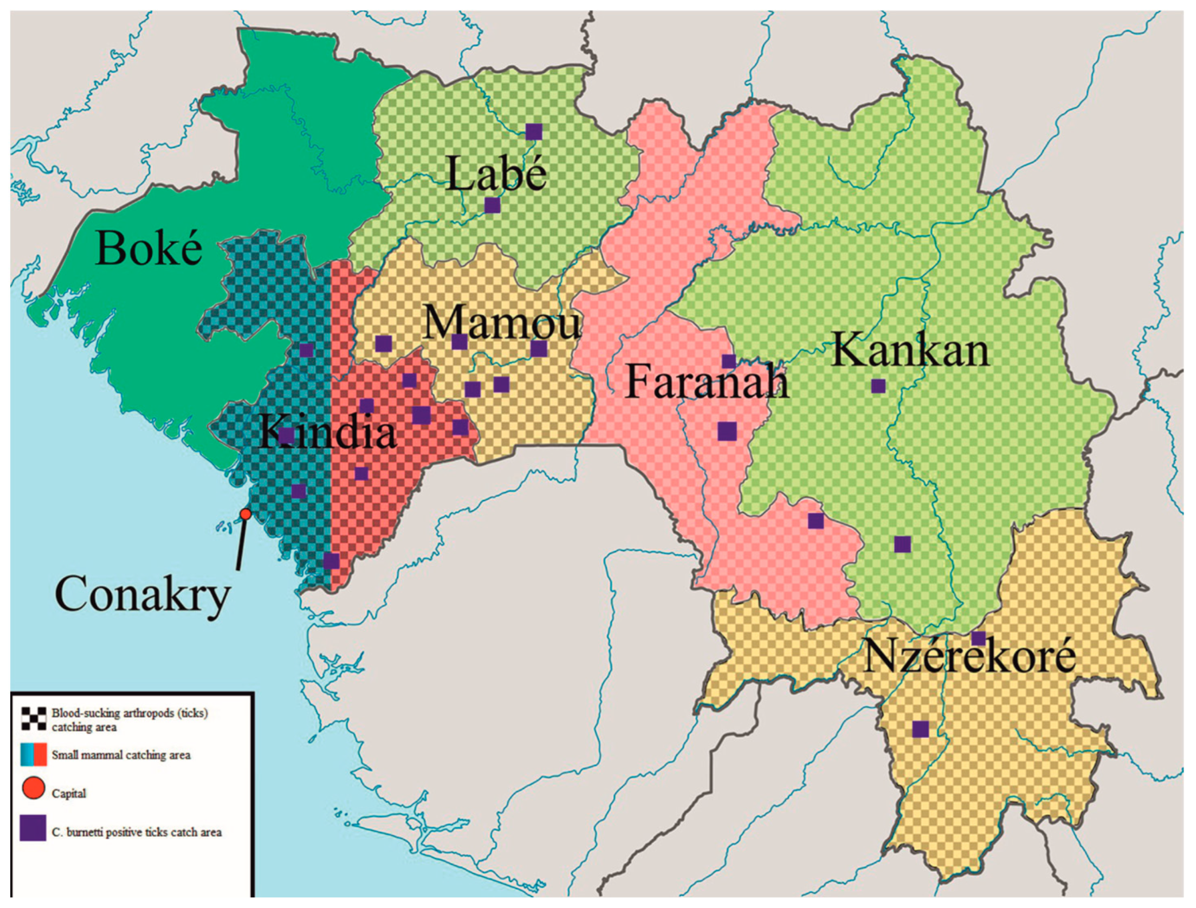
Small mammal and rodent samples: Between 2019 and 2020, 250 small mammals were captured on farms in the Kindia region of RG and separated by species. The geographical locations of rodent trapping sites are shown in Figure 1. Blood-sucking arthropods (ticks) were collected from 7600 farm animals (cattle) in the Kindia, Mamou, Faranah, Labe, Kankan, and N’Zerekore regions of RG from 2016 to 2020. A total of 9620 Ixodidae ticks of eight species were identified.
Animal rights compliance: The study was conducted in accordance with the requirements of Sanitary Rules 1.3.3118-13, titled “Safety of work with microorganisms of risk (hazard) groups 1 and 2” [32]. It adhered to the legislation of the Russian Federation as well as reference documents issued by the Federal State Budgetary Institution of Science and Research Center “Vector” of Rospotrebnadzor, Russia, and followed the International Ethical Standards. Euthanasia of animals was performed using cervical dislocation. Brain, lung, kidney, liver, and spleen samples were collected from each small mammal. Suspensions of the collected tissues and ticks were prepared by homogenization in saline solution and used immediately for DNA extraction.
DNA extraction: The collected samples were subjected to lysis using a solution (RIBO-prep, InterLabService, Moscow, Russia), which resulted in the breakdown of cellular membranes and other biopolymer complexes, releasing nucleic acids and cellular components. After the addition of a precipitation solution and centrifugation, the dissolved DNA precipitated, while other components of the lysed clinical material remained in solution and were removed through subsequent washing. In the final step of the extraction, the precipitate was dissolved in an elution buffer, resulting in purified DNA in solution. This procedure yielded a purified DNA preparation that was free from amplification reaction inhibitors, ensuring high analytical sensitivity for PCR analysis.
Real-time polymerase chain reaction (RT-PCR) was performed using the AmpliSens Coxiella burnetii-FL reagent kit (InterLabService, Moscow, Russia) following the manufacturer’s specifications. This test targets the groEL gene of the microbe and is specifically designed for the detection of C. burnetii [31,33,34]. The test has demonstrated no false-positive results when using a closely related bacterial DNA template [35]. According to the manufacturer’s instructions, total DNA was extracted from the suspensions using the RIBO-prep reagent kit (InterLabService, Moscow, Russia). All testing was conducted at the Russian-Guinean Scientific and Clinical Diagnostic Center for Epidemiology and Microbiology, located in Kindia, the Republic of Guinea (RG).
Statistical analysis: The prevalence index (PI) was calculated using the equation [36]:
PI = (ni/N) × 100.
where PI is the prevalence index; ni is the number of individuals; and N is the total number of individuals in the population. The abundance index (AI) was calculated using the equation [37]:
AI = m/N.
where m is the number of ticks and N is the number of individuals. Statistical analysis was performed in the R environment [38]. Statistically significant differences between comparison groups were accepted as p < 0.05, as assessed by the Kruskal-Wallis test with Benjamini-Hochberg (BH) adjustment for multiple comparisons. Correlations were analyzed using the R psych package, and p-values were adjusted with the Benjamini-Hochberg method based on Spearman’s rank correlation coefficient.

3. Results

3.1. Coxiella Burnetii DNA Prevalence in Small Mammal Tissue

We used 750 small mammal tissue samples in our study, identified as black rats (Rattus rattus) (73 mammals), Guinea multimammate mice (Mastomys erythroleucus) (85 mammals), Natal multimammate mice (Mastomys natalensis) (27 mammals), Guinean gerbil (Gerbilliscus guinea) (4 mammals), Dalton’s mouse (Praomys daltoni) (6 mammals), Temminck’s mouse (Mus musculoides) (16 mammals), house mouse (Mus musculus) (28 mammals), Sudanian grass rat (Arvicanthis androgen) (4 mammals), and the African giant shrew (Crocidura olivieri) (11 mammals). Five organs (brain, spleen, liver, kidney, and lungs) were obtained from each small mammal. Coxiella burnetii DNA was detected in the organs of 11 captured mammals (Table 1).
Coxiella burnetii DNA was mainly detected in the liver, spleen, lungs, and kidney among the tissues studied. The bacterial DNA was frequently found in the M. erythroleucus rodent species, with a PI of 8.2%. In addition, C. burnetii DNA was detected in a single organ sample collected from G. guineae, M. musculus, and R. rattus. The prevalence index for the detection of bacterial DNA did not exceed 9.1% in these species.
Bacterial nucleic acid was also isolated from ticks of the Laelapidae family that were collected from small mammals. Interestingly, a significant number of C. burnetii DNA-positive ticks were found on M. erythroleucus, which also exhibited the highest prevalence index for bacterial DNA detection in its organs. These findings suggest a potential role for ticks in the transmission of C. burnetii among these small mammals.
The small mammals were trapped in six different natural and satellite biotypes, including savannah, areas near water sources, agroecosystems, houses, stores/warehouses, and coniferous forests (Table 2). Small mammals that tested positive for C. burnetii DNA were identified in three biotypes: savannah (83.5%), stores/warehouses (9.7%), and agroecosystems (6.8%). It is noteworthy that the majority of small mammals with detectable bacterial DNA were captured in wilderness areas.

3.2. Coxiella burnetii DNA Prevalence in Ticks Collected from Cattle

We conducted an examination of 303 cattle to assess the presence of blood-sucking arthropods during both the dry and rainy seasons. Overall, we collected a total of 9620 ticks from the cattle. The presence of C. burnetii DNA was observed in 563 ticks collected during the dry season and 132 ticks collected during the rainy season, as shown in Table 3.

3.3. Abundance of Ticks in Investigated Cattle

Ixodid ticks were the most dominant species found in 220 (72.6%) cattle. Out of the 695 positive ixodid ticks, the most frequently found arthropods were Amblyomma variegatum (544; 78.2%), Rhipicephalus decoloratus (62; 8.9%), Rhipicephalus annulatus (38; 5.4%), Hyalomma truncatum Koch (26; 3.7%), Rhipicephalus sanguineus (16; 2.3%), Rhipicephalus (Boophilus) geigyi (7; 1%), and Haemophysalis leachi (2; 0.28%) species (Table 4).
The AI, representing the number of ticks per animal, ranged from 2.1 in the dry season to 3.5 in the rainy season. These data indicate seasonal variations in tick abundance. It appears that there are more ticks on cattle during the rainy season as compared to the dry season.
Variations in tick species PI were found between the rainy and dry seasons. H. leachi, Am. variegatum, Hy. truncatum, Rh. annulatus, and Rh. sanguineus ticks were commonly found during the dry season. However, these species were less frequent during the rainy season. Instead, Rh. decoloratus and Rh. geigyi were most frequently found during the rainy season. It should be noted that Am. variegatum was the dominant tick species collected in both seasons.
Next, we analyzed the prevalence of C. burnetii in each tick species (Figure 1). Ticks of the following species were found to be positive for bacterium DNA in the dry season: Am. variegatum, Hy. truncatum, Rh. geigyi, Baeph. Sp, Rh. annulatus, Rh. sanguineus, H. leachi, and Rh. decoloratus. A smaller group of tick species, including Am. variegatum, Hy. truncatum, Rh. geigyi, and Rh. decoloratus, were positive in the rainy season.

4. Discussion

Coxiella burnetii is a gram-negative bacterium that resides within cells and is responsible for causing Q fever [39]. This infectious disease can be transmitted to humans through the consumption of contaminated products or the inhalation of aerosols containing the bacteria [40]. Ticks have been identified as susceptible to C. burnetii, and the bacteria are often found in the cells of their middle gut or stomach, which may explain their presence in tick feces [41]. Some studies have proposed ticks as potential biological vectors for transmitting the pathogen to mammals [42]. However, in recent years, a new member of the Coxiella genus called Coxiella-like endosymbiont (Coxiella-LE) has been discovered in ticks [43]. These microbes share morphological similarities with C. burnetii [44] and are commonly found in ticks [45], which complicates our understanding of the tick’s role in transmitting C. burnetii. Notably, the LE microbes lack virulence factors, suggesting a lower likelihood of being pathogenic [46]. Therefore, accurate identification of the Coxiella genus bacterium requires techniques like PCR and sequencing. In this study, we employed RT-PCR-based testing to determine the prevalence of C. burnetii in ticks collected from RG. This method exhibits high specificity in detecting C. burnetii DNA, ensuring accurate results.
Our findings align with previous studies that have reported the presence of C. burnetii in ticks in Africa [47] and contribute to our understanding of its circulation in RG. Previously, epidemiological data on this bacterium in RG had been limited to the detection of bacterium-specific antibodies in humans and animals [39,48]. Furthermore, we confirm earlier observations that C. burnetii can be found in ticks collected from small mammals [7,49]. The prevalence index (PI) we observed (7.2%) was higher than that reported in a systematic review of bacterial prevalence in hard ticks in Europe [50]. It appears that the prevalence of C. burnetii varies significantly across different regions. For instance, Ni et al. found C. burnetii in 55.66% of ticks [51], while Gonzalez et al. reported a high prevalence of 55.66% in the Meso-Mediterranean ecosystem [52]. A study conducted in Rome documented a C. burnetii prevalence of 22% in ticks [53]. In a meta-analysis by Yessinou et al., variations in C. burnetii prevalence were observed between Africa, Europe, and the Middle East [54]. In Africa, the prevalence of the bacteria in ticks ranged from 2.91% to 13.97%, which includes the value of 7.2% found in our study. Thus, it seems that the percentage of ticks carrying this human pathogen in RG is within the range commonly observed in other countries. The substantial variations in C. burnetii prevalence reported in different studies could be attributed to the gene targets used for PCR analysis, such as IS1111, ompA, and icd [55,56]. Similarly, Korner et al. reported variations in C. burnetii prevalence in ticks due to the gene target used for PCR analysis [57]. It has been demonstrated that the icd gene has high similarity between C. burnetii and Coxiella-LE, which could potentially lead to an overestimation of the tick’s role in pathogen transmission [58]. Additionally, factors such as climate, season, humidity, and the small number of ticks included in the study may contribute to the prevalence of C. burnetii in ticks. In a study by Troupin et al., a higher seroprevalence of C. burnetii was found in cattle in most of the studied prefectures [54]. Among cattle, seroprevalence was higher in females compared to males, and a higher frequency of antibody detection was observed in mature animals compared to young ones.
In our study, we employed a PCR method that specifically detects C. burnetii in ticks. This highly specific method was chosen to minimize the potential for false-positive results due to the presence of Coxiella-LE microbes. The discovery of Coxiella-LE microbes has prompted a re-evaluation of the role of ticks as vectors for C. burnetii [45]. Coxiella-LE is known to dominate in ticks and is believed to provide them with vitamin B [59]. Additionally, the genome of Coxiella-LE shares up to 97% identity with that of C. burnetii [60]. Such high identity in nucleic acid sequences can lead to misidentification.
To detect C. burnetii, several gene targets have been used, including IS1111, icd, com1, sodB, and GroEL/htpB [61,62,63]. However, some of these genes exhibit significant similarity between the bacterium and Coxiella-LE. Duron et al. demonstrated that approximately 30% of Coxiella-LE-carrying ticks tested positive for the IS1111 gene [64]. Furthermore, the icd sequence of Coxiella-LE shares 90% identity with the same gene in C. burnetii [58]. Studies have indicated that the most conserved gene, groEL, is the optimal target for C. burnetii detection using PCR methods [43,65]. In our study, we utilized the AmpliSens Coxiella burnetii-FL reagent kit, which targets the groEL gene. Therefore, the specificity of the PCR method employed in our study is sufficient to ensure accurate detection of C. burnetii.
Our data reveals variations in the tick population on cattle, with Amblyomma being the most prevalent species, followed by Rhipicephalus and Hyalomma. These findings are consistent with previous observations made by Bayer and Maina [52]. We also observed that the tick abundance index (AI) was higher during the rainy season compared to the dry season. This supports the findings of Bayer and Maina, who demonstrated lower tick loads in dry seasons compared to rainy seasons in Nigeria [66]. Seasonal variations in tick abundance on cattle have been documented in other studies, such as the work by Babayani and Makati in Botswana [67]. It has been suggested that humidity supports tick survival and growth [68]. Therefore, the rainy season may pose a higher risk of tick-borne pathogen exposure for cattle.
Additionally, we observed changes in tick species between the two seasons. During the dry season, we collected more H. leachi, Am. variegatum, Hy. truncatum, Rh. annulatus, and Rh. sanguineus ticks. In contrast, these species were less frequent during the rainy season, while Rh. decoloratus and Rh. geigyi were more frequently found. Several factors have been identified to contribute to variations in tick numbers and species, including fur color, sex, and food preferences [69,70]. It has been demonstrated that males have fewer ticks than females during the dry season [67]. Additionally, cattle with darker fur tend to have more ticks compared to those with white fur. Another study found higher tick loads in calves during both dry and wet seasons, while seasonal changes were observed in adult calves, with fewer ticks during the dry season [71]. Interestingly, changes in food taste, such as sweet and sour, can affect tick species, as shown by Marufu et al. [72]. They reported that Hyalomma species were more prevalent in sour food. These changes in food taste could be influenced by seasonal variations in grass composition due to differences in water content.
We observed variations in the prevalence of C. burnetii in different tick species depending on the season. There were more tick species carrying the bacterium’s DNA during the dry season compared to the wet season. Limited data are available on C. burnetii prevalence during the dry and wet seasons in Africa. For instance, a study by Titcomb et al. in Kenya did not find any effect of rainfall on pathogen prevalence in ticks [73]. It is important to note that the method of tick collection can influence the interpretation of the results. Collecting ticks through dragging and flagging may not capture ticks that are actively feeding on animals [74]. Climate change can also impact the distribution of arthropod vectors. The tick population is dependent on the survival of larvae, which requires moisture [75]. Reductions in the duration and intensity of the rainy season [76,77] can affect tick survival. Additionally, the abundance of hosts is crucial for tick survival. Climate change, particularly longer dry seasons, can result in the migration of herbivores [25]. This could explain the higher species variations observed during the dry season. These findings suggest that a greater variety of tick species may contribute to the epidemiology of C. burnetii during the dry season.
We also discovered that ticks collected from small mammals tested positive for C. burnetii DNA. Interestingly, the highest prevalence of bacterial DNA was found in ticks collected from small mammals captured in the savannah. We also detected C. burnetii in ticks collected from small mammals in stores, warehouses, and agroecosystems, but the prevalence of bacterial DNA-positive mice was significantly lower. Notably, several organs of M. erythroleucus, a small mammal inhabiting the savannah, tested positive for bacterial DNA. These findings suggest that ticks may play a more significant role in the epidemiology of C. burnetii among wild small mammals.

5. Conclusions

Our findings significantly contribute to our understanding of the role of ticks in the epidemiology of C. burnetii in RG. This study represents the first analysis of C. burnetii epidemiology in ticks collected from both small mammals and cattle in the RG. We employed a highly specific PCR method to detect bacterial DNA, ensuring accurate identification. Our results revealed that multiple tick species can harbor the bacterium. Interestingly, we observed a higher prevalence of C. burnetii in ticks collected from small mammals inhabiting the savannah. This finding suggests that ticks may play a significant role in the epidemiology of C. burnetii. Furthermore, we noted a greater abundance of ticks collected from cattle during the wet season compared to the dry season. Additionally, we observed more variations in tick species during the dry season compared to the wet season. Similarly, a higher number of tick species tested positive for C. burnetii DNA in the dry season compared to the wet season.

Author Contributions

Conceptualization, methodology, investigation, writing—original draft preparation, O.V.O. and S.A.Y.; methodology, investigation, A.A.K. (Alexey A. Kabanov), D.A.O., I.V.T., N.Y.N. and A.A.K. (Andrey A. Kritsky); review and editing, visualization, data curation, K.E., A.D.M., M.A.A., M.Y.K., Y.N.D. and S.F.K.; formal analysis, obtaining funding to publish the results, S.P.M.; project administration, resources, M.N., S.B., M.F.C. and M.Y.B.; funding acquisition, project administration, A.P.A., E.V.G. and R.A.M. All authors have read and agreed to the published version of the manuscript.

Funding

The study was conducted in the framework of the Order of the Government of the Russian Federation dated 22 December 2017 No. 2904-r (2018–2020) on the Russian-Guinean scientific and technical cooperation in the field of epidemiology, prevention, and monitoring of bacterial and viral infections in the Republic of Guinea.

Data Availability Statement

All data are presented in the text.

Acknowledgments

We would like to acknowledge the Government of the Republic of Guinea, the Research Institute of Applied Biology of Guinea, the Federal Service for Surveillance on Consumer Rights Protection and Human Welfare (Russia), as well as Vladimir Viktorovich Kutyrev, Director of the Russian Research Anti-Plague Institute Microbe, and Svetlana Anatolyevna Shcherbakova. This work was supported by the Kazan Federal University Strategic Academic Leadership Program (PRIORITY-2030).

Conflicts of Interest

The authors declare no conflict of interest.

References

  1. Sykes, J.E.; Norris, J.M. Coxiellosis and Q Fever. In Canine and Feline Infectious Diseases; Elsevier Inc.: Amsterdam, The Netherlands, 2014; pp. 320–325. [Google Scholar]
  2. Wilson, K.H.; Blitchington, R.; Shah, P.; McDonald, G.; Gilmore, R.D.; Mallavia, L.P. Probe directed at a segment of Rickettsia rickettsii rRNA amplified with polymerase chain reaction. J. Clin. Microbiol. 1989, 27, 2692–2696. [Google Scholar] [CrossRef]
  3. Stein, A.; Saunders, N.; Taylor, A.; Raoult, D. Phylogenic homogeneity of Coxiella burnetii strains as determinated by 16S ribosomal RNA sequencing. FEMS Microbiol. Lett. 1993, 113, 339–344. [Google Scholar] [CrossRef]
  4. Brann, K.R.; Fullerton, M.S.; Voth, D.E. Coxiella burnetii Requires Host Eukaryotic Initiation Factor 2α Activity for Efficient Intracellular Replication. Infect. Immun. 2020, 88, e00096-20. [Google Scholar] [CrossRef]
  5. Ullah, Q.; Jamil, T.; Saqib, M.; Iqbal, M.; Neubauer, H. Q Fever—A Neglected Zoonosis. Microorganisms 2022, 10, 1530. [Google Scholar] [CrossRef]
  6. Gürtler, L.; Bauerfeind, U.; Blümel, J.; Burger, R.; Drosten, C.; Gröner, A.; Heiden, M.; Hildebrandt, M.; Jansen, B.; Offergeld, R.; et al. Coxiella burnetii—Pathogenic Agent of Q (Query) Fever. Transfus. Med. Hemother. Off. Organ Der Dtsch. Fur Transfus. Und Immunhamatol. 2014, 41, 60–72. [Google Scholar]
  7. Mediannikov, O.; Fenollar, F.; Socolovschi, C.; Diatta, G.; Bassene, H.; Molez, J.F.; Sokhna, C.; Trape, J.F.; Raoult, D. Coxiella burnetii in humans and ticks in rural Senegal. PLoS Negl. Trop. Dis. 2010, 4, e654. [Google Scholar] [CrossRef]
  8. Valkovska, L.; Mališevs, A.; Kovaļenko, K.; Bērziņš, A.; Grantiņa-Ieviņa, L. Coxiella burnetii DNA in Milk, Milk Products, and Fermented Dairy Products. J. Vet. Res. 2021, 65, 441–447. [Google Scholar] [CrossRef]
  9. Shapiro, A.; Bosward, K.; Mathews, K.; Vincent, G.; Stenos, J.; Tadepalli, M.; Norris, J. Molecular detection of Coxiella burnetii in raw meat intended for pet consumption. Zoonoses Public Health 2020, 67, 443–452. [Google Scholar] [CrossRef] [PubMed]
  10. Sireci, G.; Badami, G.D.; Di Liberto, D.; Blanda, V.; Grippi, F.; Di Paola, L.; Guercio, A.; de la Fuente, J.; Torina, A. Recent Advances on the Innate Immune Response to Coxiella burnetii. Front. Cell. Infect. Microbiol. 2021, 11, 754455. [Google Scholar] [CrossRef]
  11. Malov, V.A.; Ponomarev, S.V.; Tarasevich, I.V.; Kubensky, E.N.; Gorobchenko, A.N.; Pantyukhina, A.N.; Nemilostiva, E.A.; Bogdanova, M.V.; Makhmutov, Y.I. Description of a case of severe Q fever. Ter. Arkhiv 2015, 87, 84–91. [Google Scholar] [CrossRef]
  12. Melenotte, C.; Protopopescu, C.; Million, M.; Edouard, S.; Carrieri, M.P.; Eldin, C.; Angelakis, E.; Djossou, F.; Bardin, N.; Fournier, P.-E.; et al. Clinical Features and Complications of Coxiella burnetiid Infections From the French National Reference Center for Q Fever. JAMA Netw. Open 2018, 1, e181580. [Google Scholar] [CrossRef] [PubMed]
  13. Nielsen, S.Y.; Mølbak, K.; Henriksen, T.B.; Krogfelt, K.A.; Larsen, C.S.; Villumsen, S. Adverse pregnancy outcomes and Coxiella burnetii antibodies in pregnant women, Denmark. Emerg. Infect. Dis. 2014, 20, 925–931. [Google Scholar] [CrossRef] [PubMed]
  14. Maurin, M.; Raoult, D. Q fever. Clin. Microbiol. Rev. 1999, 12, 518–553. [Google Scholar] [CrossRef] [PubMed]
  15. Celina, S.S.; Cerný, J. Coxiella burnetii in ticks, livestock, pets and wildlife: A mini-review. Front. Vet. Sci. 2022, 9, 1068129. [Google Scholar] [CrossRef]
  16. Mori, M.; Roest, H.-J. Farming, Q fever and public health: Agricultural practices and beyond. Arch. Public Health 2018, 76, 2. [Google Scholar] [CrossRef]
  17. Dijkstra, F.; Van Der Hoek, W.; Wijers, N.; Schimmer, B.; Rietveld, A.; Wijkmans, C.J.; Vellema, P.; Schneeberger, P.M. The 2007–2010 Q fever epidemic in the Netherlands: Characteristics of notified acute Q fever patients and the association with dairy goat farming. FEMS Immunol. Med. Microbiol. 2012, 64, 3–12. [Google Scholar] [CrossRef] [PubMed]
  18. Whelan, J.; Schimmer, B.; Schneeberger, P.; Meekelenkamp, J.; Van Der Hoek, W.; Holle, M.R.R.V.B. Q Fever among Culling Workers, the Netherlands, 2009–2010. Emerg. Infect. Dis. 2011, 17, 1719–1723. [Google Scholar] [CrossRef]
  19. Rodríguez-Alonso, B.; Almeida, H.; Alonso-Sardón, M.; López-Bernus, A.; Pardo-Lledias, J.; Velasco-Tirado, V.; Carranza-Rodríguez, C.; Pérez-Arellano, J.L.; Belhassen-García, M. Epidemiological scenario of Q fever hospitalized patients in the Spanish Health System: What’s new. Int. J. Infect. Dis. 2020, 90, 226–233. [Google Scholar] [CrossRef]
  20. Clark, N.J.; Magalhães, R.J.S. Airborne geographical dispersal of Q fever from livestock holdings to human communities: A systematic review and critical appraisal of evidence. BMC Infect. Dis. 2018, 18, 218. [Google Scholar] [CrossRef]
  21. Dedkov, V.G.; Magassouba, N.; Stukolova, O.A.; Savina, V.A.; Camara, J.; Soropogui, B.; Safonova, M.V.; Semizhon, P.; Platonov, A.E. Differential Laboratory Diagnosis of Acute Fever in Guinea: Preparedness for the Threat of Hemorrhagic Fevers. Int. J. Environ. Res. Public Health 2021, 18, 6022. [Google Scholar] [CrossRef]
  22. World Health Organization. 2020. Available online: https://apps.who.int/gho/data/node.main.688 (accessed on 26 April 2023).
  23. Liu, L.; Xu, B.; Fu, Y.; Li, M.; Yang, Y.; Hou, Y.; Wang, S.; Hu, M.; Guo, T.; Jiang, C.; et al. Coxiella burnetii in rodents on Heixiazi Island at the Sino-Russian border. Am. J. Trop. Med. Hyg. 2013, 88, 770–773. [Google Scholar]
  24. Walker, A.R.; Bouattour, A.; Camicas, J.-L.; Estrada-Peña, A. Ticks of Domestic Animals in Africa: A Guide to Identification of Species; Bioscience Reports: Edinburgh, UK, 2003; p. 221. [Google Scholar]
  25. Goheen, J.R.; Palmer, T.M.; Charles, G.K.; Helgen, K.M.; Kinyua, S.N.; MacLean, J.E.; Turner, B.L.; Young, H.S.; Pringle, R.M. Piecewise Disassembly of a Large-Herbivore Community across a Rainfall Gradient: The UHURU Experiment. PLoS ONE 2013, 8, e55192. [Google Scholar] [CrossRef]
  26. Duron, O.; Sidi-Boumedine, K.; Rousset, E.; Moutailler, S.; Jourdain, E. The importance of ticks in Q fever transmission: What has (and has not) been demonstrated? Trends Parasitol. 2015, 31, 536–552. [Google Scholar] [CrossRef]
  27. Eklund, C.M.; Parker, R.R.; Lackman, D.B.; Ecklund, C.M.; Lockman, D.B. A Case of Q Fever Probably Contracted by Exposure to Ticks in Nature. Public Health Rep. 1947, 62, 1413–1416. [Google Scholar] [CrossRef] [PubMed]
  28. Pascual-Velasco, F.; Carrascosa-Porras, M.; Martínez-Bernal, M.A.; Jado-García, I. Fiebre Q tras picadura de garrapatas. Enferm. Infecc. Microbiol. Clín. 2007, 25, 360. [Google Scholar] [CrossRef]
  29. Dubourg, G.; Socolovschi, C.; Del Giudice, P.; Fournier, P.E.; Raoult, D. Scalp eschar and neck lymphadenopathy after tick bite: An emerging syndrome with multiple causes. Eur. J. Clin. Microbiol. Infect. Dis. 2014, 33, 1449–1456. [Google Scholar] [CrossRef]
  30. Beaman, M.H.; Hung, J. Pericarditis associated with tick-borne Q fever. Aust. N. Z. J. Med. 1989, 19, 254–256. [Google Scholar] [CrossRef] [PubMed]
  31. Nett, R.J.; Book, E.; Anderson, A.D. Q Fever with Unusual Exposure History: A Classic Presentation of a Commonly Misdiagnosed Disease. Case Rep. Infect. Dis. 2012, 2012, 916142. [Google Scholar] [CrossRef]
  32. Buaro, M.E. Seroepidemiological and epizootological studies of tick-borne rickettsioses in the Republic of Guinea. Pest Manag. 2013, 1, 22–28. [Google Scholar]
  33. Chief State Sanitary Physician of the Russian Federation Resolution. Safety of Work with Microorganisms of Risk (Hazard) Groups 1 and 2; Sanitary Rules 1.3.3118-13; Chief state sanitary physician of the Russian Federation resolution: Moscow, Russia, 2014. [Google Scholar]
  34. Panferova, Y.A.; Freilikhman, O.A.; Tokarevich, N.K.; Karpenko, S.F.; Galimzyanov, K.M. Comparison of diagnostic effectiveness of methods of detection of Coxiella burnetii in blood of patients with Q fever based on amplification of 16s rRNA gene fragments (standard PCR) and groel gene (realtime PCR). J. Microbiol. Epidemiol. Immunobiol. 2016, 93, 70–74. [Google Scholar]
  35. Freilikhman, O.A.; Tokarevich, N.K.; Panferova, Y.A. Kit for Detection of cu Fever Pathogen in Biological Material by Real-Time Polymerase Chain Reaction (PCR-PV). RU2525059C2, 19 July 2012. [Google Scholar]
  36. AmpliSence Coxiella burnetii-FL. Detection of the Causative Agent of Q Fever (Coxiella burnetii). Available online: https://www.amplisens.ru/upload/iblock/352/Coxiella%20burnetii-FL.pdf (accessed on 28 April 2023).
  37. Dedu, I. Ecological Encyclopedic Dictionary. In Ecological Encyclopedic Dictionary; Main Editorial Board of the Moldavian Soviet Encyclopedia (ITU): G. Asaki, Moldavia, 1989. [Google Scholar]
  38. Centers of the State Sanitary and Epidemiological Surveillance of the Russian Federation. Collecting, Recording and Preparing for Laboratory Examination of Blood-Sicking Arthropod Vectors of Natural Focal Pathogens; Methodological Guidelines 3.1.1027-01; Centers of the State Sanitary and Epidemiological Surveillance of the Russian Federation: Moscow, Russia, 2019. [Google Scholar]
  39. Naydenova, E.V.; Kalivogui, S.; Kartashov, M.Y.; Boyko, A.V.; Boumbaly, S.; Safronov, V.A.; Zakharov, K.S.; Nassour, A.A.; Drame, F.; Konstantinov, O.K.; et al. New data on the level of immune stratum against Q fever agent in population of the of Republic of Guinea. Russ. J. Infect. Immun. 2021, 11, 165–170. [Google Scholar] [CrossRef]
  40. Panferova, Y.A. Coxiella burnetii pathogenicity molecular basis. Russ. J. Infect. Immun. Infektsiyaiimmun. 2016, 6, 7–24. [Google Scholar] [CrossRef]
  41. Körner, S.; Makert, G.R.; Mertens-Scholz, K.; Henning, K.; Pfeffer, M.; Starke, A.; Nijhof, A.M.; Ulbert, S. Uptake and fecal excretion of Coxiella burnetii by Ixodes ricinus and Dermacentor marginatus ticks. Parasites Vectors 2020, 13, 75. [Google Scholar] [CrossRef]
  42. Almeida, A.P.; Marcili, A.; Leite, R.C.; Nieri-Bastos, F.A.; Domingues, L.N.; Martins, J.R.; Labruna, M.B. Coxiella symbiont in the tick Ornithodoros rostratus (Acari: Argasidae). Ticks Tick-Borne Dis. 2012, 3, 203–206. [Google Scholar] [CrossRef]
  43. Duron, O.; Jourdain, E.; McCoy, K.D. Diversity and global distribution of the Coxiella intracellular bacterium in seabird ticks. Ticks Tick-Borne Dis. 2014, 5, 557–563. [Google Scholar] [CrossRef]
  44. Bayer, W.; Maina, J.A. Seasonal pattern of tick load in Bunaji cattle in the subhumid zone of Nigeria. Vet. Parasitol. 1984, 15, 301–307. [Google Scholar] [CrossRef]
  45. Duron, O.; Noël, V.; McCoy, K.D.; Bonazzi, M.; Sidi-Boumedine, K.; Morel, O.; Vavre, F.; Zenner, L.; Jourdain, E.; Durand, P.; et al. The Recent Evolution of a Maternally-Inherited Endosymbiont of Ticks Led to the Emergence of the Q Fever Pathogen, Coxiella burnetii. PLoS Pathog. 2015, 11, e1004892. [Google Scholar] [CrossRef]
  46. Smith, T.A.; Driscoll, T.; Gillespie, J.J.; Raghavan, R. A Coxiella-Like Endosymbiont Is a Potential Vitamin Source for the Lone Star Tick. Genome Biol. Evol. 2015, 7, 831–838. [Google Scholar] [CrossRef]
  47. Spyridaki, I.; Psaroulaki, A.; Vranakis, I.; Tselentis, Y.; Gikas, A. Bacteriostatic and Bactericidal Activities of Tigecycline against Coxiella burnetii and Comparison with Those of Six Other Antibiotics. Antimicrob. Agents Chemother. 2009, 53, 2690–2692. [Google Scholar] [CrossRef]
  48. Troupin, C.; Ellis, I.; Doukouré, B.; Camara, A.; Keita, M.; Kagbadouno, M.; Bart, J.-M.; Diallo, R.; Lacôte, S.; Marianneau, P.; et al. Seroprevalence of brucellosis, Q fever and Rift Valley fever in domestic ruminants in Guinea in 2017–2019. BMC Vet Res. 2022, 18, 64. [Google Scholar] [CrossRef]
  49. Balashov, Y.S.; Daiter, A.B. Bloodsucking arthropods and rickettsiae. Sci. Leningr. 1973, 251. [Google Scholar]
  50. Körner, S.; Makert, G.R.; Ulbert, S.; Pfeffer, M.; Mertens-Scholz, K. The Prevalence of Coxiella burnetii in Hard Ticks in Europe and Their Role in Q Fever Transmission Revisited—A Systematic Review. Front. Vet. Sci. 2021, 8, 655715. [Google Scholar] [CrossRef] [PubMed]
  51. Ni, J.; Lin, H.; Xu, X.; Ren, Q.; Aizezi, M.; Luo, J.; Luo, Y.; Ma, Z.; Chen, Z.; Tan, Y.; et al. Coxiella burnetii is widespread in ticks (Ixodidae) in the Xinjiang areas of China. BMC Vet. Res. 2020, 16, 317. [Google Scholar] [CrossRef]
  52. González, J.; González, M.G.; Valcárcel, F.; Sánchez, M.; Martín-Hernández, R.; Tercero, J.M.; Olmeda, A.S. Prevalence of Coxiella burnetii (Legionellales: Coxiellaceae) Infection among Wildlife Species and the Tick Hyalomma lusitanicum (Acari: Ixodidae) in a Meso-Mediterranean Ecosystem. J. Med. Entomol. 2020, 57, 551–556. [Google Scholar] [CrossRef]
  53. Mancini, F.; Di Luca, M.; Toma, L.; Vescio, F.; Bianchi, R.; Khoury, C.; Marini, L.; Rezza, G.; Ciervo, A. Prevalence of tick-borne pathogens in an urban park in Rome, Italy. Ann. Agric. Environ. Med. 2014, 21, 723–727. [Google Scholar] [CrossRef] [PubMed]
  54. Yessinou, R.E.; Katja, M.-S.; Heinrich, N.; Farougou, S. Prevalence of Coxiella-infections in ticks—Review and meta-analysis. Ticks Tick-Borne Dis. 2022, 13, 101926. [Google Scholar] [CrossRef] [PubMed]
  55. Pilloux, L.; Baumgartner, A.; Jaton, K.; Lienhard, R.; Ackermann-Gäumann, R.; Beuret, C.; Greub, G. Prevalence of Anaplasma phagocytophilum and Coxiella burnetii in Ixodes ricinus ticks in Switzerland: An underestimated epidemiologic risk. New Microbes New Infect. 2018, 27, 22–26. [Google Scholar] [CrossRef]
  56. Knap, N.; Žele, D.; Biškup, U.G.; Avšič-Županc, T.; Vengušt, G. The prevalence of Coxiella burnetii in ticks and animals in Slovenia. BMC Vet. Res. 2019, 15, 368. [Google Scholar] [CrossRef]
  57. Sprong, H.; Fonville, M.; van Leeuwen, A.D.; Devillers, E.; Ibañez-Justicia, A.; Stroo, A.; Hansford, K.; Cull, B.; Medlock, J.; Heyman, P.; et al. Detection of pathogens in Dermacentor reticulatus in northwestern Europe: Evaluation of a high-throughput array. Heliyon 2019, 5, e01270. [Google Scholar] [CrossRef]
  58. Reeves, W.K.; Loftis, A.D.; Sanders, F.; Spinks, M.D.; Wills, W.; Denison, A.M.; Dasch, G.A. Borrelia, Coxiella, and Rickettsia in Carios capensis (Acari: Argasidae) from a brown pelican (Pelecanus occidentalis) rookery in South Carolina, USA. Exp. Appl. Acarol. 2006, 39, 321–329. [Google Scholar] [CrossRef]
  59. Buysse, M.; Plantard, O.; McCoy, K.D.; Duron, O.; Menard, C. Tissue localization of Coxiella-like endosymbionts in three European tick species through fluorescence in situ hybridization. Ticks Tick-Borne Dis. 2019, 10, 798–804. [Google Scholar] [CrossRef]
  60. Gottlieb, Y.; Lalzar, I.; Klasson, L. Distinctive Genome Reduction Rates Revealed by Genomic Analyses of Two Coxiella-Like Endosymbionts in Ticks. Genome Biol. Evol. 2015, 7, 1779–1796. [Google Scholar] [CrossRef]
  61. Klee, S.R.; Tyczka, J.; Ellerbrok, H.; Franz, T.; Linke, S.; Baljer, G.; Appel, B. Highly sensitive real-time PCR for specific detection and quantification of Coxiella burnetii. BMC Microbiol. 2006, 6, 2. [Google Scholar] [CrossRef]
  62. Brennan, R.E.; Samuel, J.E. Evaluation of Coxiella burnetii Antibiotic Susceptibilities by Real-Time PCR Assay. J. Clin. Microbiol. 2003, 41, 1869–1874. [Google Scholar] [CrossRef]
  63. Stein, A.; Raoult, D. Detection of Coxiella burnetii by DNA amplification using polymerase chain reaction. J. Clin. Microbiol. 1992, 30, 2462–2466. [Google Scholar] [CrossRef]
  64. Duron, O. The IS1111 insertion sequence used for detection of Coxiella burnetii is widespread in Coxiella-like endosymbionts of ticks. FEMS Microbiol. Lett. 2015, 362, fnv132. [Google Scholar] [CrossRef]
  65. Jourdain, E.; Duron, O.; Barry, S.; González-Acuña, D.; Sidi-Boumedine, K. Molecular methods routinely used to detect Coxiella burnetii in ticks cross-react with Coxiella-like bacteria. Infect. Ecol. Epidemiol. 2015, 5, 29230. [Google Scholar] [CrossRef]
  66. Babayani, N.D.; Makati, A. Predictive Analytics of Cattle Host and Environmental and Micro-Climate Factors for Tick Distribution and Abundance at the Livestock–Wildlife Interface in the Lower Okavango Delta of Botswana. Front. Vet. Sci. 2021, 8, 698395. [Google Scholar] [CrossRef]
  67. Marufu, M.C.; Chimonyo, M.; Mapiye, C.; Dzama, K. Tick loads in cattle raised on sweet and sour rangelands in the low-input farming areas of South Africa. Trop. Anim. Health Prod. 2011, 43, 307–313. [Google Scholar] [CrossRef]
  68. Faccini, J.L.H.; De Almeida, T.K.; Sousa, I.C.; Costa, L.M., Jr.; Luz, H.R. Temperature effects on the non-parasitic phase of Amblyomma parvum (Acari:Ixodidae) Syst. Appl. Acar. 2021, 1168–1176. [Google Scholar]
  69. Mapholi, N.O.; Banga, C.; Dzama, K.; Matika, O.; Riggio, V.; Nyangiwe, N.; Maiwashe, A. Prevalence and tick loads in Nguni cattle reared in different environmental conditions across four provinces of South Africa. Vet. World 2022, 15, 1943–1953. [Google Scholar] [CrossRef]
  70. Okwuonu, E.S.; Andong, F.A.; Ugwuanyi, I.K. Association of ticks with seasons, age, and cattle color of North-Western region of Nigeria. Sci. Afr. 2021, 12, e00832. [Google Scholar] [CrossRef]
  71. Chepkwony, R.; van Bommel, S.; van Langevelde, F. Interactive effects of biological, human and environmental factors on tick loads in Boran cattle in tropical drylands. Parasit Vectors 2021, 14, 188. [Google Scholar] [CrossRef] [PubMed]
  72. Russia Federal Service for Supervision in the field of consumer rights protection and human well-being (hereinafter—Rospotrebnadzor). Prevention of Coxiellosis (Q Fever); Sanitary Rules 3.1.7.2811-10; Russia Federal Service for Supervision in the field of consumer rights protection and human well-being (hereinafter—Rospotrebnadzor): Moscow, Russia, 2010. [Google Scholar]
  73. Titcomb, G.; Allan, B.F.; Ainsworth, T.; Henson, L.; Hedlund, T.; Pringle, R.M.; Palmer, T.; Njoroge, L.; Campana, M.; Fleischer, R.C.; et al. Interacting effects of wildlife loss and climate on ticks and tick-borne disease. Proc. R. Soc. B Biol. Sci. 2017, 284, 20170475. [Google Scholar] [CrossRef]
  74. Dantas-Torres, F.; Lia, R.P.; Capelli, G.; Otranto, D. Efficiency of flagging and dragging for tick collection. Exp. Appl. Acarol. 2013, 61, 119–127. [Google Scholar] [CrossRef] [PubMed]
  75. Randolph, S.E. Climate, satellite imagery and the seasonal abundance of the tick Rhipicephalus appendiculatus in southern Africa: A new perspective. Med. Vet. EÈntomol. 1993, 7, 243–258. [Google Scholar] [CrossRef]
  76. Cumming, G.S. Comparing Climate and Vegetation as Limiting Factors for Species Ranges of African Ticks. Ecology 2002, 83, 255–268. [Google Scholar] [CrossRef]
  77. Camberlin, P.; Philippon, N. The East African March–May rainy season: Associated atmospheric dynamics and predictability over the 1968–1997 period. J. Clim. 2002, 15, 1002–1019. [Google Scholar] [CrossRef]
Figure 1. Shows the geographical locations of rodent trapping and blood-sucking arthropod (tick) collection sites.
Microorganisms 11 01433 g001
Table 1. Small mammal species and prevalence index.
Small Mammals SpeciesNumber of Small MammalsPI (%)
Mastomys natalensis11.3
Mastomys erythroleucus78.2
Gerbilliscus guineae125
Rattus rattus11.3
Mus musculus11.3
Total: 11
Table 2. The distribution of traps in different biotypes set up in the RG areas.
BiotypeSetting Up the Trap (Times)% Traps in Biotype Number of Traps% Traps in Biotypes/Total TrapsSmall Mammals Captured (n)
Savannah2235.499351.886
Neer water11.670.40
Agrocenoses58.119910.416
Houses2540.360031.3143
Stores/warehouses711.3472,410
Coniferous forest23.2703.62
Total:62 1916 250
Table 3. The abundance index by season shows the number of positive cattle for ticks and C. burnetii DNA-positive ticks.
Cattle (n)Cattle Positive for TicksNumber of Ticks Yearly AI
Total during the period of study
3032206952.3
AI by season
Dry season
2661835632.1
Rainy season
37371323.5
Table 4. Coxiella burnetii DNA-positive Ixodid tick dominance indices by species composition and seasons.
SpeciesTotalPIRainy SeasonDry Deason
TotalPI% PositiveTotalPI% Positive
Amblyomma ariegatum54478.277657.5742.146883.1216.0
Haemophysalis leachi20.28000.020.3550.0
Hyalomma truncatum263.7410.75100.0254.4420.0
Rhipicephalu annulatus385.46000,0386.7426.3
Rhipicephalus decoloratus628.924836.3652.1142.4828.6
Rhipicephalus geigyi71.0075.3057.1000.0
Rhipicephalus sanguineus162.30000.0162.8425.0
Total positive695 132 563
Disclaimer/Publisher’s Note: The statements, opinions and data contained in all publications are solely those of the individual author(s) and contributor(s) and not of MDPI and/or the editor(s). MDPI and/or the editor(s) disclaim responsibility for any injury to people or property resulting from any ideas, methods, instructions or products referred to in the content.

