Communication

Age-Related Subgingival Colonization of Aggregatibacter actinomycetemcomitans, Porphyromonas gingivalis and Parvimonas micra—A Pragmatic Microbiological Retrospective Report

by
Rolf Claesson
1,*,
Anders Johansson
1 and
Georgios N. Belibasakis
2
1
Department of Odontology, Umeå University, 901 87 Umeå, Sweden
2
Division of Oral Diseases, Department of Dental Medicine, Karolinska Institutet, 14152 Huddinge, Sweden
*
Author to whom correspondence should be addressed.
Microorganisms 2023, 11(6), 1434; https://doi.org/10.3390/microorganisms11061434
Submission received: 27 March 2023 / Revised: 25 May 2023 / Accepted: 27 May 2023 / Published: 29 May 2023

Abstract

:
The aim of this study was to compare data about the prevalence and proportions of the bacterial species Aggregatibacter actinomycetemcomitans, Porphyromonas gingivalis, and Parvimonas micra in periodontitis pocket samples collected from young, <35 years, and old, >35-year-old patients, YP and OP, respectively. The results from the analyses of a total of 3447 subgingival plaque samples analyzed for clinical diagnosis purposes by cultivation regarding the proportions of these species were collected from a database and elucidated. The prevalence of A. actinomycetemcomitans was found to be more than twice as high (OR = 2.96, 95% CI; 2.50–3.50) in samples from the younger (42.2%) than the older group (20.4%) (p < 0.001). The prevalence of P. micra was significantly lower in samples from the younger age group (OR = 0.43, 95%) (p < 0.001), whereas P. gingivalis was similarly distributed (OR = 0.78, 95%) in the two age groups (p = 0.006). A similar pattern was noticed for A. actinomycetemcomitans and P. gingivalis when high proportions (>50%) of the samples of these bacterial species were elucidated. In contrast, the proportion of samples containing >50% with P. micra was lower compared with the two other bacterial species. Furthermore, it was noted that the proportion of samples from old patients containing A. actinomycetemcomitans in combination with P. micra was almost three times higher than in samples when P. micra was replaced by P. gingivalis. In conclusion, A. actinomycetemcomitans showed an increased presence and proportion in samples from young patients compared with the old patients, while P. gingivalis was similarly distributed in the two age groups. P. micra showed an increased presence and proportion in samples from old patients compared with the young patients.

1. Introduction

Periodontitis is a multifactorial disease characterized by microbial-induced inflammatory destruction of the periodontal (tooth-supporting) tissues [1]. Oral hygiene status, genetic factors, and also physiological factors, such as gradual aging, contribute to the risk of the initiation and progression of the disease [2]. Several bacterial species, organized within biofilm communities that attach and grow on the tooth surface, are considered the major instigating factor of the disease [3,4,5]. While molecular and high-throughput methods have enhanced our appreciation of the vast diversity of microorganisms present in biofilms, a handful of those have been more extensively studied than others, mainly due to the fact that they are able to grow under laboratory conditions [6,7]. Among these are the gram-negative capnophilic rod Aggregatibacter actinomycetemcomitans [8] and the anaerobic rod Porphyromonas gingivalis [9]. The gram-positive anaerobic coccus Parvimonas micra (earlier Peptostreptococcus micros and Micromonas micra) has also been studied [10]. While A. actinomycetemcomitans is associated with aggressive forms of the periodontitis (grade C) affecting adolescents and young adults, P. gingivalis and P. micra are mainly associated with chronic, slow progressing forms of the disease that occur later in life [11].
The association of these bacterial species to disease may be, at least partly, due to a carriage of different virulence factors [2]. The leukotoxin of A. actinomycetemcomitans activates and kills immune cells by different mechanisms [12]. Individuals carrying the JP2 genotype of the bacterium, which produces increased levels of the leukotoxin, are at an increased risk of periodontitis [13,14]. Recently A. actinomycetemcomitans carrying the cageE gene was found to produce increased levels of this toxin [15]. P. gingivalis produces gingipains, which dysregulate a number of normally tightly controlled pathways [16]. P. micra activates those gingipains and also produces high amounts of hydrogen sulfide [17,18].
Rising age is an important host-related factor that influences the microbial ecology of the oral cavity. Subsequent changes in the oral microbial community over time may become detrimental for the oral and systemic health of an older (aging) individual [19,20,21].
When studying factors associated with the initiation and progression of periodontitis, the clinical parameters collected from the patients are crucial factors. However, when we recently pooled together 15 years (2000–2014) of long data from our database to investigate the presence of A. actinomycetemcomitans in dental plaque collected for routine clinical laboratory analysis, the clinical diagnosis of the patients was not homogeneously reported in the patient information attached to the referral to the laboratory for microbiological diagnostics. Thus, the classification of the patients was digitomized only and was based on the old definition of early onset periodontitis, which distinguished patients ≤35 years versus those >35 years of age [22].
That study showed an increased prevalence and proportion of A. actinomycetemcomitans in samples from patients <35 years compared to those of the in the samples from patients >35 years. Our hypothesis is that the presence of A. actinomycetemcomitans bacteria is gradually replaced by the obligate anaerobic bacterial species as the patient ages, and the microecology of the periodontal is modified to anoxic conditions.
Therefore, in this report, we aimed to compare and summarize clinical microbiological registry data on the presence of the capnophilic A. actinomycetemcomitans and of the obligate anaerobes P. gingivalis and P. micra in subgingival plaque samples sent by external referral for routine diagnostic analysis. The clinical diagnosis of the patients was not considered, although the data was age-stratified in a binary manner (≤35 years and >35 years).

2. Materials and Methods

2.1. Study Population and Collections

The study was performed at the Dental School, Umeå University, Sweden, and includes samples which were received from the undergraduate or/and specialist clinics of the dental school and from external clinics. Collected samples were microbiologically analyzed at the Clinical Oral Microbiology laboratory of the Dental School for periodontal routine diagnostic purposes over a period of 15 years. We underline that this study is a summary of data obtained from analyses of clinical samples sent to the clinical laboratory for identification and characterization. No sample has been originally taken for research purposes, and the data cannot be traced to any of the sampled individuals. In addition, no clinical data from the patients were included. From the database results from all analyzed subgingival plaque samples, a total of 3447, collected from 1505 unique patients (range 9–92 years), were used. Among these, 1087 samples were collected from patients ≤ 35 years, young patients (YP, n = 425 patients), whereas 2360 samples were collected from patients >35 years, old patients (OP, n = 1082 patients). Based on these data, this study was carried out according to the aim.

2.2. Cultivation and Quantification of Aggregatibacter actinomycetemcomitans, Porphyromonas gingivalis, and Parvimonas micra

The samples were routinely produced by using paper points and transported in a viable anaerobic medium (VMGAIII) to the clinical laboratory and handled as described previously [23]. Briefly, samples were serially diluted and spread on blood agar plates containing Columbia agar base (CBA), 5% defibrinated horse blood, 5 mg hemin/L, and 10 mg Vitamin K/L), followed by a seven-day incubation under anaerobic conditions (10% H2, 5% CO2, 85% N2). The colonies of P. gingivalis and P. micra colonies were macroscopically identified as being black pigmented and showing a positive BAPNA test, while P. micra colonies were identified as having a white smooth or rough morphology, surrounded by a halo [6,24]. For the detection of A. actinomycetemomitans, the sample were spread on trypticase–bacitracin–vancomycin (TBV) plates and incubated for three days under aerobic condition in the presence of 5% CO2, following macroscopic identification. The colonies of A. actinomycetemcomitans were identified as having a starlike structure and showing a positive catalase test [25]. Since usage of paper points does not provide a sample size with an internal standard, total viable count (TVC) was used. This allowed normalization of the results of the analyses between the samples. This parameter was also used for calculation of the proportions (%) of the bacterial species.

2.3. Comparison of Proportions and Concentrations of A. actinomycetemcomitans in Periodontal Pockets Samples

Since the proportion and concentration of A. actinomycetemcomitans are different parameters, in this report, we presented these parameters in relation to each other. The concentration values of the bacterium of each sample were like the proportion values collected from the database.

2.4. Statistical Analysis

Chi-square tests were used to identify significant differences in the distribution of the individuals and samples positive for the analyzed bacterial species in relation to the age group. The differences in the proportions of the bacterium in patients from the two age groups were calculated with the Mann–Whitney test.

3. Results

The clinical laboratory analysis indicated that A. actinomycetemcomitans was detected in 27% of the dental plaque samples from the individuals and the same proportion (27%) was positive for P. gingivalis. Conversely, P. micra was about twice as prevalent (49%) compared to the other two species (Table 1A). We further investigated the distribution of those three species according to age categorization in younger (≤35 years) or older patients (>35 years). Perhaps not unexpectedly, the prevalence of A. actinomycetemcomitans was found to be more than twice as high (OR = 2.84, 95% CI; 2.23–3.64) in individuals from the younger than the older group (p < 0.001) (Table 1A). The prevalence of P. micra was significantly lower in individuals from the younger age group (OR = 0.50, 95% CI; 0.40–0.63) (p < 0.001), whereas P. gingivalis was slightly lower distributed (OR = 0.74, 95% CI; 0.59–1.00) in the younger age group (p = 0.047) (Table 1A). When the species’ distribution was analyzed according to sample number, the prevalence distribution followed a similar trend as the patient-based distribution (Table 1B). The prevalence of A. actinomycetemcomitans was found to be more than twice as high (OR = 2.96, 95% CI; 2.50–3.50) in samples from the younger than the older group (p < 0.001) (Table 1A). The prevalence of P. micra was significantly lower in samples from the younger age group (OR = 0.43, 95% CI; 0.37–0.50) (p < 0.001), whereas P. gingivalis was similarly distributed (OR = 0.78, 95% CI; 0.78–0.93) in the two age groups (p = 0.006) (Table 1B).
The distribution of the three species among younger and older patients was also stratified according to their proportions among the total bacterial population in the samples (Table 2). Higher subgingival proportions of A. actinomycetemcomitans (0.1–25%) were observed in the older group, whereas this species was more dominant in proportions >25% in the younger group (p < 0.001). When the two other bacterial species were considered individually, the proportions of P. gingivalis were similarly distributed in the two age groups (p = 0.547), while P. micra were more dominant in the older age group (p = 0.001) (Table 2). P. gingivalis represented >25% of the total bacterial community in >60% of the plaque samples, irrespective of the age group. On the contrary, P. micra represented <25% of the total bacterial community in >80% of the plaque samples, irrespective of the age group (Table 2).
Due to the difference in the proportion dynamics between P. gingivalis and P. micra, their co-existence was further investigated according to the distribution of A. actinomycetemcomitans in the samples (Figure 1). Subgingival plaque samples from younger patients (YP) displayed a gradual increase in P. gingivalis and P. micra with increasing A. actinomycetemcomitans, to 1 and 5% of the total bacterial load, respectively (Figure 1A). When A. actinomycetemcomitans exceeded 5% of the total bacterial load, both bacterial species displayed a decrease in proportions. On the contrary, in the older patient group (OP), the proportions of P. gingivalis remained unchanged, with increasing A. actinomycetemcomitans proportions. Instead, P. micra proportions co-increased with ascending proportions of A. actinomycetmcomitans but declined when the latter exceeded 50% (Figure 1B).
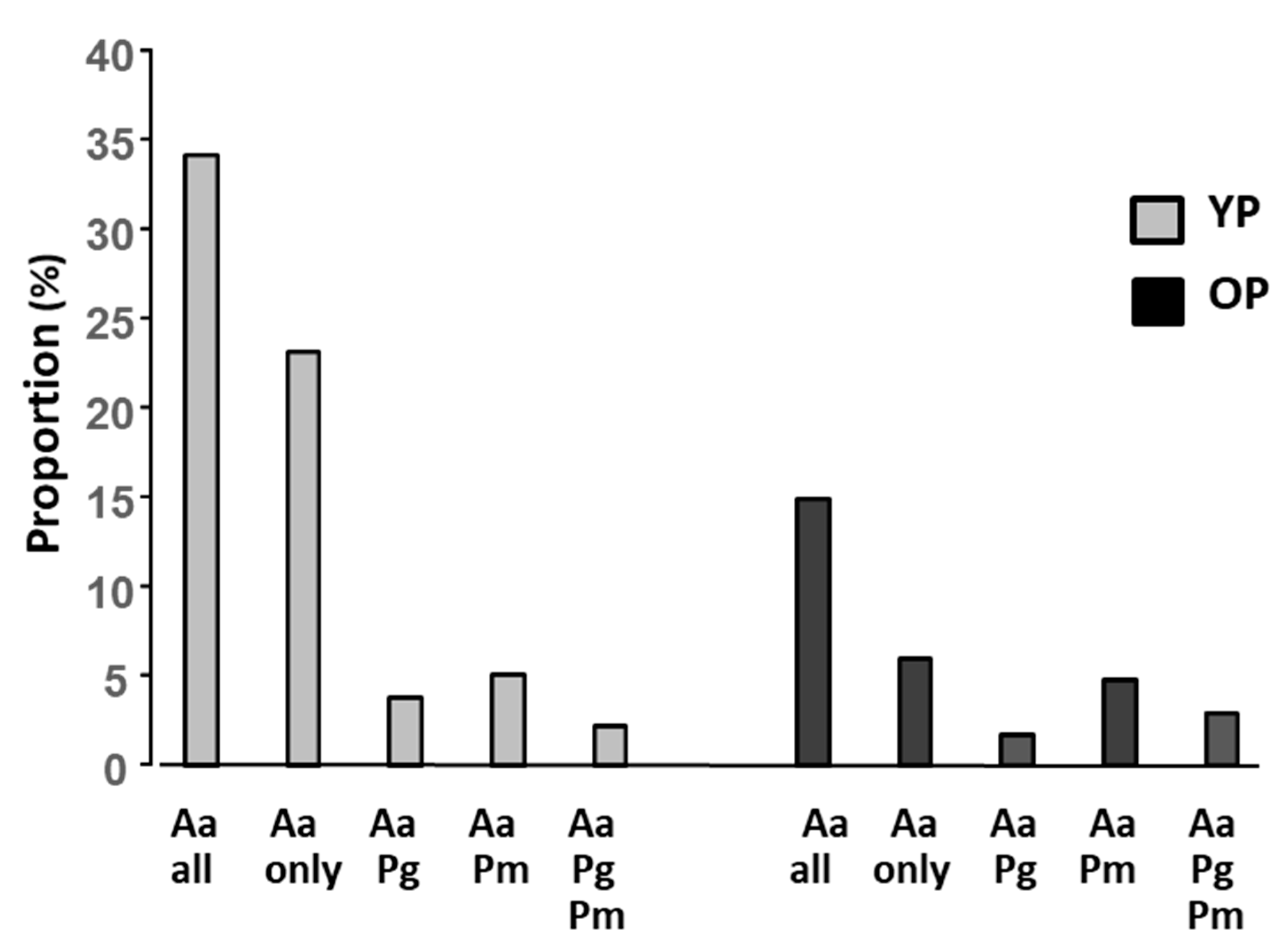
In a polymicrobial infection site the bacterial species may affect each other. To obtain an overview of co-colonization, the presence of the bacterial species was presented alone and in different combination (Figure 2). Among the samples from YP, the proportion of samples containing only A. actinomycetemcomitans was around two times higher than the samples containing A. actinomycetemcomitans in combination with the other bacterial species. Among the OP samples, the opposite was observed, i.e., the proportion of samples containing only A. actinomycetemcomitans was about two times lower than the samples containing A. actinomycetemcomitans in combination with the other bacterial species. Furthermore, it was noted that the proportion OP samples containing A. actinomycetemcomitans in combination with P. micra was almost three times higher than in samples when P. micra was replaced by P. gingivalis.
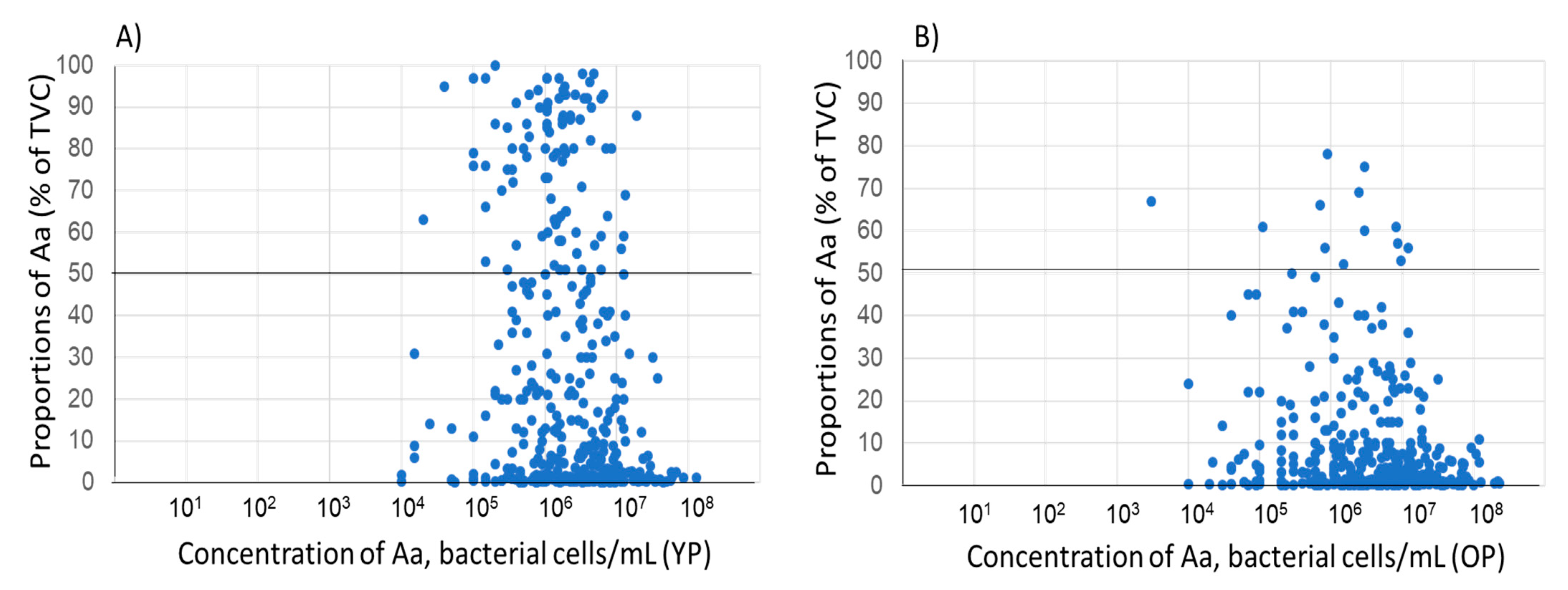
When the proportion and concentration of A. actinomycetemcomitans in the individual samples were compared, no overall relation could be observed, neither for the samples collected from young patients nor from old patients. (Figure 3A,B). However, in YP, the number of samples with a high proportion (>50%) was higher compared with that in the samples from the OP.

4. Discussion

The present study examined the dynamics of three well-established periodontal pathogens in the subgingival dental plaque of younger and older individuals. Aging is indeed a physiological process and is accompanied by natural changes in the composition of the oral microbiota [21]. In the present study, a higher prevalence of A. actinomycetemcomitans is shown in younger rather than older adults, whereas the reverse was the case for P. micra, and there was no differences in the prevalence of P. gingivalis between age groups. Our findings converge with earlier findings demonstrating that the prevalence of A. actinomycetemcomitans decreases with age but diverge in that the prevalence of P. gingivalis by increases with age [26,27,28]. When looking at the proportions of P. gingivalis in dental plaque, we did not find variations between the two age groups. Of note, in a cohort of, on average, 68-year-old patients, P. gingivalis has been subgingivally detected with higher frequency in periodontitis than health, but with no statistically significant differences in the bacterial counts [11].
In the case of A. actinomycetemcomitans, the current and earlier findings clearly agree that it is found in greater proportions in the subgingival dental plaque of younger individuals [11,23,26,27]. In an adolescent population in Ghana, the JP2 genotype of A. actinomycetemcomitans showed higher concentrations in subgingival samples compared with other genotypes of this bacterium [29]. In addition, salivary samples from Moroccan adolescents showed significantly increased concentrations of the JP2 genotype in individuals with periodontal attachment loss compared with that in saliva from periodontally healthy adolescents [30].
The case of P. micra is somehow less studied in the literature. We hereby show that P. micra is twice as prevent in subgingival plaque in the older compared to the younger population, whereas the proportions of this species in subgingival plaque do not vary according to the age grouping. While we have not identified studies reporting the subgingival prevalence of P. micra according to age grouping, there is evidence that the subgingival proportion of this species does not vary with age, but rather with periodontal heath status [20,31]. Of interest, in a cohort of, on average, 67-year-old patients, P. micra was subgingivally detected at higher numbers in active compared to stable periodontitis [32,33]. Similar results were noticed for the periodontitis-associated bacterial species Tannerella forythus and Prevotella intermedius [32].
In this study we also observed altered dynamics between the proportions of A. actinomycetemcomitans and P. gingivalis or P. micra in subgingival plaque. The gradual decrease in the proportions of the latter two species with increasing proportions of A. actinomycetemcomitans in the younger patient group converges with microbial clustering observations, implying that inherent factors promote the coexistence of P. gingivalis with P. micra, but not with A. actinomycetemcomitans [34]. Hydrogen sulfide produced by both P. micra and P. gingivalis may be a such factor, on the biochemical level [35]. Another factor which may have impact on the relationship between the bacterial species is P. micra-induced activation of gingipains [17]. Furthermore, experimental studies using biofilms have also shown a competitive interaction between A. actinomycetemcomitans and P. gingivalis [36,37]. However, in the older population our findings show that the presence of A. actinomycetemcomitans does not affect the proportions of P. gingivalis, but there is a positive interrelationship with P. micra instead.
The introduction of PCR-based quantification of periodontitis-associated bacterial species has in many aspects facilitated the work. However, quantification of the total amount of bacteria using PCR-based methods encounters problem [38], which in turn results in problems when determining proportions of the present bacterial species. In addition, normalization of the results requires determination of the sample size. Methods for this are scarcely reported. However, a method described by Thorbert Mros and co-workers [39] is based on usage of a curette corresponding to 1 μL (1 μL inoculation loop; Sarstedt).
In this study it is shown that there is no or a weak association between proportion and concentration of A. actiniomycetemcomitans in the samples. This further indicates that a high proportion of the bacterium in a sample does not necessarily mean that the sample contains a high concentration of the bacterium, or vice versa.
Clinical laboratory diagnostics is today recommended as the relevant treatment for aggressive or refractory forms of periodontitis. In addition, to analyze the samples for presence/proportions of periodontitis-associated bacterial species, it would be beneficial to screen the samples for the presence of JP2- and cage E carriers of A. actinomycetemcomitans [15,40]. Carriers of the JP2 genotype have shown a significantly increased risk of developing periodontal attachment loss compared to adolescents without this bacterium [13,14,41].
Based on the findings in this study of A. actinomycetemcomitans, P. gingivalis and P. micra, alone and in combination with each other among young and old patients, this could be inferred to ecological interdependencies between these species in the subgingival milieu, under the factor of aging. However, there is no concrete biological evidence at present to support this hypothesis.
A limitation of this study is the lack of clinical parameters; however, a sample from a periodontal pocket sent for microbial analysis is indicative for a diagnosis of periodontitis.

5. Conclusions

Periodontal plaque samples from young patients showed an increased presence and proportion of A. actinomycetemcomutans compared with samples from old patients, while P. gingivalis was similarly distributed in the two age groups. P. micra showed an increased presence and proportion in samples from old patients compared with the young patients.

Author Contributions

R.C. and G.N.B.; formal analysis, R.C.; investigation, R.C., A.J. and G.N.B.; resources, A.J.; data curation, R.C., A.J. and G.N.B.; writing—original draft preparation, R.C. and G.N.B.; writing—review and editing, A.J. and G.N.B.; visualization, R.C. and A.J.; project administration, R.C.; funding acquisition, A.J. All authors have read and agreed to the published version of the manuscript.

Funding

This work was supported by Karolinska Institutet Strategic Funds and the KI/SLL Styrgruppen för Odontologisk Forskning (SOF) Dnr 2-705/2022 (GB) and by TUA grants from the County Council of Västerbotten, Sweden (A.J.) Dnr 7003193.

Data Availability Statement

Data are available from the corresponding author (R.C.).

Acknowledgments

We thank Ewa Strömqvist Engbo for the valuable technical assistance.

Conflicts of Interest

The authors declare no conflict of interest.

References

  1. Könönen, E.; Gursoy, M.; Gursoy, U.K. Periodontitis: A Multifaceted Disease of Tooth-Supporting Tissues. J. Clin. Med. 2019, 8, 1135. [Google Scholar] [CrossRef]
  2. Dahlen, G.; Basic, A.; Bylund, J. Importance of Virulence Factors for the Persistence of Oral Bacteria in the Inflamed Gingival Crevice and in the Pathogenesis of Periodontal Disease. J. Clin. Med. 2019, 8, 1339. [Google Scholar] [CrossRef] [PubMed]
  3. Belibasakis, G.N.; Belstrøm, D.; Eick, S.; Gursoy, U.K.; Johansson, A.; Könönen, E. Periodontal microbiology and microbial etiology of periodontal diseases: Historical concepts and contemporary perspectives. Periodontology 2000, 2023, 1–17. [Google Scholar] [CrossRef] [PubMed]
  4. Caroline daSilva, F.; Piazzetta, C.M.; Torres-Pereira, C.C.; Schussel, J.L.; Amenábar, J.M. Gingival proliferative lesions in children and adolescents in Brazil: A 15-year-period cross-sectional study. J. Indian Soc. Periodontol. 2016, 20, 63–66. [Google Scholar]
  5. van der Velden, U.; Amaliya, A.; Loos, B.G.; Timmerman, M.F.; van der Weijden, F.A.; Winkel, E.G.; Abbas, F. Java project on periodontal diseases: Causes of tooth loss in a cohort of untreated individuals. J. Clin. Periodontol. 2015, 42, 824–831. [Google Scholar] [CrossRef] [PubMed]
  6. Claesson, R.; Johansson, A.; Belibasakis, G.N. Clinical laboratory diagnostics in dentistry: Application of microbiological methods. Front. Oral Health 2022, 3, 983991. [Google Scholar] [CrossRef]
  7. Bostanci, N.; Bao, K.; Greenwood, D.; Silbereisen, A.; Belibasakis, G.N. Periodontal disease: From the lenses of light microscopy to the specs of proteomics and next-generation sequencing. Adv. Clin. Chem. 2019, 93, 263–290. [Google Scholar]
  8. Hbibi, A.; Bouziane, A.; Lyoussi, B.; Zouhdi, M.; Benazza, D. Aggregatibacter actinomycetemcomitans: From Basic to Advanced Research. Adv. Exp. Med. Biol. 2022, 1373, 45–67. [Google Scholar]
  9. Bostanci, N.; Belibasakis, G.N. Porphyromonas gingivalis: An invasive and evasive opportunistic oral pathogen. FEMS Microbiol. Lett. 2012, 333, 1–9. [Google Scholar] [CrossRef]
  10. Dahlén, G. Role of suspected periodontopathogens in microbiological monitoring of periodontitis. Adv. Dent. Res. 1993, 7, 163–174. [Google Scholar] [CrossRef]
  11. Öztürk, V.Ö.; Belibasakis, G.N.; Emingil, G.; Bostanci, N. Impact of aging on TREM-1 responses in the periodontium: A cross-sectional study in an elderly population. BMC Infect. Dis. 2016, 16, 429. [Google Scholar] [CrossRef] [PubMed]
  12. Johansson, A. Aggregatibacter actinomycetemcomitans leukotoxin: A powerful tool with capacity to cause imbalance in the host inflammatory response. Toxins 2011, 3, 242–259. [Google Scholar] [CrossRef] [PubMed]
  13. Haubek, D.; Ennibi, O.K.; Poulsen, K.; Væth, M.; Poulsen, S.; Kilian, M. Risk of aggressive periodontitis in adolescent carriers of the JP2 clone of Aggregatibacter (Actinobacillus) actinomycetemcomitans in Morocco: A prospective longitudinal cohort study. Lancet 2008, 371, 237–242. [Google Scholar] [CrossRef] [PubMed]
  14. Höglund Åberg, C.; Kwamin, F.; Claesson, R.; Dahlén, G.; Johansson, A.; Haubek, D. Progression of attachment loss is strongly associated with presence of the JP2 genotype of Aggregatibacter actinomycetemcomitans: A prospective cohort study of a young adolescent population. J. Clin. Periodontol. 2014, 41, 232–241. [Google Scholar] [CrossRef] [PubMed]
  15. Johansson, A.; Claesson, R.; Höglund Åberg, C.; Haubek, D.; Lindholm, M.; Jasim, S.; Oscarsson, J. Genetic Profiling of Aggregatibacter actinomycetemcomitans Serotype B Isolated from Periodontitis Patients Living in Sweden. Pathogens 2019, 8, 153. [Google Scholar] [CrossRef]
  16. Hočevar, K.; Potempa, J.; Turk, B. Host cell-surface proteins as substrates of gingipains, the main proteases of Porphyromonas gingivalis. Biol. Chem. 2018, 399, 1353–1361. [Google Scholar] [CrossRef]
  17. Neilands, J.; Davies, J.R.; Bikker, F.J.; Svensäter, G. Parvimonas micra stimulates expression of gingipains from Porphyromonas gingivalis in multi-species communities. Anaerobe 2019, 55, 54–60. [Google Scholar] [CrossRef]
  18. Persson, S.; Claesson, R.; Carlsson, J. The capacity of subgingival microbiotas to produce volatile sulfur compounds in human serum. Oral Microbiol. Immunol. 1989, 4, 169–172. [Google Scholar] [CrossRef]
  19. Proctor, G.B.; Shaalan, A.M. Disease-Induced Changes in Salivary Gland Function and the Composition of Saliva. J. Dent. Res. 2021, 100, 1201–1209. [Google Scholar] [CrossRef]
  20. Feres, M.; Teles, F.; Teles, R.; Figueiredo, L.C.; Faveri, M. The subgingival periodontal microbiota of the aging mouth. Periodontol. 2000 2016, 72, 30–53. [Google Scholar] [CrossRef] [PubMed]
  21. Belibasakis, G.N. Microbiological changes of the ageing oral cavity. Arch. Oral Biol. 2018, 96, 230–232. [Google Scholar] [CrossRef]
  22. Loesche, W.J.; Grossman, N.S. Periodontal disease as a specific, albeit chronic, infection: Diagnosis and treatment. Clin. Microbiol. Rev. 2001, 14, 727–752. [Google Scholar] [CrossRef] [PubMed]
  23. Claesson, R.; Höglund-Åberg, C.; Haubek, D.; Johansson, A. Age-related prevalence and characteristics of Aggregatibacter actinomycetemcomitans in periodontitis patients living in Sweden. J. Oral Microbiol. 2017, 9, 1334504. [Google Scholar] [CrossRef]
  24. Kaman, W.E.; Galassi, F.; de Soet, J.J.; Bizzarro, S.; Loos, B.G.; Veerman, E.C.; van Belkum, A.; Hays, J.P.; Bikker, F.J. Highly specific protease-based approach for detection of porphyromonas gingivalis in diagnosis of periodontitis. J. Clin. Microbiol. 2012, 50, 104–112. [Google Scholar] [CrossRef]
  25. Nørskov-Lauritsen, N.; Claesson, R.; Jensen, A.B.; Åberg, C.H.; Haubek, D. Aggregatibacter Actinomycetemcomitans: Clinical Significance of a Pathobiont Subjected to Ample Changes in Classification and Nomenclature. Pathogens 2019, 8, 243. [Google Scholar] [CrossRef]
  26. Savitt, E.D.; Kent, R.L. Distribution of Actinobacillus actinomycetemcomitans and Porphyromonas gingivalis by subject age. J. Periodontol. 1991, 62, 490–494. [Google Scholar] [CrossRef]
  27. Rodenburg, J.P.; Van Winkelhoff, A.J.; Winkel, E.G.; Goene, R.J.; Abbas, F.; De Graaff, J. Occurrence of Bacteroides gingivalis, Bacteroides intermedius and Actinobacillus actinomycetemcomitans in severe periodontitis in relation to age and treatment history. J. Clin. Periodontol. 1990, 17, 392–399. [Google Scholar] [CrossRef]
  28. Jepsen, K.; Falk, W.; Brune, F.; Fimmers, R.; Jepsen, S.; Bekeredjian-Ding, I. Prevalence and antibiotic susceptibility trends of periodontal pathogens in the subgingival microbiota of German periodontitis patients: A retrospective surveillance study. J. Clin. Periodontol. 2021, 48, 1216–1227. [Google Scholar] [CrossRef]
  29. Razooqi, Z.; Höglund Åberg, C.; Kwamin, F.; Claesson, R.; Haubek, D.; Oscarsson, J.; Johansson, A. Aggregatibacter actinomycetemcomitans and Filifactor alocis as Associated with Periodontal Attachment Loss in a Cohort of Ghanaian Adolescents. Microorganisms 2022, 10, 2511. [Google Scholar] [CrossRef] [PubMed]
  30. Ennibi, O.K.; Claesson, R.; Akkaoui, S.; Reddahi, S.; Kwamin, F.; Haubek, D.; Johansson, A. High salivary levels of JP2 genotype of Aggregatibacter actinomycetemcomitans is associated with clinical attachment loss in Moroccan adolescents. Clin. Exp. Dent. Res. 2019, 5, 44–51. [Google Scholar] [CrossRef] [PubMed]
  31. Alazemi, A.M.; Jamal, W.; Al Khabbaz, A.; Rotimi, V.O. Prevalence of target anaerobes associated with chronic periodontitis. Access Microbiol. 2020, 2, acmi000177. [Google Scholar] [CrossRef] [PubMed]
  32. Swoboda, J.R.; Kiyak, H.A.; Darveau, R.; Persson, G.R. Correlates of periodontal decline and biologic markers in older adults. J. Periodontol. 2008, 79, 1920–1926. [Google Scholar] [CrossRef] [PubMed]
  33. Chapple, I.L.; Mealey, B.L.; Van Dyke, T.E.; Bartold, P.M.; Dommisch, H.; Eickholz, P.; Geisinger, M.L.; Genco, R.J.; Glogauer, M.; Goldstein, M.; et al. Periodontal health and gingival diseases and conditions on an intact and a reduced periodontium: Consensus report of workgroup 1 of the 2017 World Workshop on the Classification of Periodontal and Peri-Implant Diseases and Conditions. J. Periodontol. 2018, 89 (Suppl. S1), S74–S84. [Google Scholar] [CrossRef] [PubMed]
  34. López, R.; Dahlén, G.; Retamales, C.; Baelum, V. Clustering of subgingival microbial species in adolescents with periodontitis. Eur. J. Oral. Sci. 2011, 119, 141–150. [Google Scholar] [CrossRef] [PubMed]
  35. Reid, H.W.; Barlow, R.M.; Pow, I. Isolation of louping-ill virus from red deer (Cervus elaphus). Vet. Rec. 1978, 102, 463–464. [Google Scholar] [CrossRef] [PubMed]
  36. Bao, K.; Bostanci, N.; Thurnheer, T.; Grossmann, J.; Wolski, W.E.; Thay, B.; Belibasakis, G.N.; Oscarsson, J. Aggregatibacter actinomycetemcomitans H-NS promotes biofilm formation and alters protein dynamics of other species within a polymicrobial oral biofilm. NPJ Biofilms Microbiomes 2018, 4, 12. [Google Scholar] [CrossRef]
  37. Bao, K.; Bostanci, N.; Selevsek, N.; Thurnheer, T.; Belibasakis, G.N. Quantitative proteomics reveal distinct protein regulations caused by Aggregatibacter actinomycetemcomitans within subgingival biofilms. PLoS ONE 2015, 10, e0119222. [Google Scholar] [CrossRef]
  38. Větrovský, T.; Baldrian, P. The variability of the 16S rRNA gene in bacterial genomes and its consequences for bacterial community analyses. PLoS ONE 2013, 8, e57923. [Google Scholar] [CrossRef]
  39. Thorbert-Mros, S.; Ali, N.; Ali, M.; Ayas, M.; Trullenque-Eriksson, A.; Dahlén, G. A comparative study on periodontitis and periodontitis-associated bacteria in Somali and non-Somali children and adolescents living in Trollhättan, Sweden. Eur. J. Oral. Sci. 2022, 130, e12843. [Google Scholar] [CrossRef]
  40. Johansson, A.; Claesson, R.; Höglund Åberg, C.; Haubek, D.; Oscarsson, J. The cagE gene sequence as a diagnostic marker to identify JP2 and non-JP2 highly leukotoxic Aggregatibacter actinomycetemcomitans serotype b strains. J. Periodontal. Res. 2017, 52, 903–912. [Google Scholar] [CrossRef]
  41. Haubek, D.; Ennibi, O.K.; Poulsen, K.; Poulsen, S.; Benzarti, N.; Kilian, M. Early-onset periodontitis in Morocco is associated with the highly leukotoxic clone of Actinobacillus actinomycetemcomitans. J. Dent. Res. 2001, 80, 1580–1583. [Google Scholar] [CrossRef] [PubMed]
Figure 1. Distribution of P. gingivalis and P. micra in subgingival plaque samples, according to various proportions of A. actinomycetemcomitans present in the samples collected from young patients (YP) (A) or old patients (OP) (B).
Microorganisms 11 01434 g001
Figure 2. Prevalence of A. actinomycetemcomitans, P. gingivalis, and P. micra (n; %) in samples from young (YP) (grey) and old (OP) (black) patients, alone or in various combinations.
Microorganisms 11 01434 g002
Figure 3. Proportion of A. actinomycetemcomitans in relation to concentration of the bacterium in the corresponding sample (cells/mL, log 10). (n = 723). (A) Young patients (YP) (n = 371). (B) Old patients (OP) (n = 352).
Microorganisms 11 01434 g003
Table 1. Prevalence of of A. actinomycetemcomitans (Aa), P. gingivalis (Pg), and P. micra (Pm), (n; %) in subgingival plaque samples based on number of patients (A) and samples (B) (YP: younger patients <35 years. OP: older patients >35 years).
(A)
AgenAa, n (%)Pg, n (%)Pm, n (%)negative
YP 424179 (42.2) 99 (23.3) 156 (36.8) 122 (28.8)
OP1081221 (20.4)307 (28.4) 582 (53.8) 286 (26.4)
All patients1505400 (26.6)406 (27.0) 738 (49.0) 408 (27.1)
(B)
AgenAa, n (%)Pg, n (%)Pm, n (%)negative
YP1087371 (34.1)203 (18.7) 295 (27.1) 426 (39.2)
OP2360352 (14.9)539 (22.8)1095 (46.4) 907 (38.4)
All patients3447723 (21.0)742 (22.1)1390 (40.3)1333 (38.7)
Table 2. Distribution of A. actinomycetemcomitans, P. gingivalis, and P. micra in subgingival plaque samples according to their proportions (%) of the TVC. Both age group (young patients ≤35 years; YP and older patients; OP) and total patient (YP + OP) distributions are presented.
≥0.1–1%>1–5%>5–25%>25–50%>50%
Aa
YP78 (21.0)66 (17.8)87 (23.5)45 (12.1)95 (25.6)371
OP116 (33.0)84 (23.9)112 (31.8)27 (7.6)13 (3.7)352
YP + OP194 (26.9)150 (20.7)199 (27.5)72 (10.0)108 (14.9)723
Pg
YP9 (4.4)26 (12,8)49 (24.1)44 (21.7)75 (36.9)203
OP16 (3.03)59 (10.9)142 (26.3)116 (21.5)206 (38.2)539
YP + OP25 (3.4)85 (11.4)191 (25.7)160 (21.6)281 (37.9)742
Pm
YP37 (12.5)102 (34.6)106 (35.9)37(12.5)13 (4.4)295
OP91 (8.3)297 (27.1)495 (45.2)150 (13.7)62 (5.7)1095
YP + OP128 (9.2)399 (28.7)601 (43.2)187 (13.5)75 (5.4)1390
Disclaimer/Publisher’s Note: The statements, opinions and data contained in all publications are solely those of the individual author(s) and contributor(s) and not of MDPI and/or the editor(s). MDPI and/or the editor(s) disclaim responsibility for any injury to people or property resulting from any ideas, methods, instructions or products referred to in the content.