Article

16S-23S rRNA Internal Transcribed Spacer Region (ITS) Sequencing: A Potential Molecular Diagnostic Tool for Differentiating Lactococcus garvieae and Lactococcus petauri

by
Nadia Stoppani
1,
Silvia Colussi
1,*,
Paolo Pastorino
1,
Marino Prearo
1,
Simona Sciuto
1,
Ilhan Altinok
2,
Rafet Çağrı Öztürk
2,
Mustafa Ture
3,
Ana Isabel Vela
4,
Maria del Mar Blanco
4,
Charalampos Kotzamanidis
5,
Konstantina Bitchava
6,
Andigoni Malousi
7,
Lucio Fariano
8,
Donatella Volpatti
9,
Pier Luigi Acutis
1 and
Jose Francisco Fernández-Garayzábal
4
1
Istituto Zooprofilattico Sperimentale del Piemonte, Liguria e Valle d’Aosta, 10154 Turin, Italy
2
Faculty of Marine Sciences, Karadeniz Technical University, Sürmene, 61530 Trabzon, Turkey
3
Central Fisheries Research Institute (SUMAE), 61250 Trabzon, Turkey
4
VISAVET and Department of Animal Health, Universidad Complutense de Madrid, 28040 Madrid, Spain
5
Veterinary Research Institute, ELGO-DIMITRA, 54124 Thessaloniki, Greece
6
School of Animal Biosciences, Agricultural University of Athens, 11855 Athens, Greece
7
Laboratory of Biological Chemistry, Medical School, Aristotle University of Thessaloniki, 54124 Thessaloniki, Greece
8
Azienda Agricola Canali Cavour, 12044 Centallo, Italy
9
Department of Agricultural, Food, Environmental and Animal Sciences (DI4A), University of Udine, 33100 Udine, Italy
*
Author to whom correspondence should be addressed.
Microorganisms 2023, 11(5), 1320; https://doi.org/10.3390/microorganisms11051320
Submission received: 25 March 2023 / Revised: 12 May 2023 / Accepted: 15 May 2023 / Published: 17 May 2023

Abstract

:
Lactococcus garvieae is the etiological agent of lactococcosis, a clinically and economically significant infectious disease affecting farmed rainbow trout. L. garvieae had been considered the only cause of lactococcosis for a long time; however, L. petauri, another species of the genus Lactococcus, has lately been linked to the same disease. The genomes and biochemical profiles of L. petauri and L. garvieae have a high degree of similarity. Traditional diagnostic tests currently available cannot distinguish between these two species. The aim of this study was to use the transcribed spacer (ITS) region between 16S rRNA and 23S rRNA as a potential useful molecular target to differentiate L. garvieae from L. petauri, saving time and money compared to genomics methods currently used as diagnostic tools for accurate discrimination between these two species. The ITS region of 82 strains was amplified and sequenced. The amplified fragments varied in size from 500 to 550 bp. Based on the sequence, seven SNPs were identified that separate L. garvieae from L. petauri. The 16S-23S rRNA ITS region has enough resolution to distinguish between closely related L. garvieae and L. petauri and it can be used as a diagnostic marker to quickly identify the pathogens in a lactococcosis outbreak.

1. Introduction

Lactococcosis is a serious septicemic fish disease in Mediterranean countries. Lactococcus garvieae was considered the sole causative agent of lactococcosis until recently. L. garvieae has been isolated as a causative agent of the disease in several freshwater and marine species such as rainbow trout (Oncorhynchus mykiss), tilapia (Oreochromis sp.), Japanese eels (Anguilla japonica), olive flounders (Paralichthys olivaceous), grey mullet (Mugil cephalus), catfish (Ameiurus spp.), wild wrasses (Coris aygula), black rockfish (Sebastes schlegeli), amberjacks (Seriola dumerili), kingfish (Seriola lalandi), and giant freshwater prawns (Macrobrachium rosenbergii). Mortalities associated with the disease have been reported in America, Africa, Europe, Asia, and Oceania [1].
It is one of the most important diseases in farmed rainbow trout, causing serious economic losses with high mortality rates [2]. Other species such as the common carp (Cyprinus carpio) seem to be resistant to the disease.
There are several aquatic environmental factors such as fish stress, overcrowding, mishandling, poor water quality, and water temperature that influence the occurrence of lactococcosis outbreaks. Indeed, water temperature is one of the most important predisposing factors that increases susceptibility to infection. Most acute outbreaks occur when the water temperature is over 18 °C, although acute outbreaks have been described at a water temperature of 14–15 °C. Moreover, a decreased oxygen availability in the water can emerge, leading to stressful conditions for trout and increasing their susceptibility to infection [3]. Transmission of the disease occurs by horizontal mechanisms, mainly through water, fish injuries, and by the fecal–oral route [1]. Infected fish exhibit nonspecific clinical signs such as lethargy, erratic swimming, dark skin pigmentation, loss of orientation, early anorexia, and a marked monolateral or bilateral exophthalmia [1]. Macroscopic signs include external and internal hemorrhages (mainly in the eyes, liver, and intestine), splenomegaly, and cerebral congestion. This pathogen causes direct economic losses due to elevated rates of mortality (up to 50%), but also indirect economic losses due to the decrease in growing rates, the increased labor for fish management, and treatment costs. As a result of the development of antibiotic resistance, the use of chemotherapeutic agents in the control of lactococcosis is an unsustainable strategy. In trout farms, preventative biosecurity measures such as the destruction of dead fish, regular and appropriate disinfection of equipment, improvement of health management measures, and immunization of healthy fish are highly recommended [1].
Lactococcus petauri was first isolated from a facial abscess in a sugar glider (Petaurus breviceps) by Goodman et al. [4], who characterized it as a new member of the genus Lactococcus. The first report of an L. petauri outbreak in fish was described in farmed rainbow trout imported from Spain to Greece [5]. The pathogen was initially identified as L. garvieae before being reclassified as L. petauri in a retrospective analysis. Fish infected by L. petauri exhibit the same clinical signs and symptoms as L. garvieae-infected fish. As a result, both species can be considered causative agents of lactococcosis in fish. Recent retrospective studies have revealed that L. petauri, not L. garvieae, is responsible for the majority of lactococcosis outbreaks. In recent years, an outbreak of lactococcosis caused by L. petauri in California, USA, resulted in >50% mortality rate and the culling of more than 3.2 million farmed rainbow trout [6]. Moreover, in Brazil, an outbreak of lactococcosis caused by L. petauri resulted in a high mortality rate in farmed Nile tilapia (Oreochromis niloticus) [7]. The real relevance of L. petauri as a bacterial pathogen for Nile tilapia is still to be assessed, considering that in recent years, infections of L. garvieae have been frequently reported; however, this was before L. petauri’s description [7]. Based on these findings, greater attention should be given to this novel pathogen.
Lactococcosis is traditionally diagnosed in the laboratory by using conventional microbiological techniques or commercial phenotypic and biochemical tests. The most widely used commercial phenotypic method for a routinary identification of bacteria is the API (Analytical Profile Index). The API identifies bacteria based on sugar fermentation (carbohydrates), the assimilation of certain other carbon sources, and the production of certain unique metabolites and enzymes. Both L. garvieae and L. petauri are lactic acid bacteria (LAB) and they have similar phenotypic and biochemical properties [4]. Matrix-assisted laser desorption ionization time-of-flight mass spectrometry (MALDI-TOF MS) generally allows an accurate identification at the species level of most microorganisms and can be an important technique to rapidly identify etiological agents [8]. MALDI-TOF MS, however, does not distinguish between these two species [7].
Advantages in molecular methods have provided a novel set of tools that characterize pathogens in ever greater detail [9]. The 16S rRNA gene has been widely used in medical microbiology to identify bacteria [10]. The sequence contains hypervariable regions that can provide species-specific signature sequences useful for bacteria identification due to the presence of divergent regions in the 16S rRNA gene among various bacteria, while conserved regions allow bacteria to be categorized within the same species [11]. A species-specific PCR assay for the identification of L. garvieae was developed by Zlotkin et al. [12]. Yet, a specific 16S rRNA gene analysis performed on L. garvieae and L. petauri also failed to distinguish between Lactococcus species [7,13]. This confirms the literature findings, since the analysis of the 16S rRNA gene is insufficient to distinguish between L. petauri and L. garvieae [13,14].
Whole genome sequencing (WGS) performed by Kotzamanidis et al. [15] was the first diagnostic technique that allowed differentiation between these two bacterial species. WGS is the most modern technique used for species characterization and identification [16]. It identifies sequence fragments specific to the taxonomic level and provides access to the full genetic content of microorganisms. The average nucleotide identity (ANI) and digital DNA–DNA hybridization (dDDH) values are extensively used for reliable species differentiation [17]. The ANI score exceeded the recommended cut-off value of ~96% and showed the highest similarity (98.393%) with the LG_SAV_20 strain, isolated in 2007 from rainbow trout affected by lactococcosis in Greece [15] with L. petauri 159,469, described as a representative of the species [4]. In addition, dDDH revealed an 82.3% (cut-off point of 70%) similarity to L. petauri 159,469 [18]. This result led to the classification of the LG_SAV_20 strain as the representative sequence of the new species L. petauri isolated from rainbow trout. The adoption of WGS as a routine technique to discriminate between L. garvieae and L. petauri incurs a high expense per sample. It is therefore unrealistic to use this technique as a routine standard procedure for diagnosing lactococcosis. Developing a new rapid method would pave the way for a faster and more cost-effective diagnosis, making it simpler to use in routine diagnostic practice.
An efficient characterization system applied in Californian and Brazilian outbreaks of L. petauri is multilocus sequence typing (MLST); only three genes were used by Shahin et al. [6], these were Carbamate kinase (arcC), Gyrase B (gyrB), and RNA polymerase B (rpoB). Seven genes were instead used for Brazilian outbreaks [7]: α-acetolactate synthase (als), α-subunit of ATP synthase (atpA), elongation factor EF-Tu (tuf), glyceraldehyde-3-phosphate dehydrogenase (gapC), DNA gyrase β-subunit (gyrB), RNA polymerase β’-subunit (rpoC), and galactose permease (galP). In particular, the main differences were related to the gyrB gene. It presented similarities higher than 99.6% with L. petauri references strains (B1726, PAQ102015-99, CF11, and 159469) and 99.9% similarities with L. garvieae reference strains (ATCC49156, Lg2, and JJJN1) [7].
The amplification of a short region (290 bp) of ITS has already been suggested as a useful tool for L. garvieae diagnoses [19]. The ITS region has been considered a good tool for species specific bacterial identification among related organisms due to its genetic variability in size and sequence compared to 16S rRNA and 23S rRNA genes [20]. This method was recently used for molecular diagnosis in piscine lactococcosis [6]; however, its role in distinguishing between L. garvieae and L. petauri has not been reported.
In this study, we used the internal transcribed spacer (ITS) region 16S-23S of rRNA as a tool for distinguishing and identifying L. garvieae and L. petauri isolated from Mediterranean countries such as Greece, Italy, Spain, and Turkey.

2. Materials and Methods

2.1. Strains

A total of 104 L. garvieae and L. petauri strains were included in the study (Table 1 and Table 2). Seven strains of L. garvieae (n = 3) and L. petauri (n = 4), whose genomes are publicly available, were used as control strains for analyses and further comparison of the 16S-23S rRNA ITS sequences (Table 2). The genomes of the strain types of L. garvieae and L. petauri were also included in the study.
We also retrieved additional 16S-23S transcribed spacer (ITS) region sequences from both L. garvieae and L. petauri (n = 8 and n = 5, respectively; reference strains). Sequences of L. garvieae were directly retrieved from the NCBI database: L. garvieae LMG 9472 (accession numbers HM241914), L. garvieae LMG 8162 (accession number HM241916), L. garvieae C1 (accession number MZ146920), L. garvieae 108-33 (accession number MZ146924), L. garvieae 106-30 (accession number MZ146925), L. garvieae 168 (accession number MZ146926), L. garvieae 20,684 (accession number AF225967), and L. garvieae L1-5 (accession number AF225968). Sequences of L. petauri strains were retrieved from published whole genome sequences: L. petauri LG4 (accession number CP086401; region: 1911306-1911836), L. petauri LG26 (accession number CP086595; region 2048225-2048755), L. petauri B1726 (accession number CP094882; region 1717542-1718072), L. petauri NHH01_13 (accession number JANHCX010000013; region 2696-3716), L. petauri LG_SAV_20 (accession number SIVY01000041; region 2699-3719), L. petauri LG6 (accession number JAOYNZ010000013; region 2208-3718), L. petauri LG3 (accession number JAOYNX010000011; region 1571-2101), L. petauri LG5 (accession number JAOYNY010000011; region 1571-2101), and L. petauri LG1 (accession number JAOYNW010000014; region 2208-3718).
Afterwards, 82 field clinical strains from Italy (n = 22), Turkey, Spain, and Greece (20 strains/each) isolated from rainbow trout (Oncorhynchus mykiss) were included in this study (Table 1). Eighty-one isolates were initially identified as L. garvieae through both conventional microbiological methods and PCR according to the protocol described by Zlotkin et al. [12]. The strain 20-GR was identified as L. petauri based on a genomic analysis [15]. All strains were stored at −80 °C in a cryobank until use. Strains were sent to the Aquatic Biology, Aquaculture, and Fish Disease Laboratory of the Istituto Zooprofilattico of Piemonte, Liguria and Valle d’Aosta. Then, the strains were thawed, reactivated in brain heart infusion (BHI) liquid-enriched medium (Microbiol s.n.c., CA, Italy), and subsequently streaked on Columbia Blood Agar (CBA) and incubated at 22 ± 2 °C for 24 h.

2.2. Polymerase Chain Reaction (PCR) of ITS 16S-23S Region

The boiling and freeze–thawing methods were used for DNA extraction: briefly, the colonies of interest were resuspended in DNAse-free water and boiled for 10 min at 95 °C, followed by rapid chilling at −20 °C and DNA pelleting by centrifugation for 1 min at 11,200× g [21]. The 16S-23S rRNA ITS region was amplified by PCR using the primers 16S 5′-GCTGGATCACCTCCTTTCT-3′ and 23S 5′-GGTACTTAGATGTTTCAGTTCC-3′ described by Kabadjova et al. [22]. PCR was carried out in a final volume of 25 μL, containing 2.5 μL of 10X PCR Buffer (-MgCl2), 1 μL of 50 mM MgCl2, 0.5 μL of dNTPs (VWR), 0.3 μL of each primer (20 μM), 0.2 μL of Platinum Taq DNA polymerase (Invitrogen), and 50 ng of the template DNA. The reference strains Lactococcus garvieae DSMZ20684 and Lactococcus petauri DMSZ104842 were used as amplification positive controls. Amplifications were performed using the following thermal profile: initial denaturation at 94 °C for 2 min, followed by 32 cycles of denaturation at 94 °C for 1 min, annealing at 56 °C for 1 min, extension at 72 °C for 1 min, and final extension at 72 °C for 10 min. Amplicons were run on a 2% agarose gel for quantification. Amplicons were purified with a Qiaquick purification kit (Qiagen) and bi-directionally sequenced using the Brilliant Dye Terminator (v1.1) Cycle Sequencing Kit (NimaGen) on a genetic analyzer (Applied Biosystems 3130, Thermo Fisher). DNA sequence analyses were performed in DNASTAR Lasergene Software.
A single representative ITS sequence for a strain, isolated in different countries, was deposited to GenBank (www.ncbi.nlm.nih.gov/genbank (accessed on 2 January 2023)) under the following accession numbers: 1-IT for Italian strains OQ108343; 1-GR for Greek strains OQ108344; 1-TK for Turkish strains OQ108345; and 1-SP for Spanish strains OQ108346.

2.3. Phylogenetic Analysis

The 16S-23S rRNA ITS region sequences of L. garvieae and L. petauri reference strains, reported in Table 2, were retrieved from GenBank and used to perform a neighbor-joining analysis [23] using MEGA X [24]. Evolutionary distances were ascertained via the maximum composite likelihood method [25]. A 1000-replicate bootstrap test was performed. Lactococcus lactis strain KCTC 3768 (accession number HM241926) was used as an outgroup.

2.4. Genome Sequencing

The genomic DNA of a subset of nine clinical strains (Table 3) from Spain (n = 1), Italy (n = 2), Turkey (n = 2), and Greece (n = 4) was extracted using a QIAGEN DNeasy Blood and Tissue kit (Qiagen, Hilden, Germany). The concentration of genomic DNA was quantified using a Qubit® dsDNA BR Assay kit and a Qubit 3.0 Fluorometer (Thermo Fisher Scientific, Waltham, MA, USA). Illumina sequencing libraries were generated with a Nextera XT DNA Library Preparation Kit and sequenced on an Illumina MiSeq platform with 2 × 300-bp paired-end reads (Illumina, San Diego, CA, USA). The genomes of the nine isolates were assembled into contigs and scaffolds using the SPAdes algorithm with default parameters [26], and the quality of the assembly was checked by QUAST software [27].

2.5. Genetic Identification

Genome sequences of the strain types of L. garvieae (DSM 20684T) and L. petauri (159469T) were retrieved from GenBank (accession numbers JXJV01000001 and NZ_MUIZ0000000, respectively). The Genome-to-Genome Distance Calculator tool (GGDC; https://ggdc.dsmz.de/ggdc.php# (accessed on 8 May 2023)) was used to determine the digital DNA–DNA hybridization (dDDH) between the genomes of the seven control strains, nine field clinical strains, and those of the type strains. In addition, the average nucleotide identity (ANI) values were also calculated using the ANI Calculator tool (https://www.ezbiocloud.net/tools/ani (accessed on 8 May 2023)). Both ANI and dDDH are methods routinely used to delineate bacterial species using threshold values of ≥70% for dDDH and 95–96% for ANI [17,28].

3. Results

The alignment of the 16S-23S ITS sequences of the seven control strains and that of L. garvieae and L. petauri strain types revealed a different SNP pattern for L. garvieae and L. petauri. Thus, six diagnostic sites distinguishing L. garvieae from L. petauri were found (Table 2 and Figure 1): an adenine (A) insertion at position g.218_219 insA, 5 SNPs at position g.358G>T, g.440T>G, g.442T>A, g.469C>A, g.478G>T, and a deletion (-) at position g.443 delT. These SNP patterns were consistently detected between L. garvieae and L. petauri reference and clinical field strains (Table 2). A phylogenetic analysis of the strains included in this study revealed two well-differentiated clusters that included all the L. garvieae and L. petaurid strains, respectively. Both clusters were supported by significant bootstrap values. Italian strains clustered together with the L. garvieae control and reference strains, while Greek, Spanish, and Turkish strains clustered with the L. petauri control and reference strains (Figure 2).
The data for dDDH and ANI values for the seven control strains and nine clinical field strains are shown in Table 3. The three L. garvieae control strains displayed average dDDH and ANI similarity values with L. garvieae DSM20684T of between 85.1 and 99.9% and 98.3 and 99.9%, respectively. The average dDDH and ANI values of the four L. petauri control strains with L. petauri 159469T ranged between 85.4 and 89.5% and 98.3 and 98.9%, respectively. Among the field clinical strains, both Italian isolates exhibited dDDH and ANI similarity values with L. garvieae DSM20684T higher than the threshold values stablished for species delineation. On the other hand, the Spanish, Turkish, and Greek strains exhibited dDDH and ANI similarity values with L. petauri 159469T higher than those stablished for species delineation (Table 3).

4. Discussion

L. garvieae had been considered until recently the only etiological agent of lactococcosis [1]. However, recent studies have demonstrated the implication of a new lactococcal species, L. petauri, in the etiology of this disease [4,5,6,7]. Therefore, both L. petauri and L. garvieae are responsible for lactococcosis outbreaks. Differentiation of both species in common microbiological diagnostic laboratories is difficult due to their phenotypic and genetic similarity, which results in identification errors. Thus, commercial identification systems such as MALDI-TOF or PCR assays targeting the 16S rRNA gene cannot discriminate between isolates of both species [7,13,14]. Nevertheless, the interspecies divergence in the 16S–23S ITS sequences makes this region a potentially good candidate for bacterial discrimination and identification. Therefore, in this study, we investigate the polymorphism in the 16S rRNA and 23S rRNA transcribed spacer (ITS) region as a potentially useful molecular target to differentiate L. garvieae from L. petauri.
As there was the possibility that the identification of some of the L. garvieae and L. petauri strains available in public databases was not correct, the identification of a subset of seven strains (control strains) was corroborated by calculating their dDDH and ANI values by comparing their genomes with those of the L. garvieae- and L. petauri-type strains. The control strains displayed dDDH and ANI values higher than the threshold values considered for species delineation with their respective species, confirming therefore their correct identification (Table 2 and Table 3). Their 16S-23S ITS sequences were aligned, revealing two different ITS patterns for L. garvieae and L. petauri characterized by differences in five SNP and two indel variations (Table 2). Later on, these variations were confirmed in all the reference and field clinical strains when their 16S-23S ITS sequences were matched to those of the control strains (Table 2). Thus, the reference strains exhibited an ITS pattern according to their available identification. Regarding clinical field strains, the Italian isolates exhibited the ITS pattern characteristic of L. garvieae, while the Spanish, Greek, and Turkish isolates displayed the ITS pattern of L. petauri. A phylogenetic analysis based on the 16S-23S ITS sequences revealed two well-defined clusters (Figure 2), one cluster including all control, reference, and field clinical strains of L. garvieae, while the other cluster included all L. petauri strains. These data indicate that the identification available in GenBank of the reference strains is correct. Regarding clinical field strains, the Italian strains correspond to L. garvieae while the Spanish, Turkish, and Greek strains correspond to L. petauri. These results are consistent with previous results based on WGS and confirm the relevant role of L. petauri in the epidemiology of lactococcosis in some Mediterranean countries [5,13]. Moreover, genetic identification results support the identification based on the analysis of the 16S-23 ITS sequences. Thus, the genomes of the seven control and nine clinical field strains analyzed in this study showed respective strain type dDDH and ANI values higher than the threshold values stablished for species delineation (≥70% for dDDH and 95–96% for ANI; Table 3), confirming therefore their identification. This genetic identification was performed in about 11% of the L. garvieae and L. petauri clinical strains, but the clear phylogenetic differentiation of L. garvieae and L. petauri based on their 16S-23 ITS sequences together with the consistence of their different SNP patterns makes it reasonable to assume the identification for the remaining field clinical strains.
An analysis based on the sequencing of the gyrB gene, already used as a phylogenetic marker for several closely related genera [29], has been proposed for differentiating L. petauri and L. garvieae, grouping isolates of these species in different clusters [6,7]. In this sense, sequencing of the 16S-23S ITS regions should be as accurate, efficient, and cost effective as gyrB sequencing. Overall, the results of this study indicate the great potential of the identified SNP markers for the differentiation of L. garvieae from L. petauri.

5. Conclusions

The results of this study show that L. garvieae and L. petauri strains display two well-differentiated patterns in their 16S-23S ITS regions, characterized by five SNP differences and two indel variations that were consistently identified in all the strains investigated. Therefore, sequencing of the 16S-23S rRNA ITS region can be a useful tool for discriminating between L. garvieae and L. petauri.

Author Contributions

Conceptualization, N.S., S.C., M.P., I.A., P.L.A. and J.F.F.-G.; methodology, N.S., S.S., P.P., R.Ç.Ö. and C.K.; investigation, P.P., M.T., A.I.V., M.d.M.B., K.B. and A.M.; resources, P.L.A., J.F.F.-G., C.K. and I.A.; data curation, N.S., S.S., S.C. and P.P.; writing—original draft preparation, N.S., S.C., M.P., I.A. and J.F.F.-G.; writing—review and editing, L.F., D.V., A.I.V., J.F.F.-G., M.P., C.K., I.A. and R.Ç.Ö.; supervision, P.L.A.; project administration, P.L.A. and S.C.; funding acquisition, P.L.A. All authors have read and agreed to the published version of the manuscript.

Funding

This research was funded by SUPERTROUT (Improving SUstainability and PERformance of aquaculture farming systems: breeding for lactococcosis resistance in rainbow TROUT), a European project supported by PRIMA (Partnership for Research and Innovation in the Mediterranean Area) (project ID: 1433).

Data Availability Statement

A single representative ITS sequence for a strain, isolated in different countries, was deposited in GenBank (www.ncbi.nlm.nih.gov/genbank (accessed on 2 January 2023)) under the following accession numbers: 1-IT for Italian strains OQ108343; 1-GR for Greek strains OQ108344; 1-TK for Turkish strains OQ108345; and 1-SP for Spanish strains OQ108346.

Conflicts of Interest

The authors declare no conflict of interest or personal relationships that could have appeared to influence the work reported in this paper.

References

  1. Vendrell, D.; Balcázar, J.L.; Ruiz-Zarzuela, I.; De Blas, I.; Gironés, O.; Múzquiz, J.L. Lactococcus garvieae in fish: A review. Comp. Immunol. Microbiol. Infect. Dis. 2006, 29, 177–189. [Google Scholar] [CrossRef] [PubMed]
  2. Algöet, M.; Roberts, E.G.; Feist, S.W.; Wheeler, R.W.; Verner-Jeffreys, D.W. Susceptibility of selected freshwater fish species to a UK Lactococcus garvieae isolate. Dis. Aquat. Org. 2009, 36, 227–231. [Google Scholar] [CrossRef]
  3. Castro, R.; Reguera-Brito, M.; López-Campos, G.H.; Blanco, M.M.; Aguado-Urda, M.; Fernández-Garayzábal, J.F.; Gibello, A. How does temperature influence the development of lactococcosis? Transcriptomic and immunoproteomic in vitro approaches. J. Fish Dis. 2017, 40, 1285–1297. [Google Scholar] [CrossRef]
  4. Goodman, L.B.; Lawton, M.R.; Franklin-Guild, R.J.; Anderson, R.R.; Schaan, L.; Thachil, A.J.; Wiedmann, M.; Miller, C.B.; Alcaine, S.D.; Kovac, J. Lactococcus petauri sp. nov., isolated from an abscess of a sugar glider. Int. J. Syst. Evol. Microbiol. 2017, 67, 4397–4404. [Google Scholar] [CrossRef]
  5. Savvidis, K.; Anatoliotis, C.; Kanaki, Z.; Vafeas, G. Epizootic outbreaks of lactococcosis disease in rainbow trout, Oncorhynchus mykiss (Walbaum), culture in Greece. Bull. Eur. Assoc. Fish Pathol. 2007, 27, 223–228. [Google Scholar]
  6. Shahin, K.; Mukkatira, K.; Yazdi, Z.; Richey, C.; Kwak, K.; Heckman, T.I.; Mohammed, H.H.; Ortega, C.; Avendaño-Herrera, R.; Keleher, B.; et al. Development of a quantitative polymerase chain reaction assay for detection of the aetiological agents of piscine lactococcosis. J. Fish. Dis. 2021, 45, 847–859. [Google Scholar] [CrossRef]
  7. Egger, R.C.; Câmara Rosa, J.C.; Lara Resende, L.F.; de Pádua, S.B.; de Oliveira Barbosa, F.; Zerbini, M.T.; Tavares, G.C.; Pereira Figueiredo, H.C. Emerging fish pathogens Lactococcus petauri and L. garvieae in Nile tilapia (Oreochromis niloticus) farmed in Brazil. Aquaculture 2023, 565, 739093. [Google Scholar] [CrossRef]
  8. Assis, G.B.N.; Pereira, F.L.; Zegarra, A.U.; Tavares, G.C.; Leal, C.A.; Figueiredo, H.C.P. Use of MALDI-TOF Mass Spectrometry for the fast identification of Gram-positive fish pathogens. Microbiology 2017, 8, 1492. [Google Scholar] [CrossRef] [PubMed]
  9. Sciuto, S.; Colli, L.; Fabris, A.; Pastorino, P.; Stoppani, N.; Esposito, G.; Prearo, M.; Esposito, G.; Ajmone-Marsan, P.; Acutis, P.L.; et al. What can genetics do for the control of infectious diseases in aquaculture? Animals 2022, 12, 2176. [Google Scholar] [CrossRef]
  10. Clarridge, J.E. Impact of 16S rRNA gene sequence analysis for identification of bacteria on clinical microbiology and infectious diseases. Clin. Microbiol. Rev. 2004, 17, 840–862. [Google Scholar] [CrossRef]
  11. Janda, J.M.; Abbott, S.L. 16S rRNA gene sequencing for bacterial identification in the diagnostic laboratory: Pluses, perils, and pitfalls. J. Clin. Microbiol. 2007, 45, 2761–2764. [Google Scholar] [CrossRef]
  12. Zlotkin, A.; Eldar, A.; Ghittino, C.; Bercovier, H. Identification of Lactococcus garvieae by PCR. J. Clin. Microbiol. 1998, 36, 983–985. [Google Scholar] [CrossRef]
  13. Altinok, I.; Ozturk, R.C.; Ture, M. NGS analysis revealed that Lactococcus garvieae Lg-Per was Lactococcus petauri in Turkey. J. Fish. Dis. 2022, 45, 1839–1843. [Google Scholar] [CrossRef] [PubMed]
  14. Ou, Y.J.; Ren, Q.Q.; Fang, S.T.; Wu, J.G.; Jiang, Y.X.; Chen, Y.R.; Zhong, Y.; Wang, D.D.; Zhang, G.X. Complete genome insights into Lactococcus petauri CF11 isolated from a healthy human gut using second- and third-generation sequencing. Front. Genet. 2020, 26, 11–119. [Google Scholar] [CrossRef]
  15. Kotzamanidis, C.; Malousi, A.; Bitchava, K.; Vafeas, G.; Chatzidinitriou, D.; Skoura, L.; Papadimitriou, E.; Chatzopoulou, F.; Zdragas, A. First report of isolation and genome sequence of L. petauri strain from a rainbow trout lactococcosis outbreak. Curr. Microbiol. 2020, 77, 1089–1096. [Google Scholar] [CrossRef] [PubMed]
  16. Quinoo, S.; Coolen, J.P.M.; Van Hijum, S.A.F.T.; Huynen, M.A.; Melches, W.J.G.; van Schaik, W.; Wertheim, H.F.L. Whole-Genome sequencing of bacterial pathogens: The future of nosocomial outbreak analysis. Clin. Microbiol. Rev. 2017, 30, 1015–1063. [Google Scholar] [CrossRef]
  17. Chun, J.; Oren, A.; Ventosa, A.; Christensen, H.; Arahal, D.R.; da Costa, M.S.; Rooney, A.P.; Yi, H.; Xu, X.W.; de Meyer, S.; et al. Proposed minimal standards for the use of genome data for the taxonomy of prokaryotes. Int. J. Syst. Evol. Microbiol. 2018, 68, 461–466. [Google Scholar] [CrossRef]
  18. Martinovic, A.; Cabal, A.; Nisic, A.; Sucher, J.; Stöger, A.; Allerberger, F.; Ruppitsch, W. Genome sequences of Lactococcus garvieae and Lactococcus petauri strains isolated from traditional Montenegrin brine cheeses. Microbiol. Resour. Announc. 2021, 10, e0054621. [Google Scholar] [CrossRef]
  19. Dang, H.T.; Park, H.K.; Myung, S.C.; Kim, W. Development of a novel PCR assay based on the 16S-23S rRNA internal transcribed spacer region for the detection of Lactococcus garvieae. J. Fish. Dis. 2012, 35, 481–487. [Google Scholar] [CrossRef] [PubMed]
  20. Thanh, H.D.; Park, H.K.; Kim, W.; Shin, H.S. Development of a 16S-23S rRNA intergenic spacer-based quantitative PCR assay for improved detection and enumeration of Lactococcus garvieae. FEMS Microbiol. Lett. 2013, 339, 10–16. [Google Scholar] [CrossRef]
  21. Pastorino, P.; Colussi, S.; Pizzul, E.; Varello, K.; Menconi, V.; Mugetti, D.; Tomasoni, M.; Esposito, G.; Bertoli, M.; Bozzetta, E.; et al. The unusual isolation of carnobacteria in eyes of healthy salmonids in high-mountain lakes. Sci. Rep. 2021, 11, 2314. [Google Scholar] [CrossRef] [PubMed]
  22. Kabadjova, P.; Dousset, X.; Le Cam, V.; Prevost, H. Differentiation of closely related Carnobacterium food isolates based on 16S-23S ribosomal DNA intergenic spacer region polymorphism. Appl. Environ. Microbiol. 2002, 68, 5358–5366. [Google Scholar] [CrossRef]
  23. Saitou, N.; Nei, M. The neighbour-joining method: A new method for reconstructing phylogenetic trees. Mol. Biol. Evol. 1987, 4, 406–425. [Google Scholar] [CrossRef] [PubMed]
  24. Kumar, S.; Stecher, G.; Li, M.; Knyaz, C.; Tamura, K. MEGA X: Molecular Evolutionary Genetics Analysis across computing platforms. Mol. Biol. Evol. 2018, 35, 1547–1549. [Google Scholar] [CrossRef] [PubMed]
  25. Tamura, K.; Nei, M.; Kumar, S. Prospects for inferring very large phylogenies by using the neighbour-joining method. Proc. Natl. Acad. Sci. USA 2004, 101, 11030–11035. [Google Scholar] [CrossRef]
  26. Bankevich, A.; Nurk, S.; Antipov, D.; Gurevich, A.A.; Dvorkin, M.; Kulikov, A.S.; Lesin, V.M.; Nikolenko, S.I.; Pham, S.; Prjibelski, A.D.; et al. SPAdes: A new genome assembly algorithm and its applications to single-cell sequencing. J. Comput. Biol. 2012, 19, 455–477. [Google Scholar] [CrossRef]
  27. Gurevich, A.; Saveliev, V.; Vyahhi, N.; Tesler, G. QUAST: Quality assessment tool for genome assemblies. Bioinformatics 2013, 29, 1072–1075. [Google Scholar] [CrossRef]
  28. Goris, J.; Konstantinidis, K.T.; Klappenbach, J.A.; Coenye, T.; Vandamme, P.; Tiedje, J.M. DNA-DNA hybridization values and their relationship to whole-genome sequence similarities. Int. J. Syst. Evol. Microbiol. 2007, 57, 81–91. [Google Scholar] [CrossRef]
  29. Peeters, K.; Willems, A. The gyrB gene is a useful phylogenetic marker for exploring the diversity of Flavobacterium strains isolated from terrestrial and aquatic habitats in Antarctica. FEMS Microbiol. Lett. 2011, 321, 130–140. [Google Scholar] [CrossRef]
Figure 1. Multiple 16S-23S ITS sequences alignment of the L. garvieae and L. petauri control strains used in this study (accession number in parentheses). Polymorphism positions refer to the strain L. garvieae LMG9472 (accession number HM241914.1).
Microorganisms 11 01320 g001
Figure 2. Evolutionary relationship of taxa based on an ITS analysis. The neighbor-joining method was used to infer the evolutionary history. The optimal tree is shown. The percentage of replicate trees in which the associated taxa clustered together in the bootstrap test (1000 replicates) is shown next to the branches. The evolutionary distances were computed using the Maximum Composite Likelihood method and are in the units of the number of base substitutions per site. This analysis involved 22 nucleotide sequences. All ambiguous positions were removed for each sequence pair (pairwise deletion option). There were a total of 535 positions in the final dataset. Evolutionary analyses were conducted in MEGA X. Lactococcus lactis strain KCTC 3768 was used as the outgroup. Strains reported in the tree as L. petauri Turkey, L. petauri Greece, and L. petauri Spain are each one representative of 20 different clinical strains per country. The L. garvieae Italy strain reported in the tree is representative of 22 different clinical strains.
Microorganisms 11 01320 g002
Table 1. Field clinical strains isolated in lactococcosis outbreaks that occurred in rainbow trout farms in Italy, Spain, Greece, and Turkey.
ItalySpainGreeceTurkey
Strain ID Geographical OriginDate of IsolationStrain IDGeographical OriginDate of IsolationStrain IDGeographical OriginDate of IsolationStrain IDGeographical OriginDate of Isolation
1-ITQuinto20165239-VISAVETGranada2019LG1Macedonia2016Y-LG1Trabzon2016
2-ITQuinto20165424-VISAVETGranada20172-ELGOMacedonia2010Y2-KTUGumushane2016
3-ITCassolnovo20165664-VISAVETGranada2017LG3Macedonia2010Y3-KTUTrabzon2016
4-ITCassolnovo20165787-VISAVETGranada20164-ELGOMacedonia2010Y6-KTUTrabzon2016
5-ITCerano201702/6071-VISAVETGranada2002LG5Ipiros2009Y7-KTUTrabzon2017
6-ITCassolnovo20178666-VISAVETGranada2016LG6Macedonia2008K2-KTUTrabzon2017
7-ITCassolnovo20188059-VISAVETLérida20167-ELGOIpiros2009K3-KTUGumushane2016
8-ITCassolnovo20188495-VISAVETMadrid20168-ELGOMacedonia2009K7-KTUGumushane2018
9-ITCassolnovo20188516-VISAVETLa Coruña20169-ELGOIpiros2008K8-KTUGumushane2018
10-ITQuinto201903/8568-VISAVETAsturias200310-ELGOMacedonia2009Kürtün-KTUGumushane2020
11-ITQuinto2019818-VISAVETGuadalajara201611-ELGOMacedonia2007Rize-KTURize2020
12-ITCerano2019820-VISAVETGuadalajara201612-ELGOMacedonia2010Antalya-KTUAntalya2019
13-ITCassolnovo2019393-VISAVETGranada201913-ELGOIpiros2010Vakfıkebir-KTUTrabzon2020
14-ITQuinto2020195-VISAVETLa Coruña201914-ELGOIpiros2010LG10Mugla2017
15-ITPreore2020ICM16/00935Granada201615-ELGOIpiros200820-KTUMugla2016
16-ITOrmelle2017307-VISAVETGranada201616-ELGOIpiros20069-KTUKayseri2018
17-ITCassolnovo20171008-VISAVETLa Coruña201717-ELGOIpiros200613-KTUIzmir2016
18-ITQuinto20198831-VISAVETLérida201718-ELGOMacedonia2007123-KTUIzmir2017
19-ITCassolnovo2019P04/8864-VISAVETGranada200419-ELGOMacedonia2008140-KTUElazig2016
20-ITQuinto20198943-VISAVETGranada201620-ELGOIpiros2007107B-KTURize2019
1683San Daniele Friuli1997
1691-2Porcia1997
Table 2. Nucleotide differences in the 16S-23S rRNA internal transcribed spacer regions (ITS) of L. garvieae and L. petauri.
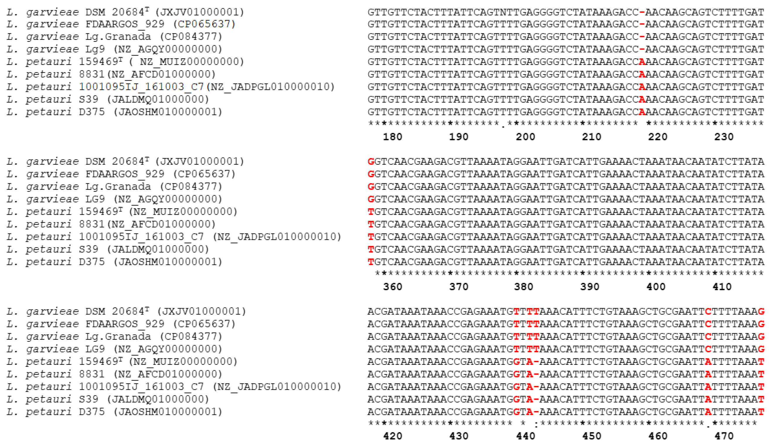
16S-23S Region Position a
219358440442443469478
Strain type
L. garvieae DSM20684T -GTTTCG
L. petauri 159469T ATGA-AT
Control strains
L. garvieae Lg.Granada-GTTTCG
L. garvieae Lg 9-GTTTCG
L. garvieae FDAARGOS_929-GTTTCG
L. petauri 1001095IJ_161003_C7ATGA-AT
L. petauri 8831ATGA-AT
L. petauri D375ATGA-AT
L. petauri S39ATGA-AT
Reference strains
L. garvieae LMG 9472-GTTTCG
L. garvieae LMG 8162-GTTTCG
L. garvieae C1-GTTTCG
L. garvieae 108-33-GTTTCG
L. garvieae 106-30-GTTTCG
L. garvieae 168-GTTTCG
L. garvieae 20684-GTTTCG
L. garvieae L1-5-GTTTCG
L. petauri LG4ATGA-AT
L. petauri LG26ATGA-AT
L. petauri B1726ATGA-AT
L. petauri NHH01_13ATGA-AT
L. petauri LG_SAV_20ATGA-AT
Clinical field strains
Italian (n = 22) b-GTTTCG
Spanish (n = 20) cATGA-AT
Greek (n = 20) cATGA-AT
Turkish (n = 20) cATGA-AT
a Positions of reported polymorphisms were inferred from the 16S-23S ribosomal RNA intergenic spacer sequence of the strain L. garvieae LMG 9472 (accession number HM241914.1). b This SNP pattern was consistently detected in all the Italian field clinical strains. c This SNP pattern was consistently detected in all the Spanish, Greek, and Turkish field clinical strains.
Table 3. ANI and dDDH values (%) of the control and the subset of clinical strains examined in this study.
IsolateL. petauri 159469T (NZ_MUIZ0000000)L. garvieae DSM 20684T (JXJV01000001)Accession Number
%ANI%DDH%ANI%DDH
Control strains
1001095IJ_161003_C798.889.593.150.9NZ_JADPGL010000010
883198.585.593.050.5NZ_AFCD01000000
D37598.485.493.150.7JAOSHM010000001
S3998.385.593.050.8JALDMQ010000000
Lg. Granada93.251.798.385.1CP084377
Lg 993.251.298.385.3NZ_AGQY00000000
FDAARGOS_92992.950.899.999.9CP065637
Field clinical strains
ICM16/00935 a98.485.792.050.5JARHWC000000000
1683 b93.351.198.485.4JARHWV000000000
1691-2 b93.351.198.485.4JARHWU000000000
Y-LG1 c98.485.693.050.8JAQPON000000000
LG10 c98.485.593.150.8JAQPOM000000000
LG6 d98.585.493.150.4JAOYNZ010000001
LG5 d98.485.492.450.4JAOYNY010000001
LG3 d98.485.492.550.4JAOYNX010000001
LG1 d98.485.392.950.5JAOYNW010000001
a, b, c, d Spanish, Italian, Turkish, and Greek clinical field strains, respectively.
Disclaimer/Publisher’s Note: The statements, opinions and data contained in all publications are solely those of the individual author(s) and contributor(s) and not of MDPI and/or the editor(s). MDPI and/or the editor(s) disclaim responsibility for any injury to people or property resulting from any ideas, methods, instructions or products referred to in the content.

