Article

Prevalence and Antibiotic Resistance of Bacillus sp. Isolated from Raw Milk

by
Patryk Adamski
,
Zuzanna Byczkowska-Rostkowska
,
Joanna Gajewska
*,
Arkadiusz Józef Zakrzewski
and
Lucyna Kłębukowska
Department of Industrial and Food Microbiology, Faculty of Food Science, University of Warmia and Mazury, Plac Cieszyński 1, 10-726 Olsztyn, Poland
*
Author to whom correspondence should be addressed.
These authors contributed equally to this work.
Microorganisms 2023, 11(4), 1065; https://doi.org/10.3390/microorganisms11041065
Submission received: 26 February 2023 / Revised: 24 March 2023 / Accepted: 17 April 2023 / Published: 19 April 2023

Abstract

:
Milk, due to its diversity in terms of its nutritional content, is an important element of the human diet, as well as a good medium for the development of bacteria. The genus Bacillus contains ubiquitous aerobic, rod-shaped, endospore-producing gram-positive bacteria. Representatives of the Bacillus cereus group and the Bacillus subtilis group contribute to shortening the shelf life of milk and dairy products by degrading milk components and its additives. They also produce a number of heat-stable toxins and can cause a number of ailments, mainly in the digestive system. The aim of this research was to identify Bacillus sp. strains isolated from raw milk and to determine their antibiotic resistance. Strains isolated from raw milk samples (n = 45) were identified by MALDI-TOF MS. Ninety strains of Bacillus sp. were identified, for which the antibiotic resistance phenotype was determined. A total of 90 strains of Bacillus were classified in five groups (the Bacillus cereus group (n = 35), B. licheniformis (n = 7), the B. subtilis group (n = 29), B. pumilus (n = 16), and Bacillus sp. (n = 3). All isolates were susceptible to chloramphenicol and meropenem. The antibiotic resistance profiles of the tested groups of Bacillus spp. differed from each other, which is of particular concern in relation to multidrug-resistant representatives of the B. cereus group resistant to cefotaxime (94.29%), ampicillin (88.57%), rifampicin (80%), and norfloxacin (65.71%). Our study provides data on the prevalence and antibiotic sensitivity of Bacillus sp. In raw milk, suggesting a potential risk to health and the dairy industry.

1. Introduction

Milk, due to its diversity in terms of its nutritional content, is an important element of the human diet and at the same time a good medium for the development of bacteria [1]. Milk that has not been processed is an important source of bacterial infection. Non-compliance with the hygiene standards of its acquisition makes it difficult to avoid the contamination of milk with microorganisms [2]. The level of microbiological contamination of milk depends on several factors such as animal health, farm sanitary conditions, milking hygiene, and milk storage temperature [3,4]. In most cases, unless the animal is suffering from a mammary gland infection or systemic disease, the milk produced by a mammary gland should not contain bacteria, although it is easily contaminated with microbes living on the animal’s skin during milking. In Europe, according to the Commission Regulation (EC) No. 1662/2006 of 6 November 2006, raw milk should not contain >1 × 10⁵ microorganisms per mL [5]. Good quality raw milk determines its technological suitability and the appropriate quality and shelf life of dairy products. The presence of pathogens in milk is a potential threat to public health, especially among consumers of raw milk [6,7].
The dominant microbiota of chilled raw milk is psychrotrophic bacteria, capable of producing undesirable proteolytic and lipolytic enzymes, causing adverse changes in dairy products [3,4,8]. Among the most significant bacteria causing spoilage of dairy products are bacteria belonging to the genus of psychrotrophic Bacillus.
The genus Bacillus is a ubiquitous aerobic, rod-shaped, endospore-producing gram-positive bacteria. The most important Bacillus species contaminating raw milk are Bacillus cereus, Bacillus licheniformis, and Bacillus pumilus [9]. B. cereus and the listed members of the B. subtilis group contribute to shortening the shelf life of milk and dairy products by degrading milk components and produce a number of thermostable toxins that can also cause digestive system ailments [10,11,12]. According to literature data, a B. cereus count above 5.0 CFU/mL may cause taste and flavour defects in pasteurized milk. With a higher presence of B. cereus, the products show defects, such as sweet curd and bitty cream, due to high proteolytic activity and lecithinase production [13]. Some B. cereus strains can potentially grow at 8 °C and below to a concentration that may be detrimental to human health [14]. However, most strains of Bacillus spp. are not pathogenic for humans, but some may infect humans incidentally. B. cereus enterotoxins were associated with the highest number of foodborne outbreaks among bacterial toxins, exceeding outbreaks caused by Clostridium perfringens and Staphylococcus aureus [15]. It is most commonly associated with gastrointestinal disease manifested as vomiting. Rarely, it can also cause eye infection, meningitis, pneumonia, periodontitis, necrotizing fasciitis, and osteomyelitis [9,16,17,18,19].
In addition to the defects of dairy products causing economic losses in the food industry, contaminated milk can cause infections and contribute to the widespread problem of antibiotic resistance due to the extensive use of pharmaceutics in cattle farming. Bacterial resistance to antimicrobial substances may be intrinsic, acquired, and/or adaptive [20]. Selective pressure exerted on microorganisms using antimicrobial agents has so far been defined as the main mechanism of antibiotic resistance. Antibiotics affect susceptible bacteria, while resistant ones survive by passing on the resistance gene to daughter cells [21]. Another form of transmission of resistance genes to microorganisms is horizontal gene transfer (HGT) [22].
Antimicrobial agents at low doses (sublethal or subtherapeutic) in the form of residues in feed might also be factors influencing bacterial antibiotic resistance, as they are able to induce genetic and phenotypic variability in exposed bacteria [23,24]. Thus, antibiotic residues in the food chain can cause antibiotic resistance to transfer not only in pathogens but also in commensal bacteria or lactic acid bacteria [25,26,27].
Although Bacillus spp. are often isolated from milk and dairy products [28], and although this industry, based on animal production, has a prominent place in the process of the development and dissemination of drug resistance in the environment, there are few reports on the antibiotic resistance profiles of Bacillus spp. isolated from milk. Therefore, the aim of this research was to identify Bacillus spp. strains isolated from raw milk and to determine their antibiotic resistance.

2. Materials and Methods

2.1. Isolation of Bacillus spp. Strains

Raw milk samples (n = 45) obtained from farms located in the Warmia-Masuria Province were subjected to analysis. All milk specimens were transported to the laboratory immediately after collection. Ten milliliters of each raw milk sample was pasteurized for 15 min at 80 °C to remove non-sporing bacteria. Next, the sample was streaked in Mannitol Egg Yolk Polymyxin Agar (MYP) (Merck, Darmstadt, Germany) and nutrient agar (Merck, Darmstadt, Germany), then incubated for 48 h at 30 °C.

2.2. Identification by MALDI-TOF

Strains were identified using VITEK®MS (bioMérieux, Marcy l’Etoile, France) according to the manufacturer’s protocol, described previously [29]. Briefly, characteristic colonies from the MYP agar and nutrient agar plates were cultured for 48 h at 30 °C on Tryptic Soy Agar (TSA) (Merck, Darmstadt, Germany). After incubation, a small portion of bacterial colonies were transferred to the MALDI target plates. Then, 1 μL of MALDI matrix VitekMS-CHCA (α-Cyano-4-hydroxycinnamic acid) (bioMérieux, Marcy l’Etoile, France) was added to the spots and then dried at room temperature. Strains were analyzed by the VITEK®MS v2.0 MALDI-TOF mass spectrometry systemV2.0 (RUO; SARAMIS version 4.13) databases (bioMérieux, Marcy l’Etoile, France). We considered the effectiveness of the MALDI-TOF identification method when the significance level was ≥90% [30]. For calibration and quality control, Escherichia coli ATCC 8739 was used.

2.3. Phenotypic Antibiotic Resistance Analysis

Antimicrobial susceptibility was determined using the Kirby–Bauer disc diffusion method according to the standard procedure described by EUCAST (European Committee on Antimicrobial Susceptibility Testing) [31]. Twelve antibiotics (Oxoid, UK) commonly used in human and animal infections were used. The selected antibiotics belong to eleven classes of antimicrobials: aminoglycosides: gentamicin (CN, 10 µg) and amikacin (AK, 30 µg); aminopenicillins: ampicillin (AMP, 10 µg); carbapenems: meropenem (MEM, 10 µg); lincosamides: clindamycin (DA, 2 µg); macrolides: erythromycin (E, 15 µg); glycopeptides: vancomycin (VA, 30 µg); third-generation cephalosporins: cefotaxime (CTX, 30 µg); phenicols: chloramphenicol (C, 30 µg); rifampicins: rifampicin (RD, 5 µg); sulfonamides–trimethoprims: trimethoprim/sulfamethoxazole (SXT, 25 µg); and fluoroquinolones: norfloxacin (NOR, 10 µg).
Firstly, suspensions in sterile saline (0.9%) were prepared from bacterial colonies on TSA (Merck, Darmstadt, Germany) cultured for 24 h until they reached a 0.5 McFarland standard concentration. A sterile swab was used to inoculate the suspension on Mueller–Hinton agar (Merck, Darmstadt, Germany) [32]. Antibiotic discs were then placed on the plates and incubated at 37 °C for 24 h. The inhibition zone diameters were recorded after the incubation period. Strains were categorized as resistant (R), intermediate-resistance (I), and susceptible (S) according to the criteria in EUCAST [31] for Bacillus sp. Additionally, due to the lack of standards for antibiotics not included in EUCAST for Bacillus sp., the standards for staphylococci were used [32]. Staphylococcus aureus 29213 was used as quality control (QC) for most of the tested antibiotics, Enterococcus feacalis ATCC 29212 was used as QC for vancomycin, and Escherichia coli ATCC 29212 as QC for meropenem [31].
The multiple antibiotic resistance (MAR) index was calculated for each isolate as: number of antibiotics to which the isolate is resistant/total number of antibiotics against which the isolate was tested [18,33]. In this study, we defined multidrug resistance (MDR) as resistance to at least one antibiotic from three or more classes of antibiotics [33].

2.4. Statistical Analysis

Statistical analyses were performed using GraphPad Prism software version 8.0 (GRAPH PAD Software Inc, San Diego, CA, USA) and p ≤ 0.05 was considered significant.

3. Results

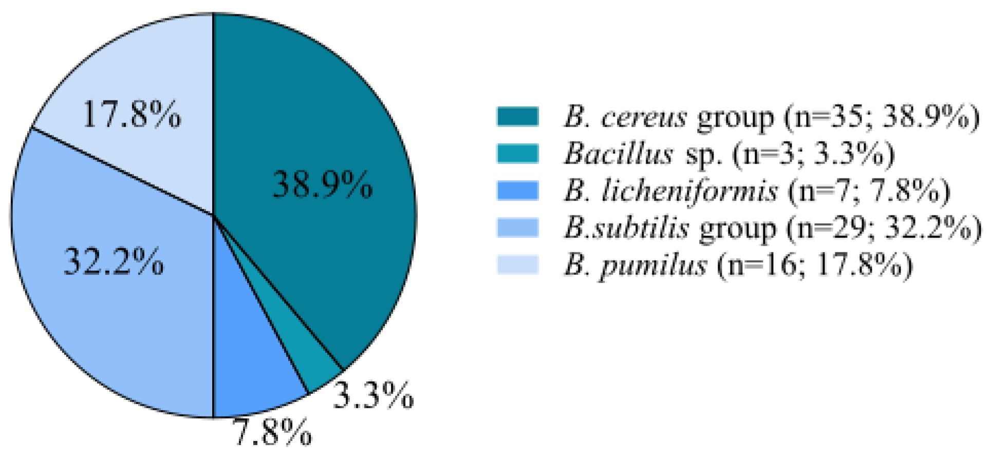
3.1. Isolation and Identification of Bacillus spp.

From 45 raw milk samples, a total of 90 strains of Bacillus sp. were isolated. Their identification using MALDI-TOF allowed for them to be classified into five groups: the Bacillus cereus group (38.9%; n = 35), B. licheniformis (7.8%; n = 7), the B. subtilis group (32.2%; n = 29), B. pumilus (17.8%; n = 16), and Bacillus sp. (3.3%; n = 3). We found that the B. cereus group was dominant (Figure 1).

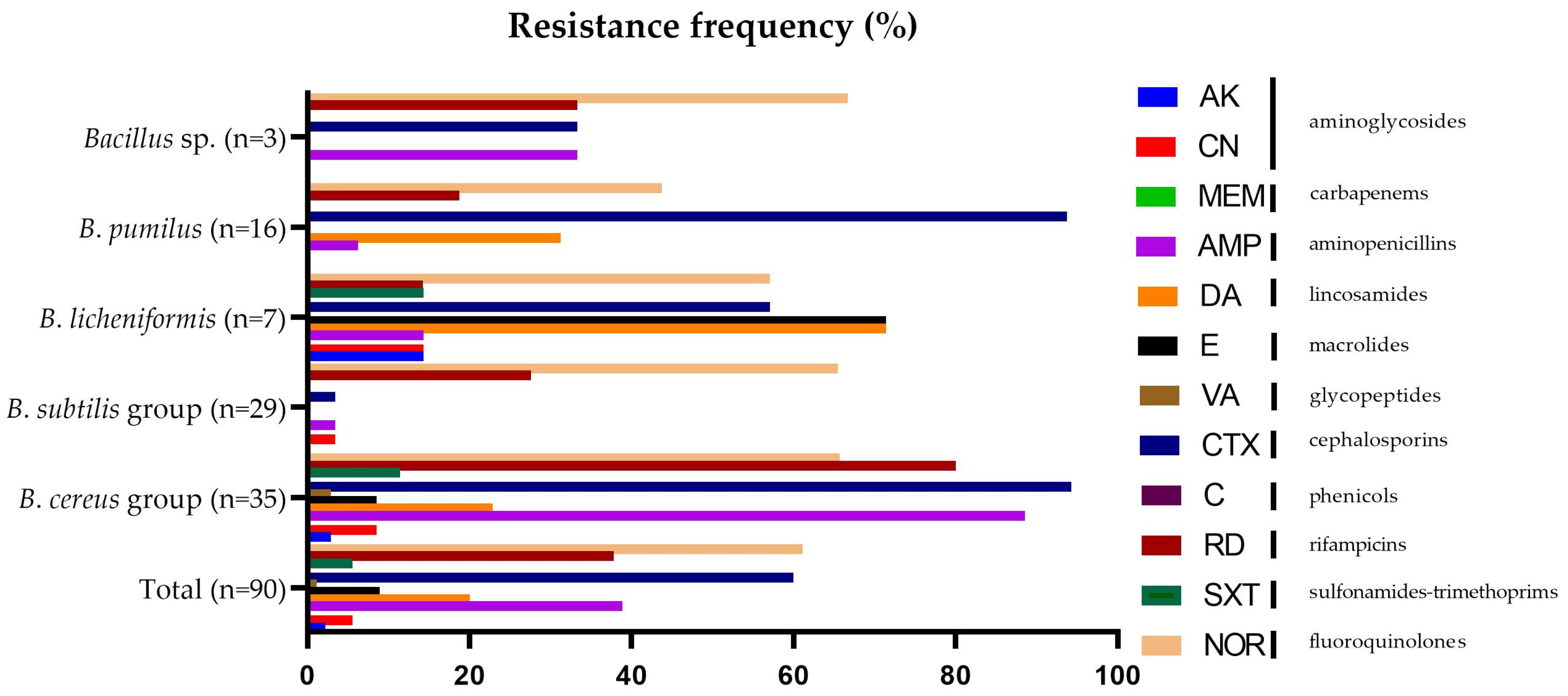
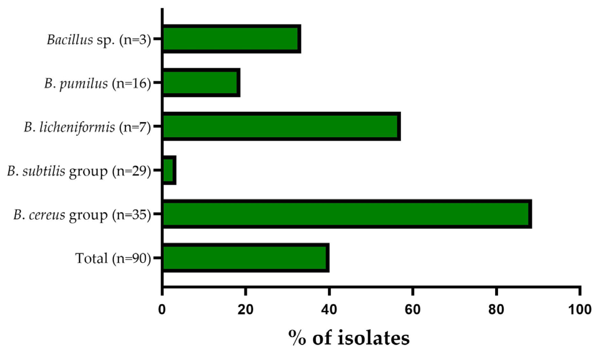
3.2. Antibiotic Resistance Pattern

The results of our study showed that the tested Bacillus spp. showed resistance to antimicrobial agents from different classes (Figure 2). The resistance frequency of the tested antibiotics among all the investigated strains ranged from 0 to 94.29%. All isolates were susceptible to chloramphenicol and meropenem, and most of the studied strains were susceptible to vancomycin (98.89%), amikacin (97.8%), gentamicin (94.44%), trimethoprim/sulfamethoxazole (94.44%), erythromycin (91.12%), ampicillin (61.11%), and rifampicin (54.44%). The highest resistance was observed for norfloxacin (61.11%) and cefotaxime (60.00%) (Figure 2). Resistance profiles differed between the groups.
All of the B. subtilis group strains (n = 29) were susceptible to chloramphenicol, meropenem, amikacin, vancomycin, trimethoprim/sulfamethoxazole, clindamycin, and erythromycin, and most of the studied strains were susceptible to cefotaxime (96.55%), gentamicin (96.55%), ampicillin (96.55%), and rifampicin (72.41%). Within the B. cereus group, all of the studied strains were susceptible to chloramphenicol and meropenem, and most of them were susceptible to amikacin (97.14%), vancomycin (97.14%), gentamicin (91.43%), and sulfamethoxazole (88.57%). All of the B. licheniformis strains (n = 7) were susceptible to chloramphenicol, meropenem, and vancomycin, and most of the studied strains were susceptible to amikacin (85.71%), gentamicin (85.71%), trimethoprim/sulfamethoxazole (85.71%), rifampicin (85.71%), and ampicillin (85.71%). B. pumilus strains (n = 16) were completely susceptible to chloramphenicol, meropenem, amikacin, trimethoprim/sulfamethoxazole, vancomycin, erythromycin, and gentamicin, and most of the studied strains were susceptible to ampicillin (93.75%), rifampicin (81.25%), clindamycin (68.75%), and norfloxacin (56.25%). Non-identified Bacillus strains (n = 3) were susceptible to chloramphenicol, meropenem, amikacin, erythromycin, trimethoprim/sulfamethoxazole, vancomycin, clindamycin, and gentamicin, and most of the studied strains were susceptible to ampicillin (66.67%), cefotaxime (66.67%), and rifampicin (66.67%). Based on the Kruskal–Wallis ANOVA, it was noted that the resistance profile is species dependent for five antibiotics: ampicillin (p < 0.000001), clindamycin (p = 0.00035), erythromycin (p = 0.034479), cefotaxime (p < 0.000001), and rifampicin (p = 0.000014).
Most of the B. cereus group strains were resistant to cefotaxime (94.29%), ampicillin (88.57%), rifampicin (80%), and norfloxacin (65.71%). The B. subtilis group strains were resistant to norfloxacin (65.52%). Most of the studied B. licheniformis strains (n = 7) were resistant to erythromycin (71.43%), clindamycin (71.43%), cefotaxime (57.14%), and norfloxacin (57.14%). Meanwhile, most of the studied B. pumilus strains (n = 16) were found to be resistant to cefotaxime (93.75%). Two of the three tested non-identified Bacillus sp. strains (n = 3) were resistant to norfloxacin (66.67%).
Among all the B. cereus group isolates, 31/35 (88.57%) were found to be multidrug resistant (Figure 3). Within the B. cereus group, the multiple antibiotic resistance (MAR) index was found to range from 0.08 to 0.66 and the overall mean was 0.34. Among the B. subtilis group, one of the 29 strains (3.45%) was defined as multidrug resistant (Figure 3), with the multiple antibiotic resistance (MAR) index calculated as 0.25. However, 57.14% (4/7) strains of B. licheniformis were defined as multidrug resistant (Figure 3). The multiple antibiotic resistance (MAR) index among isolates was found to range from 0.17 to 0.50, with an overall mean of 0.33. Within B. pumilus strains, 18.75% (3/16) were specified as multidrug resistant (Figure 2). The multiple antibiotic resistance (MAR) index of the studied strains was found to range from 0.08 to 0.33 and the overall mean was 0.13 (Figure 3). Among non-identified Bacillus strains, 33.33% (1/3) was defined as multidrug resistant (Figure 3), with the multiple antibiotic resistance (MAR) index calculated as 0.33.
Our results showed 26 antibiotic resistance profiles for all isolates. Among the tested strains, 12.2% (11/90) belonging to B. licheniformis (n = 1), B. pumilus (n = 1), the B. subtilis group (n = 8), and Bacillus sp. (n = 1) were sensitive to all tested antibiotics. Next, 22.2% (20/90) were resistant to only one antibiotic, mainly norfloxacin (14/90; 15.6%). Moreover, 20% (18/90) of the strains were resistant to two antibiotics, mainly rifampicin and norfloxacin (7/90; 7.8%). The other strains showed resistance to three or more antibiotics. The most resistant strain was resistant to eight antibiotics (AK, AMP, DA, E, VA, CTX, RD, NOR). The results showed that two antibiotic resistance profiles occurred most frequently: AMP–CTX–RD (11/90; 12.2%) and AMP–CTX–RD–NOR (11/90; 12.2%) (Table 1).

4. Discussion

The presence of Bacillus sp. in milk can cause spoilage in dairy products and food poisoning due to the enterotoxins produced by these microorganisms. B. cereus and other Bacillus spp. are common etiologic agents of foodborne diseases worldwide. Global statistics on food poisoning caused by B. cereus are underestimated due to the occurrence of vomiting symptoms similar to those of S. aureus poisoning and diarrheal symptoms similar to those caused by Clostridium perfringens type A. A significant proportion of people affected by food poisoning caused by Bacillus sp. do not seek medical help due to the short duration of the symptoms [34]. Species other than B. cereus are not indicated in clinical diagnostics as the etiological factors of food poisoning. However, studies have confirmed the production and action of thermolabile toxins and cereulide-like toxins produced by B. circulans, B. lentus, B. subtilis, B. licheniformis, B. pumilus, and B. amyloliquefaciens. Significantly, outbreaks caused by B. pumilus and B. subtilis are often wrongly assigned to B. cereus [35]. Bacillus spp. are also known to form biofilms resistant to commonly used technological processes, which is a serious problem for the dairy industry. Bacillus spp. are generally capable of producing extracellular or intracellular thermostable proteo- and lipolytic enzymes that are involved in the spoilage of milk and dairy products, leading to unfavorable organoleptic changes [34,36,37].
Some non-toxic strains of Bacillus sp. are used as probiotics in animal feed and additives in the food industry, including the dairy industry. In recent years, Bacillus spp. have gained interest in research on functional foods related to human health due to their increased tolerance and ability to survive in the unfavorable environment of the digestive tract. In addition, the bacteria are more stable during the processing and storage of foodstuffs and even pharmaceutical preparations. However, due to the their potential pathogenicity, the safety of individual strains of the genus Bacillus should be studied and a deeper analysis should be carried out in order to select the strains used as probiotics [38].
Hornik et al., based on the conducted research, found that Bacillus sp. ranges from 10 to 17% of the milk microbiome. The results differ depending on the origin of the sample collection [39]. Studies show that B. licheniformis, along with B. cereus, is one of the most widespread Bacillus species found in raw milk and in the entire milk-processing chain [35]. In addition, previous studies [40] indicate that it is the dominant species of spore-forming bacteria (68%) found in powdered skimmed milk. Other authors indicate that B. licheniformis was the second most common species of spore-forming bacteria detected in a study of 28 milk powder samples from 18 different countries, with a total prevalence of 39.2% [41]. In our research, B. licheniformis accounted for only 7.78% (7/90) of Bacillus sp. strains isolated from raw milk. Referring to the work of Heyndrickx and Scheldeman [42], B. licheniformis was the dominant species, found in greater abundance over B. subtilis and B. pumilus in pasteurized milk and its products, which is inconsistent with our results. B. licheniformis strains accounted for 7.78% (7/90), while B. pumilus and the B. subtilis group accounted for 17.77% (16/90) and 32.22% (29/90), respectively. Nieminen et al. [37] identified 21.74% (5/23) strains of B. pumilus in a study of milk from cows with mastitis.
In addition, in a study conducted by Sarkar and Kumari [43], strains of the B. cereus group were isolated from six out of eight different dairy products sold in India. Their occurrence in cheese, ice cream, powdered milk, and pasteurized/sterilized milk was relatively high (33–55%). In the work of Rahnam et al., B. cereus was present in 60% of raw milk samples, constituting 75.00% (34/44) of all Bacillus sp. isolates [28]. The results obtained in this study show that species from the B. cereus group are the most common representatives of Bacillus sp. in raw milk, constituting 38.89% (35/90) of isolated Bacillus sp. strains.
The World Health Organization warns that the increasing prevalence of antibiotic resistance is a serious threat and one of the greatest public health problems of the 21st century [44]. Bacteria that have developed mechanisms of resistance against individual antibiotics, using the horizontal gene transfer (HGT), can transfer their resistance genes to other bacteria, including the microbiome of the human digestive tract [39], intensifying the problem of antibiotic resistance. Toth et al. [26] confirm that ARGs can be found in raw milk. In addition, the use of antimicrobials is widespread in the farm environment, which contributes to the phenomenon of the milk microbiome acquiring resistance to these substances. Raw milk that has not undergone heat treatment is a convenient environment for microbial proliferation, and this affects the amplification of ARGs. Their intensity increases the risk of horizontal gene transfer.
Gundogan and Avci [45] reported that B. cereus isolates recovered from raw milk and dairy products in Turkey samples were resistant to ampicillin (91.1%) and trimethoprim/sulfamethoxazole (27.8%). Chang et al., studying resistance in raw and pasteurized milk, indicated resistance to ampicillin (96.00%) and trimethoprim/sulfamethoxazole (10.40%) strains of B. cereus [46]. Our results show a similar level of resistance to ampicillin (88.57%) and trimethoprim/sulfamethoxazole (11.43%). Hu et al. [47] isolated B. cereus, B. subtilis, B. licheniformis, and B. pumilus from food samples from local markets and restaurants. All the tested strains of B. cereus showed resistance to ampicillin. In contrast, none of the isolates showed resistance to rifampicin and vancomycin. However, our research showed that among the tested strains, only one strain from the Bacillus cereus group was resistant to vancomycin, and 80% were resistant to rifampicin. Kong et al. [48] identified B. cereus in 26.37% (159/603) samples of meat and meat products. All of the studied strains showed resistance to ampicillin and most of them were resistant to rifampicin (86.29%). In this study, 88.57% (31/36) strains of the B. cereus group were resistant to ampicillin and 80.00% (28/35) showed resistance to rifampicin. In contrast, most strains were susceptible to gentamicin, chloramphenicol, and trimethoprim-sulfamethoxazole, which is supported by our study. In their study, all of the studied strains were resistant to at least three classes of antibiotics, with the multiple antibiotic resistance (MAR) index ranging from 0.15 to 0.50.
In another study, Yang et al. [49] described the antibiotic resistance profile of common bacteria strains isolated from various environments (water, digestive tract, soil, animal products). They indicated that a B. subtilis isolate (n = 1) was resistant to ampicillin and gentamicin. However, the results obtained in this study show high sensitivity to ampicillin (96.55%) and gentamicin (96.55%) among the tested strains of the B. subtilis group (n = 29). It is worth noting that B. subtilis, despite the confirmed cases of contamination of dairy products and posing a health risk to consumers [35,50], still remains marginalized in terms of its presence in dairy products.
Pasteurization is carried out to kill unwanted microorganisms present in raw milk. There is a risk that this process will not eliminate the spores produced by bacteria, including those of the Bacillus genus. Zhui et al. [51] isolated strains of Bacillus sp. from pasteurized milk. In their study, 80% of the strains showed resistance to ampicillin; in our study, 35/90 strains (38.88%) were resistant to ampicillin. In the cited study, 10/114 (8.77%) strains were resistant to trimethoprim/sulfamethoxazole, while our study showed 5/90 (5.55%) strains resistant to this agent. The authors indicate 8/114 (7.01%) strains resistant to clindamycin and 2/114 resistant to erythromycin. In our case, the values of resistance to these two substances were 20.00% (18/90) and 5.55% (5/90), respectively.
The B. subtilis group also includes the B. licheniformis and B. pumilus species, however, due to their frequency of occurrence, they were included in the study separately. Jeong et al. showed more than four times the breakpoint resistance to clindamycin in 70.2% of 74 strains of B. licheniformis derived from fermented soybean products [52]. Hu et al. indicated 100% susceptibility to gentamicin of the tested strains of B. pumilus isolated from dairy products, probiotics, fermented food, rice products, raw or cooked meat, fermented soy beverage, and snacks from different local markets and restaurants in China [47]. In this study, all of the studied B. pumilus strains (n = 16) were susceptible to gentamicin and the majority of them were susceptible to clindamycin (68.75%).
The study showed that a high percentage (40/90 (40.11%)) of the tested isolates were multidrug resistant (resistant to at minimum three antibiotics from different chemical classes of antibiotic), with 100% of isolates having a MAR index >0.20. MAR index values higher than 0.2 suggest a high level of antibiotic resistance among strains isolated from milk [18,33]. Nevertheless, it is worthy of attention that in our study, 77.50% (31/40) of the multidrug-resistant Bacillus strains were from the B. cereus group. The high incidence of multidrug-resistant strains indicates the need to introduce an antibiotic surveillance program in the dairy industry. In addition, determination of the MAR index may be useful, especially in cases of nosocomial infections, allowing for the introduction of effective antibiotic therapy [18].
Previous studies focused mainly on determining the presence, identifying sources of contamination, and characterizing B. cereus as one of the most important microorganisms affecting the quality and safety of dairy products [12,16,46]. Nevertheless, the presented studies also raise the aspect of antibiotic resistance of other species of the genus Bacillus.
In addition, it is important to remember the correct storage conditions for raw milk before further processing. Awasti et al. [53] conducted a study on strains of Bacillus licheniformis, which was also present in our samples. Their results indicate that factors such as temperature and storage time of raw milk affected changes in the growth of Bacillus sp. Changes occurred in the activity of spore production and spore germination, as well as in the proliferation of bacterial cells. According to the authors, storing raw milk for no more than 72 h at 8 °C can ensure that bacterial populations do not increase by 1.0 log CFU/mL. Increased temperature and extended storage can result in the development of potentially pathogenic microorganisms, including Bacillus sp.
Our research has some limitations which need to be addressed here. VITEK®MS (bioMérieux, Marcy l’Etoile, France) can not distinguish some species because of high similarities among them, including Bacillus fordii and Bacillus fortis, identified as B. fordii/B. fortis; Bacillus subtilis, Bacillus amyloliquefaciens, and Bacillus vallismortis, identified as B. subtilis/amyloliquefaciens/vallismortis; and members of the B. cereus group, which are identified as a group. The Bacillus cereus group includes several species, phylogenetically organized into three broad clades. Bacillus cereus sensu stricto and Bacillus thuringiensis occur in all clades. In the first clade are B. anthracis and B. wiedmannii. The second clade includes B. mycoides, B. pseudomycoides, B. toyonensis, B. cytotoxicus, and B. weihenstephanensis (now classified as B. mycoides) [54,55]. The third clade includes B. bingmayongensis, B. gaemokensis, and B. manliponensis. In the past, a number of studies have been carried out regarding the possibility of using MALDI-TOF MS for the species identification of members of the B. cereus group. Undoubtedly, MALDI-TOF MS has a large diagnostic potential, however, its limitation is the fact that, at the species level, the obtained mass spectra are almost identical and distinguishing them is more complicated, which makes it difficult to identify species within closely related microorganisms within the group of B. cereus [54].

5. Conclusions

The conducted research showed the presence of representatives of Bacillus spp. in raw milk. We found that all isolates were sensitive to chloramphenicol and meropenem. In addition, the B. cereus group strains were mostly sensitive to multiple antibiotics such as vancomycin, gentamicin, amikacin, and trimethoprim/sulfamethoxazole. However, most of them were identified as multidrug resistant with a high percentage of resistance to ampicillin, cefotaxime, rifampicin, and norfloxacin. The high level of multidrug resistance observed in B. licheniformis strains should also be considered at high risk. In contrast, the B. subtilis group strains showed a high percentage of resistance to norfloxacin but a low value of the multiple antibiotic resistance (MAR) index. The obtained results confirm the need for further research on Bacillus spp. present in raw milk in order to prevent the spread of antibiotic resistance among human pathogenic strains, which is a growing public health problem.

Author Contributions

Conceptualization: A.J.Z. and L.K.; methodology: A.J.Z., J.G. and L.K; investigation: A.J.Z. and L.K.; data curation: A.J.Z. and J.G.; writing—original draft preparation: P.A. and Z.B.-R.; writing—review and editing: A.J.Z., J.G. and L.K.; visualization: P.A., Z.B.-R., A.J.Z. and J.G.; supervision: L.K. All authors have read and agreed to the published version of the manuscript.

Funding

This research received no external funding.

Data Availability Statement

Not applicable.

Conflicts of Interest

The authors declare no conflict of interest.

References

  1. Von Neubeck, M.; Baur, C.; Krewinkel, M.; Stoeckel, M.; Kranz, B.; Stressler, T.; Fischer, L.; Hinrichs, J.; Scherer, S.; Wenning, M. Biodiversity of refrigerated raw milk microbiota and their enzymatic spoilage potential. Int. J. Food Microbiol. 2015, 211, 57–65. [Google Scholar] [CrossRef] [PubMed]
  2. Velázquez-Ordoñez, V.; Valladares-Carranza, B.; Tenorio-Borroto, E.; Talavera-Rojas, M.; Antonio Varela-Guerrero, J.; Acosta-Dibarrat, J.; Puigvert, F.; Grille, L.; González Revello, Á.; Pareja, L. Microbial Contamination in Milk Quality and Health Risk of the Consumers of Raw Milk and Dairy Products. In Nutrition in Health and Disease—Our Challenges Now and Forthcoming Time; BoD—Books on Demand: Paris, France, 2019. [Google Scholar]
  3. Adamiak, A.; Górska, A.; Mróz, B. Bakterie psychrotrofowe w mleku surowym i jego przetworach. Zywn. Nauk. Technol. Jakosc/Food. Sci. Technol. Qual. 2015, 22, 36–48. [Google Scholar] [CrossRef]
  4. Yuan, H.; Han, S.; Zhang, S.; Xue, Y.; Zhang, Y.; Lu, H.; Wang, S. Microbial Properties of Raw Milk throughout the Year and Their Relationships to Quality Parameters. Foods 2022, 11, 3077. [Google Scholar] [CrossRef] [PubMed]
  5. Commission, T.H.E.; The, O.F.; Communities, E. European Commission Regulation (EC) no. 1662/2006 amending Regulation no. 853/2004. Regulation 2006, 2005, L320/0–L320/10. [Google Scholar]
  6. Wysok, B.; Wiszniewska-Łaszczych, A.; Uradziński, J.; Szteyn, J. Prevalence and antimicrobial resistance of Campylobacter in raw milk in the selected areas of Poland. Pol. J. Vet. Sci. 2011, 14, 473–477. [Google Scholar] [CrossRef] [PubMed]
  7. Rolbiecki, D.; Harnisz, M. Mikroorganizmy lekooporne w środowisku przemysłu mleczarskiego. Pol. Dairy J. 2022, 10, 22–29. [Google Scholar]
  8. Lucey, J.A. Raw Milk Consumption: Risks and Benefits. Nutr. Today 2015, 50, 189–193. [Google Scholar] [CrossRef]
  9. De Jonghe, V.; Coorevits, A.; Vandroemme, J.; Heyrman, J.; Herman, L.; De Vos, P.; Heyndrickx, M. Intraspecific genotypic diversity of Bacillus species from raw milk. Int. Dairy J. 2008, 18, 496–505. [Google Scholar] [CrossRef]
  10. Hutchings, C.; Rajasekharan, S.K.; Reifen, R.; Shemesh, M. Mitigating milk-associated bacteria through inducing zinc ions antibiofilm activity. Foods 2020, 9, 1094. [Google Scholar] [CrossRef]
  11. Lücking, G.; Stoeckel, M.; Atamer, Z.; Hinrichs, J.; Ehling-Schulz, M. Characterization of aerobic spore-forming bacteria associated with industrial dairy processing environments and product spoilage. Int. J. Food Microbiol. 2013, 166, 270–279. [Google Scholar] [CrossRef]
  12. Granum, P.E. Spotlight on Bacillus cereus and its food poisoning toxins. FEMS Microbiol. Lett. 2017, 364, fnx071. [Google Scholar] [CrossRef] [PubMed]
  13. Berthold-Pluta, A.; Pluta, A.; Garbowska, M.; Stefańska, I. Prevalence and Toxicity Characterization of Bacillus cereus in Food Products from Poland. Foods 2019, 8, 269. [Google Scholar] [CrossRef] [PubMed]
  14. Webb, M.D.; Barker, G.C.; Goodburn, K.E.; Peck, M.W. Risk presented to minimally processed chilled foods by psychrotrophic Bacillus cereus. Trends Food Sci. Technol. 2019, 93, 94–105. [Google Scholar] [CrossRef] [PubMed]
  15. European Union. The European Union One Health 2020 Zoonoses Report. EFSA J. 2021, T. 19, e06971. [Google Scholar]
  16. Dubey, S.; Sharma, N.; Thakur, S.; Patel, R.; Reddy, B.M. Bacillus cereus food poisoning in Indian perspective: A review. J. Food Sci. Technol. 2021, 10, 970–975. [Google Scholar]
  17. Dietrich, R.; Jessberger, N.; Ehling-Schulz, M.; Märtlbauer, E.; Granum, P.E. The Food Poisoning Toxins of Bacillus cereus. Toxins 2021, 13, 98. [Google Scholar] [CrossRef]
  18. Jovanovic, J.; Ornelis, V.F.M.; Madder, A.; Rajkovic, A. Bacillus cereus food intoxication and toxicoinfection. Compr. Rev. Food Sci. Food Saf. 2021, 20, 3719–3761. [Google Scholar] [CrossRef]
  19. Stenfors Arnesen, L.P.; Fagerlund, A.; Granum, P.E. From soil to gut: Bacillus cereus and its food poisoning toxins. FEMS Microbiol. Rev. 2008, 32, 579–606. [Google Scholar] [CrossRef]
  20. Wash, P.; Batool, A.; Mulk, S.; Nazir, S.; Yasmin, H.; Mumtaz, S.; Alyemeni, M.N.; Kaushik, P.; Hassan, M.N. Prevalence of Antimicrobial Resistance and Respective Genes among Bacillus spp., a Versatile Bio-Fungicide. Int. J. Environ. Res. Public Health 2022, 19, 14997. [Google Scholar] [CrossRef]
  21. Zablotni, A.; Jaworski, A. Źródla Antybiotyków W SRodowiskach Naturalnych I Ich Rola Biologiczna. Postepy Hig. Med. Dosw. 2014, 68, 1040–1049. [Google Scholar] [CrossRef]
  22. Chajęcka-Wierzchowska, W.; Zadernowska, A.; Zarzecka, U.; Zakrzewski, A.; Gajewska, J. Enterococci from ready-to-eat food—horizontal gene transfer of antibiotic resistance genes and genotypic characterization by PCR melting profile. J. Sci. Food Agric. 2019, 99, 1172–1179. [Google Scholar] [CrossRef] [PubMed]
  23. Wall, B.A.; Mateus, A.; Marshall, L.; Pfeiffer, D.; Lubroth, J.; Ormel, H.J.; Otto, P.; Patriarchi, A. The Emergence of Antimicrobial Resistance in Bacteria. In Drivers, Dynamics and Epidemiology of Antimicrobial Resistance in Animal Production; Veterinary Epidemiology, Economics and Public Health Group, Department of Production and Population Health, The Royal Veterinary College: London, UK, 2016; ISBN 9789251094419. [Google Scholar]
  24. Byczkowska-Rostkowska, Z.; Gajewska, J.; Chajęcka-Wierzchowska, W. Antybiotyki i antybiotykooporność w przemyśle mleczarskim. Pol. Dairy J. 2022, 12, 10–15. [Google Scholar]
  25. Roca, I.; Akova, M.; Baquero, F.; Carlet, J.; Cavaleri, M.; Coenen, S.; Cohen, J.; Findlay, D.; Gyssens, I.; Heuer, O.E.; et al. The global threat of antimicrobial resistance: Science for intervention. New Microbes New Infect. 2015, 6, 22–29. [Google Scholar] [CrossRef] [PubMed]
  26. Tóth, A.G.; Csabai, I.; Krikó, E.; Tőzsér, D.; Maróti, G.; Patai, Á.V.; Makrai, L.; Szita, G.; Solymosi, N. Antimicrobial resistance genes in raw milk for human consumption. Sci. Rep. 2020, 10, 7464. [Google Scholar] [CrossRef] [PubMed]
  27. Danilenko, V.; Devyatkin, A.; Marsova, M.; Shibilova, M.; Ilyasov, R.; Shmyrev, V. Common inflammatory mechanisms in covid-19 and Parkinson’s diseases: The role of microbiome, pharmabiotics and postbiotics in their prevention. J. Inflamm. Res. 2021, 14, 6349–6381. [Google Scholar] [CrossRef]
  28. Rahnama, H.; Azari, R.; Yousefi, M.H.; Berizi, E.; Mazloomi, S.M.; Hosseinzadeh, S.; Derakhshan, Z.; Ferrante, M.; Conti, G.O. A systematic review and meta-analysis of the prevalence of Bacillus cereus in foods. Food Control 2023, 143, 109250. [Google Scholar] [CrossRef]
  29. Zakrzewski, A.J.; Zarzecka, U.; Chajęcka-Wierzchowska, W.; Zadernowska, A. A Comparison of Methods for Identifying Enterobacterales Isolates from Fish and Prawns. Pathogens 2022, 11, 410. [Google Scholar] [CrossRef]
  30. Chajęcka-Wierzchowska, W.; Gajewska, J.; Zadernowska, A.; Randazzo, C.L.; Caggia, C. A Comprehensive Study on Antibiotic Resistance among Coagulase-Negative Staphylococci (CoNS) Strains Isolated from Ready-to-Eat Food Served in Bars and Restaurants. Foods 2023, 12, 514. [Google Scholar] [CrossRef]
  31. EUCAST: Breaking Point Tables for Interpretation of MICs and Zone Diameters v_12.0_Breakpoint_Tables. 2022, 1–110. Available online: http://www.eucast.org/clinical_breakpoints/ (accessed on 20 February 2023).
  32. Wiśniewski, P.; Zakrzewski, A.J.; Zadernowska, A.; Chajęcka-Wierzchowska, W. Antimicrobial Resistance and Virulence Characterization of Listeria monocytogenes Strains Isolated from Food and Food Processing Environments. Pathogens 2022, 11, 1099. [Google Scholar] [CrossRef]
  33. Gajewska, J.; Chajęcka-Wierzchowska, W.; Zadernowska, A. Occurrence and Characteristics of Staphylococcus aureus Strains along the Production Chain of Raw Milk Cheeses in Poland. Molecules 2022, 27, 6569. [Google Scholar] [CrossRef]
  34. Grutsch, A.A.; Nimmer, P.S.; Pittsley, R.H.; McKillip, J.L. Bacillus spp. as Pathogens in the Dairy Industry; Elsevier Inc.: Amsterdam, The Netherlands, 2018; ISBN 9780128114445. [Google Scholar]
  35. Gopal, N.; Hill, C.; Ross, P.R.; Beresford, T.P.; Fenelon, M.A.; Cotter, P.D. The prevalence and control of Bacillus and related spore-forming bacteria in the dairy industry. Front. Microbiol. 2015, 6, 1418. [Google Scholar] [CrossRef] [PubMed]
  36. Tirloni, E.; Stella, S.; Celandroni, F.; Mazzantini, D.; Bernardi, C.; Ghelardi, E. Bacillus cereus in Dairy Products and Production Plants. Foods 2022, 11, 2572. [Google Scholar] [CrossRef] [PubMed]
  37. Elegbeleye, J.A.; Buys, E.M. Potential spoilage of extended shelf-life (ESL) milk by Bacillus subtilis and Bacillus velezensis. LWT 2022, 153, 112487. [Google Scholar] [CrossRef]
  38. Elshaghabee, F.M.F.; Rokana, N.; Gulhane, R.D.; Sharma, C.; Panwar, H. Bacillus as potential probiotics: Status, concerns, and future perspectives. Front. Microbiol. 2017, 8, 1490. [Google Scholar] [CrossRef]
  39. Hornik, B.; Czarny, J.; Staninska-Pięta, J.; Wolko, Ł.; Cyplik, P.; Piotrowska-Cyplik, A. The raw milk microbiota from semi-subsistence farms characteristics by NGS analysis method. Molecules 2021, 26, 5029. [Google Scholar] [CrossRef]
  40. Li, F.; Hunt, K.; Van Hoorde, K.; Butler, F.; Jordan, K.; Tobin, J.T. Occurrence and identification of spore-forming bacteria in skim-milk powders. Int. Dairy J. 2019, 97, 176–184. [Google Scholar] [CrossRef]
  41. Rückert, A.; Ronimus, R.S.; Morgan, H.W. A RAPD-based survey of thermophilic bacilli in milk powders from different countries. Int. J. Food Microbiol. 2004, 96, 263–272. [Google Scholar] [CrossRef]
  42. Heyndrickx, M.; Scheldeman, P. Bacilli Associated with Spoilage in Dairy Products and Other Food. Appl. Syst. Bacillus Relat. 2008, 64–82. [Google Scholar] [CrossRef]
  43. Kumari, S.; Sarkar, P.K. Prevalence and characterization of Bacillus cereus group from various marketed dairy products in India. Dairy Sci. Technol. 2014, 94, 483–497. [Google Scholar] [CrossRef]
  44. World Health Organization. The World is Running Out of Antibiotics, WHO Report Confirms. 2017. Available online: https://www.who.int/news/item/20-09-2017-the-world-is-running-out-of-antibiotics-who-report-confirms (accessed on 22 May 2022).
  45. Gundogan, N.; Avci, E. Occurrence and antibiotic resistance of Escherichia coli, Staphylococcus aureus and Bacillus cereus in raw milk and dairy products in Turkey. Int. J. Dairy Technol. 2014, 67, 562–569. [Google Scholar] [CrossRef]
  46. Chang, Y.; Xie, Q.; Yang, J.; Ma, L.; Feng, H. The prevalence and characterization of Bacillus cereus isolated from raw and pasteurized buffalo milk in southwestern China. J. Dairy Sci. 2021, 104, 3980–3989. [Google Scholar] [CrossRef] [PubMed]
  47. Hu, Q.; Fang, Y.; Zhu, J.; Xu, W.; Zhu, K. Characterization of Bacillus species from market foods in Beijing, China. Processes 2021, 9, 866. [Google Scholar] [CrossRef]
  48. Kong, L.; Yu, S.; Yuan, X.; Li, C.; Yu, P.; Wang, J.; Guo, H.; Wu, S.; Ye, Q.; Lei, T.; et al. An Investigation on the Occurrence and Molecular Characterization of Bacillus cereus in Meat and Meat Products in China. Foodborne Pathog. Dis. 2021, 18, 306–314. [Google Scholar] [CrossRef] [PubMed]
  49. Yang, Y.; Babich, O.O.; Sukhikh, S.A.; Zimina, M.I.; Milentyeva, I.S. Antibiotic activity and resistance of lactic acid bacteria and other antagonistic bacteriocin-producing microorganisms. Foods Raw Mater. 2020, 8, 377–384. [Google Scholar] [CrossRef]
  50. Pasvolsky, R.; Zakin, V.; Ostrova, I.; Shemesh, M. Butyric acid released during milk lipolysis triggers biofilm formation of Bacillus species. Int. J. Food Microbiol. 2014, 181, 19–27. [Google Scholar] [CrossRef] [PubMed]
  51. Zhai, Z.; Cui, C.; Li, X.; Yan, J.; Sun, E.; Wang, C.; Guo, H.; Hao, Y. Prevalence, antimicrobial susceptibility, and antibiotic resistance gene transfer of Bacillus strains isolated from pasteurized milk. J. Dairy Sci. 2023, 106, 75–83. [Google Scholar] [CrossRef]
  52. Jeong, D.W.; Lee, B.; Heo, S.; Oh, Y.; Heo, G.; Lee, J.H. Two genes involved in clindamycin resistance of Bacillus licheniformis and Bacillus paralicheniformis identified by comparative genomic analysis. PLoS ONE 2020, 15, e0231274. [Google Scholar] [CrossRef]
  53. Awasti, N.; Anand, S.; Djira, G. Sporulating behavior of Bacillus licheniformis strains influences their population dynamics during raw milk holding. J. Dairy Sci. 2019, 102, 6001–6012. [Google Scholar] [CrossRef]
  54. Manzulli, V.; Rondinone, V.; Buchicchio, A.; Serrecchia, L.; Cipolletta, D.; Fasanella, A.; Parisi, A.; Difato, L.; Iatarola, M.; Aceti, A.; et al. Discrimination of Bacillus cereus group members by maldi-tof mass spectrometry. Microorganisms 2021, 9, 1202. [Google Scholar] [CrossRef]
  55. Liu, Y.; Lai, Q.; Shao, Z. Genome analysis-based reclassification of Bacillus weihenstephanensis as a later heterotypic synonym of Bacillus mycoides. Int. J. Syst. Evol. Microbiol. 2018, 68, 106–112. [Google Scholar] [CrossRef]
Figure 1. Distribution of Bacillus spp. isolates in different farm types (n = 90).
Microorganisms 11 01065 g001
Figure 2. Antimicrobial resistance frequency (%) for the studied Bacillus strains (n = 90) isolated from raw milk. The isolated strains belonged to the following groups: Bacillus sp., B. subtilis group, B. cereus group, or/and represented species B. licheniformis and B. pumilus. Abbreviations: CN—gentamicin, AK—amikacin, AMP—ampicillin, MEM—meropenem, DA—clindamycin, E—erythromycin, VA—vancomycin, CTX—cefotaxime, C—chloramphenicol, RD—rifampicin, SXT—trimethoprim/sulfamethoxazole, and NOR—norfloxacin.
Microorganisms 11 01065 g002
Figure 3. Multidrug-resistance frequency (%) for the studied Bacillus strains (n = 90) isolated from raw milk. The isolated strains belonged to the following groups: Bacillus sp., B. subtilis group, B. cereus group, or/and represented species B. licheniformis and B. pumilus.
Microorganisms 11 01065 g003
Table 1. Antimicrobial susceptibility profile of Bacillus spp.
No. (%) of IsolatesAntibiotic ProfilesMAR * Index
B. cereus group (n = 35)1 (2.9%) NOR0.08
1 (2.9%)RD, NOR0.17
2 (5.7%)AMP, CTX0.17
1 (2.9%)AMP, CTX, NOR0.25
8 (22.9%)AMP, CTX, RD0.25
1 (2.9%)DA, CTX, NOR0.25
10 (28.6%)AMP, CTX, RD, NOR0.33
2 (5.7%)AMP, CTX, RD, SXT0.33
1 (2.9%)AMP, DA, CTX, NOR0.33
1 (2.9%)AMP, CTX, RD, SXT, NOR0.42
1 (2.9%)CN, DA, E, CTX, NOR0.42
2 (5.7%)AMP, DA, CTX, RD, NOR0.42
1 (2.9%)AMP, DA, CTX, RD, SXT, NOR0.5
1 (2.9%)CN, AMP, DA, CTX, RD, NOR0.5
1 (2.9%)CN, AMP, E, CTX, RD, NOR0.5
1 (2.9%)AK, AMP, DA, E, VA, CTX, RD, NOR0.67
B. licheniformis (n = 7)2 (28.6%)DA, NOR0.17
1 (14.3%)DA, CTX, NOR0.25
1 (14.3%)DA, E, CTX0.25
1 (14.3%)AK, DA, E, CTX 0.33
1 (14.3%)CN, AMP, CTX, RD, SXT, NOR0.5
1 (14.3%)--
B. pumilus (n = 16)5 (31.3%)CTX0.08
4 (25.0%)CTX, NOR0.17
2 (12.5%)DA, CTX0.17
1 (6.3%)AMP, CTX, RD0.25
1 (6.3%)DA, CTX, NOR0.25
2 (12.5%)DA, CTX, RD, NOR0.33
1 (6.3%)--
B. subtilis group (n = 29)12 (41.4%)NOR0.08
1 (3.4%)RD0.08
6 (20.7%)RD, NOR0.17
1 (3,4%)CN, NOR0.17
1 (3.4%)AMP, CTX, RD0.25
8 (27.6%)--
Bacillus sp. (n = 3)1 (33.3%)NOR0.08
1 (33.3%)AMP, CTX, RD, NOR0.33
1 (33.3%)--
* MAR—Multiple antimicrobial resistance index. CN—gentamicin, AK—amikacin, AMP—ampicillin, DA—clindamycin, E—erythromycin, VA—vancomycin, CTX—cefotaxime, RD—rifampicin, SXT—trimethoprim/sulfamethoxazole, and NOR—norfloxacin.
Disclaimer/Publisher’s Note: The statements, opinions and data contained in all publications are solely those of the individual author(s) and contributor(s) and not of MDPI and/or the editor(s). MDPI and/or the editor(s) disclaim responsibility for any injury to people or property resulting from any ideas, methods, instructions or products referred to in the content.

