Article

In Vitro Anti-Candida albicans Mode of Action of Enterococcus mundtii and Enterococcus faecium

by
Svetoslav Dimitrov Todorov
1,2,*,
Richard Weeks
3,
Igor Popov
4,
Bernadette Dora Gombossy de Melo Franco
2 and
Michael Leonidas Chikindas
3,4,5
1
ProBacLab, Laboratório de Microbiologia de Alimentos, Departamento de Alimentos e Nutrição Experimental, Faculdade de Ciências Farmacêuticas, Universidade de São Paulo, São Paulo 05508-000, SP, Brazil
2
Food Research Center (FoRC), Departamento de Alimentos e Nutrição Experimental, Faculdade de Ciências Farmacêuticas, Universidade de São Paulo, São Paulo 05508-000, SP, Brazil
3
Health Promoting Naturals Laboratory, School of Environmental and Biological Sciences, Rutgers State University, 65 Dudley Road, New Brunswick, NJ 08901, USA
4
Center for Agrobiotechnology, Don State Technical University, Gagarin Square 1, Rostov-on-Don 344002, Russia
5
Department of General Hygiene, I.M. Sechenov First Moscow Medical University, Bolshaya Pirogovskaya Str., 19/1, Moscow 119146, Russia
*
Author to whom correspondence should be addressed.
Microorganisms 2023, 11(3), 602; https://doi.org/10.3390/microorganisms11030602
Submission received: 20 January 2023 / Revised: 14 February 2023 / Accepted: 22 February 2023 / Published: 27 February 2023
(This article belongs to the Special Issue Bacteriocins: Academic Advances and Immediate Applications)

Abstract

:
Candida albicans is an important vaginosis causative agent, affecting several women worldwide each year. This study reports on two strains of lactic acid bacteria (Enterococcus mundtii CRL35 and Enterococcus faecium ST88Ch) expressing bacteriocin-like inhibitor substances (BLIS) active against C. albicans 1281. Both strains were γ-hemolytic and not affected by numerous antibiotics, contraceptives, and commercial drugs, suggesting safety for human use. The recorded antimicrobial activity of semi-purified BLIS was 25,600 AU/mL for E. mundtii CRL35 and 800 AU/mL for E. faecium ST88Ch. Treatment of BLIS with 1 mg/mL proteinase K resulted in complete loss of antimicrobial activity against Listeria monocytogenes ATCC 15313 and partial loss of activity against C. albicans 1281. The killing effect of the semi-purified BLIS on cell suspensions of C. albicans 1281 after 9 h of contact was dose-dependent: for E. mundtii CRL35, 400 AU/mL to 25,600 AU/mL caused 63.61% to 79.35% lysis, while for E. faecium ST88Ch, 200 AU/mL to 800 AU/mL caused 29.32% to 31.25% cell lysis. The effects of temperature, pH, and presence of the contraceptive Nordette-28 on the adsorption levels of the BLIS to C. albicans 1281 were also evaluated. Nordette-28 (10% or 20%) promoted increased adsorption of both studied BLIS to the cells of C. albicans 1281 at pH 5.0, while a minor effect was observed at pH 3.0. Different levels of aggregation between C. albicans 1281 and E. mundtii CRL35 or E. faecium ST88Ch were recorded, and optimal adsorption levels were recorded at 37 °C. Appropriate BLIS-producing strains can effectively contribute to the equilibrium of vaginal microbial status quo and reduce negative consequences from the development of C. albicans infections.

1. Introduction

Representatives of lactic acid bacteria (LAB), including Enterococcus spp., are natural inhabitants of the urogenital area (UGA) of humans and other animals [1]. Enterococci in the UGA can play beneficial and/or harmful roles in a strain-specific manner. Some strains of Enterococcus faecalis were previously reported as associated with urinary tract infections [2,3], especially in HIV-infected patients [2]. Moreover, some representatives of Enterococcus spp. were also linked to secondary bacteremia in patients [4]. Several strains of Candida albicans have been identified as infectious agents in 80–90% of reported cases of Candida-associated vaginosis [5], affecting more than one billion women worldwide each year [6]. Approximately half of those affected experience recurrent infections [7]. Inadequately treated or untreated urogenital infections (UGI) may lead to additional secondary clinical complications, including kidney infection (pyelonephritis), impaired renal function, complications related to pregnancy such as premature delivery, infertility, and fetal mortality, pelvic inflammatory diseases, cervical cancer, and even increased transmission of HIV [8,9,10,11,12].
Pathogenic microorganisms, including those associated with urological clinical complications, can form biofilms, and colonize a targeted ecological niche in the host, thus enhancing the microbial community’s survival levels and tolerance to antibiotic treatment [13,14,15,16]. According to recommendations from the Centers for Disease Control and Prevention (CDC), uncomplicated vaginal candidiasis can be treated with a short course of topical formulations using over-the-counter intravaginal agents, such as clotrimazole, miconazole, and tioconazole, or with prescription intravaginal agents, such as butoconazole, terconazole, and fluconazole. In 80–90% of cases, symptoms and cultures become negative in three days (https://www.cdc.gov/std/treatment-guidelines/candidoasis.htm, accessed on 1 February 2023). When complications appear, antibiotic therapy may be required.
Generally, physicians will prescribe antibiotic treatments in addition to azole containing intravaginal creams [17]. However, extensive antibiotic treatment can negatively influence the healthy commensal microbiota and may lead to a decrease in the natural population of LAB in the vagina, which may in turn result in the colonization of the environment by vaginal pathogens, with an increase in the acquisition of sexually transmitted diseases [18,19], bacterial vaginosis [20,21,22] and UGI [23].
From a microbial balance point of view, vaginal infections can be treated by re-colonizing (restoring) the UGA with LAB, which are an integral part of the natural microbiome present in the vagina [6]. If carefully selected and correctly administered, these bacterial strains may adhere to mucus or epithelial cells and play a primary role in forming a physical barrier against invasive pathogens [24]. These strains can produce a variety of antimicrobial metabolites, such as hydrogen peroxide, organic acids, and antimicrobial peptides (bacteriocins or bacteriocins-like inhibitory substances) [25,26]. It has been suggested that LAB, including different strains of Enterococcus spp., can play beneficial roles as probiotics and contribute to human and other animals’ health [21,23,26]. Moreover, the production of bacteriocins/BLIS can be an additional beneficial characteristic of these probiotic strains and contribute to the health-promoting properties of the probiotics and in combating relevant pathogens [21,23,26].
Contraceptives, such as hormonal and non-hormonal intrauterine devices, can influence the vaginal microbiota and increase the risk of developing acute or recurrent vaginal infections: they can facilitate colonization by Candida spp. [27] and increase susceptibility to sexually transmitted infections, including herpes simplex virus (HSV), human papilloma virus (HPV), and HIV [28]. Moreover, negative consequences of the application of different contraceptives have been reported, including increased risks of vaginal dysbiosis, a decrease of lactobacilli, and a reduction of natural defenses, facilitating an increase in the numbers of some pathogens—all of which were reported along with the relevant impact of the applied drugs on the immune response [28].
This study aimed to screen for LAB with activity against C. albicans activity and explore their killing effect under simplified and favorable in vitro microbial growth conditions at a specific pH and in the presence of contraceptive agents.

2. Materials and Methods

2.1. Strains and Growth Conditions

LAB evaluated in this study as producers of antimicrobial metabolites or as control test organisms in the following experiments were cultured in MRS broth (Difco, Franklin Lakes, NJ, USA) [29]. C. albicans cultures were grown in Sabouraud broth (yeast extract, 10 g/L; peptone, 20 g/L; glucose, 20 g/L; all from Difco) [30]. Incubation of all strains was at 37 °C in aerobic conditions, without agitation, for 24 h. Microbial (bacterial and C. albicans) stocks were stored at −80 °C in the presence of 40% (v/v) glycerol. One specific vaginal human isolate of C albicans, C. albicans 1281, was selected as a reference microorganism based on its sensitivity to antimicrobials.

2.2. Screening of LAB for Anti-Candida Activity

A total of 75 LAB strains, isolated from plants, animals, fermented food products, and humans (culture collections, USP, Sao Paulo, SP, Brazil and CERELA, Tucuman, Argentina) belonging to the species Lactiplantibacillus plantarum, Lactiplantibacillus paraplantarum, Lactiplantibacillus pentosus, Lacticaseibacillus paracasei, Lacticaseibacillus casei, Limosilactobacillus fermentum, Latilactobacillus curvatus, Latilactobacillus sakei, Pediococcus pentosaceus, Pediococcus acidilactici, Enterococcus faecium, E. faecalis, Enterococcus hirae, Enterococcus mundtii, Enterococcus casseliflavus, Leuconostoc mesenteroides, Leuconostoc lactis, Lactococcus lactis, Streptococcus thermophilus, and Streptocococus infantarius subsp. infantarius were evaluated for the production of metabolites with activity against clinically associated C. albicans strains (used in the preliminary screening process for selection of appropriate test microorganisms for further studies) by the agar-spot test method [29]. The tests were performed by spotting 10 μL of cell-free supernatants (CFS) of the LAB strains onto Sabouraud medium supplemented with 1% agar (Difco, w/v) plates and seeded individually with different C. albicans strains (106 CFU/mL, final concentration). Antimicrobial activity was expressed as arbitrary units (AU) per mL, taking into consideration the highest serial two-fold dilution showing a clear zone of growth inhibition and the volume of spotted antimicrobial material and using the following formula:
AU / mL   =   D n   ×   1000 p
where D is the type of serial dilution, n represents the highest dilution where at least 3 mm of inhibition was recorded, p is the volume of spotted BLIS containing material in μL and 1000 is the conversion factor between μL and mL [29].
Inhibition due to H2O2 production was ruled out by culturing the LAB strains on MRS agar plates supplemented with 0.25 mg/mL 3′,3′-5′,5′-tetramethylbenzidine (TMB) (Sigma-Aldrich, St. Louis, MO, USA) and 0.01 mg/mL horseradish peroxidase (Roche Diagnostics, Basel, Switzerland). The presence of blue colonies indicated H2O2 production [24].

2.3. Purification of the Antimicrobial Metabolites (BLIS) Produced by the Selected LAB Strains

For the partial purification of the produced by studied LAB antimicrobial proteins, recommendations from Bughaloo-Vial et al. [31], Song et al. [32], and Surovtsev et al. [33] were followed. Two LAB strains selected in the screening tests for activity against C. albicans 1281 (E. mundtii CRL35 and E. faecium ST88Ch) were cultured in 500 mL MRS broth for 24 h at 37 °C. CFS were collected via centrifugation (5000× g, 10 min, 4 °C), and the pH was corrected to 6.0–6.5 with sterile 1 M NaOH, and then heat treated for 10 min at 80 °C to prevent the lytic effect of potentially produced extracellular proteases. Proteins were precipitated from the CFS with 70% saturated ammonium sulphate. Precipitates were collected after centrifugation (10,000× g, 1 h, 4 °C) and suspended in 10 mL 25 mM ammonium acetate buffer (pH 6.5). The obtained suspensions were dialyzed against 5 L of sterile distilled water using a SpectraPor® membrane with a 1000 Da cut-off (Spectrum Inc., Anaheim, CA, USA). In the next steps, suspensions containing antimicrobial peptides were separated on a SepPakC18 hydrophobicity chromatography column by step gradients of 20, 40, 60 and 80% isopropanol in 25 mM ammonium acetate buffer (pH 6.5). At all steps of the purification process, samples were collected and tested for activity against C. albicans 1281 and L. monocytogenes ATCC 15313. As controls, 20, 40, 60, and 80% isopropanol in 25 mM ammonium acetate buffer (pH 6.5) solutions and pure buffer were tested for potential antimicrobial activity against C. albicans 1281 and L. monocytogenes ATCC 15313.

2.4. Effect of Proteinase K on the Activity of Antimicrobial Metabolites (BLIS) Produced by the Selected LAB

CFS obtained from E. mundtii CRL35 and E. faecium ST88Ch as described above were treated for 10 min at 80 °C. Each sample of heat-treated CFS was separated into two equal portions: one was treated with proteinase K (1 mg/mL) for 2 h at 37 °C, while the second aliquot was used as a control. To stop enzymatic reactions, samples were heated to 98 °C for 5 min [34]. Residual antimicrobial activity against L. monocytogenes ATCC 15313 and C. albicans 1281 was evaluated as previously described.

2.5. Evaluation of the Hemolytic Activity of the Selected LAB

Hemolytic activity was evaluated according to the recommendations of Fugaban et al. [35] and Reuben et al. [36]. Aliquots of overnight cultures of the selected LAB propagated in MRS broth at 37 °C for 24 h were streaked on Trypticase Soy Agar (TSA) (Difco) supplemented with 5% defibrinated sheep blood (v/v), and plates were incubated at 37 °C for 24 h. The positive control for β-hemolytic activity (associated with the destruction of red blood cells and formation of distinct halos around the colonies) was Bacillus cereus ATCC 27348. Streptococcus pneumoniae ATCC 49619 served as a positive control for α-hemolytic activity (partial reduction of red blood cells and formation of green or brown halos around the colonies). Lb. plantarum ATCC 14917 was used as a positive control for γ-hemolytic activity (lack of hemolysis and no discoloration around the colonies). All three experiments were performed in triplicate on two independent occasions.

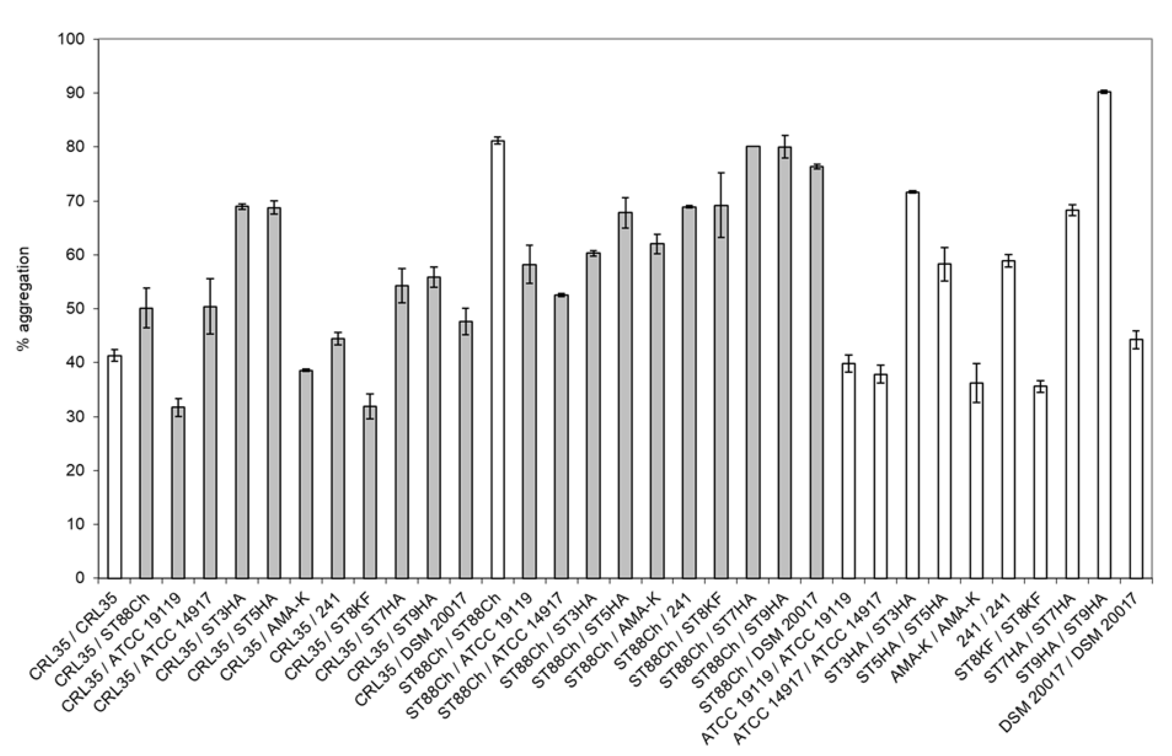
2.6. Aggregation Properties of E. mundtii CRL35 and E. faecium ST88Ch

Previously applied experimental approaches for the evaluation of aggregation (auto- and co-aggregation) properties suggested by Pingitore et al. [29] and Collado et al. [37] were adapted for current experiments. E. mundtii CRL35 and E. faecium ST88Ch were evaluated for their auto-aggregation and co-aggregation properties. For co-aggregation partners, strains with pathogenic and beneficial properties (listed in Figure 1) were selected from the culture collection of USP, Sao Paulo, SP, Brazil. The cultures were grown in 10 mL MRS broth (Difco) or brain heart infusion (BHI broth; Difco) for 24 h at 37 °C without agitation, and cells were harvested by centrifugation (7000× g, 10 min, 4 °C). The cells were washed twice, resuspended in sterile saline (0.85% NaCl, w/v), and the density of the suspension was adjusted to OD660 nm = 0.3.
The efficiency of auto-aggregation was calculated according to Pingitore et al. [29]:
%   Auto - aggregation   =   O D 0 O D 60 O D 0   ×   100 ,
where OD0 represents the initial optical density of the suspension, measured at 660 nm. OD60 refers to the optical density of the upper fraction recorded after the cells were incubated for 60 min at 37 °C and centrifuged for 2 min at 300× g.
Co-aggregation properties were determined using equal volumes of paired cell suspensions, prepared as described before and after incubation for 60 min at 37 °C. OD660 nm was determined for the upper phase after separation by centrifugation for 2 min at 300× g [29]. The efficiency of co-aggregation was calculated according to Pingitore et al. [29] as:
%   Co - aggregation   =   O D 0 O D 60 O D 0   ×   100 ,
where OD0 represents the initial optical density of the suspension, measured at 660 nm. OD60 refers to the optical density of the upper fraction recorded after the cells were incubated for 60 min at 37 °C and centrifuged for 2 min at 300× g.
The experiments were conducted twice, each time in triplicate.
In addition, auto- and co-aggregation between E. mundtii CRL35, E. faecium ST88Ch, and C. albicans 1281 in sterile saline (0.85% NaCl, w/v) were determined after 60 min at 37 °C, at pH 3.0, 5.0, and 7.0, with the aim of partially simulating the growing conditions of a healthy and a C. albicans-infected vaginal environment, both in the absence or presence of 10% or 20% (m/v) Nordette-28 contraceptive (Teva Pharmaceuticals, Basel, Switzerland).

2.7. Adsorption of BLIS to C. albicans 1281

Adsorption of the semi-purified BLIS produced by E. mundtii CRL35 and E. faecium ST88Ch to C. albicans 1281 was tested according to Pingitore et al. [29] and Agaliya and Jeevaratnam [38]. C. albicans 1281 was grown in Sabouraud broth for 18 h at 37 °C without agitation, and cells were harvested by centrifugation (8000× g, 15 min, 4 °C). The obtained cells were washed with sterile 5 mM phosphate buffer (pH 6.5) two times and resuspended in the same buffer to an OD600 nm equal to 1.0. The obtained suspension was mixed with an equal volume of semi-purified BLIS (corresponding to the 60% isopropanol fraction obtained from chromatography on SepPakC18) prepared as previously described from CRL35 or ST88Ch (with 25,600 AU/mL and 800 AU/mL, pH 6.5, respectively) and incubated for 1 h at 37 °C. After removal of the cells (centrifugation at 8000× g, 15 min, 25 °C), the antimicrobial activity against C. albicans 1281 of unbound extracellular BLIS produced by CRL35 and ST88Ch in the supernatants was estimated as previously described. All experiments were performed in duplicate at minimum.
The efficiency of BLIS adsorption to the target cells was estimated according to the following formula:
%   adsorption   =   [ 100     AU / m L 1 AU / m L 0 ]   ×   100 ,
where AU/mL1 represents the BLIS activity after treatment, and AU/mL0 the original (before treatment) BLIS activity.
The effect of temperature and pH on BLIS adsorption to C. albicans 1281 was estimated as described above, with incubation for 1 h at 30, 37, and 45 °C, and at pH 3.0, 5.0, and 7.0, simulating the changes of vaginal pH in Candida-associated infections [17]. CFS were obtained (8000× g, 15 min, 25 °C), and the pH was adjusted to 6.0 with sterile 1 M NaOH. BLIS activity in the supernatant against C. albicans 1281 was determined as previously described. The experiments were performed at least in duplicate.

Effect of the Contraceptive Nordette-28 on Adsorption of the Studied BLIS to C. albicans 1281

Cell suspensions of C. albicans obtained as previously described were supplemented with 10% (w/v) and 20% (w/v) Nordette-28. The pH of cell suspensions was adjusted to 3.0, 5.0 and 7.0 with 1 M NaOH or 1 M HCl. The studied BLIS were added to the treated cell suspensions, as described previously, and incubated for 1 h at 37 °C without agitation. The CFS were obtained by centrifugation (8000× g, 15 min, 25 °C) and the activity of the BLIS were determined as previously described against C. albicans. The experiments were performed in at least in duplicate.

2.8. Effect of Antibiotics and Generic Drugs, including Selected Contraceptives, on the Growth of E. mundtii CRL35 and E. faecium ST88Ch

Experimental approaches for the evaluation of the effect of commercial non-antibiotic drugs and antibiotics on growth of LAB previously suggested by Todorov et al. [39] and Amaral et al. [40] were applied. Overnight cultures of the two enterococcal strains were embedded into BHI supplemented with 1% (w/v) agar to a final concentration of approximately 106 CFU/mL. Antibiotic disks, listed in Table 1, were placed on the agar surface, and the plates were incubated for 24 h at 37 °C. The effect of antibiotics was recorded by measuring the diameter of the growth inhibition zones, expressed in millimeters.
In a similar experimental set-up, the effect of selected medicaments applied in specific concentrations, summarized in Table 2, and the commercially available contraceptives Nordette-28, Triphasil (Pfizer, Sydney, Australia), Diphasil (Bayer, Germany), and MicroVal (Saint-Just-Malmont, France) on the growth of E. mundtii CRL35 and E. faecium ST88Ch were determined. For the listed contraceptives (Nordette-28, Triphasil, Diphasil and MicroVal), tablets numbered 1, 8, 15 and 22 were dissolved in 5 mL of sterile distilled water. An amount of 15 μL of each commercial drug solution was spotted on the surface of BHI agar plates containing E. mundtii CRL35 or E. faecium ST88Ch (approximately 106 CFU/mL) and incubated for 24 h at 37 °C. Growth inhibition was recorded by measuring the diameter of the zones.

2.9. Cell Lysis of C. albicans by BLIS from E. mundtii CRL35 and E. faecium ST88Ch

An experimental approach previously suggested by Todorov and Dicks [41] and Pingitore et al. [29] for the evaluation of the cell lysis of target strains by the effect of antimicrobial agents was applied. Cell lysis experiments were performed using sterile flat-bottom 96-well microtiter plates (Nunc, Sigma Aldrich, St. Luis, MO, USA). C. albicans 1281 was cultured in 20 mL Sabouraud broth for 18 h at 37 °C without agitation, and the cells harvested by centrifugation (8000× g, 10 min, 4 °C), and washed twice and resuspended in 10 mL of potassium phosphate buffer (20 mM, pH 6.5). A yeast suspension (100 μL) was placed in the microtiter plate wells and supplemented with 50 μL semi-purified BLIS of CRL35 or ST88Ch (corresponding to the 60% isopropanol fraction obtained from chromatography on SepPakC18) solubilized in potassium phosphate buffer (25 mM, pH 6.5) at different serial two-fold dilutions. Microtiter plates were incubated for 24 h at 37 °C and at selected intervals, the absorbance at 655 nm was measured spectrophotometrically (Thermo Fisher Scientific, Waltham, MA, USA). Levels of cell lysis of C. albicans 1281, expressed as %, were calculated as [100 − (At/Ao × 100)], where Ao and At were absorbance measured at 0 and 3, 6, 9 or 24 h of incubation, respectively [29].

2.10. Statistical Analysis

IBM SPSS 26.0 (SPSS, Inc., Chicago, IL, USA) was used for data analysis. Data were not normally distributed according to the Shapiro–Wilk test. The Kruskal-Wallis test, followed by the Dunn-Bonferroni post-hoc test, was used to evaluate differences among results of obtained experimental data. Results were presented as mean ± standard deviation (SD). The statistical significance was determined as p < 0.05.

3. Results

3.1. Screening of LAB for Anti-Candida Activity

From a total of 75 tested LAB, the CFS (pH 6.0–5.5) of only two strains, E. mundtii CRL35 and E. faecium ST88Ch, which were previously isolated, identified, and characterized as bacteriocins producers [29], presented inhibitory activity against C. albicans 1281, a clinical isolate (6400 AU/mL and 800 AU/mL, respectively). This and all other tests were performed with planktonic C. albicans 1281 cells. However, when the CFS was submitted to ammonium sulphate precipitation and elution with the 60% isopropanol fraction after SepPakC18 hydrophobic chromatography, the activity increased to 12,800 AU/mL and 25,600 AU/mL for BLIS produced by E. mundtii CRL35, and remained unchanged for BLIS produced by E. faecium ST88Ch. It is important to emphasize that the same BLIS activity was recorded after evaluation of the 60% isopropanol fraction that was lyophilized and resuspended in 25 mM phosphate buffer (pH 6.5) as the initial volume. At the same time, neither the controls, 40%, 60% and 80% isopropanol solutions, nor the applied buffer, showed inhibitory properties against C. albicans 1281 or L. monocytogenes ATCC 15313. The 40% and 80% isopropanol fractions after SepPakC18 chromatography presented lower activity against C. albicans 1281—1600 AU/mL and 800 AU/mL for E. mundtii CRL35, and 400 AU/mL and 200 AU/mL for E. faecium ST88Ch, respectively.
E. mundtii CRL35 and E. faecium ST88Ch did not produce H2O2 when grown on modified MRS agar.
Treatment of the CFS of E. mundtii CRL35 and E. faecium ST88Ch with 1 mg/mL proteinase K at pH 6.0–6.5 resulted in complete loss of BLIS activity when tested against L. monocytogenes ATCC 15313. However, when C. albicans 1281 was used as a test organism, a low level of inhibitory effect was still observed. This is an indication that both LAB strains (E. mundtii CRL35 and E. faecium ST88Ch) are BLIS producers. However, some other non-proteinaceous antimicrobial(s) may also be present in the CFS with activity against C. albicans 1281.

3.2. Hemolytic Activity

E. mundtii CRL35 and E. faecium ST88Ch were evaluated as γ-hemolytic since no discoloration around the colonies was observed. The bacterial growth of E. mundtii CRL35 and E. faecium ST88Ch in the performed test was similar to that observed for Lb. plantarum ATCC 14917, a γ-hemolytic control.

3.3. Aggregation Properties

Different levels of aggregation were recorded between C. albicans 1281 and E. mundtii CRL35 or E. faecium ST88Ch, and additional strains were utilized as co-aggregation partners in a strain-specific manner (Figure 1). High levels of co-aggregation between E. mundtii CRL35 and E. faecium ST88Ch were recorded with C. albicans 1281 (Figure 2), compared to the other test organisms. This improved co-aggregation can be regarded as evidence of a predisposition for better antimicrobial effect of E. mundtii CRL35 and E. faecium ST88Ch against C. albicans 1281.

3.4. Adsorption of BLIS to C. albicans

In the present study, we evaluated the effects of temperature, pH, and the presence of contraceptives (Nordette-28) on the adsorption of the studied BLIS to C. albicans 1281 cells. The experimental conditions were selected to provide a simplified version of the favorable growth conditions of a healthy vaginal environment (37 °C and mildly acidic pH), and conditions when affected by the presence of Candida spp., where the pH was decreased. For the purpose of comparing the adsorption of the studied BLIS to C. albicans 1281, increased and decreased temperatures (40 °C and 30 °C) were also applied. Moreover, the presence of the contraceptive was considered a factor that may influence BLIS adsorption. Temperature, pH, and the presence of the contraceptive Nordette-28 affected the adsorption of the studied BLIS to cells of C. albicans 1281 (Table 3). Interestingly, at higher pH, representing conditions resulting from increased levels of C. albicans in the vaginal environment, the adsorption of both BLIS was higher (Table 3). Optimal adsorption levels were recorded at 37 °C, compared to the evaluated temperature controls of 30 °C and 40 °C.
The presence of 10% or 20% Nordette-28 promoted increased adsorption of both studied BLIS to the cells of C. albicans 1281 at pH 5.0 (Table 3). A minor effect was observed at pH 3.0, where only supplementation with 20% Nordette-28 increased adsorption of BLIS produced by E. mundtii CRL35. At pH 7.0, adsorption of BLIS produced by E. mundtii CRL35 was slightly reduced, while the adsorption of BLIS produced by E. faecium ST88Ch was not affected at neutral pH (Table 3).

3.5. Effect of Antibiotics and Other Medicaments, including Selected Contraceptives, on the Growth of E. mundtii CRL35 and E. faecium ST88Ch

The two BLIS-producing strains showed moderate resistance to some of the evaluated antibiotics in the present study (Table 1). None of the contraceptives evaluated in this study (Nordette-28, Triphasil, Diphasil, MicroVal) inhibited the growth of E. mundtii CRL35 or E. faecium ST88Ch. Moreover, additional drugs evaluated as potential inhibitors of the two studied strains showed no antagonism. Only thioridazine (neuroleptic) and Voltaren (anti-inflammatory and anti-rheumatic) showed moderate inhibitory effect, and a slightly stronger inhibition by Diuretidin (diuretic) on E. mundtii CRL35 and E. faecium ST88Ch was also observed (Table 2).

3.6. Cell Lysis of C. albicans by BLIS from E. mundtii CRL35 and E. faecium ST88Ch

The application of semi-purified BLIS from E. mundtii CRL35 and E. faecium ST88Ch on cell suspensions of C. albicans 1281 showed a dose-dependent killing effect (Figure 3). When applied from 400 AU/mL to 25,600 AU/mL, the BLIS produced by E. mundtii CRL35 caused 63.61% and 79.35% lysis of C. albicans 1281 cells after 9 h of contact (Figure 3A). After 24 h of contact, similar results were observed, with a slight decline of cell lysis (Figure 3A). However, when BLIS produced by E. faecium ST88Ch was applied in concentrations from 200 AU/mL to 800 AU/mL, levels of cell lysis of C. albicans 1281 were between 29.32% and 31.25%, respectively, after 9 h of contacts (Figure 3B).

4. Discussion

BLIS production is a characteristic that is considered to be an additional advantageous property for beneficial therapeutical strains [42]. BLIS such as antimicrobial peptides play an essential role in microbial interactions [43]. In a previous study, E. mundtii CRL35 and E. faecium ST88Ch were reported as BLIS-producing strains that play an important role in the biopreservation and control of Listeria monocytogenes in fresh cheese [29]. In the present study, these two strains, in addition to other LAB strains previously described as producers of antimicrobial peptides and/or presenting probiotic properties and belonging to the culture collections of USP (Brazil) and CERELA (Argentina), were explored for their antimicrobial effect against strains of C. albicans isolated from patients diagnosed with vaginosis and positively identified by API 20 C AUX fermentation reactions according to the recommendations of Singh et al. [17]. The majority of the evaluated strains showed no inhibitory activity against the tested C. albicans strains assessed in the preliminary screening process for the selection of BLIS producer/s and an appropriate sensitive strain (data not shown), even though most of them presented BLIS activity against L. monocytogenes ATCC 15313. The aim of the performed screening process was to select BLIS producers from previously identified bacteriocinogenic LAB in the culture collections of USP and CERELA with unusual anti-Candida activity. Taking into consideration the specificity of this task, the screening was also focused on selecting Candida spp. that would be sensitive to the BLIS of the LAB candidates. Thus, the screening process needed to be regarded as a dual-task challenge for the selection of the optimal combination of BLIS producer/s and sensitive organisms. Despite the current knowledge that BLIS produced by LAB are more active against genetically related bacterial species than other types of microorganisms [44], the BLIS produced by E. mundtii CRL35 and E. faecium ST88Ch were active against C. albicans. The proteinaceous nature of the expressed antimicrobials was confirmed based on the treatment of the CFS obtained from E. mundtii CRL35 and E. faecium ST88Ch with proteolytic enzymes at pH adjusted to 6.0. This experimental approach was previously reported as a simple, but reliable, test for confirming the proteinaceous nature of LAB-expressed antimicrobial metabolites [29,34,35]. The fact that a loss of antimicrobial activity was recorded when L. monocytogenes ATCC 15313 was applied as test microorganism supports the idea that the expressed antimicrobial metabolite is of proteinaceous nature. However, when C. albicans 1281 was used as a test organism in the same experiment, a low residual activity was recorded. This indicates that an additional antimicrobial compound that is different from previously recorded and evaluated bacteriocins produced by E. mundtii CRL35 and E. faecium ST88Ch [29] may be produced and play a role in the observed unusual anti-Candida activity. The fact that antagonistic tests were performed with CFS that was corrected to a neutral pH is an additional piece of evidence showing that the observed inhibitory activity cannot be associated with the direct effects of produced organic acids. Moreover, the specific nature of this additional antimicrobial agent produced by E. mundtii CRL35 and E. faecium ST88Ch merits a deeper investigation in order to answer the question of whether the observed activity is a consequence of a single mode of action or the result of synergetic interactions together with bacteriocins produced by the two enterococci [29]. Despite being considered unusual [45], Hefzy et al. [46] also reported that BLIS produced by Lb. pentosus, Lb. paracasei subsp. paracasei, Lactobacillus delbrueckii subsp. lactis and Streptococcus uberis strains were active against C. albicans. Pentocin TV35b, a bacteriocin produced by L. pentosus TV35b, was reported as having activity against C. albicans [47]. The results given by the controls (lactic acid solution at pH 4.0, CFS treated with proteinase K, and CFS untreated with proteolytic enzymes) indicated that the observed inhibitory effects are likely due to an antimicrobial compound of proteinaceous nature. Antimicrobials involved in killing and/or inhibitory effects against C. albicans can be BLIS or acids or combinations of them. Nieminen et al. [48] reported that D,L-2-hydroxyisocaproic acid (HICA) is an active substance associated with the inhibition of planktonic cells of C. albicans and biofilm formation in vitro. Fugaban et al. [26] evaluated the production of different organic compounds, including organic acids, as inhibitors of the growth of filamentous fungal species and suggested that bacteriocins may only have a secondary effect. Ishijima et al. [49] suggested that BLIS produced by Streptococcus salivarius K12 can be actively involved in the growth control of C. albicans, as shown in an oral candidiasis model. Lum et al. [50] suggested the use of KABT-AMP (lysine-rich peptide) and the antimicrobial peptide Uperin 3.6 from the Uperoleia mjobergii frog as potential templates for the development of effective anti-Candida peptides. The proposed hybrid peptides, containing a mixed backbone of KABT-AMP and Uperin 3.6, showed potent anti-Candida activity with an estimated MIC of around 8–16 mg/L. Scillato et al. [51] reported on the probiotic potential of the vaginal isolates Lactobacillus gasseri 1A-TV, Lb. fermentum 18A-TV, and Lb. crispatus 35A-TV, including antimicrobial effects of their metabolites in CFS with activity against C. albicans and C. glabrata. These authors evaluated the probiotic properties of the studied LAB, including their co-aggregation, adhesion to HeLa cells, and biofilm formation properties. Moreover, Lb. gasseri 1A-TV and Lb. crispatus 35A-TV were shown to harbor genes for the production of the helveticin J and acidocin A bacteriocins. Salari et al. [52] evaluated the antifungal effects of Lb. acidophilus and Lb. plantarum (cells and CFS) on different Candida species, including co-aggregation properties and antimicrobial activity. Lb. acidophilus and Lb. plantarum in the range of 102 to 1010 CFU/mL or as CFS showed inhibitory effects against the evaluated Candida species.
E. mundtii CRL35 and E. faecium ST88Ch did not produce H2O2. Hydrogen peroxide-producing LAB were suggested to be associated with the antimicrobial properties of the vaginal environment and are involved in spermicidal effects, such as those reported for drugs containing nonoxynol-9 [53]. Different spermicides are used as contraceptive drugs and are involved in the protection of the urogenital tract of women against sexually transmitted pathogens such as Neisseria gonorrhoea, Chlamydia trachomatis, and HIV [54]. To be effective probiotics for vaginal health, E. mundtii CRL35 and E. faecium ST88Ch need to be resistant to different contraceptives and spermicides and thus should be verified as such in appropriate in vivo experiments.
The relationship between gastrointestinal tract (GIT) and vaginal health was previously reported, and oral administration of a combination of Lacticaseibacillus rhamnosus GR-1 and Lb. fermentum RC-14 was shown to be effective in the treatment of bacterial vaginosis and supported restorations of the urogenital tract microbiota to normal levels. Reid et al. [6,55,56] reported that Lb. rhamnosus GR-1 and Lb. fermentum RC-14 were able to adhere to uroepithelial and vaginal cells and were actively involved in the inhibition of growth and adhesion of urogenital pathogens. Lb. rhamnosus GR-1 was not associated with the production of therapeutic levels of H2O2 and was resistant to different spermicide drugs, but on the other hand, Lb. fermentum RC-14, evaluated as a producer of H2O2, was sensitive to the effect of tested spermicides [57]. The potential application of E. mundtii CRL35 or E. faecium ST88Ch as putative urogenital probiotics will probably be more effective if they are taken in combination with H2O2-producing LAB, and a resulting assortment of different antimicrobial agents may also have synergistic activity.
Aggregation of E. mundtii CRL35 and E. faecium ST88Ch to C. albicans may also be regarded as a valid tool for the reduction of C. albicans adherence to epithelial cells. Aggregation between enterococci and C. albicans can be considered the primary step in the killing process of C. albicans, as a consequence of the close contact between the microbial cells and facilitated delivery of antimicrobial peptides to the target cells. The fact that clinical strains of E. faecalis and C. albicans are frequently found to co-exist in the infected urogenital tract of patients [51,52,58,59] supports the application of E. mundtii CRL35 and E. faecium ST88Ch, or their BLIS against C. albicans.
Aggregation properties between different microbial species needed to be evaluated considering the complexity of the environment due to the potential existence of a variety of different recorded antimicrobial metabolites. The human vaginal microbiome is a complex system where interactions between different species provide health benefits and contribute to the normal reproductive processes of the host. In natural processes, pathogens face the defense mechanisms of the immune system, but microorganisms considered as beneficial for the vaginal environment also play a significant role in maintaining microbial balance and in reducing the development of clinical complications associated with effective or opportunistic pathogens [1,2,6,7]. Aggregation between different microbial species (called co-aggregation) can facilitate biofilm formation and thereby improve the survival of specific microbial species as part of formed biofilm in different ecological environments, including the vagina and other parts of human and other animals’ bodies. From this point of view, high aggregation (auto- or co-aggregation) needs to be considered a beneficial characteristic when recorded for strains with clear positive effects for the host; such is the case for probiotics. However, when pathogens (effective or opportunistic) are involved in the formation of a stable aggregation complex, this needs to be regarded as facilitating their pathogenicity, since they will be protected by the biofilm and are more present in the environment due to their interactions with other aggregation partners. From a different point of view, if antimicrobial interaction is a clearly established fact between co-aggregation partners, then the co-aggregation processes will facilitate this antimicrobial action since it will provide close contact between a producer of antimicrobials and the target cell and increase the efficacity of killing properties of produced BLIS or other antagonistic metabolites. Moreover, environmental conditions (pH, temperature, different chemicals) can influence these aggregation processes, and appropriate evaluation through in vitro preliminary experiments can help to predict the efficacity of suggested antimicrobials of putative probiotics strains in further in vivo or in situ studies, as has been previously shown by Pingitore et al. [29].
One of the major mechanisms of action of bacteriocins and/or BLIS is associated with an interruption of the cellular integrity of the target organisms via pore formation in the cell wall [44,60]. This process may be associated with the adsorption of BLIS to a specific receptor [60] or simply based on affinity and electro-physical interactions [61]. Thus, the adsorption of BLIS to the cell surface of target organisms is considered a key step in their mode of action.
In the present study, we have evaluated the effects of temperature, pH, and the presence of a contraceptive (Nordette-28) on the adsorption of the studied BLIS produced by E. mundtii CRL35 and E. faecium ST88Ch to cells of C. albicans in conditions modeling healthy and candidiasis-associated vaginal conditions. We observed an increase in the adsorption of the studied BLIS produced by E. mundtii CRL35 and E. faecium ST88Ch to C. albicans 1281 in the presence of Nordette-28 at the evaluated pH levels. However, what needs to be further explored is our understanding of how the presence of the contraceptive improves the antimicrobial activity of the studied BLIS, and whether the contraceptive works synergistically with the BLIS or assists its antimicrobial activity in a complementary manner.
Optimal adsorption levels were recorded at 37 °C, compared to the evaluated temperature controls of 30 °C and 40 °C. In vitro tests for evaluating the effects of BLIS are essential in assessing their effectiveness. An in vitro research approach can be a potential indicator of the effectiveness of the bacteriocinogenic strains in future evaluations in situ, as has been shown in the study of Pingitore et al. [29], where the bioprotective effect and the control of L. monocytogenes in cheeses by the same bacteriocinogenic strains (E. mundtii CRL35 and E. faecium ST88Ch) was demonstrated.
The presence of the contraceptive Nordette-28 at a final concentration of 10% or 20% increased the adsorption of both studied BLIS to cells of C. albicans at pH 5.0 (Table 3), but very little effect was observed at pH 3.0. However, at pH 7.0, adsorption of the BLIS produced by E. mundtii CRL35 was slightly reduced and was not affected by adsorption of BLIS produced by E. faecium ST88Ch (Table 3). These results are interesting since they indicate that the use of the contraceptive will not significantly affect the prevention or active treatment of vaginal candidiasis cases by the BLIS or BLIS-producing strains.
The safety of E. mundtii CRL35 and E. faecium ST88Ch must be thoroughly tested using in vivo studies, since their use as beneficial therapeutical cultures for human consumption is still questionable. Some strains of Enterococcus spp. (such as representatives of species E. faecalis) have been associated with nosocomial infections [62]. Furthermore, some enterococci are resistant to glycopeptides and antibiotics, produce biogenic amines, and exhibit virulence factors [63]. Future in vivo studies will not only evaluate the safety of E. mundtii CRL35 and E. faecium ST88Ch, but will also provide information on their efficiency and ability to attach to uroepithelial cells [64].
The safety properties of beneficial microbial strains are of utmost importance and the evaluation of antibiotics resistance is considered a basic requirement in the selection of strains for the practical applications of beneficial therapeutical cultures [65]. The two studied BLIS-producing strains showed moderate resistance to some of the evaluated antibiotics (Table 1). In general, the scientific consensus is that beneficial therapeutical cultures and/or probiotics candidates cannot be resistant to clinically applied antibiotics [65], an argument to avoid the spread of antibiotic-resistant genetic determinates. However, Suvorov [66] defended the hypothesis that the spread of antibiotic-resistant genes (and other virulence factors) is an unlikely scenario in natural environmental conditions. On the other hand, we need to consider whether these genetic determinates are associated with bacterial chromosomes or located on plasmids, since this can influence horizontal gene transfer [66]. It is clear that before the two studied strains (E. mundtii CRL35 and E. faecium ST88Ch) can be recommended for the application of beneficial therapeutical cultures in the control of C. albicans-associated vaginal disorders, additional safety evaluations will need to be performed, including an investigation into the presence of specific antibiotics genes and their locations. Only once the appropriate safety assessments have been conducted can E. mundtii CRL35 and E. faecium ST88Ch be considered for application as life-protective cultures, or a determination made on whether their antimicrobial peptides (postbiotics) will be more suitable for the control of vaginal candidiasis.
Probiotics aimed at vaginal health can be applied orally or as vaginal suppositories. The relationship between GIT balance and general health has been well described [67]. Several arguments are provided in support of the idea that a healthy GIT has a positive effect on different parts of the body, including the vagina [68,69]. The viability of orally delivered probiotics can be influenced not only by stomach acidity but by the presence of bile salts. They are also exposed to the complex environmental condition of the GIT, including the presence of a multitude of different microorganisms, the immunological response of the host, and different drugs associated with episodical or chronic diseases.
None of the contraceptives evaluated in this study inhibited the growth of E. mundtii CRL35 or E. faecium ST88Ch. Previously, it was reported that the application of contraceptives could influence the ecological balance of the vaginal microbiota and even increase risks of the development of sexually transmitted diseases because of the reduction of natural protections by LAB and its influence on immune defense [28]. Moreover, it will be interesting for further studies to evaluate the effects of the investigated contraceptives on both the immune response and the vaginal microbiota, including the effect on E. mundtii CRL35 or E. faecium ST88Ch as determined using an appropriate animal model. Furthermore, most of the generic drugs evaluated as potential inhibitors of the two studied strains cannot be considered antagonists. Only thioridazine, Voltaren, and Diuretidin affected the growth of E. mundtii CRL35 or E. faecium ST88Ch (Table 2). The inhibitory effect of episodically or permanently applied drugs on the viability of probiotic strains needs to be evaluated and considered to determine the suitability of their combined applications. It was previously shown that several painkillers and drugs used for the treatment of chronic diseases can have an inhibitory effect on some LAB [70,71]. Such interactions merit deeper evaluation and the construction of an appropriate database to avoid negative interactions between generic drugs and probiotics, and optimize both effects.
The effect of most bacteriocins and/or BLIS on test microorganisms is associated with the destabilization of the cell wall, pore formation, and, consequently, bacterial lysis [60]. Application of semi-purified BLIS produced by E. mundtii CRL35 and E. faecium ST88Ch on cell suspension of C. albicans showed a dose-dependent effect in the killing of test organisms (Figure 3). The observed differences between the effect of BLIS produced by E. mundtii CRL35 and E. faecium ST88Ch on the lysis of C. albicans were expected and are in agreement with observed levels of adsorption between the studied BLIS and applied test microorganism (Table 3). Thus, the results agree with previously reported similar experiments evaluating the effectiveness of BLIS produced by E. mundtii CRL35 and E. faecium ST88Ch against L. monocytogenes ATCC 15313 and their possible application in biopreservation processes of fresh cheeses [29]. Appropriate in vitro experiments can serve as an indicator for the selection of appropriate strains for further in situ or in vivo evaluation of their effectiveness as putative probiotics strains.

5. Conclusions

Beneficial microorganisms with therapeutical properties (probiotics) can be regarded as effective alternatives for reducing the use of chemical and designed drugs, as some are associated with several negative side effects. Appropriate probiotic microorganisms effectively modulate the vaginal microbiome and reduce negative consequences due to the development of yeast infections (including C. albicans). Probiotics with therapeutical properties can interfere with pathogens via cell-to-cell interaction or via the production of several antimicrobial metabolites, such as BLIS, including the establishment of cumulative or synergetic interactions between different factors. Moreover, the application of contraceptives can modulate the beneficial properties of beneficial microorganisms with therapeutical properties and reinforce their antimicrobial effects.

Author Contributions

Conceptualization, S.D.T. and M.L.C.; resources, S.D.T. and B.D.G.d.M.F.; writing—original draft preparation, S.D.T.; writing—review and editing, S.D.T., M.L.C., R.W., I.P. and B.D.G.d.M.F.; language corrections: R.W. All authors have read and agreed to the published version of the manuscript.

Funding

This research was funded by FAPESP (Grant 2013/07914-8), and USP, for providing scholarships for visiting professors (2016.1.920.93). IP and MLC were supported, in part, by the Ministry of Science and Higher Education of the Russian Federation (Project Number 075-15-2019-1880).

Data Availability Statement

All data generated by the present project is available upon request.

Acknowledgments

To Fernando Sesma (in memoriam), for providing some of the strains used in this study. To Mia Miau, for her unconditional support.

Conflicts of Interest

The authors declare no conflict of interest.

References

  1. Álvarez-Artero, E.; Campo-Nuñez, A.; García-García, I.; García-Bravo, M.; Cores-Calvo, O.; Galindo-Pérez, I.; Pendones-Ulerio, J.; López-Bernus, A.; Belhassen-García, M.; Pardo-Lledías, J. Urinary tract infection caused by Enterococcus spp.: Risk factors and mortality. An observational study. Rev. Clin. Esp. 2021, 221, 375–383. [Google Scholar] [CrossRef] [PubMed]
  2. Giannakopoulos, X.; Sakkas, H.; Ragos, V.; Tsiambas, E.; Bozidis, P.M.; Evangelou, A.; Papadopoulou, C.; Petrogiannopoulos, L.; Sofikitis, N. Impact of enterococcal urinary tract infections in immunocompromised - neoplastic patients. J. Buon. 2019, 24, 1768–1775. [Google Scholar] [PubMed]
  3. Li, M.; Yang, F.; Lu, Y.; Huang, W. Identification of Enterococcus faecalis in a patient with urinary-tract infection based on metagenomic next-generation sequencing: A case report. BMC Infect. Dis. 2020, 20, 467. [Google Scholar] [CrossRef] [PubMed]
  4. Lerma, A.F.; Palomar, M.; Insausti, J.; Olaechea, P.; Alcala, M.A.; Blanco, A. Enterococcal infections in critically ill patients admitted at ICU. Med. Clin. 2003, 13, 281–286. [Google Scholar] [CrossRef]
  5. Sobel, J.D. Candidal vulvovaginitis. Clin. Obstet. Gynaecol. 1993, 36, 153–165. [Google Scholar] [CrossRef]
  6. Reid, G.; Bruce, A.W. Urogenital infections in women: Can probiotics help? Post. Grad. Med. J. 2003, 79, 428–432. [Google Scholar] [CrossRef] [PubMed] [Green Version]
  7. Ferrer, J. Vaginal candidosis: Epidemiology and etiological factors. Int. J. Gynaecol. Obstet. 2000, 71, 21–27. [Google Scholar] [CrossRef]
  8. Foxman, B. Epidemiology of urinary tract infections: Incidence, morbidity, and economic costs. Dis. Mon. 2003, 49, 53–70. [Google Scholar] [CrossRef]
  9. Jung, W.W.; Chun, T.; Sul, D.; Hwang, K.W.; Kang, H.S.; Lee, D.J.; Han, I.K. Strategies against human papillomavirus infection and cervical cancer. J. Microbiol. 2004, 42, 255–266. [Google Scholar] [PubMed]
  10. Parratt, J.R.; Hay, D.P. Sexually transmitted infections. Curr. Obstet. Gynaecol. 2003, 13, 224–231. [Google Scholar] [CrossRef]
  11. Rhoton-Vlasak, A. Infections and infertility. Prim. Care Update. Ob./Gyns. 2000, 7, 200–206. [Google Scholar] [CrossRef] [PubMed]
  12. Romero, R.; Chaiworapongsa, T.; Kuivaniemi, H.; Tromp, G. Bacterial vaginosis, the inflammatory response and the risk of preterm birth: A role for genetic epidemiology in the prevention of preterm birth. Am. J. Obstet. Gynaecol. 2004, 190, 1509–1519. [Google Scholar] [CrossRef] [PubMed]
  13. Greiner, L.L.; Edwards, J.L.; Shao, J.; Rabinak, C.; Entz, D.; Apicella, M.A. Biofilm formation by Neisseria gonorrhoeae. Infect. Immun. 2005, 73, 1964–1970. [Google Scholar] [CrossRef] [PubMed] [Green Version]
  14. Mukherjee, P.K.; Zhou, G.; Munyon, R.; Ghannoum, M.A. Candida biofilm: A well-designed protected environment. Med. Mycol. 2005, 43, 191–208. [Google Scholar] [CrossRef] [Green Version]
  15. Swidsinski, A.; Mendling, W.; Loening-Baucke, V.; Ladhoff, A.; Swidsinski, S.; Hale, L.P.; Lochs, H. Adherent biofilms in bacterial vaginosis. Obstet. Gynaecol. 2005, 106, 1013–1023. [Google Scholar] [CrossRef] [Green Version]
  16. Tendolkar, P.M.; Baghdayan, A.S.; Shankar, N. The N-terminal domain of enterococcal surface protein, Esp, is sufficient for Esp-mediated biofilm enhancement in Enterococcus faecalis. J. Bacteriol. 2005, 187, 6213–6222. [Google Scholar] [CrossRef] [Green Version]
  17. Singh, A.; Verma, R.; Murari, A.; Agrawal, A. Oral candidiasis: An overview. J. Oral Maxillofac. Pathol. 2014, 18 (Suppl. 1), S81–S85. [Google Scholar] [CrossRef]
  18. Mbizvo, E.M.; Msuya, S.E.; Stray-Pedersen, B.; Sundby, J.; Chirenje, M.Z.; Hussain, A. HIV seroprevalence and its associations with the other reproductive tract infections in asymptomatic women in Harare, Zimbabwe. Int. J. STD AIDS 2001, 12, 524–531. [Google Scholar] [CrossRef]
  19. van De Wijgert, J.H.; Mason, P.R.; Gwanzura, L.; Mbizvo, M.T.; Chirenje, Z.M.; Iliff, V.; Shiboski, S.; Padian, N.S. Intravaginal practices, vaginal flora disturbances, and acquisition of sexually transmitted diseases in Zimbabwean women. J. Infect. Dis. 2000, 181, 587–594. [Google Scholar] [CrossRef]
  20. Eschenbach, D.A.; Davick, P.R.; Williams, B.L.; Klebanoff, S.J.; Young-Smith, K.; Critchlow, C.M.; Holmes, K.K. Prevalence of hydrogen peroxide-producing Lactobacillus species in normal women and women with bacterial vaginosis. J. Clin. Microbiol. 1989, 27, 251–256. [Google Scholar] [CrossRef] [Green Version]
  21. Klebanoff, S.J.; Hillier, S.L.; Eschenbach, D.; Waltersdorph, A.M. Control of the microbial flora of the vagina by H2O2-generating lactobacilli. J. Infect. Dis. 1991, 164, 94–100. [Google Scholar] [CrossRef] [PubMed]
  22. Nagy, E.; Petterson, M.; Mardh, P.A. Antibiosis between bacteria from the vagina of women with and without signs of bacterial vaginosis. APMIS 1991, 99, 739–744. [Google Scholar] [CrossRef] [PubMed]
  23. Gupta, K.; Stapleton, A.E.; Hooton, T.M.; Roberts, P.L.; Fennell, C.L.; Stamm, W.E. Inverse association of H2O2-producing lactobacilli and vaginal Escherichia coli colonization in women with recurrent urinary tract infections. J. Infect. Dis. 1998, 178, 446–450. [Google Scholar] [CrossRef] [PubMed] [Green Version]
  24. Song, Y.L.; Kato, N.; Matsumiya, Y.; Liu, C.X.; Kato, H.; Watanabe, K. Identification of and hydrogen peroxide production by fecal and vaginal lactobacilli isolated from Japanese women and newborn infants. J. Clin. Microbiol. 1999, 37, 3062–3064. [Google Scholar] [CrossRef] [Green Version]
  25. Hoesl, C.E.; Altwein, J.E. The probiotic approach: An alternative treatment option in urology. Eur. Urol. 2005, 47, 288–296. [Google Scholar] [CrossRef]
  26. Fugaban, J.I.I.; Holzapfel, W.H.; Todorov, S.D. The overview of natural by-products of beneficial lactic acid bacteria as promising antimicrobial agents. Appl. Food Biotechnol. 2022, 9, 127–143. [Google Scholar] [CrossRef]
  27. Donders, G.; Bellen, G.; Janssens, D.; Van Bulck, B.; Hinoul, P.; Verguts, J. Influence of contraceptive choice on vaginal bacterial and fungal microflora. Eur. J. Clin. Microbiol. Infect. Dis. 2017, 36, 43–48. [Google Scholar] [CrossRef]
  28. Achilles, S.L.; Hillier, S.L. The complexity of contraceptives: Understanding their impact on genital immune cells and vaginal microbiota. AIDS 2013, 27 (Suppl. 1), S5–S15. [Google Scholar] [CrossRef] [Green Version]
  29. Pingitore, E.V.; Todorov, S.D.; Sesma, F.; Franco, B.D.G.M. Application of bacteriocinogenic Enterococcus mundtii CRL35 and Enterococcus faecium ST88Ch in the control of Listeria monocytogenes in fresh Minas cheese. Food Microbiol. 2012, 32, 38–47. [Google Scholar] [CrossRef]
  30. Silveira-Gomes, F.; Sarmento, D.N.; Espírito-Santo, E.P.; Souza, N.d.O.; Pinto, T.M.; Marques-da-Silva, S.H. Differentiation between Candida albicans and Candida dubliniensis using hypertonic Sabouraud broth and tobacco agar. Rev. Soc. Bras. Med. Trop. 2011, 44, 457–460. [Google Scholar] [CrossRef] [Green Version]
  31. Bhugaloo-Vial, P.; Grajek, W.; Dousset, X.; Boyaval, P. Continuous bacteriocin production with high cell density bioreactors. Enzym. Microb. Technol. 1997, 21, 450–457. [Google Scholar] [CrossRef]
  32. Song, D.; Zhu, M.; Gu, Q. Purification and characterization of plantaricin ZJ5, a new bacteriocin produced by Lactobacillus plantarum ZJ5. PLoS ONE 2014, 9, e105549. [Google Scholar] [CrossRef] [PubMed]
  33. Surovtsev, V.; Borzenkov, V.; Levchuk, V. Purification of bacteriocins by chromatographic methods. Appl. Biochem. Microbiol. 2015, 51, 881–886. [Google Scholar] [CrossRef]
  34. dos Santos, K.M.O.; de Matos, C.R.; Salles, H.O.; Franco, B.D.G.M.; Arellano, K.; Holzapfel, W.H.; Todorov, S.D. Exploring beneficial/virulence properties of two dairy related strains of Streptococcus infantarius subsp. infantarius. Prob. Antimicrob. Prot. 2020, 12, 1524–1541. [Google Scholar] [CrossRef] [PubMed]
  35. Fugaban, J.I.I.; Jung, E.S.; Todorov, S.D.; Holzapfel, W.H. Evaluation of antifungal metabolites produced by lactic acid bacteria. Prob. Antimicrob. Prot. 2022, in press. [Google Scholar] [CrossRef]
  36. Reuben, R.C.; Roy, P.C.; Sarkar, S.L.; Alam, R.-U.; Jahid, I.K. Isolation, characterization, and assessment of lactic acid bacteria toward their selection as poultry probiotics. BMC Microbiol. 2019, 19, 253. [Google Scholar] [CrossRef] [PubMed] [Green Version]
  37. Collado, M.C.; Meriluoto, J.; Salminen, S. Measurement of aggregation properties between probiotics and pathogens: In vitro evaluation of different methods. J. Microbiol. Meth. 2007, 71, 71–74. [Google Scholar] [CrossRef] [PubMed]
  38. Agaliya, P.J.; Jeevaratnam, K. Characterisation of the bacteriocins produced by two probiotic Lactobacillus isolates from idli batter. Ann. Microbiol. 2013, 63, 1525–1535. [Google Scholar] [CrossRef]
  39. Todorov, S.D.; Furtado, D.N.; Saad, S.M.; Tome, E.; Franco, B.D. Potential beneficial properties of bacteriocin-producing lactic acid bacteria isolated from smoked salmon. J. Appl. Microbiol. 2011, 110, 971–986. [Google Scholar] [CrossRef] [Green Version]
  40. Amaral, D.M.F.; Silva, L.F.; Casarotti, S.N.; Nascimento, L.C.S.; Penna, A.L.B. Enterococcus faecium and Enterococcus durans isolated from cheese: Survival in the presence of medications under simulated gastrointestinal conditions and adhesion properties. J. Dairy Sci. 2017, 100, 933–949. [Google Scholar] [CrossRef] [Green Version]
  41. Todorov, S.D.; Dicks, L.M. Bacteriocin production by Pediococcus pentosaceus isolated from marula (Scerocarya birrea). Int. J. Food Microbiol. 2009, 132, 117–126. [Google Scholar] [CrossRef] [PubMed]
  42. Gillor, O.; Etzion, A.; Riley, M.A. The dual role of bacteriocins as anti- and probiotics. Appl. Microbiol. Biotechnol. 2008, 81, 591–606. [Google Scholar] [CrossRef] [Green Version]
  43. Chikindas, M.L.; Weeks, R.; Drider, D.; Chistyakov, V.A.; Dicks, L.M. Functions and emerging applications of bacteriocins. Curr. Opin. Biotechnol. 2018, 49, 23–28. [Google Scholar] [CrossRef] [PubMed]
  44. Heng, N.; Tagg, J. What’s in a name? Class distinction for bacteriocins. Nat. Rev. Microbiol. 2006, 4, 160. [Google Scholar] [CrossRef] [Green Version]
  45. Todorov, S.D.; Franco, B.D.G.M.; Tagg, J.R. Bacteriocins of Gram-positive bacteria having activity spectra extending beyond closely-related species. Benef. Microbs. 2019, 10, 315–328. [Google Scholar] [CrossRef] [PubMed]
  46. Hefzy, E.M.; Khalil, M.A.F.; Amin, A.A.I.; Ashour, H.M.; Abdelaliem, Y.F. Bacteriocin-like inhibitory substances from probiotics as therapeutic agents for Candida vulvovaginitis. Antibiotics 2021, 10, 306. [Google Scholar] [CrossRef]
  47. Okkers, D.J.; Dicks, L.M.T.; Silvester, M.; Joubert, J.J.; Odendaal, H.J. Characterization of pentocin TV35b, a bacteriocin-like peptide isolated form Lactobacillus pentosus with a fungistic effect on Candida albicans. J. Appl. Microbiol. 1991, 87, 726–734. [Google Scholar] [CrossRef]
  48. Nieminen, M.T.; Novak-Frazer, L.; Rautemaa, W.; Rajendran, R.; Sorsa, T.; Ramage, G.; Bowyer, P.; Rautemaa, R. A novel antifungal is active against Candida albicans biofilms and inhibits mutagenic acetaldehyde production in vitro. PLoS One 2014, 9, e101859, Erratum in PLoS One 2014, 9, e97864. [Google Scholar] [CrossRef]
  49. Ishijima, S.A.; Hayama, K.; Burton, J.P.; Reid, G.; Okada, M.; Matsushita, Y.; Abe, S. Effect of Streptococcus salivarius K12 on the in vitro growth of Candida albicans and its protective effect inan oral candididiasis model. Appl. Environ. Microbiol. 2012, 78, 2190–2199. [Google Scholar] [CrossRef] [Green Version]
  50. Lum, K.; Tay, S.; Le, C.; Lee, V.S.; Sabri, N.H.; Velayuthan, R.D.; Hassan, H.; Sekaran, S.D. Activity of novel synthetic peptides against Candida albicans. Sci. Rep. 2015, 5, 9657. [Google Scholar] [CrossRef] [Green Version]
  51. Scillato, M.; Spitale, A.; Mongelli, G.; Privitera, G.F.; Mangano, K.; Cianci, A.; Stefani, S.; Santagati, M. Antimicrobial properties of Lactobacillus cell-free supernatants against multidrug-resistant urogenital pathogens. Microbiol. Open 2021, 10, e1173. [Google Scholar] [CrossRef] [PubMed]
  52. Salari, S.; Ghasemi Nejad Almani, P. Antifungal effects of Lactobacillus acidophilus and Lactobacillus plantarum against different oral Candida species isolated from HIV/ AIDS patients: An in vitro study. J. Oral Microbiol. 2020, 12, 1769386. [Google Scholar] [CrossRef] [PubMed]
  53. McGregor, J.A.; Chong, S.; Reid, G.; Bruce, A.W. Influence of the spermicidal compound nonoxynol-9 on the growth and adhesion of urogenital bacteria in vitro. Curr. Microbiol. 1990, 21, 219–223. [Google Scholar]
  54. Richardson, B.A. Nonoxynol-9 as a vaginal microbicide for prevention of sexually transmitted infections. JAMA 2002, 287, 1171–1172. [Google Scholar] [CrossRef]
  55. Reid, G.; Beuermann, D.; Heinemann, C.; Bruce, A.W. Probiotic Lactobacillus dose required to restore and maintain a normal vagina flora. FEMS Immunol. Med. Microbiol. 2001, 32, 37–41. [Google Scholar] [CrossRef] [Green Version]
  56. Reid, G.; Bruce, A.W.; Fraser, N.; Heinemann, C.; Owen, J.; Henning, B. Oral probiotics can resolve urogenital infections. FEMS Immunol. Med. Microbiol. 2001, 30, 49–52. [Google Scholar] [CrossRef]
  57. Reid, G.; Bruce, A.W.; Taylor, M. Instillation of Lactobacillus and stimulation of indigenous organisms to prevent recurrence of urinary tract infections. Microecol. Ther. 1995, 23, 32–45. [Google Scholar]
  58. Bayo, M.; Berlanga, M.; Agut, M. Vaginal microbiota in healthy pregnant women and prenatal screening of group B streptococci. Int. Microbiol. 2002, 5, 87–90. [Google Scholar] [CrossRef]
  59. Lifshitz, D.A.; Winkler, H.Z.; Gross, M.; Sulkes, J.; Baniel, J.; Livne, P.M. Predictive value of urinary cultures in assessment of microbial colonization of ureteral stents. J. Endourol. 1999, 13, 735–738. [Google Scholar] [CrossRef]
  60. Kumariya, R.; Garsa, A.K.; Rajput, Y.S.; Sood, S.K.; Akhtar, N.; Patel, S. Bacteriocins: Classification, synthesis, mechanism of action and resistance development in food spoilage causing bacteria. Microb. Pathog. 2019, 128, 171–177. [Google Scholar] [CrossRef]
  61. Putra, R.D.; Lyrawati, D. Interactions between bacteriophages and eukaryotic cells. Scientifica 2020, 2020, 3589316. [Google Scholar] [CrossRef]
  62. Fiore, E.; Van Tyne, D.; Gilmore, M.S. Pathogenicity of enterococci. Microbiol. Spectr. 2019, 7. [Google Scholar] [CrossRef] [PubMed]
  63. Pillay, S.; Zishiri, O.T.; Adeleke, M.A. Prevalence of virulence genes in Enterococcus species isolated from companion animals and livestock. Onderstepoort J. Vet. Res. 2018, 85, e1–e8. [Google Scholar] [CrossRef] [PubMed] [Green Version]
  64. Foulquié Moreno, M.R.; Sarantinopoulos, P.; Tsakalidou, E.; De Vuyst, L. The role and application of enterococci in food and health. Int. J. Food Microbiol. 2006, 106, 1–24. [Google Scholar] [CrossRef] [PubMed]
  65. Ouwehand, A.C.; Forssten, S.; Hibberd, A.A.; Lyra, A.; Stahl, B. Probiotic approach to prevent antibiotic resistance. Ann. Med. 2016, 48, 246–255. [Google Scholar] [CrossRef]
  66. Suvorov, A. What is wrong with enterococcal probiotics? Prob. Antimicrob Prot. 2020, 12, 1–4. [Google Scholar] [CrossRef]
  67. Osoba, M.Y.; Rao, A.K.; Agrawal, S.K.; Lalwani, A.K. Balance and gait in the elderly: A contemporary review. Laryngoscope Investig. Otolaryngol. 2019, 4, 143–153. [Google Scholar] [CrossRef] [Green Version]
  68. Chen, X.; Lu, Y.; Chen, T.; Li, R. The female vaginal microbiome in health and bacterial vaginosis. Front. Cell Infect. Microbiol. 2021, 11, 631972. [Google Scholar] [CrossRef]
  69. Nisaa, A.A.; Oon, C.-E.; Sreenivasan, S.; Balakrishnan, V.; Tan, J.J.; Teh, C.S.-J.; Sany, S.; Todorov, S.D.; Liu, G.; Park, Y.-H.; et al. Breast milk from healthy women has higher anti-Candida properties than women with vaginal infections during pregnancy. Food Sci. Biotechnol. 2022, in press. [Google Scholar] [CrossRef]
  70. Paula, A.T.; Jeronymo-Ceneviva, A.B.; Silva, L.F.; Todorov, S.D.; Franco, B.D.G.M.; Penna, A.L.B. Leuconostoc mesenteroides SJRP55: A potential probiotic strain isolated from Brazilian water buffalo mozzarela cheese. Ann. Microbiol. 2015, 65, 899–910. [Google Scholar] [CrossRef]
  71. Todorov, S.D.; Furtado, D.N.; Saad, S.M.I.; Franco, B.D.G.M. Bacteriocin production and resistance to drugs are advantageous features for Lactobacillus acidophilus La-14, a potential probiotic strain. New Microbiol. 2011, 34, 357–370. [Google Scholar] [PubMed]
Figure 1. Aggregation (auto-aggregation (white bars) and co-aggregation (grey bars)) properties between CRL35: E. mundtii CRL35; ST88Ch: E. faecium ST88Ch; ATCC 19119: L. ivanovii subsp. ivanovii ATCC 19119; ATCC 14917: Lb. plantarum ATCC 14917; ST3HA: P. pentosaceus ST3HA; ST5HA: E. faecium ST5HA; AMA-K: Lb. plantarum AMA-K; 241: Lb. curvatus 241; ST8KF: Lb. plantarum ST8KF; ST7HA: Enterococcus spp. ST7HA; ST9HA: Pediococcus spp. ST9HA; DSM 20017: Lb. sakei subsp. sakei DSM20017. The results represent the average of three independent experiments, and SD are included.
Microorganisms 11 00602 g001
Figure 2. Aggregation properties between CRL35 (E. mundtii CRL35), ST88Ch (E. faecium ST88Ch) and C. albicans 1281 in presence of Nordette-28 (contraceptive) at 10% and 20% and at pH 3.0 (A), 5.0 (B) and 7.0 (C). Aggregation between the two BLIS-producing enterococci and C. albicans 1281 was evaluated in the presence of 10 and 20% Nordette-28 (contraceptive) and at a pH representing simplified models of the healthy and infected vaginal environments. Results are the average of three independent experiments, and SD are included. * p < 0.05.
Microorganisms 11 00602 g002aMicroorganisms 11 00602 g002b
Figure 3. Cell lysis (%) of C. albicans 1281 evaluated after 3, 6, 9 and 24h incubation time, in presence of semi-purified BLIS-produced E. mundtii CRL35 applied from 400 AU/mL to 25,600 AU/mL (A) and semi-purified BLIS-produced E. faecium ST88Ch applied from 400 AU/mL to 800 AU/mL (B), illustrating the effect of different concentrations of the studied BLIS on killing of C. albicans 1281 and the role of contact time between the studied antimicrobials and evaluated test microorganism. Results are the average of three independent experiments, and SD are included. * p < 0.05.
Microorganisms 11 00602 g003
Table 1. Effect of selected antibiotics on the growth of E. mundtii CRL35 and E. faecium ST88Ch. Results are expressed as the size of inhibition zones in millimeters.
Antibioitcs (μg per Disk)CRL35ST88Ch
Nitrofurantoin (300)3022
Ciprofloxacin (5)2420
Fusidic acid (10)2520
Furazolidone (10)2112
Rifampicin (30)3025
Tetracycline (30)3838
Ofloxacin (5)1616
Cephazolin (30)1315
Ceftriaxone (30)018
Erithromicin (15)230
Chloramphenicol (30)3330
Vancomycin (30)240
Sulphamethoxazole/trimethorpin (23.75 and 1.25)170
Trimethoprim (5)1612
Nalidixic acid (30), Neomycin (10), Tobramycin (10), Cefuroxime (30), Clindamycin (2), Cefotaxime (30), Oxacillin (1), Compound sulphonamides (300), Cefepime (30), Amikacin (30), Ceftazidim (30), Streptomycin (10), Metronidazole (50)00
Table 2. Effect of commercial drugs on bacterial growth of Enterococcus mundtii CRL35 and Enterococcus faecium ST88Ch.
Commercial NameOriginalFinal ConcentrationComposition
(Active Substance)
Medication FormProducerApplied Quantity of Drugs
(10 μL) and Diameter of the Inhibition Zone (mm)
Medication Group
      CRL35ST88Ch 
Dimenhydrinat50 mg10 mg/mLDimenhydrinat 50 mgTabletsPharmacia AD, Bulgaria00Antihistamine
Dehydratin Neo25 mg5 mg/mLHydrochlorothiazidin 25 mgTabletsPharmacia AD, Bulgaria00Moderate diuretic
Famotidine20 mg4 mg/mLFamotidine 20 mgFilm tabletsMedika AD, Sandansky, Bulgaria00Antiacid
Thioridazin10 mg2 mg/mLThioridazine hydrochloride 10 mg; starch, colorant E110Film tabletsBalkanpharma AD, Dupnitza, Bulgaria52Neuroleptic
Diclamina 70 mg14 mg/mLCinarizina 20 mg; Heptaminol Acetilinato 50 mgTabletsLaboratorios Dr. Esteve S.A. Barcelona, Spain00 
Acetylcystein 600 Stada® Tabs600 mg120 mg/mLAcetylcystein 600 mgTabletsSTADA Arzneimittel AG, Bad Vilder, Germany00Antitusiva, anti-inflammatory
Duoventrinetten N500 mg100 mg/mLAntazidum 500 mgTabletsPharma Schworer, Wiesenbach, Germany00 
Bisalax5 mg1 mg/mLBisacodylum 5 mgFilm tabletsPharmacia AD, Bulgaria00Treatment of constipation
Diuretidin 37.5 mg7.5 mg/mLTriamterenum 25 mg; hydrochlorothiazidum 12.5 mgTabletsPharmacia AD, Bulgaria127Moderate diuretic
Oleum Jecoris37.5 mg7.5 mg/mLRetinol palmitas (Vit A) 3750 IU
Ergocalciferolum (Vit D2) 375 IU
Oleum Jecoris Aselli to 37.5 mg
CapsulesTroyaPharma AD, Bulgaria00Hepatic and pancreatic
Atarax25 mg5 mg/mLHidroxizina diclorhidrato 25 mgTabletsUCB Pharma S.A.00Nevrolepta,
anti-inflammatory
Ambro 100 mg20 mg/mLAmbroxol 100 mgTabletsHexal AG00Anti-inflammatory
Voltaren 50 mg10 mg/mlSodium diclofenac 50 mgSuppositoryNovartis48Antirheumatic
Anti-inflammatory
Proalgin500 mg100 mg/mLMetamizole sodium 500 mgTabletsBalkanpharma AD, Dupnitza, Bulgaria00Analgesic, anti-inflammatory
Novphyllin100 mg10 mg/mLAminophylline 100 mgTabletsBalkanpharma AD, Dupnitza, Bulgaria00Anti-asthmatic
Cerucal10.54 mg2.5 mg/mLMetoclopramide hydrochloride 10.54 mgTabletsArzneimittelwerk Dresden GmbH, Radebeul, Germany00Anti-inflammatory
Espumisan (Simethicon)40 mg8 mg/mLSimethicon 40 mg; methyl-4-hydroxybenzoat 0.28 mgTabletsBerlin-Chemie (Menarini Group), Berlin, Germany00Analgesic
Hepcarsil70 mg14 mg/mLSilymarin 70 mgCapsulesMedica, Sandansky, Bulgaria00Hepatic and pancreatic
Table 3. Evaluation of the adsorption of BLIS produced by E. mundtii CRL35 and E. faecium ST88Ch to C. albicans 1281 at pH 3.0, 5.0 and 7.0 and 30 °C, 37 °C and 40 °C (A), and in presence of 10% and 20% Nordette-28 (B). The presented results are representative of the three repetitions of the mentioned experiments.
(A)CRL35ST88Ch
pH 3.0 at 30 °C33%50%
pH 3.0 at 37 °C33%50%
pH 3.0 at 40 °C33%25%
   
pH 5.0 at 30 °C33%75%
pH 5.0 at 37 °C33%25%
pH 5.0 at 40 °C33%25%
   
pH 7.0 at 30 °C67%100%
pH 7.0 at 37 °C83%75%
pH 7.0 at 40 °C67%75%
(B)CRL35ST88Ch
37 °C  
pH 3.033%50%
pH 3.0 and 10% contraceptive33%50%
pH 3.0 and 20% contraceptive50%50%
   
pH 5.033%25%
pH 5.0 and 10% contraceptive50%75%
pH 5.0 and 20% contraceptive50%50%
   
pH 7.083%75%
pH 7.0 and 10% contraceptive67%75%
pH 7.0 and 20% contraceptive67%75%
Disclaimer/Publisher’s Note: The statements, opinions and data contained in all publications are solely those of the individual author(s) and contributor(s) and not of MDPI and/or the editor(s). MDPI and/or the editor(s) disclaim responsibility for any injury to people or property resulting from any ideas, methods, instructions or products referred to in the content.

