Case Report

Isolation and Identification of Aeromonas veronii in Sheep with Fatal Infection in China: A Case Report

College of Veterinary Medicine, Northwest A&F University, Yangling, Xianyang 712100, China
*
Authors to whom correspondence should be addressed.
Microorganisms 2023, 11(2), 333; https://doi.org/10.3390/microorganisms11020333
Submission received: 13 December 2022 / Revised: 12 January 2023 / Accepted: 15 January 2023 / Published: 29 January 2023
(This article belongs to the Special Issue Domestic Animals and Wildlife Zoonotic Microorganisms)

Abstract

:
According to the findings of a sheep breeding farm in Shaanxi, China, 2.53% (15/594) of sheep exhibited respiratory (clinical) symptoms such as dyspnoea, nasal discharge, wet cough, fever, and progressive emaciation. Although multi-drug treatment strategies (including ampicillin, tylosin, florfenicol, and ceftiofur) have been attempted to improve clinical outcomes, they have only been met with limited success, with a mortality rate of 40%. Ultimately, Aeromonas veronii (A. veronii) was identified as the causative pathogen for respiratory disease. The rates of symptomatic and asymptomatic sheep positive to A. veronii were 64.28% (95% CI 52.25–76.31%) and 8.02% (95% CI 6.96–9.08%), respectively. Pathogenicity tests demonstrated that the A. veronii is pathogenic to sheep and mice. The results of the antibiotic susceptibility tests revealed that the strain was sensitive to cefotaxime, gentamicin, and enrofloxacin and resistant to ampicillin, ceftiofur, amoxicillin, kanamycin, neomycin, streptomycin, tetracycline, florfenicol, and tylosin. We suggest that the combination of cefotaxime and gentamicin is an effective treatment based on the results of an antimicrobial susceptibility test, which exhibited good therapeutic efficacy. To the best of our knowledge, this is the first report in which pathogenic A. veronii has been documented as the cause of death in sheep in China. We concluded that pathogenic A. veronii poses a potential risk to the industry of sheep husbandry. This study’s findings can help guide prevention and treatment plans for A. veronii infection in sheep.

1. Introduction

A. veronii, an emerging opportunistic pathogen, is widely present in natural environments, predominantly in freshwater and estuaries [1]. It has been reported that A. veronii causes diarrhea, wound infections, pneumonia, peritonitis, uremic syndrome, and hemorrhagic septicemia in humans [2,3,4]. Furthermore, A. veronii causes mass mortalities in Nile tilapia and Sea Bass and catastrophic losses to the fish farm industry [5,6]. A. veronii infections have also caused the deaths of ducks and foxes [7,8]. However, there is no documentation of clinical symptoms and pathological changes in A. veronii causing disease in sheep in China.
Respiratory diseases lead to high economic losses in the sheep industry worldwide. Several pathogens cause respiratory disease, such as Pasteurella multocida, Mycoplasmosis, Mannheimia haemolytica, and Staphylococcus aureus [9,10,11]. In this report, A. veronii was first isolated in a sheep that died from a respiratory disease. This intensive sheep farm did not have any sheep trade in recent months. Because of our findings, the list of the pathogenic bacteria that cause respiratory diseases in sheep has been enriched.

2. Case Description

One intensive commercial Hu sheep farm in north Shaanxi Province, China, with an altitude range of 1266 m, a longitude range of 109.02°, and a latitude range of 38.20°, reported that about 2.53% (15/594) of the sheep experienced respiratory (clinical) symptoms such as dyspnoea, runny nose, wet cough, and hyperthermia (the average body temperature of the diseased was 41.2 °C), with a mortality rate higher than 40% in the diseased animals, shown in Table S1. The herd had been vaccinated with the Foot and Mouth Disease Vaccine (Xinjiang TianKang Bio-technique Co., Ltd., Urumqi, China, strain OHM/02 and AKT-III, inactivated) and the Peste des Petits Ruminants Vaccine (Xinjiang TianKang Bio-technique Co., Ltd. strain Clone9, inactivated). Multi-drug treatment strategies (such as penicillin 20,000 U/Kg, ceftiofur 5 mg/kg, and tylosin 10 mg/kg, repeated twice per day for 7 days) have been attempted to improve clinical outcomes, but with limited success.
On 15 April 2022, we went to the farm and happened to have a 15-month-old mature ewe die from respiratory disease. The sheep had been treated with ceftiofur sodium (5 mg/kg body weight, intramuscular, Qilu Animal Health Products Co., Ltd., Jinan, China) for five consecutive days. After the necropsy, we examined all of the organs and found hemorrhage in multiple organs, such as the congestion of the conjunctiva and nasal discharge shown in Figure 1A, a congestive tracheal ring in Figure 1B, the pulmonary consolidation and congestion in Figure 1C, and intestinal mucosal hemorrhage in Figure 1D. In addition, 14 and 187 blood samples were collected and centrifuged (5000× g, 10 min) to gather serum in symptomatic and asymptomatic sheep, respectively [12]. The lung from the dead sheep and serum samples were sent to our laboratory under cold-chain standards.
The lung tissues were fixed in a 4% formaldehyde solution. Tissue paraffin embedding, sectioning, and H&E staining were performed by Y&KBio Co., Ltd., Xi’an, China. The sections were observed with an Olympus BX51 microscope (Olympus Corporation, Tokyo, Japan) [13], shown in Figure 2. The pathological findings included infiltrating lymphocytes, fibrinous exudation, thickened alveolar space, and interstitial lung congestion in different parts of the lung from the dead sheep.
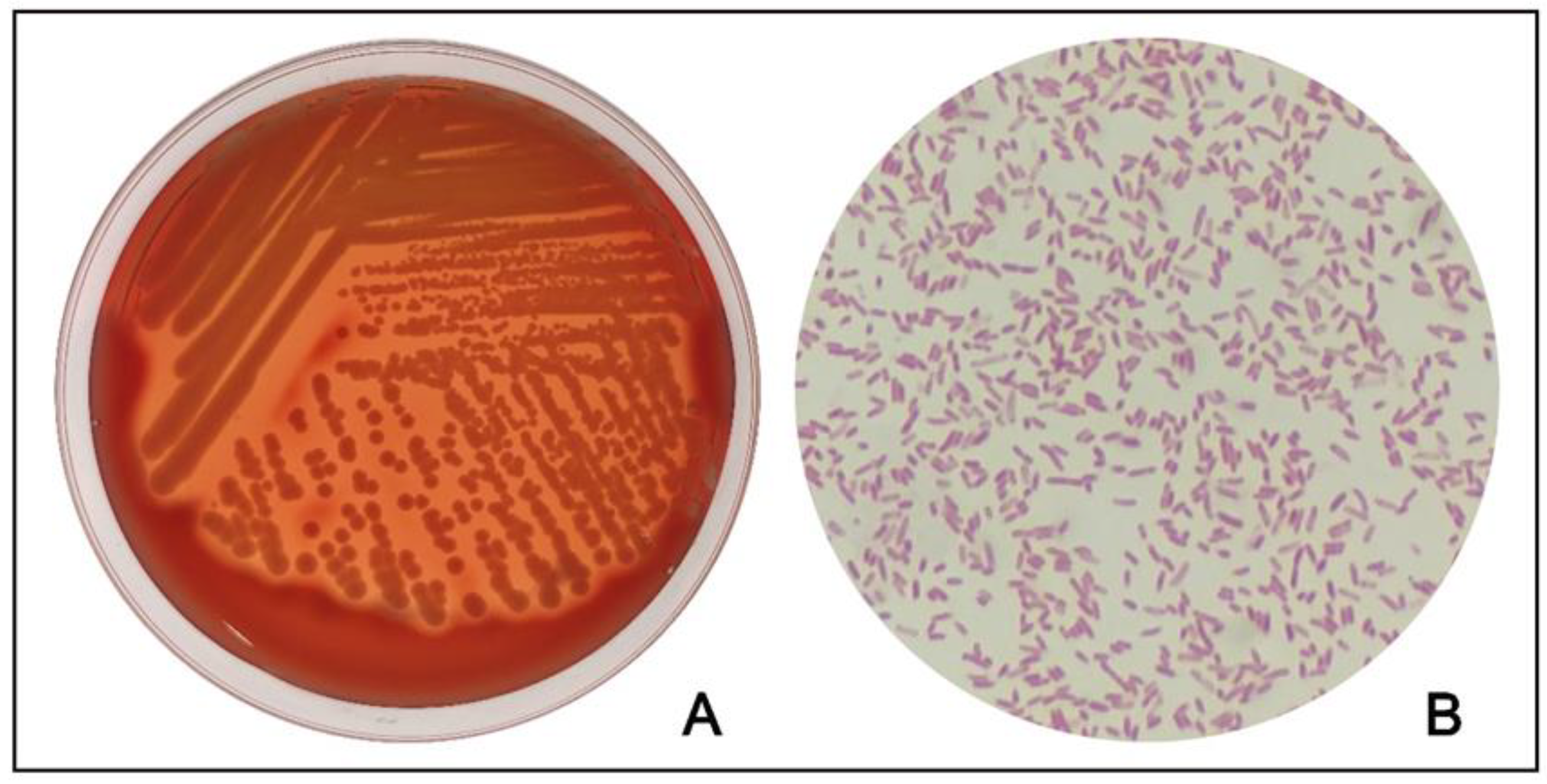
A lung sample was used for testing for common respiratory pathogens such as Mycoplasma [14], peste des petits ruminant’s virus (PPRV) [15], foot and mouth disease virus (FMDV) [15], Maedi-Visna (MV) [16], parainfluenza virus type 3 (PIV3) [17], and ovine respiratory syncytial virus (ORSV) [18] using previously reported methods (primer sequences are available in Table S2), and the results were negative. At the same time, small pieces of lungs were used for pathogenic bacterial isolation. Specifically, inoculation on nutrient agar containing 5% sheep blood was performed using a sterilized inoculation loop dipped in the fresh-cut surface of the lung. After anaerobic and aerobic incubation at 37 °C for 24 h, single-strain isolates were subjected to three rounds of single colony purification. A bacterium strain was isolated in the lung, which is a gram-negative, rod-shaped strain with β-hemolytic, shown in Figure 3.
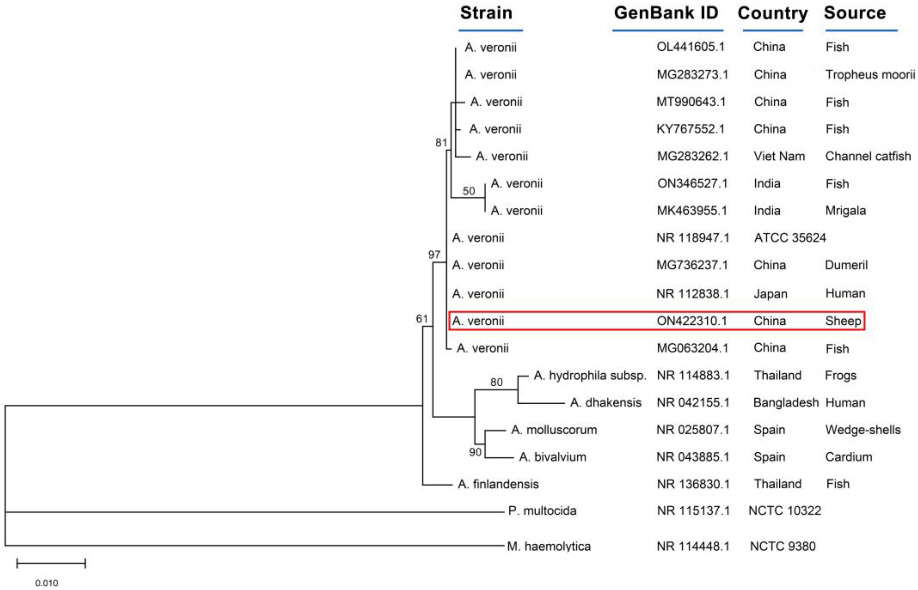
The 16S rRNA gene of the isolated strain was amplified by the universal primers, 27F:5′-AGAGTTTGATCCTGGCTCAG-3′ and 1492R: 5′-GGTTACCTTGTTACGACTT-3′. The PCR conditions consisted of 94 °C for 5 min, followed by 35 cycles of 94 °C for 30 s, 55 °C for 45 s, 72 °C for 90 s, and a final extension at 72 °C for 6 min. The obtained sequences were sequenced at Beijing Tsingke Biotechnology Co., Ltd, Xi’an, China. A BLAST search for sequences was carried out via the NCBI public database (BLAST: Basic Local Alignment Search Tool). The sequence showed 100% identity (Query cover 98%) with the strain isolated from humans (NR112838) and the standard strain ATCC35624. Moreover, the sequence shares 99.84% identity with the strain isolated from the environment (NR044845). The sequence was submitted to the database at NCBI under accession number ON442310.1. MEGA X software was used to align the sequences and generate the phylogenetic tree with the Maximum Likelihood method in Figure 4 [19]. The strain was closely related to the strains isolated from infected fish (MG063204.1), humans (NR112838.1), and dumevil (MG736237.1).
To further confirm the strain, a series of biochemical experiments were performed using commercial tubes (Hangzhou Binhe Microorganism Reagent Co., Ltd., Hangzhou, China) with three independent experiments (see Table S3). The results of peptone, Simmons citrate, catalase, lactose, D-mannose, Voges-Proskauer test, glucose (gas), indole, lysine, nitrate reduction, and glycerin tests were positive, and the results were negative for urea, esculin, sucrose, arabinose, xylose, sorbitol, H2S, phenylalanine, and ornithine, which matched the biochemical characteristics of A. veronii [20]. According to the phenotypic features, biochemical characteristics, and 16SrRNA gene phylogenetic analysis, the strain YL4077 was identified as A. veronii.
The agglutination test is widely used in the diagnosis of bacterial infections, especially those caused by Gram-negative organisms such as Aeromonas spp. [21]. A total of 14 and 187 serum samples from symptomatic and asymptomatic sheep were tested by rapid agglutination test (RAT) with antigen prepared from A. veronii YL4077. RATs were performed according to previous studies [22]. Surprisingly, the raters of symptomatic and asymptomatic positivity in sheep were 64.28% (95% CI 52.25–76.31%) and 8.02% (95% CI 6.96–9.08%), respectively, suggesting that A. veronii was one of the pathogens causing respiratory disease in a sheep farm.
Thirty-two Balb/c mice were divided into four groups, G1–G4, with 8 mice/group. The A. veronii YL4077 overnight culture was serially diluted to obtain the concentrations of 5.5 × 10⁷ CFU/mL, 5.5 × 10⁶ CFU/mL, 5.5 × 10⁵ CFU/mL, and 5.5 × 10⁴ CFU/mL for G1–G4, respectively. The mice were injected intraperitoneally with 200 μL and monitored for mortality for 7 days (see Table 1). The 50% lethal dose (LD50) of bacteria was calculated by the modified Kärber method [23]. Finally, the LD50 in Balb/c mice exposed to A. veronii YL4077 was determined to be 1.10 × 10⁶ CFU. The anatomical experiments on the dead mice showed obvious congestion in the lungs and liver, and all of the mice developed severe abscesses at the injection site. The bacteria were isolated in the lungs, liver, and blood of the dead mice again. The above data indicate that the strain has a certain pathogenicity.
To further confirm the pathogenicity of A. veronii YL4077 in sheep, 1 mL of the A. veronii YL4077 overnight culture (1.10 × 10⁹ CFU/mL) was exposed to an 8-month-old lamb through intraperitoneal injection (the sheep was provided by The Animal Experimental Center, Northwest A&F University). The body temperature was increased to 40.5 °C, accompanied by dyspnoea and bloody diarrhea 4 h post-infection, and the sheep died after 19 h. Upon postmortem examination, severe congestion and edema of the lungs and intestinal tract was observed. A. veronii YL4077 was again cultured from the blood, lungs, and liver. We concluded that pathogenic A. veronii is the cause of respiratory disease and death in sheep.
We then examined the bacterial resistance of A. veronii YL4077 by utilizing the disc diffusion assay (the disc was purchased from Hangzhou Binhe Microorganism Reagent Co., Ltd., Hangzhou, China), while Escherichia coli ATCC 25922 was used as a quality control. The results were interpreted following Clinical and Laboratory Standards Institute (CLSI) guidelines. A. veronii YL4077 was sensitive to cefotaxime, gentamicin, and enrofloxacin; intermediately sensitive to doxycycline; and resistant to ampicillin, ceftiofur, amoxicillin, kanamycin, neomycin, streptomycin, tetracycline, florfenicol, and tylosin, as shown in Table 2.
We suggest the combination of cefotaxime and gentamicin as an effective treatment based on the results of the antimicrobial susceptibility test. To our delight, the mortality rate of diseased sheep was reduced to 0.

3. Discussion

Aeromonas spp. was first isolated from pneumonia, wound infections, septicemia, and abortion in horses, cattle, and pigs in 1987 [24]. It is known as a threat to aquaculture systems and human health, causing a high death rate [3,25]. As an important member of Aeromonas, A. veronii is pathogenic to both aquatic and terrestrial animals [7,8,26]. To date, there has been no documentation of sheep infected with A. veronii. Here, we identified an A. veronii strain, YL4077, in a sheep that died from severe respiratory illnesses in Shaanxi Province, China.
In this study, the strain was resistant to ampicillin, ceftiofur, amoxicillin, kanamycin, neomycin, streptomycin, tetracycline, florfenicol, and tylosin, consistent with previous findings. It has been shown that almost 96% of Aeromonas isolates from humans and aquatic animals are resistant to the antibiotics ampicillin, ciprofloxacin, chloramphenicol, tetracycline, and cotrimoxazole. [27,28]. From a therapeutic standpoint, multi-drug-resistant A. veronii makes treatment options more limited. Pathogenicity results revealed that the strain is pathogenic to both sheep and mice. Thus, A. veronii is not only an important threat to the sheep industry, but also poses a potential threat to human health, as the infection may spread to humans via the consumption of undercooked meat contaminated with A. veronii [25].
Respiratory diseases are a widespread problem in sheep flocks [29], with presentations varying from acute deaths to chronic ill-thrift and being either sporadic or involving whole groups of animals. This could be attributed to the full roles of other pathogenic viruses such as PPRV, FMDV, MV, PIV3, and ORSV, especially in young sheep. These viruses can, however, form complex infections with other bacterial species (co-infection), such as Pasteurella spp., Mannheimia spp., and Mycoplasma spp., which could come as a secondary infection leading to acute disease outbreaks [30]. However, all the targeted pathogenic viruses and Mycoplasma test results were negative, and no other bacteria were isolated in lung samples except A. veronii. One possible cause is that the sheep had been treated with ceftiofur sodium for five consecutive days, which inhibited the proliferation of ceftiofur-sensitive bacteria. We deem that ceftiofur-resistant A. veronii infection is the leading cause of sheep death. According to the results of the RAT, 64.28% (9/14) and 7.81% (15/187) of the serums from the symptomatic and asymptomatic sheep were positive, respectively. These results further showcase A. veronii as the pathogen causing respiratory disease in sheep.
Previous studies revealed that animals exposed to a high level of ammonia experience irritation of mucous membranes and the respiratory tract [31]. Emerging evidence suggests that even low levels of gaseous ammonia have toxic effects on the respiratory tract and cause an imbalance in nasal microbiota in growing pigs and broiler chickens [32,33]. According to the sheep owner, this area experienced a sudden temperature drop in late March, and the sheepfold doors were closed to keep warm, which resulted in ammonia concentrations exceeding 31 ppm. This would cause adverse effects on the respiratory tract of sheep. Studies have shown that the immunity of animals can be reduced under cold stimulation [34]. Cold-induced stress impaired the production of Th1 protective cytokines and can reduce the level of IL-2 in the blood of mice [35,36]. Borsoi et al. [37] indicated that cold, stress-challenged birds have a decreased capacity to overcome the induced infection. Guo H et al. [38] demonstrated that cold stimulation can weaken the immunity of sheep. Furthermore, alveolar macrophages are the first line of defense against inhaled bacteria [39], and overcrowding stress decreases macrophage activity and increases bacterial infections in broiler chickens [40]. Taken together, these stress stimuli may induce A. veronii to infect the sheep and cause respiratory disease. However, the source of the A. veronii remains unclear. This intensive sheep farm did not have any trade in recent months, thereby excluding a possibility that A. veronii was transmitted via the sheep trade. Previous studies have suggested that A. veronii is widely distributed in aquatic environments and soil [5,6,7,8]. We speculate that the source of A. veronii was the potable water and soil. An important clue in Figure 1D is intestinal mucosal hemorrhage. Therefore, we boldly speculate that the sheep were infected with A. veronii through the digestive tract. Our next step will be to clarify the source of the strain and verify this conjecture.
Considered together, we make the following recommendations: First, a major emphasis should be placed on heightened, strict biosecurity rules on sheep farms, including thorough cleaning and disinfection of sheepfolds. Secondly, make sure that every sheep can drink warm, clean, and safe water, especially in the cold season. Thirdly, attention should be paid to ventilation. Finally, and most importantly, the combination of cefotaxime with gentamicin was an effective treatment for A. veronii infection. In conclusion, the strain has a certain pathogenicity that may cause serious, even life-threatening diseases in domestic animals under specific conditions. To our knowledge, this is the first case report of pathogenic A. veronii isolated from sheep in China. It is necessary to pay attention to the potential threat of A. veronii infection to the sheep industry.

Supplementary Materials

The following supporting information can be downloaded at: https://www.mdpi.com/article/10.3390/microorganisms11020333/s1, Table S1: Description of sick and dead sheep affected by this outbreak; Table S2: Primers used in this case report; Table S3: Biochemical characteristic of strain YL4077.

Author Contributions

Y.M., J.W., and Z.Y. handled the case; Y.M., Q.X., H.F., J.D., and X.B. performed the experiments; Y.M., X.Z., and F.E.A.A. wrote the manuscript. All authors have read and agreed to the published version of the manuscript.

Funding

This research was funded by the China Wool sheep Industry and Technology System project (No. CARS-39-14).

Institutional Review Board Statement

Written informed consent was obtained from the rancher for the publication of this case report. Pathogenicity test in mice and sheep were reviewed and approved by the Ethics Committee at Northwest A&F University. The ethic approval number: NWAFU2021-0910. Experiments were carried out in accordance with the approved guidelines.

Data Availability Statement

The authors confirm that all data are fully available without restriction.

Conflicts of Interest

The authors declare no conflict of interest.

References

  1. Fernández, B.A.; Figueras, M.J. An Update on the Genus Aeromonas: Taxonomy, Epidemiology, and Pathogenicity. Microorganisms. 2020, 8, 129. [Google Scholar] [CrossRef] [PubMed] [Green Version]
  2. Ku, Y.; Yu, W. Extensive community-acquired pneumonia with hemophagocytic syndrome caused by Aeromonas veronii in an immunocompetent patient. J. Microbiol. Immunol. Infect. 2017, 50, 555–556. [Google Scholar] [CrossRef] [PubMed] [Green Version]
  3. Xu, C.; Lin, Q.; Zhao, Y.; Zhu, G.; Jiang, E.; Li, S.; Mi, Y. Clinical characteristics and risk factors of Aeromonas bloodstream infections in patients with hematological diseases. BMC Infect. Dis. 2022, 22, 303. [Google Scholar] [CrossRef] [PubMed]
  4. Monti, M.; Torri, A.; Amadori, E.; Rossi, A.; Bartolini, G.; Casadei, C. Aeromonas veronii biovar veronii and sepsis-infrequent complication of biliary drainage placement: A case report. World. J. Clin. Cases 2019, 7, 759–764. [Google Scholar] [CrossRef]
  5. Raj, N.S.; Swaminathan, T.R.; Dharmaratnam, D.A. Aeromonas veronii caused bilateral exophthalmia and mass mortality in cultured Nile tilapia, Oreochromis niloticus in India. Aquaculture 2019, 51, 734278. [Google Scholar] [CrossRef]
  6. Wang, B.; Mao, C.; Feng, J.; Li, Y.; Hu, J.; Jiang, B. A First Report of Aeromonas veronii Infection of the Sea Bass, Lateolabrax maculatus in China. Front Vet. Sci. 2021, 7, 600587. [Google Scholar] [CrossRef]
  7. Su, X.; Zhong, Y.; Wang, X.; Qin, T.; Chen, S.J.; Peng, D.X. Isolation and identification of Aeromonas veronii from duck and preliminary study on its pathogenicity. Chin. J Pre-Vet. Med. 2020, 42, 1008–1589. [Google Scholar]
  8. LI, W.J.; Zhao, Y.; Liu, Y.; Qi, X.X.; Kang, K.; Chen, M. Isolation and identification of pathogenic Aeromonas veronii from fox. Chin. J Pre-Vet. Med. 2012, 34, 290–292. [Google Scholar]
  9. Getnet, K.; Abera, B.; Getie, H.; Molla, W.; Mekonnen, S.A.; Megistu, B.A.; Abat, A.S.; Dejene, H.; Birhan, M. Serotyping and Seroprevalence of Mannheimia haemolytica, Pasteurella multocida, and Bibersteinia trehalose and Assessment of Determinants of Ovine Pasteurellosis in West Amhara Sub-region, Ethiopia. Front. Vet. Sci. 2022, 9, 866206. [Google Scholar] [CrossRef]
  10. Bell, S. Respiratory disease in sheep: Differential diagnosis and epidemiology. In. Pract. 2008, 30, 200–207. [Google Scholar] [CrossRef] [Green Version]
  11. Besser, T.E.; Frances, C.E.; Highland, M.A. Bighorn sheep pneumonia: Sorting out the cause of a polymicrobial disease. Prev. Vet. Med. 2013, 108, 85–93. [Google Scholar] [CrossRef] [PubMed]
  12. Shi, S.Y.; Lu, S.Y.; Si, T. DJ-1 links muscle ROS production with metabolic reprogramming and systemic energy homeostasis in mice. Nat Commun. 2015, 6, 7415. [Google Scholar] [CrossRef]
  13. Calvi, L.M.; Bromberg, O.; Rhee, Y. Osteoblastic expansion induced by parathyroid hormone receptor signaling in murine osteocytes is not sufficient to increase hematopoietic stem cells. Blood. 2012, 119, 2489–2499. [Google Scholar] [CrossRef] [PubMed]
  14. McAuliffe, L.; Hatchell, F.M.; Ayling, R.D.; King, A.I.; Nicholas, R.A. Detection of Mycoplasma ovipneumoniae in Pasteurella-vaccinated sheep flocks with respiratory disease in England. Vet. Rec. 2003, 153, 687–688. [Google Scholar] [CrossRef]
  15. He, Y.P.; Zhang, Q.; Fu, M.Z.; Xu, X.G. Development of multiplex PCR for simultaneous detection and differentiation of six DNA and RNA viruses from clinical samples of sheep and goats. J. Virol. Methods 2017, 243, 44–49. [Google Scholar] [CrossRef]
  16. Singh, R.; Kumar, P.; Singh, R. Pathology and polymerase chain reaction detection of ovine progressive pneumonia (maedi) cases in slaughtered sheep in India. Vet. World 2017, 10, 1401–1406. [Google Scholar] [CrossRef] [PubMed] [Green Version]
  17. Li, W.; Mao, L.; Cheng, S.; Wang, Q.; Huang, J.; Deng, J.; Wang, Z.; Zhang, W.; Yang, L.; Hao, F.; et al. A novel parainfluenza virus type 3 (PIV3) identified from goat herds with respiratory diseases in eastern China. Vet. Microbiol. 2014, 174, 100–106. [Google Scholar] [CrossRef]
  18. Eleraky, N.Z.; Kania, S.A.; Potgieter, L.N. The ovine respiratory syncytial virus F gene sequence and its diagnostic application. J. Vet. Diagn. Invest. 2001, 13, 455–461. [Google Scholar] [CrossRef] [Green Version]
  19. Kumar, S.; Stecher, G.; Li, M.; Knyaz, C.; Tamura, K. MEGA X: Molecular Evolutionary Genetics Analysis across Computing Platforms. Mol. Biol. Evol. 2018, 35, 1547–1549. [Google Scholar] [CrossRef]
  20. Holt, J.H.; Krieg, N.R.; Sneath, P.H. Bergey’s Manual of Determinative Bacteriology, 9th ed.; Williams & Wilkins: Baltimore, MD, USA, 1994. [Google Scholar]
  21. Leblanc, D.; Mittal, K.R.; Olivier, G.; Lallier, R. Serogrouping of motile Aeromonas species isolated from healthy and moribund fish. Appl. Environ. Microbiol. 1987, 42, 56–60. [Google Scholar] [CrossRef] [Green Version]
  22. Lhan, Z.; Gülhan, T.; Aksakal, A. Aeromonas hydrophila associated with ovine abortion. Small Rumin. Res. 2006, 61, 73–78. [Google Scholar]
  23. Xia, Y.; Li, H.; Shen, Y. Antimicrobial Drug Resistance in Salmonella enteritidis Isolated from Edible Snakes with Pneumonia and Its Pathogenicity in Chickens. Front Vet Sci. 2020, 7, 463. [Google Scholar] [CrossRef] [PubMed]
  24. Lallier, R.; Higgins, R. Biochemical and toxigenic characteristics of Aeromonas spp. isolated from diseased mammals, moribund and healthy fish. Vet. Microbiol. 1988, 18, 63–71. [Google Scholar] [CrossRef] [PubMed]
  25. Pessoa, R.B.; Oliveira, W.F.; Correia, M.; Fontes, A.; Coelho, L. Aeromonas and Human Health Disorders: Clinical Approaches. Front. Microbiol. 2022, 13, 868–890. [Google Scholar] [CrossRef] [PubMed]
  26. Sun, J.; Zhang, X.; Gao, X.; Jiang, Q.; Wen, Y.; Lin, L. Characterization of Virulence Properties of Aeromonas veronii Isolated from Diseased Gibel Carp (Carassius gibelio). Int. J. Mol. Sci. 2016, 17, 496. [Google Scholar] [CrossRef]
  27. Skwor, T.A.; Shinko, J.; Augustyniak, A.; Gee, C.; Andraso, G. Aeromonas hydrophila and Aeromonas veronii Predominate among Potentially Pathogenic Ciprofloxacin- and Tetracycline-Resistant Aeromonas Isolates from Lake Erie. Appl. Environ. Microbiol. 2014, 80, 841–848. [Google Scholar] [CrossRef] [Green Version]
  28. Vila, J.; Gallardo, F.; Vargas, M.; Solerk, L.; Figueras, M.J.; Gascon, J. Aeromonas spp. and traveler’s diarrhea: Clinical features and antimicrobial resistance. Emerg. Infect. Dis. 2003, 9, 552–555. [Google Scholar] [CrossRef]
  29. Kumar, A.; Tikoo, S.K.; Malik, P.; Kumar, A.T. Respiratory diseases of small ruminants. Vet. Med. Int. 2014, 2014, 373642. [Google Scholar] [CrossRef]
  30. Brogden, K.A.; Lehmkuhl, H.D.; Cutlip, R.C. Pasteurella haemolytica complicated respiratory infections in sheep and goats. Vet. Res. 1998, 29, 233–254. [Google Scholar]
  31. Kearney, G.D.; Shaw, R.; Prentice, M.; Tutor-Marcom, R. Evaluation of respiratory symptoms and respiratory protection behavior among poultry workers in small farming operations. J. Agromed. 2014, 19, 162–170. [Google Scholar] [CrossRef]
  32. Wang, T.; He, Q.; Yao, W.; Shao, Y.; Li, J.; Huang, F. The Variation of Nasal Microbiota Caused by Low Levels of Gaseous Ammonia Exposure in Growing Pigs. Front. Microbiol. 2019, 10, 1083. [Google Scholar] [CrossRef] [PubMed]
  33. Zhou, Y.; Zhang, M.; Liu, Q.; Feng, J. The alterations of tracheal microbiota and inflammation caused by different levels of ammonia exposure in broiler chickens. Poult. Sci. 2021, 100, 685–696. [Google Scholar] [CrossRef] [PubMed]
  34. Lavoy, E.C.P.; McFarlin, B.K.; Simpson, R.J. Immune Responses to Exercising in a Cold Environment. Wild. Environ. Med. 2011, 22, 343–351. [Google Scholar]
  35. Belay, T. Cold-induced stress impairs the production of Th1 protective cytokines during co-culturing of dendritic cells and naive T helper cells of Chlamydia muridarum infected mice. J. Immunol. 2018, 200, 117–135. [Google Scholar] [CrossRef]
  36. Makarova, O.V.; Trunova, G.V.; Diatroptov, M.E.; Serebryakov, S.N.; Kondashevskaya, M.V.; Malaitsev, V.V. Comparative characterization of cytokine production by concanavalin A-activated splenocytes from BALB/c and C57BL/6 mice after cold exposure. Bull. Exp. Biol. Med. 2005, 139, 220–222. [Google Scholar] [CrossRef]
  37. Borsoi, A.; Quinteiro-Filho, W.M.; Calefi, A.S. Effects of cold stress and Salmonella Heidelberg infection on bacterial load and immunity of chickens. Avian. Pathol. 2015, 44, 490–497. [Google Scholar] [CrossRef] [PubMed] [Green Version]
  38. Guo, H.; Zhou, G.; Tian, G. Changes in Rumen Microbiota Affect Metabolites, Immune Responses and Antioxidant Enzyme Activities of Sheep under Cold Stimulation. Animals 2021, 11, 712. [Google Scholar] [CrossRef]
  39. Goldstein, E.; Lippert, W.; Warshauer, D. Pulmonary alveolar macrophage. Defender against bacterial infection of the lung. J. Clin. Investig. 1974, 54, 519–528. [Google Scholar] [CrossRef] [Green Version]
  40. Gomes, A.V.; Quinteiro-Filho, W.M.; Ribeiro, A. Overcrowding stress decreases macrophage activity and increases Salmonella Enteritidis invasion in broiler chickens. Avian. Pathol. 2014, 43, 82–90. [Google Scholar] [CrossRef]
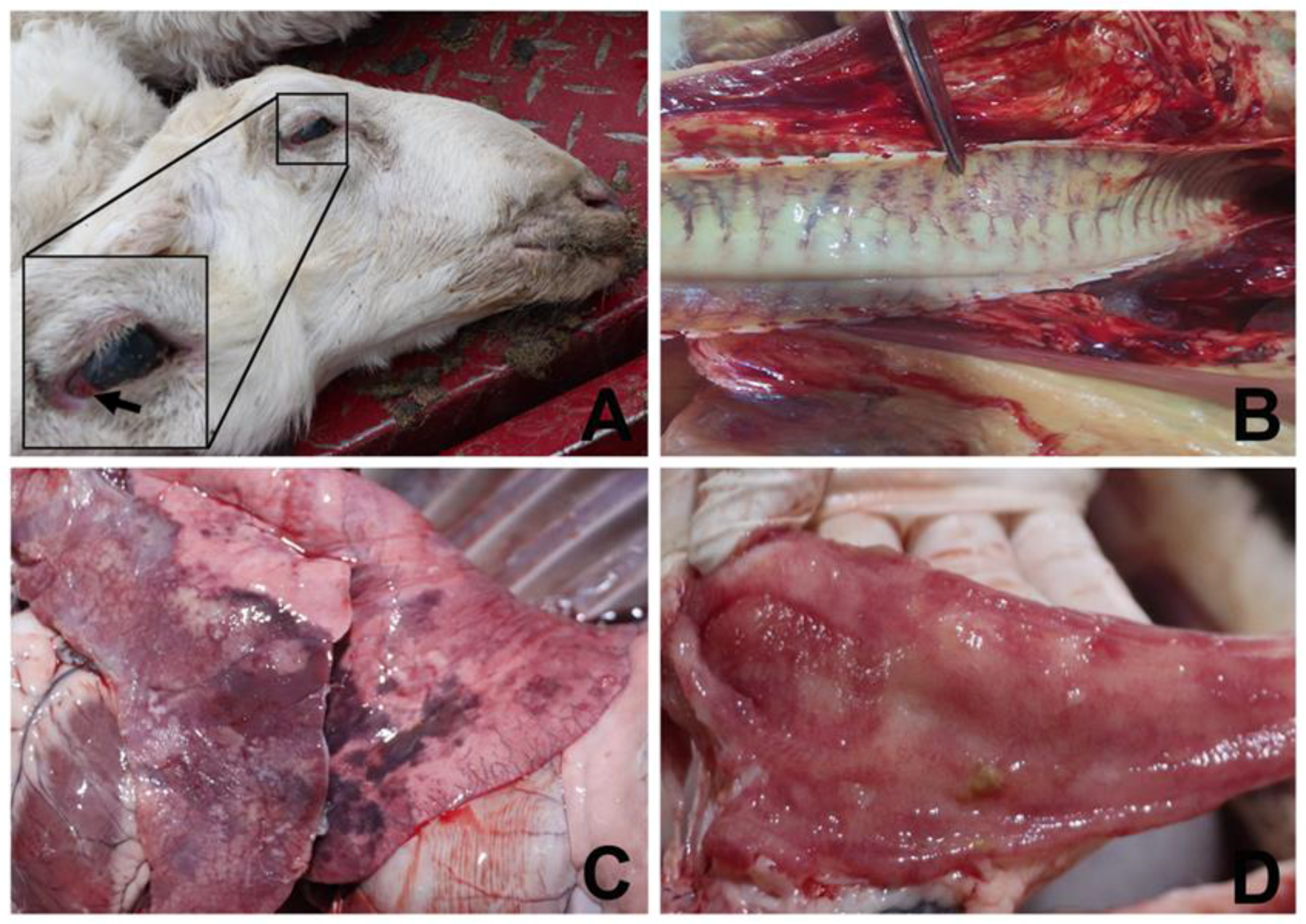
Figure 1. Macroscopic appearance of dead sheep, trachea, lung, and mesentery. (A) Congestion of the conjunctiva and nasal discharge were observed in dead sheep; (B) Congestive tracheal ring; (C) Pulmonary consolidation and congestion; (D) Intestinal mucosal hemorrhage.
Microorganisms 11 00333 g001
Figure 2. HE-stained pathological sections of different parts of the lungs from the sheep. (A) Infiltrating lymphocytes (black arrow): the alveolar space of the lung tissue was thickened, and the interstitial lung was congested (black triangle) (bar = 200 µm, 200×); (B) fibrinous exudation (black arrow): vacuoles were visible in the alveolar cells (black triangle) (bar = 200 µm, 200×).
Microorganisms 11 00333 g002
Figure 3. This bacterium is rod-shaped and Gram-negative and shows β-hemolytic activity on ovine blood agar plates. (A) This bacterium was incubated on the ovine blood agar plates overnight at 37 °C; (B) gram staining showed the bacterium is rod-shaped and Gram-negative.
Microorganisms 11 00333 g003
Figure 4. Phylogenetic tree of 16SrRNA in A. veronii YL4077 isolated from dead sheep in China. A red line indicates the isolate from this study. The numbers in each node are the confidence values. The distance bar is the number of nucleotide substitutions per site.
Microorganisms 11 00333 g004
Table 1. Experimental data on the pathogenicity of A. veronii YL4077 in mice.
GroupDoseNumbers (n)Death Number (n)Mortality (P)P2
CFU/MouseLog
G11.1 × 1077.048811
G21.1 × 1066.04860.750.5625
G31.1 × 1055.04820.250.0625
G41.1 × 1044.048000
lgLD50 = X − d(∑Pi − 0.5);
Standard error SlogLD50 = d√((∑P − ∑P2)/(n − 1))
∑P = 2.00∑P2 = 1.625
“X” means the maximum logarithmic dose, “d” means the difference in logarithmic dose between two adjacent groups, “Pi” means the mortality rate, and “i” means the group number.
Table 2. Results of the drug susceptibility test.
DrugsDiameter
(mm)
SensitivityJudging Criteria (mm)Dose
(ug/disc)
RIS
Amp0R≤1819–21≥2210
Cef13R≤1415–22≥2330
Cet17S≤1415–22≥2330
Amo0R≤1314–17≥1810
Kan12R≤1314–17≥1830
Gen19S≤14->1410
Neo10R≤1213–16≥1730
Str9R≤1112–14≥1510
Tet13R≤1415–18≥1930
Dox13I≤1213–15≥1630
Flo12R≤1213–17≥1830
Enr23S≤1213–16≥1710
Tyl13R≤1414–15≥1630
Note: Amp Ampicillin; Cef, Ceftiofur; Cet, Cefotaxime; Amo, Amoxicillin; Kan, Kanamycin; Gen, Gentamicin; Neo, Neomycin; Str, Streptomycin; Tet, Tetracycline; Dox, Doxycycline; Flo, Florfenicol; Enr, Enrofloxacin; Tyl, Tylosin; R, Resistance; I, Intermediate; S Sensitive.
Disclaimer/Publisher’s Note: The statements, opinions and data contained in all publications are solely those of the individual author(s) and contributor(s) and not of MDPI and/or the editor(s). MDPI and/or the editor(s) disclaim responsibility for any injury to people or property resulting from any ideas, methods, instructions or products referred to in the content.