Article

Role of Probiotics in Preventing Carbapenem-Resistant Enterobacteriaceae Colonization in the Intensive Care Unit: Risk Factors and Microbiome Analysis Study

1
Division of Gastroenterology, Department of Internal Medicine, Inha University Hospital, Inha University School of Medicine, Incheon 22332, Republic of Korea
2
Department of Hospital Medicine, Inha University Hospital, Inha University School of Medicine, Incheon 22332, Republic of Korea
3
Department of Neurology, Kangdong Sacred Heart Hospital, Hallym University College of Medicine, Seoul 05355, Republic of Korea
*
Authors to whom correspondence should be addressed.
These authors contributed equally to this work.
Microorganisms 2023, 11(12), 2970; https://doi.org/10.3390/microorganisms11122970
Submission received: 18 October 2023 / Revised: 5 December 2023 / Accepted: 6 December 2023 / Published: 12 December 2023

Abstract

:
Older patients with multiple comorbidities often necessitate prolonged hospital stays and antibiotic treatment in the intensive care unit (ICU), leading to a rise in multidrug-resistant organisms like carbapenem-resistant Enterobacteriaceae (CRE). This study examined risk factors for carbapenem-resistant Enterobacteriaceae colonization in the ICU and assessed probiotics’ preventive role. In this single-center, retrospective study, 9099 ICU patients were tested for stool CRE culture from March 2017 to April 2022. We excluded 136 patients with CRE colonization within one week post-admission and 26 who received probiotics before CRE colonization. Ultimately, 8937 CRE-negative patients were selected. Logistic analysis identified CRE colonization risk factors and evaluated probiotics’ influence, including Saccharomyces boulardii or Lactobacillus rhamnosus, used by 474 patients (5.3%) in the ICU. Compared with data on initial admission, 157 patients (1.7%) had newly discovered CRE colonization before discharge. In a multivariate analysis, coronavirus disease 2019, the ICU, tube feeding, antibiotics such as aminoglycoside, extended-spectrum penicillin, stool vancomycin-resistance enterococci colonization, and chronic kidney disease were significantly associated with de novo CRE infection. However, probiotic use was negatively correlated with CRE infection. Managing risk factors and administering probiotics in the ICU may help prevent CRE colonization; large randomized prospective studies are needed.

1. Introduction

Carbapenem-resistant Enterobacteriaceae (CRE) are antibiotic-resistant bacteria that are increasingly common in healthcare settings. According to the Centers for Disease Control and Prevention, the incidence of CRE infections in the USA has increased in recent years. The percentage of Enterobacteriaceae isolates resistant to carbapenems increased from 1.2% in 2001 to 4.2% in 2017 [1]. Furthermore, patients in the intensive care unit (ICU) have a higher risk of developing healthcare-associated infections owing to a variety of factors, such as underlying illnesses, use of antibiotics, and prolonged hospital stays. In Korea, the incidence of CRE colonization in ICUs varied, with a carriage rate upon admission ranging from 2.8% to 6.3% [2]. A systematic review indicates an approximate 16.5% risk of infection with CRE among patients colonized with CRE, accompanied by a high mortality rate associated with CRE infections [3]. Therefore, preventing or managing CRE in the ICU is significantly associated with patient safety.
CRE colonization in feces is difficult to treat and can cause severe infections. Notably, fecal microbiota transplantation (FMT) was hypothesized to be a possible treatment option for CRE decolonization [4,5,6]. These previous studies, including our published FMT study, have shown promising results in using FMT for CRE decolonization, with others reporting a successful eradication of CRE from the gastrointestinal tract in a significant proportion of treated patients. However, FMT use for CRE decolonization remains an investigational treatment and has not yet been approved for routine clinical use. Additionally, FMT is a complex medical procedure that should only be performed by qualified healthcare professionals in appropriate clinical settings.
A potential strategy to prevent the spread of CRE is using probiotics, which are live microorganisms that confer health benefits to a host when administered in sufficient quantities; specifically, they are believed to promote gut health by balancing the microbiota and preventing the overgrowth of harmful bacteria [7]. The use of probiotics to prevent CRE fecal colonization has been the subject of several studies; however, most have not demonstrated a significant effect [8]. Nonetheless, these studies had small sample sizes and lacked long-term follow-ups and microbiome analyses. Therefore, this study aimed to analyze the risk factors for CRE fecal colonization in the ICU and to investigate the effectiveness of probiotics in preventing colonization while exploring the potential mechanisms of the action of probiotics in preventing multi-drug-resistant organism (MDRO) colonization.

2. Materials and Methods

We performed a retrospective analysis of 12,355 patients initially admitted to the ICU of Inha University Hospital between March 2017 and April 2022 (Figure 1). Specifically, we included patients who had undergone a stool CRE culture within one week after admission and those who had undergone an initial stool CRE-negative test admission. Therefore, we selected 9099 patients who underwent a stool CRE culture during admission. A total of 136 and 26 patients with CRE colonization who underwent a stool CRE test 1 week after admission and a stool CRE test before probiotic administration, respectively, were excluded. Finally, 8937 patients with a negative stool CRE test within one week after admission were enrolled. This study was reviewed and approved by the ethics committee of Inha University Hospital (approval No. IUH 2023-07-023). Patients were not required to provide informed consent because this retrospective analysis used anonymous clinical and microbiologic data that were obtained after each patient agreed to treatment by written consent.
Saccharomyces boulardii and Lactobacillus rhamnosus were selected as probiotics because they have been shown to decolonize MDRO [7,9]. Furthermore, Inha University Hospital has operated a CRE screening program since 2017; therefore, all patients admitted to the ICU have been screened from admission to discharge every 15 days.
The change in stool CRE culture was defined as follows: (1) negative conversion, a switch from positive to two subsequent negative stool cultures until the last follow-up; (2) no change, the persistence of either a positive or negative test result; and (3) new onset CRE, a transition from negative to positive stool culture during admission. The following risk factors affecting new CRE colonization were considered: age, sex, ICU category, length of stay, tube feeding, probiotics administration and duration, proton pump inhibitor administration and duration, carbapenem administration and duration, antibiotics other than carbapenem, and comorbidity.
Further, we selected patients with CRE colonization with or without probiotics for microbiome analysis using 16s RNA metagenomics sequencing. Notably, these samples were obtained from another MDRO study (KCT0004423). Moreover, fecal samples from 20 patients with CRE colonization were divided into the probiotic (12) and non-probiotic (8) groups. We conducted this study to investigate the effect of FMT on decolonizing CRE or vancomycin-resistant enterococci (VRE) [4]. From these samples, we selected the metagenomics data of 20 pre-FMT CRE stool samples. We analyzed α-diversity using two distinct methods: the species richness (CHAO1) and the diversity index (Shannon). The β-diversity analysis for both groups was executed using unweighted UniFrac and weighted UniFrac. Lastly, statistical analysis to determine the differences in β-diversities was conducted using the PERMANOVA method. This approach characterized the microbiome, including α- and β-diversities, and the relative abundance of specific taxonomic units associated with CRE, such as the Enterobacteriaceae family and the genera Klebsiella, Escherichia, Citrobacter, and Enterobacter.

2.1. Stool Bacterial Analysis for Metagenomics Analysis

A 16S rRNA amplicon sequencing with v3–4 primers was performed using a MiSeq sequencer on an Illumina platform (Macrogen Inc., Seoul, Republic of Korea) according to the manufacturer’s specifications. The original library and single long reads were obtained by assembling the paired-end reads created via sequencing in both directions, and FLASH (version 1.2.11) was used for this process [10]. Data containing sequence errors were removed for precise operational taxonomic unit (OTU) analysis. Reads containing ambiguous bases and chimeric sequences were removed because of the implied sequencing errors.
Among the assembled reads, those shorter than 400 or longer than 500 bp were excluded. Next, clustering was performed based on sequence similarity. The OTUs of the remaining reads were created using a cluster cutoff value of 97% in the CD-HIT-OTU program [10]. Moreover, data containing sequence errors were removed for precise OTU analysis and reads containing ambiguous bases and chimeric sequences were removed because of the implied sequencing errors.
Next, clustering was performed based on sequence similarity. The OTUs of the remaining reads were created using a cluster cutoff value of 97% in the CD-HIT-OTU program [11].
QIIME 2 and EzBioClould (CJ Bioscience Inc, Seoul, Republic of Korea; https://www.ezbiocloud.net/contents/16smtp (accessed on 23 August 2023) were used for OTU analysis and taxonomy [12,13]. The major sequence of each OTU was identified, and high-quality sequence reads were assigned to the “species group” at 97% sequence similarity using the PKSSU4.0 database.
To confirm the diversity and evenness of the microbial community, the Shannon and Chao1 indices were calculated. β-diversity (diversity among samples within a group) was calculated based on the unweighted UniFrac distance and UniFrac. The genetic relationships among samples were visualized using principal coordinate analysis (PCoA).

2.2. Statistical Analysis

Using logistic analysis, we identified risk factors for CRE colonization and assessed the relationship between probiotics and CRE. A multivariate logistic regression with group assignment as a predictor variable and decolonization as the outcome variable was performed to calculate the odds ratios (OR) and 95% confidence intervals (CI). Fisher’s exact test was used to analyze categorical variables. Furthermore, differences in microbiota between the groups were analyzed using the Wilcoxon rank-sum test for two independent samples or the Wilcoxon signed-rank test for two related or matched samples. Data were analyzed using the R software (R Foundation, Vienna, Austria; https://www.r-project.org (accessed on 22 August 2023) or SPSS version 26 software (IBM Corp., Armonk, NY, USA). Finally, a permutational multivariate analysis of variance (PERMANOVA) was used to determine the significance of the PCoA plot results. Statistical significance was set at p < 0.05.

3. Results

The baseline characteristics of the enrolled patients are shown in Table 1. The patients were divided into two groups: CRE colonization and non-CRE colonization. A total of 157 patients (1.7%) showed new CRE colonization during admission. Age, ICU category, total length of stay, VRE colonization, Clostridium difficile infection, proton pump inhibitor usage and duration, carbapenem usage and duration, and tube feeding proportion significantly differed between the two groups.
Among antibiotics, the frequencies of aminoglycosides, extended-spectrum penicillin, glycopeptides, second- and third-generation cephalosporins, quinolone, metronidazole, aztreonam, colistimate, tigecylcline, trimethoprim–sulfamethoxazole, azoles, and antibiotics duration were significantly different between the groups. Acute myocardial infarction and chronic kidney disease were also independent risk factors for new CRE colonization. Analyzing the genus composition of CRE colonization, the Klebsiella genus (120, 76.3%) constituted the largest portion of CRE.

3.1. Multivariable Analysis of Risk Factors Affecting New CRE Colonization

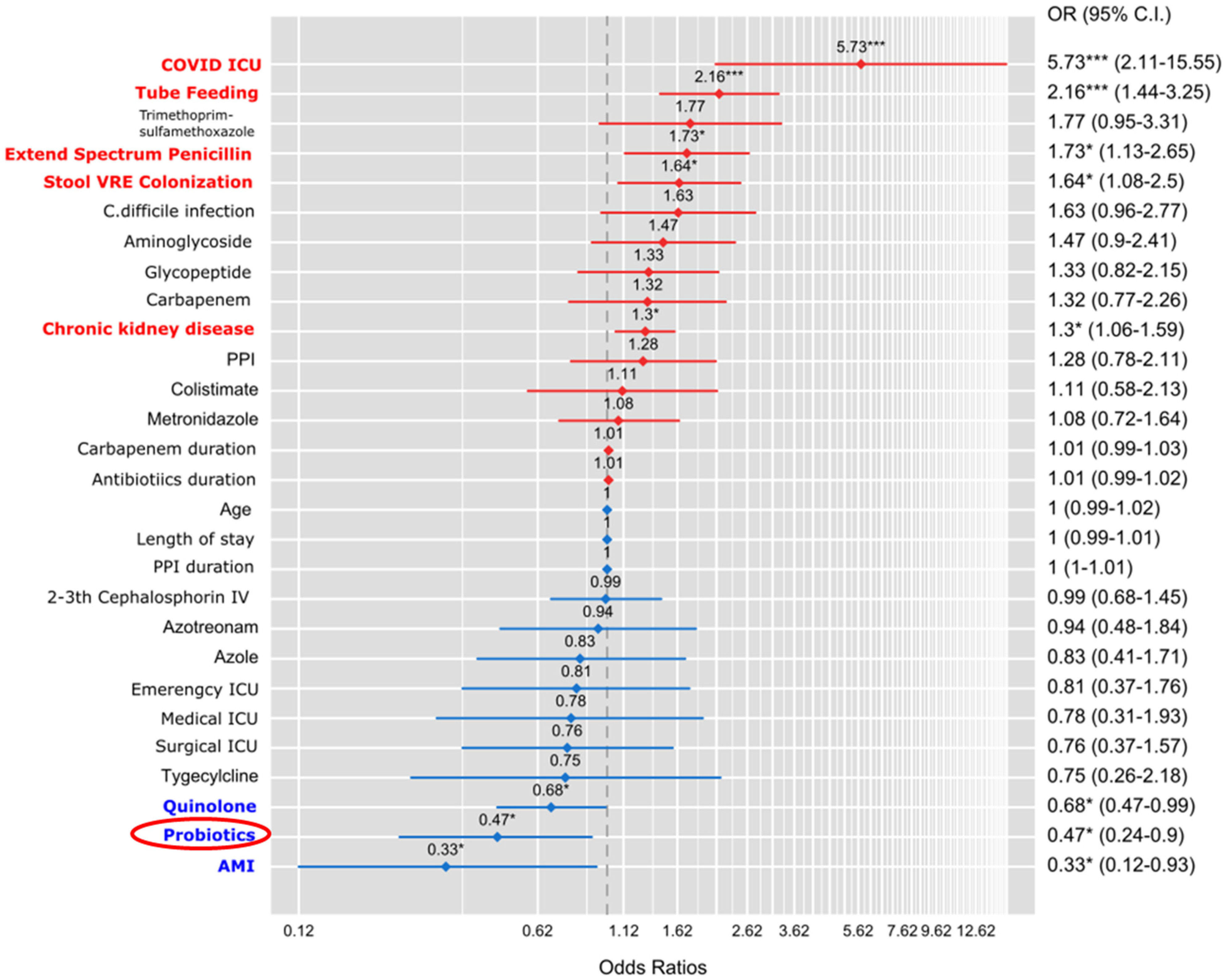
We determined the risk factors for new CRE colonization (Figure 2). In multivariate analysis, coronavirus disease 2019 (COVID-19) ICU (OR, 5.73; 95% CI, 2.11–15.55), tube feeding (OR, 2.16; 95% Cl: 1.44–3.25), extend spectrum penicillin (OR, 1.73; 95% Cl: 1.13–2.65), stool VRE colonization (OR, 1.64; 95% Cl: 1.08–2.5), and chronic kidney disease (OR, 1.3; 95% Cl: 1.06–1.59) were positively correlated with new CRE colonization. Moreover, quinolone (OR, 0.68; 95% Cl: 0.47–0.99), acute myocardial infarction (OR, 0.33; 95% Cl: 0.12–0.93), and probiotics administration (OR, 0.47; 95% Cl: 0.24–0.9) significantly and negatively correlated with new CRE colonization.

3.2. Analysis of the Microbiome’s Response to Probiotics in Patients with CRE Colonization

We analyzed the family and genus composition of CRE-colonized stool according to probiotics administration (Supplementary Figure S1). The abundance of the Enterobacteriaceae family, to which CRE belongs, was lower in the probiotics group (3.43%) than in the non-probiotics group (20.85%). In contrast to the CRE genus composition, the abundances of the Klebsiella genus (<1 vs. 6.15%), Escherichia (2.51 vs. 1.76%), and Citrobacter (<1 vs. 4.04%) were different between the probiotics group and the non-probiotics group.
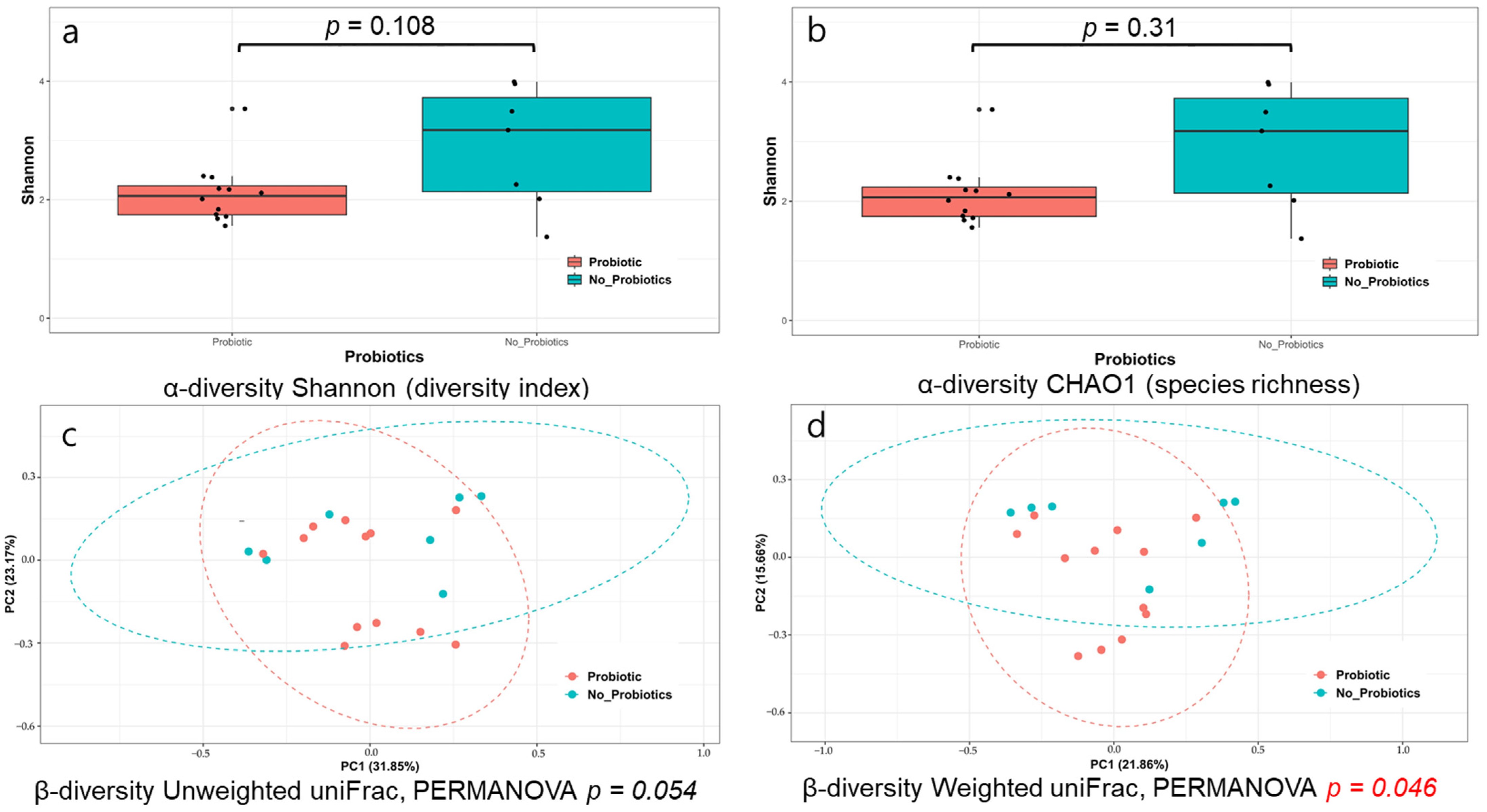
All α-diversities demonstrated using the Shannon and CHAO1 methods showed no significant differences between the probiotic and non-probiotic groups (p = 0.108 and p = 0.31, respectively) (Figure 3a,b). The β-diversity shown using unweighted UniFrac and weighted UniFrac was marginally different between the two groups (p = 0.054 and p = 0.046, PERMANOVA, respectively) (Figure 3c,d).
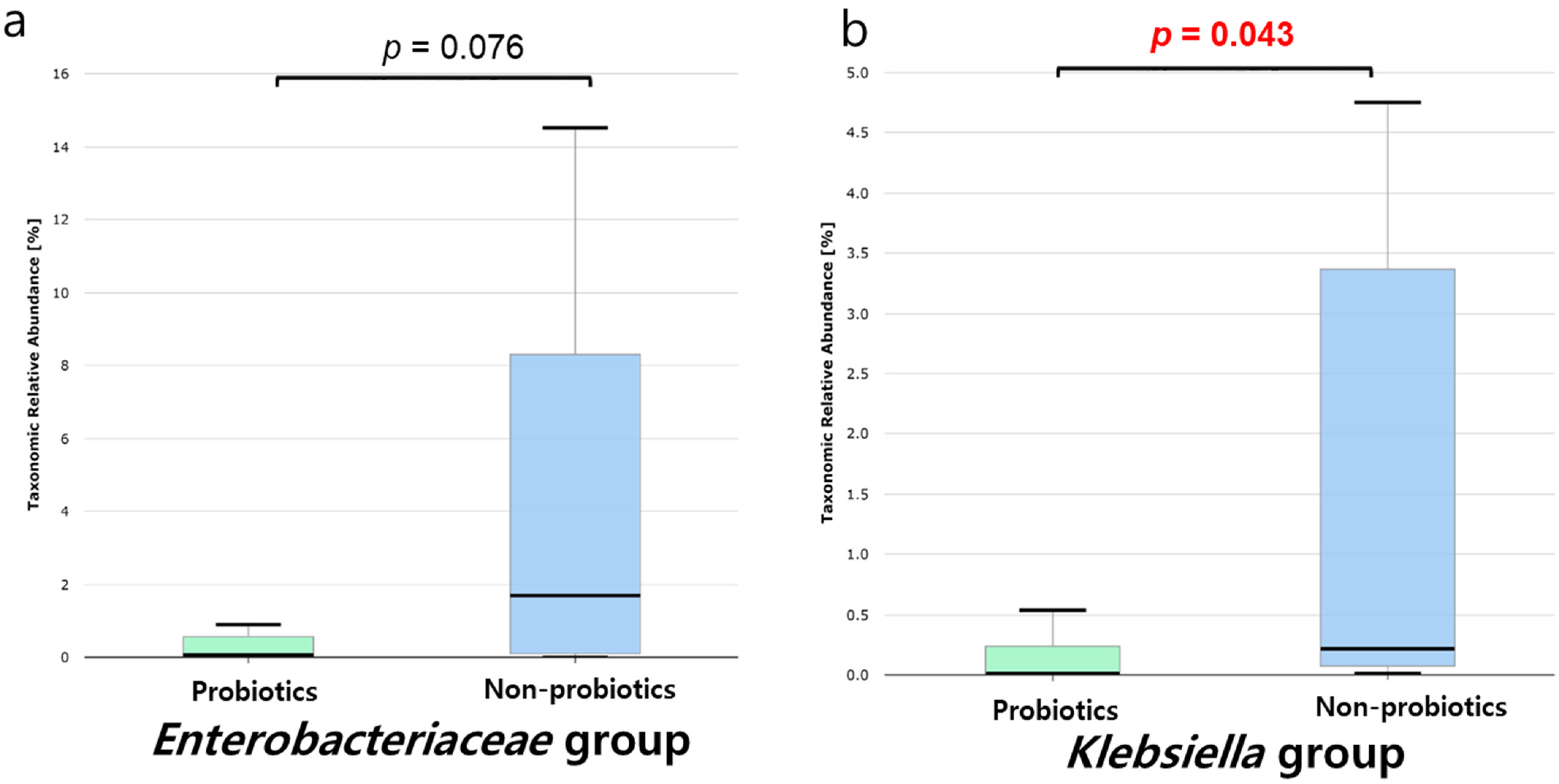
Further, we analyzed the relative abundances of both groups, including the Enterobacteriaceae family (Figure 4a) and the Klebsiella genus (Figure 4b), which are the main taxa of CRE. The relative abundances of the Enterobacteriaceae family in the probiotics group were lower than those in the non-probiotics group (p = 0.076). Similarly, the relative abundance of the Klebsiella genus in the probiotics group was significantly lower than that in the non-probiotics group (p = 0.043). However, the relative abundances of the Escherichia, Citrobacter, and Enterobacter genera were not significantly different (p = 0.673, p = 0.316, and p = 0.190, respectively) (Supplementary Figure S3).

4. Discussion

To our knowledge, this is the first study to demonstrate that probiotics had a preventive effect on CRE colonization using microbiome analysis in ICU patients. After adjusting for other risk factors, COVID-19 ICU, tube feeding, antibiotics such as aminoglycoside, extended-spectrum penicillin, glycopeptide, chronic kidney disease, and length of stay were significantly associated with de novo CRE infection in multivariate analysis. However, probiotics were negatively correlated with new CRE colonization. Moreover, we analyzed the microbiome associated with CRE abundance. Although we cannot directly demonstrate a decrease in the amount of CRE, the relative abundance of the Enterobateriaceae family in patients who received probiotics showed a reduction in the number of these taxa. In particular, the abundance of the Klebsiella genus, the main genus in CRE, significantly decreased. Therefore, probiotic administration may prevent new CRE colonization.
CRE colonization in the ICU is a significant concern due to the potential for subsequent infections and limited treatment options. Particularly, in previous studies, several risk factors contributed to the colonization of CRE in ICU patients, such as antibiotics exposure, hospitalization, ICU stay, indwelling medical devices, and transfer from other healthcare facilities [14,15], consistent with our results. Our study proved COVID-19 ICU to be the most significant risk factor for CRE colonization. Prolonged hospitalization, broad-spectrum antibiotics, immunosuppressed status, shared environments, and close proximity were known as CRE-acquisition risk factors [16,17]. Longer lengths of ICU stay, carbapenem treatment, corticosteroid treatment, and invasive mechanical ventilation were frequently applied to COVID-19 ICU patients, increasing their susceptibility to CRE acquisition [17]. Additionally, elderly patients admitted to the COVID ICU were likely to be initially undetectable CRE carriers from long-term care facilities in the Korean medical environment [18]. Infection prevention management in the COVID ICU was also challenging due to the wearing of protective clothing and the risk of COVID-19 infection. Therefore, we should be more careful about CRE colonization in COVID-19 patients in the ICU. Various well-established prevention strategies can be implemented, such as hand hygiene, contact precautions, device control, environmental cleaning, and the early detection of CRE carriers using active surveillance methods [2]. In our hospital, UV light decontamination was routinely performed in the COVID-19 ICU to prevent the spread of CRE [19]. Additionally, stool VRE colonization has been identified as a risk factor for CRE colonization in ICU patients, as demonstrated in our current study [20]. Patients with acute myocardial infarction were admitted to isolated rooms in the cardiovascular ICU during the period following the intervention, thus potentially reducing their risk of CRE colonization.
Probiotics exert their effects by producing short-chain fatty acids from metabolic precursors, leading to similar downstream effects, such as immune modulation and increased mucosal barrier function [18]. Moreover, they may have the added effect of producing their own antimicrobial compounds, physically occupying the epithelial niche, and limiting the ability of other pathogens to colonize the enteric microbiome; specifically, their main effect on the clearance or decolonization of MDRO is dysbiosis [7], which possesses the primary mechanism of CRE acquisition and expansion. Furthermore, patients with CRE showed low α-diversity, reduced anaerobic bacteria producing short-chain free acids, and increased Enterobacteriaceae [19]. Our previous study showed that patients with CRE exhibited dysbiosis associated with a higher relative abundance of Enterobacteriaceae [5]. Significantly, the reduction in dysbiosis through FMT can decrease the levels of CRE [6], and the effectiveness of FMT in clearing MDRO is influenced by the extent of dysbiosis [4,5]. Probiotics may serve as a therapeutic alternative for MDRO colonization by addressing dysbiosis, even in ICU patients [7,20].
Numerous clinical trials have shown the clearance effects of probiotics. However, many prior studies have not successfully demonstrated the preventive and clearing effects of MDRO [7,21,22]. Notably, three studies have shown the decolonizing effect of probiotics [23,24,25,26]. Regarding Enterobacteriaceae, only one randomized control trial showed eradication of ESBL Enterobacteriaceae, but the result was not statistically significant [27]. Furthermore, some nonrandomized studies have demonstrated clearance of CRE [9,28]. However, these studies did not encompass a large number of patients or an adequate follow-up period to effectively demonstrate the MDRO clearance efficacy of probiotics. Probiotics’ effectiveness might be less than that of FMT; hence, studies comparing them would benefit from larger sample sizes and longer durations. Yet, implementing such a methodology seemed impractical within the limits of our current study design. Furthermore, numerous risk factors for CRE colonization may have been impacted during the study period; thus, the studies were unable to conclusively establish the probiotics’ effectiveness in clearing or preventing CRE. In contrast, unlike others, our study showed the preventive effect of CRE through multivariable analysis, adjusting for additional factors, and reinforced our hypothesis with microbiome analysis.
However, the administration of probiotics to patients in the ICU remains a subject of debate. Nevertheless, a meta-analysis demonstrated that probiotics, when used in critically ill patients, decreased the length of ICU stay and the incidence of ventilator-associated pneumonia and antibiotic-induced diarrhea, including C. difficile colitis. This finding, though supported by low-quality studies, showed no impact on mortality [20,29]. Probiotics are believed to mitigate gut dysbiosis induced by broad-spectrum antibiotics, thereby inhibiting the proliferation of pathogenic bacteria in the ICU. This contributes to the prevention of nosocomial infections, sepsis, and organ failure. Additionally, they curb gut inflammation and the incursion of pathogens resulting from dysbiosis [20]. Moreover, some cases of fungemia have been reported, followed by Saccharocmyces boulardii administration. Nonetheless, no fungemia has been observed in ICU patients who were administered Saccharocmyces boulardii at our hospital [30]. For MDRO prevention, probiotic administration to patients was believed to be a reasonable option.
Limited information is available regarding the comparative efficacy of Saccharomyces boulardii and Lactobacillus rhamnosus in MDRO decolonization. Research on rats demonstrated that neither of the probiotic strains, including Lactobacillus rhamnosus GG, Saccharomyces boulardii, nor Pediococcus acidilacticii C69, eradicated VRE colonization. However, they offered protection against VRE-associated epithelial damage, with Saccharomyces boulardii providing the most effective protection among the three probiotic microorganisms [31]. Another study focusing on Danish adults who traveled to India for 10–28 days revealed that Lactobacillus rhamnosus (Dicoflor®) did not significantly diminish the colonization rates of ESBL-Ent [8]. However, further research is necessary to ascertain the optimal probiotic regimen for the prevention and decolonization of MDRO colonization. A recent study demonstrated that Saccharomyces boulardii CNCM I-745 significantly diminished the incidence of travelers’ diarrhea [32]. Specifically, Saccharomyces boulardii exerts its preventive effect on antibiotic-associated diarrhea through three primary actions: the luminal action, the trophic action, and the anti-toxin effects [33,34]. The luminal action refers to the capacity to alter the gut environment by generating short-chain fatty acids, which may inhibit the growth of pathogenic bacteria. The trophic action pertains to the ability to encourage the proliferation of beneficial bacteria, such as Lactobacillus and Bifidobacterium, which may outcompete pathogenic bacteria for nutrients and space. The anti-toxin effects involve the capability to disrupt pathogenesis within the intestinal lumen by obstructing pathogen toxin receptor sites, serving as a decoy receptor for the pathogenic toxin, or directly neutralizing the pathogenic toxin.
Our study had several strengths. First, we demonstrated the risk of CRE colonization after adjusting for multiple risk factors during admission. Because our hospital has a CRE screening program every 15 days, new CRE colonizations were identified. Second, we revealed differences in the microbiome analysis of fecal samples from patients with CRE who were administered probiotics. Therefore, probiotics effectively inhibited dysbiosis as CRE colonization increased in the feces, thereby decreasing the abundance of CRE. We confirmed that the abundance of Enterobacteriaceae and Klebsiella decreased in patients treated with probiotics.
However, this study had limitations. First, it was a retrospective study. Therefore, the results may have some bias and lower statistical power than those of prospective designs. However, we enrolled many patients and analyzed risk factors for CRE colonization using multivariate analysis. Second, there was a small number of patients with microbiome analysis, and there was no follow-up analysis after probiotics. Third, we could not identify the difference according to the probiotic type because of the number of Lactobacillus rhamnosus.

5. Conclusions

In conclusion, this study suggests that probiotics, such as Saccharomyces boulardii or Lactobacillus rhamnosus, may have a role in preventing new CRE colonization in the ICU, demonstrating the preventive effect through multivariable analysis and microbiome analysis. However, the administration of probiotics to ICU patients remains controversial, and further large randomized prospective studies are warranted to validate these findings. Finally, the study highlights the importance of managing risk factors to prevent CRE colonization in the ICU, emphasizing the need for a comprehensive approach to controlling CRE in healthcare settings.

Supplementary Materials

The following supporting information can be downloaded at: https://www.mdpi.com/article/10.3390/microorganisms11122970/s1. Supplementary Figure S1. Comparison of family (a) and genus (b) abundance in patients with carbapenem-resistant Enterobacteriaceae colonization according to probiotics administration; Supplementary Figure S2. Comparison of species abundance in patients colonized with carbapenem-resistant Enterobacteriaceae according to probiotics administration; Supplementary Figure S3. Microbiome analysis for patients with carbapenem-resistant Enterobacteriaceae colonization according to probiotics administration.

Author Contributions

Conceptualization and methodology: J.-H.L., D.-H.L. and K.S.K.; data curation and resources: J.-H.L., J.S. and S.-H.P.; formal analysis: J.-H.L., J.S., S.-H.P., B.C. and J.-T.H.; writing—original draft: J.-H.L.; writing—review and editing: J.S., B.C., J.-T.H., D.-H.L. and K.S.K.; funding acquisition: J.-H.L. and J.S.; project administration and supervision: D.-H.L. All authors have read and agreed to the published version of the manuscript.

Funding

This study was supported by a grant from the Daewoong Pharmaceutical Company (DFY2115P), Seoul Clinical Laboratories (2023AR05), and the National Research Foundation of Korea (2021R1G1A1095241).

Institutional Review Board Statement

The study was conducted in accordance with the Declaration of Helsinki, and approved by the Institutional Review Board of Inha University Hospital (approval No. IUH 2023-07-023).

Informed Consent Statement

Patients’ consents were waived due to a retrospective study. However, written informed consents were obtained in the patients who underwent microbiome analysis for current study.

Data Availability Statement

The dataset presented in this study is available from the corresponding author upon reasonable request.

Acknowledgments

The authors are grateful to all the patients whose data were used in this study. The funders had no role in the design of the study.

Conflicts of Interest

The authors declare no conflict of interest.

References

  1. Antibiotic Resistance Threats in the United States, 2019, US Department of Health Human Service: Washington, DC, USA, 2019.
  2. Yi, J.; Kim, K.H. Identification and infection control of carbapenem-resistant Enterobacterales in intensive care units. Acute Crit. Care 2021, 36, 175–184. [Google Scholar] [CrossRef] [PubMed]
  3. Tischendorf, J.; de Avila, R.A.; Safdar, N. Risk of infection following colonization with carbapenem-resistant Enterobactericeae: A systematic review. Am. J. Infect. 2016, 44, 539–543. [Google Scholar] [CrossRef] [PubMed]
  4. Seong, H.; Lee, S.K.; Cheon, J.H.; Yong, D.E.; Koh, H.; Kang, Y.K.; Jeong, W.Y.; Lee, W.J.; Sohn, Y.; Cho, Y.; et al. Fecal Microbiota Transplantation for multidrug-resistant organism: Efficacy and Response prediction. J. Infect. 2020, 81, 719–725. [Google Scholar] [CrossRef] [PubMed]
  5. Shin, J.; Lee, J.H.; Park, S.H.; Cha, B.; Kwon, K.S.; Kim, H.; Shin, Y.W. Efficacy and Safety of Fecal Microbiota Transplantation for Clearance of Multidrug-Resistant Organisms under Multiple Comorbidities: A Prospective Comparative Trial. Biomedicines 2022, 10, 2404. [Google Scholar] [CrossRef] [PubMed]
  6. Hyun, J.; Lee, S.K.; Cheon, J.H.; Yong, D.E.; Koh, H.; Kang, Y.K.; Kim, M.H.; Sohn, Y.; Cho, Y.; Baek, Y.J.; et al. Faecal microbiota transplantation reduces amounts of antibiotic resistance genes in patients with multidrug-resistant organisms. Antimicrob. Resist. Infect. Control 2022, 11, 20. [Google Scholar] [CrossRef] [PubMed]
  7. Newman, A.M.; Arshad, M. The Role of Probiotics, Prebiotics and Synbiotics in Combating Multidrug-Resistant Organisms. Clin. Ther. 2020, 42, 1637–1648. [Google Scholar] [CrossRef]
  8. Campos-Madueno, E.I.; Moradi, M.; Eddoubaji, Y.; Shahi, F.; Moradi, S.; Bernasconi, O.J.; Moser, A.I.; Endimiani, A. Intestinal colonization with multidrug-resistant Enterobacterales: Screening, epidemiology, clinical impact, and strategies to decolonize carriers. Eur. J. Clin. Microbiol. Infect. Dis. 2023, 42, 229–254. [Google Scholar] [CrossRef]
  9. Resat Sipahi, O.; Quliyeva, G.; Cilli, F.; Deniz Kucukler, N.; Dikis, D.; Bilgili-Korkmaz, N.; Barik-Aksit, S.; Kepeli, N.; Arda, B.; Ulusoy, S. 532. Can Saccharomyces boulardii Therapy Be Effective in Decolonizing Rectal Carbapenem-Resistant Enterobacteriaceae (CRE) Colonization? Open Forum Infect Dis. 2019, 6 (Suppl. S2), S255–S256. [Google Scholar] [CrossRef]
  10. Magoc, T.; Salzberg, S.L. FLASH: Fast length adjustment of short reads to improve genome assemblies. Bioinformatics 2011, 27, 2957–2963. [Google Scholar] [CrossRef]
  11. Li, W.; Fu, L.; Niu, B.; Wu, S.; Wooley, J. Ultrafast clustering algorithms for metagenomic sequence analysis. Brief. Bioinform. 2012, 13, 656–668. [Google Scholar] [CrossRef]
  12. Bolyen, E.; Rideout, J.R.; Dillon, M.R.; Bokulich, N.A.; Abnet, C.C.; Al-Ghalith, G.A.; Alexander, H.; Alm, E.J.; Arumugam, M.; Asnicar, F.; et al. Reproducible, interactive, scalable and extensible microbiome data science using QIIME 2. Nat. Biotechnol. 2019, 37, 852–857. [Google Scholar] [CrossRef] [PubMed]
  13. Yoon, S.H.; Ha, S.M.; Kwon, S.; Lim, J.; Kim, Y.; Seo, H.; Chun, J. Introducing EzBioCloud: A taxonomically united database of 16S rRNA gene sequences and whole-genome assemblies. Int. J. Syst. Evol. Microbiol. 2017, 67, 1613–1617. [Google Scholar] [CrossRef] [PubMed]
  14. Aleidan, F.A.S.; Alkhelaifi, H.; Alsenaid, A.; Alromaizan, H.; Alsalham, F.; Almutairi, A.; Alsulaiman, K.; Abdel Gadir, A.G. Incidence and risk factors of carbapenem-resistant Enterobacteriaceae infection in intensive care units: A matched case-control study. Expert. Rev. Anti Infect. Ther. 2021, 19, 393–398. [Google Scholar] [CrossRef] [PubMed]
  15. Kang, J.S.; Yi, J.; Ko, M.K.; Lee, S.O.; Lee, J.E.; Kim, K.H. Prevalence and Risk Factors of Carbapenem-resistant Enterobacteriaceae Acquisition in an Emergency Intensive Care Unit in a Tertiary Hospital in Korea: A Case-Control Study. J. Korean Med. Sci. 2019, 34, e140. [Google Scholar] [CrossRef] [PubMed]
  16. Porretta, A.D.; Baggiani, A.; Arzilli, G.; Casigliani, V.; Mariotti, T.; Mariottini, F.; Scardina, G.; Sironi, D.; Totaro, M.; Barnini, S.; et al. Increased Risk of Acquisition of New Delhi Metallo-Beta-Lactamase-Producing Carbapenem-Resistant Enterobacterales (NDM-CRE) among a Cohort of COVID-19 Patients in a Teaching Hospital in Tuscany, Italy. Pathogens 2020, 9, 635. [Google Scholar] [CrossRef] [PubMed]
  17. Tiri, B.; Sensi, E.; Marsiliani, V.; Cantarini, M.; Priante, G.; Vernelli, C.; Martella, L.A.; Costantini, M.; Mariottini, A.; Andreani, P.; et al. Antimicrobial Stewardship Program, COVID-19, and Infection Control: Spread of Carbapenem-Resistant Klebsiella Pneumoniae Colonization in ICU COVID-19 Patients. What Did Not Work? J. Clin. Med. 2020, 9, 2744. [Google Scholar] [CrossRef]
  18. Vlad, N.D.; Cernat, R.C.; Carp, S.; Mitan, R.; Dumitru, A.; Nemet, C.; Voidazan, S.; Rugina, S.; Dumitru, I.M. Predictors of carbapenem-resistant Enterobacteriaceae (CRE) strains in patients with COVID-19 in the ICU ward: A retrospective case-control study. J. Int. Med. Res. 2022, 50, 3000605221129154. [Google Scholar] [CrossRef] [PubMed]
  19. Park, Y.; Son, E.; Choe, Y.J.; Kang, C.R.; Roh, S.; Hwang, Y.O.; Cho, S.; Bang, J. Outbreak of carbapenem-resistant Enterobacterales at a long-term care facility in Seoul, Korea: Surveillance and intervention mitigation strategies. Epidemiol. Health 2023, 45, e2023057. [Google Scholar] [CrossRef]
  20. Lowman, W.; Etheredge, H.R.; Gaylard, P.; Fabian, J. The novel application and effect of an ultraviolet light decontamination strategy on the healthcare acquisition of carbapenem-resistant Enterobacterales in a hospital setting. J. Hosp. Infect. 2022, 121, 57–64. [Google Scholar] [CrossRef]
  21. Papadimitriou-Olivgeris, M.; Spiliopoulou, I.; Christofidou, M.; Logothetis, D.; Manolopoulou, P.; Dodou, V.; Fligou, F.; Marangos, M.; Anastassiou, E.D. Co-colonization by multidrug-resistant bacteria in two Greek intensive care units. Eur. J. Clin. Microbiol. Infect. Dis. 2015, 34, 1947–1955. [Google Scholar] [CrossRef]
  22. Patel, R.; DuPont, H.L. New approaches for bacteriotherapy: Prebiotics, new-generation probiotics, and synbiotics. Clin. Infect. Dis. 2015, 60 (Suppl. S2), S108–S121. [Google Scholar] [CrossRef]
  23. Korach-Rechtman, H.; Hreish, M.; Fried, C.; Gerassy-Vainberg, S.; Azzam, Z.S.; Kashi, Y.; Berger, G. Intestinal Dysbiosis in Carriers of Carbapenem-Resistant Enterobacteriaceae. mSphere 2020, 5, e00173-20. [Google Scholar] [CrossRef] [PubMed]
  24. Wischmeyer, P.E.; McDonald, D.; Knight, R. Role of the microbiome, probiotics, and ‘dysbiosis therapy’ in critical illness. Curr. Opin. Crit. Care 2016, 22, 347–353. [Google Scholar] [CrossRef] [PubMed]
  25. Dall, L.B.; Lausch, K.R.; Gedebjerg, A.; Fuursted, K.; Storgaard, M.; Larsen, C.S. Do probiotics prevent colonization with multi-resistant Enterobacteriaceae during travel? A randomized controlled trial. Travel. Med. Infect. Dis. 2019, 27, 81–86. [Google Scholar] [CrossRef] [PubMed]
  26. Kwon, J.H.; Bommarito, K.M.; Reske, K.A.; Seiler, S.M.; Hink, T.; Babcock, H.M.; Kollef, M.H.; Fraser, V.J.; Burnham, C.A.; Dubberke, E.R.; et al. Randomized Controlled Trial to Determine the Impact of Probiotic Administration on Colonization With Multidrug-Resistant Organisms in Critically Ill Patients. Infect. Control Hosp. Epidemiol. 2015, 36, 1451–1454. [Google Scholar] [CrossRef] [PubMed]
  27. Buyukeren, M.; Yigit, S.; Buyukcam, A.; Kara, A.; Celik, H.T.; Yurdakok, M. A new use of Lactobacillus rhamnosus GG administration in the NICU: Colonized vancomycin-resistant enterococcus eradication in the gastrointestinal system. J. Matern. Fetal Neonatal Med. 2022, 35, 1192–1198. [Google Scholar] [CrossRef] [PubMed]
  28. Hua, X.T.; Tang, J.; Mu, D.Z. Effect of oral administration of probiotics on intestinal colonization with drug-resistant bacteria in preterm infants. Chin. J. Contemp. Pediatr. 2014, 16, 606–609. [Google Scholar]
  29. Manley, K.J.; Fraenkel, M.B.; Mayall, B.C.; Power, D.A. Probiotic treatment of vancomycin-resistant enterococci: A randomised controlled trial. Med. J. Aust. 2007, 186, 454–457. [Google Scholar] [CrossRef]
  30. Szachta, P.; Ignys, I.; Cichy, W. An evaluation of the ability of the probiotic strain Lactobacillus rhamnosus GG to eliminate the gastrointestinal carrier state of vancomycin-resistant enterococci in colonized children. J. Clin. Gastroenterol. 2011, 45, 872–877. [Google Scholar] [CrossRef]
  31. Ljungquist, O.; Kampmann, C.; Resman, F.; Riesbeck, K.; Tham, J. Probiotics for intestinal decolonization of ESBL-producing Enterobacteriaceae: A randomized, placebo-controlled clinical trial. Clin. Microbiol. Infect. 2020, 26, 456–462. [Google Scholar] [CrossRef]
  32. Nouvenne, A.; Ticinesi, A.; Meschi, T. Carbapenemase-producing Klebsiella pneumoniae in elderly frail patients admitted to medical wards. Ital. J. Med. 2015, 9, 116–119. [Google Scholar] [CrossRef]
  33. Sharif, S.; Greer, A.; Skorupski, C.; Hao, Q.; Johnstone, J.; Dionne, J.C.; Lau, V.; Manzanares, W.; Eltorki, M.; Duan, E.; et al. Probiotics in Critical Illness: A Systematic Review and Meta-Analysis of Randomized Controlled Trials. Crit. Care Med. 2022, 50, 1175–1186. [Google Scholar] [CrossRef] [PubMed]
  34. Poncelet, A.; Ruelle, L.; Konopnicki, D.; Miendje Deyi, V.Y.; Dauby, N. Saccharomyces cerevisiae fungemia: Risk factors, outcome and links with S. boulardii-containing probiotic administration. Infect. Dis. Now 2021, 51, 293–295. [Google Scholar] [CrossRef] [PubMed]
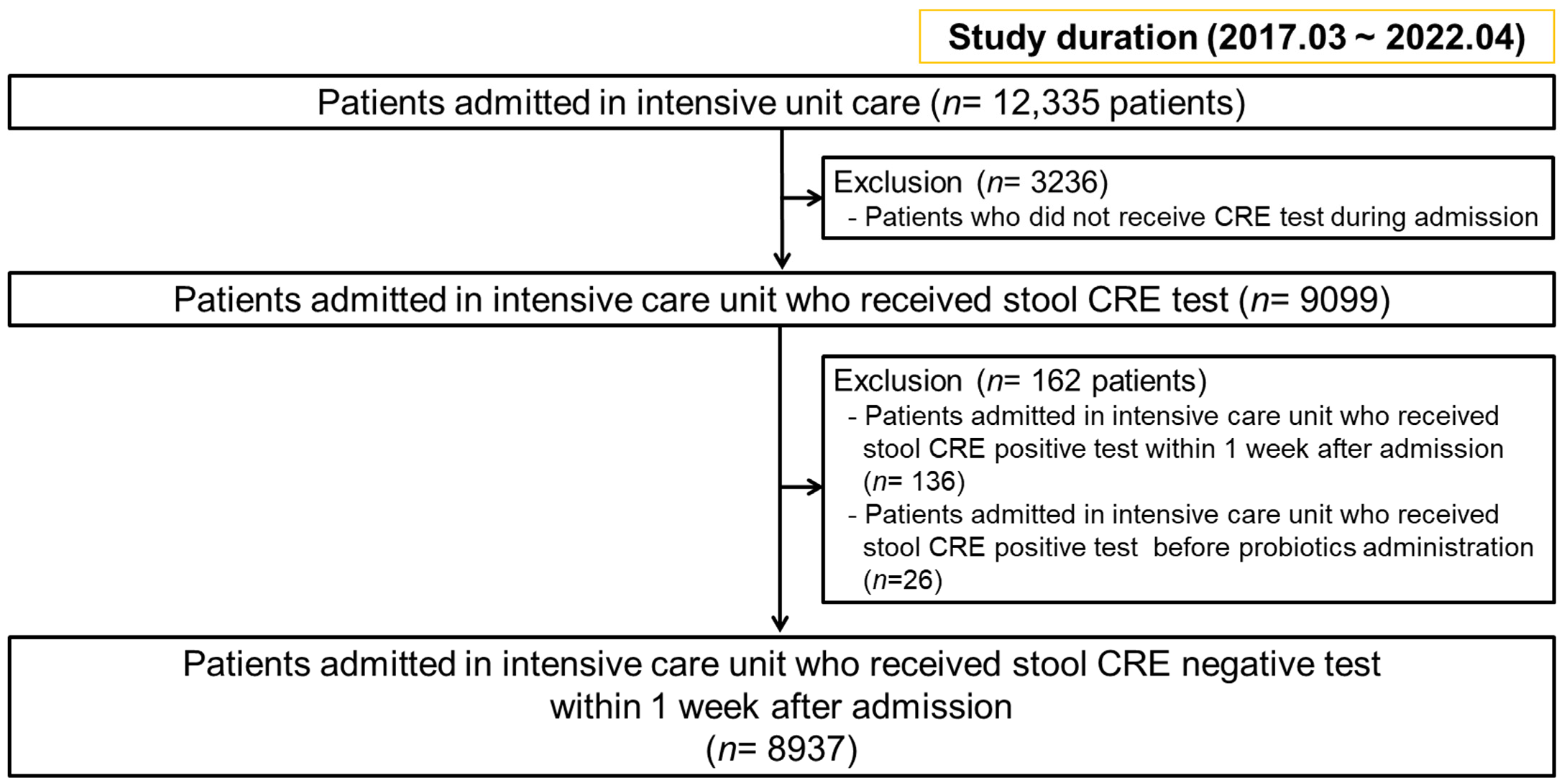
Figure 1. Flow chart of patients’ selection.
Microorganisms 11 02970 g001
Figure 2. Multivariable analysis of the risk factors associated with carbapenem-resistant Enterobacteriaceae (CRE) colonization.; *, p-value < 0.05; and ***, p-value < 0.001; red color texts indicated the significant factors positively correlated with CRE colonization; blue color texts indicated the significant factors negatively correlated with CRE colonization; red oval emphasizes probiotics as a significant negative correlated factor.
Microorganisms 11 02970 g002
Figure 3. Microbiome analysis of 20 patients’ samples with CRE colonization, divided into probiotics (12 samples) and non-probiotics (8 samples) groups. (a) α-diversity (Shannon) between the probiotic and non-probiotic groups. (b) α-diversity (CHAO1) between the probiotic and non-probiotic groups. (c) β-diversity (unweighted uniFrac) between the probiotics group and non-probiotics group. (d) β-diversity (weighted uniFrac) between the probiotic and non-probiotic groups. The red color indicated a significant difference between the groups. Abbreviations: PERMANOVA, permutational multivariate analysis of variance.
Microorganisms 11 02970 g003
Figure 4. Microbiome analysis for patients with CRE colonization according to probiotics administration. (a) Relative abundance of the Enterobacteriaceae family between the probiotics and non-probiotics groups. (b) Relative abundance of the Klebsiella genus between the probiotic and non-probiotic groups. The red color indicated a significant difference between the groups.
Microorganisms 11 02970 g004
Table 1. Baseline characteristics between carbapenem-resistant Enterobacteriaceae (CRE) colonization and no CRE colonization.
CharacteristicsCRE Colonization (n = 157)No CRE Colonization (n = 8780)p-Value
Age, years, median (IQR)73.0 (61.0–80.0)69.0 (56.0–79.0)0.006
Sex >0.999
Male91 (58.0)5119(58.3)
Female73 (42.0)3661 (41.7)
ICU Category <0.001
Cardiology ICU (CCU)9 (5.2)770 (8.8)
Emergency ICU (EA and EB)88 (56.1)5778 (65.8)
Medical ICU (MA and MB)36 (22.9)1491 (17.0)
Surgical ICU (SB)12 (7.6)681 (7.8)
COVID ICU (IICU)12 (7.6)58 (0.7)
Length of stay, days, median (IQR)33.0 (21.0–63.0)12.0 (6.0–21.0)<0.001
Stool VRE Positive725 (8.3)41 (26.1)<0.001
Clostridium difficile infection 29 (18.5)397 (4.5)<0.001
CRE-colonized genus
Citrobacter3 (1.9)
Enterobacter9 (5.7)
Escherichia25 (15.9)
Klebsiella120 (76.3)
Tube feeding116 (66.7)2558 (29.2)<0.001
Probiotics13 (8.3)461 (5.3)0.264
Probiotics duration, days, median (IQR)0.0 (0.0–0.0)0.0 (0.0–0.0)0.165
Saccharomyces boulardii10 (6.4)340 (3.9)0.455
Lactobacillus rhamnosus3 (1.7)121 (1.4)0.955
PPI135 (86.0)6236 (71.0)<0.001
PPI duration, days, median (IQR)17.0 (2.0–42.0)3.0 (0.0–10.0)<0.001
Carbapenem55 (35.0)883 (10.1)<0.001
Carbapenem duration, days, median (IQR)14.0 (8.0–26.5)9.0 (5.0–18.0)<0.001
Antibiotics
Aminoglycoside32 (20.4)548 (6.2)<0.001
Penicillin6 (3.8)178 (2.0)0.116
Extend Spectrum Penicillin115 (73.2)3675 (41.9)<0.001
Macrolide9 (5.7)466 (5.3)0.931
Glycopeptide53 (33.8)742 (8.5)<0.001
Cephalosphorin PO7 (4.5)330 (3.8)>0.999
1st Cephalosphorin IV16 (10.2)685 (7.8)0.167
2nd–3rd Cephalosphorin IV106 (67.5)4662 (53.1)<0.001
Quinolone75 (47.8)2967 (33.8)<0.001
Metronidazole75 (47.8)2317 (26.4)<0.001
Clindamycin5 (3.2)357 (4.1)0.726
Azotreonam16 (10.2)264 (3.0)<0.001
Colistimate18 (11.5)158 (1.8)<0.001
Tygecylcline8 (5.1)76 (0.9)<0.001
Trimethoprim–Sulfamethoxazole20 (12.7)181 (2.1)<0.001
Rifamycin6 (3.8)169 (1.9)0.248
Azole13 (7.5)193 (2.2)<0.001
Antibiotics duration, days, median (IQR)8 (1–16)27 (17–50)<0.001
Comorbidity
Diabetes4 (2.3)351 (4.0)0.344
Diabetes complication1 (0.6)73 (0.8)0.484
Liver disease (Mild)2 (1.3)276 (3.1)0.269
Liver disease (moderate to severe)3 (1.9)225 (2.6)0.125
Acute myocardial infarction4 (2.3)908 (10.3)0.002
Congestive heart failure15 (9.6)932 (10.6)0.766
PAOD6 (3.8)229 (2.6)0.490
Cerebral vascular attack28 (17.8)1842 (21.0)0.389
Hemiplegia22 (14.0)1249 (14.2)>0.999
Dementia2 (1.3)75 (0.9)0.898
COPD2 (1.3)289 (3.3)0.236
Rheumatic disease3 (1.9)46 (0.5)0.074
Peptic ulcer 6 (3.8)450 (5.1)0.580
Chronic kidney disease37 (23.6)967 (11.0)<0.001
Localized solid tumor16 (10.2)836 (9.5)0.884
Lymphoma or Leukemia2 (1.3)60 (0.7)0.690
Metastasis of tumor2 (1.3)243 (2.8)0.374
HIV0 (0.0)7 (0.1)>0.999
Charlson comorbidity index, median (IQR)1 (0–2) 1 (0–2)0.894
Data are presented as median (interquartile range) or number (). Abbreviations: IQR, interquartile range; COVID-19, coronavirus disease 2019; CRE, carbapenem-resistant Enterobacteriaceae; ICU, intensive care unit; VRE, vancomycin-resistance enterococci; PPI, proton pump inhibitor; HIV, human immunodeficiency virus; PAOD, peripheral arterial occlusive disease; and COPD, chronic obstructive pulmonary disease.
Disclaimer/Publisher’s Note: The statements, opinions and data contained in all publications are solely those of the individual author(s) and contributor(s) and not of MDPI and/or the editor(s). MDPI and/or the editor(s) disclaim responsibility for any injury to people or property resulting from any ideas, methods, instructions or products referred to in the content.