Article

Motility in Periweissella Species: Genomic and Phenotypic Characterization and Update on Motility in Lactobacillaceae

1
National Research Council of Italy, Institute of Sciences of Food Production (CNR-ISPA), 70126 Bari, Italy
2
Department of Microbiology and Biotechnology, Max Rubner-Institut, 24103 Kiel, Germany
*
Authors to whom correspondence should be addressed.
Microorganisms 2023, 11(12), 2923; https://doi.org/10.3390/microorganisms11122923
Submission received: 30 October 2023 / Revised: 24 November 2023 / Accepted: 28 November 2023 / Published: 5 December 2023

Abstract

:
The genus Weissella and the recently described genus Periweissella, to which some previously named Weissella species have been reclassified as a result of a taxogenomic assessment, includes lactic acid bacteria species with high biotechnological and probiotic potential. Only one species, namely, Periweissella (P.) beninensis, whose type strain has been shown to possess probiotic features, has so far been described to be motile. However, the availability of numerous genome sequences of Weissella and Periweissella species prompted the possibility to screen for the presence of the genetic determinants encoding motility in Weissella and Periweissellas spp. other than P. beninensis. Herein, we performed a comprehensive genomic analysis to identify motility-related proteins in all Weissella and Periweissella species described so far, and extended the analysis to the recently sequenced Lactobacillaceae spp. Furthermore, we performed motility assays and transmission electron microscopy (TEM) on Periweissella type strains to confirm the genomic prediction. The homology-based analysis revealed genes coding for motility proteins only in the type strains of P. beninensis, P. fabalis, P. fabaria and P. ghanensis genomes. However, only the P. beninensis type strain was positive in the motility assay and displayed run-and-tumble behavior. Many peritrichous and long flagella on bacterial cells were visualized via TEM, as well. As for the Lactobacillaceae, in addition to the species previously described to harbor motility proteins, the genetic determinants of motility were also found in the genomes of the type strains of Lactobacillus rogosae and Ligilactobacillus salitolerans. This study, which is one of the first to analyze the genomes of Weissella, Periweissella and the recently sequenced Lactobacillaceae spp. for the presence of genes coding for motility proteins and which assesses the associated motility phenotypes, provides novel results that expand knowledge on these genera and are useful in the further characterization of lactic acid bacteria.

1. Introduction

The genus Periweissella was described by Bello et al. in 2022 [1]. They used 16S rRNA gene phylogenetics as well as a phylogenomic analyses (the latter based on the concatenated sequences of 498 core proteins from the genera Convivina, Fructobacillus, Leuconostoc, Oenococcus and Weissella) to transfer five species that clustered in a separate clade from the genus Weissella (W.) into the proposed novel genus Periweissella (P.). These five species included P. cryptocerci (basonym W. cryptocerci [2]), P. beninensis (basonym W. beninensis [3]), P. fabalis (basonym W. fabalis [4]), P. fabaria (basonym W. fabaria [5]) and P. ghanensis (basonym W. ghanensis [6]). As is the case for the genus Weissella, the novel Periweissella genus is also included in the family Lactobacillaceae, which was recently revised by Zheng et al. [7], who merged this family together with the Leuconostocaceae family.
Species of the genera Weissella and Periweissella are widespread in nature and have been isolated from several food matrices, including meat, fish, vegetables and fermented foods, as well as from soil, sewage and the gastrointestinal tracts of humans and animals [8,9]. Despite their probiotic potential, which has recently been demonstrated by several studies [10,11], to date, none of the Weissella and Periweissella strains are recognized as GRAS (Generally Recognized as Safe) by the Food and Drug Administration (FDA) in the United States of America, nor have they been included in the Qualified Presumption of Safety (QPS) list by the European Food Safety Authority (EFSA).
Within Weissella and Periweissella spp., only P. beninensis was previously described as being motile [3]. Recently, during the publication process of this manuscript, Qiao et al. [12] reported on the motility of Periweissella strains. Bacterial motility, combined with chemotaxis, provides the ability for bacterial dissemination and surface exploration, enabling movement towards favorable conditions to reach resources and allow for surface colonization. These features are also relevant for the probiotic properties of bacterial strains, as motility is important for the initial steps of adhesion to intestinal epithelial cells and biofilm formation [13]. It, furthermore, provides certain advantages regarding survivability within this specific ecological niche and persistence of the microorganisms in the gut mucosa, and is crucial for enabling them to exert their protective effects and for interaction with the host [14].
The recent availability of genome sequences of all Periweissella and Weissella strains allows for investigations into the presence of genes coding for motility proteins in other species in addition to P. beninensis, and if found, these could point towards a potential motile phenotype. In addition, the probiotic potential of some Weissella and Periweissella strains, recently confirmed by Fanelli et al. [11], also encourages further investigation of functional traits of these strains. This study aimed to perform a comprehensive genomic analysis to identify motility proteins in all Weissella and Periweissella species described so far, and extended the analysis to the recently sequenced Lactobacilliaceae spp. Furthermore, motility assays and transmission electron microscopy (TEM) were carried out on Periweissella type strains to confirm our genomic predictions.

2. Materials and Methods

2.1. Strain Info and Culture Conditions

P. beninensis LMG 25373T, P. fabalis LMG 26217T and P. fabaria LMG 24289T were purchased from the Belgian Coordinated Collection of Microorganisms (BCCM/LMG, Ghent, Belgium), while P. ghanensis DSM 19935T and W. diestrammenae DSM 27940T were purchased from the German Collection of Microorganisms and Cell Cultures (© DSMZ, Braunschweig, Germany). The purity of each strain was confirmed as described in Fanelli et al. [11] via streaking on de Man, Rogosa and Sharpe (MRS; Oxoid, Milan, Italy) agar plates and through microscopic observation. Strains were maintained at −80 °C as pure stock cultures in MRS broth (Oxoid) supplemented with 30% glycerol (v/v). Strains were routinely grown in MRS broth at 30° under aerobic and static conditions, unless specified otherwise.

2.2. Motility Test

Motility was evaluated according to the method described by Shield and Cathcart [15]. Periweissella strains were cultured on MRS agar medium (Oxoid). Subsequently, different media, including MRS, PCA (Oxoid) and motility-indole-lysine (MIL) medium [15], were tested to find the one that could best show the strain motility. By using a sterile needle, a colony of each strain was picked and stabbed into the relevant sterile agar medium in tubes to within 1 cm of the bottom. Tubes were incubated at 30 °C until growth became evident. The motility test was considered positive if a diffuse cloud of growth was visibly extending away from the line of inoculation. The test was considered negative if the growth was limited along the inoculation line.

2.3. Transmission Electron Microscopy

For transmission electron microscopy, all samples were essentially prepared as described by Sørensen et al. [16], with the modification that 2% (v/v) glutaraldehyde was used for fixation and 1% (w/v) uranyl acetate for negative staining. Electron micrographs were taken using a Talos L120C transmission electron microscope (Thermo Fisher Scientific, Eindhoven, The Netherlands) at an accelerating voltage of 80 kV. Digital micrographs were taken using a 4 k × 4 k Ceta camera (Thermo Fisher Scientific).

2.4. Comparative Analysis of Flagellar Locus and Motility Proteins

2.4.1. Identification of Motility Protein Genes in Some Periweissella Type Strains

The whole-genome sequencing and the evaluation of the quality of the assemblies were described in Fanelli et al. [17]. The whole-genome shotgun project and GenBank accession number used for the identification of the motility proteins are listed in Supplementary Table S1. Proteins were predicted as described in Fanelli et al. [17] by using the Prokaryotic Genome Annotation Pipeline [18] and PROKKA pipeline [19] implemented in the Galaxy platform (Galaxy Version 1.14.6 + galaxy0; [20]. Functional classification was performed using the RAST server [21]. Genes related to motility and flagellar systems were retrieved through a keyword search within the UniProtID entry list obtained via functional annotation (by using as a query the terms ‘flagella’ and ‘motility’, and by using the RAST server [21]), and then, manually curated.

2.4.2. In Silico Identification of Motile Species in the Lactobacillaceae Family

The identification of motile species in the family Lactobacillaceae was performed according to the strategy used by Cousin et al. [22], with some modification. Sequences of the predicted filament protein flagellin FliC, the flagellar hook–basal body complex protein FliE, the flagellar motor switch protein FliG, the flagellar biosynthesis protein FlhA and the chemotaxis/stator protein MotA were identified in the proteomes of Periweissella spp. Consensus sequences of the selected proteins, generated using EMBOSS Cons [23], were used as query for a TBLASTN search performed against all publicly available Weissella species genomes, and then, extended to all recently sequenced and publicly available Lactobacillaceae species genomes described after the analysis performed by Cousin et al. [22], according to the taxonomic description amended by Zheng et al. [7], with a cut off E-value of 1 × 10⁻¹⁰ and % identity > 30%. Where positive hits were retrieved, motility protein sequences were extracted from the respective proteomes. The homology-based relationship among proteins was determined and confirmed using the BLASTP algorithm on the NCBI site (http://blast.ncbi.nlm.nih.gov/Blast.cgi accessed on 1 October 2023).

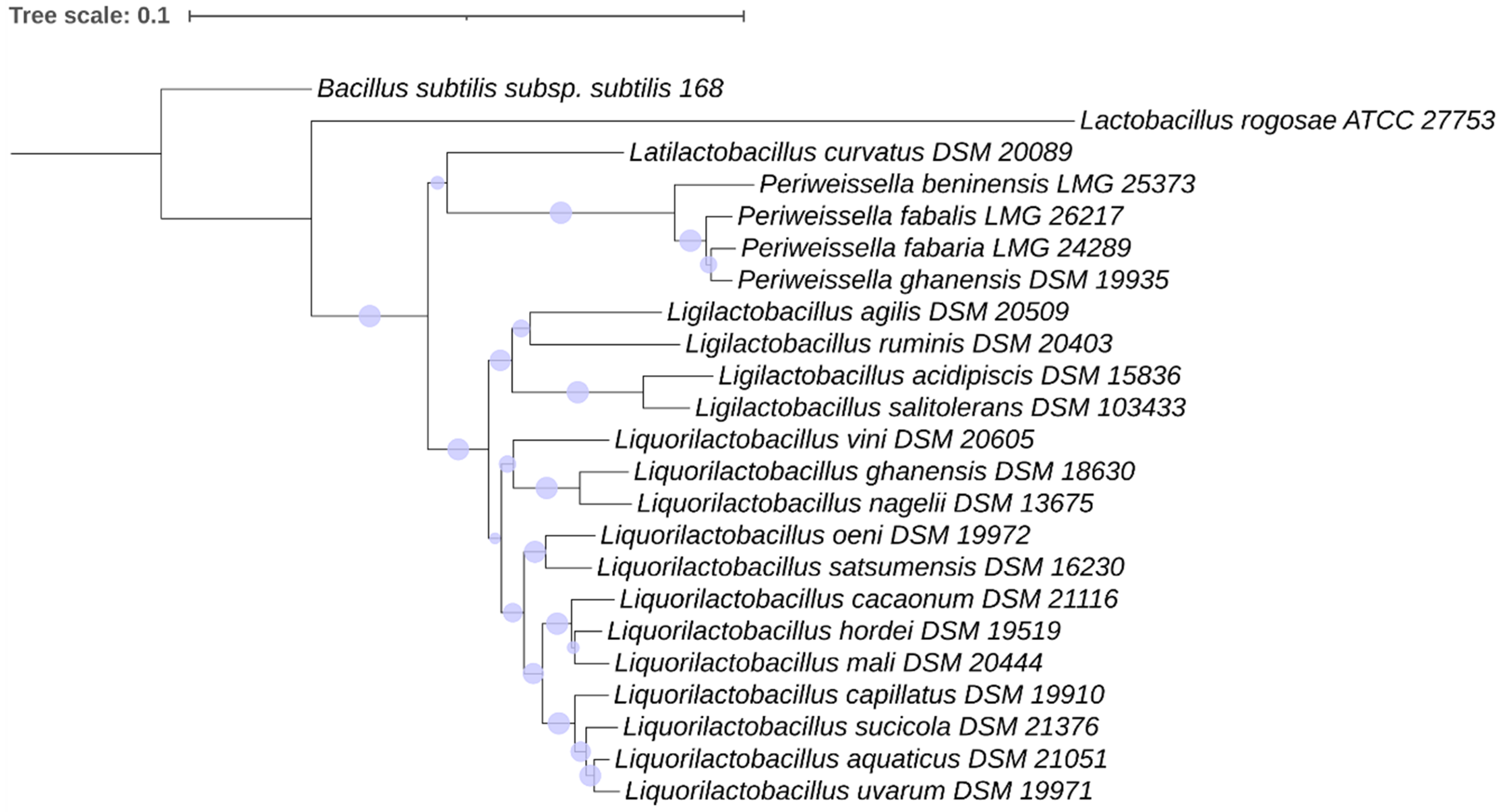
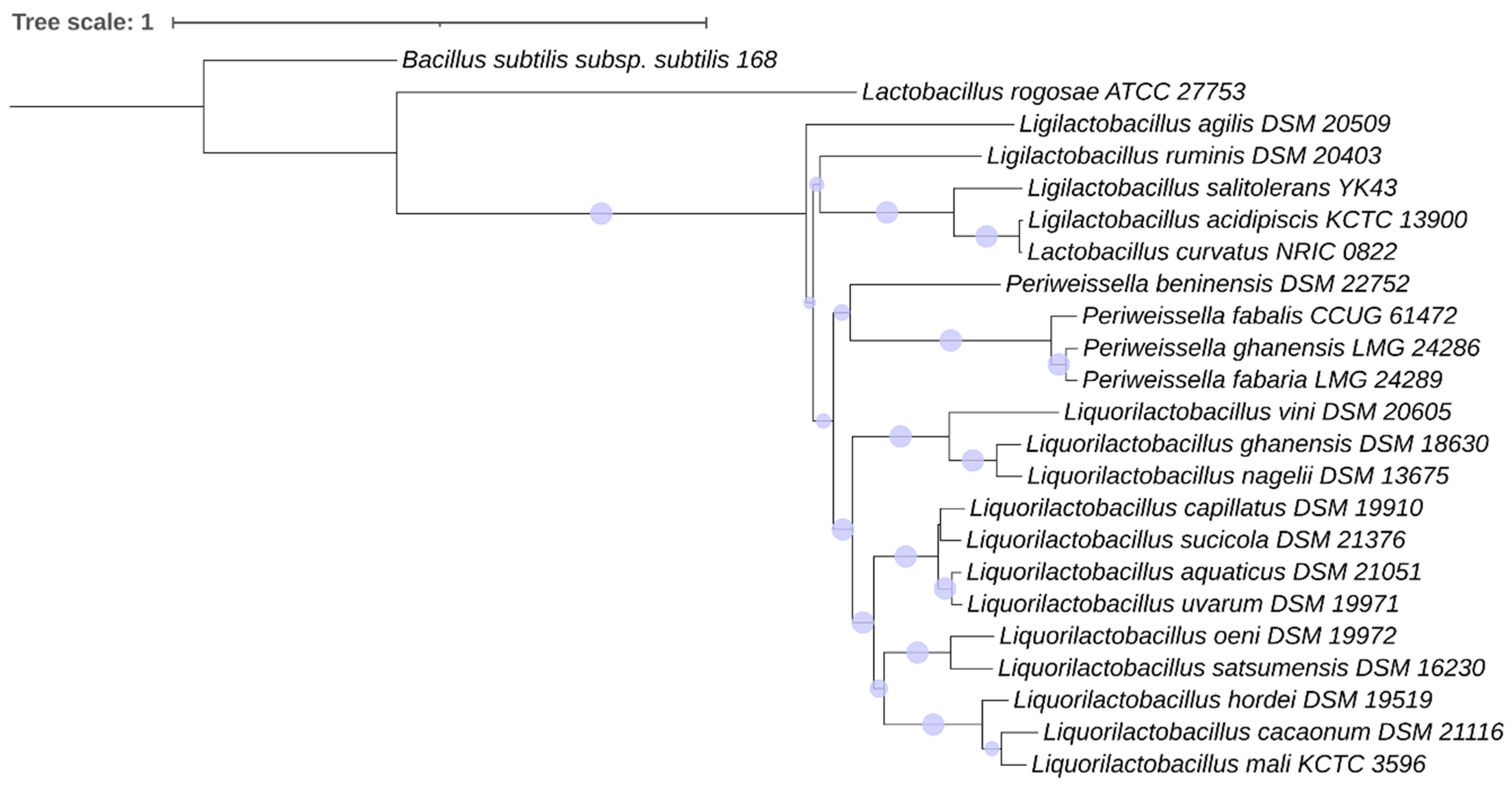
2.4.3. Phylogenetic Analysis

The DNA sequences of the 16S rRNA gene of the type strain of each Lactobacillaceae species positive for the presence of motility proteins were downloaded from the NCBI GenBank database. Phylogenetic analysis was performed by using (i) the 16S rRNA genes, (ii) a concatenated dataset of all the motility proteins identified in each species assumed as being motile by the in silico analysis, and (iii) concatenated datasets of clusters of motility proteins grouped according to their function or position in the flagellum: chemotaxis proteins (concatenated dataset of CheA, CheB, CheC, CheD, CheW and CheY); basal body–hook proteins (concatenated dataset of FlhA, FlhB, FliE, FliF, FliG, FliH, FliI, FliJ, FliM, FliO, FliP, FliQ, FliR and FliK); filament junction proteins (concatenated dataset of FlgL, FlgK, FliC, FliD and FliS); rod, ring and hook/filament junction proteins (concatenated dataset of FlgB, FlgC, FlgD, FlgE, FlgF and FlgG). Phylogeny was inferred by using an “A la Carte” Mode workflow implemented in the NGphylogeny.fr platform [24] (https://ngphylogeny.fr/workflows/alacarte accessed on 1 October 2023) integrated with MAFFT (version 7.407), BMGE (version 1.12) and FastMe (version 2.1.6.1) with default parameters. The phylogenetic reconstruction was performed using the BIONJ method; the phylogenetic robustness was inferred via a bootstrapping procedure with 1000 replications to obtain the confidence value for the aligned sequence dataset. The trees were graphically generated using iTOL version 6.6 [25]. Bacillus subtilis subsp. subtilis strain 168 was used as an outgroup.

2.5. Brightfield Microscopy

Bacteria grown for 24 h in MRS broth under anaerobic conditions were diluted 1:10 in sterile MRS broth. A total of 200 µL was pipetted into 8-well ibiTreat chambered coverslips (Ibidi, Gräfelfing, Germany) and used for microscopy at 1000× magnification with oil immersion using a Nikon Eclipse TS100 (Nikon, Tokyo, Japan) inverted microscope. The bacteria were allowed to settle for at least 30 min, and videos were recorded using the NIS-Elements D software (version 4.13.03, Nikon). The videos were processed using ImageJ to remove artefacts, and the final videos were compressed to 2× speed and combined.

3. Results

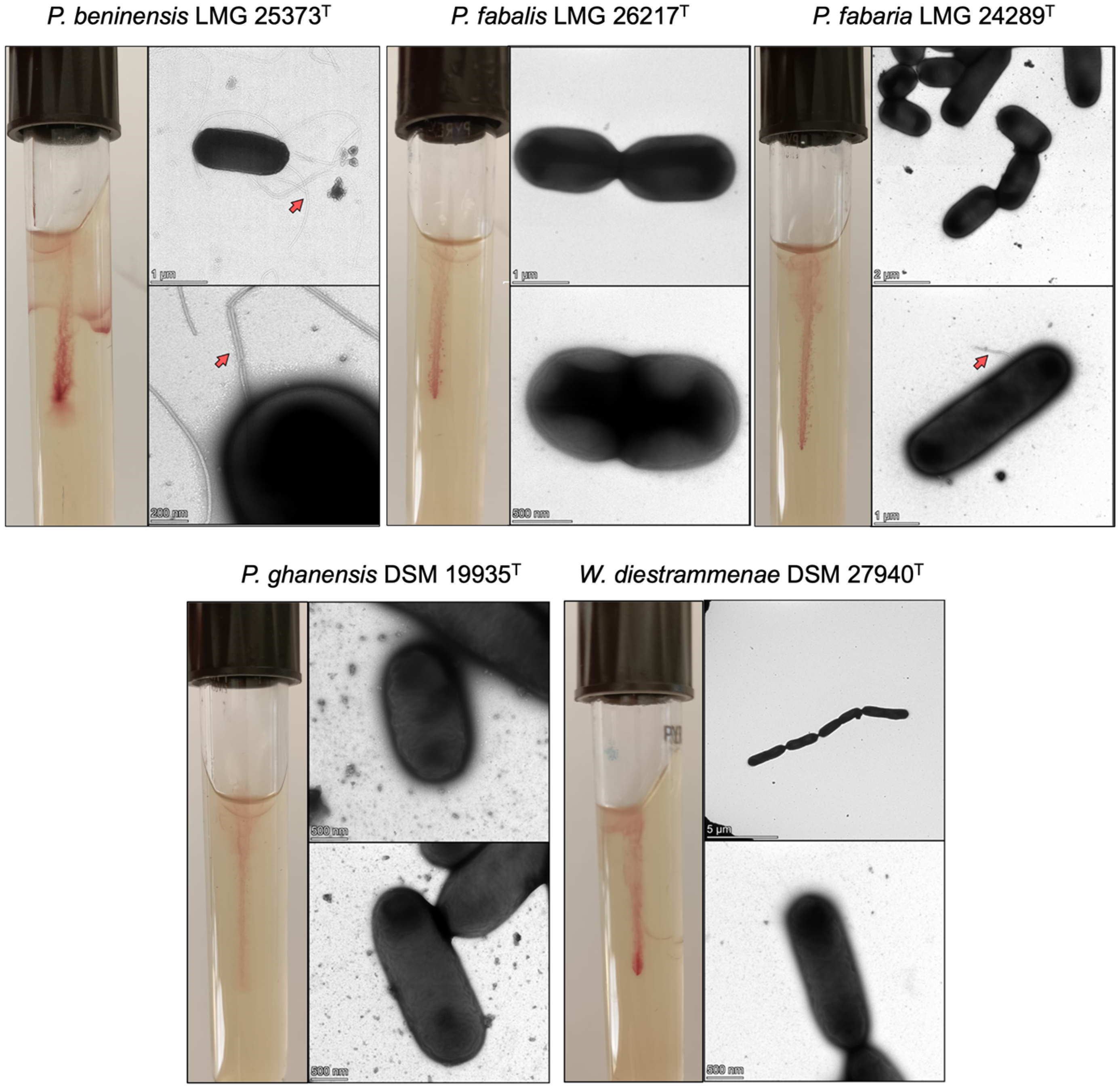
3.1. Motility Behavior

As reported in the Material and Methods section, different media were tested for the motility assay. P. beninensis grew in MRS and PCA, but without the possibility to take a photo and underline its motility, whereas it very clearly showed its motility in MIL. Therefore, MIL was used as a medium for the assay of the motility of all the target strains. Among the four Periweissella type strains tested, only the P. beninensis type strain was able to grow and spread diffusely within the MIL medium (Figure 1). Negative results were obtained for the P. fabalis LMG 26217T, P. fabaria LMG 24289T and P. ghanensis DSM 19935T type strains (Figure 1). P. beninensis LMG 25373T was also the only type strain demonstrating overt motility when observed using brightfield microscopy, showing run-and-tumble movements (Supplementary Movie S1).
The other four strains, P. fabalis LMG 26217T, P. fabaria LMG 24289T, P. ghanensis DSM 19935T and W. diestrammenae DSM 27940T, did not demonstrate motility beyond vibrations, which did not appear directional in the observed time span (Supplementary Movie S1).

3.2. Transmission Electron Microscopy

Figure 1 also shows the electron micrographs of Periweissella type strains analyzed in this study. As indicated by the red arrows, only in the P. beninensis type strain was it possible to visualize numerous peritrichous and long flagella. In P. fabaria LMG 24289T, only a rudimental piece of flagellum was visible (red arrow), and such short pieces were visible only in a very minor portion of cells. W. diestrammenae DSM 27940T was used as a negative control since its genome was found not to harbor any motility protein genes.

3.3. Comparative Analysis of Flagellar Locus and Motility Proteins

3.3.1. Presence of Motility Proteins in Lactobacillaceae

The homology-based analysis of the P. beninensis LMG 25373T, P. fabalis LMG 26217T, P. fabaria LMG 24289T and P. ghanensis DSM 19935T genomes revealed the presence of motility protein-encoding genes in all four strains. The chemotaxis- and flagella-related proteins identified in Periweissella species are listed in Table 1.
FlgN was predicted to be encoded in the genomes of all Periweissella putative motile species as a hypothetical protein (indicated by hp gene near to flgM gene in Figure 2). This protein is the flagellum synthesis protein required for the efficient initiation of filament assembly. The FlgN putative homolog proteins identified in P. beninensis LMG 25373T (WP_205144167.1) and P. ghanensis DSM 19935T (WP_230098022) were classified by InterProScan as having a FlgN-like domain (IPR036679), while no Geno Ontology (GO) term was associated with the putative FlgN homolog of P. fabalis LMG 26217T (WP_168721789.1) and P. fabaria LMG 24289T (WP_230097033.1), indicating no functional prediction, although the MotifFinder analysis retrieved, for all four hypothetical proteins, the PAFM domain PF05130 associated with the FlgN protein.
Some main differences between the Periweissella species positive for the presence of motility proteins could be determined. For instance, the flaG gene coding for the FlaG protein (flagella-associated protein) was identified only in P. beninensis LMG 25373T (WP_205144163.1); the flaG gene was located in the same operon as the fliD and fliS genes reported in other species (e.g., Camphylobacter jejuni), or in the vicinity of the fliD operon on the chromosome of many different species of bacteria [26].
With respect to the Weissella genus, only in the draft genome sequences of Weissella confusa L3 and L9 strains, isolated from human feces, were partial sequences of flagellin proteins detected, and they have been deposited under the accession nos. MBS7551942.1, MBS7552003.1, MCQ8173872.1 and MCQ8173814.1. No homolog of the motility proteins listed in Table 1 was identified in the other Weissella species, nor in the P. cryptocerci type strain, the only remaining species within the Periweissella genus so far described. In Leuconostoc spp., Oenococcus spp., Convivina spp. and Fructobacillus spp., no motility proteins could be determined.
In addition to the Lactobacillaceae species described by Cousin et al. [22] to harbor motility proteins, they were also identified in the genomes of the recently sequenced Lactobacillus (L.) rogosae ATCC 27753T (and identical to those retrieved from the genome of the L. rogosae isolate avicel METABAT 177 assembled from the human gut microbiome) and Ligilactobacillus salitolerans DSM 103433T (Figure 2).

3.3.2. Comparative Analysis of Motility Locus in Periweissella Species

The organization of the flagellar locus of the Periweissella type strains analyzed in this study is illustrated in Figure 2, while motility protein IDs are listed in Table 1.
In P. fabaria LMG 24289T, P. fabalis LMG 26217T and P. ghanensis DSM 19935T, the motility gene locus was annotated on a unique contig, while in P. beninensis LMG 25373T the fliD operon (fliD operon; following nomenclature of corresponding operons in Bacillus subtilis and Salmonella enterica Typhimurium, containing the genes fliD, fliS and fliT) [27,28] was annotated in a separated contig (Figure 2). The organization of the flagellar operon was conserved in the Periweissella species, while the gene content and arrangement within the fliD operon showed some differences: (i) Downstream of the FliS coding gene in P. fabaria LMG 24289T and P. ghanensis DSM 19935T were two genes (gtfA) annotated to code for a UDP-N-acetylglucosamine-peptide N-acetylglucosaminyltransferase GtfA subunit; (ii) in P. fabaria LMG 24289T and P. fabalis LMG 26217T, there were two genes coding for two hypothetical proteins, both harboring a NEAT domain, which was found in a group of iron-regulated surface determinant proteins. In addition, one of these was predicted to have an internaline surface protein domain with a leucine-rich repeat (LRR) that allowed it to provide a structural framework for the formation of protein–protein interactions. (iii) The fliD operon of P. fabalis LMG 26217T also harbored two hag genes coding for the two flagellins FliC1 and FliC2, as previously described, while only one hag gene was present in the other Periweissella spp. (iv) In the fliD operon in P. beninensis LMG 25373T one gene was annotated that codes for FlaG and one for a glycosyltransferase located between the flgL and hag gene, respectively, and three methyl-accepting chemotaxis protein (Mcp)-encoding genes were located upstream of the flgM operon (Figure 2), which exceeded the count observed in the loci of the other Periweissella species by one.
Another difference in the motility genes of the different Periweissella species was that in P. beninensis LMG 25373T, there were the putative homologs of proteins, annotated as FliH and FliJ (WP_205144100.1 and WP_205144102.1, respectively; Table 1 and boxed parts in Figure 2). These were two cytoplasmic ATPase complex proteins that are part of the flagellum export apparatus [29]. Meanwhile, in P. fabalis LMG 26217T, P. fabaria LMG 24289T and P. ghanensis DSM 19935T, they were annotated as hypothetical proteins. The similarity among these hypothetical proteins and the respective homolog in P. beninensis LMG 25373T was, on average, ca. 35%, while between P. fabaria LMG 24289T and the respective homolog in P. ghanensis DSM 19935T, it reached 99%, and between P. fabalis LMG 26217T and the respective homolog in P. fabaria LMG 24289T and P. ghanensis DSM 19935T, it was around 75–80%. The MotifFinder pipeline predicted, in the three hypothetical proteins homologous to FliH of P. fabalis LMG 26217T, P. fabaria LMG 24289T and P. ghanensis DSM 19935T, the PFAM domain PF02108 associated with the flagellar assembly protein FliH, and in all three hypothetical protein homologs of FliJ, the PFAM domain PF02050 associated with the flagellar FliJ protein.
As mentioned before, a difference in the motility genes among the Periweissella species is that only in P. fabalis LMG 26217T was a second hag (fliC2) gene coding for the flagellin FliC2 annotated in the flagellar operon (WP_168721785.1): the predicted FliC2 shares 75% similarity with FliC1 (WP_168721786.1), and 78% and 65% with the homologs in P. fabaria LMG 24289T (WP_230097036.1) and Ligilactobacillus ruminis AM12-58A (WP_270413121.1), respectively. In Ligilactobacillus ruminis AM12-58A, the fliC2 flagellin gene was 98% identical at nucleotide level to the fliC1 gene encoded in the same strain; however, gene expression studies showed that only fliC2 was expressed at a high level in the motile strains [30]. Multiple flagellin proteins were also described to occur in several species, such as Sinorhizobium meliloti, Rhizobium leguminosarum, Agrobacterium tumefaciens and Caulobacter crescentus, the latter of which has a single polar flagellum and six flagellin proteins [31,32,33,34].
The main differences between the flagellar loci of the non-motile Periweissella strains P. fabalis LMG 26217T, P. fabaria LMG 24289T and P. ghanensis DSM 19935T with respect to the motile P. beninensis LMG 25373T thus appears to be the absence of the flaG gene and the presence of hypothetical genes showing low homology to the corresponding (fliH, fliJ, flgN) genes. The identity of the hypothetical genes was validated using HHpred, which allows for the detection of remote homologs by comparing protein profiles based on Hidden Markov Models rather than only relying on sequence homology. The hypothetical protein sequences were screened against the RCSB Protein Data Bank database (https://www.rcsb.org/ accessed on 1 October 2023) and yielded hits for FliH, FliJ and FlgN, respectively.
The ATPase complex promoting the flagellar protein export machinery is composed of FliH, FliJ and FliI, where the latter exhibits enzymatic ATPase activity. The characteristic Walker A (GxxxxGK(S/T)—where x is any amino acid) and Walker B (hhhhDE—where h is any hydrophobic amino acid) motifs found in ATPases were detected in all Periweissella homologs (Supplementary Figure S1), suggesting that all homologs are catalytically active. Surprisingly, there are major differences in the conservation of the three proteins forming the ATPase complex between the analyzed Periweissella species. While FliI is highly conserved between the here determined non-motile species P. fabaria, P. fabalis and P. ghanensis (protein similarity 98.2–98.5%) and rather well conserved in P. beninensis (protein similarity 72.4–72.9%), the other two proteins forming the ATPase complex (FliH and FliJ) display a decreased homology between P. beninensis and the three herein determined as non-motile Periweissella species. Among only the latter species, FliH displays an amino acid sequence similarity of 71.5% and FliJ of 79.2–81.9%. Compared to P. beninensis, protein similarity decreases to 25.9–33.2% for FliH and 31.7–33.7% for FliJ, thus demonstrating unequal conservation of the ATPase complex. Interestingly, these genes are all present and share higher homology in all the other motile lactic acid bacteria examined in this study (L. acidipisces DSM 15836T, L. salitotolerans DSM 103433T and L. rogosae ATCC 27753T (Figure 2)), the only exception being that the flaG gene is not present in the motility locus of L. salitotolerans DSM 103433T. On the other hand, the fliD gene encoding the filament cap protein, which is a protein for chaperoning and sorting flagellin (FliC) proteins after they traverse the hollow filament and exit the growing flagellum tip [35], was only found in the flagellar loci of the P. fabalis LMG 26217T, P. fabaria LMG 24289T and P. ghanensis DSM 19935T strains, but not in the corresponding locus of the P. beninensis strain LMG 25373T (Figure 2). As shown in Figure 2, the content and organization of the flagellar locus in Periweissella species compared to other Lactobacillaceae, such as the L. acidipiscis type strain and L. salitolerans type strain, is quite different. In L. salitolerans DSM 103433T, motA and motB genes were located within the fliD operon, which comprised two flagellin genes and was annotated in a separate contig with respect to the flagellar operon. Also, in Lactobacillus rogosae ATCC 27753T, the fliD operon was annotated on a separate contig with respect to the flagellar operon, which misses flbD, fliK and fliL and harbors the fapA gene coding for the flagellar assembly protein FapA, and ycgR coding for PliZ, a cyclic-di-GMP (c-di-GMP) associated with flagellar and pili-based motility. In the L. acidipiscis type strain fliD operon, there are four genes annotated as transposase, motB and motA genes, the fliS and fliD gene interspaced by a gene coding for a hypothetical protein, the flaG gene, one RNA polymerase-coding gene, two flagellin hag genes, a gene coding for the peptidoglycan-binding protein LysM and one integrase.

3.3.3. Phylogeny of the Motility Proteins in the Lactobacillaceae Species

Figure 3 and Figure 4 show the concordant phylogeny of 16S rRNA genes and motility proteins in the Lactobacillaceae family. P. beninensis LMG 25373T, P. fabalis LMG 26217T, P. fabaria LMG 24289T and P. ghanensis DSM 19935T were always clustered in one separate clade, with P. fabaria LMG 24289T and P. ghanensis DSM 19935T being on the same node, and slightly divergent from P. fabalis LMG 26217T and more divergent from P. beninensis LMG 25373T (Figure 3 and Figure 4). P. beninensis LMG 25373T was clustered together with the other Periweissella species in the phylogenetic tree of the concatenated dataset of all the motility proteins, and of the groups of chemotaxis proteins, filament junction proteins, and rod–ring–hook proteins, although, interestingly, it was also grouped apart from these, showing a clear difference in the motility gene content outlined above. Furthermore, in a few cases, the phylogeny of individual proteins, such as CheB, CheR, CheC, CheD, FlgB, FlgC, etc., as well as of those occurring in the cluster of the basal body and hook proteins (shown in Supplementary Figure S2B), underlines a higher proximity of P. beninensis LMG 25373T with species outside of the Periweissella genus.
The phylogenetic analysis of the motility proteins mostly overlapped with the 16S rRNA gene sequence phylogeny, and in both trees (Figure 3 and Figure 4), almost all the Lactobacillaceae species clustered in the same clade: (1) L. capillatus, L. sucicola, L. acquaticus and L. uvarum; (2) L. cacaonum, L. hordei and L. mali; (3) L. oeni and L. satsumensis; (4) L. ghanensis, L. nagelii and L. vini; (5) L. acidipiscis and L. salitolerans; (6) P. beninensis, P. ghanensis, P. fabalis and P. fabaria.
L. rogosae ATCC 27753T (as well as L. rogosae isolate avicel METABAT 177) was phylogenetically apart from the other Lactilobacillaceae species in each motility protein analyzed, as well as in relation to the 16S rRNA gene sequence phylogeny (Figure 3 and Figure 4).

4. Discussion

The bacterial flagellum is organized into three basic components [36,37]: (1) The basal body, anchored in the cell membrane and comprising a rotary motor, connected to a rod, which powers the flagellum, the export apparatus machinery, and bearing structures. (2) The hook [38], which functions like a universal joint and is a tubular helical structure made via the polymerization of multiple copies of a protein, FlgE; its length is controlled by FliK. The hook transmits the torque produced by the motor to the (3) filament, which is the propeller of the flagellum, with FliC as the core unit and FliD acting as a cap. The junction between the hook and the filament is the charger of FlgL and FlgK.
The flagellar apparatus in Salmonella enterica subsp. enterica serovar Typhimurium is one of the best characterized thanks to the work by Minamino and coworkers [29], and the crystal structures of most of the protein complexes that comprise the flagellar apparatus have been resolved and deposited in the RCSB Protein Data Bank. Both FliH and FliJ, which are also present in the genetic locus of motile P. beninensis LMG 25373T, have been described to be part of the FliH(12)-FliI(6)-FliJ(1) cytoplasmic ATPase export ring complex, which acts as an active protein transporter. Its structure resembles the cytoplasmic part of FOF1 ATP synthase, a rotary motor that couples proton (H+) flow through FO with ATP synthesis by F1 [39]. We found that the conservation of the subunits of the ATPase involved in protein export is dissimilar between the Periweissella strains. While FliI is rather highly conserved, FliH and FliJ display considerably lower sequence homology between P. beninensis and the three non-motile species. This could indicate that the complex might not form properly in the non-motile species due to the high evolutionary divergence, resulting in an impaired flagellar export mechanism, and thus, could explain the difference in motility.
In addition to that, FlgN was also poorly conserved. FlgN is one of the flagellar chaperones, together with FliS and FliT, specific to FlgK and FlgL, which, in Bacillus subtilis, is required for flagellum-based motility [40]. The crystal structure of FlgN in Salmonella was solved in 2016 by Kinoshita et al. [41] and consists of three helices. The authors showed that a conformational change in FlgN with rearrangements of α-helices is critical for the association and dissociation of FlgK [41]. Minamino et al. [42] demonstrated that FlgN promotes the docking of FlgK and FlgL to the FlhA platform of the export gate complex to facilitate the rapid and efficient export of these proteins [43]. The impairing of this activity reduces the secretion levels of FlgK and FlgL, resulting in a considerable reduction in the probability of filament formation at the tip of the hook–basal body [44]. In addition to its chaperone activity, FlgN interacts with FlhA, which is the export engine of the apparatus, generating a conformational change in FlhA that allows it to function as a Na+ channel.
The flaG gene was identified only in P. beninensis LMG 25373T. FlaG is one of the flagellar proteins that are actually absent in well-studied model organisms such as Escherichia coli and Salmonella enterica [45,46], but has been detected and characterized in other motile species such as Aeromonas caviae, Campylobacter jejuni, Pseudomonas aeruginosa and Pseudomonas fluorescens [47,48,49]. Divergence of the protein sequences and mutational behaviors in different species suggested that FlaG had a possible role in regulating the export of flagellin proteins, filament length and the number or the expression of other flagellin genes [50,51,52].
Our study shows that hypothetical proteins with putative homology to FliH, FliJ and FlgN in the three non-motile Periweissella species have only low similarity (35–41%) when compared to those comprising the flagellar locus of P. beninensis LMG 25373T. As FliH and FliJ are essential for the export of flagellar proteins, it is possible that a low homology of counterpart proteins in the non-motile strains implies that the flagellar components cannot be sufficiently exported for flagellar biosynthesis. Similarly, FlgN is a flagellum synthesis protein and is required for the export of the hook/filament junction proteins FlgK and FlgL. If this is absent or nonfunctional, again, this could prevent the assembly of a functional flagellum, although we have evidence that in P. fabaria LMG 24289T, a partial flagellum could possibly be assembled, as seen in the results of electron microscopy (Figure 1).
Despite the fact that P. fabalis LMG 26217T, P. fabaria LMG 24289T and P. ghanensis DSM 19935T possess a complete flagellar operon, motility could be demonstrated only for P. beninensis LMG 25373T. It is very clear from live cell video microscopy that P. beninensis LMG 25373T bacteria are remarkably motile compared to the other Periweisella species (Supplementary Movie S1).
Our data are in agreement with previous papers describing Periweissella spp. In particular P. fabalis [4] and P. fabaria [5] were described as not motile, and for P. ghanensis, gliding motility was not observed [6], while, conversely, P. beninensis [3] was defined as motile with peritrichous flagella that can occur singly, in pairs or in short chains.
A recent paper published by Qiao et al. [12] confirmed the presence of the flagellar operon in P. beninensis, P. fabalis and P. fabaria, and the authors described all these species as motile. However, the only phenotypic assessment to prove their assumption was the stab test in semisolid MRS agar medium, which, for us, was not as adequately informative for assessing motility compared to using MIL medium. Furthermore, surprisingly, P. beninensis was not able to grow under the condition tested by Qiao et al. [12]. In our work, the TEM experiments showed that with the exception of the P. beninensis type strain, the other Periweissella species were not able to assemble any visible flagellar apparatus under the tested condition. Only in P. fabaria LMG 24289T was a rudimentary piece of flagellum visible, and very few pieces of flagella could be found only in a very minor portion of cells. This was supported by microscopic observations, which showed that only P. beninensis had high motility. Hypothetically the genomic differences reported here in the flagellar operons of the other Periweissela when compared to P. beninensis LMG 25373T might lead to a lower expression of flagellar proteins under the condition that we tested in this study. This should, however, be investigated for media and test conditions in further studies.
It is interesting to note that, in addition to other Weissella species, in the P. cryptocerci type strain, no motility proteins could be determined. Previous work by our group [11,17] demonstrated that P. beninensis LMG 25373T displayed the best probiotic behavior, with the highest adhesion capacity, as well as a varied carbohydrate utilization profile, and was the only one capable of using d-galactose. Although motility is not essential for gut colonization, it might provide certain advantages in survivability and persistence for these organisms in the gut mucosa, and favor colonization in the gastrointestinal tract, as occurred for motile lactobacilli compared to not-motile ones [53].
Flagella are composed, together with pili, of surface layer proteins (SLPs), capsular polysaccharide (CPS), lipoteichoic acid and lipopolysaccharide, the surface components of probiotics that constitute microbe-associated molecular patterns (MAMPs) [54], which can specifically bind to pattern recognition receptors and regulate nuclear factor kappa B, mitogen-activated protein kinases, peroxisome proliferator-activated receptor gamma, and other signaling pathways in intestinal epithelial cells [55], in addition to activating the cellular protease-dependent signaling cascade to produce a variety of cytokines and chemokines that alleviate inflammation and enhance intestinal epithelial function [56]. In the probiotic E. coli Nissle 1917, the flagellum is not just responsible for motility and capable of inducing the production of human antimicrobial peptide β-defensin 2, but is the major adhesin mediating binding to human mucus [57], which enables this probiotic strain to compete efficiently for binding sites on host tissue with several pathogenic bacteria. The ability of P. beninensis LMG 25373T to assemble a functional flagellum thus confirms the distinctiveness of this strain with respect to the other Periweissella type strains analyzed, and suggests a correlation with optimal probiotic activity, in particular related to its adhesion ability, that was recently demonstrated in vitro by Fanelli and coworkers [17].

5. Conclusions

The comprehensive genomic analysis herein carried out to detect the genetic determinants of motility in Weissella, Periweissella and the recently sequenced Lactobacillaceae spp. allowed us to detect genes coding for motility proteins only in the type strains of P. beninensis, P. fabalis, P. fabaria and P. ghanensis genomes, as well as in the recently genome-sequenced type strains of Lactobacillus rogosae and Ligilactobacillus salitolerans. Among the Weissella and Periweissella type strains analyzed in this study, only the P. beninensis type was positive in the motility assay, and its many peritrichous and long flagella were visualized via TEM, while, despite the type strains of P. fabalis, P. fabaria and P. ghanensis possessing a complete flagellar operon, no motility was observed in these strains under the conditions used in this study, and only the P. fabaria type strain showed a rudimentary piece of flagellum on few cells. The type strain of P. beninesis showed the highest in vitro adhesion capacity and resistance to simulated gastrointestinal digestion [17] (Fanelli et al., 2023), indicating a possible correlation between the motile behavior and the probiotic potential of this strain.
This study provides novel results that are useful in the further characterization of these genera.

Supplementary Materials

The following supporting information can be downloaded at: https://www.mdpi.com/article/10.3390/microorganisms11122923/s1, Table S1. List of Lactobacillaceae type strains and WGS reference accession used in this study. Movie S1. Brightfield microscopy videos (2× speed) depicting run-and-tumble motility in P. beninensis LMG 25373T in comparison to the lack thereof in other Periweissella and Weissella strains. Figure S1. Protein sequence alignments of different Periweissella FliI homologs. Conservation of each residue is represented by colored bars, where tall yellow bars indicate a conservation of 100%. Amino acids that constitute the putative Walker A and Walker B motifs are labeled in red. Figure S2. Phylogenetic tree of motility proteins concatenated and grouped according to their function or position in the flagellum. (A) Chemotaxis proteins; (B) basal body–hook proteins; (C) filament junction proteins; (D) rod, ring and hook/filament junction proteins. Phylogenetic robustness was inferred via a bootstrapping procedure from 1000 replications to obtain the confidence value for the aligned sequence dataset. Bacillus subtilis subsp. subtilis str. 168 was used as an outgroup. The trees were drawn to scale, with branch lengths measured as the number of substitutions per site. Scaled circles are representative of bootstrap values.

Author Contributions

Conceptualization: F.F. and V.F.; Methodology: F.F., C.M.A.P.F. and V.F.; Investigation: F.F., M.M. and C.M.A.P.F.; Resources: V.F. and C.M.A.P.F.; Data Curation: F.F. and C.M.A.P.F.; Writing—Original Draft: F.F., V.F. and C.M.A.P.F.; Writing—Review and Editing: F.F., M.M., D.C., G.-S.C., H.-Z.L., F.H., C.M.A.P.F. and V.F.; Visualization: F.F., C.M.A.P.F. and H.-Z.L.; Supervision: C.M.A.P.F. and V.F.; Funding Acquisition: C.M.A.P.F. and V.F. All authors have read and agreed to the published version of the manuscript.

Funding

This research did not receive any specific grants from funding agencies in the public, commercial or not-for-profit sectors.

Data Availability Statement

All data generated or analyzed during this study are included in this published article and its Supplementary Material.

Conflicts of Interest

The authors declare no conflict of interest.

References

  1. Bello, S.; Rudra, B.; Gupta, R.S. Phylogenomic and comparative genomic analyses of Leuconostocaceae species: Identification of molecular signatures specific for the genera Leuconostoc, Fructobacillus and Oenococcus and proposal for a novel genus Periweissella gen. nov. Int. J. Syst. Evol. Microbiol. 2022, 72, 005284. [Google Scholar] [CrossRef] [PubMed]
  2. Heo, J.; Hamada, M.; Cho, H.; Weon, H.Y.; Kim, J.S.; Hong, S.B.; Kim, S.J.; Kwon, S.W. Weissella cryptocerci sp. nov., isolated from gut of the insect Cryptocercus kyebangensis. Int. J. Syst. Evol. Microbiol. 2019, 69, 2801–2806. [Google Scholar] [CrossRef] [PubMed]
  3. Padonou, S.W.; Schillinger, U.; Nielsen, D.S.; Franz, C.M.A.P.; Hansen, M.; Hounhouigan, J.D.; Nago, M.C.; Jakobsen, M. Weissella beninensis sp. nov., a motile lactic acid bacterium from submerged cassava fermentations, and emended description of the genus Weissella. Int. J. Syst. Evol. Microbiol. 2009, 60, 2193–2198. [Google Scholar] [CrossRef] [PubMed]
  4. Snauwaert, I.; Papalexandratou, Z.; De Vuyst, L.; Vandamme, P. Characterization of strains of Weissella fabalis sp. nov. and Fructobacillus tropaeoli from spontaneous cocoa bean fermentations. Int. J. Syst. Evol. Microbiol. 2013, 63, 1709–1716. [Google Scholar] [CrossRef]
  5. De Bruyne, K.; Camu, N.; De Vuyst, L.; Vandamme, P. Weissella fabaria sp. nov., from a Ghanaian cocoa fermentation. Int. J. Syst. Evol. Microbiol. 2010, 60, 1999–2005. [Google Scholar] [CrossRef]
  6. De Bruyne, K.; Camu, N.; Lefebvre, K.; De Vuyst, L.; Vandamme, P. Weissella ghanensis sp. nov., isolated from a Ghanaian cocoa fermentation. Int. J. Syst. Evol. Microbiol. 2008, 58, 2721–2725. [Google Scholar] [CrossRef]
  7. Zheng, J.; Wittouck, S.; Salvetti, E.; Franz, C.M.A.P.; Harris, H.M.B.; Mattarelli, P.; O’Toole, P.W.; Pot, B.; Vandamme, P.; Walter, J.; et al. A taxonomic note on the genus Lactobacillus: Description of 23 novel genera, emended description of the genus Lactobacillus Beijerinck 1901, and union of Lactobacillaceae and Leuconostocaceae. Int. J. Syst. Evol. Microbiol. 2020, 70, 2782–2858. [Google Scholar] [CrossRef]
  8. Fusco, V.; Quero, G.M.; Cho, G.S.; Kabisch, J.; Meske, D.; Neve, H.; Bockelmann, W.; Franz, C.M.A.P. The genus Weissella: Taxonomy, ecology and biotechnological potential. Front. Microbiol. 2015, 6, 155. [Google Scholar] [CrossRef]
  9. Fusco, V.; Chieffi, D.; Fanelli, F.; Montemurro, M. The Weissella and Periweissella genera: Up to date taxonomy, ecology, safety, biotechnological and probiotic potential. Front. Microbiol. 2023, 14, 2023. [Google Scholar] [CrossRef]
  10. Lakra, A.K.; Domdi, L.; Hanjon, G.; Tilwani, Y.M.; Arul, V. Some probiotic potential of Weissella confusa MD1 and Weissella cibaria MD2 isolated from fermented batter. LWT 2020, 125, 109261. [Google Scholar] [CrossRef]
  11. Fanelli, F.; Montemurro, M.; Verni, M.; Garbetta, A.; Bavaro, A.R.; Chieffi, D.; Cho, G.-S.; Franz, C.M.A.P.; Rizzello, C.G.; Fusco, V. Probiotic Potential and Safety Assessment of Type Strains of Weissella and Periweissella Species. Microbiol. Spectr. 2023, 11, e0304722. [Google Scholar] [CrossRef] [PubMed]
  12. Qiao, N.; Bechtner, J.; Cnockaert, M.; Depoorter, E.; Díaz-Muñoz, C.; Vandamme, P.; De Vuyst, L.; Gänzle, M.G. Comparative genomic analysis of Periweissella and the characterization of novel motile species. Appl. Environ. Microbiol. 2023, 89, e0103423. [Google Scholar] [CrossRef] [PubMed]
  13. Haiko, J.; Westerlund-Wikström, B. The role of the bacterial flagellum in adhesion and virulence. Biology 2013, 2, 1242–1267. [Google Scholar] [CrossRef] [PubMed]
  14. Liu, Q.; Yu, Z.; Tian, F.; Zhao, J.; Zhang, H.; Zhai, Q.; Chen, W. Surface components and metabolites of probiotics for regulation of intestinal epithelial barrier. Microb. Cell Fact. 2020, 19, 23. [Google Scholar] [CrossRef] [PubMed]
  15. Shields, P.; Cathcart, L. Motility Test Medium Protocol; American Society for Microbiology: Washington, DC, USA, 2011. [Google Scholar]
  16. Sørensen, M.C.; Gencay, Y.E.; Birk, T.; Baldvinsson, S.B.; Jäckel, C.; Hammerl, J.A.; Vegge, C.S.; Neve, H.; Brøndsted, L. Primary isolation strain determines both phage type and receptors recognised by Campylobacter jejuni bacteriophages. PLoS ONE 2015, 10, e0116287. [Google Scholar] [CrossRef]
  17. Fanelli, F.; Montemurro, M.; Chieffi, D.; Cho, G.S.; Franz, C.M.A.P.; Dell’Aquila, A.; Rizzello, C.G.; Fusco, V. Novel Insights into the Phylogeny and Biotechnological Potential of Weissella Species. Front. Microbiol. 2022, 13, 914036. [Google Scholar] [CrossRef]
  18. Tatusova, T.; DiCuccio, M.; Badretdin, A.; Chetvernin, V.; Nawrocki, E.P.; Zaslavsky, L.; Lomsadze, A.; Pruitt, K.D.; Borodovsky, M.; Ostell, J. NCBI prokaryotic genome annotation pipeline. Nucleic Acids Res. 2016, 44, 6614–6624. [Google Scholar] [CrossRef]
  19. Seemann, T. Prokka: Rapid prokaryotic genome annotation. Bioinformatics 2014, 30, 2068–2069. [Google Scholar] [CrossRef] [PubMed]
  20. Afgan, E.; Baker, D.; Batut, B.; van den Beek, M.; Bouvier, D.; Čech, M.; Chilton, J.; Clements, D.; Coraor, N.; Grüning, B.A.; et al. The Galaxy platform for accessible, reproducible and collaborative biomedical analyses: 2018 update. Nucleic Acids Res. 2018, 46, W537–W544. [Google Scholar] [CrossRef] [PubMed]
  21. Aziz, R.K.; Bartels, D.; Best, A.A.; DeJongh, M.; Disz, T.; Edwards, R.A.; Formsma, K.; Gerdes, S.; Glass, E.M.; Kubal, M.; et al. The RAST Server: Rapid annotations using subsystems technology. BMC Genom. 2008, 9, 75. [Google Scholar] [CrossRef]
  22. Cousin, F.J.; Lynch, S.M.; Harris, H.M.; McCann, A.; Lynch, D.B.; Neville, B.A.; Irisawa, T.; Okada, S.; Endo, A.; O’Toole, P.W. Detection and genomic characterization of motility in Lactobacillus curvatus: Confirmation of motility in a species outside the Lactobacillus salivarius clade. Appl. Environ. Microbiol. 2015, 81, 1297–1308. [Google Scholar] [CrossRef] [PubMed]
  23. Madeira, F.; Pearce, M.; Tivey, A.R.N.; Basutkar, P.; Lee, J.; Edbali, O.; Madhusoodanan, N.; Kolesnikov, A.; Lopez, R. Search and sequence analysis tools services from EMBL-EBI in 2022. Nucleic Acids Res. 2022, 50, W276–W279. [Google Scholar] [CrossRef] [PubMed]
  24. Lemoine, F.; Correia, D.; Lefort, V.; Doppelt-Azeroual, O.; Mareuil, F.; Cohen-Boulakia, S.; Gascuel, O. NGPhylogeny.fr: New generation phylogenetic services for non-specialists. Nucleic Acids Res. 2019, 47, W260–W265. [Google Scholar] [CrossRef] [PubMed]
  25. Letunic, I.; Bork, P. Interactive Tree Of Life (iTOL) v5: An online tool for phylogenetic tree display and annotation. Nucleic Acids Res. 2021, 49, W293–W296. [Google Scholar] [CrossRef] [PubMed]
  26. Aizawa, S.-I. The Flagellar World: Electron Microscopic Images of Bacterial Flagella and Related Surface Structures from More than 30 Species; Academic Press: New York, NY, USA; Oxford, UK, 2014. [Google Scholar]
  27. Chen, Y.F.; Helmann, J.D. The Bacillus subtilis flagellar regulatory protein sigma D: Overproduction, domain analysis and DNA-binding properties. J. Mol. Biol. 1995, 249, 743–753. [Google Scholar] [CrossRef] [PubMed]
  28. Yokoseki, T.; Kutsukake, K.; Ohnishi, K.; Iino, T. Functional analysis of the flagellar genes in the fliD operon of Salmonella typhimurium. Microbiology 1995, 141 Pt 7, 1715–1722. [Google Scholar] [CrossRef] [PubMed]
  29. Minamino, T.; Kinoshita, M.; Inoue, Y.; Morimoto, Y.V.; Ihara, K.; Koya, S.; Hara, N.; Nishioka, N.; Kojima, S.; Homma, M.; et al. FliH and FliI ensure efficient energy coupling of flagellar type III protein export in Salmonella. Microbiologyopen 2016, 5, 424–435. [Google Scholar] [CrossRef] [PubMed]
  30. Neville, B.A.; Forde, B.M.; Claesson, M.J.; Darby, T.; Coghlan, A.; Nally, K.; Ross, R.P.; O’toole, P.W. Characterization of Pro-Inflammatory Flagellin Proteins Produced by Lactobacillus ruminis and Related Motile Lactobacilli. PLoS ONE 2012, 7, e40592. [Google Scholar] [CrossRef]
  31. Scharf, B.; Schuster-Wolff-Bühring, H.; Rachel, R.; Schmitt, R. Mutational analysis of the Rhizobium lupini H13-3 and Sinorhizobium meliloti flagellin genes: Importance of flagellin A for flagellar filament structure and transcriptional regulation. J. Bacteriol. 2001, 183, 5334–5342. [Google Scholar] [CrossRef]
  32. Tambalo, D.D.; Bustard, D.E.; Del Bel, K.L.; Koval, S.F.; Khan, M.F.; Hynes, M.F. Characterization and functional analysis of seven flagellin genes in Rhizobium leguminosarum bv. viciae. Characterization of R. leguminosarum flagellins. BMC Microbiol. 2010, 10, 219. [Google Scholar] [CrossRef]
  33. Mohari, B.; Thompson, M.A.; Trinidad, J.C.; Setayeshgar, S.; Fuqua, C. Multiple Flagellin Proteins Have Distinct and Synergistic Roles in Agrobacterium tumefaciens Motility. J. Bacteriol. 2018, 200, e00327-18. [Google Scholar] [CrossRef] [PubMed]
  34. Faulds-Pain, A.; Birchall, C.; Aldridge, C.; Smith, W.D.; Grimaldi, G.; Nakamura, S.; Miyata, T.; Gray, J.; Li, G.; Tang, J.; et al. Flagellin redundancy in Caulobacter crescentus and its implications for flagellar filament assembly. J. Bacteriol. 2011, 193, 2695–2707. [Google Scholar] [CrossRef] [PubMed]
  35. Postel, S.; Deredge, D.; Bonsor, D.A.; Yu, X.; Diederichs, K.; Helmsing, S.; Vromen, A.; Friedler, A.; Hust, M.; Egelman, E.H.; et al. Bacterial flagellar capping proteins adopt diverse oligomeric states. Elife 2016, 5, e18857. [Google Scholar] [CrossRef] [PubMed]
  36. Wadhwa, N.; Sassi, A.; Berg, H.C.; Tu, Y. A multi-state dynamic process confers mechano-adaptation to a biological nanomachine. Nat. Commun. 2022, 13, 5327. [Google Scholar] [CrossRef] [PubMed]
  37. Mondino, S.; San Martin, F.; Buschiazzo, A. 3D cryo-EM imaging of bacterial flagella: Novel structural mechanistic insights into cell motility. J. Biol. Chem. 2022, 298, 102105. [Google Scholar] [CrossRef] [PubMed]
  38. Matsunami, H.; Barker, C.S.; Yoon, Y.-H.; Wolf, M.; Samatey, F.A. Complete structure of the bacterial flagellar hook reveals extensive set of stabilizing interactions. Nat. Commun. 2016, 7, 13425. [Google Scholar] [CrossRef] [PubMed]
  39. Minamino, T.; Kinoshita, M.; Namba, K. Insight into Distinct Functional Roles of the Flagellar ATPase Complex for Flagellar Assembly in Salmonella. Front. Microbiol. 2022, 13, 864178. [Google Scholar] [CrossRef]
  40. Cairns, L.S.; Marlow, V.L.; Kiley, T.B.; Birchall, C.; Ostrowski, A.; Aldridge, P.D.; Stanley-Wall, N.R. FlgN is required for flagellum-based motility by Bacillus subtilis. J. Bacteriol. 2014, 196, 2216–2226. [Google Scholar] [CrossRef]
  41. Kinoshita, M.; Hara, N.; Imada, K.; Namba, K.; Minamino, T. Interactions of bacterial flagellar chaperone-substrate complexes with FlhA contribute to co-ordinating assembly of the flagellar filament. Mol. Microbiol. 2013, 90, 1249–1261. [Google Scholar] [CrossRef]
  42. Minamino, T.; Kinoshita, M.; Morimoto, Y.V.; Namba, K. The FlgN chaperone activates the Na+-driven engine of the Salmonella flagellar protein export apparatus. Commun. Biol. 2021, 4, 335. [Google Scholar] [CrossRef]
  43. Minamino, T.; Kinoshita, M.; Hara, N.; Takeuchi, S.; Hida, A.; Koya, S.; Glenwright, H.; Imada, K.; Aldridge, P.D.; Namba, K. Interaction of a bacterial flagellar chaperone FlgN with FlhA is required for efficient export of its cognate substrates. Mol. Microbiol. 2012, 83, 775–788. [Google Scholar] [CrossRef] [PubMed]
  44. Kutsukake, K.; Okada, T.; Yokoseki, T.; Iino, T. Sequence analysis of the flgA gene and its adjacent region in Salmonella typhimurium, and identification of another flagellar gene, flgN. Gene 1994, 143, 49–54. [Google Scholar] [CrossRef] [PubMed]
  45. Minamino, T.; Imada, K. The bacterial flagellar motor and its structural diversity. Trends Microbiol. 2015, 23, 267–274. [Google Scholar] [CrossRef] [PubMed]
  46. Mukherjee, S.; Kearns, D.B. The structure and regulation of flagella in Bacillus subtilis. Annu. Rev. Genet. 2014, 48, 319–340. [Google Scholar] [CrossRef] [PubMed]
  47. Capdevila, S.; Martínez-Granero, F.M.; Sánchez-Contreras, M.; Rivilla, R.; Martín, M. Analysis of Pseudomonas fluorescens F113 genes implicated in flagellar filament synthesis and their role in competitive root colonization. Microbiology 2004, 150 Pt 11, 3889–3897. [Google Scholar] [CrossRef] [PubMed]
  48. Kalmokoff, M.; Lanthier, P.; Tremblay, T.L.; Foss, M.; Lau, P.C.; Sanders, G.; Austin, J.; Kelly, J.; Szymanski, C.M. Proteomic analysis of Campylobacter jejuni 11168 biofilms reveals a role for the motility complex in biofilm formation. J. Bacteriol. 2006, 188, 4312–4320. [Google Scholar] [CrossRef] [PubMed]
  49. Rabaan, A.A.; Gryllos, I.; Tomás, J.M.; Shaw, J.G. Motility and the polar flagellum are required for Aeromonas caviae adherence to HEp-2 cells. Infect. Immun. 2001, 69, 4257–4267. [Google Scholar] [CrossRef]
  50. Tsai, J.-Y.; Yeh, Y.-H.; Lin, L.-D.; Sun, Y.-J.; Hsiao, C.-D. Crystal structure of the flagellin protein FlaG from Helicobacter pylori. J. Chin. Chem. Soc. 2019, 66, 1178–1185. [Google Scholar] [CrossRef]
  51. Kurniyati, K.; Liu, J.; Zhang, J.-R.; Min, Y.; Li, C. A pleiotropic role of FlaG in regulating the cell morphogenesis and flagellar homeostasis at the cell poles of Treponema denticola. Cell. Microbiol. 2019, 21, e12886. [Google Scholar] [CrossRef]
  52. Inoue, T.; Barker, C.S.; Matsunami, H.; Aizawa, S.I.; Samatey, F.A. The FlaG regulator is involved in length control of the polar flagella of Campylobacter jejuni. Microbiology 2018, 164, 740–750. [Google Scholar] [CrossRef]
  53. Kajikawa, A.; Suzuki, S.; Igimi, S. The impact of motility on the localization of Lactobacillus agilis in the murine gastrointestinal tract. BMC Microbiol. 2018, 18, 68. [Google Scholar] [CrossRef] [PubMed]
  54. Lebeer, S.; Bron, P.A.; Marco, M.L.; Van Pijkeren, J.P.; O’Connell Motherway, M.; Hill, C.; Pot, B.; Roos, S.; Klaenhammer, T. Identification of probiotic effector molecules: Present state and future perspectives. Curr. Opin. Biotechnol. 2018, 49, 217–223. [Google Scholar] [CrossRef] [PubMed]
  55. Siciliano, R.A.; Mazzeo, M.F. Molecular mechanisms of probiotic action: A proteomic perspective. Curr. Opin. Microbiol. 2012, 15, 390–396. [Google Scholar] [CrossRef] [PubMed]
  56. Sharma, R.; Young, C.; Neu, J. Molecular modulation of intestinal epithelial barrier: Contribution of microbiota. J. Biomed. Biotechnol. 2010, 2010, 305879. [Google Scholar] [CrossRef]
  57. Troge, A.; Scheppach, W.; Schroeder, B.O.; Rund, S.A.; Heuner, K.; Wehkamp, J.; Stange, E.F.; Oelschlaeger, T.A. More than a marine propeller—The flagellum of the probiotic Escherichia coli strain Nissle 1917 is the major adhesin mediating binding to human mucus. Int. J. Med. Microbiol. 2012, 302, 304–314. [Google Scholar] [CrossRef]
Figure 1. Motility test tubes and TEM of Periweissella spp. W. diestrammenae DSM 27940T was used as negative control. Flagella are indicated by red arrows.
Microorganisms 11 02923 g001
Figure 2. Organization of flagellar locus in Lactobacillaceae species. Schematic representation of the flagellar locus in P. fabalis LMG 26217T, P. fabaria LMG 24289T, P. ghanensis DSM 19935T, P. beninensis LMG 25373T, L. acidipiscis DSM 15836T, L. salitolerans DSM 103433T, L. rogosae ATCC 27753T and B. subtilis subsp. subtilis str. 168. Genes are not drawn to scale. Rectangles highlight the fliJ, fliH and flgM locus, where the main differences among P. beninensis LMG 25373T and the other Periweissella species were identified (see text for details).
Microorganisms 11 02923 g002
Figure 3. 16S rRNA gene-based phylogeny of Lactobacillaceae species. Phylogeny was performed using the neighbor-joining method; phylogenetic robustness was inferred via a bootstrapping procedure from 1000 replications to obtain the confidence value for the aligned sequence dataset. Bacillus subtilis subsp. subtilis str. 168 was used as an outgroup. The tree was drawn to scale, with branch lengths measured as the number of substitutions per site. Scaled circles are representative of bootstrap values.
Microorganisms 11 02923 g003
Figure 4. Phylogenetic analysis of motility proteins in Lactobacillaceae species. The tree is based on the concatenated dataset of motility proteins (see text for details). Phylogenetic robustness was inferred via a bootstrapping procedure from 1000 replications to obtain the confidence value for the aligned sequence dataset. Bacillus subtilis subsp. subtilis str. 168 was used as an outgroup. The tree was drawn to scale, with branch lengths measured as the number of substitutions per site. Scaled circles are representative of bootstrap values.
Microorganisms 11 02923 g004
Table 1. Flagellar and chemotaxis proteins predicted in Periweissella strains a.
ProductP. beninensis LMG 25373TP. fabalis
LMG 26217T
P. fabaria
LMG 24289T
P. ghanensis DSM 19935T
flagellar motor protein MotBWP_205144093.1
KAK10_03145
WP_168721826.1
KAR41_09605
WP_230096995.1
KAR50_04000
WP_230096995.1
KAR53_05550
MotA/TolQ/ExbB proton channel family proteinWP_205144094.1
KAK10_03150
WP_168721825.1
KAR41_09610
WP_230096996.1
KAR50_04005
WP_230097992.1
KAR53_05545
flagellar basal body rod protein FlgBWP_205144095.1
KAK10_03155
WP_168721824.1
KAR41_09615
WP_230096997.1
KAR50_04010
WP_230096997.1
KAR53_05540
flagellar basal body rod protein FlgCWP_205144096.1
KAK10_03160
WP_168721823.1
KAR41_09620
WP_230096998.1
KAR50_04015
WP_230097993.1
KAR53_05535
flagellar hook–basal body complex protein FliEWP_205144097.1
KAK10_03165
WP_210726990.1
KAR41_09625
WP_230096999.1
KAR50_04020
WP_230097994.1
KAR53_05530
flagellar M-ring protein FliFWP_205144098.1
KAK10_03170
WP_168721822.1
KAR41_09630
WP_230097000.1
KAR50_04025
WP_230097995.1
KAR53_05525
flagellar motor switch protein FliGWP_205144099.1
KAK10_03175
WP_168721821.1
KAR41_09635
WP_230097001.1
KAR50_04030
WP_230097996.1
KAR53_05520
flagellar assembly protein FliHWP_205144100.1
KAK10_03180
WP_168721820.1
KAR41_09640 b
WP_230097002.1
KAR50_04035 b
WP_230097997.1
KAR53_05515 b
flagellar protein export ATPase FliIWP_205144101.1
KAK10_03185
WP_168721819.1
KAR41_09645
WP_230097003.1
KAR50_04040
WP_230097998.1
KAR53_05510
flagellar export protein FliJWP_205144102.1
KAK10_03190
WP_168721818.1
KAR41_09650 b
WP_230096704.1
KAR50_04045 b
WP_230097999.1
KAR53_05505 b
flagellar hook–length control protein FliKWP_205144103.1
KAK10_03195
WP_168721817.1
KAR41_09655
WP_230097005.1
KAR50_04050
WP_230098000.1
KAR53_05500
flagellar basal body rod modification protein FlgDWP_205144104.1
KAK10_03200
WP_210726989.1
KAR41_09660
WP_230097006.1
KAR50_04055
WP_230097006.1
KAR53_05495
flagellar hook–basal body complex protein FlgEWP_205144105.1
KAK10_03205
WP_168721816.1
KAR41_09665
WP_230097007.1
KAR50_04060
WP_230098001.1
KAR53_05490
flagellar FlbD family proteinWP_205144106.1
KAK10_03210
WP_168721815.1
KAR41_09670
WP_230097008.1
KAR50_04065
WP_230098002.1
KAR53_05485
flagellar basal body-associated FliL family proteinWP_205144107.1
KAK10_03215
WP_168721814.1
KAR41_09675
WP_230097009.1
KAR50_04070
WP_230098003.1
KAR53_05480
flagellar biosynthetic protein FliOWP_205144108.1
KAK10_03220
WP_168721813.1
KAR41_09680
WP_230097010.1
KAR50_04075
WP_230098004.1
KAR53_05475
flagellar type III secretion system pore protein FliPWP_205144109.1
KAK10_03225
WP_168721812.1
KAR41_09685
WP_230097011.1
KAR50_04080
WP_230097011.1
KAR53_05470
flagellar biosynthesis protein FliQWP_205144110.1
KAK10_03230
WP_168721811.1
KAR41_09690
WP_230097012.1
KAR50_04085
WP_230097012.1
KAR53_05465
flagellar biosynthetic protein FliRWP_205144111.1
KAK10_03235
WP_168721810.1
KAR41_09695
WP_230097013.1
KAR50_04090
WP_230098005.1
KAR53_05460
flagellar biosynthesis protein FlhBWP_205144112.1
KAK10_03240
WP_168721809.1
KAR41_09700
WP_230096714.1
KAR50_04095
WP_230098006.1
KAR53_05455
flagellar biosynthesis protein FlhAWP_205144113.1
KAK10_03245
WP_168721808.1
KAR41_09705
WP_230097015.1
KAR50_04100
WP_230098007.1
KAR53_05450
FliA/WhiG family RNA polymerase sigma factorWP_205144114.1
KAK10_03250
WP_168721807.1
KAR41_09710
WP_230097016.1
KAR50_04105
WP_230098008.1
KAR53_05445
flagellar hook–basal body complex protein FlgEFGWP_205144115.1
KAK10_03255
WP_168721806.1
KAR41_09715
WP_230097017.1
KAR50_04110
WP_230098009.1
KAR53_05440
flagellar hook–basal body protein FlgEFGWP_205144116.1
KAK10_03260
WP_168721805.1
KAR41_09720
WP_230097018.1
KAR50_04115
WP_230098010.1
KAR53_05435
methyl-accepting chemotaxis proteinWP_205144117.1
KAK10_03265
WP_168721804.1
KAR41_09725
WP_230097019.1
KAR50_04120
WP_230098011.1
KAR53_05430
chemotaxis protein CheWWP_205144118.1
KAK10_03270
WP_168721803.1
KAR41_09730
WP_230097020.1
KAR50_04125
WP_230097020.1
KAR53_05425
chemotaxis protein CheDWP_205144119.1
KAK10_03275
WP_168721802.1
KAR41_09735
WP_230097021.1
KAR50_04130
WP_230098012.1
KAR53_05420
chemotaxis-specific protein-glutamate methyltransferase CheBWP_205144120.1
KAK10_03280
WP_168721801.1
KAR41_09740
WP_230097022.1
KAR50_04135
WP_230098013.1
KAR53_05415
protein-glutamate O-methyltransferase CheRWP_205144121.1
KAK10_03285
WP_168721800.1
KAR41_09745
WP_230097057.1
KAR50_04140
WP_230098035.1
KAR53_05410
chemotaxis protein CheAWP_205144122.1
KAK10_03290
WP_168721799.1
KAR41_09750
WP_230097023.1
KAR50_04145
WP_230098014.1
KAR53_05405
chemotaxis protein CheCWP_205144123.1
KAK10_03295
WP_168721798.1
KAR41_09755
WP_230097024.1
KAR50_04150
WP_230098015.1
KAR53_05400
response regulatorWP_205144124.1
KAK10_03300
WP_168721797.1
KAR41_09760
WP_230097025.1
KAR50_04155
WP_230097025.1
KAR53_05395
chemotaxis protein CheWWP_205144125.1
KAK10_03305
WP_168721796.1
KAR41_09765
WP_230097026.1
KAR50_04160
WP_230097026.1
KAR53_05390
flagellar motor switch protein FliMWP_205144126.1
KAK10_03310
WP_168721795.1
KAR41_09770
WP_230097027.1
KAR50_04165
WP_230098016.1
KAR53_05385
flagellar motor switch phosphatase FliYWP_205144127.1
KAK10_03315
WP_168721794.1
KAR41_09775
WP_230097028.1
KAR50_04170
WP_230098017.1
KAR53_05380
methyl-accepting chemotaxis proteinWP_205144169.1
KAK10_03445
WP_168721793.1
KAR41_09780
WP_230097029.1
KAR50_04175
WP_230098018.1
KAR53_05375
methyl-accepting chemotaxis proteinWP_239517065.1
KAK10_03450
WP_168721792.1
KAR41_09785
WP_230097030.1
KAR50_04180
WP_230098019.1
KAR53_05370
methyl-accepting chemotaxis proteinWP_239517064.1
KAK10_03455
nanana
flagellar biosynthesis anti-sigma factor FlgMWP_205144168.1
KAK10_03460
WP_168721790.1
KAR41_09795
WP_230097032.1
KAR50_04190
WP_230098021.1
KAR53_05360
Flagella synthesis protein FlgNWP_205144167.1 b
KAK10_03465
WP_168721789.1 b
KAR41_09800
WP_230097033.1 b
KAR50_04195
WP_230098022.1 b
KAR53_05355
flagellar hook-associated protein FlgKWP_205144166.1
KAK10_03470
WP_168721788.1
KAR41_09805
WP_230097034.1
KAR50_04200
WP_230098023.1
KAR53_05350
flagellar hook-associated protein FlgLWP_205144165.1
KAK10_03475
WP_168721787.1
KAR41_09810
WP_230097035.1
KAR50_04205
WP_230098024.1
KAR53_05345
Flagellin FliC1WP_205144164.1
KAK10_03485
WP_168721786.1
KAR41_01540
WP_230097036.1
KAR50_04210
WP_230098025.1
KAR53_05340
FliC2naWP_168721785.1
KAR41_01545
nana
flagellar protein FlaGWP_205144163.1
KAK10_03490
nanana
flagellar filament-capping protein FliDWP_205144162.1
KAK10_03495
WP_168721784.1
KAR41_06490
WP_230097037.1
KAR50_04215
WP_230098026.1
KAR53_05335
flagellar export chaperone FliSWP_205144160.1
KAK10_03505
WP_168721782.1
KAR41_01555
WP_230097038.1
KAR50_04225
WP_230098028.1
KAR53_05325
a in the columns, protein ID and locus tag are indicated; b annotated as hypothetical protein; na = not annotated.
Disclaimer/Publisher’s Note: The statements, opinions and data contained in all publications are solely those of the individual author(s) and contributor(s) and not of MDPI and/or the editor(s). MDPI and/or the editor(s) disclaim responsibility for any injury to people or property resulting from any ideas, methods, instructions or products referred to in the content.

