Article

Unveiling the Probiotic Potential of the Anaerobic Bacterium Cetobacterium sp. nov. C33 for Enhancing Nile Tilapia (Oreochromis niloticus) Cultures

by
Mario Andrés Colorado Gómez
1,2,
Javier Fernando Melo-Bolívar
1,
Ruth Yolanda Ruíz Pardo
1,
Jorge Alberto Rodriguez
1 and
Luisa Marcela Villamil
1,*
1
Doctorado en Biociencias, Facultad de Ingeniería, Universidad de La Sabana, Chía 250001, Colombia
2
Fundación Clínica Shaio, Bogotá 110121, Colombia
*
Author to whom correspondence should be addressed.
Microorganisms 2023, 11(12), 2922; https://doi.org/10.3390/microorganisms11122922
Submission received: 19 September 2023 / Revised: 14 November 2023 / Accepted: 16 November 2023 / Published: 5 December 2023
(This article belongs to the Special Issue Dietary Components and Gut Microbes in Fish)

Abstract

:
The bacterium strain Cetobacterium sp. C33 was isolated from the intestinal microbial content of Nile tilapia (O. niloticus) under anaerobic conditions. Given that Cetobacterium species are recognized as primary constituents of the intestinal microbiota in cultured Nile tilapia by culture-independent techniques, the adaptability of the C33 strain to the host gastrointestinal conditions, its antibacterial activity against aquaculture bacterial and its antibiotic susceptibility were assessed. The genome of C33 was sequenced, assembled, annotated, and subjected to functional inference, particularly regarding pinpointed probiotic activities. Furthermore, phylogenomic comparative analyses were performed including closely reported strains/species relatives. Comparative genomics with closely related species disclosed that the isolate is not phylogenetically identical to other Cetobacterium species, displaying an approximately 5% sequence divergence from C. somerae and a 13% sequence divergence from Cetobacterium ceti. It can be distinguished from other species through physiological and biochemical criteria. Whole-genome annotation highlighted that Cetobacterium sp. nov. C33 possesses a set of genes that may contribute to antagonism against competing bacteria and has specific symbiotic adaptations in fish. Additional in vivo experiments should be carried out to verify favorable features, reinforcing its potential as a probiotic bacterium.

1. Introduction

The aquaculture industry is considered the fastest-growing industry in several countries worldwide and represents about 17% of global protein intake as reported by the FAO [1]. The increased demand for fish in the world market has led to production intensification with the increase in stocking densities being associated with stress factors and fish susceptibility to pathogens [2]. Therefore, in recent years strategies based on the manipulation of intestinal microbiota balance have been proposed to improve fish survival and growth [3]. Among these strategies, probiotics selection with mechanisms of action such as modulatory effect on the intestinal microbiota, the production of antimicrobial metabolites, vitamins, and enzymes, and immune regulation have been employed.
Numerous studies have highlighted the significance of the composition of intestinal bacteria for animal health due to its crucial role in protecting against infectious diseases and in maintaining the host’s immune and metabolic homeostasis [3,4,5]. Due to the complexity of the intestinal microbiota of humans and other animals, probiotics composed of more than one strain have mainly been developed with aerobic bacteria and yeast [6,7]. Even though most probiotic studies have been conducted to evaluate aerobic bacteria, it is known, mostly by culture-independent studies, that anaerobic bacteria are significant members of the fish intestinal microbiota.
Tsuchiya et al. [8] isolated vitamin B12-producing Cetobacterium somerae from the intestines of freshwater fish: goldfish, common carp, and Mozambique tilapia. Later, Ramírez et al. [9] reported C. somerae as a major component of the intestinal microbiota of the giant Amazonian freshwater fish, Arapaima gigas, using sequenced microbial DNA-based techniques.
Likewise, Melo-Bolivar et al. [10] reported the significant presence of Fusobacteria, represented mainly by Cetobacterum species, in O. niloticus microbial content from two different farms, and also in a continuous-flow competitive exclusion culture from the intestinal content of Nile tilapia juveniles using a metataxonomic DNA-based approach.
In addition, LaFrentz et al. [11] reported a C. somerae genome sequence from intestinal isolates of pond-raised channel catfish, Ictalurus punctatus. Recently, Xie et al. [12] evaluated the effects of stabilized C. somerae XMX-1 fermentation products on gut and liver health and zebrafish survival during a viral challenge; C. somerae XMX-1, was isolated from zebrafish intestines, cultured in anaerobic conditions, and added to the diet in a four-week feeding trial. It was found that dietary administration of C. somerae (XMX-1) improved the gut and liver health of zebrafish, reducing the intestinal inflammatory score, reducing proinflammatory cytokines, and increasing the antiviral gene expression; it also altered the composition of gut microbiota, reducing proteobacteria and increasing Firmicutes and Actinobacteria.
Zhang et al. [13] found that Cetobacterium was the core genus in the foregut, midgut, and hindgut of tilapia. They isolated Cetobacterium sp. NK01 from Nile tilapia foreguts, and sequenced the whole genome of the isolate, which indicated it to be a novel candidate species of the Cetobacterium sp. The genome analysis showed the production of amino acids, participating in various metabolic activities, and synthesizing vitamins, which indicated that Cetobacterium plays a key role in fish nutrition. However, the functions of Cetobacterium in the fish gut need to be further explored through in vivo and in vitro experiments [12,13].
In summary, probiotics are widely used in aquafeeds and exhibit beneficial effects in fish by improving host health and resistance to pathogens. Nevertheless, probiotics applied to aquaculture are mostly from terrestrial sources rather than the host animal and are mostly aerobic [14].
The purpose of the work was to isolate and characterize anaerobic bacteria from the gastrointestinal tract of Nile tilapia (O. niloticus) and to evaluate the probiotic potential in vitro.

2. Materials and Methods

2.1. Ethical Statement

The project followed the regulations of the Colombian national government. The Permit for access to genetic resources was issued by the Colombian Ministry of the Environment Number 117 (Otrosi N 4 RGE0154-4), on the 8 May 2018.

2.2. Bacteria Isolation

The Cetabacterium C33 strain was isolated from the intestinal microbiota of cultured Nile tilapia. Forty-seven samples were analyzed in which serial dilutions were conducted in a phosphate buffer (pH 7.3) containing 0.05% hydrochlorinated L-cysteine and 0.001% resazurin under anaerobic conditions [15]. Then, 100 μL was plated on Columbia agar at pH 7.22 with 5% lamb red blood cells, and incubated overnight under anaerobic conditions (O2: less than 1%; CO2: 9–13%; 28 °C) in an anaerobic jar (2.5 L AnaeroJar, Oxoid, Hampshire, UK). The colonies that showed different morphologies were sub-cultured in Columbia media following gram staining. Among the anaerobes isolated, the colonies that had a bacillus morphology and were gram-negative were selected. These selected samples were sub-cultured in Columbia media following the same method, and cryopreserved in 20% (v/v) glycerol under anaerobic conditions [16].

2.3. Phenotypic Characterization

The physiological and biochemical indices of the pure cultured C33 strain were evaluated using API 20A (BioMérieux, S.A., Marcy l’Etoile, France) according to the manufacturer’s instructions [13,17].

2.4. Whole-Genome Sequencing and Bacterial Identification

2.4.1. DNA Extraction, Library Preparation, and Sequencing

Bacterial genomic DNA from the bacterial isolate C33 was extracted using the DNeasy® UltraClean® Microbial Kit (Qiagen, Hilden, Germany) following the manufacturer’s instructions. Briefly, bacteria were grown in Columbia Broth at 28 °C for 24 h. The DNA extraction method was optimized to reach a DNA concentration of 100 ng/DNA of C33 isolate. The sequencing library was prepared using the TruSeq Nano DNA kit. Finally, the paired reads were sequenced using the Illumina platform NovaSeq 6000.

2.4.2. Quality Control, Trimming, Assembly of Paired Ends Reads, and Contigs Selection

The Paired-End Reads were assembled with Shovill v1.1.0 (SPAdes, v3.15.3; Velvet, v1.2.10; Megahit, v1.2.9; Skesa, v2.4.0) using default arguments (https://github.com/tseemann/shovill (accessed on 18 June 2023)). After obtaining the assembled contigs, a Quast comparison was used to select the assembly with the fewest contigs and an N50 length close to 50% of the total genome length [6,18].

2.4.3. Bacterial Identification

JSpeciesWS v3.8.5 was used to identify the species (default parameters) through a Tetra correlation search along with ANIb (average nucleotide identity, calculated with the BLAST algorithm) and ANIm (average nucleotide identity, calculated with the alignment tool MUMmer) [19]. In addition to this approach, whole genome drafts were analyzed using the Type (Strain) Genome Server (TYGS) [20] to define species-level taxonomic affiliation.
RefSeq (NCBI Reference Sequence Database) was used to obtain the reference genomes of the bacteria. The assembled contigs were then loaded with their respective reference genomes into Medusa (http://combo.dbe.unifi.it/medusa (accessed on 20 July 2023)) [21] to determine the orientation and the order among the contigs to produce longer scaffolds [22,23]. The genome sequence data was uploaded to the Type Strain Genome Server (TYGS), a free bioinformatics platform available at https://tygs.dsmz.de (accessed on 26 July 2023), for full genome-based taxonomic analysis [24] and, for the determination of closely related type strains, a pairwise comparison of genomic sequences, phylogenetic inferences, and grouping of species and subspecies based on the type [24].

2.4.4. Functional Annotation

Functional annotation of the genome and establishment of probiotic characteristics of the isolated and identified anaerobic bacteria was performed using programs such as the RAST (Rapid Annotation using Subsystem Technology) server for the identification of putative genes involved in tolerance to acid and bile salts, proteins potentially implicated in adhesion and aggregation, and genes important to intestinal survival, intestinal adhesion, and probiotic potential (https://rast.nmpdr.org/, accessed on 20 August 2023) [25]. The Resfinder program was also used for the identification of acquired resistance genes [26,27,28]. Regarding the detection of virulence genes, the Virulence Finder program was used [29]. The Mobile Element Finder tool enabled rapid detection of mobile genetic elements (MME) and their genetic context in assembled sequence data. MMEs are screened for sequence similarity against a database of 4452 known elements augmented with resistance gene annotation, virulence factors, and plasmid analysis [30]. Likewise, the online resource Virulence Factor Database (VFDB) for virulence factors [31] and antiSMASH were used for the rapid identification, annotation, and analysis of genes that biosynthesize secondary metabolites [32,33]. Finally, the BAGEL4 web server enabled the identification and visualization of gene clusters involved in the biosynthesis of ribosomal-synthesized post-translationally modified peptides (RiPP) and bacteriocins [34].
The raw reads used to assemble the draft genome were deposited in the sequence read archive (SRA) as PRJNA1010509. The genome sequence data was deposited under accession number JAVIKH000000000.

2.5. Evaluation of Probiotic Potential In Vitro

2.5.1. Enzymatic Activity

To evaluate the enzymatic activity of C33 the Api-Zym galleries (BioMérieux, S.A., Marcy l’Etoile, France) were used [17,35].

2.5.2. Vitamin B12 Production

The C33 strain was cultured in 100 mL of Columbia Broth medium under anaerobic conditions at 28 °C for 48 h, then the sample was filtered through 0.22 μm Millipore filters and transferred to an amber container at 4 °C; the sample was sent to the AOXLAB S.A.S laboratory, NIT 900.567.821-9 in Medellin Colombia; the Sample Code was Sample ID 9480-22. The analysis method used was AOAC 2011.09. Determination of vitamin B12 was evaluated using HPLC purification on an immunoaffinity column (1st Action) [36].

2.5.3. Hemolytic Activity

The hemolytic activity was assessed following Melo-Bolívar et al. [6]. Briefly, the blood agar was prepared using Columbia agar (Condalab, Madrid, Spain) at pH 7.22, containing 0.05% hydro chlorinated L-cysteine and 0.001% resazurin under anaerobic conditions, with 5% (v/v) and sterile defibrinated sheep blood. The bacteria were seeded (100 μL at 1 × 10⁸ CFU/mL) onto the agar after the culture medium was solidified in the continuous flow of CO2. The Petri dishes were incubated at 28 °C for 48 h under anaerobic conditions (O2: below 1%; CO2: 9–13%) in an anaerobic jar (2.5 L AnaeroJar, Oxoid, Hampshire, UK).

2.5.4. Bile Salts and pH Survival

This test was conducted following Melo-Bolívar et al. [6], with certain modifications. First, the Columbia broth (Condalab, Madrid, Spain) culture medium was prepared for pH resistance by adding 1 N HCl to a final pH of 2.0 or 3.0, containing 0.05% hydro chlorinated L-cysteine) and 0.001% resazurin under anaerobic conditions.
Bacterial survival in bile salts was evaluated in Columbia broth culture medium (Condalab, Madrid, Spain) adjusted to pH 7.0, containing 0.05% hydrochlorinated L-cysteine and 0.001% resazurin under anaerobic conditions, then a salt was added. Then, 0.3 percent w/v bile salts (Sigma-Aldrich, St. Louis, MO, USA) was added, and the medium was autoclaved. A saline solution (0.9 w/v) was used as a control. C33 isolate was then inoculated at 4.2 × 10⁷ CFU/mL in each treatment and incubated at 28 °C at 50 rpm. Agar plate counts were carried out every hour for three hours by inoculating 20 μL onto Columbia agar (Condalab, Madrid, Spain) containing 0.05% hydrochlorinated L-cysteine and 0.001% resazurin, and incubated in anaerobic conditions (O2: below 1%; CO2: 9–13%; 28 °C) in an anaerobic jar (2.5 L AnaeroJar, Oxoid, Hampshire, UK) at 28 °C for 36 h. The percentage of survival over time was estimated according to Equation (1) [6].
%   S u r v i v a l = b a c t e r i a l   c o n c e n t r a t i o n   e a c h   t r e a t m e n t   p e r   h o u r   ( C F U / m L )   b a c t e r i a l   c o n c e n t r a t i o n   c o n t r o l   a t   t i m e   0   ( C F U / m L ) × 100

2.5.5. Hydrophobicity Evaluation

The hydrophobicity of the isolates, as an indirect measure of adhesion ability, was determined using the Darilmaz et al., protocol [37]. Briefly, 2 mL of the bacteria (OD 0.08–0.10 at 600 nm in saline solution) (0.9 percent w/v) was vortexed for 1 min with 0.5 mL of chloroform in treatment one, or 0.5 mL ethyl acetate in treatment two. Then, the mixture was incubated for 10 min at 37 °C, the aqueous phase was removed, and the absorbance value was measured. The percentage of hydrophobicity was calculated using the following equation (2) [6]:
%   H y d r o p h o b i c i t y =   O D 600 n m   b e f o r e   m i x i n g O D 600 n m   a f t e r   m i x i n g O D 600 n m   b e f o r e   m i x i n g × 100 %
The hydrophobic activity of the evaluated strains was classified as high (51–100%), medium (30–50%), and low (0–29%), as proposed by Nader-Macías [38].

2.5.6. Antibiotic Resistance

The minimum inhibitory concentration of antibiotics such as tetracycline, ampicillin, vancomycin, gentamicin, and chloramphenicol against the C33 bacterial strain was evaluated according to Florez et al. [39] and Melo-Bolívar et al. [6]. Colonies were suspended in sterile glass or plastic tubes containing 2 to 5 mL of sterile saline to a density corresponding to McFarland standard 1, or its spectrophotometric equivalent (approximately 108 CFU/mL). A sterile cotton swab of the above McFarland suspension was spread on Columbia agar plates. After approximately 15 min, the E-test strips (BioMérieux, Durham, NC, USA) were applied. Following 48 h of incubation at 28 °C in anaerobic conditions, the results were classified as resistant (R) or susceptible (S) using the cut-off point recommended by the European Food Safety Authority (EFSA) according to the respective species and the inhibition zone [14].

2.5.7. Antibacterial Activity against Streptococcus agalactiae and Aeromonas hydrophila

The antimicrobial activity of Extracellular Products (ECPs) obtained from C33 anaerobic bacteria was assessed against S. agalactiae and A. hydrophila, following the methodology previously described by Melo-Bolívar et al. [6]. The supernatants were centrifuged at 10,000× g for 30 min, and the ECPs were subsequently filtered, first with 0.45 µm Syringe Filters and then with 0.22 µm Syringe Filters to eliminate any remaining cells. The thermal stability of the ECPs was evaluated through two treatments: one involving heating at 80 °C for 3 min [40], and the other without a heating process. The dose–effect relationship was determined by diluting the ECPs to 50%, 25%, and 12%.
The experiments were conducted in 96-well plates, where pathogenic bacterial suspensions at a concentration of 6 × 10⁸ cells/mL (100 μL) were incubated with 100 μL of ECPs at different concentrations. Absorbance readings at 600 nm were taken every 60 min for a duration of 12 h, at 28 °C.
The survival rate was calculated as the absorbance percentage using Equation (3) [41]:
                                      %   I n h i b i t i o n = ( A b s o r b a n c e   o f   c o n t r o l A b s o r b a n c e   o f   t e s t ) A b s o r b a n c e   o f   c o n t r o l × 100

3. Results

3.1. Bacteria Characterization

Of the 47 samples of bacteria obtained from the tilapia intestinal content [10], sample C33 presented the typical characteristics of the Cetobacterium genus, therefore, it was selected for investigation due to the high presence reported in the intestinal microbiota in fish [8,9,10,11,12,13] and the beneficial properties that this genus has for Nile tilapia reported by several authors [12,13]. The C33 isolate was found to be a gram-negative bacillus, see Figure 1b, taken with a bright field microscope, and see Figure 1c,d, taken with a scanning electron microscope (SEM) (JEOL JSM-6460LV), which generates a round, white colony of 1.0 to 2.0 mm in diameter (Figure 1a) and grows in Columbia agar anaerobic medium as small colonies with an irregular shape, curved edge, umbonate elevation, moderate size, smooth texture, shiny appearance, and an opaque optical property.
Regarding the phenotypic characterization, the indole production test and gelatinase test for the C33 strain were negative, and the glucosidase test was positive. The urease reaction was negative and the fermentation of glucose, sucrose, maltose, salicin, mannitol, and trehalose was positive. However, the tests for mannitol, lactose, xylose, arabinose, glycerol, cellobiose, pine triose, raffinose, sorbitol, and rhamnose were negative, and the catalase reaction was negative (Table 1).

3.2. Whole-Genome Sequencing and Bacterial Identification

3.2.1. Whole-Genome Sequencing

The counting assembly resulted in a scaffold level with a length of 2,830,091 bp (Figure 2a) and 2753 features were annotated: 2754 were protein-coding sequences and 40 were RNA genes, of which 22 were tRNA genes (Figure 2b). The G + C content of the C33 strain was 28.2 mol%.

3.2.2. Bacterial Identification

When the whole-genome sequence of the C33 strain was compared to closely related strains, both the Average Nucleotide Identity, ANIb, and the digital DNA–DNA hybridization, dDDH, values were low (Table 2). The closest relative to the C33 strain in the analyses was Cetobacterium somerae ATCC BAA-474, with ANIb, ANIm, dDDH, and Tetranucleotide Signature Correlation Indices (Tetra) of 83.64, 86.21, 28.8%, and 0.9832, respectively. Similarly, in the analyses of Cetobacterium sp. NK01, also isolated from Nile tilapia, the ANIb, ANIm, dDDH, and Tetra were 83.97, 87,73, 28.9%, and 0.9822, respectively. Analysis of the C33 strain, using genome–genome comparisons with state-of-the-art approaches and up-to-date genomic and taxonomic reference databases such as the Genome Type (Strain) Server (TYGS), indicates that it belongs to the Cetobacterium genus, but its genome and 16S rRNA gene sequence phylogeny shows that it represents a new, undescribed species, having as its closest relatives both C. somerae and C. ceti (Figure 3). Therefore, we propose the name of Candidatus Cetobacterium colombiensis sp. nov. for the C33 strain.

3.2.3. Functional Annotation as an In-Silico Tool for Probiotic Screening in Aquaculture

Moreover, the predicted genes were assigned to clusters of orthologous groups (COGs); these COG functional categories are compiled in Figure 2b. Based on the COG annotation results, the most gene-rich COG classifications were principally Carbohydrates (231), followed by Amino Acids and Derivatives (192), Protein Metabolism (159), Cell Wall and Capsule (141), Cofactors, Vitamins, Prosthetic Groups, Pigments (115), RNA Metabolism (102), Fatty Acids, Lipids, and Isoprenoids (61), Stress Response (58), Virulence, Disease and Defense (50) (Table S1). The present article determines the possible presence of genes related to the following subsystems: amino acid and vitamin biosynthesis, adherent ability, carbohydrate utilization, and bacteriocin production. Amino acid biosynthesis of glutamine, glutamate, aspartate, asparagine, polyamine, methionine, threonine, homoserine, lysine, tryptophan, phenylalanine, tyrosine, proline, glycine, alanine, serine; and degradation of urea, histidine, arginine, ornithine, threonine, methionine, lysine, creatine, and creatinine was observed. Additionally, the vitamin biosynthesis of compounds like biotin, thiamin, cobalamin, heme, siroheme, riboflavin, flavodoxin, folate, and coenzyme A was also observed. Furthermore, Table S1 shows the possible presence of genes related to the metabolism of carbohydrates in the C33 bacteria strain.
The C33 strain featured the bacteriocin Linocin M18 and Zoocin A and two ribosomal sactipeptides (Table 3). C33 presented mobile genetic elements such as the tetA and tetB genes and Lnu(C) gene (Table 4). In addition, the C33 strain presented the plasmid named rep7a (Table 5).

3.3. Evaluation of Probiotic Potential In Vitro

3.3.1. Enzymatic Activity

Enzyme activities were determined using an API-ZYM kit (BioMerieux) according to the manufacturer’s instructions. Positive enzymatic activities were determined for Esterase (C4) (2-naphthyl butyrate) and Acid Phosphatase (2-naphthyl phosphate) (Table 6).

3.3.2. Vitamin B12 Production

The reported result indicates that the analyzed sample did not produce vitamin B12 as the value was below 0.05 µg/100 mL (quantification limit for this specific method).

3.3.3. Hemolytic Activity

C33 is non-hemolytic; the hemolysis range (γ) was present.

3.3.4. Bile Salts and pH Survival

The C33 strain survived in the presence of 0.3% bile salts (Figure 4). After three hours of incubation, a higher survival rate was observed in the treatment with pH 3.0 (48%) followed by pH 2.0 (39%) (Figure 5a,b).

3.3.5. Hydrophobicity Evaluation

The hydrophobicity percentages of the C33 strain were higher in chloroform than when ethyl acetate was used, at 64% ± 1.15 and 8% ± 0.60, respectively.

3.3.6. Antibiotic Resistance

The results of the Minimum Inhibitory Concentrations (MIC), expressed in μg/mL, obtained for the C33 strain are shown in Table 7. There is no report of a cut-off point for the Cetobacterium genus established by the European Food Safety Authority [14]. However, the breakpoints of a gram-negative bacillus (Escherichia coli) were used as a reference. In the context of probiotics, antibiotic susceptibility testing is performed to find the sensitivity and resistance of the probiotic strain against certain antibiotics that may be administered. The antibiotic susceptibility profile of the C33 isolate indicates that the strain was sensitive to all antibiotics evaluated in comparison to the cut-off value.
The C33 strain was more sensitive to Ampicillin, followed by Vancomycin, and Gentamicin, and less sensitive to Chloramphenicol (1.5 μg/mL) and Tetracycline (3.0 μg/mL) (Table 7).

3.3.7. Antibacterial Activity against S. agalactiae and A. hydrophila

The pH of the extracellular products (ECPs) was at the value of 5.7. The ECPs of C33 showed antibacterial activity against S. agalactiae and A. hydrophila. In both pathogenic bacteria; a higher percentage of inhibition was observed in the ECPs that were not heated to 80 °C. However, no significant difference was observed between the different concentrations of ECPs. In contrast, the ECPs that were heated to 80 °C showed a significant difference between the different concentrations (Figure 6 and Figure 7).

4. Discussion

In the present study, we isolated the bacterial strain C33 from the intestinal content of Nile tilapia. It was previously found, in a continuous-flow competitive exclusion culture (CFCEC) obtained from Nile tilapia intestinal content, that Fusobacteria, primarily represented by the Cetobacterum genus, were highly abundant in the first days of the CFCEC [10].
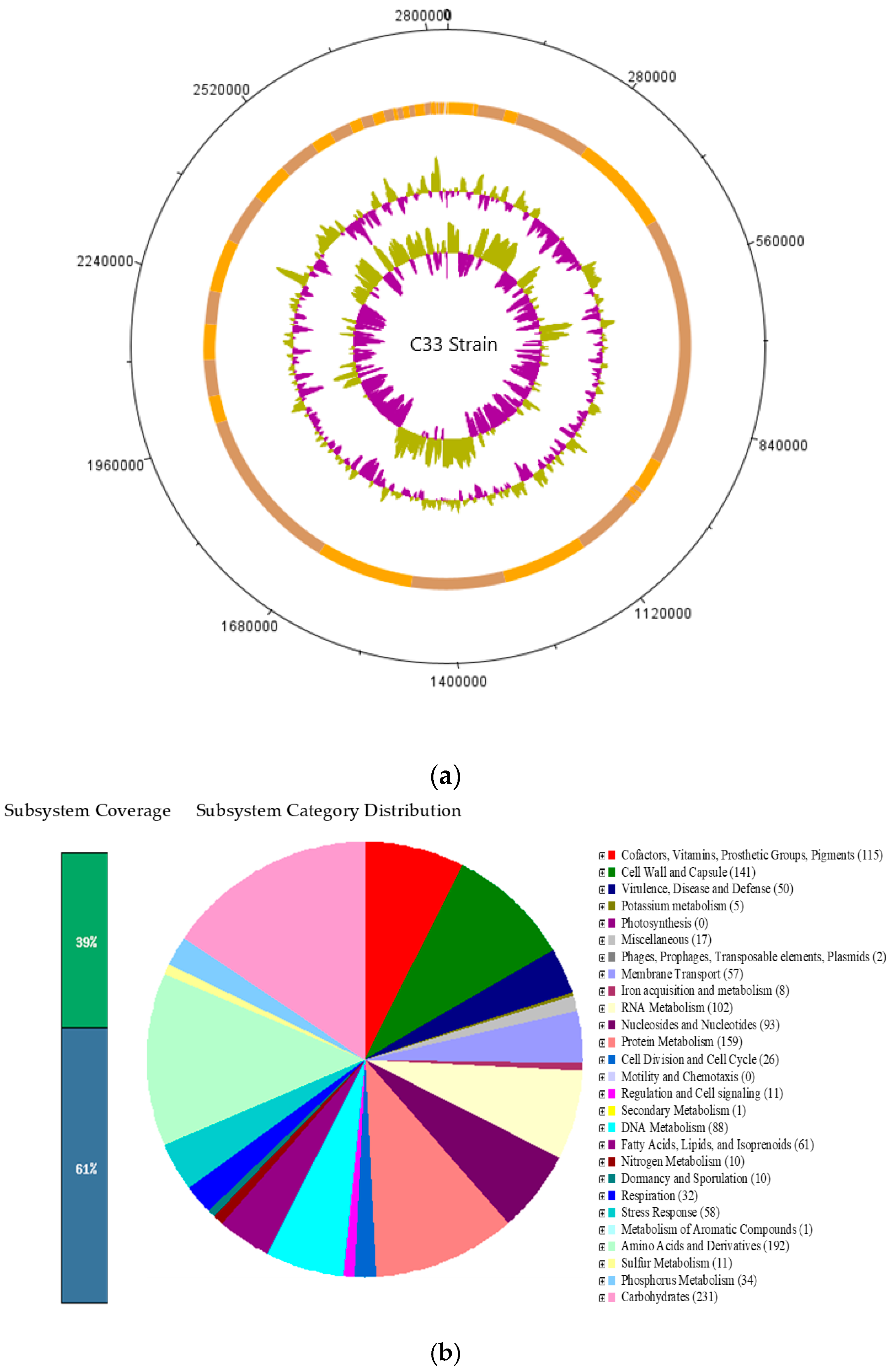
C33 is an anaerobic gram-negative bacterium, that, according to the genome annotation, was identified as Cetobacterium sp. nov C33. Since it was found to be a dominant species, it in vitro analyses that could determine its potential use as probiotic were conducted.
The results of the phenotypic characterization of C33 showed gelatinase negative activity, which is favorable since it has been suggested that the presence of this activity in probiotic microorganisms could be detrimental to the health of the host due to the possible damage it can cause in the extracellular protein matrices of intestinal tissue [42]. In addition, the positive activity of esterase is related to the breaking activity of the ester bonds of polysaccharides, favoring the action of hydrolases of high molecular weight compounds such as carbohydrates and proteins. Likewise, the positive activity of acid phosphatase is important for the degradation of organic phosphorus found primarily in plant and animal protein sources [43]. In addition to these characteristics, the C33 strain has the possibility of using different energy sources such as glucose, sucrose, maltose, salicin, mannitol, and trehalose, a fact that may favor the fish’s nutrition, improving the absorption of nutrients by solubilizing the elements of the diets through extracellular enzymes; this facilitates the absorption of individual molecules through the intestinal epithelium of the animal host [44,45] and provides enzymes that the animal host does not have [44,
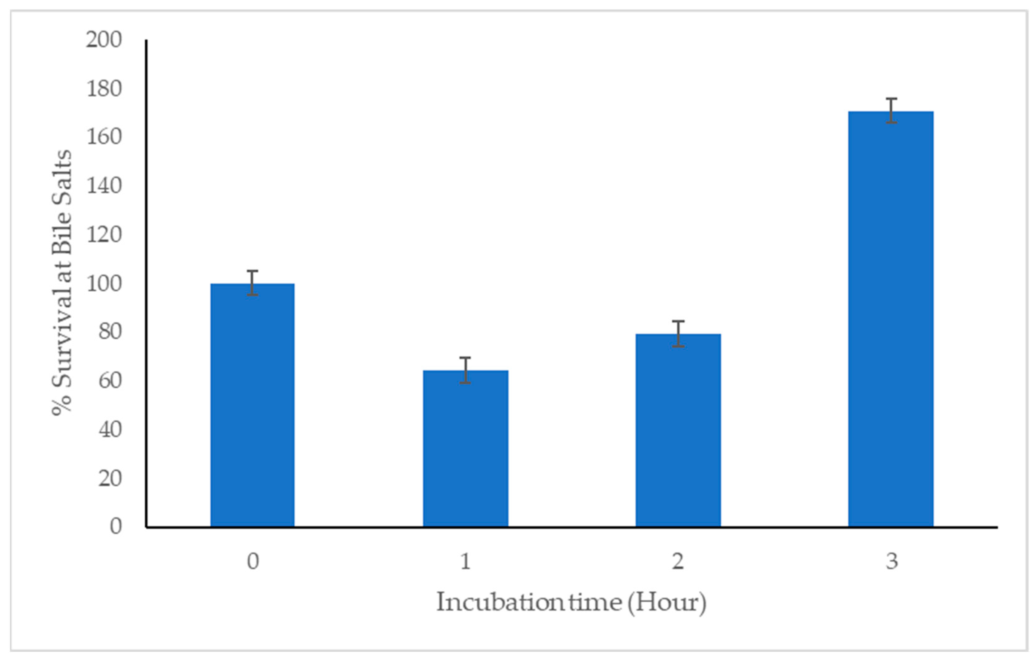
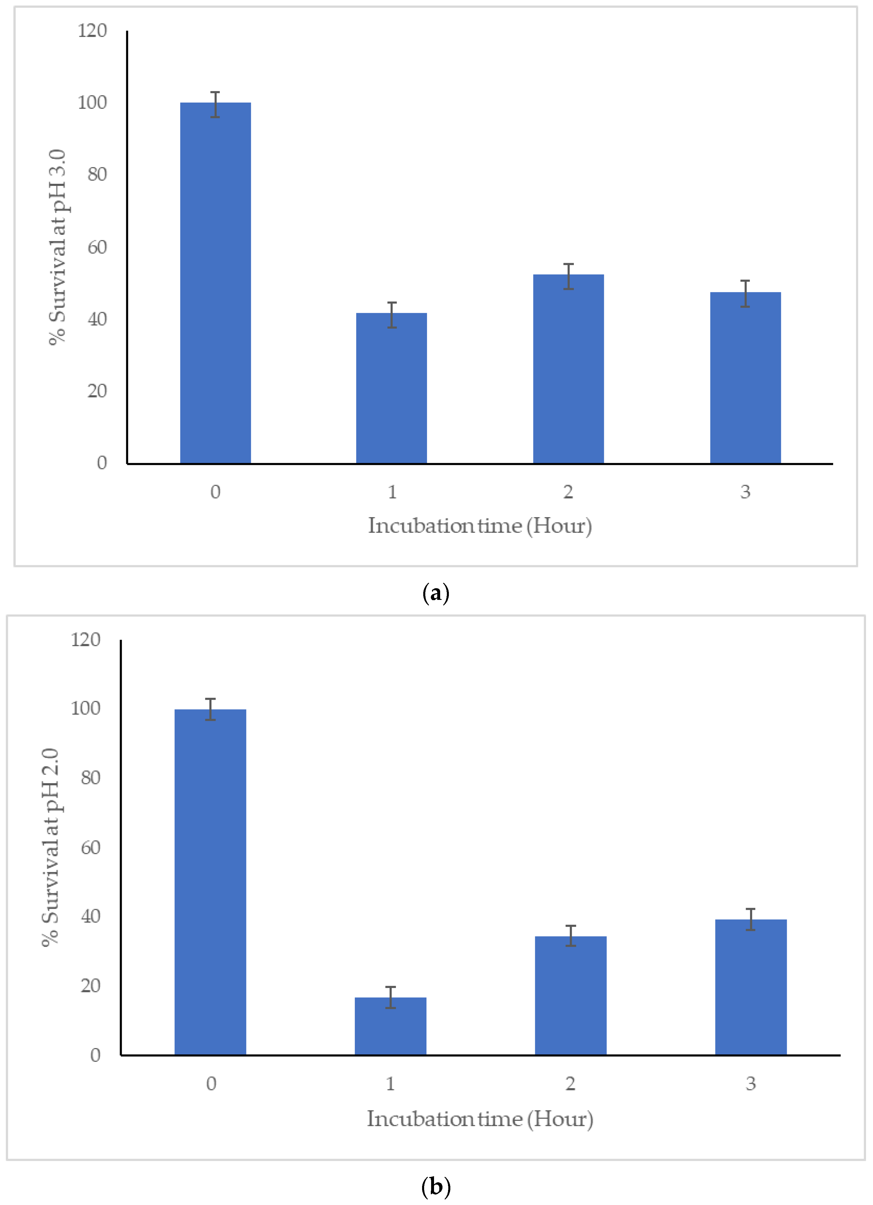
46]. Furthermore, we have identified the C33 strain as a gamma (γ) hemolytic bacterium. In addition, for a bacterium to qualify as a probiotic, it must be able to survive and ideally colonize the intestine. This involves overcoming the stress generated by the low pH of the stomach and contact with bile salts, which are inhibitory for multiple microorganisms because they cause cell lysis [47,48,49,50,51]. Here we report the significant capacity of C33 to survive even at pH 2.0.
On the other hand, after passing through the digestive tract of the host, for a strain candidate to be used as a probiotic it must be able to colonize the gut. The C33 strain showed hydrophobicity percentages higher than 50% with a non-polar solvent (chloroform), a result that indicates a hydrophobic character of the cell surface, a factor that contributes to the interaction of the bacteria with the cells of the gastrointestinal tract. This indicates the potential ability of this isolate to colonize and persist in the gastrointestinal tract [6,41].
Antibiotic resistance and a growing reluctance to administer antibiotics have led to an increase in the use of probiotics [6,52]. Bacterial antibiotic resistance mechanisms can be innate, natural, or acquired. There is no horizontal transferability of the intrinsic resistance; nevertheless, the acquired resistance can be gained by mutations or the acquisition of genes through mobile genetic elements into their genomes [6,53,54]. The resistance to antimicrobial agents in a potential bacterial probiotic should be considered with caution. On the other hand, some authors mention that resistance to given antibiotics may be acceptable because, in the event of the use of any of these compounds being required in the fish culture, the probiotic will not be eliminated [6,55].
Nevertheless, the cutoff value of all the antibiotics tested in C33 indicated that this bacterium is sensitive. Additionally, in the RASfinder genome annotation, no resistance genes were found for any antibiotics (amoxicillin, ertapenem, aztreonam, amoxicillin, ampicillin, piperacillin, cefixime, ceftriaxone, ticarcillin, penicillin, cefotaxime, temocillin, metronidazole, doxycycline, minocycline, tigecycline, teicoplanin, and vancomycin). Due to the potential migration of antibiotic resistance factors, we consider that it is better to use a probiotic free of any acquired antibiotic resistance to avoid further changes in the microbial community induced by the overuse of the probiotic bacteria.
C33 presents with the plasmid rep7a, belonging to the group of glutathione S-transferases (GST), which are dimeric proteins that can conjugate glutathione (GSH) with a variety of compounds containing electrophilic centers. On the other hand, C33 also presented with the rep7a gene, the tetA and tetB genes related with tetracycline resistance, and the Lnu(C) gene that confers resistance to lincomycin [56].
C33 has two ribosomal sactipeptides (peptides cross-linked from sulfur to carbon alpha thioether) belonging to RiPP, that show various biological activities such as antibacterial properties and post-translationally modified peptides (RiPPs) [57]. Nevertheless, this condition requires further research for better understanding of its bacterial physiology.
The C33 strain has a gene encoding the Linocin M18 bacteriocin; a protein that forms nano compartments within the pathogenic bacterium, it also contains ferritin-like proteins or peroxidases and enzymes involved in the oxidative stress response. Various authors have reported that Linocin M18 has bacteriostatic activity against strains of Arthrobacter, Bacillus, Brevibacterium, Corynebacterium, and Listeria [58]. In addition, C33 presents with genes encoding Zoocin A, a peptidoglycan hydrolase, which, combined with lauricidin, a cell membrane active lipid, has been reported to selectively suppress the growth of Streptococcus mutans [59].
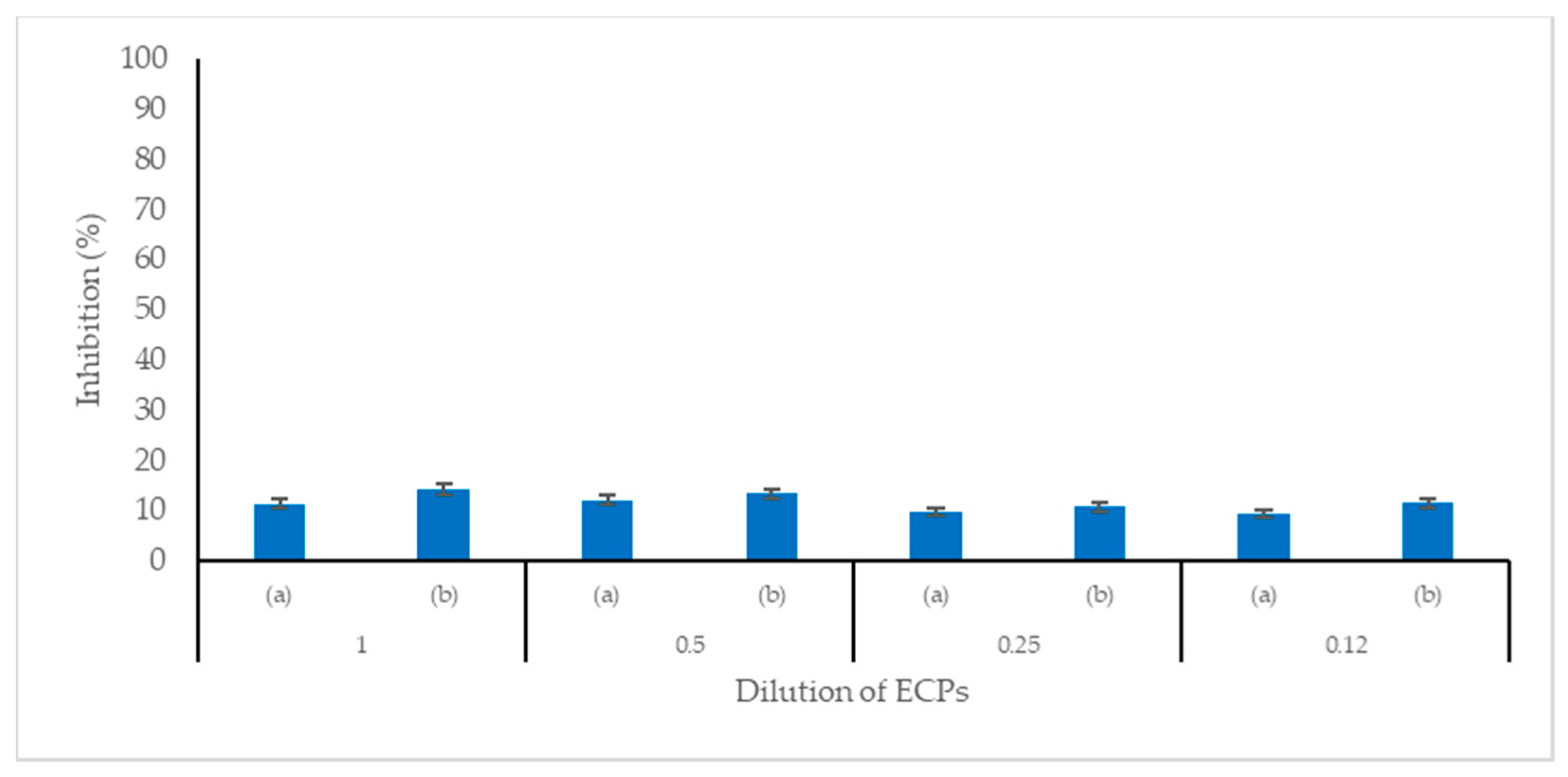
Here cell-free extracellular products (ECPs) of Cetobacterium sp. C33 are found to have antimicrobial activity against S. agalactiae and A. hydrophila, two pathogenic bacteria responsible for high mortality in Nile tilapia cultures and substantial economic losses in tilapia farms [48,60,61,62].
Probiotic use has been reported as an alternative to the use of antibiotics which has caused antimicrobial resistance in the aquaculture industry [63]. They contribute to natural resistance and a higher survival rate of the fish [64,65].
C33 has the capacity to biosynthesize amino acids, such as glutamine, glutamate, aspartate, asparagine, polyamine, methionine, threonine, homoserine, lysine, tryptophan, phenylalanine, tyrosine, proline, glycine, alanine, and serine, that benefit fish growth [66]. In addition, biotin production is important for the synthesis of fatty acids, the oxidation of carbohydrates, and the synthesis of purines. Biotin deficiency causes loss of appetite, dark coloration, and seizures in fish. Thiamine acts in Nile tilapia as a coenzyme in various metabolic decarboxylation reactions (pyruvic acid, alpha-ketoglutaric acid). Thiamine deficiency in fish results in weakness, terminal convulsions, degeneration, and fish fin paralysis. Tetrapyrrole compounds like heme, cobalamin, etc., have multiple essential functions in fish. Its synthesis begins with the formation of aminolaevulinic acid from glutamine [67]. Riboflavin and FAD are components of two enzymes (FMN and FAD) that are oxidases and reductases that participate in the metabolic degradation of proteins, carbohydrates, and lipids. Its deficiency in tilapia generates loss of appetite, dark skin, cataracts, and photophobia. Deficiencies in B vitamins, such as folate and vitamin B12, affect the offspring’s resistance and fertility [68].
In general, the C33 strain breaks down and uses Mannose, Chitin, N-acetylglucosamine, Sucrose, Maltose, Maltodextrin, Lactose, Galactose, Lactate, Glycerol, Glycerol-3-phosphate, D-ribose utilization, L-ascorbate, and Fructose. The genus Cetobacterium provides extracellular enzymes to degrade complex carbohydrates [69]. The presence of Cetobacterium may be associated with better glucose utilization [70] and could improve glucose homeostasis and increase insulin expression [71]. The above supports the role of Cetobacterium in carbohydrate regulation.
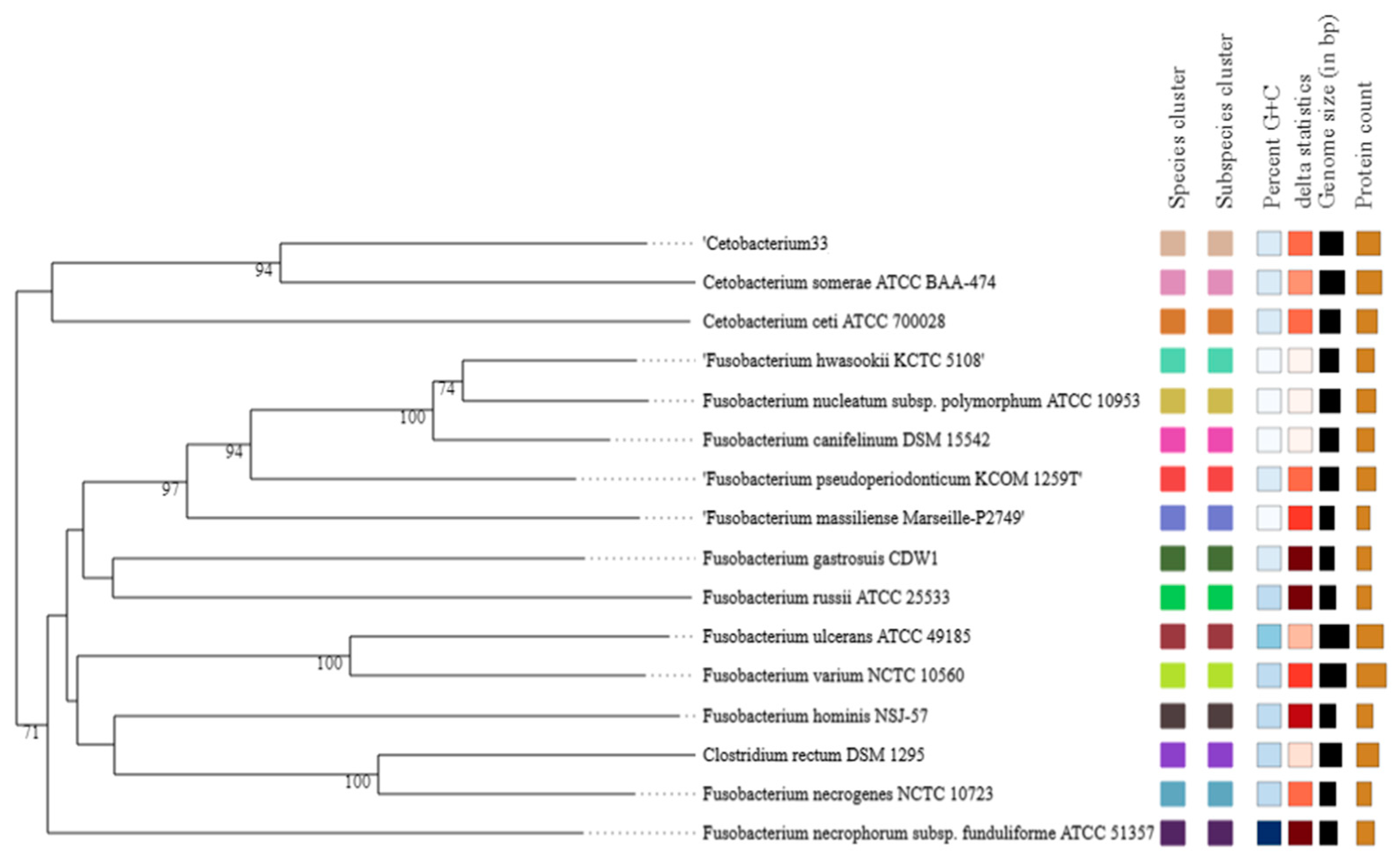
Finally, by comparing the sequencing results of the whole genome with the NCBI database, it was found that the C33 strain can be classified within the genus Cetobacterium. Similarly, the phylogenetic analysis indicates that the C33 strain has the greatest similarity with the species Cetobacterium somerae ATCC BAA-474, with a tetranucleotide signature correlation index (Tetra) result of 0.98712, a value that was less than 0.99 (Minimum value to consider it to be the same species [72]), which suggests that it can be considered a new species. In addition, the C33 strain differs from C. somerae WAL 14325T Cetobacterium sp. NK01 and C. ceti M 3333T in other aspects, such as no indole production, positive glucosidase reaction, positive enzyme activities for esterase (C4) (2-naphthyl butyrate) and acid phosphatase (2-naphthyl phosphate), and finally, it did not present any vitamin B12 production.
Consequently, the physiological characteristics and the phylogenetic analysis suggest that the C33 strain represents a new species of the genus Cetobacterium with high probiotic potential for Nile tilapia cultures based on the in vitro analysis shown here. Nevertheless in vivo experiments should be conducted in other studies to verify the effects on immune regulation, microbiota modulation fish growth, and resistance.

5. Conclusions

The anaerobic bacterial strain C33, isolated from the intestinal microbial content of Nile tilapia was sequenced, showing that it was a new candidate species of the genus Cetobacterium that could be named Cetabacterium colombiensis sp. nov C33.
Isolated C33 has probiotic characteristics which are high adaptability to gastrointestinal conditions, and a potential capacity to adhere to epithelial intestinal cells and produce antimicrobial substances.
To continue with the development of the probiotic product, the next step is to incorporate these bacteria into the fish feed and evaluate the effect on growth performance, microbiota modulation, and immunomodulation.

Supplementary Materials

The following supporting information can be downloaded at: https://www.mdpi.com/article/10.3390/microorganisms11122922/s1, Table S1: Whole genome annotation of bacteria strain C33 with features of interest.

Author Contributions

Conceptualization, M.A.C.G., R.Y.R.P. and L.M.V.; methodology, M.A.C.G., J.A.R. and L.M.V.; software, M.A.C.G., J.F.M.-B. and L.M.V.; validation, M.A.C.G., R.Y.R.P. and L.M.V.; formal analysis, M.A.C.G. and L.M.V.; research, M.A.C.G.; Resources, M.A.C.G. and L.M.V.; data curation, M.A.C.G. and J.F.M.-B.; writing—preparation of original draft, M.A.C.G.; writing: proofreading and editing, M.A.C.G. and L.M.V.; visualization, M.A.C.G.; supervision, R.Y.R.P. and L.M.V.; project management, M.A.C.G. and L.M.V.; acquisition of funds, M.A.C.G. and L.M.V. All authors have read and agreed to the published version of the manuscript.

Funding

Sistema Nacional de Regalias (SGR): Ministerio De Ciencia, Tecnología e Innovación, Colombia. Project “Implementación de herramientas biotecnológicas y sistemas de recirculación para lograr la sostenibilidad del cultivo de tilapia como estrategia productiva para la seguridad e inocuidad alimentaria en La Guajira”. BPIN: 2020000100487, SGR—Asignación para la inversión en ciencia, tecnología e innovación—Departamento de La Guajira. Mario Andrés Colorado Gómez was financed by Fundación Clínica Shaio.

Data Availability Statement

The raw reads used to assemble the draft genome were deposited in the Sequence Read Archive (SRA) under bio project accession number PRJNA1010509. Genome sequence data for C33 was deposited under accession number JAVIKH000000000.

Acknowledgments

Patrimonio Autónomo Fondo Nacional de Financiamiento para la Ciencia, la Tecnología y la Innovación Francisco José de Caldas for the grant number 808-2018 and Universidad de La Sabana. The authors thank the fish farmers for donating the fish specimens and Servicio Nacional de Aprendizaje SENA.

Conflicts of Interest

The authors declare that they have no conflict of interest.

References

  1. Food and Agriculture Organization of the United Nations. The State of World Fisheries and Aquaculture 2022; Food and Agriculture Organization of the United Nations: Rome, Italy, 2022. [Google Scholar] [CrossRef]
  2. Virapat, C. The Current Challenges of Sustainable Aquaculture in the Asia-Pacific Region and the Measures Recommended. Asia-Pac. J. Rural Dev. 2023, 32, 207–216. [Google Scholar] [CrossRef]
  3. Luan, Y.; Li, M.; Zhou, W.; Yao, Y.; Yang, Y.; Zhang, Z.; Ringø, E.; Olsen, R.E.; Clarke, J.L.; Xie, S.; et al. The Fish Microbiota: Research Progress and Potential Applications. Engineering 2023. [Google Scholar] [CrossRef]
  4. Ringø, E.; Zhou, Z.; Vecino, J.L.G.; Wadsworth, S.; Romero, J.; Krogdahl, Å.; Olsen, R.E.; Dimitroglou, A.; Foey, A.; Davies, S.; et al. Effect of dietary components on the gut microbiota of aquatic animals. A never-ending story? Aquac. Nutr. 2016, 22, 219–282. [Google Scholar] [CrossRef]
  5. Llewellyn, M.S.; Boutin, S.; Hoseinifar, S.H.; Derome, N. Teleost microbiomes: The state of the art in their characterization, manipulation and importance in aquaculture and fisheries. Front. Microbiol. 2014, 5, 207. [Google Scholar] [CrossRef]
  6. Melo-Bolívar, J.F.; Pardo, R.Y.R.; Junca, H.; Sidjabat, H.E.; Cano-Lozano, J.A.; Díaz, L.M.V. Competitive Exclusion Bacterial Culture Derived from the Gut Microbiome of Nile Tilapia (Oreochromis niloticus) as a Resource to Efficiently Recover Probiotic Strains: Taxonomic, Genomic, and Functional Proof of Concept. Microorganisms 2022, 10, 1376. [Google Scholar] [CrossRef] [PubMed]
  7. Wilms, E.; Gerritsen, J.; Smidt, H.; Besseling-van der Vaart, I.; Rijkers, G.T.; Fuentes, A.R.G.; Masclee, A.A.M.; Troost, F.J. Effects of Supplementation of the Synbiotic Ecologic® 825/FOS P6 on Intestinal Barrier Function in Healthy Humans: A Randomized Controlled Trial. PLoS ONE 2016, 11, e0167775. [Google Scholar] [CrossRef]
  8. Tsuchiya, C.; Sakata, T.; Sugita, H. Novel ecological niche of Cetobacterium somerae, an anaerobic bacterium in the intestinal tracts of freshwater fish. Lett. Appl. Microbiol. 2008, 46, 43–48. [Google Scholar] [CrossRef]
  9. Ramírez, C.; Coronado, J.; Silva, A.; Romero, J. Cetobacterium Is a Major Component of the Microbiome of Giant Amazonian Fish (Arapaima gigas) in Ecuador. Animals 2018, 8, 189. [Google Scholar] [CrossRef]
  10. Melo-Bolívar, J.F.; Ruiz Pardo, R.Y.; Hume, M.E.; Nisbet, D.J.; Rodríguez-Villamizar, F.; Alzate, J.F.; Junca, H.; Villamil Díaz, L.M. Establishment and characterization of a competitive exclusion bacterial culture derived from Nile tilapia (Oreochromis niloticus) gut microbiomes showing antibacterial activity against pathogenic Streptococcus agalactiae. PLoS ONE 2019, 14, e0215375. [Google Scholar] [CrossRef]
  11. LaFrentz, B.R.; LaFrentz, S.A.; Beck, B.H.; Arias, C.R. Draft Genome Sequences of Cetobacterium somerae 2G Large and Two Novel Cetobacterium Isolates from Intestines of Channel Catfish (Ictalurus punctatus). Microbiol. Resour. Announc. 2020, 9, 44. [Google Scholar] [CrossRef]
  12. Xie, M.; Xie, Y.; Li, Y.; Zhou, W.; Zhang, Z.; Yang, Y.; Olsen, R.E.; Ringø, E.; Ran, C.; Zhou, Z. Stabilized fermentation product of Cetobacterium somerae improves gut and liver health and antiviral immunity of zebrafish. Fish Shellfish Immunol. 2022, 120, 56–66. [Google Scholar] [CrossRef] [PubMed]
  13. Zhang, Z.; Fan, Z.; Yi, M.; Liu, Z.; Ke, X.; Gao, F.; Cao, J.; Wang, M.; Chen, G.; Lu, M. Characterization of the core gut microbiota of Nile tilapia (Oreochromis niloticus): Indication of a putative novel Cetobacterium species and analysis of its potential function on nutrition. Arch. Microbiol. 2022, 204, 690. [Google Scholar] [CrossRef] [PubMed]
  14. European Food Safety Authority (EFSA). Panel on Additives and Products or Substances Used in Animal Feed (FEEDAP) guidance on the assessment of bacterial susceptibility to antimicrobials of human and veterinary importance. EFSA J. 2012, 10, 2740–2750. [Google Scholar]
  15. Amachi, S.; Kawaguchi, N.; Muramatsu, Y.; Tsuchiya, S.; Watanabe, Y.; Shinoyama, H.; Fujii, T. Dissimilatory iodate reduction by marine Pseudomonas sp. strain SCT. Appl. Environ. Microbiol. 2007, 73, 5725–5730. [Google Scholar] [CrossRef] [PubMed]
  16. Li, Y.; Lu, C.; Yu, Z.; Ma, Q. Isolation of Enterococcus faecium with Feeding Attractant Function from Pacific White Shrimp (Litopenaeus vannamei) Intestine. J. Ocean. Univ. China 2020, 19, 931–940. [Google Scholar] [CrossRef]
  17. Monteagudo Mera, A. Selección In Vitro de Microorganismos con Potencial Probiótico. Ph.D. Thesis, University of León, León, Spain, 2010. [Google Scholar] [CrossRef]
  18. Bankevich, A.; Nurk, S.; Antipov, D.; Gurevich, A.A.; Dvorkin, M.; Kulikov, A.S.; Lesin, V.M.; Nikolenko, S.I.; Pham, S.; Prjibelski, A.D.; et al. SPAdes: A new genome assembly algorithm and its applications to single-cell sequencing. J. Comput. Biol. J. Comput. Mol. Cell Biol. 2012, 19, 455–477. [Google Scholar] [CrossRef] [PubMed]
  19. Richter, M.; Rosselló-Móra, R.; Oliver Glöckner, F.O.; Peplies, J. JSpeciesWS: A web server for prokaryotic species circumscription based on pairwise genome comparison. Bioinformatics 2016, 32, 929–931. [Google Scholar] [CrossRef]
  20. Meier-Kolthoff, J.P.; Göker, M. TYGS is an automated high-throughput platform for state-of-the-art genome-based taxonomy. Nat. Commun. 2019, 10, 2182. [Google Scholar] [CrossRef]
  21. Seemann, T. Prokka: Rapid prokaryotic genome annotation. Bioinformatics 2014, 30, 2068–2069. [Google Scholar] [CrossRef]
  22. Luo, J.; Jin, F.Q.; Yin, M.; Jin, Z.G. Regulation of SRF protein stability by an autophagy-dependent pathway. Biochem. Biophys. Res. Commun. 2020, 521, 279–284. [Google Scholar] [CrossRef]
  23. Bosi, E.; Donati, B.; Galardini, M.; Brunetti, S.; Sagot, M.-F.; Lió, P.; Crescenzi, P.; Fani, R.; Fondi, M. MeDuSa: A multi-draft based scaffolder. Bioinformatics 2015, 31, 2443–2451. [Google Scholar] [CrossRef]
  24. Meier-Kolthoff, J.P.; Carbasse, J.S.; Peinado-Olarte, R.L.; Göker, M. TYGS and LPSN: A database tandem for fast and reliable genome-based classification and nomenclature of prokaryotes. Nucleic Acids Res. 2022, 50, D801–D807. [Google Scholar] [CrossRef] [PubMed]
  25. Kapse, N.G.; Engineer, A.S.; Gowdaman, V.; Wagh, S.; Dhakephalkar, P.K. Functional annotation of the genome unravels probiotic potential of Bacillus coagulans HS243. Genomics 2019, 111, 921–929. [Google Scholar] [CrossRef] [PubMed]
  26. Bortolaia, V.; Kaas, R.S.; Ruppe, E.; Roberts, M.C.; Schwarz, S.; Cattoir, V.; Philippon, A.; Allesoe, R.L.; Rebelo, A.R.; Florensa, A.F.; et al. ResFinder 4.0 for predictions of phenotypes from genotypes. J. Antimicrob. Chemother. 2020, 75, 3491–3500. [Google Scholar] [CrossRef] [PubMed]
  27. Clausen, P.T.L.C.; Aarestrup, F.M.; Lund, O. Rapid and precise alignment of raw reads against redundant databases with KMA. BMC Bioinform. 2018, 19, 307. [Google Scholar] [CrossRef] [PubMed]
  28. Zankari, E.; Allesøe, R.; Joensen, K.G.; Cavaco, L.M.; Lund, O.; Aarestrup, F.M. PointFinder: A novel web tool for WGS-based detection of antimicrobial resistance associated with chromosomal point mutations in bacterial pathogens. J. Antimicrob. Chemother. 2017, 72, 2764–2768. [Google Scholar] [CrossRef] [PubMed]
  29. Joensen, K.G.; Scheutz, F.; Lund, O.; Hasman, H.; Kaas, R.S.; Nielsen, E.M.; Aarestrup, F.M. Real-time whole-genome sequencing for routine typing, surveillance, and outbreak detection of verotoxigenic Escherichia coli. J. Clin. Microbiol. 2014, 52, 1501–1510. [Google Scholar] [CrossRef] [PubMed]
  30. Johansson, M.H.K.; Bortolaia, V.; Tansirichaiya, S.; Aarestrup, F.M.; Roberts, A.P.; Petersen, T.N. Detection of mobile genetic elements associated with antibiotic resistance in Salmonella enterica using a newly developed web tool: MobileElementFinder. J. Antimicrob. Chemother. 2021, 76, 101–109. [Google Scholar] [CrossRef]
  31. Yılmaz Çolak, Ç. Computational Design of a Multi-epitope Vaccine Against Clostridium chauvoei: An Immunoinformatics Approach. Int. J. Pept. Res. Ther. 2021, 27, 2639–2649. [Google Scholar] [CrossRef]
  32. Chen, L.; Zheng, D.; Liu, B.; Yang, J.; Jin, Q. VFDB 2016: Hierarchical and refined dataset for big data analysis—10 years on. Nucleic Acids Res. 2016, 44, D694–D697. [Google Scholar] [CrossRef]
  33. Medema, M.H.; Blin, K.; Cimermancic, P.; De Jager, V.; Zakrzewski, P.; Fischbach, M.A.; Weber, T.; Takano, E.; Breitling, R. antiSMASH: Rapid identification, annotation and analysis of secondary metabolite biosynthesis gene clusters in bacterial and fungal genome sequences. Nucleic Acids Res. 2011, 39 (Suppl. 2), W339–W346. [Google Scholar] [CrossRef] [PubMed]
  34. van Heel, A.J.; de Jong, A.; Song, C.; Viel, J.H.; Kok, J.; Kuipers, O.P. BAGEL4: A user-friendly web server to thoroughly mine RiPPs and bacteriocins. Nucleic Acids Res. 2018, 46, W278–W281. [Google Scholar] [CrossRef] [PubMed]
  35. Patel, D.; Gismondi, R.; Alsaffar, A.; Tiquia-Arashiro, S.M. Applicability of API ZYM to capture seasonal and spatial variabilities in lake and river sediments. Environ. Technol. 2018, 40, 3227–3239. [Google Scholar] [CrossRef] [PubMed]
  36. Latimer, G.W., Jr. (Ed.) Official Methods of Analysis of AOAC INTERNATIONAL; AOAC International: Rockville, MD, USA, 2023. [Google Scholar] [CrossRef]
  37. Darilmaz, D.O.; Beyatli, Y.; Yuksekdag, Z.N. Aggregation and Hydrophobicity Properties of 6 Dairy Propionibacteria Strains Isolated from Homemade Turkish Cheeses. J. Food Sci. 2012, 77, M20–M24. [Google Scholar] [CrossRef] [PubMed]
  38. Nader-Macías, M.E.F.; Otero, M.C.; Espeche, M.C.; Maldonado, N.C. Advances in the design of probiotic products for the prevention of major diseases in dairy cattle. J. Ind. Microbiol. Biotechnol. 2008, 35, 1387–1395. [Google Scholar] [CrossRef] [PubMed]
  39. Flórez, A.B.; Danielsen, M.; Korhonen, J.; Zycka, J.; von Wright, A.; Bardowski, J.; Mayo, B. Antibiotic survey of Lactococcus lactis strains to six antibiotics by Etest, and establishment of new susceptibility-resistance cut-off values. J. Dairy Res. 2007, 74, 262–268. [Google Scholar] [CrossRef] [PubMed]
  40. Cabo, M.L.; Murado, M.A.; Gonzalez, M.P.; Pastoriza, L. A method for bacteriocin quantification. J. Appl. Microbiol. 1999, 87, 907–914. [Google Scholar] [CrossRef]
  41. Adnan, M.; Patel, M.; Hadi, S. Functional and health promoting inherent attributes of Enterococcus hirae F2 as a novel probiotic isolated from the digestive tract of the freshwater fish Catla catla. PeerJ 2017, 5, e3085. [Google Scholar] [CrossRef]
  42. de Oliveira, C.G.; López-Olmeda, J.F.; Costa, L.S.; do Espirito Santo, A.H.; dos Santos FA, C.; Luz, R.K.; Ribeiro, P.A.P. Gastrointestinal emptying and daily patterns of activity of proteinolytic enzymes in Nile tilapia (Oreochromis niloticus). Aquaculture 2022, 546, 737338. [Google Scholar] [CrossRef]
  43. Jiang, Z.; Su, W.; Yang, M.; Li, W.; Gong, T.; Zhang, Y.; Wen, C.; Wang, X.; Wang, Y.; Jin, M.; et al. Screening of Bacteria Inhibiting Clostridium perfringens and Assessment of Their Beneficial Effects In Vitro and In Vivo with Whole Genome Sequencing Analysis. Microorganisms 2022, 10, 2056. [Google Scholar] [CrossRef]
  44. Butt, R.L.; Volkoff, H. Gut Microbiota and Energy Homeostasis in Fish. Front. Endocrinol. 2019, 10, 9. [Google Scholar] [CrossRef]
  45. Ikeda-Ohtsubo, W.; Brugman, S.; Warden, C.H.; Rebel, J.M.J.; Folkerts, G.; Pieterse, C.M.J. How Can We Define “Optimal Microbiota?”: A Comparative Review of Structure and Functions of Microbiota of Animals, Fish, and Plants in Agriculture. Front. Nutr. 2018, 5, 90. [Google Scholar] [CrossRef]
  46. Zhou, W.-H.; Wu, C.-C.; Limbu, S.M.; Li, R.-X.; Chen, L.-Q.; Qiao, F.; Luo, Y.; Zhang, M.-L.; Han, T.; Du, Z.-Y. More simple more worse: Simple carbohydrate diets cause alterations in glucose and lipid metabolism in Nile tilapia (Oreochromis niloticus). Aquaculture 2022, 550, 737857. [Google Scholar] [CrossRef]
  47. Basri, L.; Nor, R.M.; Salleh, A.; Yasin, I.S.M.; Saad, M.Z.; Rahaman, N.Y.A.; Barkham, T.; Amal, M.N.A. Co-Infections of Tilapia Lake Virus, Aeromonas hydrophila and Streptococcus agalactiae in Farmed Red Hybrid Tilapia. Animals 2020, 10, 2141. [Google Scholar] [CrossRef]
  48. Kusdarwati, R.; Amin, M.; Wardana, A.B. DNase and Gelatinase Activities Of β-Hemolysin Aeromonas hydrophila Isolated from Catfish (Clarias batrachus). J. Aquac. Fish Health 2021, 10, 331–340. [Google Scholar] [CrossRef]
  49. Vidhya Hindu, S.; Chandrasekaran, N.; Mukherjee, A.; Thomas, J. A review on the impact of seaweed polysaccharide on the growth of probiotic bacteria and its application in aquaculture. Aquac. Int. 2019, 27, 227–238. [Google Scholar] [CrossRef]
  50. Liu, H.; Wang, S.; Cai, Y.; Guo, X.; Cao, Z.; Zhang, Y.; Liu, S.; Yuan, W.; Zhu, W.; Zheng, Y.; et al. Dietary administration of Bacillus subtilis HAINUP40 enhances growth, digestive enzyme activities, innate immune responses and disease resistance of tilapia, Oreochromis niloticus. Fish Shellfish Immunol. 2017, 60, 326–333. [Google Scholar] [CrossRef]
  51. Kumar, V.; Roy, S.; Meena, D.K.; Sarkar, U.K. Application of Probiotics in Shrimp Aquaculture: Importance, Mechanisms of Action, and Methods of Administration. Rev. Fish. Sci. Aquac. 2016, 24, 342–368. [Google Scholar] [CrossRef]
  52. Sankar, H.; Philip, B.; Philip, R.; Singh, I.S.B. Effect of probiotics on digestive enzyme activities and growth of cichlids, Etroplus suratensis (Pearl spot) and Oreochromis mossambicus (Tilapia). Aquac. Nutr. 2017, 23, 852–864. [Google Scholar] [CrossRef]
  53. Slizovskiy, I.B.; Mukherjee, K.; Dean, C.J.; Boucher, C.; Noyes, N.R. Mobilization of Antibiotic Resistance: Are Current Approaches for Colocalizing Resistomes and Mobilomes Useful? Front. Microbiol. 2020, 11, 1376. [Google Scholar] [CrossRef]
  54. Perry, J.A.; Wright, G.D. The antibiotic resistance “mobilome”: Searching for the link between environment and clinic. Front. Microbiol. 2013, 4, 138. [Google Scholar] [CrossRef]
  55. Reda, R.M.; Selim, K.M.; El-Sayed, H.M.; El-Hady, M.A. In Vitro Selection and Identification of Potential Probiotics Isolated from the Gastrointestinal Tract of Nile Tilapia, Oreochromis niloticus. Probiotics Antimicrob. Proteins 2018, 10, 692–703. [Google Scholar] [CrossRef]
  56. Mores, C.R.; Montelongo, C.; Putonti, C.; Wolfe, A.J.; Abouelfetouh, A. Investigation of Plasmids Among Clinical Staphylococcus aureus and Staphylococcus haemolyticus Isolates From Egypt. Front. Microbiol. 2021, 12, 659116. [Google Scholar] [CrossRef]
  57. Chen, Y.; Wang, J.; Li, G.; Yang, Y.; Ding, W. Current Advancements in Sactipeptide Natural Products. Front. Chem. 2021, 9, 595991. [Google Scholar] [CrossRef]
  58. Babar, T.K.; Glare, T.R.; Hampton, J.G.; Hurst, M.R.H.; Narciso, J.; Sheen, C.R.; Koch, B. Linocin M18 protein from the insect pathogenic bacterium Brevibacillus laterosporus isolates. Appl. Microbiol. Biotechnol. 2023, 107, 4337–4353. [Google Scholar] [CrossRef]
  59. Lester, K.; Simmonds, R.S. Zoocin A and Lauricidin in Combination Reduce Streptococcus mutans Growth in a Multispecies Biofilm. Caries Res. 2012, 46, 185–193. [Google Scholar] [CrossRef]
  60. Zhang, D.; Liu, Z.; Ren, Y.; Wang, Y.; Pan, H.; Liang, D.; Bei, W.; Chang, O.; Wang, Q.; Shi, C. Epidemiological characteristics of Streptococcus agalactiae in tilapia in China from 2006 to 2020. Aquaculture 2022, 549, 737724. [Google Scholar] [CrossRef]
  61. Wang, B.; Gan, Z.; Wang, Z.; Yu, D.; Lin, Z.; Lu, Y.; Wu, Z.; Jian, J. Integrated analysis neurimmiRs of tilapia (Oreochromis niloticus) involved in immune response to Streptococcus agalactiae, a pathogen causing meningoencephalitis in teleosts. Fish Shellfish Immunol. 2017, 61, 44–60. [Google Scholar] [CrossRef]
  62. Iwashita, M.K.P.; Nakandakare, I.B.; Terhune, J.S.; Wood, T.; Ranzani-Paiva, M.J.T. Dietary supplementation with Bacillus subtilis, Saccharomyces cerevisiae and Aspergillus oryzae enhance immunity and disease resistance against Aeromonas hydrophila and Streptococcus iniae infection in juvenile tilapia Oreochromis niloticus. Fish Shellfish Immunol. 2015, 43, 60–66. [Google Scholar] [CrossRef]
  63. Preena, P.G.; Arathi, D.; Raj, N.S.; Arun Kumar, T.V.; Arun Raja, S.; Reshma, R.N.; Raja Swaminathan, T. Diversity of antimicrobial-resistant pathogens from a freshwater ornamental fish farm. Lett. Appl. Microbiol. 2020, 71, 108–116. [Google Scholar] [CrossRef]
  64. Park, Y.; Lee, S.; Hong, J.; Kim, D.; Moniruzzaman, M.; Bai, S.C. Use of probiotics to enhance growth, stimulate immunity and confer disease resistance to Aeromonas salmonicida in rainbow trout (Oncorhynchus mykiss). Aquac. Res. 2017, 48, 2672–2682. [Google Scholar] [CrossRef]
  65. Gupta, A.; Gupta, P.; Dhawan, A. Dietary supplementation of probiotics affects growth, immune response and disease resistance of Cyprinus carpio fry. Fish Shellfish Immunol. 2014, 41, 113–119. [Google Scholar] [CrossRef] [PubMed]
  66. Xie, S.; Tian, L.; Niu, J.; Liang, G.; Liu, Y. Effect of N-acetyl cysteine and glycine supplementation on growth performance, glutathione synthesis, and antioxidative ability of grass carp, Ctenopharyngodon idella. Fish Physiol. Biochem. 2017, 43, 1011–1020. [Google Scholar] [CrossRef] [PubMed]
  67. Bryant, D.A.; Hunter, C.N.; Warren, M.J. Biosynthesis of the modified tetrapyrroles-the pigments of life. J. Biol. Chem. 2020, 295, 6888–6925. [Google Scholar] [CrossRef]
  68. Skjærven, K.H.; Jakt, L.M.; Dahl, J.A.; Espe, M.; Aanes, H.; Hamre, K.; Fernandes, J.M.O. Parental vitamin deficiency affects the embryonic gene expression of immune-, lipid transport- and apolipoprotein genes. Sci. Rep. 2016, 6, 34535. [Google Scholar] [CrossRef]
  69. Larsen, A.M.; Mohammed, H.H.; Arias, C.R. Characterization of the gut microbiota of three commercially valuable warmwater fish species. J. Appl. Microbiol. 2014, 116, 1396–1404. [Google Scholar] [CrossRef]
  70. Gong, Y.; Lu, Q.; Liu, Y.; Xi, L.; Zhang, Z.; Liu, H.; Jin, J.; Yang, Y.; Zhu, X.; Xie, S.; et al. Dietary berberine alleviates high carbohydrate diet-induced intestinal damages and improves lipid metabolism in largemouth bass (Micropterus salmoides). Front. Nutr. 2022, 9, 1010859. [Google Scholar] [CrossRef]
  71. Wang, A.; Zhang, Z.; Ding, Q.; Yang, Y.; Bindelle, J.; Ran, C.; Zhou, Z. Intestinal Cetobacterium and acetate modify glucose homeostasis via parasympathetic activation in zebrafish. Gut Microbes 2021, 13, 1–15. [Google Scholar] [CrossRef]
  72. Richter, M.; Rosselló-Móra, R. Shifting the genomic gold standard for the prokaryotic species definition. Proc. Natl. Acad. Sci. USA 2009, 106, 19126–19131. [Google Scholar] [CrossRef]
Figure 1. Cetobacterium sp. nov C33. (a) Colony morphology, 24 h culture in Columbia agar; (b) Gram staining, bright field microscopy (100×); SEM images of morphology and arrangement of C33 after 24 h culture in Columbia broth. (c) 5000× magnification and (d) 7000× magnification.
Microorganisms 11 02922 g001
Figure 2. Cetobacterium sp. nov C33 whole-genome analysis. (a) Genomic atlas. Circles illustrate the following, from outermost to innermost rings: the scaffolds; the location of the contigs; the local % GC plot, and the innermost ring represents the GC skew. (b) Subsystem distribution of Cetobacterium sp. nov C33 based on Rapid Annotation using Subsystem Technology (RAST). Numbers mean: Number of Coding Sequences.
Microorganisms 11 02922 g002
Figure 3. Cetobacterium sp. nov. C33 whole-genome sequence-based phylogenomic tree. The tree was inferred using FastME 2.1.6.1 with Genome Blast Distance Phylogeny approach (GBDP) distances calculated from genome sequences. The branch lengths were scaled in terms of GBDP-distance formula d5. The numbers below the branches are GBDP pseudo-bootstrap support values > 60% from 100 replications, with an average branch support of 73.1%. For the Percent G + C and Delta statistics columns, Darker color means higher value.
Microorganisms 11 02922 g003
Figure 4. Evaluation of Cetobacterium sp. nov. C33 survival at 0.3 percent w/v% bile salts). Data represents mean + SEM (n = 3).
Microorganisms 11 02922 g004
Figure 5. Evaluation of Cetobacterium sp. nov. C33 survival at (a) pH 3.0; (b) pH 2.0. Data represents mean + SEM (n = 3).
Microorganisms 11 02922 g005
Figure 6. Antibacterial activity of the extracellular products (ECPs) of Cetobacterium sp. nov C33 against Streptococcus agalactiae: (a) ECPs heated at 80 °C; (b) ECPs without heating at 80 °C. Data represents mean + SEM (n = 3).
Microorganisms 11 02922 g006
Figure 7. Antibacterial activity of the extracellular products (ECPs) of Cetobacterium sp. nov C33 against Aeromonas hydrophila: (a) ECPs heated at 80 °C; (b) ECPs without heating at 80 °C. Data represents mean + SEM (n = 3).
Microorganisms 11 02922 g007
Table 1. Cetobacterium sp. nov. C33 phenotypic properties. (−) negative reaction, and (+) positive reaction.
PropertiesCetobacterium sp. nov. C33
Indole formation
Urease
Acidification (GLUcose)+
Acidification (MANnitol)
Acidification (LACtose)
Acidification (SACcharose)+
Acidification (MALtose)+
Acidification (SALicin)+
Acidification (XYLose)
Acidification (ARAbinose)
Hydrolysis (protease) (GELatin)
Hydrolysis (ß-glucosidase) (ESCulin)+
Acidification (GLYcerol)
Acidification (CELlobiose)+
Acidification (ManNosE)+
Acidification (MeLeZitose)+
Acidification (RAFfinose)
Acidification (SORbitol)
Acidification (RHAmnose)
Acidification (TREhalose)
Catalase
Vitamin B12
Spores
Gram reaction
MorphologyRod
Table 2. Cetobacterium sp. nov. C33 pairwise genome comparisons vs. type strain genomes. Average Nucleotide Identity based on BLAST (ANIb); Average Nucleotide Identity values based on MUMmer algorithm (ANIm); digital DNA-DNA hybridization (dDDH).
Subject StrainNCBI RefSeqANIbANImdDDH (d4, in %)Tetra
Cetobacterium somerae ATCC BAA-474GCA_000479045.183.6486.2128.80.98320
Cetobacterium sp. NK01NCBI:txid299353083.9787.7328.90.98225
Cetobacterium ceti ATCC 700028GCA_900167275.173.3383.6319.20.93057
Table 3. Gene clusters involved in biosynthesis in Cetobacterium sp. nov. C33 obtained from BAGEL4 web server.
Areas Of Interest (AOI)Contig PositionClass
NODE_14_length_68805_cov_11741857.1.AOI_019860–29,860Sactipeptides
NODE_14_length_68805_cov_11741857.1.AOI_0221,395–41,395Sactipeptides
NODE_12_length_73494_cov_14215396.25.AOI_0124,074–44,8273.3; Bacteriocin
NODE_13_length_71663_cov_18402088.3.AOI_0138,945–59,28493.3; Zoocin_A
Table 4. Cetobacterium sp. nov. C33 genetic elements obtained from Mobile Element Finder tool.
GenPhenotypeAccessionCountingContig PositionCoverageIdentity
tetB(P)doxycycline, tetracycline, minocyclineNC_010937NODE_39_length_5959_cov_111.589261_pilon1788–3746100.00%97.54%
tetA(P)doxycycline, tetracyclineAB054980NODE_39_length_5959_cov_111.589261_pilon1804–54299.92%92.72%
Inu(C)LincomycinAY928180NODE_61_length_1005_cov_102.149888_pilon429–923100.00%98.99%
Table 5. Cetobacterium sp. nov. C33 plasmid obtained from Mobile Element Finder tool.
Name of PlasmidDatabaseAccessionCountingPosition in ContigCoverageIdentity
rep7aGram-positiveSAU83488NODE_53_length_2280_cov_1.059013_pilon1408–197695.31%90.53%
Table 6. Cetobacterium sp. nov. C33 enzymatic activity analysis using Api Zym. (+) Positive result, (−) Negative result.
Enzyme AnalyzedCetobacterium sp. nov. C33
Phosphatase alkaline
Esterase (C 4)+
Esterase Lipase (C 8)
Lipase (C 14)
Leucine arylamidase
Valine arylamidase
Cystine arylamidase
Trypsine
α-chymotrypsine
Phosphatase acide+
Naphtol-AS-BI-phosphohydrolase
α-galactosidase
ß-galactosidase
ß-glucuronidase
α-glucosidase
ß-glucosidase
N-acetyl-ß-glucosaminidase
α-mannosidase
α-fucosidase
Table 7. Antibiotic Minimum Inhibitory Concentrations (MIC) were obtained in Cetobacterium sp. nov. C33.
AntibioticMIC C33 Strain* Escherichia coli
(μg/mL)(μg/mL)
Tetracycline3 (S)8
Ampicillin0.016 (S)8
Vancomycin0.250 (S)not reported
Gentamicin0.750 (S)2
Chloramphenicol1.5 (S)16
* Cut off values by EFSA (µg/mL) for Escherichia coli. Resistant (R) MIC > cut-off value; Susceptible (S) MIC < cut-off value.
Disclaimer/Publisher’s Note: The statements, opinions and data contained in all publications are solely those of the individual author(s) and contributor(s) and not of MDPI and/or the editor(s). MDPI and/or the editor(s) disclaim responsibility for any injury to people or property resulting from any ideas, methods, instructions or products referred to in the content.

