
Acute Pyelonephritis with Bacteremia in an 89-Year-Old Woman Caused by Two Slow-Growing Bacteria: Aerococcus urinae and Actinotignum schaalii


Abstract
1. Introduction
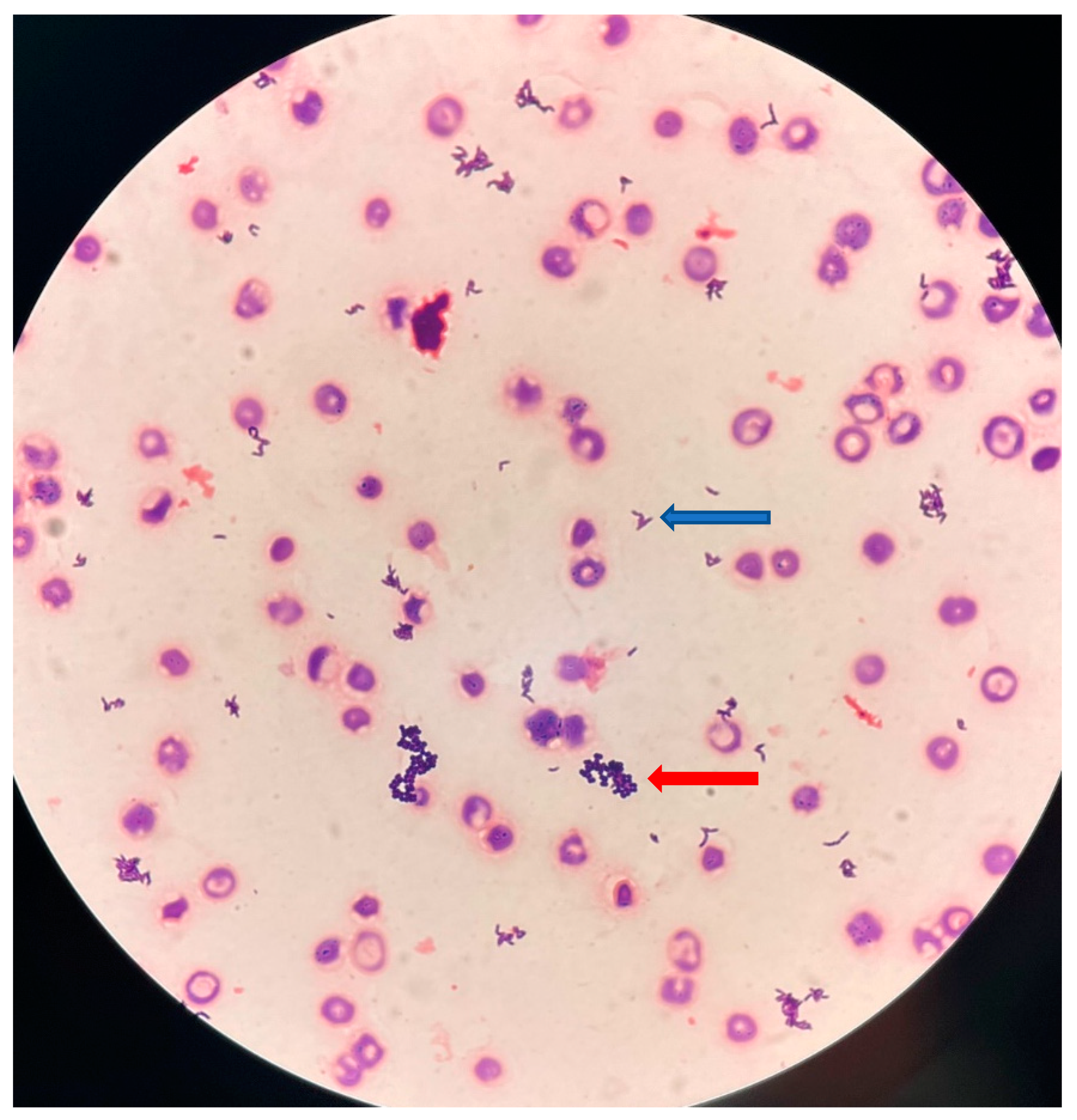
2. Case Report
3. Discussion
4. Conclusions
Author Contributions
Funding
Institutional Review Board Statement
Informed Consent Statement
Data Availability Statement
Acknowledgments
Conflicts of Interest
References
- Rasmussen, M. Aerococcus: An Increasingly Acknowledged Human Pathogen. Clin. Microbiol. Infect. 2016, 22, 22–27. [Google Scholar] [CrossRef] [PubMed]
- Lotte, R.; Lotte, L.; Ruimy, R. Actinotignum schaalii (Formerly Actinobaculum schaalii): A Newly Recognized Pathogen—Review of the Literature. Clin. Microbiol. Infect. 2016, 22, 28–36. [Google Scholar] [CrossRef] [PubMed]
- Siddiqui, H.; Nederbragt, A.J.; Lagesen, K.; Jeansson, S.L.; Jakobsen, K.S. Assessing Diversity of the Female Urine Microbiota by High Throughput Sequencing of 16S rDNA Amplicons. BMC Microbiol. 2011, 11, 244. [Google Scholar] [CrossRef] [PubMed]
- Lotte, R.; Durand, M.; Mbeutcha, A.; Ambrosetti, D.; Pulcini, C.; Degand, N.; Loeffler, J.; Ruimy, R.; Amiel, J. A Rare Case of Histopathological Bladder Necrosis Associated with Actinobaculum schaalii: The Incremental Value of an Accurate Microbiological Diagnosis Using 16S rDNA Sequencing. Anaerobe 2014, 26, 46–48. [Google Scholar] [CrossRef] [PubMed]
- Sturm, P.D.J.; Van Eijk, J.; Veltman, S.; Meuleman, E.; Schülin, T. Urosepsis with Actinobaculum schaalii and Aerococcus urinae. J. Clin. Microbiol. 2006, 44, 652–654. [Google Scholar] [CrossRef] [PubMed]
- Senneby, E.; Göransson, L.; Weiber, S.; Rasmussen, M. A Population-Based Study of Aerococcal Bacteraemia in the MALDI-TOF MS-Era. Eur. J. Clin. Microbiol. Infect. Dis. 2016, 35, 755–762. [Google Scholar] [CrossRef] [PubMed]
- Pedersen, H.; Senneby, E.; Rasmussen, M. Clinical and Microbiological Features of Actinotignum Bacteremia: A Retrospective Observational Study of 57 Cases. Eur. J. Clin. Microbiol. Infect. Dis. 2017, 36, 791–796. [Google Scholar] [CrossRef] [PubMed]
- Simon, L.; Ughetto, E.; Gaudart, A.; Degand, N.; Lotte, R.; Ruimy, R. Direct Identification of 80 Percent of Bacteria from Blood Culture Bottles by Matrix-Assisted Laser Desorption Ionization-Time of Flight Mass Spectrometry Using a 10-Minute Extraction Protocol. J. Clin. Microbiol. 2019, 57, e01278-18. [Google Scholar] [CrossRef] [PubMed]
- Senneby, E.; Nilson, B.; Petersson, A.-C.; Rasmussen, M. Matrix-Assisted Laser Desorption Ionization–Time of Flight Mass Spectrometry Is a Sensitive and Specific Method for Identification of Aerococci. J. Clin. Microbiol. 2013, 51, 1303–1304. [Google Scholar] [CrossRef] [PubMed][Green Version]
- Lotte, L.; Lotte, R.; Durand, M.; Degand, N.; Ambrosetti, D.; Michiels, J.-F.; Amiel, J.; Cattoir, V.; Ruimy, R. Infections Related to Actinotignum schaalii (Formerly Actinobaculum schaalii): A 3-Year Prospective Observational Study on 50 Cases. Clin. Microbiol. Infect. 2016, 22, 388–390. [Google Scholar] [CrossRef] [PubMed]
- Roy, F.E.; Berteau, T.; Bestman-Smith, J.; Grandjean Lapierre, S.; Dufresne, S.F.; Domingo, M.-C.; Leduc, J.-M. Validation of a Gradient Diffusion Method (Etest) for Testing of Antimicrobial Susceptibility of Aerococcus urinae to Fluoroquinolones. J. Clin. Microbiol. 2021, 59, e00259-21. [Google Scholar] [CrossRef] [PubMed]


| Actinotignum schaalii | Aerococcus urinae c | |||
|---|---|---|---|---|
| Antimicrobial Agent | MIC (mg/L) | Susceptibility Categories b | MIC (mg/L) | Susceptibility Categories |
| Amoxicillin | 0.25 | S | 0.032 | S |
| Amoxicillin-clavulanic acid | 0.064 | S | NA | |
| Piperacillin-tazobactam | 1 | S | NA | |
| Ciprofloxacin | NA | 0.25 | S | |
| Levofloxacin | NA | 1 | S | |
| Moxifloxacin | 2 | I | NA | |
| Metronidazole | 256 | R | NA | |
| Rifampicin | NA | 0.064 | S | |
Disclaimer/Publisher’s Note: The statements, opinions and data contained in all publications are solely those of the individual author(s) and contributor(s) and not of MDPI and/or the editor(s). MDPI and/or the editor(s) disclaim responsibility for any injury to people or property resulting from any ideas, methods, instructions or products referred to in the content. |
© 2023 by the authors. Licensee MDPI, Basel, Switzerland. This article is an open access article distributed under the terms and conditions of the Creative Commons Attribution (CC BY) license (https://creativecommons.org/licenses/by/4.0/).
Share and Cite
Lotte, L.; Durand, C.; Chevalier, A.; Gaudart, A.; Cheddadi, Y.; Ruimy, R.; Lotte, R. Acute Pyelonephritis with Bacteremia in an 89-Year-Old Woman Caused by Two Slow-Growing Bacteria: Aerococcus urinae and Actinotignum schaalii. Microorganisms 2023, 11, 2908. https://doi.org/10.3390/microorganisms11122908
Lotte L, Durand C, Chevalier A, Gaudart A, Cheddadi Y, Ruimy R, Lotte R. Acute Pyelonephritis with Bacteremia in an 89-Year-Old Woman Caused by Two Slow-Growing Bacteria: Aerococcus urinae and Actinotignum schaalii. Microorganisms. 2023; 11(12):2908. https://doi.org/10.3390/microorganisms11122908
Chicago/Turabian StyleLotte, Laurène, Claire Durand, Alicia Chevalier, Alice Gaudart, Yousra Cheddadi, Raymond Ruimy, and Romain Lotte. 2023. "Acute Pyelonephritis with Bacteremia in an 89-Year-Old Woman Caused by Two Slow-Growing Bacteria: Aerococcus urinae and Actinotignum schaalii" Microorganisms 11, no. 12: 2908. https://doi.org/10.3390/microorganisms11122908
APA StyleLotte, L., Durand, C., Chevalier, A., Gaudart, A., Cheddadi, Y., Ruimy, R., & Lotte, R. (2023). Acute Pyelonephritis with Bacteremia in an 89-Year-Old Woman Caused by Two Slow-Growing Bacteria: Aerococcus urinae and Actinotignum schaalii. Microorganisms, 11(12), 2908. https://doi.org/10.3390/microorganisms11122908