Review

Emerging Infectious Diseases Are Virulent Viruses—Are We Prepared? An Overview

1
Division of Gynecologic Oncology, Department of Gynecologic Surgery and Obstetrics, Department of Clinical Investigation, Brooke Army Medical Center, San Antonio, TX 78234, USA
2
Department of Bioengineering, University of California, Los Angeles, CA 90024, USA
3
Department of Clinical Investigation, Brooke Army Medical Center, San Antonio, TX 78234, USA
4
Gynecologic Oncology & Clinical Investigation, Department of Clinical Investigation, Brooke Army Medical Center, San Antonio, TX 78234, USA
*
Authors to whom correspondence should be addressed.
Microorganisms 2023, 11(11), 2618; https://doi.org/10.3390/microorganisms11112618
Submission received: 1 September 2023 / Revised: 10 October 2023 / Accepted: 20 October 2023 / Published: 24 October 2023
(This article belongs to the Special Issue Emerging Viruses: New Challenges in Detection, Prevention and Control)

Abstract

:
The recent pandemic caused by SARS-CoV-2 affected the global population, resulting in a significant loss of lives and global economic deterioration. COVID-19 highlighted the importance of public awareness and science-based decision making, and exposed global vulnerabilities in preparedness and response systems. Emerging and re-emerging viral outbreaks are becoming more frequent due to increased international travel and global warming. These viral outbreaks impose serious public health threats and have transformed national strategies for pandemic preparedness with global economic consequences. At the molecular level, viral mutations and variations are constantly thwarting vaccine efficacy, as well as diagnostic, therapeutic, and prevention strategies. Here, we discuss viral infectious diseases that were epidemic and pandemic, currently available treatments, and surveillance measures, along with their limitations.

1. Introduction

Throughout human history, viruses have played a complex role. They can lie dormant for extended periods, while in other instances, they can surge forth with astonishing potency to reshape the very course of history. The fragile balance between humans and the microbial world is forever vulnerable to disruption, a lesson engraved into our memory by past devastating outbreaks and pandemics. Since the beginning of the 21st century, several viruses have challenged public health and global communities. The emergence of the Severe Acute Respiratory Syndrome Coronavirus (SARS-CoV) in 2002 [1], followed by the H1N1 influenza outbreak in 2009 [2] and the most recent SARS-CoV-2 pandemic, shook the world and underscored the potential of viruses to swiftly disrupt global health and economies. These events illuminated the concept of “emerging viruses”, those that suddenly breach the barrier between animals and humans, and the challenges they present regarding recognition, containment, and mitigation. In addition, re-emerging viruses, such as avian influenza and coronavirus, showed us their evolutionary powers with renewed vigor. As we stand at the crossroads of scientific discovery and global interconnectivity, the tools we employ to detect and monitor these invisible adversaries play a fundamental role. During this challenge, a Nobel-worthy mRNA vaccine was developed, and the bioaerosol detection method has emerged as a cutting-edge approach that holds the promise of revolutionizing our ability to prevent pandemic infections and identify and track viruses with enhanced precision and efficiency. An overview of the current emerging virulent viruses, preventive methods of vaccination, currently available treatment modalities, and surveillance tools is presented in this study. The information summarized will aid authorities in designing and adopting effective prevention and control strategies to counter the next emerging or re-emerging virus. The viruses reviewed are shown in Figure 1 for their transmission routes and characteristics.

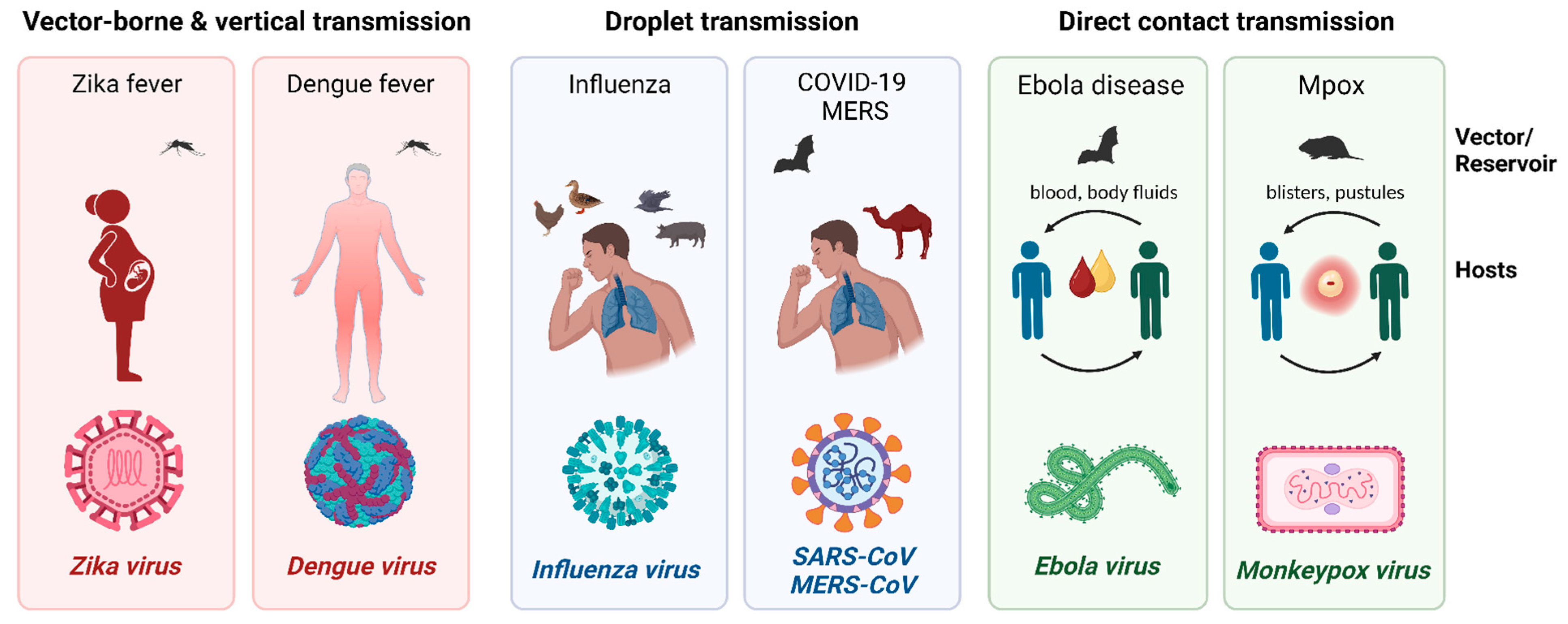
2. Emerging and Re-Emerging Viruses

2.1. Filoviridae: Ebola Virus and Marburg Virus

Ebola and Marburg viruses are single-stranded RNA filoviruses, which are zoonotic and are often transmitted through direct contact with infected fluids. Both Ebola and Marburg viruses have historically high mortality rates, which have varied from 25% to 90% and 24% to 88%, respectively [3,4]. The largest outbreak of Ebola virus disease (EVD) occurred in West Africa from 2014 to 2016 with more than 28,000 cases [5]. However, both viruses’ MVD case fatality rate is known to be 50%. Both viruses are transmitted through direct contact with infected blood and other bodily fluids. The Ebola virus is believed to have been first contracted by wild animals (such as fruit bats, porcupines, and non-human primates), then it spread to humans, in which human-to-human transmission is possible [3]. The first outbreak of Ebola was recorded in 1976 with an 88% mortality rate [5] and there continue to be outbreaks, mainly in Africa [3]. Marburg virus was first identified in Germany and Serbia in 1967, where laboratory workers were exposed to infected monkeys from Uganda [4].
The wide and fluctuating range of case fatality rate (CFR) data is not truly understood; possible reasons include differences in health status (nutrition, immunity, and co-infection status), genetics (ethnicity-dependent haplotypes or random polymorphisms), health-seeking behavior, case recognition, and the accessibility of supportive care at health-care facilities [5].

2.2. Flaviviridae: Zika Virus and Dengue Virus

Zika and Dengue viruses are single-stranded RNA flaviviruses often transmitted by arthropods, such as mosquitos and ticks. Both viruses are transmitted through mosquitos. Although the Zika virus was first identified in 1947 in Uganda, it became more well known when an outbreak occurred in Brazil in 2015, leading to its spread to other parts of the Americas. Before then, it was only sporadically prevalent in Africa and Asia. Its primary concern for public health is how the virus affects pregnancy and newborn babies. A total of 5–15% of infants of women who were infected with Zika while pregnant suffered from Zika-related problems, like the following: microcephaly in the newborn, which is where the baby is born with an underdeveloped head and brain, resulting in neurological disorders and other developmental problems [6]. Dengue has become more prevalent in the past decade, from over 500,000 cases in 2000 to 5.2 million in 2019. Dengue mostly affects Southeast Asia, which accounts for 70% of the global burden. It also seriously affects the Americas and the Western Pacific region. The largest recorded dengue outbreak occurred in 2019, in which the Americas reported 3.1 million cases, and Asian countries, like Bangladesh, Malaysia, Philippines, and Vietnam, recorded many cases as well. Dengue’s severity ranges from being asymptomatic to fatal. The majority of those who contract dengue have mild to no symptoms, in which case they will get better in 1–2 weeks. Common symptoms include high fever, severe headaches, body aches, and rash. Rarely, dengue can become more severe, in which case, symptoms can also include bleeding and even cause death. One who has had dengue in the past is more likely to contract severe dengue the second time [7].

2.3. Coronaviruses: MERS-CoV, SARS-CoV, and SARS-CoV-2

MERS-CoV, SARS-CoV, and SARS-CoV-2 are single-stranded RNA coronaviruses and transmitted via airborne particulates. Middle East Respiratory Syndrome (MERS) was first reported in Saudi Arabia in 2012, and all cases of MERS have been traced back to the Middle East. MERS-CoV is first contracted from camels, mainly in the Middle East, Africa, and South Asia, and is then transmitted to humans. However, its specific transmission route between camels and humans still needs to be determined. Due to its limited person-to-person transmission, though possible, it is not considered an epidemic; however, it still poses a global threat because of its high mortality rate [8]. Both SARS-CoV and SARS-CoV-2 originate from China, emerging in 2002 and 2019, respectively. Both viruses caused and continue to cause a global threat due to their high transmission rates [9]. COVID-19, caused by SARS-CoV-2, is still considered a pandemic as it continues to mutate and spread worldwide.

2.4. Avian Influenza (H5N1, H7N9)

Avian influenza virus is a strain of the single-stranded RNA influenza A virus that is transmitted between birds through airborne transmission, thus being able to be detected in the form of aerosols. Avian influenza includes the strains H5N1 and H7N9, which are transmissible to humans from birds and considered the most common strains to infect people [10]. Although it cannot be transmitted from human to human, nor is it common for it to be transmitted to humans from birds, it has the potential to become a global threat due to its high fatality rate in humans. With increasing numbers of mammals with avian influenza being detected, it poses risks of the virus being able to infect humans faster and easier since mammals are biologically closer to humans than birds are. Scientists have found mammal-to-mammal airborne transmission possible in ferrets, and only a few mutations take place before it is possible [11]. Scientists have also found a strong possibility of co-infection with avian influenza and the more common human influenza due to the high seroprevalence of both viruses in minks [12]. Thus, the possibility of new mutations arising from co-infection could lead to a greater risk to public health.

2.5. Monkeypox Virus

Since May 2022, there have been multiple outbreaks of mpox (previously known as monkeypox) worldwide. It has been declared a public health emergency by the World Health Organization (WHO) as it becomes increasingly transmissible among humans. The monkeypox virus is a zoonotic double-stranded DNA virus in the Orthopoxvirus genus, which also includes smallpox. Although its fatality rate is not high, it spreads rapidly, resulting in many hospitalized patients. Mpox was first discovered in Denmark in 1958. Since 2005, mpox outbreaks have been sporadically occurring, mainly in Africa. Thus, the sudden spread of mpox throughout Europe and the Americas caused concern. The monkeypox virus’ primary mode of transmission is through direct contact, particularly affecting men who have sexual contact with other men [13]. A summary of emerging viruses is described in Table 1.

3. Vaccination

3.1. History

The term vaccine derives from the cowpox virus Vaccinia (which comes from the Latin name for cow, vacca [40]), which was the earliest laboratory-maintained virus. Variolation was introduced by Dr. Edward Jenner in 1796, who observed that milkmaids previously infected with cowpox showed no symptoms of smallpox after being exposed [41]. Almost 2 centuries after the first smallpox vaccine was introduced, the WHO officially announced the eradication of this disease in 1980 [42]. Sequencing studies have determined that the vaccinia virus is more closely related to horsepox than cowpox, either suggesting an initial misidentification or that subsequent mutations likely accumulated during laboratory propagation. This continued to alter the virus to the extent that vaccinia is no longer known to be a naturally occurring strain [43].

3.2. Vaccination, Herd Immunity, and Public Awareness

The widely practiced childhood vaccination program provided immunity before children were exposed to potentially life-threatening diseases. In 2000, measles was declared eliminated in the United States, with a high vaccination rate that resulted in societal herd immunity [44]. However, the decreased vaccination rate driven by misinformation about measles and measles, mumps, and rubella (MMR) vaccines resulted in outbreaks in 2014 with 667 cases and in 2019 with 1274 cases [45]. The outbreaks were transmitted by unvaccinated international travelers acquiring measles abroad and returning to low-vaccinated close-knit vulnerable communities. High coverage with the MMR vaccine is the most effective strategy to limit transmission and obtain herd immunity to maintain the elimination of measles in the United States [46].
Since 2000, multiple outbreaks of vaccine-derived poliovirus (VDPV) have been recorded worldwide in unvaccinated communities [47]. VDPV is a strain related to the weakened live poliovirus contained in the oral polio vaccine (OPV). OPV is a live attenuated combination of three poliovirus serotypes and may have borne out the risk of reversion or gain-of-function mutation if allowed to circulate in under- or unimmunized populations for long enough [48]. Studies show that VDPV type 2 has the highest risk for paralytic disease [49]. In July 2022, an unvaccinated individual from Rockland County, New York, was infected by VDPV type 2. VDPVs emerge when insufficient individuals are vaccinated against polio, and the weakened strain of the poliovirus from OPV spreads among under-immunized populations, causing gain-of-function mutation. The United States has used the inactivated poliovirus vaccine (IPV) since 2000 and protects against paralytic diseases caused by all types of polioviruses, including VDPV. Thus, increasing the population vaccination rate to reach herd immunity is paramount in primary prevention to eradicate polio [48].

3.3. Vaccine Hesitancy

Almost counterintuitively, the success of modern vaccines at reducing the morbidity and mortality of once-common illnesses has led to periodic downturns in segments of the public’s willingness to accept vaccines. Despite decades of data from controlled studies collectively supporting the efficacy and benefit of vaccination, small but dedicated movements continue to resist or outright refuse vaccination. Historically, reluctance to vaccinate can be attributed to one of three general biases: religious/moral; cultural; or social/political objections. Even then, the degree of hesitancy tends to lie somewhere along a continuum and can be a complex mix of personal and outside reasons [49]. In the most recent pandemic period, the amplification of messaging using traditional and social media platforms has increased the proportion of those identifying as anti-vax for social and political reasons. Shifting attitudes with respect to government control, collective public health and safety, trustworthiness of science, and the medical profession can all contribute to fluctuations in the willingness to vaccinate.
In a self-fulfilling manner, the reduction in vaccine coverage caused by hesitancy has led to failures in protective immunity. The primary example is the effort launched by the World Health Assembly in 1988 to eradicate poliovirus. At the outset, there was every reason for optimism; smallpox had been declared eradicated in 1977 after a massive global vaccination campaign. There was an effective vaccine readily available for deployment, and teams of medical personnel were committed to the effort. Polio, like smallpox, is an exclusively human disease; there is no other natural reservoir from which a re-introduction could occur. However, twenty-five years on, the global community now declares individual countries as polio-free and regularly endures outbreaks of the disease due to hesitancy but also to regional political and resource instability. Without a widespread collective memory of how destructive some infectious diseases can be, the willingness of individuals to participate for the wider benefit is no longer a certainty.

3.4. Vaccine Development in Emerging Viruses

The COVID-19 vaccine was first available to the public on 11 December 2020, under emergency use authorization (EUA), one year after the identification of the SARS-CoV-2 virus in China. This nucleic acid-based mRNA technology yielded the Pfizer/BioNTech COVID-19 vaccine Comirnaty, which received U.S. Food and Drug Administration (FDA) approval on 23 August 2021 [50].
Moderna’s Spikevax, also an mRNA vaccine, received FDA approval in 2022. On the contrary, the Johnson & Johnson COVID-19 (Ad26.COV2.S) vaccine is an adenovirus-based vaccine and was deployed with success under EUA during the declared public health emergency. This adenovirus type 26 is a naturally occurring virus with the replication gene deleted, so it is incapable of replicating within humans but expresses the SARS-CoV-2 spike antigen to induce an immune response and produce the antibodies to protect from future infection. However, clinical trial results in the United States showed the Pfizer and Moderna vaccines to be 95% effective and the Johnson & Johnson vaccine to be 66% effective in protecting against moderate and symptomatic SARS-CoV-2 infection [51].
Multiple Ebola vaccine candidates have entered phase I-III clinical trials since the major West African outbreak in 2013. More than 10 of these were live-attenuated constructs based on the backbone of the vesicular stomatitis virus, VSV [5,52]. The FDA-approved ERVEBO® vaccine, which is a recombinant virus vaccine against Ebola virus disease (EVD), will begin phase 4 trials in 2024, [53]. Several Marburg vaccine trials are underway, but none are approved. Currently, a phase 2 double-blind, placebo-controlled trial using a monovalent chimpanzee adenoviral-vectored Marburg virus vaccine is open for recruitment [54]. For Zika virus, there is no approved vaccine available, but currently, there is a phase 2 trial of the purified inactivated Zika virus vaccine (PIZV) in healthy participants [55].
The most common avian influenza strains that are transmitted to humans are H5N1 and H7N9 viruses [56]. After the emergence of the H5N1 virus in poultry that eventually infected people in China in 2013, it became of great importance to find a vaccine for avian influenza viruses and begin to vaccinate poultry. Due to this, by 2018, China was able to successfully eliminate the spread and infection of the H7N9 avian influenza virus in humans by vaccinating its poultry [57]. In 2007, the first H5N1 avian influenza vaccine was FDA-approved [58], but there is no vaccine available for H7N9. Seasonal influenza vaccines consist of two strains of influenza type A viruses and two strains of influenza type B viruses [59].

4. Clinical Manifestation and Treatment

4.1. Filoviridae: Ebolavirus and Marburg Virus

Ebola virus disease (EVD) develops abruptly with fever and chills after 6–12 days after exposure [60]. During the incubation period, the infected individual is not contagious to others. However, all symptomatic individuals are assumed to have the virus in the blood and other body fluids. Initial common symptoms include fever, fatigue, headache, and a loss of appetite [61]. The patient can also develop a diffuse nonpruritic rash by day 5 to 7. Diarrhea and vomiting are very common and usually develop within the first few days of illness, which may result in severe fluid loss, leading to dehydration, hypotensive shock, and multi-organ failure [62]. Bleeding can also occur, mostly manifesting as blood in the stool (about 6%), and significant hemorrhage may be seen in the terminal phase of the illness [63].
Marburg virus disease has symptoms like EVD but a higher fatality rate of 80–90% [64]. The symptoms begin abruptly with high fever, severe headache, and malaise. Diarrhea and vomiting can begin on the third day. Many patients develop severe hemorrhage within seven days [65] due to systemic inflammatory response triggering coagulation cascades, leading to disseminated intravascular coagulation [41]. In severe cases, death usually occurs 8 to 9 days after the onset of symptoms, preceded by shock and severe hemorrhage [66].
Management and treatment for the filovirus infections of EVD and Marburg virus disease heavily rely on aggressive supportive care to prevent the development of shock and to maintain adequate organ functions while the host immune system mobilizes an adaptive response to eliminate the virus [67,68]. For Ebola-specific therapies, there are the triple-monoclonal antibody (mAb) REGN-EB3 made of atoltivimab, maftivimab, and odesivimab (Inmazeb) and the monoclonal antibody ansuvimab (Ebanga). In late 2020, the U.S. Food and Drug Administration (FDA) approved these therapies [69,70], which can be used for adult and pediatric patients and are administered as a single dose. On the contrary, there is no approved treatment for Marburg virus disease. mAb therapy against the ebolavirus is ineffective for the Marburg virus because the therapy consists of mAb against the surface glycoprotein of the specific ebolavirus.

4.2. Zika Virus

Zika virus infection presents with mild clinical disease and is mostly asymptomatic. Symptoms of Zika virus infection include low-grade fever; pruritic rash on the face, trunk, extremities, palms, and soles; arthralgia; and conjunctivitis [71,72], with other common viral symptoms of myalgia and headache after the incubation period of 2–14 days from the mosquito bite [73]. Although the initial symptoms may be mild, there is a strong association between Guillain–Barré syndrome [74] and other neurological disorders, such as optic nerve damage caused by increased intraocular pressure, which may result in glaucoma [75]. Moreover, maternal infection during pregnancy results in congenital microcephaly, stillbirth, and miscarriages [76,77,78]. This maternal-to-fetal vertical transmission of Zika virus was 65% or even higher in a cohort study of 130 infants because not all infants performed serial testing [79]. This vertical transmission has serious fetal complications of fetal growth restriction, extremity abnormalities [80], and a plethora of central nervous system sequelae, including but not limited to ventriculomegaly, microcephaly, and intracranial calcifications [81].
Currently, the treatment of the infection is still supportive, although researchers developing an antiviral drug have made enormous efforts. Management consists of rest and symptomatic treatment, including fluids to prevent dehydration and the administration of acetaminophen to relieve fever and pain, but avoiding aspirin and other nonsteroidal anti-inflammatory drugs until dengue infection can be ruled out to reduce the risk of hemorrhage [82].

4.3. Coronavirus: MERS-CoV, SARS-CoV, and SARS-CoV-2 (COVID-19)

4.3.1. MERS-CoV

Middle East respiratory syndrome coronavirus (MERS-CoV) has an estimated incubation period of 9 to 12 days [83] with common viral infection symptoms of fever and chills with cough and shortness of breath. Most reported patients were severe cases of pneumonia, acute respiratory distress syndrome requiring mechanical ventilation, and acute kidney injury. MERS-CoV infection treatment is supportive, and the World Health Organization provides guidelines for severe acute respiratory infection (SARI) when MERS-CoV is suspected. Early recognition of SARI and the immediate implementation of infection prevention with respiratory support and shock management are critical for effective treatment [84].

4.3.2. SARS-CoV

Severe acute respiratory syndrome (SARS), caused by the SARS-associated coronavirus (SARS-CoV) outbreak in 2003, was treated with supportive measures, and about 90% (7322/8096) of patients survived. However, half of the patients who required mechanical ventilation died [85]. This global SARS epidemic was contained, and there have been no known cases of SARS since 2004.

4.3.3. SARS-CoV-2

The incubation period for COVID-19 is 14 days after exposure, with most cases occurring after 4 to 5 days [86]. Among symptomatic COVID-19 patients, cough, myalgias, and headache are the most reported symptoms [87]. Other symptoms are low-grade fever, cough, smell and taste abnormalities, rash, and conjunctivitis. Loss of smell was less common in patients infected during Omicron’s prevalence, supporting the idea that patients with recent variants are at a lower risk of developing associated chemosensory loss [88,89]. In early studies, respiratory failure due to acute respiratory distress syndrome developed in 20% after 8 days of symptom onset, and 12.3% of patients had mechanical ventilation [90]. Thromboembolic complication is common, with a reported 31% incidence in ICU patients [91].
The treatment recommendation for COVID-19 disease caused by SARS-CoV-2 has evolved as many randomized controlled trials have been completed. The current guideline by WHO recommends treatments based on a disease severity classification of non-severe, severe, or critical based on respiratory status and oxygen saturation. Based on the WHO severity criteria, treatments of Remdesivir, Nirmatrelvir, corticosteroids, IL-6 receptor blockers, and Baricitinib are recommended [92].

4.4. Avian Influenza (H5N1, H7N9)

4.4.1. H5N1

Prior H5N1 infections in humans remain very rare, involving predominantly children and young adults, but they can cause severe disease with a high mortality rate of up to 50% [93]. The most common clinical symptoms are similar to other viral infections, including fever, cough, and dyspnea. Risk factors for severe disease include the presence of bilateral pneumonia. Neutropenia and increased liver enzyme of ALT predicted fatal outcomes [94].

4.4.2. H7N9

Patients with H7N9 infection would show symptoms of fever, cough, dyspnea, headache, myalgia, and malaise, causing severe illness, including pneumonia and ARDS, with a high 78% ICU admission rate and a 28% death rate [95]. Most patients with H7N9 are severely ill, but mild and moderate cases have been reported [96]. For outpatients without fever and symptoms, close monitoring is recommended for the progression of the illness.
For any influenza treatment, oral oseltamivir is recommended, and the peramivir intravenous route is recommended if the patient cannot tolerate the oral therapy. These medications are in the neuraminidase inhibitor category. These therapies have proven efficacy when started early in the diagnosis. However, the emergence of antiviral resistance was also noted, which should be considered in pandemic preparedness planning [97].

4.5. Monkeypox Virus (Mpox)

Monkeypox typically presents with systemic symptoms attributable to a viremic phase of illness lasting 1 to 5 days with fever, headache, myalgia, fatigue, sore throat, and back pain after the incubation period from 5 to 13 days. However, during the recent outbreak in May 2022, some patients presented with genital, anal, and/or oral lesions without systemic illness. Proctitis and tonsillitis were more common during the recent outbreak than previously reported [98]. Also, ocular involvement is rare but requires urgent treatment since corneal scarring and vision loss are potential complications [99]. In addition, there is evidence of severe neurological complications of encephalitis and seizure [100]. Most patients with mpox will recover without medical intervention, but antiviral therapy is recommended in severe disease. Currently, tecovirimat is available for mpox treatment, which was approved for smallpox [101]. In addition to tecovirimat, trifluridine can be used as eye drops or ointment for ocular lesions [102].

5. Surveillance Tools

5.1. Air Surveillance System

The COVID-19 pandemic highlighted the importance of science-based decision making and exposed global vulnerabilities in preparedness and response systems. One of the biggest challenges faced during the COVID-19 pandemic has been the lack of an effective surveillance system that can provide early warnings about outbreaks. The development and usage of a vaccine have been proven effective at delivering valid protection to individuals; however, the threat of the appearance of a new variant is constantly challenging the vaccine’s effectiveness. Under these circumstances, the means that can prepare us for the next infectious disease challenge become critical. More importantly, where we should conduct surveillance for emerging diseases is another fundamental question regarding how fast we can detect the source of potential outbreaks.
Robust evidence has shown that inhaling bioaerosol particles containing viable respiratory viruses is the primary route of COVID-19 human-to-human transmission [103,104,105]. Viruses can become airborne through various mechanisms such as talking, sneezing, coughing, the breathing of an infected individual, medical procedures, and flushing a toilet containing infectious particles. Once the viral-laden aerosols (<100 µm) are generated, they can linger in the air for hours and travel beyond 1 to 2 m from the source, causing new infections at both short and long ranges [100]. Currently, we are facing not only the threat from the re-emergence of SARS-CoV-2 but also other respiratory viruses such as Middle East respiratory syndrome coronavirus (MERS-CoV), respiratory syncytial virus (RSV), and influenza. Effectively identifying the viral particles in bioaerosols will be the key to spotting the location of the next novel respiratory virus threat. Since the COVID pandemic started in 2019, the number of studies regarding bioaerosol monitoring has been rapidly increasing, with most studies focused on SARS-CoV-2 in hospital settings. The fundamental step for in-air pathogen detection is the collection of bioaerosols. Several air biosensor collection methods are widely used by researchers: sedimentation, filters, impactors, cyclones, impingers, electrostatic precipitators, and microfluid platforms [106]. Each method can collect bioaerosols from air samples. Furthermore, researchers should carefully choose the method based on the specific research aim or application. The advantages and disadvantages of the commonly used aerosol collection methods are summarized in Table 2.
Bioaerosol particles not only contain pathogenic and/or non-pathogenic live/dead microorganisms but also contain environmental components, including dust, droplets, salt, and other particles [118]. These non-pathogenic components can potentially affect the effectiveness of airborne microorganisms’ collection, isolation, and purification. In addition, the concentration of the virus in bioaerosol particles, room humidity, room temperature, room size, and airflow rate all play a role in air sample collection and virus isolation [119].

5.2. Viral Detection Methods

Detecting viruses in air samples can be challenging due to the small size of viral particles and relatively low viral load. In general, all available virus detection methods can be used in bioaerosol samples after elution. The most common in-air viral detection method is the polymerase chain reaction (PCR). This includes the regular PCR to detect DNA viruses, reverse-transcription (RT) PCR to detect RNA viruses, real-time PCR (qPCR) to quantify viruses, and digital PCR to precisely identify the copy number variations. As a widely used molecular DNA and RNA amplification method, the PCR offers many detection advantages, such as sensitivity (especially digital PCR), specificity, and speed (results are usually available within hours). The PCR method can also be automated, simultaneously allowing the high-throughput testing of large-scale samples. On the other hand, the major limitation of the PCR method in emerging virus detection is that this technique requires prior knowledge of the target sequence in the viral genome. Thus, this method cannot be used for unknown viruses. In addition, if the target sequence mutates significantly, the PCR may not be able to detect the new strains that diverged from the target virus. Other than PCR, other virus detection methods include virus culture [120], loop-mediated isothermal amplification (LAMP) [121], enzyme-linked immunosorbent assay (ELISA) [122], immunofluorescence assay (IFA) [123], and next-generation sequencing (NGS) [124]. Due to the ability to sequence genetic material from samples, NGS is widely used to identify unknown viruses [125].

5.3. Potential Surveillance Location Identification

Many virus outbreaks of emerging infections originate in animals. These zoonotic viruses include SARS-CoV-2, HIV, Zika, MERS, Ebola, and monkeypox. Humans can acquire zoonotic viruses in various settings where there is close contact between humans and animals, especially wild animals. Although not all interactions between humans and animals lead to virus transmission, certain conditions, such as overcrowding, poor sanitation, close contact between different animal species and humans, and inadequate biosafety measures, can increase the risk of virus spillover. Thus, to reduce the risk of zoonotic virus transmission, environments where human–animal contact occurs may require more frequent monitoring or constant surveillance. Here, we list some places that can act as high-risk disease transmission environments.
A wildlife market/wet market, where live animals or wild animals are sold for food, can be a typical example of this environment. These markets often involve a mix of animal species from different regions, which provide opportunities for zoonotic viruses to cross species barriers and facilitate the transmission of viruses from animals to humans. In December 2019, the first case of COVID-19 was identified in a seafood market in Wuhan, China; then, the virus spread worldwide. According to the World Health Organization (WHO), as of August 2023, over 6.9 million deaths were due to COVID-19 [126].
Farms where animals are raised for food production, including poultry, pigs, and other livestock, can be breeding grounds for viruses. The H1N1 influenza (swine flu) pandemic in 2009 is one example. This novel strain of the H1N1 influenza virus contained genetic elements from pigs, birds, and human influenza viruses. The virus was initially identified in pigs, particularly in North America, but it quickly spread to humans. The close contact between humans and pigs on pig farms played a role in the transmission of the virus to humans. The 2009 H1N1 pandemic that was first detected in California, United States [127], resulted in widespread illness and affected countries around the world.
Tropical and subtropical regions with warm and humid climates are often connected with a high risk of vector-borne viruses. Mosquitos thrive in this environment, increasing the risk of transmitting diseases like Zika, dengue, West Nile fever, and chikungunya [128]. Forests and rural areas have high biodiversity and host a variety of animal species, and their associated vectors can pose an increased risk of virus spillover to humans.

6. Discussion

Historically, virus outbreaks have initiated the public response to vaccine development and clinical trials for treatment. The recent COVID-19 pandemic and the recent mpox outbreak showcased the possible effects of constantly changing viruses on society. A genomic mutation develops a new deadly variant by changing its route of transmission and becoming highly infectious with a low viral load. However, our ability to anticipate, respond to, and mitigate the impacts of emerging and re-emerging viruses is more promising than ever due to scientific and medical advancements in this modern era. In addition, with technological advancements in bioinformatics, a rapid, streamlined means of sequencing newly emerging viruses is possible through an automated workflow pipeline [129]. This will facilitate the expedited identification of emerging viruses, resulting in earlier public and medical responses to confine their outbreak. The continuously evolving mutation of viruses may challenge the effectiveness of available vaccines.
In addition, non-invasive environmental surveillance tools for early detection have been developed that are currently used by either research groups or the government, such as wastewater monitoring [130,131] and vector sampling [132]. This method was supported by the provided evidence that non-water-borne viruses can be detected during wastewater monitoring. For example, airborne viruses (coronavirus [121] and influenza [133]) and vector-borne viruses (Zika virus [134] and West Nile virus [135]) were detected via wastewater-based epidemiological studies.
However, whether non-airborne viruses can be detected via bioaerosol monitoring is unclear. Since virus-containing aerosols can be generated through various mechanisms, such as respiratory activities (talking, coughing, and sneezing), medical procedures (intubation, dental procedures, and bronchoscopy), environmental disturbance (vacuuming, cleaning, and sweeping), animal activities, and industrial processes, bioaerosol surveillance can be considered a powerful tool to screen viruses that linger in the air, especially indoor air. Admittedly, this method has some limitations; for example, the potential for a virus to become aerosolized depends on various factors, the virus detection rate can be very low, and some viruses are more capable of remaining infectious in aerosols and surviving airborne conditions than others. In the era characterized by the swift advancement of science and technology, technological limitations are expected to be addressed soon.
Under these circumstances, the means that can prepare us for the next infectious disease challenge become critical. International collaboration, data sharing, and interdisciplinary research are key to staying ahead of viral threats. To prepare for the next outbreak in global public health, research and public effort ought to focus on the availability of reliable surveillance systems for emerging threats, expedited vaccine development as the viral infection emerges and becomes epidemic, public awareness and receptivity towards the benefits of mass vaccination, and practicing good citizenship surrounding the public health measures of testing and quarantines. The information summarized in this review will aid authorities in designing and adopting effective prevention and control strategies to counter the next emerging or re-emerging virus.
The topic that we summarized is an overview of a quickly changing field that expands exponentially with new research findings. Here, we attempted to provide the current research topics and past knowledge of emerging and re-emerging viruses for readers to further explore their interested field of study. Thus, the limitation of this approach lies in the lack of an in-depth description of each topic and virus discussed.

7. Conclusions

COVID-19 disease highlighted the importance of science-based decision making and exposed global vulnerabilities in the prevention and preparedness of pandemic infection and response systems. During the pandemic, medical advancements in developing a new vaccine in a timely manner and improved treatment methods becoming available for viral infections were successes. The development and usage of a vaccine have been proven effective at providing valid protection to individuals, but public awareness and receptivity towards mass vaccination are a challenge. Furthermore, the threat of new variants is constantly challenging the vaccine’s effectiveness. One of the major challenges faced during the last pandemic was the lack of an effective surveillance system that can provide early warnings about outbreaks. From past experiences, we learned that proactive surveillance, rapid diagnostics, and effective communication networks are pivotal in containing and managing emerging threats. In a world bound by shared vulnerabilities, our collective action and unwavering determination hold the key to safeguarding future generations against the threats that emerge from the virus realm. As we peer into the future, preparedness is not an option but a mandate.

Author Contributions

Conceptualization, J.J.H. writing—original draft preparation, J.J.H., H.A.S., Q.X. and S.L.P. writing—review and editing, J.J.H., H.A.S., Q.X., S.L.P. and J.S.-G. All authors have read and agreed to the published version of the manuscript.

Funding

The APC was funded by the Department of Clinical Investigation at Brooke Army Medical Center, Fort Sam Houston, Texas.

Data Availability Statement

Not applicable.

Acknowledgments

We thank Amanda W. Pierson for supporting the illustration draft.

Conflicts of Interest

The views expressed herein are those of the author(s) and do not necessarily reflect the official policy or position of the Defense Health Agency, Brooke Army Medical Center, Department of Defense, nor any agencies under the U.S. Government.

References

  1. LeDuc, J.W.; Barry, M.A. SARS, the First Pandemic of the 21st Century1. EID J. 2004, 10, e26. [Google Scholar] [CrossRef]
  2. Sullivan, S.J.; Jacobson, R.M.; Dowdle, W.R.; Poland, G.A. 2009 H1N1 Influenza. Mayo Clin. Proc. 2010, 85, 64–76. [Google Scholar] [CrossRef] [PubMed]
  3. Ebola Virus Disease. Available online: https://www.who.int/health-topics/ebola#tab=tab_1 (accessed on 23 August 2023).
  4. Marburg Virus Disease. Available online: https://www.who.int/news-room/fact-sheets/detail/marburg-virus-disease (accessed on 23 August 2023).
  5. Jacob, S.T.; Crozier, I.; Fischer, W.A., 2nd; Hewlett, A.; Kraft, C.S.; Vega, M.A.; Soka, M.J.; Wahl, V.; Griffiths, A.; Bollinger, L.; et al. Ebola virus disease. Nat. Rev. Dis. Prim. 2020, 6, 13. [Google Scholar] [CrossRef] [PubMed]
  6. WHO Zika Virus. Available online: https://www.who.int/news-room/fact-sheets/detail/zika-virus (accessed on 23 August 2023).
  7. World Health Organization (WHO). Dengue and Severe Dengue. Key Facts. 2019. Available online: https://www.who.int/news-room/fact-sheets/detail/dengue-and-severe-dengue#:~:text=Dengue%20(break%2Dbone%20fever),body%20aches%2C%20nausea%20and%20rash (accessed on 17 September 2023).
  8. WHO. Middle East Respiratory Syndrome Coronavirus (MERS-CoV). MERS Monthly Summary, November 2019. Available online: https://www.who.int/emergencies/mers-cov/en/ (accessed on 20 August 2023).
  9. Severe Acute Respiratory Syndrome (SARS). Available online: https://www.who.int/health-topics/severe-acute-respiratory-syndrome#tab=tab_1 (accessed on 20 August 2023).
  10. Influenza Type A Viruses|Avian Influenza. Available online: https://www.cdc.gov/flu/avianflu/influenza-a-virus-subtypes.htm (accessed on 15 August 2023).
  11. Sutton, T.C.; Finch, C.; Shao, H.; Angel, M.; Chen, H.; Capua, I.; Cattoli, G.; Monne, I.; Perez, D.R. Airborne transmission of highly pathogenic H7N1 influenza virus in ferrets. J. Virol. 2014, 88, 6623–6635. [Google Scholar] [CrossRef]
  12. Sun, H.; Li, F.; Liu, Q.; Du, J.; Liu, L.; Sun, H.; Li, C.; Liu, J.; Zhang, X.; Yang, J.; et al. Mink is a highly susceptible host species to circulating human and avian influenza viruses. Emerg. Microbes Infect. 2021, 10, 472–480. [Google Scholar] [CrossRef]
  13. Mpox (Monkeypox). Available online: https://www.who.int/news-room/fact-sheets/detail/monkeypox (accessed on 20 August 2023).
  14. Noda, T.; Sagara, H.; Suzuki, E.; Takada, A.; Kida, H.; Kawaoka, Y. Ebola virus VP40 drives the formation of virus-like filamentous particles along with GP. J. Virol. 2002, 76, 4855–4865. [Google Scholar] [CrossRef]
  15. Baseler, L.; Chertow, D.S.; Johnson, K.M.; Feldmann, H.; Morens, D.M. The Pathogenesis of Ebola Virus Disease. Annu. Rev. Pathol. Mech. Dis. 2017, 12, 387–418. [Google Scholar] [CrossRef]
  16. Ebola Disease Distribution Map: Cases of Ebola Disease in Africa Since 1976. Available online: https://www.cdc.gov/vhf/ebola/history/distribution-map.html (accessed on 24 August 2023).
  17. Martin, P.; Laupland, K.B.; Frost, E.H.; Valiquette, L. Laboratory diagnosis of Ebola virus disease. Intensiv. Care Med. 2015, 41, 895–898. [Google Scholar] [CrossRef]
  18. Dudas, G.; Carvalho, L.M.; Bedford, T.; Tatem, A.J.; Baele, G.; Faria, N.R.; Park, D.J.; Ladner, J.T.; Arias, A.; Asogun, D.; et al. Virus genomes reveal factors that spread and sustained the Ebola epidemic. Nature 2017, 544, 309–315. [Google Scholar] [CrossRef]
  19. Tong, Y.-G.; The China Mobile Laboratory Testing Team in Sierra Leone; Shi, W.-F.; Liu, D.; Qian, J.; Liang, L.; Bo, X.-C.; Liu, J.; Ren, H.-G.; Fan, H.; et al. Genetic diversity and evolutionary dynamics of Ebola virus in Sierra Leone. Nature 2015, 524, 93–96. [Google Scholar] [CrossRef]
  20. World Health Organization. WHO Emergency Use Assessment and Listing for Ebola Virus Disease IVDs. Product: ReEBOV Antigen Rapid Test Kit; WHO: Geneva, Switzerland, 2015. [Google Scholar]
  21. Broadhurst, M.J.; Kelly, J.D.; Miller, A.; Semper, A.; Bailey, D.; Groppelli, E.; Simpson, A.; Brooks, T.; Hula, S.; Nyoni, W.; et al. ReEBOV Antigen Rapid Test kit for point-of-care and laboratory-based testing for Ebola virus disease: A field validation study. Lancet 2015, 386, 867–874. [Google Scholar] [CrossRef]
  22. Layqah, L.A.; Eissa, S. An electrochemical immunosensor for the corona virus associated with the Middle East respiratory syndrome using an array of gold nanoparticle-modified carbon electrodes. Microchim. Acta 2019, 186, 224. [Google Scholar] [CrossRef]
  23. Brauburger, K.; Hume, A.J.; Mühlberger, E.; Olejnik, J. Forty-five years of marburg virus research. Viruses 2012, 4, 1878–1927. [Google Scholar] [CrossRef]
  24. Marburg Virus Disease Distribution Map. Available online: https://www.cdc.gov/vhf/marburg/outbreaks/distribution-map.html (accessed on 24 August 2023).
  25. Coelho, S.V.A.; Neris, R.L.S.; Papa, M.P.; Schnellrath, L.C.; Meuren, L.M.; Tschoeke, D.A.; Leomil, L.; Verçoza, B.R.F.; Miranda, M.; Thompson, F.L.; et al. Development of standard methods for Zika virus propagation, titration, and purification. J. Virol. Methods 2017, 246, 65–74. [Google Scholar] [CrossRef]
  26. Woolsey, C.; Cross, R.W.; Agans, K.N.; Borisevich, V.; Deer, D.J.; Geisbert, J.B.; Gerardi, C.; Latham, T.E.; Fenton, K.A.; Egan, M.A.; et al. A highly attenuated Vesiculovax vaccine rapidly protects nonhuman primates against lethal Marburg virus challenge. PLoS Negl. Trop. Dis. 2022, 16, e0010433. [Google Scholar] [CrossRef] [PubMed]
  27. Herrada, C.A.; Kabir, A.; Altamirano, R.; Asghar, W. Advances in Diagnostic Methods for Zika Virus Infection. J. Med. Devices 2018, 12, 040802. [Google Scholar] [CrossRef]
  28. Madanayake, N.H.; Rienzie, R.; Adassooriya, N.M. Electron microscopic methods for virus diagnosis. In Viral Infections and Antiviral Therapies; Elsevier: Amsterdam, The Netherlands, 2023; pp. 121–140. [Google Scholar] [CrossRef]
  29. Lim, J.K.; Seydou, Y.; Carabali, M.; Barro, A.; Dahourou, D.L.; Lee, K.S.; Nikiema, T.; Namkung, S.; Lee, J.-S.; Shin, M.Y.; et al. Clinical and epidemiologic characteristics associated with dengue during and outside the 2016 outbreak identified in health facility-based surveillance in Ouagadougou, Burkina Faso. PLoS Negl. Trop. Dis. 2019, 13, e0007882. [Google Scholar] [CrossRef] [PubMed]
  30. Needs, S.H.; Sirivisoot, S.; Jegouic, S.; Prommool, T.; Luangaram, P.; Srisawat, C.; Sriraksa, K.; Limpitikul, W.; Mairiang, D.; Malasit, P.; et al. Smartphone multiplex microcapillary diagnostics using Cygnus: Development and evaluation of rapid serotype-specific NS1 detection with dengue patient samples. PLoS Negl. Trop. Dis. 2022, 16, e0010266. [Google Scholar] [CrossRef]
  31. Masters, P.S. The molecular biology of coronaviruses. Adv. Virus Res. 2006, 66, 193–292. [Google Scholar] [CrossRef]
  32. Chong, Z.X.; Liew, W.P.P.; Ong, H.K.; Yong, C.Y.; Shit, C.S.; Ho, W.Y.; Ng, S.Y.; Yeap, S.K. Current diagnostic approaches to detect two important betacoronaviruses: Middle East respiratory syndrome coronavirus (MERS-CoV) and severe acute respiratory syndrome coronavirus 2 (SARS-CoV-2). Pathol.-Res. Pract. 2021, 225, 153565. [Google Scholar] [CrossRef]
  33. Singh, B.; Datta, B.; Ashish, A.; Dutta, G. A comprehensive review on current COVID-19 detection methods: From lab care to point of care diagnosis. Sens. Int. 2021, 2, 100119. [Google Scholar] [CrossRef] [PubMed]
  34. Massart, S.; Chiumenti, M.; De Jonghe, K.; Glover, R.; Haegeman, A.; Koloniuk, I.; Komínek, P.; Kreuze, J.; Kutnjak, D.; Lotos, L.; et al. Virus detection by high-throughput sequencing of small RNAs: Large-scale performance testing of sequence analysis strategies. Phytopathology 2019, 109, 488–497. [Google Scholar] [CrossRef] [PubMed]
  35. Al Ahmad, M.; Mustafa, F.; Panicker, N.; Rizvi, T.A. Optical Detection of SARS-CoV-2 Utilizing Antigen-Antibody Binding Interactions. Sensors 2021, 21, 6596. [Google Scholar] [CrossRef]
  36. Moreira, E.A.; Yamauchi, Y.; Matthias, P. How Influenza Virus Uses Host Cell Pathways during Uncoating. Cells 2021, 10, 1722. [Google Scholar] [CrossRef] [PubMed]
  37. Korteweg, C.; Gu, J. Pathology, molecular biology, and pathogenesis of avian influenza A (H5N1) infection in humans. Am. J. Pathol. 2008, 172, 1155–1170. [Google Scholar] [CrossRef]
  38. Lansiaux, E.; Jain, N.; Laivacuma, S.; Reinis, A. The virology of human monkeypox virus (hMPXV): A brief overview. Virus Res. 2022, 322, 198932. [Google Scholar] [CrossRef]
  39. Chelsky, Z.L.; Dittmann, D.; Blanke, T.; Chang, M.; Vormittag-Nocito, E.; Jennings, L.J. Validation Study of a Direct Real-Time PCR Protocol for Detection of Monkeypox Virus. J. Mol. Diagn. 2022, 24, 1155–1159. [Google Scholar] [CrossRef]
  40. Moss, B. “Poxviridae.” Fields Virology, 6th ed.; Knipe, D.M., Howley, P.M., Eds.; Lippincott Williams & Wilkins: Philadelphia, PA, USA, 2013; pp. 2129–2159. [Google Scholar]
  41. Turley, P.K. Vaccine: From vacca, a cow. AJO-DO Clin. Companion 2021, 1, 5–6. [Google Scholar] [CrossRef]
  42. History of Smallpox. CDC. Available online: https://www.cdc.gov/smallpox/history/history.html (accessed on 15 September 2023).
  43. Carroll, D.S.; Emerson, G.L.; Li, Y.; Sammons, S.; Olson, V.; Frace, M.; Nakazawa, Y.; Czerny, C.P.; Tryland, M.; Kolodziejek, J.; et al. Chasing Jenner’s vaccine: Revisiting cowpox virus classification. PLoS ONE 2011, 6, e23086. [Google Scholar] [CrossRef]
  44. Patel, M.; Lee, A.D.; Redd, S.B.; Clemmons, N.S.; McNall, R.J.; Cohn, A.C.; Gastañaduy, P.A. Increase in Measles Cases—United States, January 1–April 26, 2019. MMWR Morb. Mortal. Wkly. Rep. 2019, 68, 402–404. [Google Scholar] [CrossRef]
  45. Centers for Disease Control and Prevention. Measles Cases and Outbreaks. Available online: https://www.cdc.gov/measles/cases-outbreaks.html (accessed on 16 September 2023).
  46. Patel, M.; Lee, A.D.; Clemmons, N.S.; Redd, S.B.; Poser, S.; Blog, D.; Zucker, J.R.; Leung, J.; Link-Gelles, R.; Pham, H.; et al. National Update on Measles Cases and Outbreaks—United States, January 1–October 1, 2019. MMWR Morb. Mortal. Wkly. Rep. 2019, 68, 893–896. [Google Scholar] [CrossRef]
  47. O’reilly, K.M.; Lamoureux, C.; Molodecky, N.A.; Lyons, H.; Grassly, N.C.; Tallis, G. An assessment of the geographical risks of wild and vaccine-derived poliomyelitis outbreaks in Africa and Asia. BMC Infect. Dis. 2017, 17, 367. [Google Scholar] [CrossRef] [PubMed]
  48. Polio Vaccine: Vaccine-Derived Poliovirus. Available online: https://www.cdc.gov/vaccines/vpd/polio/hcp/vaccine-derived-poliovirus-faq.html (accessed on 15 September 2023).
  49. Dubé, E.; Laberge, C.; Guay, M.; Bramadat, P.; Roy, R.; Bettinger, J.A. Vaccine hesitancy: An overview. Hum. Vaccines Immunother. 2013, 9, 1763–1773. [Google Scholar] [CrossRef] [PubMed]
  50. FDA News Release: FDA Approves First COVID-19 Vaccine. Available online: https://www.fda.gov/news-events/press-announcements/fda-approves-first-covid-19-vaccine (accessed on 1 September 2023).
  51. Patel, R.; Kaki, M.; Potluri, V.S.; Kahar, P.; Khanna, D. A comprehensive review of SARS-CoV-2 vaccines: Pfizer, Moderna & Johnson & Johnson. Hum. Vaccines Immunother. 2022, 18, 2002083. [Google Scholar] [CrossRef]
  52. Halperin, S.A.; Das, R.; Onorato, M.T.; Liu, K.; Martin, J.; Grant-Klein, R.J.; Nichols, R.; Coller, B.-A.; Helmond, F.A.; Simon, J.K. Immunogenicity, lot consistency, and extended safety of rVSVΔG-ZEBOV-GP vaccine: A phase 3 randomized, double-blind, placebo-controlled study in healthy adults. J. Infect. Dis. 2019, 220, 1127–1135. [Google Scholar] [CrossRef]
  53. Schieffelin, J. REVIVE (Response to the Ebola Virus Vaccine). 15 August 2023. Available online: https://clinicaltrials.gov/study/NCT05992480?cond=Ebola%20Virus%20Disease&intr=Vaccine&rank=3 (accessed on 28 August 2023).
  54. Study Record. Available online: https://clinicaltrials.gov/study/NCT05817422?cond=Marburg%20Virus%20Disease&term=vaccine&limit=25&page=1&rank=1 (accessed on 11 September 2023).
  55. Study Record. Available online: https://clinicaltrials.gov/study/NCT05469802?cond=Zika%20Virus&term=vaccine&limit=25&aggFilters=status:not%20rec%20act&rank=2 (accessed on 11 September 2023).
  56. Current U.S. Bird Flu Situations in Humans. Available online: https://www.cdc.gov/flu/avianflu/inhumans.htm#:~:text=Six%20main%20hemagglutinin%20(HA)%20subtypes,majority%20of%20infections%20in%20people (accessed on 15 August 2023).
  57. Zeng, X.; Tian, G.; Shi, J.; Deng, G.; Li, C.; Chen, H. Vaccination of poultry successfully eliminated human infection with H7N9 virus in China. Sci. China Life Sci. 2018, 61, 1465–1473. [Google Scholar] [CrossRef]
  58. Puthanakit, T.; Anugulruengkitt, S.; Jantarabenjakul, W. Prevention of Emerging Infections in Children. Pediatr. Clin. N. Am. 2022, 69, 185–202. [Google Scholar] [CrossRef]
  59. Key Facts about Seasonal Flu Vaccines. Available online: https://www.cdc.gov/flu/prevent/keyfacts.htm (accessed on 15 August 2023).
  60. Bah, E.I.; Lamah, M.C.; Fletcher, T.; Jacob, S.T.; Brett-Major, D.M.; Sall, A.A.; Shindo, N.; Fischer, W.A.; Lamontagne, F.; Saliou, S.M.; et al. Clinical presentation of patients with Ebola virus disease in Conakry, Guinea. N. Engl. J. Med. 2015, 372, 40. [Google Scholar] [CrossRef]
  61. Schieffelin, J.S.; Shaffer, J.G.; Goba, A.; Gbakie, M.; Gire, S.K.; Colubri, A.; Sealfon, R.S.; Kanneh, L.; Moigboi, A.; Momoh, M.; et al. Clinical illness and outcomes in patients with Ebola in Sierra Leone. N. Engl. J. Med. 2014, 371, 2092. [Google Scholar] [CrossRef]
  62. Chertow, D.S.; Kleine, C.; Edwards, J.K.; Scaini, R.; Giuliani, R.; Sprecher, A. Ebola virus disease in west africa—Clinical manifestations and management. N. Engl. J. Med. 2014, 371, 2054–2057. [Google Scholar] [CrossRef]
  63. Batra, S.; Ochani, R.K.; Diwan, M.N.; Yasmin, F.; Qureshi, S.S.; Bhimani, S.; Shaikh, S.; Tariq, M.A.; Ashraf, M.A.; Farooqi, H.A.; et al. Clinical aspects of Ebola virus disease: A review. Infez. Med. 2020, 28, 212–222. [Google Scholar] [PubMed]
  64. Comer, J.E.; Brasel, T.; Massey, S.; Beasley, D.W.; Cirimotich, C.M.; Sanford, D.C.; Chou, Y.-L.; Niemuth, N.A.; Novak, J.; Sabourin, C.L.; et al. Natural History of Marburg Virus Infection to Support Medical Countermeasure Development. Viruses 2022, 14, 2291. [Google Scholar] [CrossRef] [PubMed]
  65. Marburg Virus Disease. Available online: https://www.who.int/health-topics/marburg-virus-disease#tab=tab (accessed on 20 July 2023).
  66. Geisbert, T.W.; Young, H.A.; Jahrling, P.B.; Davis, K.J.; Kagan, E.; Hensley, L.E. Mechanisms underlying coagulation abnormalities in Ebola hemorrhagic fever: Overexpression of tissue factor in primate monocytes/macrophages is a key event. J. Infect. Dis. 2003, 188, 1618–1629. [Google Scholar] [CrossRef] [PubMed]
  67. Rougeron, V.; Feldmann, H.; Grard, G.; Becker, S.; Leroy, E. Ebola and Marburg haemorrhagic fever. J. Clin. Virol. 2015, 64, 111–119. [Google Scholar] [CrossRef]
  68. Martines, R.B.; Ng, D.L.; Greer, P.W.; Rollin, P.E.; Zaki, S.R. Tissue and cellular tropism, pathology and pathogenesis of Ebola and Marburg viruses. J. Pathol. 2015, 235, 153–174. [Google Scholar] [CrossRef]
  69. Rayaprolu, V.; Fulton, B.O.; Rafique, A.; Arturo, E.; Williams, D.; Hariharan, C.; Callaway, H.; Parvate, A.; Schendel, S.L.; Parekh, D.; et al. Structure of the Inmazeb cocktail and resistance to Ebola virus escape. Cell Host Microbe 2023, 31, 260–272.e7. [Google Scholar] [CrossRef]
  70. Misasi, J.; Sullivan, N.J. Immunotherapeutic strategies to target vulnerabilities in the Ebolavirus glycoprotein. Immunity 2021, 54, 412–436. [Google Scholar] [CrossRef]
  71. Brasil, P.; Calvet, G.A.; Siqueira, A.M.; Wakimoto, M.; De Sequeira, P.C.; Nobre, A.; Quintana, M.D.S.B.; De Mendonça, M.C.L.; Lupi, O.; De Souza, R.V.; et al. Zika Virus Outbreak in Rio de Janeiro, Brazil: Clinical Characterization, Epidemiological and Virological Aspects. PLoS Negl. Trop. Dis. 2016, 10, e0004636. [Google Scholar] [CrossRef]
  72. Derrington, S.M.; Cellura, A.P.; McDermott, L.E.; Gubitosi, T.; Sonstegard, A.M.; Chen, S.; Garg, A. Mucocutaneous Findings and Course in an Adult With Zika Virus Infection. JAMA Dermatol. 2016, 152, 691–693. [Google Scholar] [CrossRef]
  73. Centers for Disease Control and Prevention. Zika Virus Disease Q & A. Available online: http://www.cdc.gov/zika/disease-qa.html (accessed on 20 August 2023).
  74. Parra, B.; Lizarazo, J.; Jiménez-Arango, J.A.; Zea-Vera, A.F.; González-Manrique, G.; Vargas, J.; Angarita, J.A.; Zuñiga, G.; Lopez-Gonzalez, R.; Beltran, C.L.; et al. Guillain-Barré Syndrome Associated with Zika Virus Infection in Colombia. N. Engl. J. Med. 2016, 375, 1513. [Google Scholar] [CrossRef]
  75. Adebayo, G.; Neumark, Y.; Gesser-Edelsburg, A.; Abu Ahmad, W.; Levine, H. Zika pandemic online trends, incidence and health risk communication: A time trend study. BMJ Glob. Health 2017, 2, e000296. [Google Scholar] [CrossRef] [PubMed]
  76. Kakooza-Mwesige, A.; Tshala-Katumbay, D.; Juliano, S.L. Viral infections of the central nervous system in Africa. Brain Res. Bull. 2019, 145, 2–17. [Google Scholar] [CrossRef] [PubMed]
  77. Song, B.-H.; Yun, S.-I.; Woolley, M.; Lee, Y.-M. Zika virus: History, epidemiology, transmission, and clinical presentation. J. Neuroimmunol. 2017, 308, 50–64. [Google Scholar] [CrossRef] [PubMed]
  78. Rasmussen, S.A.; Jamieson, D.J.; Honein, M.A.; Petersen, L.R. Zika virus and birth defects—Reviewing the evidence for causality. N. Engl. J. Med. 2016, 374, 1981–1987. [Google Scholar] [CrossRef]
  79. Brasil, P.; Vasconcelos, Z.; Kerin, T.; Gabaglia, C.R.; Ribeiro, I.P.; Bonaldo, M.C.; Damasceno, L.; Pone, M.V.; Pone, S.; Zin, A.; et al. Zika virus vertical transmission in children with confirmed antenatal exposure. Nat. Commun. 2020, 11, 3510. [Google Scholar] [CrossRef]
  80. Walker, C.L.; Little, M.-T.E.; Roby, J.A.; Armistead, B.; Gale, M.; Rajagopal, L.; Nelson, B.R.; Ehinger, N.; Mason, B.; Nayeri, U.; et al. Zika virus and the nonmicrocephalic fetus: Why we should still worry. Am. J. Obstet. Gynecol. 2019, 220, 45–56. [Google Scholar] [CrossRef]
  81. Vouga, M.; Baud, D. Imaging of congenital Zika virus infection: The route to identification of prognostic factors. Prenat. Diagn. 2016, 36, 799–811. [Google Scholar] [CrossRef]
  82. Zika Virus: Symptoms, Diagnosis, & Treatment. Available online: http://www.cdc.gov/zika/symptoms/index.html (accessed on 20 August 2023).
  83. Guery, B.; Poissy, J.; el Mansouf, L.; Séjourné, C.; Ettahar, N.; Lemaire, X.; Vuotto, F.; Goffard, A.; Behillil, S.; Enouf, V.; et al. Clinical features and viral diagnosis of two cases of infection with Middle East Respiratory Syndrome coronavirus: A report of nosocomial transmission. Lancet 2013, 381, 2265–2272. [Google Scholar] [CrossRef]
  84. Clinical Management of Severe Acute Respiratory Infection When Middle East Respiratory Syndrome Coronavirus (MERS-CoV) Infection Is Suspected, Updated January 2019. Available online: https://www.who.int/publications/i/item/WHO-MERS-Clinical-15-1-Revision-1 (accessed on 20 August 2023).
  85. Tai, D.Y. Pharmacologic Treatment of SARS: Current Knowledge and Recommendations. Ann. Acad. Med. Singap. 2007, 36, 438–443. [Google Scholar] [CrossRef]
  86. Wu, Y.; Kang, L.; Guo, Z.; Liu, J.; Liu, M.; Liang, W. Incubation Period of COVID-19 Caused by Unique SARS-CoV-2 Strains: A Systematic Review and Meta-analysis. JAMA Netw. Open 2022, 5, e2228008. [Google Scholar] [CrossRef]
  87. Huang, C.; Wang, Y.; Li, X.; Ren, L.; Zhao, J.; Hu, Y.; Zhang, L.; Fan, G.; Xu, J.; Gu, X.; et al. Clinical features of patients infected with 2019 novel coronavirus in Wuhan, China. Lancet 2020, 395, 497. [Google Scholar] [CrossRef] [PubMed]
  88. Menni, C.; Valdes, A.M.; Polidori, L.; Antonelli, M.; Penamakuri, S.; Nogal, A.; Louca, P.; May, A.; Figueiredo, J.C.; Hu, C.; et al. Symptom prevalence, duration, and risk of hospital admission in individuals infected with SARS-CoV-2 during periods of omicron and delta variant dominance: A prospective observational study from the ZOE COVID Study. Lancet 2022, 399, 1618. [Google Scholar] [CrossRef] [PubMed]
  89. Coelho, D.H.; Reiter, E.R.; French, E.; Costanzo, R.M. Decreasing Incidence of Chemosensory Changes by COVID-19 Variant. Otolaryngol. Head Neck Surg. 2023, 168, 704–706. [Google Scholar] [CrossRef] [PubMed]
  90. Wang, D.; Hu, B.; Hu, C.; Zhu, F.; Liu, X.; Zhang, J.; Wang, B.; Xiang, H.; Cheng, Z.; Xiong, Y.; et al. Clinical Characteristics of 138 Hospitalized Patients With 2019 Novel Coronavirus-Infected Pneumonia in Wuhan, China. JAMA 2020, 323, 1061. [Google Scholar] [CrossRef]
  91. Klok, F.A.; Kruip, M.J.; van der Meer, N.J.; Arbous, M.S.; Gommers, D.A.; Kant, K.M.; Kaptein, F.H.; van Paassen, J.; Stals, M.A.; Huisman, M.V.; et al. Incidence of thrombotic complications in critically ill ICU patients with COVID-19. Thromb. Res. 2020, 191, 145. [Google Scholar] [CrossRef]
  92. World Health Organization Therapeutics and COVID-19: Living Guideline V. 13.1, Published on 1/12/23. Available online: https://app.magicapp.org/#/guideline/nBkO1E (accessed on 20 August 2023).
  93. Oner, A.F.; Bay, A.; Arslan, S.; Akdeniz, H.; Sahin, H.A.; Cesur, Y.; Epcacan, S.; Yilmaz, N.; Deger, I.; Kizilyildiz, B.; et al. Avian influenza A (H5N1) infection in eastern Turkey in 2006. N. Engl. J. Med. 2006, 355, 2179–2185. [Google Scholar] [CrossRef]
  94. Liem, N.T.; Tung, C.V.; Hien, N.D.; Hien, T.T.; Chau, N.Q.; Long, H.T.; Hien, N.T.; Mai, L.Q.; Taylor, W.R.; Wertheim, H.; et al. Clinical features of human influenza A (H5N1) infection in Vietnam: 2004–2006. Clin. Infect. Dis. 2009, 48, 1639. [Google Scholar] [CrossRef]
  95. Gao, H.-N.; Lu, H.-Z.; Cao, B.; Du, B.; Shang, H.; Gan, J.-H.; Lu, S.-H.; Yang, Y.-D.; Fang, Q.; Shen, Y.-Z.; et al. Clinical findings in 111 cases of influenza A (H7N9) virus infection. N. Engl. J. Med. 2013, 368, 2277–2285. [Google Scholar] [CrossRef]
  96. Ip, D.K.; Liao, Q.; Wu, P.; Gao, Z.; Cao, B.; Feng, L.; Xu, X.; Jiang, H.; Li, M.; Bao, J.; et al. Detection of mild to moderate influenza A/H7N9 infection by China’s national sentinel surveillance system for influenza-like illness: Case series. BMJ 2013, 346, f3693. [Google Scholar] [CrossRef]
  97. Hu, Y.; Lu, S.; Song, Z.; Wang, W.; Hao, P.; Li, J.; Zhang, X.; Yen, H.-L.; Shi, B.; Li, T.; et al. Association between adverse clinical outcome in human disease caused by novel influenza A H7N9 virus and sustained viral shedding and emergence of antiviral resistance. Lancet 2013, 381, 2273–2279. [Google Scholar] [CrossRef]
  98. Minhaj, F.S.; Ogale, Y.P.; Whitehill, F.; Schultz, J.; Foote, M.; Davidson, W.; Hughes, C.M.; Wilkins, K.; Bachmann, L.; Chatelain, R.; et al. Monkeypox Outbreak—Nine States, May 2022. MMWR Morb. Mortal. Wkly. Rep. 2022, 71, 764–769. [Google Scholar] [CrossRef] [PubMed]
  99. Cash-Goldwasser, S.; Labuda, S.M.; McCormick, D.W.; Rao, A.K.; McCollum, A.M.; Petersen, B.W.; Chodosh, J.; Brown, C.M.; Chan-Colenbrander, S.Y.; Dugdale, C.M.; et al. Ocular Monkeypox—United States, July–September 2022. MMWR Morb. Mortal. Wkly. Rep. 2022, 71, 1343–1347. [Google Scholar] [CrossRef] [PubMed]
  100. Patel, A.; Bilinska, J.; Tam, J.C.H.; Fontoura, D.D.S.; Mason, C.Y.; Daunt, A.; Snell, L.B.; Murphy, J.; Potter, J.; Tuudah, C.; et al. Clinical features and novel presentations of human monkeypox in a central London centre during the 2022 outbreak: Descriptive case series. BMJ 2022, 378, e072410. [Google Scholar] [CrossRef]
  101. Guidance for Tecovirimat Use. Available online: https://www.cdc.gov/poxvirus/mpox/clinicians/Tecovirimat.html (accessed on 20 August 2023).
  102. Semba, R.D. The Ocular complications of smallpox and smallpox immunization. Arch. Ophthalmol. 2003, 121, 715–719. [Google Scholar] [CrossRef]
  103. Pan, M.; Lednicky, J.; Wu, C. Collection, particle sizing and detection of airborne viruses. J. Appl. Microbiol. 2019, 127, 1596–1611. [Google Scholar] [CrossRef]
  104. Wang, C.C.; Prather, K.A.; Sznitman, J.; Jimenez, J.L.; Lakdawala, S.S.; Tufekci, Z.; Marr, L.C. Airborne transmission of respiratory viruses. Science 2021, 373, 6558. [Google Scholar] [CrossRef]
  105. Leung, N.H.L. Transmissibility and transmission of respiratory viruses. Nat. Rev. Microbiol. 2021, 19, 528–545. [Google Scholar] [CrossRef] [PubMed]
  106. Li, M.; Wang, L.; Qi, W.; Liu, Y.; Lin, J. Challenges and Perspectives for Biosensing of Bioaerosol Containing Pathogenic Microorganisms. Micromachines 2021, 12, 798. [Google Scholar] [CrossRef]
  107. Li, Y.H.; Fan, Y.Z.; Jiang, L.; Wang, H.B. Aerosol and environmental surface monitoring for SARS-CoV-2 RNA in a designated hospital for severe COVID-19 patients. Epidemiol. Infect. 2020, 148, e154. [Google Scholar] [CrossRef]
  108. Li, J.; Leavey, A.; Wang, Y.; O’neil, C.; Wallace, M.A.; Burnham, C.-A.D.; Boon, A.C.; Babcock, H.; Biswas, P. Comparing the performance of 3 bioaerosol samplers for influenza virus. J. Aerosol Sci. 2017, 115, 133–145. [Google Scholar] [CrossRef]
  109. Kenny, L.C.; Thorpe, A.; Stacey, P. A collection of experimental data for aerosol monitoring cyclones. Aerosol Sci. Technol. 2017, 51, 1190–1200. [Google Scholar] [CrossRef]
  110. Sung, G.; Ahn, C.; Kulkarni, A.; Shin, W.G.; Kim, T. Highly efficient in-line wet cyclone air sampler for airborne virus detection. J. Mech. Sci. Technol. 2017, 31, 4363–4369. [Google Scholar] [CrossRef]
  111. Alonso, C.; Raynor, P.C.; Davies, P.R.; Torremorell, M. Concentration, Size Distribution, and Infectivity of Airborne Particles Carrying Swine Viruses. PLoS ONE 2015, 10, e0135675. [Google Scholar] [CrossRef]
  112. Lindsley, W.G.; Blachere, F.M.; Beezhold, D.H.; Thewlis, R.E.; Noorbakhsh, B.; Othumpangat, S.; Goldsmith, W.T.; McMillen, C.M.; Andrew, M.E.; Burrell, C.N.; et al. Viable influenza A virus in airborne particles expelled during coughs versus exhalations. Influenza Other Respir. Viruses 2016, 10, 404–413. [Google Scholar] [CrossRef]
  113. Zheng, Y.; Yao, M. Liquid impinger BioSampler’s performance for size-resolved viable bioaerosol particles. J. Aerosol Sci. 2017, 106, 34–42. [Google Scholar] [CrossRef]
  114. Kim, H.R.; An, S.; Hwang, J. An integrated system of air sampling and simultaneous enrichment for rapid biosensing of airborne coronavirus and influenza virus. Biosens. Bioelectron. 2020, 170, 112656. [Google Scholar] [CrossRef] [PubMed]
  115. Kim, H.R.; An, S.; Hwang, J. High air flow-rate electrostatic sampler for the rapid monitoring of airborne coronavirus and influenza viruses. J. Hazard. Mater. 2021, 412, 125219. [Google Scholar] [CrossRef]
  116. Hong, S.C.; Kang, J.S.; Lee, J.E.; Kim, S.S.; Jung, J.H. Continuous aerosol size separator using inertial microfluidics and its application to airborne bacteria and viruses. Lab Chip 2015, 15, 1889–1897. [Google Scholar] [CrossRef]
  117. Choi, J.; Hong, S.C.; Kim, W.; Jung, J.H. Highly Enriched, Controllable, Continuous Aerosol Sampling Using Inertial Microfluidics and Its Application to Real-Time Detection of Airborne Bacteria. ACS Sens. 2017, 2, 513–521. [Google Scholar] [CrossRef]
  118. Fronczek, C.F.; Yoon, J.-Y. Biosensors for Monitoring Airborne Pathogens. JALA J. Assoc. Lab. Autom. 2015, 20, 390–410. [Google Scholar] [CrossRef]
  119. Benbough, J.E. Some factors affecting the survival of airborne viruses. J. Gen. Virol. 1971, 10, 209–220. [Google Scholar] [CrossRef] [PubMed]
  120. Leland, D.S.; Ginocchio, C.C. Role of cell culture for virus detection in the age of technology. Clin. Microbiol. Rev. 2007, 20, 49–78. [Google Scholar] [CrossRef]
  121. Zhang, H.; Yao, Y.; Chen, Z.; Sun, W.; Liu, X.; Chen, L.; Sun, J.; Qiu, X.; Yu, D.; Zhang, L. Real-Time Detection of LAMP Products of African Swine Fever Virus Using Fluorescence and Surface Plasmon Resonance Method. Biosensors 2022, 12, 213. [Google Scholar] [CrossRef] [PubMed]
  122. Cheng, T.-Y.; Buckley, A.; Van Geelen, A.; Lager, K.; Henao-Díaz, A.; Poonsuk, K.; Piñeyro, P.; Baum, D.; Ji, J.; Wang, C.; et al. Detection of pseudorabies virus antibody in swine oral fluid using a serum whole-virus indirect ELISA. J. Vet.-Diagn. Investig. 2020, 32, 535–541. [Google Scholar] [CrossRef] [PubMed]
  123. Hajjaran, H.; Ebadizadeh, M.; Ataei-Pirkooh, A.; Mohebali, M.; Samimi-Rad, K.; Saberi, R.; Naddaf, S.R. Development of an Indirect Fluorescent Antibody (IFA) Assay for the Detection of Leishmania RNA Virus 2 (LRV2) in Leishmania Parasites. Iran. J. Parasitol. 2022, 17, 349–357. [Google Scholar] [CrossRef]
  124. Visser, M.; Burger, J.T.; Maree, H.J. Targeted virus detection in next-generation sequencing data using an automated e-probe based approach. Virology 2016, 495, 122–128. [Google Scholar] [CrossRef]
  125. Madera, S.; Kistler, A.; Ranaivoson, H.C.; Ahyong, V.; Andrianiaina, A.; Andry, S.; Raharinosy, V.; Randriambolamanantsoa, T.H.; Ravelomanantsoa, N.A.F.; Tato, C.M.; et al. Discovery and Genomic Characterization of a Novel Henipavirus, Angavokely Virus, from Fruit Bats in Madagascar. J. Virol. 2022, 96, e0092122. [Google Scholar] [CrossRef]
  126. WHO Coronavirus (COVID-19) Dashboard. Available online: https://covid19.who.int/ (accessed on 20 August 2023).
  127. Centers for Disease Control and Prevention (CDC). Swine influenza A (H1N1) infection in two children--Southern California, March-April 2009. MMWR Morb. Mortal. Wkly. Rep. 2009, 58, 400–402. [Google Scholar]
  128. Agboli, E.; Zahouli, J.B.Z.; Badolo, A.; Jöst, H. Mosquito-Associated Viruses and Their Related Mosquitoes in West Africa. Viruses 2021, 13, 891. [Google Scholar] [CrossRef]
  129. Shen-Gunther, J.; Cai, H.; Wang, Y. A Customized Monkeypox Virus Genomic Database (MPXV DB v1.0) for Rapid Sequence Analysis and Phylogenomic Discoveries in CLC Microbial Genomics. Viruses 2022, 15, 40. [Google Scholar] [CrossRef]
  130. Abdeldayem, O.M.; Dabbish, A.M.; Habashy, M.M.; Mostafa, M.K.; Elhefnawy, M.; Amin, L.; Al-Sakkari, E.G.; Ragab, A.; Rene, E.R. Viral outbreaks detection and surveillance using wastewater-based epidemiology, viral air sampling, and machine learning techniques: A comprehensive review and outlook. Sci. Total Environ. 2022, 803, 149834. [Google Scholar] [CrossRef]
  131. Mackuľak, T.; Gál, M.; Špalková, V.; Fehér, M.; Briestenská, K.; Mikušová, M.; Tomčíková, K.; Tamáš, M.; Škulcová, A.B. Wastewater-Based Epidemiology as an Early Warning System for the Spreading of SARS-CoV-2 and Its Mutations in the Population. Int. J. Environ. Res. Public Health 2021, 18, 5629. [Google Scholar] [CrossRef] [PubMed]
  132. Engler, O.; Savini, G.; Papa, A.; Figuerola, J.; Groschup, M.H.; Kampen, H.; Medlock, J.; Vaux, A.; Wilson, A.J.; Werner, D.; et al. European surveillance for west Nile virus in mosquito populations. Int. J. Environ. Res. Public Health 2013, 10, 4869–4895. [Google Scholar] [CrossRef] [PubMed]
  133. Heijnen, L.; Medema, G. Surveillance of Influenza A and the pandemic influenza A (H1N1) 2009 in sewage and surface water in the Netherlands. J. Water Health 2011, 9, 434–442. [Google Scholar] [CrossRef] [PubMed]
  134. Gourinat, A.-C.; O’connor, O.; Calvez, E.; Goarant, C.; Dupont-Rouzeyrol, M. Detection of zika virus in urine. Emerg. Infect. Dis. 2015, 21, 84–86. [Google Scholar] [CrossRef]
  135. Barzon, L.; Pacenti, M.; Franchin, E.; Pagni, S.; Martello, T.; Cattai, M.; Cusinato, R.; Palù, G. Excretion of west Nile virus in urine during acute infection. J. Infect. Dis. 2013, 208, 1086–1092. [Google Scholar] [CrossRef]
Figure 1. (Re)-emerging viral diseases: vectors, reservoirs, and routes of transmission. The causative viral agents, primary modes of transmission by vectors, reservoirs, and infectious biological materials, and hosts are shown (figure created with BioRender.com).
Microorganisms 11 02618 g001
Table 1. A summary of emerging viruses.
VirusSize (Diameter)ShapeGeo-RegionsDetection MethodReferences
Ebola80 nm,
varying length
rod-shaped17 countries
(mainly Africa)
RT-PCR, LFA, NGS, antigen test[3,14,15,16,17,18,19,20,21,22]
Marburg<14,000 nmrod-shaped11 countries
(mainly Africa)
RT-PCR, ELISA[23,24]
Zika40–43 nmsphere89 countriesRT-PCR, ELISA, RT-LAMP[25,26,27]
Dengue40–50 nmsphere>100 countriesRT-PCR, ELISA, immunoassay[7,28,29,30]
MERS-CoV80–120 nmsphere27 countriesRT-PCR, CRISPR, biosensor[8,31,32,33]
SARS-CoV80–120 nmsphereChina and four
other countries
RT-PCR, CRISPR[9,31,32]
SARS-CoV-280–120 nmsphereworldwideRT-PCR, CRISPR, ELISA, LAMP, LFA, NGS, antigen/antibody test[31,32,33,34,35]
Avian Influenza100 nmsphereworldwideRT-PCR[36,37]
Monkeypox200–250 nm (length)brick-shaped110 countriesRT-PCR[13,38,39]
RT-PCR, real-time polymerase chain reaction; LFA, lateral flow assay; NGS, next-generation sequencing; ELISA, enzyme-linked immunosorbent assay; RT-LAMP, reverse-transcription loop-mediated isothermal amplification, CRISPR, clustered regularly interspaced short palindromic repeats.
Table 2. A summary of the commonly used methods for bioaerosol collection.
Sampling MethodCollection MechanismAdvantageDisadvantageReferences
Natural sedimentationGravity. Collect the settling bioaerosol using a nutrient agar plate or swabsLow cost, easy operation, and little impact on microbial activityLow efficiency; increases microbial risk resulting from cultured pathogenic microorganisms[106,107]
FiltrationBioaerosol collected on filter media through interception, impaction, and diffusionHigh collection efficiency, low cost, and easy to operateEasily blocked, low collection velocity due to the fragility of the filters, and the air environment may increase the sampling difficulty[106,108]
Centrifugation/cycloneCentrifugal force deviates bioaerosol into the collection wall or liquidCompact size, continuous-flow collection, and ability to collect the virus in different particle sizesLow collection efficiency for small bioaerosols (<1 µm), virus deactivation upon collection, and evaporation of the thin liquid film[109,110]
ImpactionBioaerosol first drawn into a nozzle with a vacuum pump, then impacted onto a solid collection mediaCost-effective and easy to useReduced collection efficiency due to the deposition of the bioaerosol on the impactor wall; decreased bioactivity due to the shear force on the particles[103,111]
ImpingementBioaerosol is sucked into a chamber through a nozzle and captured with liquid collection mediaThe virus can be detected without the elution processReduced viability due to the shear forces in the nozzle; particles adhere to the wall of the collection chamber[112,113]
Electrostatic precipitators (ESPs)Bioaerosols are charged by metal needles at the inlet of the ESP, then travel in the direction of collecting electrodesHigh collection efficiency can enrich viral particles by more than 106-foldExtra preparation steps for additional material are needed for viral enrichment[114,115]
MicrofluidicsRelies on differently structured microfluid chips to trap and concentrate particlesLow cost, easy integration, and automatic operationSmall sampling volume[116,117]
Disclaimer/Publisher’s Note: The statements, opinions and data contained in all publications are solely those of the individual author(s) and contributor(s) and not of MDPI and/or the editor(s). MDPI and/or the editor(s) disclaim responsibility for any injury to people or property resulting from any ideas, methods, instructions or products referred to in the content.

