Article

Natural Product Aloesin Significantly Inhibits Spore Germination and Appressorium Formation in Magnaporthe oryzae

1
Institute of Crop Protection, College of Agriculture, Guizhou University, Guiyang 550025, China
2
College of Life and Health Science, Kaili University, Kaili 556000, China
*
Authors to whom correspondence should be addressed.
Microorganisms 2023, 11(10), 2395; https://doi.org/10.3390/microorganisms11102395
Submission received: 14 August 2023 / Revised: 14 September 2023 / Accepted: 21 September 2023 / Published: 26 September 2023
(This article belongs to the Special Issue Plant-Pathogenic Fungi)

Abstract

:
This study aims to determine the effects of the natural product aloesin against Magnaporthe oryzae. The results exposed that aloesin had a high inhibitory effect on appressorium formation (the EC50 value was 175.26 μg/mL). Microscopic examination revealed that 92.30 ± 4.26% of M. oryzae spores could be broken down by 625.00 μg/mL of aloesin, and the formation rate of appressoria was 4.74 ± 1.00% after 12 h. M. oryzae mycelial growth was weaker than that on the control. The enzyme activity analysis results indicated that aloesin inhibited the activities of polyketolase (PKS), laccase (LAC), and chain-shortening catalytic enzyme (Aayg1), which are key enzymes in melanin synthesis. The inhibition rate by aloesin of PKS, LAC, and Aayg1 activity was 32.51%, 33.04%, and 43.38%, respectively. The proteomic analysis showed that actin expression was downregulated at 175.62 μg/mL of aloesin, which could reduce actin bundle formation and prevent the polar growth of hyphae in M. oryzae. This is the first report showing that aloesin effectively inhibits conidia morphology and appressorium formation in M. oryzae.

1. Introduction

Magnaporthe oryzae remains the major pathogen of rice crops, affecting rice production negatively across many different geographical regions [1,2,3,4]. Disease management approaches have primarily focused on synthetic chemicals and disease-resistant rice varieties [5,6]. However, these synthetic chemicals are expensive, difficult to obtain for small-scale farmers, and have detrimental effects on the environment, farmers’ and consumers’ health, and beneficial predators and parasitoids. The rapid development of resistance against these fungi limits the efficacy of chemical treatment. Damage caused by rice blasts must be prevented, especially in countries where agriculture is the primary industry [7]. Appressorium formation is a critical step in the infection cycle of M. oryzae. Therefore, inhibiting appressorium formation may help reduce damage from the disease. The presence of melanin is necessary for M. oryzae spore germination [8,9]. The appressorium’s color gradually deepens and the cell wall thickens as a result of melanin deposition and turgor pressure, thereby becoming a mature appressorium [10]. Biological control is observed as an environmentally friendly alternative for inhibiting M. oryzae, due to the many hazards caused by chemicals.
Plant-derived compounds can be effectively applied for disease prevention. Plant extracts can inhibit many of the plant diseases caused by fungi [11,12]; therefore, it would be appropriate to use plant-derived biomolecules for their antifungal properties. For example, Azadirachta indica extracts can effectively control M. oryzae [13]. Azadirachta indica leaf extracts and seeds effectively reduce the pathogen’s radial growth in vitro, as well as in controlling M. oryzae development and spread in rice plants. Citral is a naturally occurring compound that can disrupt M. oryzae, causing physiological changes and cytotoxicity by reducing sugar, soluble protein, chitinase activity, pyruvate, malondialdehyde, and cell-wall glucans. Comparative proteomic analysis has demonstrated that citral inhibits the proteins involved in oxidative phosphorylation, the TCA cycle (the tricarboxylic acid cycle), glycolysis, and translationally related pathways [11,12,13,14]. Compared with synthetic fungicides, natural chemicals isolated from plants are cheaper, readily available, and more cost-effective for use in developing countries [13,15,16,17].
Aloe vera is the world’s most widely recognized herbal medicine because it provides preservative, anti-inflammatory, and antifungal benefits [18]. A. vera extract has a strong inhibitory effect on M. oryzae. An aqueous Aloe vera leaf extract also has high antimicrobial activity against the mycelial growth of M. oryzae, which causes leaf blast [19]. Aloe vera extracts (25% v/v) exerted a 79.45% inhibitory effect against Pyricularia grisea from rice blast disease, but these extracts had no phytotoxic effects on seed germination, shoot height, root length, dry weight, seedling growth, or the seedling vigor index [4]. These studies are the first to investigate the antimicrobial effects of specific components of Aloe vera against M. oryzae. Aloe genus plants are rich in active ingredients and display a strong antifungal action against many fungi [20,21,22,23]. In a previous study, 1 mg/mL of aloesin was the MIC obtained against Aspergillus niger, Candida albicans, and Penicillium funiculosum [24]. Specifically, a test of 1000 mg/kg body weight/day of aloesin, a functional ingredient derived from Aloe vera extracts, demonstrated no adverse effects [25]. Inhibition of the formation of thiobarbituric acid reactive substances (TBARS) was demonstrated by aloesin-rich extracts, antimicrobial effects were found against bacterial and fungal strains, and antioxidant activity was also demonstrated, but no toxicity was observed for PLP2 cells [24]. The present study reports on the antifungal properties of aloesin, derived from A. vera, against M. oryzae. This study aims to evaluate the efficacy of aloesin in inhibiting conidia morphology and appressorium formation in M. oryzae. Based on proteomic analysis, we have elucidated the mechanism by which aloesin significantly inhibits M. oryzae spore germination and appressorium formation.

2. Materials and Methods

2.1. Magnaporthe oryzae

Guy 11 was obtained from the Agricultural Products Quality Safety Laboratory of the College of Agriculture at Guizhou University, Guiyang, China. Aloesin (95%) was obtained from the Xi’an Yunyue Biotechnology Co., Ltd., Xi’an City, Shanxi Province, China. The US3 multifunction digital microscope used in the test was manufactured by Shenzhen Love Science Digital Technology Co., Ltd., Shenzhen City, Guangdong Province, China. M. oryzae was cultured on potato sucrose agar (PSA) for two to three days at 28 °C. Purified M. oryzae was cultured on OTA medium and cultured at 25–28 °C for about 10 days. The mycelia were scraped off with sterilized slides; we then opened the lid of a Petri dish and the colony was introduced, followed by culturing in the light at about 25 °C, until a layer of grayish black conidium was produced on the surface of the colony. The strains with strong sporulation ability were then illuminated in the dark for 12 h using a fluorescent lamp on an ultra-clean workbench. The strain with weak sporulation ability was illuminated by black light until it produced a short villous and gray-black spore layer.

2.2. Effects of Aloesin on the Morphology of the Appressorium

M. oryzae spore morphology and germination were observed in water using a Hitachi SU8010 scanning electron microscope. M. oryzae spore morphology and appressorium formation in an aloesin solution were observed and recorded. Conidia were prepared by softly scratching the surface of 10-day-old fungal culture plates with a spatula. Low aloesin concentrations (625.00 μg/mL) were employed to research M. oryzae spore morphology and germination. A method of direct observation of the medium was used in this experiment. We cut a 0.5 cm × 0.5 cm medium containing the M. oryzae hypha layer and fixed it with 2.5% glutaraldehyde for 2 h. Then, M. oryzae hypha and spores were scanned with a Hitachi SU8010 scanning electron microscope. We then performed the following experimental operations: for cleaning with pH 7.2 phosphate buffer: we used ethanol in a series of 30%–50%–70%–85%–95%–100% (2 times) for step-by-step dehydration, each stage lasting 10–15 min (large blocks should be properly shaken to ensure clean dehydration). Intermediate fluid substitution was performed in two steps: the first step was soaking with a mixture of acetic (isoamyl) acetate and ethanol at 1:1 for 10 min, and the second step was soaking with isoamyl acetate for 10 min, then the mixture was appropriately shaken. For critical point drying, the sample was transferred to the sample basket, put into the dryer sample room at a pre-cooled critical point, the chamber was covered, and liquid carbon dioxide was injected until the sample was submerged. It was then heated up to 15 °C for 10 min, and the temperature was increased to 35 °C for gasification until the liquid had fully vaporized and slowly released the gas. It was only when the gas was exhausted that we opened the lid and took out the sample. Paste samples: General samples were pasted with double-sided tape; if the sample block was large and the electrical conductivity was poor, they were pasted with conductive adhesive. Observation by microscope was conducted after ion sputtering.

2.3. Inhibitory Effect of Aloesin on Hyphae and Appressoria of M. oryzae

A series of solutions containing different aloesin concentrations (5, 10, 20, 40, 80, 160, and 320 × 103 μg/mL) were prepared. It was established that 95% aloesin was easily dissolved at a concentration of 320 × 103 μg/mL. After melting PSA (at 40 °C) in an electric furnace, 1 mL aliquots of test solution were mixed with 9 mL PSA agar and added to 90 mm culture dishes. The test compound concentrations were 0.50, 1.00, 2.00, 4.00, 8.00, 16.00, and 32.00 × 10³ μg/mL. The untreated medium was used as the control. A piece of fungus disc was placed on each plate with six treatments (0.50, 1.00, 2.00, 4.00, 8.00, 16.00, and 32.00 × 10³ μg/mL) and the control. Three fungus discs were placed on each plate, and each treatment was performed in quadruplicate. M. oryzae was incubated at 28 °C for seven days, and the colony diameters of the tested fungus were recorded. The percentage inhibition of fungal growth was estimated, using the formula given by Ogbebor and Adekunle [26]:
I = D c D t D c D d × 100 %
where I is the mycelial inhibition (%); Dc is the mycelial growth diameter in control (mm); Dt is the mycelial growth diameter in the treatment (mm), and Dd is the initial mycelial disc diameter (mm). According to the rate of inhibition (RI) of mycelial growth, aloesin’s EC50 (half maximal effective concentration) value was calculated using the IBM SPSS Statistics 19.0 software.

2.4. Enzyme Activity Analysis

The polyketide synthase (PKS), microcolumn ketone dehydratase (SCD), 1,3,6,8-tetrahydroxynaphthalene reductase (T4HR), 1,3,8-trihydroxynaphthalene reductase (T3HR), chain-shortening catalytic enzyme (Aayg1), and laccase (LAC) activities were determined according to the manufacturer’s instructions (Shanghai Jingkang Biological Engineering Co., Ltd., Shanghai City, China) for the samples, using the double antibody sandwich assay. The M. oryzae mycelium was treated with aloesin at different concentrations (250.00, 500.00, 1000.00, 2000.00, 4000.00, 8000.00, and 160,000.00 μg/mL), and non-treated PSA was used as a control. The M. oryzae mycelium weight was more than 50 mg. The sample was homogenized with phosphate buffer saline (0.01 mol/L, pH 7.2 to 7.4; sample:homogenate, 1:9) using a tissue homogenizer or it was ground manually. The homogenate was centrifuged at 1789× g for 15–30 min. The absorbance was measured using a microplate reader at 450 nm. The PKS, T4HR, SCD, T3HR, Aayg1, and LAC activities were calculated for the samples using a standard curve for each enzyme. All the tests were performed in triplicate.

2.5. Control Efficacy of Aloesin

The aloesin’s antifungal activity was investigated in vivo using the Becker–Ritt and Carlini method [27]. The rice variety used was “Dalixiang”. The wounding inoculation method uses mycelium blocks as inocula at a plant age of four to six leaves. After washing with water three times, the rice leaves were wounded using an anatomical needle to draw three wounds on the main vein. After spraying with 0.02% (v/v) Tween 20 solution, 9145.00 and 175.62 μg/mL of aloesin solutions were added separately to treat the wound, and mycelia (0.5 cm × 0.5 cm) were placed onto the injured part. After culturing in the dark (25 to 28 °C; 100% relative humidity (RH)) for 30 to 32 h, the leaves were placed in an illuminated incubator (25 °C, 100% RH) for 72 to 96 h. The incidence was measured using plaque size and color, and the aloesin’s antifungal activity was evaluated in vivo. The Tween 20 solution (0.02% v/v) was used as a positive control. Each group comprised two rice leaves, and all tests were performed in triplicate.

2.6. Data Analysis

An ANOVA conducted with Origin 2019b software was used to determine the least significant difference (p < 0.05) from the mean by analyzing the mean and standard deviation (SD). For models with significant fixed effects, differences between treatments were determined using the Origin 2021 at the α = 0.05 significance level, with an adjustment for Tukey’s HSD to control for family-wise error. All the measurements were repeated three times using duplicate samples.

2.7. Labeling Free Proteomic Analysis

2.7.1. Protein Extraction

The pathogenic fungus M. oryzae was cultured on PSA medium (CK, Control; EC50, 175.62 μg/mL of aloesin; EC95, 625.00 μg/mL of aloesin) for five days. The sample (200 mg) was taken and 1 mL of PBS was added. The mixture was shaken at 56 °C for 10 min and centrifuged at 14,100× g for 5 min. The supernatant was discarded, and the sample was washed repeatedly three times. Each sample was ground individually in liquid nitrogen and lysed with 500 μL lysis buffer (8 M urea, 100 mM Tris-HCl, and 10% Protease Inhibitor Cocktail), followed by 2 min of ultrasonication on ice. The lysate was centrifuged at 12,000× g for 20 min at 4 °C, then the supernatant was transferred to a clean test tube. The protein concentration was determined using the Bradford method; the rest of the sample was frozen at −80 °C.

2.7.2. Protein Digestion and Desalination

Reduced aliquots of proteins (100 g) were obtained from each sample after extraction. After incubating at 37 °C for 60 min, the solution was supplemented with 200 mM solution of dithiothreitol (DTT). To dilute the sample, 25 mM of ammonium bicarbonate (ABC) buffer was added four times. Next, trypsin (trypsin:protein =1:50) was introduced and the sample was left to incubate at a temperature of 37 °C for the entire night. On the following day, an additional 50 μL of 0.1% formic acid (FA) was introduced to halt the digestion process. To cleanse the C18 column, 100 μL of pure ACN was utilized and then subjected to centrifugation at a speed of 1200 rpm for a duration of 3 min. The column was rinsed once using 100 microliters of 0.1% formic acid and then subjected to centrifugation at a speed of 1200 revolutions per minute for a duration of 3 min. Following the replacement of the EP tube, the sample was introduced and subjected to centrifugation at a speed of 1200 rpm for a duration of 3 min. The column was rinsed two times with 100 μL of 0.1% formic acid and then spun at a speed of 1200 revolutions per minute for a duration of 3 min. The column was rinsed once using 100 microliters of water at a pH of 10. The EP tube was replaced and washed with 70% ACN. The eluents from each sample were mixed, freeze-dried, and kept at −80 °C until they were loaded.

2.7.3. Peptide Identification Using Nano UPLC-MS/MS

The acquired peptide fractions were suspended with 20 μL of buffer A (0.1% FA, 2% ACN) and centrifuged at 12,000 rpm for 10 min. Briefly, 10 μL of the supernatants were injected into a nano UPLC-MS/MS system, consisting of a Nanoflow HPLC system (EASY-nLC 1000 system from Thermo Scientific, Waltham, MA, USA) and Orbitrap Exploris™ 480 mass spectrometer (ThermoFisher Scientific). The sample was loaded onto the Acclaim PepMap100 C18 column and separated using an EASY-Spray C18 column. The mass spectrometer was operated in positive ion mode (source voltage 2.1 KV), and the full MS scans were performed in the Orbitrap over the range of 300–1500 m/z at a resolution of 120,000. Following one full MS scan, the 20 most abundant ions with multiple charge states were selected for higher-energy collisional dissociation fragmentation for MS/MS scans. The M. oryzae database was used in this experiment. Proteome Discoverer 2.4 was used to process the MS/MS data.

2.7.4. Proteomic Data Analysis

Proteome Discoverer (version 2.4, Thermo Fisher Scientific) was used to analyze all RAW files. The following parameters were used for identification: precursor ion mass tolerance, ±15 ppm; fragment ion mass tolerance, ±20 mmu; max missed cleavages, 2; static modification, carboxy amidomethylation (57.021 Da) of Cys residues; and dynamic modifications, oxidation modification (+15.995 Da) of Met residues. Peptide spectral matches were validated, and fault detection rate (FDR)verification was performed to remove peptides and proteins with an FDR of >1%. Median normalization was performed on the original data to eliminate any errors caused by the experiment. Data with more than 50% of null values were filtered in the sample. The missing values were imputed into the filtered data using the Perseus algorithm. Differentially expressed proteins (DEPs) satisfied the following conditions: average ratio-fold change > 1.2 and p-value < 0.05 (Student’s t-test). Gene ontology (GO) annotation of the proteome was derived from the GO database http://www.ebi.ac.uk/QuickGO/ (accessed on 23 September 2022) [28]. The pathway analysis was performed using the Kyoto Encyclopedia of Genes and Genomes (KEGG) pathway (http://www.kegg.jp/kegg/pathway.html) (accessed on 23 September 2022) [28] to test the enrichment of the different proteins against all identified proteins by hypergeometric distribution.

3. Results

3.1. Effects of Aloesin on the Morphology of Hyphae and Appressorium of M. oryzae

The antimicrobial activity of aloesin on M. oryzae spores revealed that 625.00 μg/mL aloesin caused one or both ends of the M. oryzae spores to break or rupture at 12 h, and the hyphae were weaker (Figure 1(B1)) than those of the control (Figure 1(A1)). When treated with 625.00 μg/mL aloesin, the M. oryzae spores collapsed compared to the polar growth of M. oryzae spores in the control, reducing the level of polar growth on spores and the formation of appressoria (Figure 1(B2)) compared to the control (Figure 1(A2)).

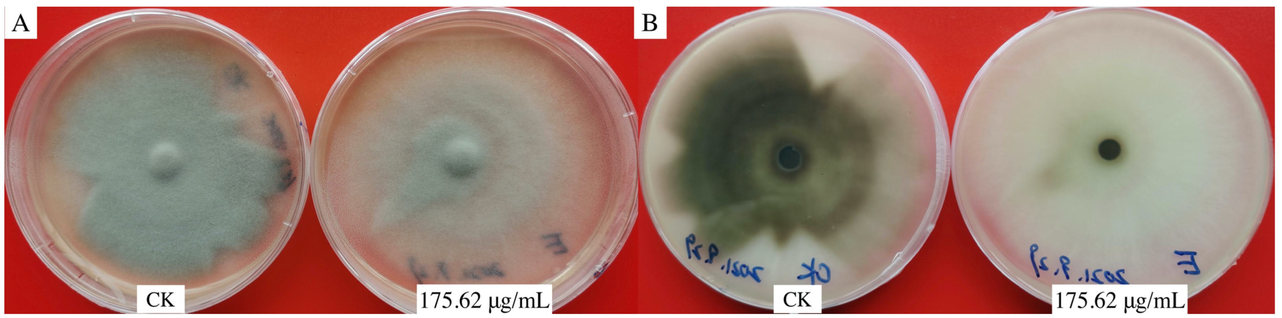
3.2. Inhibitory Effect of Aloesin on Hyphae and Appressorium of M. oryzae

The different aloesin concentrations clearly affected spore morphology, specifically appressorium formation. Only a few spores were germinated, mycelial growth was weak, and an appressorium was not formed with an aloesin concentration. We established that 625.00 μg/mL of aloesin caused the rupturing of 92.30 ± 4.26% of spores and a rate of appressorium formation of 4.74 ± 1.00% at 12 h. The percentage of appressorium formation decreased, and the spore rupturing increased, as the aloesin concentration increased above 625.00 μg/mL of aloesin (Figure 2A). The EC50 and EC95 values of the appressorium formation rate were 175.62 μg/mL and 625.00 μg/mL, respectively. According to the results, aloesin inhibited appressorium formation significantly better than it did mycelial growth in M. oryzae. The inhibition rate also increased as the aloesin test concentration increased. Specifically, 32.00 × 10³ μg/mL of aloesin revealed the lowest inhibition rate of 15.64% on day two and the highest inhibition rate of 74.45% on day seven, representing an increase in the inhibition rate of 58.81%. The inhibitory effect was continuously enhanced at the same concentration; the EC50 value was 9145.00 μg/mL after four days (Figure 2B).
Although 175.62 μg/mL of aloesin caused the M. oryzae colony to fade, mycelium growth had no apparent inhibitory effect, and the dense mycelium layer became thinner (Figure 3).

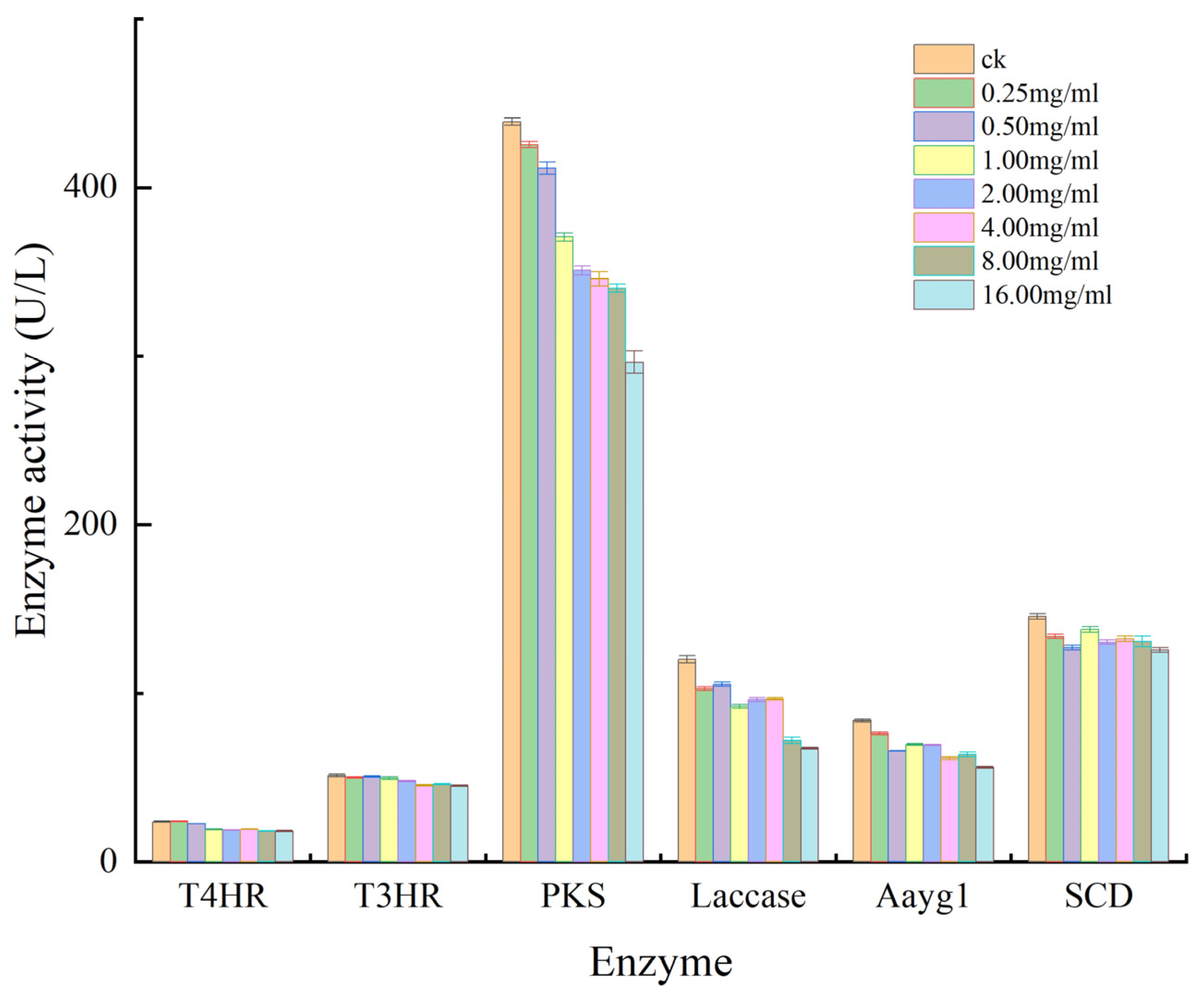
3.3. Effect of Aloesin on Enzyme Activity of M. oryzae

Regarding the inhibitory effect of aloesin on enzyme activity, PKS indicated the highest activity of 439.09 U/L in the control and the lowest activity of 296.36 U/L at 16.00 × 10³ μg/mL of aloesin, representing an increase in inhibition of 142.73 U/L and an inhibition rate of 32.51%. Laccase activity decreased by 52.70 U/L, with a 43.38% inhibition rate. The inhibition rate was 33.04%, with Aayg1 activity decreasing by 27.67 U/L and SCD activity decreasing by 19.88 U/L. These results indicated that aloesin inhibits the melanin synthase Aayg1, PKS, and LAC of M. oryzae, which is possibly one of the causes of colony fading (Figure 4).

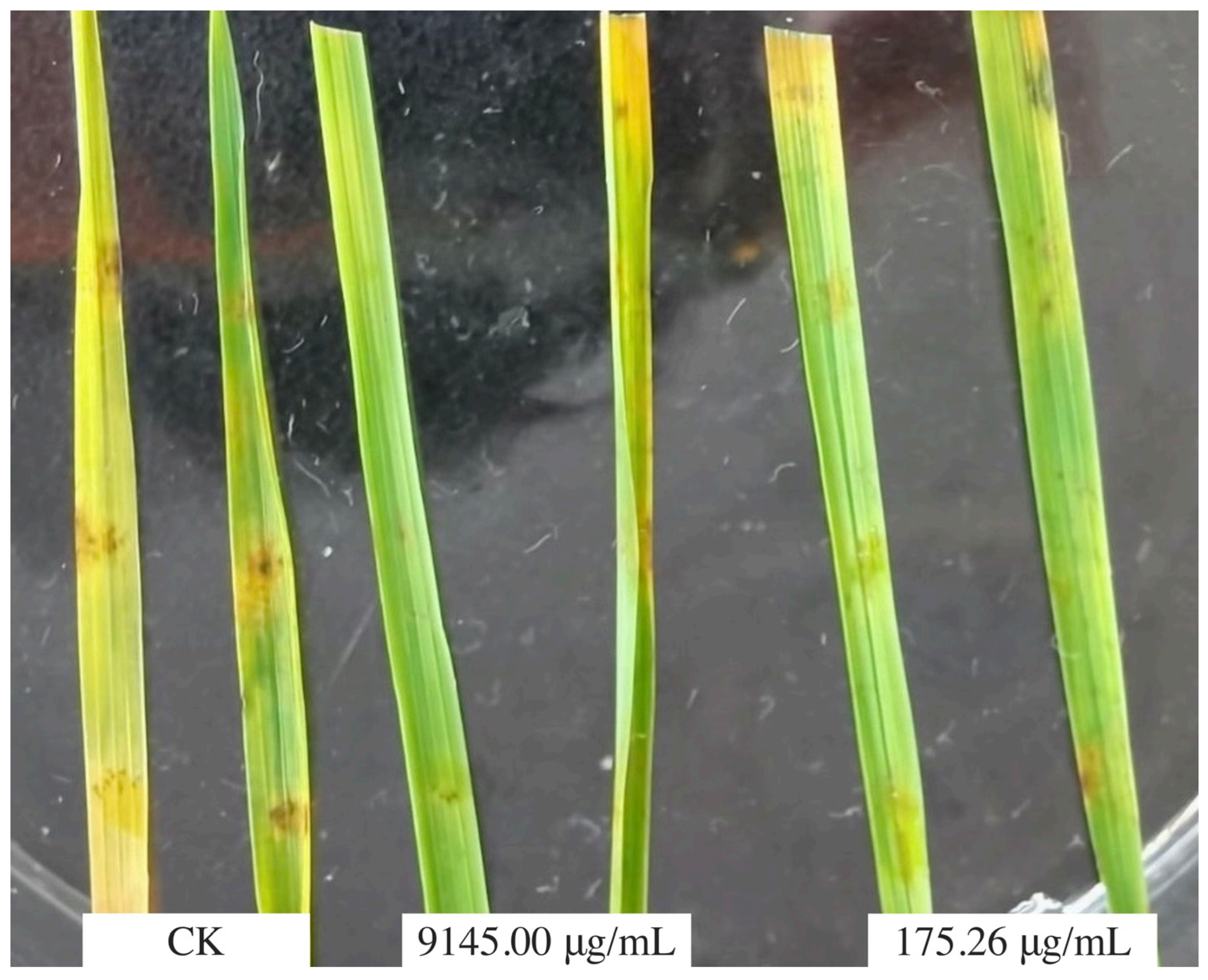
3.4. Control Efficacy of Aloesin

The virulence regression equation of aloesin to the inhibition rate of the hyphae of M. oryzae is Y = 40.855 + 0.001X (R-value = 0.9434), where EC50 = 9145.00 μg/mL. The virulence regression equation of aloesin to the inhibition rate of the appressorium of M. oryzae is Y = 1.911X + 0.7104 (R-Value = 0.8801), EC50 = 175.26 μg/mL. The inoculation results showed that the vaccination symptom in the CK was the obvious brown oval lesions and dark areas in the centers of disease spots. The results of inoculation revealed similar disease symptoms at 9145.00 μg/mL and 175.62 μg/mL of aloesin (Figure 5), the inoculated sites of the leaves showed only slight discoloration, and the disease spots were not obvious, indicating that 175.62 μg/mL of aloesin had an obvious anti-invasion effect on M. oryzae. It was also indirectly proved that the invasion of M. oryzae was closely related to the production of appressoria.

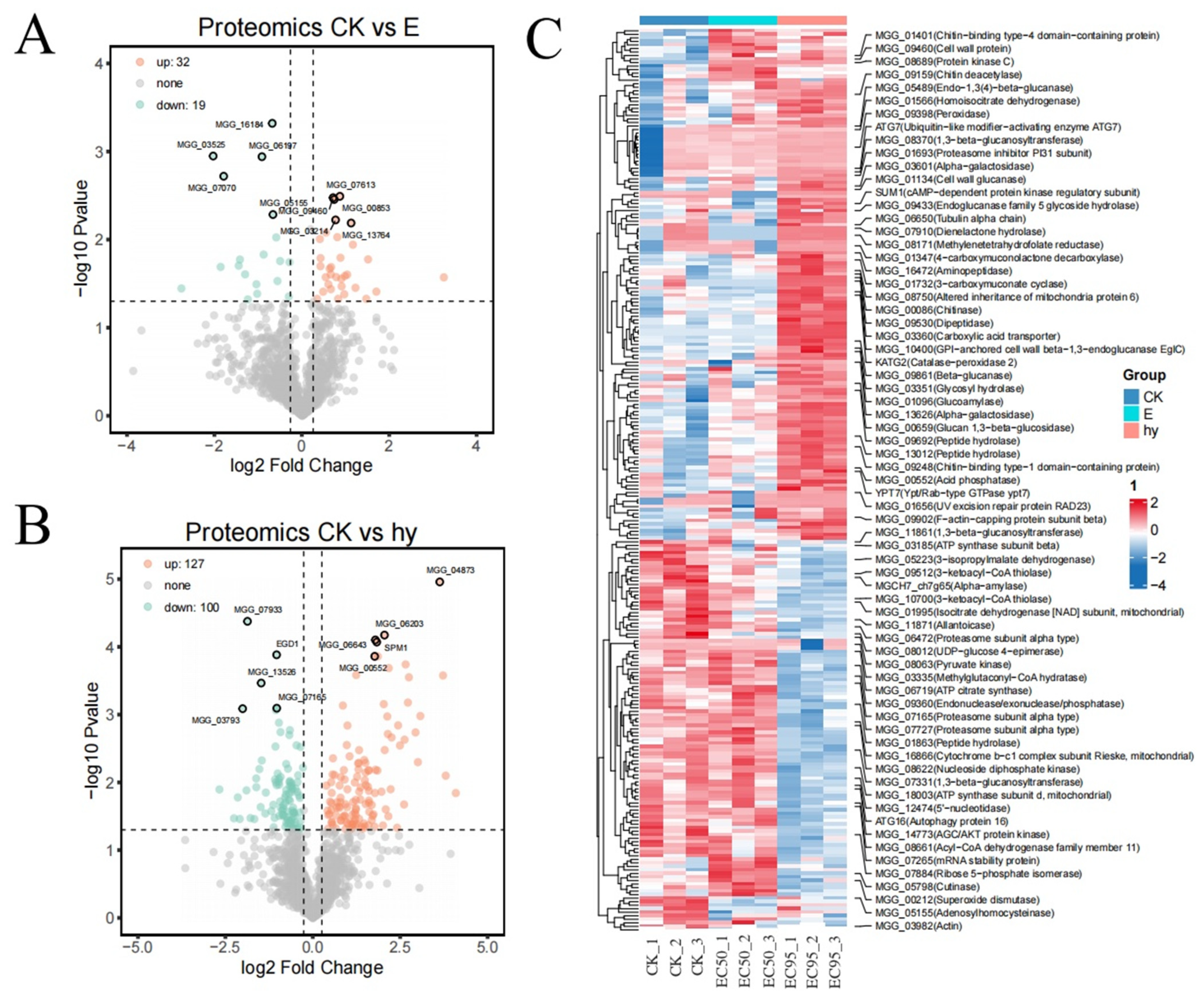
3.5. Labeling Free Proteomic Analysis

The information was converted using centered-log and regularized-log proportions. The heat map of the sample-to-sample distance produced in the principal component analysis of the top 300 variable proteins showed high levels of similarity among all three replicates (Figure 6A). According to the principal component analysis, there were no notable variances in gene expression between the biological duplicates. The proteomic analysis data were dependable and satisfied the requirements for differential expression analysis (Figure 6B).
The proteomic analysis identified 51 differentially expressed proteins (DEPs), with 32 proteins upregulated and 19 downregulated at 175.62 μg/mL of aloesin (Figure 7A). We also identified 227 DEPs, with 127 proteins upregulated and 100 proteins downregulated at 625.00 μg/mL of aloesin (Figure 7B). This proteomic analysis displayed significantly different cell expression levels compared to the non-treated cases. In our study, 175.62 μg/mL of aloesin reduced the actin (MGG_03982) expression levels (Figure 7C).
GO analysis of proteins from each cluster revealed that the downregulated proteins exhibited metabolic and biosynthetic functions at 625.00 μg/mL of aloesin, including the carbon metabolic process, canonical glycolysis, malate metabolic process, organic substance metabolic process, microtubule-based process, pyruvate biosynthetic process, and polyamine biosynthetic process between aloesin-treated and control samples, presumably reducing the amount of energy and materials that will be provided to support the basic requirements for appressorium formation by M. oryzae. However, the upregulated proteins are involved in mitochondrion organization, cellular manganese ion homeostasis, malonyl-CoA biosynthesis, cytoplasmic translational elongation, intracellular signal transduction, and fungal-type cell wall biogenesis, suggesting that these molecular events underlie the change in spore morphology (Figure 8A). The GO analysis of proteins from each cluster revealed that the downregulated proteins exhibited metabolic and biosynthetic functions at 175.62 μg/mL of aloesin, including protein translation and the cellular amino acid metabolic process, presumably reducing the biological activity and materials needed to support the basic requirements for protein synthesis in M. oryzae. However, the upregulated proteins form part of the polysaccharide catabolic and carbohydrate metabolic processes, suggesting that these molecular events degrade nutrients (Figure 8B).
The enriched GO analysis results of EC50 revealed that the most upregulated gene categories were of ribosomes, while the most downregulated gene categories were of cysteine and methionine metabolism. The enriched gene ontology analysis results of EC95 indicated that the most upregulated gene categories were glycosphingolipid biosynthesis, the globo and isoglobo series, and starch and sucrose metabolism, while the most down-regulated gene categories were ribosome and fatty acid degradation (Figure 8C).

4. Discussion

Rice blast disease, which is caused by the pathogenic fungus M. oryzae, causes widespread rice damage and yield loss [29]. Biological control technologies that reduce the use of chemical fungicides have recently received increased research attention. The prevention and control of rice blast disease is considered paramount for safeguarding the security of rice production. It is important to understand the biological characteristics of conidia germination, appressorium formation, and the disease development of M. oryzae for controlling rice blast [30,31]. Appressorium formation is a critical step in the infection cycle of M. oryzae. Therefore, inhibiting appressorium formation may help reduce damage from the disease. The presence of melanin is necessary for M. oryzae spore germination. In this study, aloesin effectively inhibited melanin synthesis and appressorium formation. Aloesin has a good inhibitory effect on the melanin synthase Aayg1, PKS, and LAC of M. oryzae. Aloesin exhibited high inhibitory effects on appressorium formation (the EC50 value was 175.26 μg/mL) and low inhibitory effects on the mycelial growth of M. oryzae (the EC50 value was 9145.00 μg/mL). The inhibitory effect was enhanced with increasing exposure from two to seven days. These results demonstrated that aloesin could effectively control the growth and proliferation of. M. oryzae. Additionally, different concentrations of aloesin affected spore morphology and appressorium formation, as was evident from the microscopic examination. At aloesin concentrations exceeding 312.50 μg/mL, no appressoria were produced, only a small number of spores germinated, mycelial growth was weak, and the average diameter of the hypanthium was less than that of the control. Appressorium formation is required for the pathogenicity of M. oryzae. Compared to spore germination in the control group, the spores were damaged at one or both ends in response to treatment with aloesin, reducing the number of germinating spores and appressorium formation. The formation of appressoria and their normal functioning are critical for M. oryzae adhesion and the penetration of host cells [32,33,34]. DHN-melanin plays an important role in the pathogenicity of appressorium, and its synthesis pathway is as follows: malonyl-CoA is found in polyketide synthase (PKS) and catalyzes the synthesis of 1,3,6,8-tetrahydroxynaphthalene (T4HN), then, with the action of a specific reductase, it is restored to 2,4-dihydro-3,6,8-trihydroxy-1-naphthalone (scytalone, SCY). SCY is dehydrated to form 1,3,8-trihydoxynaphthalene (1,3,8-THN), followed by 2,4-dihydro-3,8-dihydroxy-1-naphthalene (vermelone, VER) under the action of a second reductas and is then dehydrated to form 1,8-dihydroxynaphthalene (1,8-DHN). Finally, 1, 8-DHN polymerizes to produce DHN-melanin under the action of laccase [35]. The enzyme activity analysis results indicated that aloesin inhibited polyketolase (PKS), laccase (LAC), and chain-shortening catalytic enzyme (Aayg1) activities; these are key enzymes in melanin synthesis. The inhibition rate by aloesin of PKS, LAC, and Aayg1 activity was 32.51%, 33.04%, and 43.38%, respectively. The inhibitory effect of aloesin on enzyme activity was strongest in Aayg1, LAC, and PKS, and was least effective regarding T4HR activity. Aayg1, LAC, and PKS are important enzymes for melanin synthesis in M. oryzae spores and the appressoria [35]. Thus, aloesin exhibited high inhibitory effects on appressorium formation.
The actin cytoskeleton is believed to play a key role in the development and pathogenesis of M. oryzae [36,37,38,39]. All eukaryotes contain actin, which is essential in many cellular processes [31,40,41,42]. The actin cytoskeleton assembles as patches, cables, and rings in filamentous fungi [43,44,45,46]. The development and pathogenesis of M. oryzae largely depend on the actin cytoskeleton [37]. Xu discovered that onion epidermal cells accumulate a large amount of actin in the appressorium at the point of contact between rice blast fungus and the onion surface. An appressorium penetration peg is formed when M. oryzae septins polymerize into a heteromeric ring to scaffold a toroidal f-actin network, breaching the leaf surface. Reduced actin bundle formation inhibits plant cell polar growth, vesicle transport, and hyphal expansion [47]. This experiment revealed that aloesin could induce the downregulation of actin at a concentration of 175.26 μg/mL. Appressorium formation, rather than that of mycelia, determines M. oryzae invasion, and PKS and actin are important for melanin synthesis in the appressorium [30,35,36]. In particular, aloesin could induce the downregulation of actin at concentrations of 175.26 μg/mL. Reducing actin bundle formation would prevent polar growth, vesicle transport, and hyphal expansion in M. oryzae.

5. Conclusions

This study is the first to report that aloesin inhibits the appressorium germination and enzyme activity of melanin synthase Aayg1, PKS, and LAC of M. oryzae; the mechanism of aloesin could induce the downregulation of actin. The results presented herein support the proposition that aloesin controls mostly spore germination and appressorium formation, thereby reducing blast spot production. Aloesin is safe and environmentally friendly compared to synthetic fungicides. However, additional studies are needed to identify and characterize the active antifungal compounds in the extracts and determine their roles in rice blast disease control. Molecular docking and experimental verification will be performed for the mechanism investigation.

Author Contributions

Conceptualization, R.L. and M.L.; methodology, G.Z.; formal analysis, X.W.; resources, R.L.; data curation, G.Z. All authors have read and agreed to the published version of the manuscript.

Funding

This study was supported by the National Natural Science Foundation of China (Nos. 32160657 and 32160658), the Cultivation Project of Guizhou University (No. [2019]43), the Science and Technology Foundation Project of Guizhou Province (No. 2017-1166), and the Professor Project of Kaili University (JS201603).

Data Availability Statement

Data will be made available upon request.

Acknowledgments

We would like to thank the researcher Yue-se Ning from the Chinese Academy of Agricultural Sciences for providing the M. oryzae strain and for his advice and guidance.

Conflicts of Interest

The authors declare no conflict of interest.

References

  1. Couch, B.C.; Fudal, I.; Lebrun, M.-H.; Tharreau, D.; Valent, B.; Van Kim, P.; Notteghem, J.-L.; Kohn, L.M. Origins of host-specific populations of the blast pathogen Magnaporthe oryzae in crop domestication with subsequent expansion of pandemic clones on rice and weeds of rice. Genetics 2005, 170, 613–630. [Google Scholar] [CrossRef] [PubMed]
  2. Uda, M.N.A.; Shaari, N.H.; Said, N.S.; Ibrahim, N.H.; Akhir, M.A.M.; Hashim, M.K.R.; Salimi, M.N.; Nuradibah, M.A.; Hashim, U.; Gopinath, S.C.B. Antimicrobial activity of plant extracts from Aloe vera, Citrus hystrix, Sabah snake grass and Zingiber officinale against Pyricularia oryzae that causes rice blast disease in paddy plants. IOP Conf. Ser. Mater. Sci. Eng. 2018, 318, 012009. [Google Scholar] [CrossRef]
  3. Bussaban, B.; Lumyong, S.; Lumyong, P.; Seelanan, T.; Park, D.C.; McKenzie, E.H.C.; Hyde, K.D. Molecular and morphological characterization of Pyricularia and allied genera. Mycologia 2005, 97, 1002–1011. [Google Scholar] [CrossRef] [PubMed]
  4. Hubert, J.; Mabagala, R.B.; Mamiro, D.P. Efficacy of selected plant extracts against Pyricularia grisea, causal agent of rice blast disease. Am. J. Plant Sci. 2015, 6, 602–611. [Google Scholar] [CrossRef]
  5. Mohiddin, F.A.; Bhat, N.A.; Wani, S.H.; Bhat, A.H.; Ahanger, M.A.; Shikari, A.B.; Sofi, N.R.; Parveen, S.; Khan, G.H.; Bashir, Z.; et al. Combination of Strobilurin and Triazole Chemicals for the Management of Blast Disease in Mushk Budji -Aromatic Rice. J. Fungi 2021, 7, 1060. [Google Scholar] [CrossRef] [PubMed]
  6. Sánchez-Sanuy, F.; Mateluna-Cuadra, R.; Tomita, K.; Okada, K.; Sacchi, G.A.; Campo, S.; San Segundo, B. Iron induces resistance against the rice blast fungus Magnaporthe oryzae through potentiation of immune responses. Rice 2022, 15, 68. [Google Scholar] [CrossRef]
  7. Zhou, A.A.; Li, R.Y.; Mo, F.X.; Ding, Y.; Li, R.T.; Guo, X.; Hu, K.; Li, M. Natural product citronellal can significantly disturb chitin synthesis and cell wall integrity in Magnaporthe oryzae. J. Fungi 2022, 8, 1310. [Google Scholar] [CrossRef]
  8. Huang, P.; Li, Y.; Wang, J.; Wang, Q.; Huang, Z.; Liu, X.; Lin, F.; Lu, J. Casein Kinase 2 Mediates Degradation of Transcription Factor Pcf1 during Appressorium Formation in the Rice Blast Fungus. J. Fungi 2022, 8, 144. [Google Scholar] [CrossRef]
  9. Liu, C.; Shen, N.; Zhang, Q.; Qin, M.; Cao, T.; Zhu, S.; Tang, D.; Han, L. Magnaporthe oryzae Transcription Factor MoBZIP3 Regulates Appressorium Turgor Pressure Formation during Pathogenesis. Int. J. Mol. Sci. 2022, 23, 881. [Google Scholar] [CrossRef]
  10. Cooper, J.; Donofrio, N. Regulators unite to enable plant entry. Nat. Microbiol. 2021, 6, 1349–1350. [Google Scholar] [CrossRef]
  11. Li, R.Y.; Wu, X.M.; Yin, X.H.; Long, Y.H.; Li, M. Naturally produced citral can signifificantly inhibit normal physiology and induce cytotoxicity on Magnaporthe grisea. Pestic Biochem. Phys. 2015, 118, 19–25. [Google Scholar] [CrossRef]
  12. Song, X.; Zhao, Q.; Zhou, A.; Wen, X.; Li, M.; Li, R.; Liao, X.; Xu, T. The Antifungal Effects of Citral on Magnaporthe oryzae Occur via Modulation of Chitin Content as Revealed by RNA-Seq Analysis. J. Fungi 2021, 7, 1023. [Google Scholar] [CrossRef] [PubMed]
  13. Amadioha, A.C. Controlling rice blast in vitro and in vivo with extracts of Azadirachta indica. Crop Prot. 2000, 19, 287–290. [Google Scholar] [CrossRef]
  14. Zhao, Q.; Ding, Y.; Song, X.; Liu, S.; Li, M.; Li, R.; Ruan, H. Proteomic analysis reveals that naturally produced citral can significantly disturb physiological and metabolic processes in the rice blast fungus Magnaporthe oryzae. Pesti. Biochem. Phys. 2021, 175, 104835. [Google Scholar] [CrossRef]
  15. Bowers, J.H.; Locke, J.C. Effect of botanical extracts on the population density of Fusarium oxysporum in soil and control of Fusarium wilt in the greenhouse. Plant Dis. 2000, 84, 300–305. [Google Scholar] [CrossRef] [PubMed]
  16. Wang, W.; Ben-Daniel, B.H.; Cohen, Y. Control of plant diseases by extracts of Inula viscosa. Phytopathology 2004, 94, 1042–1047. [Google Scholar] [CrossRef] [PubMed]
  17. Lalitha, V.; Raveesha, K.A.; Kiran, B. Antimicrobial activity of Solanum torvum Swart against important seed borne pathogens of paddy. Iran. J. Energy Environ. 2010, 1, 160–164. [Google Scholar]
  18. Gurjar, M.S.; Ali, S.; Akhtar, M.; Singh, K.S. Efficacy of plant extracts in plant disease management. Agric. Sci. 2012, 3, 425–433. [Google Scholar] [CrossRef]
  19. Jyotsna, J.; Das, S.; Kumar, B. Efficacy of aqueous leaf extract of medicinal plants against blast and brown spot disease of rice. Int. J. Curr. Microbiol. Appl. Sci. 2017, 6, 4138–4144. [Google Scholar] [CrossRef]
  20. Campestrini, L.H.; Silveira, J.L.M.; Duarte, M.E.R.; Koop, H.S.; Noseda, M.D. NMR and rheological study of Aloe barbadensis partially acetylated glucomannan. Carbohydr. Polym. 2013, 94, 511–519. [Google Scholar] [CrossRef]
  21. Park, Y.I.; Jo, T.H. Perspective of industrial application of Aloe vera. In New Perspectives on Aloe; Springer: Boston, MA, USA, 2006; pp. 191–200. [Google Scholar]
  22. Lin, C.W.; Wu, C.F.; Hsiao, N.W.; Chang, C.Y.; Li, S.W.; Wan, L.; Lin, Y.J.; Lin, W.Y. Aloe-emodin is an interferon-inducing agent with antiviral activity against Japanese encephalitis virus and enterovirus 71. Int. J. Antimicrob. Agents 2008, 32, 355–359. [Google Scholar] [CrossRef]
  23. López, B.P.; Calvarro, L.M.; Piñón, D.; Esteban, A.; Tello, M.L.; Carril EP, U.; Garay, A.G. Antifungal effects of phenolic extract from industrial residues of Aloe Vera. Span. J. Agric. Res. 2019, 16, 1010. [Google Scholar]
  24. Añibarro-Ortega, M.; Pinela, J.; Ćirić, A.; Lopes, E.; Molina, A.K.; Calhelha, R.C.; Soković, M.; Ferreira, O.; Ferreira, I.C.F.R.; Barros, L. Extraction of Aloesin from Aloe vera Rind Using Alternative Green Solvents: Process Optimization and Biological Activity Assessment. Biology 2021, 10, 951. [Google Scholar] [CrossRef]
  25. Lynch, B.; Simon, R.; Roberts, A. Subchronic toxicity evaluation of aloesin. Regul. Toxicol. Pharmacol. 2011, 61, 161–171. [Google Scholar] [CrossRef]
  26. Ogbebor, N.; Adekunle, A.T. Inhibition of conidial germination and mycelial growth of Corynespora cassiicola (Berk and Curt) of rubber (Hevea brasiliensis Muell. Arg.) using extracts of some plants. Aft. J. Biotechnol. 2005, 4, 996–1000. [Google Scholar]
  27. Reyna Nathan, S.; Yang, Y. Molecular analysis of the rice MAP kinase gene family in relation to Magnaporthe grisea infection. Mol. Plant-Microbe Interact. MPMI 2006, 19, 530–540. [Google Scholar] [CrossRef] [PubMed]
  28. You, X.; Guo, B.; Wang, Z.; Ma, H.; Zhang, X. Label-free quantitative proteomic analysis of serum exosomes from patients of renal anemia: The Good and the Bad of Roxadustat. Clin. Proteom. 2022, 19, 21. [Google Scholar] [CrossRef] [PubMed]
  29. Sun, Z.; Li, L.; Qu, J.; Li, H.; Chen, H. Proteomic analysis of therapeutic effects of Qingyi pellet on rodent severe acute pancreatitis-associated lung injury. Biomed. Pharmacother. 2019, 118, 109300. [Google Scholar] [CrossRef] [PubMed]
  30. Dean, R.; Van Kan, J.A.; Pretorius, Z.A.; Hammond-Kosack, K.E.; Di Pietro, A.; Spanu, P.D.; Rudd, J.J.; Dickman, M.; Kahmann, R.; Ellis, J.; et al. The Top 10 fungal pathogens in molecular plant pathology. Mol. Plant Pathol. 2012, 13, 414–430. [Google Scholar] [CrossRef]
  31. Li, Y.-B.; Xu, R.; Liu, C.; Shen, N.; Han, L.-B.; Tang, D. Magnaporthe oryzae fimbrin organizes actin networks in the hyphal tip during polar growth and pathogenesis. PLoS Pathog. 2020, 16, e1008437. [Google Scholar] [CrossRef]
  32. Cai, X.; Zhang, X.; Li, X.; Liu, M.; Liu, X.; Wang, X.; Zhang, H.; Zheng, X.; Zhang, Z. The atypical guanylate kinase MoGuk2 plays important roles in asexual/sexual development, conidial septation, and pathogenicity in the rice blast fungus. Front. Microbiol. 2017, 8, 2467. [Google Scholar] [CrossRef]
  33. Howard, R.J.; Ferrari, M.A.; Roach, D.H.; Money, N.P. Penetration of hard substrates by a fungus employing enormous turgor pressures. Proc. Natl. Acad. Sci. USA 1991, 88, 11281–11284. [Google Scholar] [CrossRef]
  34. Saunders, D.G.O.; Aves, S.J.; Talbot, N.J. Cell cycle-mediated regulation of plant infection by the rice blast fungus. Plant Cell 2010, 22, 497–507. [Google Scholar] [CrossRef]
  35. Langfelder, K.; Streibel, M.; Jahn, B.; Haase, G.; Brakhage, A.A. Biosynthesis of fungal melanins and their importance for human pathogenic fungi. Fungal Genet. Biol. 2003, 38, 143–158. [Google Scholar] [CrossRef] [PubMed]
  36. Li, L.W.; Chen, X.L.; Zhang, S.P.; Yang, J.; Chen, D.; Liu, M.X.; Zhang, H.; Zheng, X.; Wang, P.; Peng, Y.; et al. MoCAP proteins regulated by MoArk1-mediated phosphorylation coordinate endocytosis and actin dynamics to govern development and virulence of Magnaporthe oryzae. PLoS Genet. 2017, 13, e1006814. [Google Scholar] [CrossRef] [PubMed]
  37. Li, X.; Gao, C.; Li, L.; Liu, M.; Yin, Z.; Zhang, H.; Zheng, X.; Wang, P.; Zhang, Z. MoEnd3 regulates appressorium formation and virulence through mediating endocytosis in rice blast fungus Magnaporthe oryzae. PLoS Pathog. 2017, 13, 1006449. [Google Scholar] [CrossRef]
  38. Ryder, L.S.; Dagdas, Y.F.; Mentlak, T.A.; Kershaw, M.J.; Thornton, C.R.; Schuster, M.; Chen, J.; Wang, Z.; Talbot, N. NADPH oxidases regulate septin-mediated cytoskeletal remodeling during plant infection by the rice blast fungus. Proc. Natl. Acad. Sci. USA 2013, 110, 3179–3184. [Google Scholar] [CrossRef]
  39. Wang, C.L.; Shaw, B.D. F-actin localization dynamics during appressorium formation in Colletotrichum graminicola. Mycologia 2016, 108, 506–514. [Google Scholar] [CrossRef]
  40. Garduño-Rosales, M.; Callejas-Negrete, O.A.; Medina-Castellanos, E.; Bartnicki-García, S.; Herrera-Estrella, A.; Mouriño-Pérez, R.R. F-actin dynamics following mechanical injury of Trichoderma atroviride and Neurospora crassa hyphae. Fungal Genet. Biol. 2022, 159, 103672. [Google Scholar] [CrossRef]
  41. Mooren, O.L.; Galletta, B.J.; Cooper, J.A. Roles for actin assembly in endocytosis. Annu. Rev. Biochem. 2012, 81, 661–686. [Google Scholar] [CrossRef]
  42. Li, J.; Blanchoin, L.; Staiger, C.J. Signaling to actin stochastic dynamics. Annu. Rev. Plant. Biol. 2015, 66, 415–440. [Google Scholar] [CrossRef]
  43. Echauri-Espinosa, R.O.; Callejas-Negrete, O.A.; Roberson, R.W.; Bartnicki-Garcia, S.; Mourino-Perez, R.R. Coronin Is a Component of the Endocytic Collar of Hyphae of Neurospora crassa and Is Necessary for Normal Growth and Morphogenesis. PLoS ONE 2012, 7, 38237. [Google Scholar] [CrossRef]
  44. Bartnicki-Garcia, S.; Garduno-Rosales, M.; Delgado-Alvarez, D.L.; Mourino-Perez, R.R. Experimental measurement of endocytosis in fungal hyphae. Fungal Genet. Biol. 2018, 118, 32–36. [Google Scholar] [CrossRef] [PubMed]
  45. Berepiki, A.; Lichius, A.; Shoji, J.Y.; Tilsner, J.; Read, N.D. F-actin dynamics in Neurospora crassa. Eukaryot. Cell 2010, 9, 547–557. [Google Scholar] [CrossRef] [PubMed]
  46. Delgado-Alvarez, D.L.; Callejas-Negrete, O.A.; Gomez, N.; Freitag, M.; Roberson, R.W.; Smith, L.G.; Mouriño-Pérez, R. Visualization of F-actin localization and dynamics with live cell markers in Neurospora crassa. Fungal Genet. Biol. 2010, 47, 573–586. [Google Scholar] [CrossRef] [PubMed]
  47. Xu, J.R.; Staiger, C.J.; Hamer, J.E. Inactivation of the mitogen-activated protein kinase Mps1 from the rice blast fungus prevents penetration of host cells but allows activation of plant defense responses. Proc. Natl. Acad. Sci. USA 1998, 95, 12713–12718. [Google Scholar] [CrossRef] [PubMed]
Figure 1. The spores of M. oryzae collapsed (indicated by an arrow) when aloesin was used against the M. oryzae spores (12 h). (A1,A2) Control. (B1,B2) 625.00 μg/mL aloesin. Bar = 50.00 µm.
Microorganisms 11 02395 g001
Figure 2. The inhibitory effect of aloesin on the hyphae and appressoria of M. oryzae. (A) Inhibition rate of aloesin against the morphology and germination of M. oryzae spores under microscopic examination (12 h). (B) Inhibition rate of aloesin against mycelium growth in M. oryzae (2−7 d).
Microorganisms 11 02395 g002
Figure 3. Inhibitory effect of 175.62 μg/mL of aloesin against M. oryzae Guy11. (A) Colony front view; (B) colony back view.
Microorganisms 11 02395 g003
Figure 4. The enzyme activity effect of aloesin against M. oryzae.
Microorganisms 11 02395 g004
Figure 5. Inoculation effect of aloesin against M. oryzae by the inoculation method.
Microorganisms 11 02395 g005
Figure 6. Quality control chart of the protein. Data were transformed with the centered-log and regularized-log ratios (A). The sample-to-sample distance heat map exhibited good similarity between all three replicates (B). CK, Control; EC50, 175.62 μg/mL of aloesin; EC95, 625.00 μg/mL of aloesin.
Microorganisms 11 02395 g006
Figure 7. Volcano plot and heatmaps of DEPs in M. oryzae between the aloesin-treated and control samples. (A,B) Volcano plot illustrating the upregulated (red dots) and downregulated (blue dots) DEPs and the normally expressed proteins (gray dots). (C) Heatmap of up-regulated DEPs and down-regulated DEPs. CK, Control; EC50, 175.62 μg/mL of aloesin; EC95, 625.00 μg/mL of aloesin.
Microorganisms 11 02395 g007
Figure 8. KEGG enrichment analysis of the proteins in M. oryzae between the aloesin-treated and control samples. (A,B) Pathway analysis showing the upregulated (red dots) and downregulated (green dots) DEPI. (C) Differentially expressed proteins induced by aloesin treatment, shown as an enriched gene ontology analysis of up-regulated DEPs and an enriched gene ontology analysis of down-regulated DEPs. padj: adjusted p-value. CK, Control; EC50, 175.62 μg/mL of aloesin; EC95, 625.00 μg/mL of aloesin.
Microorganisms 11 02395 g008
Disclaimer/Publisher’s Note: The statements, opinions and data contained in all publications are solely those of the individual author(s) and contributor(s) and not of MDPI and/or the editor(s). MDPI and/or the editor(s) disclaim responsibility for any injury to people or property resulting from any ideas, methods, instructions or products referred to in the content.

