Article

DNA Markers for Detection and Genotyping of Xanthomonas euroxanthea

by
Kayla Gisela Silva
1,2,3,†,
Leonor Martins
1,2,3,*,†,
Miguel Teixeira
1,2,3,‡,
Joël F. Pothier
4 and
Fernando Tavares
1,2,3,*
1
CIBIO, Centro de Investigação em Biodiversidade e Recursos Genéticos, InBIO Laboratório Associado, Campus de Vairão, Universidade do Porto, 4485-661 Vairão, Portugal
2
Departamento de Biologia, Faculdade de Ciências, Universidade do Porto, 4169-007 Porto, Portugal
3
BIOPOLIS Program in Genomics, Biodiversity and Land Planning, CIBIO, Campus de Vairão, 4485-661 Vairão, Portugal
4
Environmental Genomics and Systems Biology Research Group, Institute for Natural Resource Sciences, Zurich University of Applied Sciences (ZHAW), Einsiedlerstrasse 31, 8820 Wädenswil, Switzerland
*
Authors to whom correspondence should be addressed.
These authors contributed equally to this work.
Current address: Department of Biotechnology and Food Science, Norwegian University of Science and Technology, 7491 Trondheim, Norway.
Microorganisms 2022, 10(6), 1078; https://doi.org/10.3390/microorganisms10061078
Submission received: 29 April 2022 / Revised: 19 May 2022 / Accepted: 19 May 2022 / Published: 24 May 2022

Abstract

:
Xanthomonas euroxanthea is a bacterial species encompassing both pathogenic and non-pathogenic strains and is frequently found colonizing the same host plants as X. arboricola. This presents the need to develop a detection and genotyping assay able to track these bacteria in microbial consortia with other xanthomonads. Eight X. euroxanthea-specific DNA markers (XEA1-XEA8) were selected by comparative genomics and validated in silico regarding their specificity and consistency using BLASTn, synteny analysis, CG content, codon usage (CAI/eCAI values) and genomic proximity to plasticity determinants. In silico, the selected eight DNA markers were found to be specific and conserved across the genomes of 11 X. euroxanthea strains, and in particular, five DNA markers (XEA4, XEA5, XEA6, XEA7 and XEA8) were unfailingly found in these genomes. A multiplex of PCR targeting markers XEA1 (819 bp), XEA8 (648 bp) and XEA5 (295 bp) was shown to successfully detect X. euroxanthea down to 1 ng of DNA (per PCR reaction). The topology of trees generated with the concatenated sequences of three markers (XEA5, XEA6 and XEA8) and four housekeeping genes (gyrB, rpoD, fyuA and acnB) underlined the equal discriminatory power of these features and thus the suitability of the DNA markers to discriminate X. euroxanthea lineages. Overall, this study displays a DNA-marker-based method for the detection and genotyping of X. euroxanthea strains, contributing to monitoring for its presence in X. arboricola-colonizing habitats. The present study proposes a workflow for the selection of species-specific detection markers. Prospectively, this assay could contribute to unveil alternative host species of Xanthomonas euroxanthea; and improve the control of phytopathogenic strains.

1. Introduction

Following extensive genotyping and comparative genomics studies performed on walnut-associated bacterial isolates, it has been shown that not all the isolates could be identified as Xanthomonas arboricola pv. juglandis, the phytopathogen commonly acknowledged as causing walnut bacterial blight (WBB) [1,2,3]. Further studies showed that some walnut-associated Xanthomonas isolates were taxonomically distinct from any of the other described Xanthomonas species.
These were proposed as members of the new species Xanthomonas euroxanthea [4]. Pathogenicity assays indicated that X. euroxanthea encompasses non-pathogenic and pathogenic strains that can cause WBB-like symptoms, being in a privileged position to investigate genetic determinants of pathogenesis and its evolution [4,5,6].
Interestingly, apart from its occurrence in walnuts (Juglans regia), recent evidence was gathered reporting the isolation of X. euroxanthea from distinct plant host species, such as Carya illinoensis (pecan; strains CPBF 761 and CPBF 766), that together with walnut (Juglans regia; strains CPBF 367, CPBF 424T, CPBF 426 and CFBP 7653) belong to the Juglandaceae family; Solanum lycopersicum (tomato plants; strains BRIP 62409, BRIP 62411, BRIP 62415 and BRIP 62418) a member of the Solanaceae family [7,8,9]; and Phaseolus vulgaris (common bean; strain CFBP 7622, previously misclassified as X. arboricola [10,11]), a Fabaceae plant species.
More strikingly is that X. arboricola strains were also isolated from all the mentioned plant species, suggesting that both X. euroxanthea and X. arboricola share the same host plants, including the same plant specimen, which raises questions regarding co-colonization and niche-specific adaptations [7,8,12]. Still, taking into consideration the recent taxonomic refinement of X. arboricola species and the unearthing of six new X. euroxanthea strains (2949, 2955, 2957, 2974, 3640 and F2) [11], it is foreseeable the identification of additional X. euroxanthea isolates from plant species that have passed unnoticed so far.
Altogether, the common ecological niche of these two closely related species, plus their cosmopolitan distribution and co-occurrence in different host plant species resulted in misclassifying some X. euroxanthea isolates as X. arboricola [8,10,11], which calls for the need to develop methods for the detection and genotyping of this bacterial species. Over the years, different diagnostic and molecular typing tools have been developed for a variety of xanthomonads [13], namely for X. arboricola including their most studied pathovars juglandis [14,15] and pruni [16,17,18,19], aiming to address their diversity within a geographic or epidemiological context.
Generally, these approaches consist of culture-based detection of the phytopathogen by PCR, followed by multilocus sequence analysis (MLSA) of several housekeeping genes to define haplotypes. In fact, multiplex-PCR and dot-blot hybridization assays showed that three X. arboricola pv. juglandis specific DNA markers (XAJ1, XAJ6 and XAJ8) were absent from X. euroxanthea strains CPBF 367, CPBF 424T and CPBF 426, which formed a distinct MLSA cluster [12,14].
A robust method to detect X. euroxanthea while undoubtedly distinguishing it from X. arboricola has not been described so far. Furthermore, while several genotyping techniques have been used to assess the diversity of phytosanitary regulated Xanthomonas, as recently reviewed [13], the data currently available regarding genotyping of X. euroxanthea is scarce and limited to MLSA studies performed on Xanthomonas isolated from walnut trees [12].
In this study, comparative genomics and in silico validation tools were combined as previously described [20] to select eight X. euroxanthea-specific DNA markers located in conserved genomic regions. While XEA1, XEA5 and XEA8 DNA markers were chosen to optimize a multiplex PCR detection method for the reliable detection of the target bacteria, altogether the number of SNPs recorded for DNA markers XEA5, XEA6 and XEA8 within the studied X. euroxanthea strains, revealed an allelic variation capable to discriminate X. euroxanthea strains as efficiently as the housekeeping genes used in MLSA. Ultimately, this work may unlock the possibility to conciliate bacterial detection and diversity assessment using the same DNA markers, which may be particularly useful to survey X. euroxanthea populations in environmental samples.

2. Materials and Methods

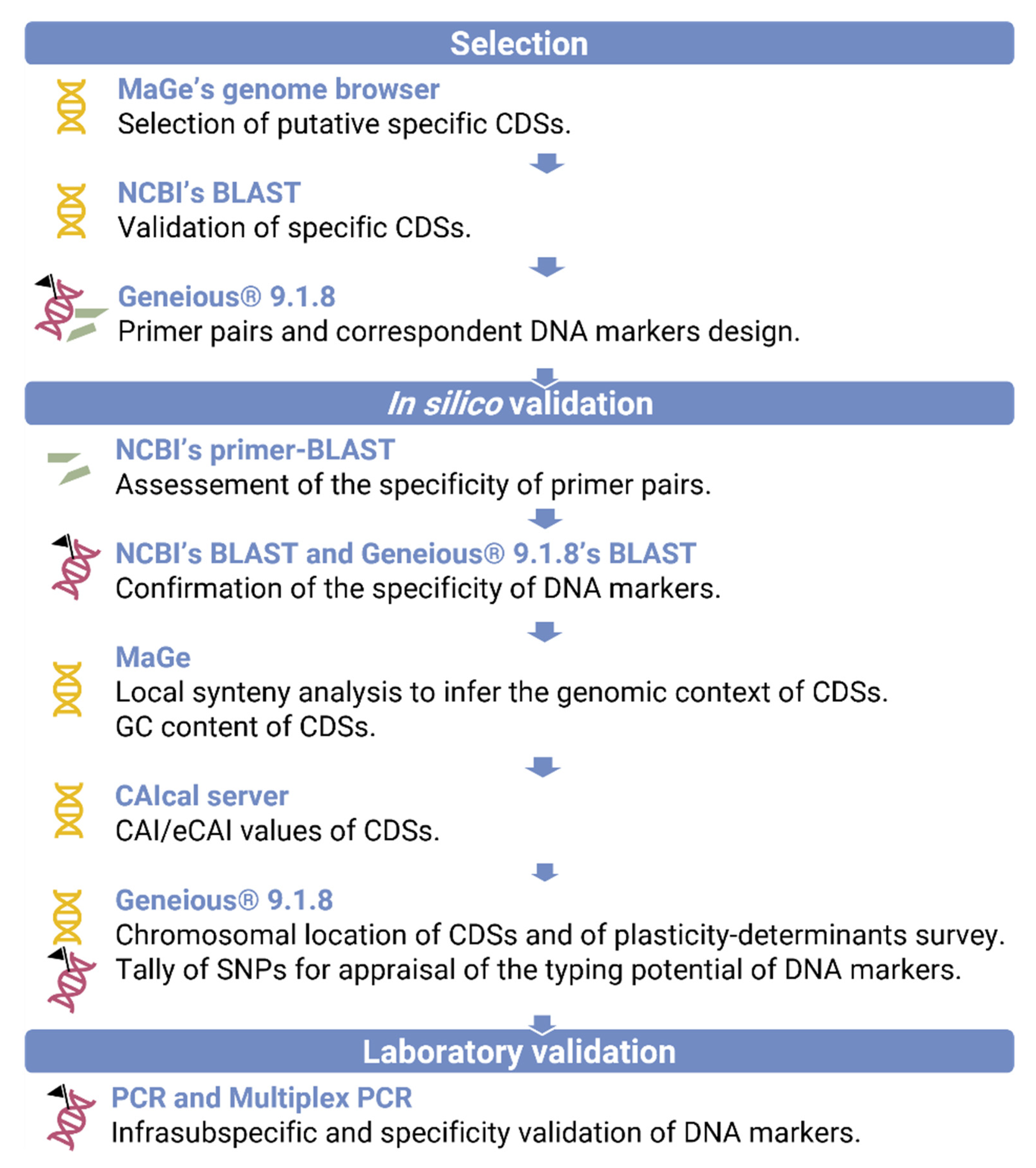
2.1. In Silico Selection and Validation of X. euroxanthea-Specific DNA Markers

A synteny analysis of 11 X. euroxanthea strains and other 24 representative xanthomonads, downloaded from the NCBI database (Table 1), was performed using MaGe v3.15.3 [21]. This led to the identification of X. euroxanthea-specific coding DNA sequences (CDSs) that are concomitantly present in X. euroxanthea genomes and absent from non-X. euroxanthea strains.
Then, a BLAST was performed in NCBI (accessed in 22 November 2021 at https://www.ncbi.nlm.nih.gov/) using as query the putative X. euroxanthea-specific CDSs and the database nr/nt to further confirm their specificity. Sequences with hits pertaining solely to X. euroxanthea were considered putative X. euroxanthea-specific genomic regions (Table S1) and were used for primer and specific DNA marker design using Geneious® 9.1.8 [22]. The affinity and complementarity of these primers to target X. euroxanthea CDSs was checked using NCBI’s Primer-BLAST tool [22,23]. The obtained eight DNA markers were then evaluated for specificity by a BLASTn analysis in Geneious® 9.1.8 and NCBI (using the database nr/nt).
The genomic context of the markers was assessed to ensure its consistency across the diversity of X. euroxanthea strains. Particularly, a comparative genomics analysis was performed by local alignment of CDSs in MaGe (Figure S1a–d), to evaluate the syntenic context. Additionally, features, such as the CAI/eCAI values [24]; GC content (deducted from MaGe); chromosomal location; and proximity to determinants of genomic plasticity, namely transposons, integrases, recombinases and phage-related ORFs (Geneious® 9.1.8), were annotated. The number of Single Nucleotide Polymorphisms (SNPs) included in the eight DNA markers and housekeeping genes of the 11 X. euroxanthea genomes considered in this study were summed up recurring to Geneious® 9.1.8 and normalized using the formula ( SNPs   number total   nucleotide   sequence   length × 100 ) . The described procedure followed for specific X. euroxanthea DNA markers design is systematized in Figure 1.

2.2. Bacterial Strains, Culture Conditions and DNA Extraction

The bacterial strains used for the validation of the eight X. euroxanthea-specific markers are listed in Table 2 and include seven X. euroxanthea strains; and other closely related and niche-sharing strains, namely 10 strains of X. arboricola, seven strains representing six pathovars of X. arboricola and 11 strains belonging to non-arboricola Xanthomonas species. Bacterial strains were cultured as previously described [14] or in peptone-sucrose-agar (PSA) medium (10 g peptone; 10 g sucrose; 1 g glutamic acid; 15 g agar and distilled water up to 1.0 L) at 28 °C. DNA was extracted from pure cultures using the EZNA Bacterial DNA Purification kit (Omega Bio-Tek, Norcross, GA, USA), according to the manufacturer’s instructions and quantified using a DS-11 microvolume spectrophotometer (DeNovix, Wilmington, DE, USA).

2.3. Experimental Validation of Putative X. euroxanthea-Specific DNA Markers by Multiplex PCR

A multiplex PCR targeting the most promising DNA markers was optimized to validate a method to rapidly identify X. euroxanthea isolates. XEA1, XEA5 and XEA8 were the chosen markers with distinct amplicon lengths of 819, 295 and 648 bp, respectively; and their broad occurrence in the tested X. euroxanthea strains, apart from XEA1 (absent in CFBP 7622). A 20 µL PCR reaction mix consisted of 1 × DreamTaq Buffer (ThermoFisher Scientific, Waltham, MA, USA), 0.2 mM of each deoxynucleotide triphosphate (dNTP) (Grisp, Porto, Portugal), 0.2 mM of each forward and reverse primers (Table 3), 1.5 U of DreamTaq DNA Polymerase (ThermoFisher Scientific, Waltham, MA, USA) and 25 ng of DNA template.
Sterile distilled water was used as the negative control. PCR cycling parameters consisted of a first amplification cycle of 5 min at 95 °C, followed by 35 cycles of 95 °C for 30 s, 61 °C for 15 s and 72 °C for 30 s as well as a final DNA extension at 72 °C for 10 min. The same DNA samples were used as template in PCR reactions using each of the markers individually (XEA1, XEA5 and XEA8) and 1.0 U of DreamTaq DNA polymerase per PCR reaction.
PCR products were separated by electrophoresis on a 0.8% agarose gel (1 × TAE buffer) and visualized using Xpert Green DNA stain (Grisp, Porto, Portugal) with a Molecular Imager Gel Doc XR+ System (Bio-Rad, Hercules, CA, USA). The obtained PCR products for each marker and each strain were purified using the Illustra GFX GEL Band Purification kit (GE Healthcare, Buckinghamshire, UK), following the reference protocol available and sequenced on both strands (STAB Vida, Caparica, Portugal) to confirm their identity and determine the number of SNPs.

2.4. PCR Detection Limit

The detection limit of the multiplex PCR was determined using 10 μL from each of the 10-fold dilutions of X. euroxanthea CPBF 424T chromosomal DNA prepared in distilled sterile water, ranging from 100 ng to 1 pg per PCR reaction. Multiplex PCR conditions were kept as described above.

2.5. Typing Potential of X. euroxanthea-Specific DNA Markers

Unrooted trees using XEA5, XEA6 and XEA8 markers (transversal to all strains of X. euroxanthea) (Table S2) and partial sequence analysis of the housekeeping genes acnB, fyuA, gyrB and rpoD (Table S3) of 11 X. euroxanthea strains were built to infer the typing potential of these DNA markers. Markers XEA5 (295 bp), XEA6 (237 bp) and XEA8 (648 bp) sequences were concatenated using the Geneious® v. 9.1.7 and used to build a maximum-likelihood tree based on Tamura-Nei model on MEGA X [25], as previously described [14].
The nucleotide sequences of housekeeping genes acnB, fyuA, gyrB and rpoD were retrieved from the 11 X. euroxanthea genomes, aligned and trimmed to 513, 640, 828 and 793 bp, respectively, and subsequently concatenated using the Geneious® v. 9.1.7 to build a maximum-likelihood tree as described for the markers. Since these three markers are X. euroxanthea specific, and no homologous marker could be found to define a coherent outgroup, the relatedness of the X. euroxanthea strains was inferred by an unrooted tree.

3. Results

3.1. In Silico Selection of DNA Markers for X. euroxanthea

The selection of X. euroxanthea-specific DNA markers was conducted according to the workflow detailed in Figure 1, which consisted on the use of a platform of comparative genomics (Mage) to screen for unique X. euroxanthea CDSs, further validated by BLASTn.
A list of CDSs exclusively present in the genomes of 11 X. euroxanthea strains and absent from other 12 Xanthomonas species, including the closely related X. arboricola, was deducted from synteny analysis between the 24 genomes (Table 1) along the full length of X. euroxanthea CPBF 424T genome used as reference (Figure S1). The candidate CDSs (from the reference genome of X. euroxanthea CPBF 424T) were submitted to a BLASTn to confirm their exclusivity in X. euroxanthea considering high stringency values of E-value, percentage of identity and query coverage and their suitability as X. euroxanthea-specific DNA markers appraised by the design of robust primers.
The data showed that, four CDSs had no significant BLAST hits; three CDSs showed no hits with other Xanthomonas sp. and solely one CDS coding for a putative conserved protein of unknown function matched with Xanthomonas sp. GW (E-value of 2 × 10⁻⁴ and query coverage of 19%) (Table 3). These seven CDSs were selected for the design of eight DNA markers, designated XEA1-XEA8; being that two consecutive and partly overlapping CDSs (XE424_v1_a1415 and XE424_v1_a1414) were used to design marker XEA8 (Table 3). Five of these CDSs are predicted to be proteins of unknown function and two to be transcriptional regulators (Table 3). Ultimately, five DNA markers, XEA4-XEA8, are unfailingly present in the genome of the 11 X. euroxanthea strains analyzed; XEA1 is present in eight genomes; and XEA2 and XEA3 are present in five strains (Figure 2).

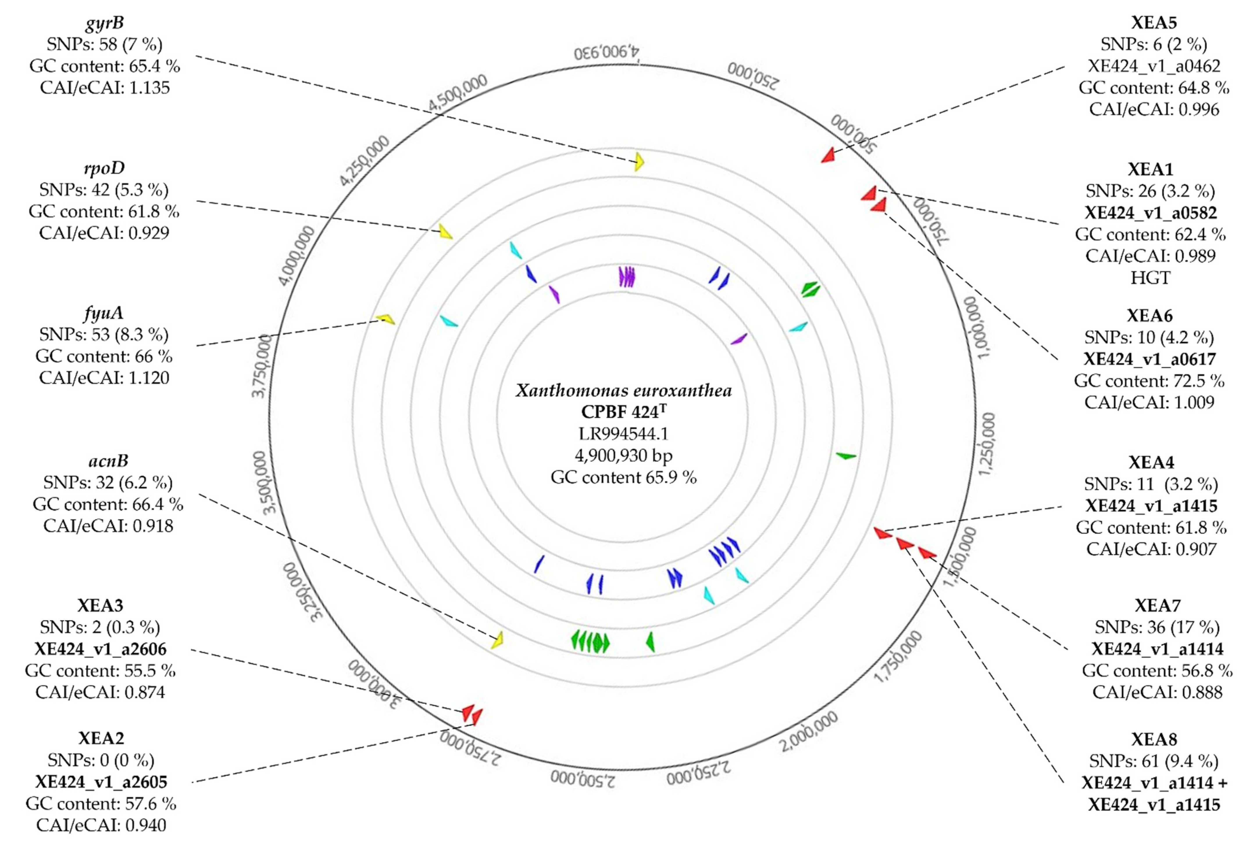
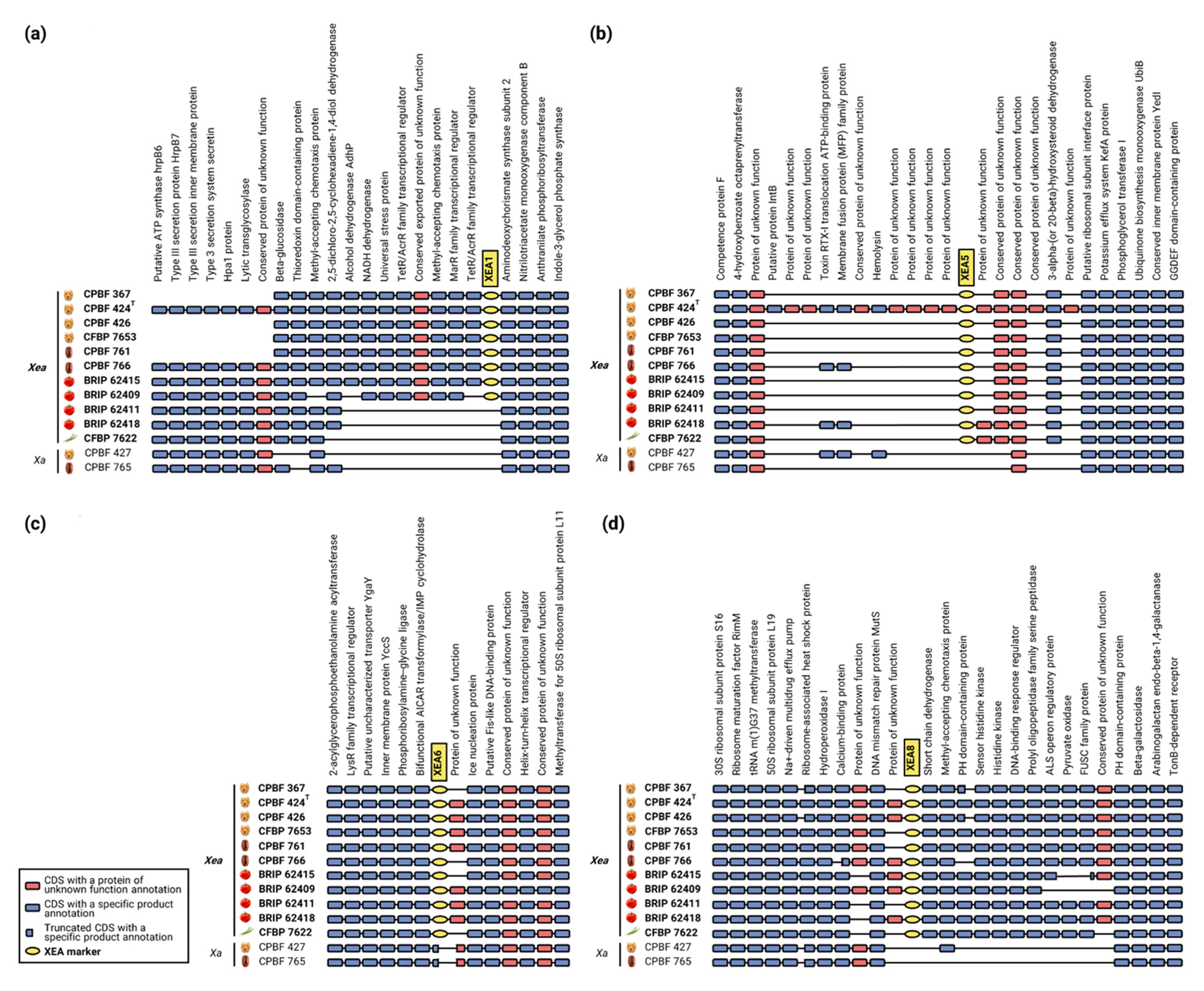
3.2. Genomic Analysis Unearths the Stability of XEA DNA Markers

To further assess the uniqueness of these X. euroxanthea-specific CDSs used for DNA marker design, several features, including SNPs number, CAI/eCAI values, GC content, chromosomal location and chromosomal proximity to genomic plasticity-determinants were surveyed (Figure 3).
Within the 11 X. euroxanthea genomes considered in this study, the number of SNPs for each marker and housekeeping gene was determined and normalized to the full length of the sequence (i.e., DNA marker or housekeeping gene) as a percentual value indicative of the allelic diversity of the sequences (Figure 3).
CAI/eCAI values of CDSs range between 0.874 (for XE424_v1_a2606 corresponding to DNA marker XEA3) to 1.009 (for XE424_v1_a0617 corresponding to DNA marker XEA6); which are values similar to the range attained with housekeeping genes, namely, 0.918 (acnB) to 1.135 (gyrB) (Figure 3). GC content values for X. euroxanthea-specific CDSs varied between 55.5% (XE424_v1_a2606, XEA3) to 72.5% (XE424_v1_a0617, XEA6), which parallels with X. euroxanthea CPBF 424T genome GC content value of 65.9% (Figure 3).
Moreover, chromosomal location studies revealed that the XEA markers are scattered throughout the first half of the chromosome and not in the vicinity of genomic plasticity-determinants, including transposases, recombinases, integrases and phage-related ORFs (Figure 3).
The synteny analysis performed with MaGe allowed to investigate the genomic context of the most representative DNA markers, i.e., XEA1, XEA5, XEA6 and XEA8, across 11 X. euroxanthea genomes and comparatively to Xanthomonas arboricola strains CPBF 427 (X. arboricola pv. juglandis) and CPBF 765 (Figure 4) and other Xanthomonas spp. representative strains (Figure S1).
The data showed that XEA1, XEA5, XEA6 and XEA8 are located in highly syntenic regions across all the X. euroxanthea genomes studied, underlining the absence of genomic rearrangements (Figure 4). Furthermore, CDSs used for the design of markers XEA5, XEA6 and XEA8 are exclusive of X. euroxanthea, being either absent (CDSs encompassing XEA5 and XEA8) or truncated (CDS encompassing XEA6) in X. arboricola strains (Figure 4).
Particularly, the region upstream and including marker XEA1 appears to have suffered erosion in the X. arboricola strains analyzed (CPBF 427 and CPBF 765) and in three of the X. euroxanthea strains studied (BRIP 62411, BRIP 62418 and CFBP 7622). In parallel, this scenario is observed for the region upstream of XEA5 for all strains, except for CPBF 424T; and downstream of XEA8 for the non-X. euroxanthea strains.
Within the flanking regions of the XEA markers, the CDSs annotated as unknown proteins are the ones where the presence/absence across strains is inconsistent, suggesting that they have been decaying. While, for XEA6 and XEA8 markers, these events are limited to a single CDS immediately flanking the markers, for XEA5 this paradigm is particularly clear as several CDSs annotated as unknown proteins are present in CPBF 424T, have been lost by the other strains.

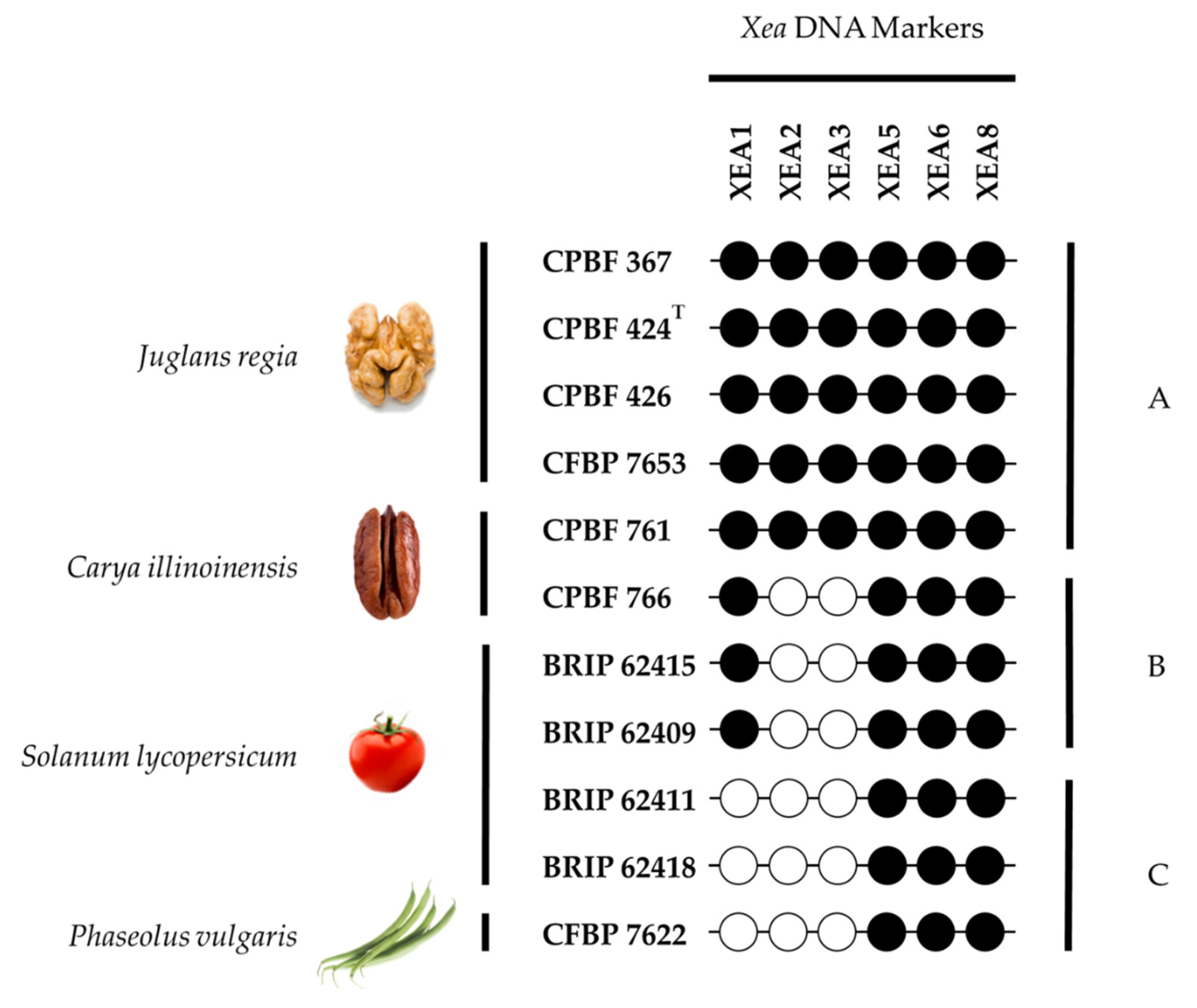
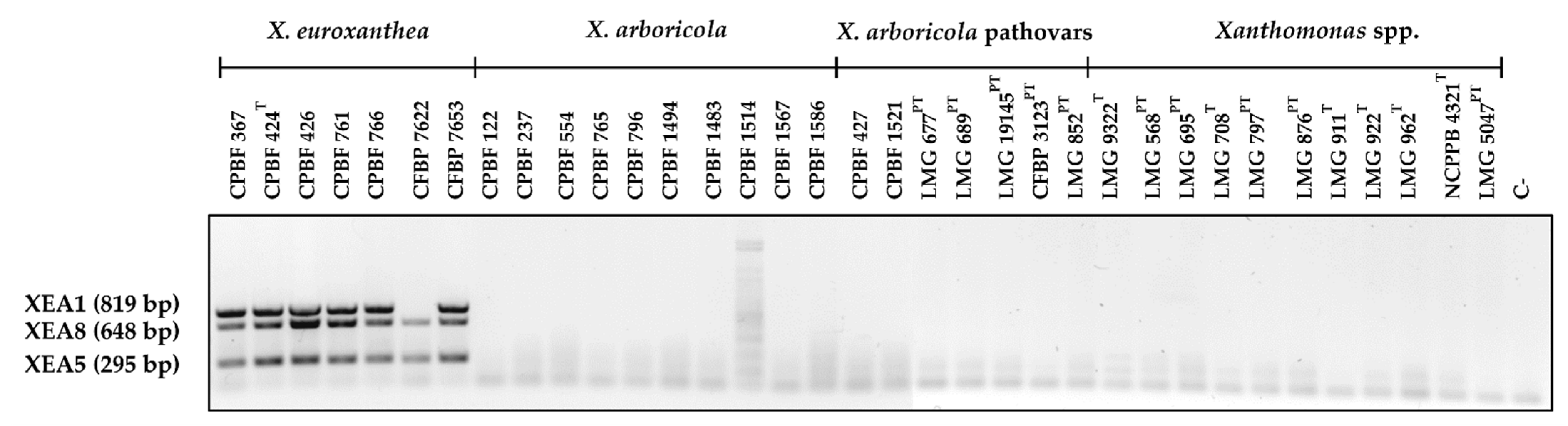
3.3. Multiplex PCR Allows for the Confident Identification of X. euroxanthea Strains

Reliable identification of X. euroxanthea isolates by multiplex PCR was optimized for DNA markers XEA1, XEA5 and XEA8, as these originate amplicons of distinguishable size (819, 295 and 648 bp, respectively) and are located in three independent genomic regions of X. euroxanthea. The three markers were successfully amplified in all X. euroxanthea strains analyzed, with exception for XEA1 in X. euroxanthea CFBP 7622 strain (Figure 5) as expected by in silico studies. In addition, no amplification was observed for any of the other 28 xanthomonads, namely 17 strains of X. arboricola, including different pathovars and 9 non-arboricola Xanthomonas species.

3.4. Detection Limit of Multiplex PCR with XEA DNA Markers

The detection limit of the multiplex PCR targeting the X. euroxanthea-specific markers XEA1, XEA5 and XEA8, determined through serial dilution of chromosomal DNA, was 1 ng for PCR reaction (Figure S2). When assessing the PCR detection limit of each marker individually, while for XEA1 and XEA8 the detection limit was identical to the multiplex PCR, i.e., 1 ng/PCR reaction, it was ascertained that the limit of detection lowered to 100 pg/PCR reaction for XEA5.

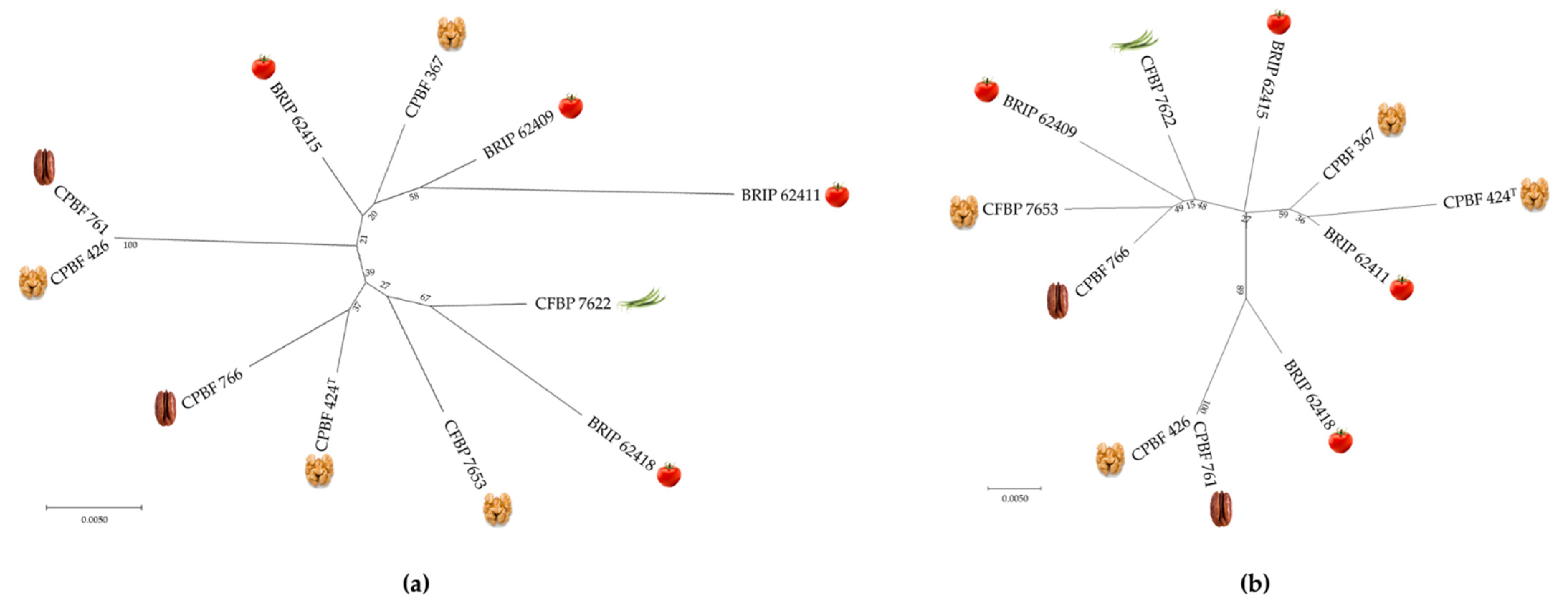
3.5. Typing Potential of Informative XEA DNA Markers

The concatenated sequences of XEA5, XEA6 and XEA8 (1180 bp) for each X. euroxanthea strain studied were aligned and used to generate a maximum-likelihood tree to investigate the discriminatory potential of these X. euroxanthea-specific markers comparatively to four housekeeping genes, corresponding to a concatenated sequence length of 2774 bp, (acnB, fyuA, gyrB and rpoD genes) commonly used for MLSA (Figure 6).
Attending that these XEA detection markers are highly specific to X. euroxanthea, no homologous sequences have been found in other bacterial taxa, and therefore the allelic diversity determined by the number of SNP (6 SNP/295nt for XEA5, 10 SNP/237nt for XEA6, and 61 SNP/648nt for XEA8; Figure 3) is represented by an unrooted Maximum-likelihood tree. For both trees, each X. euroxanthea strain is represented by an independent tree branch, with exceptions for strains CPBF 426 and CPBF 761, which clustered together in a single branch. Furthermore, the topology of both trees does not reflect any clustering according to plant–host of isolation nor by pathogenicity or non-pathogenicity phenotypes (Figure 6).

4. Discussion

It is important to investigate the distribution and role played by X. euroxanthea and the closely related X. arboricola within the plant hosts that they frequently co-colonize [12]. Therefore, it is essential to develop a reliable and efficient method for the accurate detection and identification of X. euroxanthea strains and its differentiation from X. arboricola [12].
Additionally, early detection is a critical first step towards timely sanitary intervention aimed at eradicating the pathogen and at reducing the inoculum spread to other plants; thus lessening the disease-induced damage in crops from growth to postharvest processing of products and ensuring agricultural sustainability [26]. Likewise, discriminating one bacterial species from the other would be useful in the application of suitable management procedures.
Recommended standard diagnostic protocols from OEPP/EPPO for other plant-diseases caused by Xanthomonas spp. (namely, X. arboricola pv. corylina, X. axonopodis pv. dieffenbachiae, X. arboricola pv. pruni, X. axonopodis pv. citri, X. fragariae, X. oryzae, X. axonopodis pv. allii, X. euvesicatoria, X. hortorum pv. gardneri, X. perforans and X. vesicatoria) still require several steps-observation of disease symptoms, microscopic examination, pathogen isolation, pathogenicity tests and molecular tests [27,28,29,30,31,32,33,34]. Having in mind that only recently X. euroxanthea was proposed as a new species [4], isolated from different plant hosts [7,8,9,10], for which the nature of the bacteria–plant interaction is still unknown, and no distinct symptoms have been described [4], the herein proposed multiplex PCR is the tool available for accurate detection and identification of X. euroxanthea.
While multiplex PCRs have been proposed to detect X. arboricola pv. juglandis [14] or X. arboricola pv. pruni [35], the present work describes eight X. euroxanthea-specific DNA markers (XEA1-XEA8) and proposes a methodology to detect and genotype X. euroxanthea strains. In view of that, using a comparative genomics strategy and by assessing a pool of numerous xanthomonads genomes, including several X. euroxanthea and X. arboricola, it was possible to narrow down X. euroxanthea-specific genomic regions to only seven CDSs. Genomes alignment is emerging as a fast and convenient solution to rapidly identify species-specific DNA markers to implement PCR-based detection methods, as recently evidenced for Xanthomonas campestris pv. raphani [36].
The specificity of these putative X. euroxathea-specific DNA markers were further validated by BLASTn analysis using X. euroxanthea type strain CPBF 424T as query sequence, followed by an in silico workflow essentially as described in previous studies [20,37], to determine the genomic context of each X. euroxanthea-specific loci and particularly to ensure that these putative DNA markers are within conserved and stable genomic regions.
Specifically, four CDSs showed no significant BLASTn hits outside of X. euroxanthea, three CDSs showed some similarity with non Xanthomonas species, and only one CDS matched with a Xanthomonas spp. but with a poor similarity as shown by an E-value of 2 × 10⁻⁴ and query coverage of 19%. Based on these data, eight primer pairs corresponding to eight markers (XEA1-XEA8), were designed to nest in these seven CDSs. Marker XEA8 was designed from two consecutive and partly overlapping CDSs.
Furthermore, the chromosomal distance between each of the X. euroxanthea DNA markers (XEA1-XEA8) and genomic plasticity-determinants, such as transposases, recombinases, integrases and phage-related ORFs attests to the low genomic plasticity and high stability of the DNA marker-harboring-regions, as previously suggested [20]. All seven specific CDSs on which XEA markers are founded appear to be well adapted to the codon usage and CG content, thereby, suggesting that these CDSs were not recently acquired by Horizontal Gene Transfer—HGT [20,38].
Such findings are consistent with the hypothesis that these CDSs are conserved in the X. euroxanthea genomes and likely present across all the X. euroxanthea strains regardless of infrasubspecific variability. A BLAST analysis of each DNA-marker (XEA1-XEA8) against the genomes of 11 X. euroxanthea showed that five DNA markers (XEA4-XEA8) are unfailingly present in the genome of the 11 X. euroxanthea strains analyzed.
One marker (XEA1) is absent in three out of the 11 X. euroxanthea genomes analyzed, and markers XEA2 and XEA3 are present in the four X. euroxanthea strains isolated from walnut (CPBF 367, CPBF 424T, CPBF 426 and CFBP 7653) and in one strain isolated from pecan (CPBF 766). These results suggest that the CDSs used for the design of markers XEA4-XEA8 are within the core genome of the 11 X. euroxanthea studied, whereas the CDSs corresponding to XEA1-XEA3 markers, although X. euroxanthea-specific, are part of the accessory genome [39].
An interesting attribute of X. euroxanthea is the encompassment of pathogenic (CPBF 424T) and non-pathogenic strains (CPBF 367) by the same host [4]. Considering the pathogenicity and non-pathogenicity phenotypes; different plant hosts of isolation; and the occurrence of markers XEA1-XEA8 across the 11 studied X. euroxanthea, one may conclude that these X. euroxanthea-specific markers are not biased by pathogenicity phenotype or plant host species.
To determine the allelic variation of the markers and infer their potential to discriminate X. euroxanthea strains, the number of SNPs of markers shown to be present in the 11 X. euroxanthea strains (i.e., XEA5, XEA6 and XEA8) was determined. The data indicate that XEA8 with a 9.4% of SNPs 61 SNP/648 bp) stands as the most informative X. euroxanthea-specific DNA marker for typing purposes, as it surpasses the 2% and 4.2% of SNPs recorded for XEA5 and XEA6 markers, respectively (6 SNP/295 bp for XEA5; and 10 SNP/237 bp for XEA6).
Although the allelic variation of XEA5 and XEA6 may be inferior to the allelic variation observed for the housekeeping genes commonly used for MLSA (acnB (6.2%), fyuA (8.3%), gyrB (7%) and rpoD (5.3%)), when combined with XEA8, it is nevertheless sufficient to separate the 11 X. euroxanthea strains as efficiently as the housekeeping genes. These results suggest that these three X. euroxanthea-specific DNA markers (XEA5, XEA6 and XEA8) may be used both for the detection and genotyping of X. euroxanthea. Since the allelic variation was conducted considering 11 genomes of X. euroxanthea, the number of SNPs may be underestimated, i.e., novel X. euroxanthea strains may reveal novel single nucleotide substitutions in the sequence of the mentioned markers.
The synteny analyses conducted to investigate the genomic context of the XEA markers across 11 X. euroxanthea and two strains of the closely related species X. arboricola, revealed that XEA markers and their flanking CDSs are syntenic for X. euroxanthea. It is worth mentioning that, within the flanking regions of XEA markers, CDSs annotated as unknown proteins are intermittently absent when compared to CDSs of proteins of known function, suggesting genomic erosion.
This paradigm is further supported by the genomic context of the XEA5 marker where two CDSs, namely, the toxin RTX-I translocation ATP-binding protein and the membrane fusion protein (MFP) family protein remain present in CPBF 424T, CPBF 766, BRIP 62418 and CPBF 427 despite the overall loss of the flanking unknown proteins. The fact that the CDS of the XEA6 marker is present in X. euroxanthea strains and truncated in X. arboricola strains suggests a common and recent ancestrality of these two species.
Distinctively, markers designed from CDSs annotated as a family transcriptional regulator, namely XEA5 and XEA8, are likely to portray an essential role in cell life and consequently remain conserved in X. euroxanthea, which is further supported by their inclusion as part of the core genome.
Overall, the data gathered regarding synteny, CAI/eCAI values and the GC content of X. euroxanthea-specific CDSs concerning markers XEA1, XEA5, XEA6 and XEA8, suggest that these markers and their flanking regions are not the result of recent HGT acquisitions events by X. euroxanthea, which increases its reliability for X. euroxanthea detection and identification, as has been hypothesized for other bacteria [20,37].
A multiplex PCR detection assay targeting XEA1, XEA5 and XEA8 markers was successfully developed for the identification of X. euroxanthea. The annealing temperature and extension time were optimized to steer clear of non-specific amplifications of DNA, which was also reportedly been done in previous studies [40].
This multiplex PCR was shown to be both specific and efficient given that neither non-specific amplifications (bands of unexpected sizes) in X. euroxanthea nor amplification in non-target Xanthomonas spp. tested were observed. As predicted by the in silico assessments, the amplicons corresponding to XEA1, XEA5 and XEA8 markers were obtained for all X. euroxanthea strains, with the exception of XEA1 for X. euroxanthea CFBP 7622. These results emphasize the robustness of the multiplex PCR for the detection and identification of X. euroxanthea.
When assessing the PCR detection limit of each marker individually, it was observed that XEA5 lowered the limit of detection value to 10 pg/µL, indicating that the sensitivity of the simplex PCR targeting XEA5 marker is ten-fold higher than the multiplex PCR. The detection limit of 0.1 ng/µL attained for the multiplex PCR is similar to what has been reported in other studies (0.02 and 0.5 ng/µL) [40,41].
To investigate the genotyping potential of the chosen markers, the allelic variation of the three markers present in all studied X. euroxanthea strains (XEA5, XEA6 and XEA8) was compared with four housekeeping genes commonly used for MLSA (acnB, fyuA, gyrB and rpoD) and represented as unrooted trees. The trees obtained had a similar topology, as each strain was allocated to an independent tree branch, except for strains CPBF 426 and CPBF 761, which clustered together in a single branch, and no SNPs were observed within the concatenated sequences of the XEA markers (1180 bp) nor within the partial housekeeping genes (2774 bp).
Such data reveals that markers and housekeeping genes are similarly informative in discriminating X. euroxanthea strains; and that the studied X. euroxanthea strains are not clustered according to plant–host isolation, pathogenicity or non-pathogenicity phenotypes.
By comparing marker-presence profiles to plant host species from which the X. euroxanthea strains were isolated, three patterns identified as A, B and C (Figure 2) are observed. Attending that all XEA markers are in syntenic genomic regions that overlap with homologs regions in the closely related X. arboricola strains, we may hypothesize that the X. euroxanthea strains isolated from two Juglandaceae species (Juglans regia and Carya illinoinensis) with pattern A, i.e., possessing all XEA markers (XEA1-XEA8), are ancestors of strains included in pattern B (i.e., which lost XEA2 and XEA3) and isolated from Carya illinoinensis and Solanaceaceae (Solanum lycopersicum) and pattern C (i.e., which lost XEA1, XEA2 and XEA3), isolated from Solanum lycopersicum and a Fabaceae (Phaseolus vulgaris).
Thus, rather than an acquisition of XEA2 and XEA3 markers in five of the eleven studied X. euroxanthea strains (pattern A), these markers were likely lost in the other X. euroxanthea strains, followed by XEA1 marker shown to be absent in 3 out the 11 X. euroxanthea strains, leading to the emergence of recent X. euroxanthea lineage characterized by marker-profile C (BRIP 62411, BRIP 62418) and Phaseolus vulgaris (CFBP 7622). These data suggest that the loss of markers XEA2, XEA3 and XEA1 i.e., patterns A-C, occurred progressively as X. euroxanthea lineages extended to a new host, specifically, from walnut, to pecan, to tomato, to the common bean. These results are aligned with studies describing events of genome erosion as a consequence of bacterial adaptation to a new plant–host [42,43].
To summarize, we may infer that the evolutionary trend of XEA marker loss follows progressive host-jump events from walnut, to pecan, to tomato and finally to common bean.

5. Conclusions

The present study proposes eight specific, efficient and reliable DNA markers for the detection and identification of X. euroxanthea isolates. The allelic variation of some of these markers allows to conciliate the detection and genotyping of X. euroxanthea strains, contributing to survey these bacteria in ecological niches colonized by the closely related X. arboricola. The multiplex PCR was shown to be highly specific, as solely the target DNA (i.e., X. euroxanthea) was amplified; and efficient, as an amplicon was observed with all tested X. euroxanthea strains.
The present study also provided a successful workflow for the selection of molecular markers, which is able to be implemented in the selection of species-specific genomic regions for any other taxa.
By analyzing a marker’s presence across strains of different colonizing plant host species, we may infer that X. euroxanthea colonization of different plant host species occurred at different points in time.

Supplementary Materials

The following supporting information can be downloaded at: https://www.mdpi.com/article/10.3390/microorganisms10061078/s1, Table S1. MaGe labels of the seven selected X. euroxanthea-specific CDSs of 11 X. euroxanthea genomes used to design XEA1 to XEA8 markers. Table S2. Genomic coordinates of the eight X. euroxanthea-specific DNA markers of 11 X. euroxanthea genomes. Table S3. MaGe labels of four housekeeping genes from 11 X. euroxanthea genomes used in the construction of an unrooted tree. Figure S1. Synteny map of DNA markers (a) XEA1, (b) XEA5, (c) XEA6 and (d) XEA8 across the genomes of 11 X. euroxanthea and 24 other Xanthomonas spp. strains. Figure S2. The PCR detection limits assessed using purified DNA from CPBF 424T. C-: negative control (sterile distilled water).

Author Contributions

Conceptualization: F.T., J.F.P. and L.M.; validation, writing—original draft preparation and formal analysis: K.G.S., F.T. and L.M.; investigation: K.G.S., L.M. and M.T.; writing—review and editing: K.G.S., F.T., J.F.P., L.M. and M.T.; supervision: F.T. and L.M.; project administration, funding acquisition and resources: F.T. All authors have read and agreed to the published version of the manuscript.

Funding

Funding for this work was provided by the EU Horizon 2020 Research and Innovation program (grant agreement 857251), European Structural & Investment Funds (ESIFs) through COMPETE 2020, National Funds through FCT (in the scope of the projects EVOXANT-PTDC/BIA-EVF/ 3635/2014-POCI-01-0145-FEDER-016600; and UIDB/50027/2020) and the Operational Thematic Program for Competitiveness and Internationalization (POCI) under the PORTUGAL 2020 Partnership Agreement through the European Regional Development Fund (FEDER). L.M. was supported by fellowship from FCT (SFRH/BD/137079/2018). J.F.P. acknowledges support from the Department of Life Sciences and Facility Management of the Zurich University of Applied Sciences (ZHAW) in Wädenswil. This article is based upon work from COST Action CA16107 EuroXanth, supported by COST (European Cooperation in Science and Technology). The APC was funded by EU’s Horizon 2020 Research and Innovation program through BIOPOLIS (grant agreement 857251).

Data Availability Statement

Not applicable.

Conflicts of Interest

The authors declare no conflict of interest. The funders had no role in the design of the study; in the collection, analyses, or interpretation of data; in the writing of the manuscript, or in the decision to publish the results.

References

  1. Hajri, A.; Meyer, D.; Delort, F.; Guillaumès, J.; Brin, C.; Manceau, C. Identification of a genetic lineage within Xanthomonas arboricola pv. juglandis as the causal agent of vertical oozing canker of Persian (English) walnut in France. Plant Pathol. 2010, 59, 1014–1022. [Google Scholar] [CrossRef]
  2. Moragrega, C.; Matias, J.; Aletà, N.; Montesinos, E.; Rovira, M. Apical necrosis and premature drop of Persian (English) walnut fruit caused by Xanthomonas arboricola pv. juglandis. Plant Dis. 2011, 95, 1565–1570. [Google Scholar] [CrossRef] [PubMed] [Green Version]
  3. Moragrega, C.; Özaktan, H. Apical necrosis of persian (english) walnut (Juglans regia): An update. J. Plant Pathol. 2010, 92, S67–S71. [Google Scholar]
  4. Martins, L.; Fernandes, C.; Blom, J.; Dia, N.C.; Pothier, J.F.; Tavares, F. Xanthomonas euroxanthea sp. nov., a new xanthomonad species including pathogenic and non-pathogenic strains of walnut. Int. J. Syst. Evol. Microbiol. 2020, 70, 9. [Google Scholar] [CrossRef]
  5. Fernandes, C.; Blom, J.; Pothier, J.F.; Tavares, F. High-Quality Draft Genome Sequence of Xanthomonas sp. Strain CPBF 424, a Walnut-Pathogenic Strain with Atypical Features. Microbiol. Resour. Announc. 2018, 7, e00921-18. [Google Scholar] [CrossRef] [Green Version]
  6. Teixeira, M.; Martins, L.; Fernandes, C.; Chaves, C.; Pinto, J.; Tavares, F.; Fonseca, N.A. Complete Genome Sequences of Walnut-Associated Xanthomonas euroxanthea Strains CPBF 367 and CPBF 426 Obtained by Illumina/Nanopore Hybrid Assembly. Microbiol. Resour. Announc. 2020, 9, e00902-20. [Google Scholar] [CrossRef]
  7. Martins, L.; Teixeira, M.; Fernandes, C.; Pothier, J.F.; Koebnik, R.; Fonseca, N.A.; Tavares, F. Genomic Features of Xanthomonas arboricola and X. euroxanthea Sharing the Same Plant Host Species Preferences (Walnut, Pecan and Tomato) Seems Unbiased by the Host Species; CIBIO, Universidade do Porto: Porto, Portugal, 2022; manuscript in preparation. [Google Scholar]
  8. Fernandes, C.; Martins, L.; Teixeira, M.; Blom, J.; Pothier, J.F.; Fonseca, N.A.; Tavares, F. Comparative Genomics of Xanthomonas euroxanthea and Xanthomonas arboricola pv. juglandis Strains Isolated from a Single Walnut Host Tree. Microorganisms 2021, 9, 624. [Google Scholar] [CrossRef]
  9. Roach, R.; Mann, R.; Gambley, C.G.; Shivas, R.G.; Rodoni, B. Identification of Xanthomonas species associated with bacterial leaf spot of tomato, capsicum and chilli crops in eastern Australia. Eur. J. Plant Pathol. 2018, 150, 595–608. [Google Scholar] [CrossRef] [Green Version]
  10. Kałużna, M.; Fischer-Le Saux, M.; Pothier, J.F.; Jacques, M.; Obradović, A.; Tavares, F.; Stefani, E. Xanthomonas arboricola pv. juglandis and pv. corylina: Brothers or distant relatives? Genetic clues, epidemiology, and insights for disease management. Mol. Plant Pathol. 2021, 22, 1481–1499. [Google Scholar] [CrossRef]
  11. Zarei, S.; Taghavi, S.M.; Rahimi, T.; Mafakheri, H.; Potnis, N.; Koebnik, R.; Saux, M.F.-L.; Pothier, J.F.; Palacio-Bielsa, A.; Cubero, J.; et al. Taxonomic Refinement of Xanthomonas arboricola. Phytopathology, 2022; Online ahead of print. [Google Scholar] [CrossRef]
  12. Fernandes, C.; Albuquerque, P.; Mariz-Ponte, N.; Cruz, L.; Tavares, F. Comprehensive diversity assessment of walnut-associated xanthomonads reveal the occurrence of distinct Xanthomonas arboricola lineages and of a new species (Xanthomonas euroxanthea) within the same tree. Plant Pathol. 2021, 70, 5. [Google Scholar] [CrossRef]
  13. Catara, V.; Cubero, J.; Pothier, J.F.; Bosis, E.; Bragard, C.; Ðermić, E.Ð.; Holeva, M.C.; Jacques, M.-A.; Petter, F.; Pruvost, O.; et al. Trends in Molecular Diagnosis and Diversity Studies for Phytosanitary Regulated Xanthomonas. Microorganisms 2021, 9, 862. [Google Scholar] [CrossRef] [PubMed]
  14. Fernandes, C.; Albuquerque, P.; Sousa, R.; Cruz, L.; Tavares, F. Multiple DNA Markers for Identification of Xanthomonas arboricola pv. juglandis Isolates and its Direct Detection in Plant Samples. Plant Dis. 2017, 101, 858–865. [Google Scholar] [CrossRef] [PubMed] [Green Version]
  15. Martins, L.; Fernandes, C.; Albuquerque, P.; Tavares, F. Assessment of Xanthomonas arboricola pv. juglandis Bacterial Load in Infected Walnut Fruits by Quantitative PCR. Plant Dis. 2019, 103, 2577–2586. [Google Scholar] [CrossRef] [PubMed]
  16. Pothier, J.F.; Pagani, M.C.; Pelludat, C.; Ritchie, D.F.; Duffy, B. A duplex-PCR method for species- and pathovar-level identification and detection of the quarantine plant pathogen Xanthomonas arboricola pv. pruni. J. Microbiol. Methods 2011, 86, 16–24. [Google Scholar] [CrossRef] [PubMed]
  17. Loreti, S.; Pucci, N.; Perez, G.; Catara, V.; Scortichini, M.; Bella, P.; Ferrante, P.; Giovanardi, D.; Stefani, E. Detection and identification of Xanthomonas arboricola pv. pruni from symptomless plant material: Results of an Italian test performance study. EPPO Bull. 2015, 45, 41–51. [Google Scholar] [CrossRef]
  18. Palacio-Bielsa, A.; Cubero, J.; Cambra, M.A.; Collados, R.; Berruete, I.M.; López, M.M. Development of an efficient real-time quantitative PCR protocol for detection of Xanthomonas arboricola pv. pruni in Prunus species. Appl. Environ. Microbiol. 2011, 77, 89–97. [Google Scholar] [CrossRef] [Green Version]
  19. Palacio-Bielsa, A.; López-Soriano, P.; Bühlmann, A.; van Doorn, J.; Pham, K.; Cambra, M.A.; Berruete, I.M.; Pothier, J.F.; Duffy, B.; Olmos, A.; et al. Evaluation of a real-time PCR and a loop-mediated isothermal amplification for detection of Xanthomonas arboricola pv. pruni in plant tissue samples. J. Microbiol. Methods 2015, 112, 36–39. [Google Scholar] [CrossRef]
  20. Albuquerque, P.; Caridade, C.M.R.; Rodrigues, A.S.; Marcal, A.R.S.; Cruz, J.; Cruz, L.; Santos, C.L.; Mendes, M.V.; Tavares, F. Evolutionary and experimental assessment of novel markers for detection of Xanthomonas euvesicatoria in plant samples. PLoS ONE 2012, 7, e37836. [Google Scholar] [CrossRef]
  21. Vallenet, D.; Calteau, A.; Dubois, M.; Amours, P.; Bazin, A.; Beuvin, M.; Burlot, L.; Bussell, X.; Fouteau, S.; Gautreau, G.; et al. MicroScope: An integrated platform for the annotation and exploration of microbial gene functions through genomic, pangenomic and metabolic comparative analysis. Nucleic Acids Res. 2020, 48, D579–D589. [Google Scholar] [CrossRef] [Green Version]
  22. Kearse, M.; Moir, R.; Wilson, A.; Stones-Havas, S.; Cheung, M.; Sturrock, S.; Buxton, S.; Cooper, A.; Markowitz, S.; Duran, C.; et al. Geneious Basic: An integrated and extendable desktop software platform for the organization and analysis of sequence data. Bioinformatics 2012, 28, 1647. [Google Scholar] [CrossRef]
  23. Stothard, P. The sequence manipulation suite: JavaScript programs for analyzing and formatting protein and DNA sequences. Biotechniques 2000, 28, 1102–1104. [Google Scholar] [CrossRef] [PubMed] [Green Version]
  24. Puigbò, P.; Bravo, I.G.; Garcia-Vallve, S. CAIcal: A combined set of tools to assess codon usage adaptation. Biol. Direct 2008, 3, 38. [Google Scholar] [CrossRef] [PubMed] [Green Version]
  25. Kumar, S.; Stecher, G.; Li, M.; Knyaz, C.; Tamura, K. MEGA X: Molecular evolutionary genetics analysis across computing platforms. Mol. Biol. Evol. 2018, 35, 1547–1549. [Google Scholar] [CrossRef] [PubMed]
  26. Fang, Y.; Ramasamy, R.P. Current and prospective methods for plant disease detection. Biosensors 2015, 5, 537–561. [Google Scholar] [CrossRef] [Green Version]
  27. PM 7/064 (2) Xanthomonas arboricola pv. pruni. EPPO Bull. 2021, 51, 240–266. [CrossRef]
  28. PM 7/022 (1) Xanthomonas arboricola pv. corylina. EPPO Bull. 2004, 34, 179–181. [CrossRef]
  29. PM 7/023 (2) Xanthomonas axonopodis pv. dieffenbachiae. EPPO Bull. 2009, 39, 393–402. [CrossRef]
  30. PM 7/044 (1) Xanthomonas axonopodis pv. citri. EPPO Bull. 2005, 35, 289–294. [CrossRef]
  31. PM 7/065 (1) Xanthomonas fragariae. EPPO Bull. 2006, 36, 135–144. [CrossRef]
  32. PM 7/080 (1) Xanthomonas oryzae: Diagnostics. EPPO Bull. 2007, 37, 543–553. [CrossRef]
  33. PM 7/128 (1) Xanthomonas axonopodis pv. allii. EPPO Bull. 2016, 46, 429–443. [CrossRef] [Green Version]
  34. PM 7/110 (1) Xanthomonas spp. (Xanthomonas euvesicatoria, Xanthomonas gardneri, Xanthomonas perforans, Xanthomonas vesicatoria) causing bacterial spot of tomato and sweet pepper. EPPO Bull. 2013, 43, 7–20. [CrossRef]
  35. Pothier, J.F.; Vorhölter, F.J.; Blom, J.; Goesmann, A.; Pühler, A.; Smits, T.H.M.; Duffy, B. The ubiquitous plasmid pXap41 in the invasive phytopathogen Xanthomonas arboricola pv. pruni: Complete sequence and comparative genomic analysis. FEMS Microbiol. Lett. 2011, 323, 52–60. [Google Scholar] [CrossRef] [PubMed] [Green Version]
  36. Inoue, Y.; Fujikawa, T.; Takikawa, Y. Detection and identification of Xanthomonas campestris pv. campestris and pv. raphani by multiplex polymerase chain reaction using specific primers. Appl. Microbiol. Biotechnol. 2021, 105, 1991–2002. [Google Scholar] [CrossRef]
  37. Almeida, A.; Albuquerque, P.; Araujo, R.; Ribeiro, N.; Tavares, F. Detection and discrimination of common bovine mastitis-causing streptococci. Vet. Microbiol. 2013, 164, 370–377. [Google Scholar] [CrossRef]
  38. Puigbò, P.; Bravo, I.G.; Garcia-Vallvé, S. E-CAI: A novel server to estimate an expected value of Codon Adaptation Index (eCAI). BMC Bioinform. 2008, 9, 65. [Google Scholar] [CrossRef] [Green Version]
  39. Segerman, B. The genetic integrity of bacterial species: The core genome and the accessory genome, two different stories. Front. Cell. Infect. Microbiol. 2012, 2, 116. [Google Scholar] [CrossRef] [Green Version]
  40. Fonseca, N.P.; Felestrino, É.B.; Caneschi, W.L.; Sanchez, A.B.; Cordeiro, I.F.; Lemes, C.G.C.; Assis, R.A.B.; Carvalho, F.M.S.; Ferro, J.A.; Varani, A.M.; et al. Detection and identification of Xanthomonas pathotypes associated with citrus diseases using comparative genomics and multiplex PCR. PeerJ 2019, 2019, e7676. [Google Scholar] [CrossRef] [Green Version]
  41. Bangratz, M.; Wonni, I.; Kini, K.; Sondo, M.; Brugidou, C.; Béna, G.; Gnacko, F.; Barro, M.; Koebnik, R.; Silué, D.; et al. Design of a new multiplex PCR assay for rice pathogenic bacteria detection and its application to infer disease incidence and detect co-infection in rice fields in Burkina Faso. PLoS ONE 2020, 15, e0232115. [Google Scholar] [CrossRef]
  42. Brown, B.P.; Wernegreen, J.J. Genomic erosion and extensive horizontal gene transfer in gut-associated Acetobacteraceae. BMC Genom. 2019, 20, 472. [Google Scholar] [CrossRef] [Green Version]
  43. Armbruster, C.R.; Marshall, C.W.; Garber, A.I.; Melvin, J.A.; Zemke, A.C.; Moore, J.; Zamora, P.F.; Li, K.; Fritz, I.L.; Manko, C.D.; et al. Adaptation and genomic erosion in fragmented Pseudomonas aeruginosa populations in the sinuses of people with cystic fibrosis. Cell Rep. 2021, 37, 109829. [Google Scholar] [CrossRef] [PubMed]
Figure 1. Flowchart for the selection and validation of X. euroxanthea-specific DNA markers.
Microorganisms 10 01078 g001
Figure 2. Distribution of six Xanthomonas euroxanthea (Xea)-specific DNA markers (XEA1, XEA2, XEA3, XEA5, XEA6 and XEA8) in 11 X. euroxanthea genomes. The presence/absence of six XEA DNA markers was assessed by BLASTn analysis in Geneious, allowing to disclose three patterns, A to C, that do not translate strain-host affinities.
Microorganisms 10 01078 g002
Figure 3. Circular map of Xanthomonas euroxanthea strain CPBF 424T chromosome. Outside to inner circles are showing genome coordinates (bp); X. euroxanthea-specific DNA markers XEA1-XEA8 (red); housekeeping genes gyrB, rpoD, fyuA and acnB (yellow); transposases (green); recombinases (light blue); integrases (dark blue) and phage-related ORFs (purple). For each XEA DNA marker and housekeeping gene the number of SNPs (calculated based on 11 genomes of X. euroxanthea), GC content and CAI/eCAI values are shown.
Microorganisms 10 01078 g003
Figure 4. Comparative syntenic maps of four Xanthomonas euroxanthea-specific DNA marker-harboring-regions (a) XEA1 (designed from a conserved protein of unknown function sequence), (b) XEA5 (design from a MarR family transcriptional regulator), (c) XEA6 (designed from a conserved protein of unknown function sequence) and (d) XEA8 (designed from a protein of unknown function and a TetR/AcrR family transcriptional regulator sequences) DNA markers across 11 X. euroxanthea (Xea) and two Xanthomonas arboricola (Xa) strains.
Microorganisms 10 01078 g004
Figure 5. Multiplex PCR using Xanthomonas euroxanthea-specific DNA markers XEA1 (819 bp), XEA8 (648 bp) and XEA5 (295 bp) on 7 X. euroxanthea strains, 10 Xanthomonas arboricola strains, 6 pathovars of Xanthomonas arboricola and 9 non-arboricola Xanthomonas species. Markers XEA5 and XEA8 were successful in detecting X. euroxanthea strains, while XEA1 identified all X. euroxanthea strains, except for CFBP 7622. No amplification was observed for any of the other xanthomonads tested, namely X. arboricola and other Xanthomonas species. C-: negative control.
Microorganisms 10 01078 g005
Figure 6. Maximum-likelihood phylogenetic tree based on concatenated sequences of (a) DNA markers XEA5, XEA6 and XEA8 (1180 bp); and (b) partial housekeeping gene sequences for acnB, fyuA, gyrB and rpoD (2774 bp) extracted from 11 X. euroxanthea genomes. The tree was constructed using the Tamura-Nei model using MEGA X (Kumar et al., 2018 [25]). Supporting values from 1000 bootstrap replicates are indicated near nodes.
Microorganisms 10 01078 g006
Table 1. Bacterial strains used for MaGe synteny analysis to retrieve Xanthomonas euroxanthea specific coding sequences (CDSs).
Xanthomonas Species and PathovarsStrainsGenBank, NCBI Accession/WGS Prefix
X. euroxantheaCPBF 367LR861803.1
X. euroxantheaCPBF 424TLR994544.1
X. euroxantheaCPBF 426LR861805.1
X. euroxantheaCPBF 761HG999363.1
X. euroxantheaCPBF 766HG999364.1
X. euroxantheaCFBP 7622MIGF01.1
X. euroxantheaCFBP 7653MIGK01.1
X. euroxantheaBRIP 62409QEZJ01.1
X. euroxantheaBRIP 624011QEZI01.1
X. euroxantheaBRIP 62415QEZH01.1
X. euroxantheaBRIP 62418QEZG01.1
X. arboricolaCPBF 1494HG999362.1
X. arboricolaCPBF 765HG999365.1
X. arboricola pv. juglandisCPBF 427LR861807.1
X. campestris pv. campestrisLMG 568PTNC_003902
X. campestris pv. campestris8004NC_007086
X. campestris pv. campestrisB100NC_010688
X. citri pv. citri306NC_003919
X. citri pv. bilvaeNCPPB 3213PTCDHI01
X. phaseoli pv. phaseoliCFBP 412NZ_CP020964.2
X. citri subsp. aurantifolliICPB 10535ACPY01
X. vasicola pv. musacearumBCC282RRCQ01
X. vasicola pv. musacearumNCPPB 4381ACHT01
X. oryzae pv. oryzaePX099ANC_010717.1
X. oryzae pv. oryzaeMAFF 311018NC_007705.1
X. oryzae pv. oryzaeKACC 10331NC_006834.1
X. oryzae pv. oryzicolaBLS256NZ_AAQN
X. translucens pv. translucens569VIWM01.1
X. translucens pv. translucensLMG 876PTNZ_CAPJ.1
X. sacchariNCPPB 4393AGDB01.1
X. hortorum pv. gardneriICMP 7383CP018731.1
X. hortorum pv. gardneriLMG 962TNZ_AEQX.1
X. vesicatoriaLMG 911TNZ_AEQV
X. euvesicatoria pv. perforans91-118NZ_AEQW
X. albilineansGPE PC73NC_013722
Table 2. List of bacterial strains used for validation of the Xanthomonas euroxanthea-specific DNA markers.
Xanthomonas Species and PathovarsStrains 1Geographic OriginYear of Isolation
X. euroxantheaCPBF 367Portugal (Loures)2016
X. euroxantheaCPBF 424TPortugal (Loures)2016
X. euroxantheaCPBF 426Portugal (Loures)2016
X. euroxantheaCPBF 761Portugal (Alcobaça)2016
X. euroxantheaCPBF 766Portugal (Alcobaça)2016
X. euroxantheaCFBP 7622USA1985
X. euroxantheaCFBP 7653France2008
X. arboricolaCPBF 122Portugal (Ponte da Barca)2015
X. arboricolaCPBF 237Portugal (Ponte de Lima)2015
X. arboricolaCPBF 554Portugal (Carrazeda de Ansiães)2016
X. arboricolaCPBF 765Portugal (Alcobaça)2016
X. arboricolaCPBF 796Portugal (Alcobaça)2016
X. arboricolaCPBF 1494Portugal (Alcobaça)2014
X. arboricolaCPBF 1483Portugal (Alcobaça)2014
X. arboricolaCPBF 1514Portugal (Estremoz)2014
X. arboricolaCPBF 1567Portugal (Bombarral)2015
X. arboricolaCPBF 1586Portugal (Loures)2015
X. arboricola pv. juglandisCPBF 427Portugal (Loures)2016
X. arboricola pv. juglandisCPBF 1521Portugal (Loures)2014
X. arboricola pv. celebensisLMG 677PTNew Zealand1960
X. arboricola pv. corylinaLMG 689PTUSA1939
X. arboricola pv. fragariaeLMG 19145PTItaly1993
X. arboricola pv. populiCFBP 3123PTNetherlands1979
X. arboricola pv. pruniLMG 852PTNew Zealand1953
X. citri pv. citriLMG 9322TUSA1989
X. campestris pv. campestrisLMG 568PTUnited Kingdom1957
X. axonopodis pv. dieffenbachiaeLMG 695PTBrazil1965
X. fragariaeLMG 708TUSA1960
X. oryzae pv. oryzicolaLMG 797PTMalaysia1964
X. translucens pv. translucensLMG 876PTUSA1933
X. vesicatoriaLMG 911TNew Zealand1955
X. euvesicatoria pv. euvesicatoriaLMG 922USA1939
X. hortorum pv. gardneriLMG 962TYugoslavia1953
X.euvesicatoria pv. perforansNCPPB 4321TUSA1933
X.oryzae pv. oryzaeLMG 5047PTIndia1965
1 CPBF: Portuguese Collection of Phytopathogenic Bacteria, Instituto Nacional de Investigação Agrária e Veterinária, I.P. Oeiras, Portugal. CFBP: French Collection for Plant-associated Bacteria, Institut National de la Recherche Agronomique, Angers, France. LMG: Belgian Coordinated Collections of Microorganisms/LMG Bacteria Collection, Universiteit Gent—Laboratorium voor Microbiologie, Gent, Belgium. NCPPB: National Collection of Plant Pathogenic Bacteria, Fera Science Ltd., York, UK. Superscript following strain names indicate T the type strain of a species and PT the pathotype strain for a pathovar.
Table 3. Selected Xanthomonas euroxanthea-specific DNA markers (XEA1-XEA8), corresponding primer pair sequences, expected amplicon sizes and best BLASTn hits of amplicons with non-X. euroxanthea genomes.
DNA MarkersCDS (MaGe) 1Locus Tag
(NCBI)
Gene Annotation
(MaGe)
PrimersSequences (5′→3′)Length (bp)Best BLASTn Hit with Non-X. euroxanthea
(E Value/Query Coverage)
XEA1XE424_v1_a0582XTG_000508Conserved protein of unknown functionXEA1FCTGCCGAGCGTGAAATCCAG819-
XEA1RCCTTCAGTTGCACCGAACGC
XEA2XE424_v1_a2605XTG_002379Conserved protein of unknown functionXEA2FAGTCCACCAATGCCATCGCC425-
XEA2RAGTCCACCAATGCCATCGCC
XEA3XE424_v1_a2606XTG_002380Conserved protein of unknown functionXEA3FCGGATCGGACAATGACGCTG612-
XEA3RGCTCTACATCGCCGCTGGAG
XEA4XE424_v1_a1415XTG_001287TetR/AcrR family transcriptional regulatorXEA4FGACGCATCCGCCCACGACC341Dickeya zeae A586-S18-A17 (2 × 10−50/95%)
XEA4RTAGGCGGCAGACCCCTTCC
XEA5XE424_v1_a0462XTG_000401MarR family transcriptional regulatorXEA5FAACGACGCTGACCTGGACC295Sphingomonas sp. AP4-R1 (5 × 10⁻¹³/73%)
XEA5RCGACACCGCACGACCCCG
XEA6XE424_v1_a0617XTG_000542Conserved protein of unknown functionXEA6FGCGGCTGCAGCGTCGTTG237Xanthomonas sp. GW (2 × 10⁻⁴/19%)
XEA6RTCACCTGATGATCGAAGCCTGG
XEA7XE424_v1_a1414n/aProtein of unknown functionXEA7FGGACGCGCCATGATCTGCC212-
XEA7RGGTGTCCGAGGMTCAGGTGC
XEA8222XEA8FATCGCCTCTGGATGACGGC648Dickeya zeae A586-S18-A17 (1 × 10−95/73%)
XEA8RGGTGATGTCGGCAAGCTCG
n/a: not available; 1: no significant hit has been found. 2: XEA8 DNA marker was designed within the genomic subsequent and partially overlapping CDSs for markers XEA7 and XEA4.
Publisher’s Note: MDPI stays neutral with regard to jurisdictional claims in published maps and institutional affiliations.

