Article

Clustered Regularly Interspaced Short Palindromic Repeats in Xanthomonas citri—Witnesses to a Global Expansion of a Bacterial Pathogen over Time

Plant Health Institute of Montpellier, University of Montpellier, Cirad, INRAE, Institut Agro, IRD, 34394 Montpellier, France
*
Author to whom correspondence should be addressed.
Microorganisms 2022, 10(9), 1715; https://doi.org/10.3390/microorganisms10091715
Submission received: 13 July 2022 / Revised: 9 August 2022 / Accepted: 23 August 2022 / Published: 26 August 2022

Abstract

:
Xanthomonas citri pv. citri, a Gram-negative bacterium, is the causal agent of citrus canker, a significant threat to citrus production. Understanding of global expansion of the pathogen and monitoring introduction into new regions are of interest for integrated disease management at the local and global level. Genetic diversity can be assessed using genomic approaches or information from partial gene sequences, satellite markers or clustered regularly interspaced short palindromic repeats (CRISPR). Here, we compared CRISPR loci from 355 strains of X. citri pv. citri, including a sample from ancient DNA, and generated the genealogy of the spoligotypes, i.e., the absence/presence patterns of CRISPR spacers. We identified 26 novel spoligotypes and constructed their likely evolutionary trajectory based on the whole-genome information. Moreover, we analyzed ~30 additional pathovars of X. citri and found that the oldest part of the CRISPR array was present in the ancestor of several pathovars of X. citri. This work presents a framework for further analyses of CRISPR loci and allows drawing conclusions about the global spread of the citrus canker pathogen, as exemplified by two introductions in West Africa.

1. Introduction

Xanthomonads are a large genus of Gram-negative, plant-associated bacteria that shows a high degree of host plant specificity. Pathogenic members of the genus cause diseases on over 400 host plants such as rice, citrus, cassava, pepper, wheat, banana, cabbage, tomato and bean. Many of these bacteria cause significant yield losses of economically important crops, such as cereals, solanaceous and brassicaceous plants. They cause a variety of symptoms, including necrosis, cankers, spots, and blight, and they affect different parts of the plant, including leaves, stems, and fruits. The genus currently comprises 31 validly described species [https://lpsn.dsmz.de/search?word=Xanthomonas, accessed on 12 April 2022] and is further divided into subspecies (subsp.; based on genetic criteria) or pathovars (pv.; based on phytopathological data). Global and local epidemiologic surveillance is applied to this pathogen because it represents a major threat for agricultural industries worldwide [1].
Collectively, strains of Xanthomonas citri can infect many different host plants, among which Citrus is one of the economically most important host plants. Citrus is a genus of flowering plants belonging to the family Rutaceae. It originated in Australia, New Caledonia, and Southeast Asia [2]. Citrus fruits provide an ample supply of vitamin C, folic acid, minerals, fiber, and various phytochemicals such as carotenoids, flavonoids, and limonoids, which have tremendous health benefit.
The production of citrus fruits, however, is threatened by bacterial canker disease, which is caused by X. citri pv. citri. Symptoms include leaf spotting, fruit rind blemishing, defoliation, shoot dieback, and fruit drop under favorable environmental conditions conducive to pathogen proliferation. This disease has a serious economic impact on citrus production worldwide. Citrus canker outbreaks occurred in the state of Florida in 1910, 1984, and 1995 [3].
Citrus canker is caused by three pathotypes for X. citri pv. citri, A, A*, and Aw, which have been assigned based on host specificity and defense responses triggered on some of their host plants [4]. Pathotype A is the most widely spread pathotype, which causes severe disease on Citrus and related species of the Rutaceae family. Pathotype A* and Aw have a comparatively narrower host range, infecting key lime (Citrus aurantifolia) and alemow (Citrus macrophylla), and either not producing symptoms on grapefruit (A*) or eliciting a hypersensitive response on grapefruit (Aw).
X. citri pv. citri is not the only pathovar known in X. citri. Other well-known pathovars are glycines, mangiferaeindicae, malvacearum, phaseoli, vignicola, members of which infect soybean, mango, cotton, bean and cowpea, respectively. Currently, ~30 pathovars of X. citri have been described, including bacteria that infect grapevine, pomegranate, ornamental, medicinal or woody plants [5,6].
Bacteria are exposed to infectious entities as well, such as bacteriophages or other hostile bacteria. Yet, they can persist well in a multitude of ecological systems, thereby sometimes relying on clustered regularly interspaced short palindromic repeats (CRISPRs). CRISPRs constitute defense mechanism against parasitic organisms. Since their first discovery in Escherichia coli in 1987, they were found within the genomes of ~40% of bacteria and ~90% of archaea [7]. Any particular bacterial species may contain more than one CRISPR locus whereas others do not have this defense mechanism and may rely on other resistance mechanisms.
Most CRISPR loci consist of a CRISPR array and set of CRISPR-associated (cas) genes. cas genes belong to large and polymorphic gene families, whose members encode proteins that carry nucleic acid-related domains such as nucleases, helicases, and nucleotide binding motifs. Six ‘core’ cas genes are known, cas1 to cas6, among which the strongly conserved cas1 gene can be considered a universal marker for CRISPR/cas systems [7]. CRISPR arrays typically consist of several quasi-identical noncontiguous, partially palindromic DNA sequences (direct repeats) that are separated by stretches of nonrepetitive sequences (spacers). The size of the direct repeats (DRs) and CRISPR spacers varies between 23 to 47 base pairs (bp) and 21 to 72 bp, respectively [7].
The CRISPR leader, which is usually colinear with the cas genes and found upstream of the first CRISPR repeat, acts as a promoter for the CRISPR array [8]. CRISPR repeats are usually colinear with the cas genes, while terminal repeats often degenerate at the trailer side [7,9]. When cells are exposed to a bacteriophage, phage-resistant variants can evolve that incorporate one or multiple new CRISPR repeat-spacer units at the leader end, which makes the progeny then immune against this phage [7].
Depending on the architecture of the CRISPP locus, i.e., complement of cas genes, operon structure and repeat sequences, an updated classification was introduced in 2020, which includes 2 classes (1 and 2), 6 types (I to VI) and 33 subtypes [10]. Collectively, species of Xanthomonas have been described to possess two distinct CRISPR subtypes, IC and IF (formerly known as Dvulg and Ypest subtypes [8]), where some subspecies contain one or the other subtype, few contain both subtypes, and many do not contain any CRISPR locus [11,12]. Since CRISPR arrays can be considered rapidly evolving loci, they have been employed to develop molecular typing schemes [7,9].
Here, we performed genome-wide CRISPR analyses for the species X. citri, predicted all CRISPR arrays, compared and categorized all spacers, updated the CRISPR-based genealogy of X. citri pv. citri [13] and examined the CRISPR locus of an ancient X. citri pv. citri strain that was found in an herbarium specimen [14]. Specifically, we addressed the following questions: Do all strains of X. citri pv. citri have a CRISPR locus? Are their CRISPR arrays restricted to 23 spacers, as observed before [13]? Can CRISPR arrays be reconstructed from ancient DNA samples? Is the previously proposed genealogy of spoligotypes correct and can this scheme be improved be adding more samples? Is there a geographic signal in the spoligotypes? Do other pathovars of X. citri contain CRISPR loci? And if so, do they share spacers with each other, and where do the spacers come from?

2. Materials and Methods

2.1. Genomic Resources

Genome sequences for strains of X. citri were retrieved from NCBI GenBank, using ‘xanthomonas citri[orgn]’ as a query. Since we were aware of many additional GenBank entries that should belong to the X. citri species but that have been deposited as X. campestris or X. axonopodis, we also used these two species as queries. These mis-annotations are due to the fact that for a long time, strains of Xanthomonas were generally considered to belong to different pathovars of polytypic species X. campestris [15]. Other strains were mis-classified as X. axonopodis as members of the so-called Rademaker group 9, which was formed based on DNA-DNA reassociation studies and was thought to reflect a distinct bacterial species, X. axonopodis [16,17]. Rademaker group 9 was further divided into six subgroups, 9.1 to 9.6, and it was speculated that they may form different species [18]. Ten years later, five of the six Rademaker 9 subgroups were taxonomically reclassified into four species of Xanthomonas, X. axonopodis (PG IV; subgroup 9.3), X. citri (PG I; subgroups 9.5 and 9.6), X. euvesicatoria (PG II; subgroup 9.2), and X. phaseoli (PG III; subgroup 9.4), whereas subgroup 9.1 still awaits taxonomic reevaluation [19,20].
All relevant retrieved genome sequences, i.e., those annotated as X. citri and those of X. campestris and X. axonopodis that were not clearly belonging to these species, were taxonomically re-evaluated by calculating genome-wide average nucleotide identities (ANI) using the enve-omics webserver at http://enve-omics.ce.gatech.edu/enveomics/, accessed on 11 April 2022 [21]. All sequences that were at least 95% identical to the genome of the X. citri pathotype strain LMG 9322 (acc. no. CCVY01000000) were considered to belong to the species X. citri. This analysis was complemented by reassigning taxonomic information using the Type (Strain) Genome Server at https://tygs.dsmz.de, accessed on 13 April 2022, which calculates digital DNA:DNA hybrization values [22].

2.2. Prediction of CRISPR Loci in Genome Assemblies

Genome FASTA files were retrieved from NCBI GenBank and analyzed with the CRISPRCasFinder Perl script, as downloaded from https://github.com/dcouvin/CRISPRCasFinder on 11 May 2021, release 4.2.20 [23]. Since the two known CRISPR subtypes in Xanthomonas consist of direct repeats (DRs) of 28 or 31 bp, we restricted the search to DR sizes of 27 to 32 bp, thus reducing the number of false positives. Spacer and DR sequences were parsed from JSON output files into an Excel file. To exclude false positives, we manually scrutinized all predicted CRISPR arrays for the presence of bona fide CRISPR DR sequences.

2.3. Prediction of CRISPR Loci in a Sequence Read Archive (SRA)

CRISPR-related sequences from a historical herbarium-derived dataset were identified by BLASTN at NCBI (https://blast.ncbi.nlm.nih.gov/, accessed on 29 November 2021) with the corresponding Sequence Read Archive (SRX9261163; 220,879,176 sequences) as search set. As query sequences, we used the 37 known spacer sequences from X. citri pv. citri [13] as well as the three described repeat sequences (DR1, 5′-GGCGCGCCCTCACGGGCGCGTGGATTGAAAC; DR2, 5′-TTCGCGCCCTCATGGGCGCGTGGATTGAAAC; DR, 5′-GTCGCGCCCTCACGGGCGCGTGGATTGAAAC). To define the boundaries of the CRISPR array, we also used the conserved flanking sequences (100 bp), a.k.a. leader and terminator sequences, as queries.
Parameters were kept at default, except that the maximum target sequences were increased to 5000 and the filter for low complexity regions was deactivated. Notably, we kept the automatic adjustment for short input sequences active.
All BLASTN hits had a size of 150 nucleotides, consisting of the actual herbarium-derived sequence, a bar code used for DNA sequencing, the Illumina adaptor, and a stretch of A and G nucleotides as space holders to add to the total size of 150 bp. We used an in-house Python script to trim the non-herbarium-related nucleotides, starting with the barcode, and created a library of clean CRISPR-related sequences for further analyses.

3. Results

3.1. Curated Database of X. citri Genome Sequences

First of all, we established a curated database of X. citri pv. citri strains, for which genomic information was available at NCBI GenBank. Using ‘Xanthomonas citri’ as a query, we found 243 genome entries at GenBank, representing sequences from 228 different strains. In addition, we found another 251 strains of X. citri pv. citri, all from a recent genomic study of pathotype-A strains [24], which were not reported as genome entries at GenBank.
Because of known problems with taxonomic assignments at GenBank due to several taxonomic revisions, we also examined genome sequences of the species X. axonopodis and X. campestris. All entries that were not clearly assigned to known pathovars of these two species and which could potentially belong to the species X. citri were analysed by genome-wide ANI calculations and digital DNA:DNA hybrization. For ‘Xanthomonas campestris’, we found 175 entries at GenBank, ten of which were found to represent strains of X. citri (pvs. centellae, leeana, merremiae, thespesiae, trichodesmae, vitiscarnosae, vitistrifoliae, vitiswoodrowii). Likewise, for ‘Xanthomonas axonopodis, we found 20 entries at GenBank, eight of which were found to belong to the species X. citri (pvs. bauhiniae, cajani, clitoriae, eucalyptorum, khayae, martyniicola, melhusii). In addition, we found two additional strains of X. citri at GenBank, X. campestris pv. azadirachtae strain LMG 543 and X. campestris pv. durantae strain LMG 696, which were not reported as genome entries at the GenBank, probably because of their status of an ‘anomalous assembly’. We also included two strains of Pseudomonas cissicola (CCUG 18839, LMG21719) in our dataset since this species has been assigned to the genus Xanthomonas [25,26] and clearly belongs to the species X. citri based on genome-wide ANI and digital DNA:DNA hybrization calculations. Notably, the Type (Strain) Genome Server reports ‘Pseudomonas cissicola’ for Rademaker subgroup 9.5 X. citri strains and ‘Xanthomonas fuscans’ for Rademaker subgroup 9.6 X. citri strains.
In total, our curated dataset contained 516 genome sequences, among them 366 originating from X. citri pv. citri, and the rest from another 30 pathovars (Table 1 and Table S1). Six strains of X. citri pv. citri were found to be sequenced twice, typically first by Illumina technology, followed by single-molecule long-read sequencing (LH201, LH276, JJ207-7, LL074-4, LM180, LM199). The pathotype strain LMG 9322 had even been sequenced three times, albeit once at very low quality (1143 contigs). Strain NCPPB 3615 is listed twice at GenBank but careful analysis revealed that the entry with accession number CDHC010000000 likely resulted from a mistake during the submission process and may correspond to strain LE116-1, which was another time submitted under accession number CDHD010000000. We also assumed that strains 306 and A306 as well as strains Xac29-1 and Xcc29-1 represent equivalent strains. Excluding these duplicates, we were left with 355 genome sequences of X. citri pv. citri.
A total of 150 sequences corresponded to other pathovars of X. citri, among them 48 from the bean pathovar fuscans (including seven duplicates), 18 from the soybean pathovar glycines (including three duplicates and one triplicate), twelve from the cotton pathovar malvacearum and twelve from the pomegranate pathovar punicae (including two duplicates). For 22 pathovars, only one or two draft genome sequences were available.

3.2. Inventory of CRISPR Spacers from Genome Sequences of X. citri pv. citri

FASTA files of these all 516 X. citri genomes were retrieved from GenBank and used to predict CRISPR loci and their spacer and DR sequences, using the CRISPRCasFinder Perl script. This algorithm scans nucleotide sequences for repeated sequence motifs, which are separated by distinct unique sequences (i.e., spacers). However, some proteins consist of repeated motifs as well, as for example a conserved predicted 218-aa surface protein in Xanthomonas (23-bp repeats), a filamentous haemagglutinin or the ice-nucleation protein [28], and are notoriously predicted as false positives. In order to reduce prediction of such false positives, we restricted the search to repeats between 27 and 32 base pairs in size, which includes the DR sizes of both CRISPR subtypes that are known for Xanthomonas (subtype IC, DR = 31 bp; subtype IF, DR = 28 bp) [11,12,29,30].
From those results, an inventory in the form of an Excel table was generated containing the strain name, the nucleotide sequence of the CRISPR spacer, the upstream and downstream DR sequences, and a unique ID for each CRISPR spacer that includes the name of the strain plus the order number of the spacer within the genome sequence (Table S2).
This inventory contains all the spacers for each strain, in total 6447 for 355 strains of X. citri pv. citri. Manual inspection revealed ten false positives for eight strains (LG98_001, LI070-01_019, LI070-01_020, LK142-04_020, LM057-04_020, LM057-15_020, LM088-25_019, LM095-04_020, LM095-04_021, LP029-13_023), as inferred from the upstream and downstream DR sequences (Table 2 and Table S2). Without the false positives, the average number of spacers per strain is 18.1. Except for one strain from Pakistan, CFBP 2911, none of the strains contained a spacer beyond the 23 canonical spacers that were previously described [13].
All strains of X. citri pv. citri were found to have a CRISPR array, which was in most cases encoded on a single contig. However, in a few strains, the CRISPR array was split into two parts due to the insertion of an IS element, as reported previously [13]. Since IS elements are present in several copies per genome, such events resulted in splitting of the CRISPR array into two contigs in case of Illumina-sequenced genomes (LB302, LG97, LG115, NCPPB 3608, X2003-3218). In most pathotype Aw strains (AW13, AW14, AW15, AW16, Aw12879, LB302, LG115, NCPPB 3608, TX160042, TX160197, X2003-3218), the IS element was inserted in the DR between spacers Xcc_20 and Xcc_21, whereas in strain LG97, the IS element was inserted in spacer Xcc_18.
We also observed cases of tandem duplications of single spacer-repeat units. For example, spacer Xcc_02 was sometimes found two (D07 (52), JA159-1 (52), JQ613-01 (52), LH201 (52), LH276 (52), LI214-09 (52), LJ207-7 (31), LL074-4 (52), LL082-03 (08), LL095-08 (08), LL098-02 (40), LL111-06 (52), LL124-01 (52), LL132-01 (52), LL174-05 (08), LL186-5 (08), LM089-02 (43), LM089-20 (52), LM090-02 (52), LM095-04 (19), LM096-08 (19), LM121-01 (52), LN006-18 (43)), three (D02 (08)) or even four (LL096-08 (52)) times. Spacer Xcc_12 was triplicated in strain JK004-04 (04) (spoligotypes given in brackets).

3.3. Spoligotypes of X. citri pv. citri

Since we had realized that none of the X. citri pv. citri strains contained a truly novel spacer beyond the 37 known ones [13], we used this information to tabulate the presence/absence of spacers for all genome sequences and to determine their spoligotypes, i.e., the presence/absence patterns of spacers. During evolution, CRISPR arrays ‘grow’ from the terminal side to the leader-proximal side. The oldest spacer is therefore called Xcc_01 and the youngest one Xcc_37 [13]. Since spacers Xcc_24 to Xcc_37 were only found in one strain, these spacers were not informative when comparing strains and therefore not considered for the spoligotypes.
From the CRISPRCasFinder output, we imported the strain names, the coordinates of the CRISPR arrays (including the two outer DR sequences), and the CRISPR array sequences into an Excel file (Table S3). In cases where the array was split into two parts due to an IS element insertion, we extracted the entire region including the IS element from the genome sequence. This procedure was necessary because the CRISPRCasFinder program fails at reporting spacers that are next to the IS element when they are not flanked by DRs at both sides. Likewise, when the array was encoded on two contigs, we included the sequence beyond the predicted array portions until the contig end, thus ‘sampling’ missing spacers that were not flanked by DRs at both sides.
The established Excel file compares the known spacer sequences to the CRISPR array sequences and reports a ‘1′ in case of presence and a ‘0′ in case of absence (Table S3). This pattern is then transformed into a binary code, where each code is specific to a certain spoligotype. We then compared known spoligotypes to the observed binary codes, which either assigned a spoligotype to the strain, or did not report any known spoligotype. In such a case, a novel spoligotype was assigned to this pattern. This procedure was repeated until all strains were associated with a defined spoligotype. During this procedure we observed that some spacers may have experienced mutations that typically resulted in single-nucleotide polymorphisms (Table 3). Whenever we observed such a sequence variation, we considered these variants as homologs and included them in the list of known spacer sequences as equivalents of the prototype spacer sequences.
We also observed cases where the direct repeats (DR) had undergone changes in sequence (Table 4). Whereas most of them likely resulted from point mutations, we also observed a case in strain FDC 828 where spacer Xcc_01 was lost due to recombination between the first and the second repeat, thus forming a hybrid between the two terminal, degenerate DRs.
In total, we observed 48 different spoligotypes in the set of 366 genome sequences (Tables S2 and S3). Four spoligotypes that were known from a previous PCR analysis [13], Xcc07 (LG116), Xcc13 (LE065-1), Xcc20 (JK148-10), Xcc23 (LH001-3), were not found in our dataset. Together with them, we provide evidence for the existence of 52 spoligotypes in a global collection of X. citri pv. citri strains. The distribution of spoligotypes is not uniform and likely strongly affected by sampling biases. There are two dominant spoligotypes in our dataset: spoligotype 08 was found 120 times and spoligotype 52 was found 61 times, thus representing half of all analyzed strains. Some spoligotypes were found with intermediate frequency: spoligotype 02 (18×), spoligotype 14 (37×), spoligotype 19 (16×), and spoligotype 38 (10×), whereas 20 spoligotypes were only observed once and eight spoligotypes were observed twice.

3.4. Genealogy of CRISPR Loci in X. citri pv. citri

Following a procedure that we had used before [13], we aimed at providing evolutionary scenarios of the spoligotypes for each of the three pathotypes of X. citri pv. citri, A, A* and Aw. In brief, we used genomic information to calculate a robust phylogenetic tree, based on genome-wide average nucleotide identities (ANI), and placed the various spoligotypes in the corresponding phylogenetic tree. This procedure allowed reconstructing the evolutionary history of the spoligotypes. We chose one representative strain for each spoligotype and the corresponding phylogenetic tree was then associated with the observed spoligotypes (Figure 1). This figure shows that similar spoligotypes cluster in the phylogenetic tree. Since variation in spoligotype is due to recombinational loss of spacer/repeat units, this information helps to build hypotheses about the genealogy of the spoligotypes, i.e., which spacer/repeat units were lost first, etc.
We also calculated an ANI-based phylogenetic tree for all 28 strains belonging to pathotype A* (Figure S1). In total, ten spoligotypes were observed for this pathotype, with some spoligotypes only observed once (i.e., spoligotype 11, strain CFBP 2911; spoligotype 24, strain NCPPB 3615; spoligotype 49, strain AS8), whereas others were observed up to five times (i.e., spoligotypes 10, 16 and 18). Notably, strains with the same (or similar) spoligotype were isolated in the same geographical area. For instance, strains with spoligotype 18 originate from Iran and strains with spoligotypes 16 and 17 originate from Southeast Asia (Cambodia, Singapore, Thailand) (Tables S2 and S3; Figure S1).
Surprisingly, strains from pathotype Aw belonged to only three spoligotypes (01, 02, 04), two of which were also observed for pathotype-A strains (02, 04) (Tables S2 and S3). Since both pathotypes are postulated to have diverged early in diversification of the X. çitri pv. citri pathovar, we wondered if these shared spoligotypes may have evolved independently of each other. We therefor calculated the ANI-based phylogenetic tree for all pathotype-Aw strains and also for all the other strains that share their spoligotype with them (Figure S2). This analysis demonstrates that strains of the two pathotypes cluster together. Moreover, within each pathotype, strains with the same spoligotype cluster as well. As before, there is a clear geographic signal in the data, with all pathotype-A strains from the Seychelles sharing spoligotype 02 and those from the Maldives sharing spoligotype 04. Likewise, we observed three lineages for the pathotype-Aw strains, with each lineage corresponding to another spoligotype. Notably, each lineage includes strains from the US, an ancestor of which may have been introduced from another continent.
Based on the genomic information, we developed parsimonious models for the evolutionary scenarios of spoligotypes for all three pathotypes, thereby minimizing the number of postulated recombinational events, which can be deletion of one or several spacer/repeat units at once. Since pathotype Aw has only three spoligotypes, the most parsimonious scenario starts with spoligotype 01 (containing all 23 spacers). Deletion of spacer Xcc_14 then yielded spoligotype 02, and subsequent deletion of spacer Xcc_11 may have resulted in spoligotype 04. Close linkage of spoligotypes 01 and 02 is also supported by an IS element, that had jumped into the same position between spacers Xcc_20 and Xcc_21, resulting in a target site duplication of six base pairs [13]. However, since strains with spoligotype 04 do not contain the IS element at this position, yet share absence of spacer Xcc_14, we assume that deletion of spacer Xcc_14 in spoligotypes 02* and 04 were independent events. If so, we cannot conclude which of the two spacers in spoligotype 04 was lost first, Xcc_11 or Xcc_14.
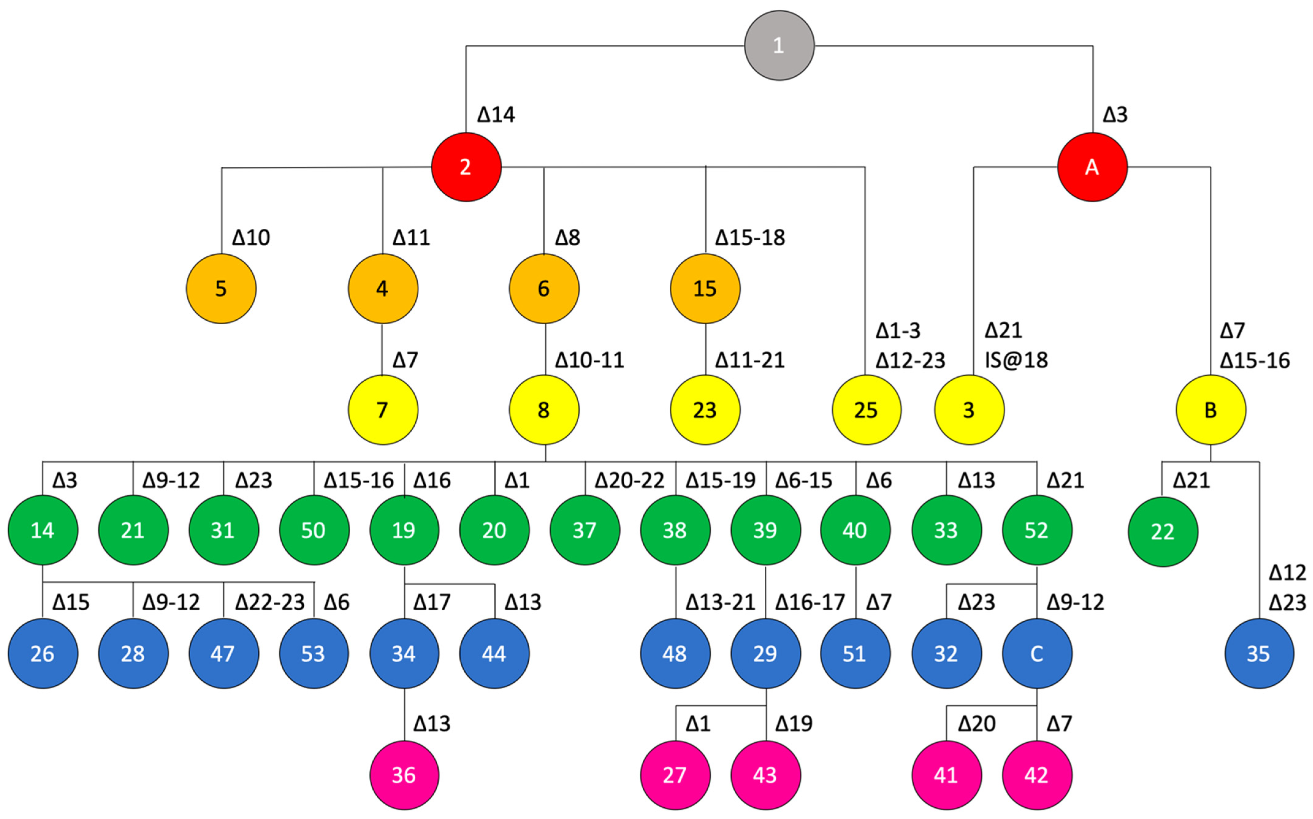
Strains of pathotype A* fall into two major clades (Figure 2). The first clade contains strains from Cambodia, Singapore and Thailand. Here, spoligotype 17 originated from spoligotype 16 by deletion of spacer Xcc_05. The second major clade can be further divided in three subclades. Subclade 2A comprises strains from India, Oman, Pakistan and Saudi Arabia with spoligotypes 10, 11, and 24, where spoligotype 10 constitutes the predecessor of the other two spoligotypes. Subclade 2B includes strains from Iran and Saudi Arabia, where spoligotype 09 (Saudi Arabia) gave rise to spoligotype 49 (Saudi Arabia) by deletion of spacers Xcc_18/Xcc_19, which then evolved to spoligotype 18 (Iran) by deletion of spacers Xcc_03/Xcc_04. Subclade 2C contains strains from Ethiopia (spoligotype 12) and Iran (spoligotype 45; deletion of spacers Xcc_19 to Xcc_23). Based on these data, one may speculate that the strains in Iran resulted from two independent introductions.
The situation with pathotype A, for which we have the largest dataset, is much more complex (Figure 3). Yet, we succeeded to update the scheme that had been published before [13]. The previous scheme contained only four layers of recombinational events, with the fourth layer consisting of spoligotypes 14, 19, 20, 21, and 26 (green circles in Figure 3). Based on the genomic data, we can now add another eight spoligotypes to layer 4 and form two additional layers (eleven observed spoligotypes in layer 5 and five spoligotypes in layer 6). Compared to the scheme from 2019, where we had only 16 spoligotypes identified among 56 analysed strains (25 pathotype-A strains), we now present a robust genealogy for 39 spoligotypes.

3.5. Characterization of the CRISPR Locus from an Ancient X. citri pv. citri

Recently, the first genome of a bacterial plant pathogen was sequenced from a herbarium specimen [14]. DNA that was extracted from a canker-symptomatic citrus leaf sample, originating from Mauritius Island in the Indian Ocean and dating back to 1937, was used to generate the first historical genome of a bacterial crop pathogen from such a specimen. When we analysed this genome sequence with CRISPRCasFinder, it was reported to belong to spoligotype 14 (deletion of spacers Xcc_14, Xcc_08, Xcc_10/Xcc_11, Xcc_03). This spoligotype is identical to the one of strain 306, which served as a reference genome in the mapping-based assembly of the herbarium-derived genome sequence. It was therefore possible that the herbarium specimen may contain spacer that were not present in the reference genome and thus belong to another spoligotype.
We wondered whether the sequence reads can be used to reconstruct the CRISPR array of that historical sample. In order to identify candidate reads, we used the 37 described spacer sequences as queries at NCBI GenBank [13]. In addition, we used the three DR sequence variants and the two flanking regions of the CRISPR array as queries. With these queries, we detected only for 19 of the described spacer sequences a homolog in the herbarium dataset. None of the 14 more recently acquired spacers, which have only been found in one strain from Pakistan (spoligotype 11), were found. Among the 23 widely conserved spacers, we missed finding a homolog for spacers Xcc_08, Xcc_10, Xcc_11, Xcc_14. This pattern thus corresponded to spoligotype 08, which had been found before in pathotype-A strains from China, Reunion Island, and USA [13].
We also observed reads which contained subsequences of two neighboring spacers, thus confirming the order of most spacers, the deletion of spacers Xcc_08 and Xcc_10/Xcc_11, and the absence of any additional, hitherto not observed spacer (Figure S3). However, in this metagenomic sample, we also found reads that contained DR sequences, which were flanked by candidate spacer sequences likely originating from other bacteria. Indeed, only 1.2% of the reads were mapped on the X. citri pv. citri reference genome, whereas at least 5.2% of the reads belonged to other bacteria [14]. This number may even be much larger because 60.1% of the reads were not assigned to any taxon and may well contain many more bacterial sequences, including pieces of CRISPR arrays.

3.6. Analysis of CRISPR Loci in Other Pathovars of X. citri

The species X. citri contains more pathovars than the one infecting Citrus, among them pathovars that were reported to belong to the species X. axonopodis or X. campestris [6]. We wondered whether the presence of the CRISPR locus is restricted to only one pathovar or if we could identify CRISPR loci in other pathovars. We used the CRISPRCasFinder tool to scrutinize all genome sequences that belong to the species X. citri (Table 1). In addition to the pathovar citri, we also detected CRISPR loci in the pathovars durantae, cajani, clitoriae, melhusi, punicae, and khayae (Figure 4). Four of these pathovars, pvs. citri, durantae, cajani, and clitoriae, are phylogenetically close to each other (Figure 4). Like strains of the pathvar citri, X. citri pv. durantae strain LMG 696 from India contains a subset of the 23 widely conserved spacers. With spacers Xcc_03, Xcc_06, Xcc_09/Xcc_10, and Xcc_19 missing, it matches to spoligotype 10, which was also found in five pathotype-A* strains of X. citri pv. citri: DAR73910 (international intercept from India at Sydney Airport), FDC 1682 (Oman), JF90-2 (Oman), JM35-2 (Saudi Arabia), NCPPB 3607 (India).
Strain LMG 558 (X. citri pv. cajani) contains 26 spacers, twelve of which are shared with X. citri pv. citri, whereas the other spacers have not been identified before. Notably, four of the novel spacers are shared with strain LMG 9045 (X. citri pv. clitoriae). In addition, this strain contains six of the 23 canonical spacers of X. citri pv. citri. Strains of the other three pathovars have also a few spacers in common with X. citri pv. citri, albeit at a smaller number, and they share spacers with each other and with pv. clitoriae. Our dataset includes ten strains of X. citri pv. punicae, for which we describe three different spoligotypes.
In total, the set of strains beyond pathovar citri contains 103 hitherto undescribed spacers; 62% of them do not have significant hits at NCBI GenBank’s non-redundant database. Among those with homologs, 30% matched to bacteriophage sequences and 75% matched to sequences in the genomes of xanthomonads. Since 35 of the novel spacers are at positions between one of the 23 canonical X. citri pv. citri spacers and the terminator, they must have been there before the divergence of the different pathovars of X. citri. Hence, the common ancestor of these pathovars contained a CRISPR array significantly larger than that of the founder of the pathovar citri lineage.

4. Discussion

4.1. Genomics-Informed Analysis of X. citri pv. citri Doubles the Number of Known Spoligotypes and Allows Reconstructing Their Probable Evolutionary Trajectory/Provides Information about Lineage Descendance of CRISPR Loci

In this study, we have made use of the wealth of genomic resources that are available for xanthomonads. We analyzed all publicly available genome sequences of strains of X. citri, including several pathovars that were taxonomically misidentified until recently [6]. We observed that all strains of X. citri pv. citri have a CRISPR locus and their CRISPR arrays were found to be restricted to 23 spacers, as observed before [13]. This work allowed us to update the genealogy of CRISPR spoligotypes for the pathovar citri. In comparison to the most recent study with 25 spoligotypes, we describe 26 novel spoligotypes and assign all of them a parsimonious position in the framework for the genealogy of the citrus canker pathogen.
In addition to the spoligotypes, we also compiled other variations at the CRISPR loci of 355 strains of X. citri pv. citri, such as presence of IS elements, SNPs in spacers (Table 3) and repeats (Table 4), which provide further information about lineage descendance. As a note of caution, however, we need to emphasize that some of these sequence variations may have resulted from sequencing artifacts and need critical review and/or independent confirmation before making use of them.

4.2. The Spoligotype Genealogy Framework Contains Information on the Global Spread of the Citrus Canker Pathogen, as Exemplified by the Two Introductions in West Africa

In general, we observed geographical signals in the dataset. Strains that originate from the same country or one of its neighboring countries often share the same or a similar spoligotype. However, we also found trans-continental commonalities. For instance, strains from West Africa contain either spoligotype 14 (Mali) or 15 (Mali, Senegal). These two spoligotypes are quite distant to each other, suggesting at least two independent introductions of the pathogen in this area. Spoligotype 14 has been observed in 10% of the genome sequences, whereas spoligotype 15 was only found in four strains (LD7-1 and LE116-1 from Mali, LH37-1 from Senegal, NCPPB 3562 from India). These data are compatible with a previous study which used micro- and minisatellites to assess the geographic spread of the citrus canker pathogen [33]. It was suggested that two groups of strains, belonging to two DAPC (discriminant analysis of principal components) clusters, were introduced in Mali. DAPC-1 strains originated from very diverse geographic origins, which made it impossible to decipher the origin of West African DAPC-1 strains. Yet, Brazil was identified as the country that shared the largest number of haplotypes with strains from West Africa [33]. Interestingly, the Malian spoligotype 15-strain LC80 belongs to DAPC cluster 1, along with six strains from Brazil and eleven strains from Argentina that contain spoligotype 15 as well (Table S1). DAPC-2 strains from Mali shared a haplotype with strains from Pakistan and it was suggested that DAPC-2 strains from Mali might have originated from the Indian subcontinent [33]. Again, the two Malian spoligotype-14 strains LD7-1 and LE116-1 belong to DAPC cluster 2, as does strain NCPPB 3562 from India, thus supporting the hypothesis of an introduction from the Indian subcontinent. In conclusion, our spoligotyping data are consistent with previous work using mini- and microsatellite analyses.

4.3. Metagenomic Data from a Herbarium Sample Enable Reconstruction of the Spoligotype of an Ancient Citrus Canker Pathogen and Its Positioning in the CRISPR-Based Genealogy

In this study, we succeeded in reconstructing the spoligotype of bacteria whose DNA was preserved in a herbarium specimen from 1937 originating from Mauritius [14]. BLAST analyses assigned this material to spoligotype 08, thus correcting the spoligotype which was assigned based on a reference-genome based assembly. Spoligotype 08 is the most dominant spoligotype in our set of 355 strains, which originate from Brazil, China, Comoros, Hong Kong, Mayotte, New Zealand, Reunion Island, Taiwan, USA, and Vietnam (Table S1). It will be interesting to decipher the spoligotypes of additional, perhaps even older samples from similar sources.

4.4. The X. citri pv. citri CRISPR Locus Is Conserved in Some Additional Pathovars of X. citri and Different Pathovars Tend to Share Their Oldest Spacers

Phylogenomic analyses revealed that several X. citri pathovars beyond citri contain CRISPR loci. Interestingly, different pathovars share common spacers, indicating that their common ancestor harbored a CRISPR array that was significantly larger than those which are nowadays found in the pathovar citri. It appears that the whole locus got lost in several other pathovars, suggesting that the CRISPR locus is not required for inhabiting different plants and environments. We did not observe any linkage to certain modes of plant colonization or host plants. It appears that the locus got lost several times and was perhaps functionally substituted for by other defense systems against bacteriophages, such as restriction/modification systems.
One of the non-citri pathovar strains, i.e., LMG 696 (pv. durantae), did not contain any spacer that was not found in the pathovar citri, and therefore qualified as spoligotype 10. This strain was isolated from Duranta repens (family: Verbenaceae) and was considered a “clonal variant” of pathovar citri strains [34]. Moreover, this strain clustered in a maximum likelihood phylogenetic tree based on concatenation of 1785 single-copy genes with pathotype-A* strains FDC 1682, JF90-2, JM35-2, and NCPPB 3607, all belonging to spoligotype 10, and with pathotype-A* strain CFBP 2911, which belongs to the derived spoligotype 11 (Figure 2) [35]. These data are supported by our ANI-based phylogenetic tree, which clusters strain LMG 696 together with the pathovar citri pathotype strain LMG 9322 (Figure 4). It is not known whether strain LMG 696 can also infect plants of the Rutaceae family, as strains of pathovar citri do, and if it shares a similar host range as pathotype-A* strains [36]. Pathotyping on different candidate host plants and resequencing of strain LMG 696 using long-read technology could help clarify whether plasmids contribute to the different host ranges, as suggested previously [35].

4.5. Some Spacers Observed in X. citri Pathovars beyond the Citri Pathovar Originated from Bacteriophages

In contrast to the 23 canonical spacers of X. citri pv. citri, none of which was found to be homolog to contemporary bacteriophages at NCBI GenBank [13], we identified twelve spacers among the non-citri pathovar strains that are homologous to sequences from bacteriophages, mainly from Xanthomonas: Phage Cf (AJ011389.1), Cf1c (M57538.1), Cf1t (U08370.1), FoX2 (NC_055836.1), FoX3 (NC_055837.1), FoX4 (NC_055839.1), KPhi1 (NC_054460.1), M29 (MZ345003.1), MYK3 (OK275494.1), phiLF (MH206184.1), phiXaf18 (NC_054461.1), phiXv2 (MH206183.1), XacF1 (AB910602.), XAJ2 (KU197014.1), Xf109 (NC_043028.1), Xf409 (NC_055055.1). In addition, we observed hits with the genomes of the two Xylella bacteriophages Salvo (NC_042345.1) and Sano (NC_042344.1), with the Stenotrophomonas phage phiSMA6 (NC_043029.1), and with the Siphoviridae sp. isolate ctZLc1 (BK020633.1). We also observed that pathovars clitoriae and melhusi contained 23 and 33 spacers, respectively, that may have been acquired after divergence from the pathovar citri, as indicated by their leader-proximal localization with respect to the spacers that are shared with X. citri pv. citri. While we assume that the CRISPR locus in X. citri pv. citri is not able to acquire new spacers (perhaps except for strain CFBP 2911, which contains 14 extra spacers, some of them with homology to bacteriophages), we do not know if the locus in some of the other pathovars can acquire new spacers and provide resistance to contemporary bacteriophages. Analyses of a larger number of strains from these pathovars will help answering this question.

5. Conclusions

CRISPR arrays provide a novel approach to elucidate the evolutionary trajectory of bacteria, which have hitherto rarely been exploited for bacterial plant pathogens. Using X. citri as a model, we have demonstrated its usefulness to understand the evolution and global spread of the citrus canker pathogen. Moreover, conservation of CRISPR spacers in other pathovars of X. citri suggests that their common ancestor had a much larger CRISPR array than nowadays observed in strains of X. citri. It will be interesting to extend this study to additional species of Xanthomonas and perhaps even beyond them, once appropriate bioinformatic tools have been developed for such a large-scale analysis.

Supplementary Materials

The following supporting information can be downloaded at: www.mdpi.com/article/10.3390/microorganisms10091715/s1, Figure S1: Phylogenetic tree of pathotype-A* X. citri pv. citri strains.; Figure S2: Phylogenetic tree of X. citri pv. citri strains with spoligotype 1, 2, or 4.; Figure S3: Schematic representation of the CRISPR array from the metagenomic herbarium specimen; Table S1: Genomic resources; Table S2: Inventory of X. citri CRISPR spacers; Table S3: Spoligotypes of X. citri pv. citri CRISPR arrays.

Author Contributions

Conceptualization, R.K.; methodology, N.B. and R.K.; software, A.D., N.B. and R.K.; validation, R.K.; formal analysis, N.B. and R.K.; investigation, N.B. and R.K.; resources, N.B., A.D. and R.K.; data curation, N.B., A.D. and R.K.; writing—original draft preparation, N.B.; writing—review and editing, N.B., A.D. and R.K.; visualization, N.B. and R.K.; supervision, R.K.; project administration, R.K. All authors have read and agreed to the published version of the manuscript.

Funding

This research received no external funding.

Data Availability Statement

Not applicable.

Conflicts of Interest

The authors declare no conflict of interest.

References

  1. Timilsina, S.; Potnis, N.; Newberry, E.A.; Liyanapathiranage, P.; Iruegas-Bocardo, F.; White, F.F.; Goss, E.M.; Jones, J.B. Xanthomonas diversity, virulence and plant-pathogen interactions. Nat. Rev. Microbiol. 2020, 18, 415–427. [Google Scholar] [CrossRef] [PubMed]
  2. Wu, G.A.; Terol, J.; Ibanez, V.; López-García, A.; Pérez-Román, E.; Borredá, C.; Domingo, C.; Tadeo, F.R.; Carbonell-Caballero, J.; Alonso, R.; et al. Genomics of the origin and evolution of Citrus. Nature 2018, 554, 311–316. [Google Scholar] [CrossRef] [PubMed]
  3. Islam, M.N.; Ali, M.S.; Choi, S.J.; Hyun, J.W.; Baek, K.H. Biocontrol of citrus canker disease caused by Xanthomonas citri subsp. citri using an endophytic Bacillus thuringiensis. Plant Pathol. J. 2019, 35, 486–497. [Google Scholar] [CrossRef]
  4. Brunings, A.M.; Gabriel, D.W. Xanthomonas citri: Breaking the surface. Mol. Plant Pathol. 2003, 4, 141–157. [Google Scholar] [CrossRef]
  5. Midha, S.; Patil, P.B. Genomic insights into the evolutionary origin of Xanthomonas axonopodis pv. citri and its ecological relatives. Appl. Environ. Microbiol. 2014, 80, 6266–6279. [Google Scholar] [CrossRef] [PubMed]
  6. Bansal, K.; Kumar, S.; Patil, P.B. Phylo-taxonogenomics supports revision of taxonomic status of twenty Xanthomonas pathovars to Xanthomonas citri. Phytopathology 2022, 112, 1201–1207. [Google Scholar] [CrossRef] [PubMed]
  7. Horvath, P.; Barrangou, R. CRISPR/cas, the immune system of bacteria and archaea. Science 2010, 327, 167–170. [Google Scholar] [CrossRef]
  8. Haft, D.H.; Selengut, J.; Mongodin, E.F.; Nelson, K.E. A guild of 45 CRISPR-associated (Cas) protein families and multiple CRISPR/Cas subtypes exist in prokaryotic genomes. PLoS Comput. Biol. 2005, 1, e60. [Google Scholar] [CrossRef]
  9. Barrangou, R.; Horvath, P. The CRISPR system protects microbes against phages, plasmids. Microbe 2009, 4, 224–230. [Google Scholar] [CrossRef]
  10. Makarova, K.S.; Wolf, Y.I.; Iranzo, J.; Shmakov, S.A.; Alkhnbashi, O.S.; Brouns, S.J.J.; Charpentier, E.; Cheng, D.; Haft, D.H.; Horvath, P.; et al. Evolutionary classification of CRISPR-Cas systems: A burst of class 2 and derived variants. Nat. Rev. Microbiol. 2020, 18, 67–83. [Google Scholar] [CrossRef]
  11. Pieretti, I.; Royer, M.; Barbe, V.; Carrere, S.; Koebnik, R.; Couloux, A.; Darrasse, A.; Gouzy, J.; Jacques, M.A.; Lauber, E.; et al. Genomic insights into strategies used by Xanthomonas albilineans with its reduced artillery to spread within sugarcane xylem vessels. BMC Genom. 2012, 13, 658. [Google Scholar] [CrossRef] [PubMed]
  12. Koebnik, R.; Burokiene, D.; Bragard, C.; Chang, C.; Saux, M.F.; Kölliker, R.; Lang, J.M.; Leach, J.E.; Luna, E.K.; Portier, P.; et al. The complete genome sequence of Xanthomonas theicola, the causal agent of canker on tea plants, reveals novel secretion systems in clade-1 xanthomonads. Phytopathology 2021, 111, 611–616. [Google Scholar] [CrossRef] [PubMed]
  13. Jeong, K.; Muñoz-Bodnar, A.; Arias Rojas, N.; Poulin, L.; Rodriguez-R, L.M.; Gagnevin, L.; Vernière, C.; Pruvost, O.; Koebnik, R. CRISPR elements provide a new framework for the genealogy of the citrus canker pathogen Xanthomonas citri pv. citri. BMC Genom. 2019, 20, 917. [Google Scholar] [CrossRef]
  14. Campos, P.E.; Groot Crego, C.; Boyer, K.; Gaudeul, M.; Baider, C.; Richard, D.; Pruvost, O.; Roumagnac, P.; Szurek, B.; Becker, N.; et al. First historical genome of a crop bacterial pathogen from herbarium specimen: Insights into citrus canker emergence. PLoS Pathog. 2021, 17, e1009714. [Google Scholar] [CrossRef]
  15. Young, J.M.; Dye, D.W.; Bradbury, J.F.; Panagopoulos, C.G.; Robbs, C.F. A proposed nomenclature and classification for plant pathogenic bacteria. N. Z. J. Agric. Res. 1978, 21, 153–177. [Google Scholar] [CrossRef]
  16. Vauterin, L.; Hoste, B.; Kersters, K.; Swings, J. Reclassification of Xanthomonas. Int. J. Syst. Bacteriol. 1995, 45, 472–489. [Google Scholar] [CrossRef]
  17. Rademaker, J.L.; Hoste, B.; Louws, F.J.; Kersters, K.; Swings, J.; Vauterin, L.; De Bruijn, F.J. Comparison of AFLP and rep-PCR genomic fingerprinting with DNA-DNA homology studies: Xanthomonas as a model system. Int. J. Syst. Evol. Microbiol. 2000, 50, 665–677. [Google Scholar] [CrossRef]
  18. Rademaker, J.L.W.; Louws, F.J.; Schultz, M.H.; Rossbach, U.; Vauterin, L.; Swings, J.; De Bruijn, F.J. A comprehensive species to strain taxonomic framework for Xanthomonas. Phytopathology 2005, 95, 1098–1111. [Google Scholar] [CrossRef]
  19. Constantin, E.C.; Cleenwerck, I.; Maes, M.; Baeyen, S.; Van Malderghem, C.; De Vos, P.; Cottyn, B. Genetic characterization of strains named as Xanthomonas axonopodis pv. dieffenbachiae leads to a taxonomic revision of the X. axonopodis species complex. Plant Pathol. 2016, 65, 792–806. [Google Scholar] [CrossRef]
  20. Oren, A.; Garrity, G.M. Notification of changes in taxonomic opinion previously published outside the IJSEM. Int. J. Syst. Evol. Microbiol. 2017, 67, 2081–2086. [Google Scholar] [CrossRef]
  21. Rodriguez-R, L.M.; Konstantinidis, K.T. The enveomics collection: A toolbox for specialized analyses of microbial genomes and metagenomes. PeerJ Prepr. 2016, 4, e1900. [Google Scholar] [CrossRef]
  22. Meier-Kolthoff, J.P.; Göker, M. TYGS is an automated high-throughput platform for state-of-the-art genome-based taxonomy. Nat. Commun. 2019, 10, 2182. [Google Scholar] [CrossRef] [PubMed]
  23. Couvin, D.; Bernheim, A.; Toffano-Nioche, C.; Touchon, M.; Michalik, J.; Néron, B.; Rocha, E.P.C.; Vergnaud, G.; Gautheret, D.; Pourcel, C. CRISPRCasFinder, an update of CRISRFinder, includes a portable version, enhanced performance and integrates search for Cas proteins. Nucleic Acids Res. 2018, 46, W246–W251. [Google Scholar] [CrossRef] [PubMed]
  24. Richard, D.; Rieux, A.; Lefeuvre, P.; Hamza, A.; Lobin, K.K.; Naiken, M.; Stravens, R.; Boyer, C.; Boyer, K.; Javegny, S.; et al. Draft genome sequences of 284 Xanthomonas citri pv. citri strains causing Asiatic citrus canker. Microbiol. Resour. Announc. 2021, 10, e01024-20. [Google Scholar] [CrossRef]
  25. Hu, F.P.; Young, J.M.; Stead, D.E.; Goto, M. Transfer of Pseudomonas cissicola (Takimoto 1939) Burkholder 1948 to the Genus Xanthomonas. Int. J. Syst. Evol. Microbiol. 1997, 47, 228–230. [Google Scholar] [CrossRef]
  26. Rudra, B.; Gupta, R.S. Phylogenomic and comparative genomic analyses of species of the family Pseudomonadaceae: Proposals for the genera Halopseudomonas gen. nov. and Atopomonas gen. nov., merger of the genus Oblitimonas with the genus Thiopseudomonas, and transfer of some misclassified species of the genus Pseudomonas into other genera. Int. J. Syst. Evol. Microbiol. 2021, 71, 9. [Google Scholar] [CrossRef]
  27. Richard, D.; Pruvost, O.; Balloux, F.; Boyer, C.; Rieux, A.; Lefeuvre, P. Time-calibrated genomic evolution of a monomorphic bacterium during its establishment as an endemic crop pathogen. Mol. Ecol. 2021, 30, 1823–1835. [Google Scholar] [CrossRef]
  28. Kajava, A.V.; Lindow, S.E. A model of the three-dimensional structure of ice nucleation proteins. J. Mol. Biol. 1993, 232, 709–717. [Google Scholar] [CrossRef]
  29. Midha, S.; Bansal, K.; Kumar, S.; Girija, A.M.; Mishra, D.; Brahma, K.; Laha, G.S.; Sundaram, R.M.; Sonti, R.V.; Patil, P.B. Population genomic insights into variation and evolution of Xanthomonas oryzae pv. oryzae. Sci. Rep. 2017, 7, 40694. [Google Scholar] [CrossRef]
  30. Gétaz, M.; Krijger, M.; Rezzonico, F.; Smits, T.H.M.; van der Wolf, J.M.; Pothier, J.F. Genome-based population structure analysis of the strawberry plant pathogen Xanthomonas fragariae reveals two distinct groups that evolved independently before its species description. Microb. Genom. 2018, 4, e000189. [Google Scholar] [CrossRef]
  31. ‪Johnson, S.C. Hierarchical clustering ‪schemes. Psychometrika 1967, 32, 241–254. [Google Scholar] [CrossRef] [PubMed]
  32. Letunic, I.; Bork, P. Interactive Tree Of Life (iTOL) v4: Recent updates and new developments. Nucleic Acids Res. 2019, 47, W256–W259. [Google Scholar] [CrossRef] [PubMed]
  33. Leduc, A.; Traoré, Y.N.; Boyer, K.; Magne, M.; Grygiel, P.; Juhasz, C.C.; Boyer, C.; Guerin, F.; Wonni, I.; Ouedraogo, L.; et al. Bridgehead invasion of a monomorphic plant pathogenic bacterium: Xanthomonas citri pv. citri, an emerging citrus pathogen in Mali and Burkina Faso. Environ. Microbiol. 2015, 17, 4429–4442. [Google Scholar] [CrossRef]
  34. Bansal, K.; Midha, S.; Kumar, S.; Patil, P.B. Ecological and evolutionary insights into Xanthomonas citri pathovar diversity. Appl. Environ. Microbiol. 2017, 83, e02993-16. [Google Scholar] [CrossRef] [PubMed]
  35. Patané, J.S.L.; Martins, J., Jr.; Rangel, L.T.; Belasque, J.; Digiampietri, L.A.; Facincani, A.P.; Ferreira, R.M.; Jaciani, F.J.; Zhang, Y.; Varani, A.M.; et al. Origin and diversification of Xanthomonas citri subsp. citri pathotypes revealed by inclusive phylogenomic, dating, and biogeographic analyses. BMC Genom. 2019, 20, 700. [Google Scholar] [CrossRef] [Green Version]
  36. Srinivasan, M.C.; Patel, M.K. Two new phytopathogenic bacteria on verbenacious hosts. Curr. Sci. 1957, 26, 90–91. [Google Scholar]
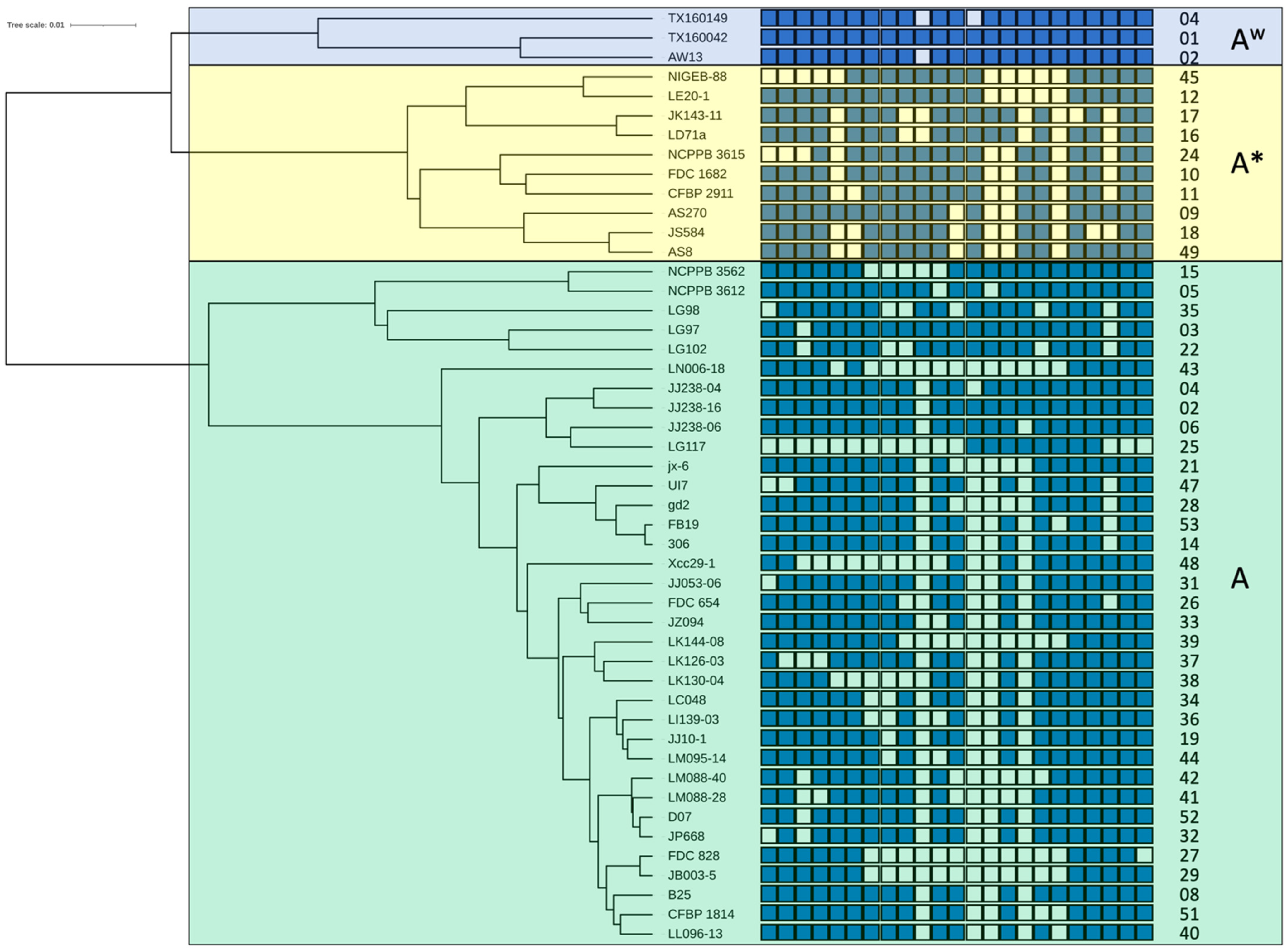
Figure 1. Phylogenetic tree of representative X. citri pv. citri strains and their associated spoligotypes. For each pathotype. i.e., A, A* and Aw, one representative strain per spoligotype was included in the dataset to calculate genome-wide pairwise average nucleotide identities and perform their phylogenetic analysis on the enve-omics platform [21]. Complete (maximum) linkage clustering was used to build the phylogenetic tree [31]. The interactive Tree Of Life (iTOL) suite was used for better visualisation of the tree [32]. CRISPR arrays are represented by a series of 23 blue and open boxes, oriented with the leader-proximal spacers on the left side. Identical spacers within the same block are vertically aligned. Detected CRISPR spacers are represented by blue boxes. Open boxes indicate the absence of the corresponding spacer. Names of representative strains are shown between the tree and the CRISPR arrays. Spoligotype numbers are given on the right side. Transparent blue, yellow and green boxes frame pathotype-Aw, pathotype-A*, and pathotype-A strains, respectively.
Microorganisms 10 01715 g001
Figure 2. Genealogy of spoligotypes from pathotype-A* strains. Postulated mutational events leading to the observed spoligotypes are indicated, starting from the ancestral spoligotype with all 23 spacers shown in grey on the top, with the colors indicating the number of events (from one to six events, colored in salmon, orange, yellow, green, blue and pink, respectively). Numbers of spoligotypes are indicated in the circles. Characters, i.e., A and B, indicate postulated intermediate spoligotypes that were not observed among the analyzed pathotype-A* strains. Spoligotype 13 was not observed in any of the analyzed genome sequences but had been found in strain LE065-1 previously [13].
Microorganisms 10 01715 g002
Figure 3. Genealogy of spoligotypes from pathotype-A strains. Postulated mutational events leading to the observed spoligotypes are indicated, starting from the ancestral spoligotype with all 23 spacers shown in grey on the top, with the colors indicating the number of events (from one to six events, colored in salmon, orange, yellow, green, blue, pink, respectively). Numbers of spoligotypes are indicated in the circles. Characters, i.e., A and B, indicate postulated intermediate spoligotypes that were not observed among the analyzed pathotype-A strains. Spoligotypes 7, 20, and 23 were not observed in any of the analyzed genome sequences but had been found before in strains LG116, JK148-10, LH001-3, respectively [13].
Microorganisms 10 01715 g003
Figure 4. Phylogenetic tree of representative strains from 33 X. citri pathovars. Genome-wide pairwise average nucleotide identities were calculated, and their phylogenetic analysis was performed on the enve-omics platform [21]. Complete (maximum) linkage clustering was used to build the phylogenetic tree [31]. The interactive Tree Of Life (iTOL) suite was used for better visualisation of the tree [32]. 22 pathovars cluster within the Rademaker 9.5 group and 11 pathovars cluster within the Rademaker 9.6 group [18]. Strain names are shown to the left of the tree, followed by the pathovar name. Pathovars with CRISPR arrays are shown in green and those without CRISPR arrays in red.
Microorganisms 10 01715 g004
Table 1. Overview of genomic resources used in this study and presence of CRISPR Cas systems.
Correct Taxonomic StatusGenBank AnnotationSubgroup aComplete Genome SequencesDraft Genome SequencesCRISPR Subtype b
X. citri pv. anacardiiX. citri pv. anacardii9.613w/o
X. citri pv. aurantifoliiX. citri pv. aurantifolii9.646w/o
X. citri pv. azadirachtaeX. campestris pv. azadirachtae9.501w/o
X. citri pv. bauhiniaeX. axonopodis pv. bauhiniae9.501w/o
X. citri pv. bilvaeX. citri pv. bilvae9.501w/o
X. citri pv. cajaniX. axonopodis pv. cajani9.501IC
X. citri pv. centellaeX. campestris pv. centellae9.501w/o
X. citri pv. citriX. axonopodis9.510IC
X. citri9.510
X. citri pv. citri9.54568
X. citri pv. citri c9.50251
X. citri pv. clitoriaeX. axonopodis pv. clitoriae9.501IC
X. citri pv. dieffenbachiae 9.6 w/o
X. citri pv. durantaeX. campestris pv. durantae9.501IC
X. citri pv. eucalyptorumX. axonopodis pv. eucalyptorum9.601w/o
X. citri pv. fuscansX. citri pv. fuscans9.61025w/o
X. citri pv. phaseoli var. fuscans9.6130
X. citri pv. glycinesX. citri pv. glycines9.5126w/o
X. citri pv. khayaeX. axonopodis pv. khayae9.501IC
X. citri pv. leeanaX. campestris pv. leeana9.501w/o
X. citri pv. malvacearumX. citri pv. malvacearum9.584w/o
X. citri pv. mangiferaeindicaeX. citri pv. mangiferaeindicae9.534w/o
X. citri pv. martyniicolaX. axonopodis pv. martyniicola9.501w/o
X. citri pv. melhusiiX. axonopodis pv. melhusii9.501IC
X. citri pv. merremiaeX. campestris pv. merremiae9.602w/o
X. citri pv. punicaeX. citri pv. punicae9.5102IC
X. citri pv. sesbaniaeX. citri pv. sesbaniae9.601w/o
X. citri pv. thespesiaeX. campestris pv. thespesiae9.501w/o
X. citri pv. thirumalachariiX. citri pv. thirumalacharii9.601w/o
X. citri pv. trichodesmaeX. campestris pv. trichodesmae9.602w/o
X. citri pv. vignicolaX. citri pv. vignicola9.630w/o
X. citri pv. viticolaX. citri pv. viticola9.502w/o
X. citri pv. vitiscarnosaeX. campestris pv. vitiscarnosae9.501w/o
X. citri pv. vitistrifoliaeX. campestris pv. vitistrifoliae9.501w/o
X. citri pv. vitiswoodrowiiX. campestris pv. vitiswoodrowii9.501w/o
X. citri [pv. cissicola?]Pseudomonas cissicola9.502w/o
X. citri not pv. citriX. citri9.505w/o
X. citri not pv. citriX. citri9.641w/o
Total 115401
a, Rademaker subgroup [18]; b, Predicted CRISPR Cas subtype, w/o = without; c, 251 strains taken from Richard et al. [27].
Table 2. Predicted false positive CRISPR arrays in X. citri pv. citri.
StrainSpacer IDDR Size (bp)Spacer Size (bp)Homolog a
Canonical sequence 3134–37CRISPR array
LG98Xanci22643128
LI070-01Xanci2535 Xanci25362728–29Hypothetical protein in Staphylococcus aureus [8 × 10⁻²³]
LK142-04Xanci35122743Hypothetical protein in Escherichia coli [1 × 10⁻¹³]
LM057-04Xanci46672960
LM057-15Xanci47062860
LM088-25Xanci48012729Hypothetical protein in Staphylococcus aureus [2 × 10⁻¹¹]
LM095-04Xanci4987 Xanci49882919–22
LP029-13Xanci58252862
Loci that correspond to each other are highlighted by the same color. a, Homologs were search at GenBank using TBLASTN, e values are given in square brackets.
Table 3. Variant spacer sequences in X. citri pv. citri.
SpacerVariant SequenceCanonical SequenceStrains
Xcc_03aAAGAAGACCAGTCTGCGGCGTCGCGGCATCCTTGGGAAGAAGACCAGTCTGCGGCGTCGCGGCATCCTGGGGJJ009-1
Xcc_03bAAGAAGACCAGTCTGCGGCGTCGCGGCATCTTGGGGAAGAAGACCAGTCTGCGGCGTCGCGGCATCCTGGGGLL098-02
Xcc_03cAAGAAGACCAGTCTGCGGCGTCGCGGCATCCTGGGGGAAGAAGACCAGTCTGCGGCGTCGCGGCATCCTGGGGLK136-05
Xcc_13aGCCATCATGCTTTGAATGCGCCTACCCACGGCGAAGCCATCATGCTTTGAATGCGCTTACCCACGGCGAAUI6, UI7
Xcc_18aGTGCCACCGACAGCGACGCACGTGGACCTGCATGTTGTGCCACCGACAGCGACGCACGTGGACCTGCAGATCLG97
Xcc_19aTCGAGCGCATCGATGACGGTCACCCATCCCC_AATGTCGAGCGCATCGATGACGGTCACCCATCCCCCAATGLK169-03
Xcc_19bGTGCCACCGATGACGGTCACCCATCCCCCAAT_TCGAGCGCATCGATGACGGTCACCCATCCCCCAATGjx4
Variant nucleotides in comparison to the canonical spacer sequence are double underlined.
Table 4. Variant DR sequences in X. citri pv. citri.
StrainsSequenceRemark
AllGTCGCGCCCTCACGGGCGCGTGGATTGAAACCanonical sequence
MostGGCGCGCCCTCACGGGCGCGTGGATTGAAACDR1, degenerate variant of terminal repeat
MostTTCGCGCCCTCATGGGCGCGTGGATTGAAACDR2, degenerate variant of the penultimate repeat
FDC 828TTCGCGCCCTCACGGGCGCGTGGATTGAAACHybrid of DR1 and DR2
LK130-09TTCGCGCCCTCATGGGCGCGTGGATTGAA_C
LK169-03GTCGCGCCCTCACGGGCGCGTGGATTGAAAAC
LM089-02, LMG 9322, LN003-10, MN10, MN11, MN12GTCGCGCCCTCACGGGCGCGTGGATTGGAAC
NCPPB 3610GTCGCGCCCTCCCGGGCGCGTGGATTGAAAC
Variant nucleotides in comparison to the canonical DR sequence are double underlined.
Publisher’s Note: MDPI stays neutral with regard to jurisdictional claims in published maps and institutional affiliations.