Brief Report

A Potential Predictive Role of the Scalp Microbiome Profiling in Patients with Alopecia Areata: Staphylococcus caprae, Corynebacterium, and Cutibacterium Species

1
Department of Laboratory Medicine, Chonnam National University Hwasun Hospital, Hwasun 58128, Jeollanam-do, Korea
2
Department of Parasitology and Tropical Medicine, Chonnam National University Medical School, Hwasun 58128, Jeollanam-do, Korea
3
Department of Biomedical Science and Engineering, Gwangju Institute of Science and Technology (GIST), Gwangju 61005, Korea
4
Department of Dermatology, Chonnam National University Hospital, Gwangju 61469, Korea
*
Author to whom correspondence should be addressed.
Microorganisms 2022, 10(5), 864; https://doi.org/10.3390/microorganisms10050864
Submission received: 24 March 2022 / Revised: 20 April 2022 / Accepted: 20 April 2022 / Published: 21 April 2022
(This article belongs to the Special Issue Infection, Immunity, and Allergy Caused by Propionibacterium acnes)

Abstract

:
Little is known about the scalp bacterial composition of alopecia areata (AA) patients. The aim of this study was to investigate the differences in the scalp microbiome of AA patients according to their prognosis, in addition to healthy controls. A total of 33 AA patients and 12 healthy controls (HC) were included in this study. The microbiomes were characterized by sequencing 16S rRNA genes on the Illumina MiSeq platform. The scalp microbiome was more diverse in AA patients compared to HC, but not significantly different according to the severity of AA. Nevertheless, the higher proportion of Corynebacterium species and the lower proportion of Staphylococcus caprae among the Staphylococcus species were noticed in severe AA patients compared to HC or mild AA. The higher ratio of Cutibacterium species to S. caprae was noticed in severe AA. We highlight the potential predictive role of scalp microbiome profiling to a worse prognosis of patients with alopecia areata.

1. Introduction

Alopecia areata (AA) is the second most common type of hair loss disorder in humans, which affects nearly 2% of the general population [1]. The most common type is a small annular or patchy bald lesion usually on the scalp [2], and these can extend to the entire scalp (Alopecia totalis) or to the entire pilar area of the body (Alopecia universalis). This disorder can have a devastating impact on the shaping of the body’s image and self-esteem, causing, in many cases, inefficient functioning in the social environment and eventually decreased quality of life [3]. However, the management of AA still remains a challenge. Several speculations on the causes of AA have been proposed, such as loss of immune privilege/immune dysregulation, genetic predisposition, hormonal imbalance, emotional or physical stress, genetic tendencies, and other local skin disorders and nutritional deficiencies [4]. Among the proposed causes, a link with the gut microbiome has also been hypothesized [5]. Cutibacterium, Staphylococcus, and Corynebacterium were the most common bacteria, while Malassezia was the most common fungus, on healthy scalps [6,7]. Due to its unique features, the scalp is expected to harbor a specific microbiome, which is expected to play a peculiar role in scalp conditions related to hair growth [7]. Several researchers studied the scalp microbiome profile regarding AA and they suggested the notion that an imbalance between skin microbes that maintain skin homeostasis causes inflammation [8,9,10,11,12]. Several microbes such as Cutibacterium acnes, Staphylococcus, and Malassezia were suggested to be implicated in AA, but it still needs to be clarified using various cohorts of different populations. Here, we aimed to investigate scalp microbiome profiling in patients with alopecia areata and to assess their prognostic implications.

2. Materials and Methods
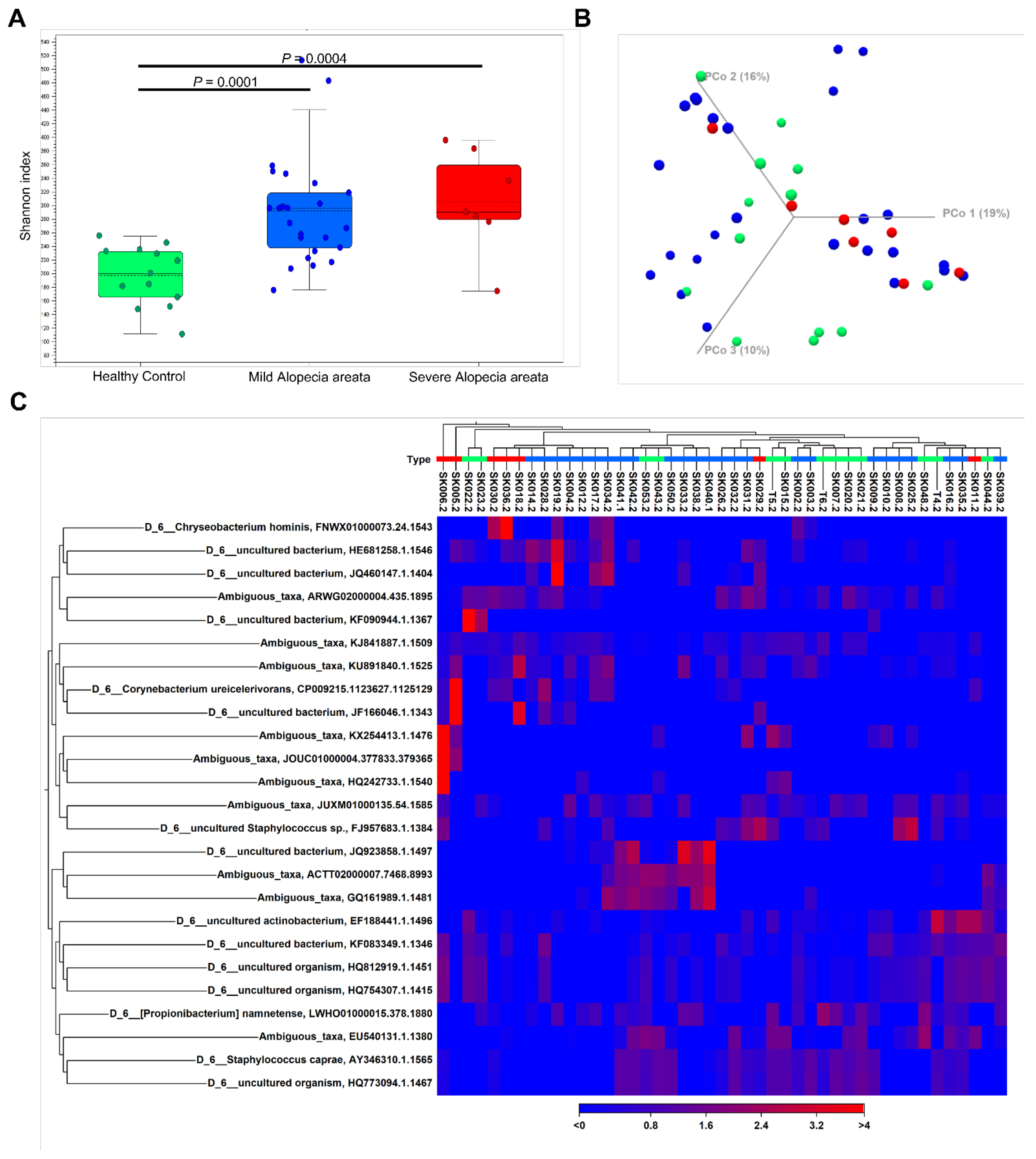
This study was carried out in accordance with all relevant institutional guidelines. The Ethics Committee of Chonnam National University Hospital approved this study (CNUH-2020-118) and written informed consent was obtained from all subjects.
All patients with AA were evaluated at a tertiary university hospital between May 2020 and February 2021. A total of 33 AA patients and 12 healthy controls were finally included in this study. Clinical and demographic information for each patient was recorded, including age, gender, age of disease onset, duration of current episode of disease, and any underlying immunological disorders. The severity of AA was evaluated based on the extent of scalp hair loss, progression of disease, and response of treatment, and was divided into mild AA (n = 26) and severe AA (n = 7) (Table 1). The scalp samples were obtained by means of swab procedure according to previously reported methods [8,9,10] with minor modifications. Sterile cotton swabs were soaked for at least 30 s in solution (0.9% NaCl and 0.1% Tween 20). After collection, the head of each swab was cut and stored in solution. The samples were transferred to our laboratory and stored at −80 °C until DNA extraction. Bacterial DNA from scalp swabs was extracted by means of the DNeasy Blood&Tissue kit (Qiagen, Hilden, Germany) according to manufacturer protocol, with minor modifications [11,12,13]. The DNA samples were amplified using 16S universal primers that target the V3–V4 region of the bacterial 16S ribosomal gene. Detection of the sequencing fragments was performed using Illumina MiSeq technology by Macrogen (Seoul, Korea). The sequences were trimmed and merged, and then clustered into operational taxonomic units (OTUs) using the CLC Genomics Workbench v. 10.1.1 and CLC Microbial Genomics Module v. 2.5 (Qiagen, Hilden, Germany). Taxonomic assignment of these sequences was carried out based on the NCBI taxonomy database, with an OTU cutoff of 3%. The most abundant sequences were considered representative of each cluster and assigned to a taxonomy level based CLC Microbial Genomics default values. A multiple sequence alignment with MUSCLE v.3.8.31 was performed to obtain an OTU table together with the pre-aligned 16S and 18S data from the SILVA v132 database. Alpha diversity metrics (richness and Total number) were calculated based on rarefied OTU counts and an exploratory analysis of beta-diversity (between-sample diversity) was performed based on the Bray-Curtis measure of dissimilarity as a principal coordinate analysis (PCoA). For the hierarchical cluster analysis, Bray-Curtis metrics and complete linkage clustering were implemented. Volcano plots were created to show estimated log2-fold differences in OTU abundance between AA and HC. A two-tailed Student’s t test and the χ2 test were used to compare the differences in scalp microbiomes between the groups using GraphPad Prism software (GraphPad Software Inc., San Diego, CA, USA). PERMANOVA analysis was carried out to ascertain the statistical significance of the clusters using the tools in the CLC Microbial Genomics Module v. 2.5 (Qiagen).

3. Results

A total of 3,692,142 good-quality reads with a mean length of 301 base pairs were generated. The results of the alpha diversity metrics (Shannon index) are shown in boxplots in Figure 1A. The Shannon index was significantly higher in the AA patients than in the HC (all groups, p < 0.001), reflecting a significantly higher species richness in the former group. In contrast, there was no significant difference in the alpha diversity according to the severity of AA. The unweighted beta diversity analysis showed the overall bacterial community structure and phylogenetic diversity between AA patients and HC (Figure 1B,C). Overall, we could not find any distinct composition of the scalp microbiome specific to AA or its severity. The statistical values obtained by the PERMANOVA test were not significant (AA vs. HC, p = 0.29543; mild AA vs. severe AA, p = 0.0825). The bacterial compositions of the scalp samples were further examined. At the family level, Staphylococcaceae and Burkholderiaceae were more abundant in HC and mild AA than in severe AA (Figure 2A). The proportion of Propionibacteriaceae increased in mild AA but not in severe AA. At the genus level, severe AA exhibited a lesser abundance of Staphylococcus species and Ralstonia species (Figure 2B). The volcano plot also presented that the genus Corynebacterium that is represented in the microbiome by the several OTU: Corynebacterium ihumii, Corynebacterium simulans, Corynebacterium sp. NML98-0116, and Corynebacterium sp. KPL 1996 were more distinctive to AA than HC (Supplementary Figure S1). Something of interest that we found was that the proportion of S. caprae among the Staphylococcus species was markedly declined in severe AA (mean, 49.5% vs. 42.1% vs. 10.5%, HC vs. mild AA vs. severe AA) (Table 2), while the proportion of Corynebacterium species was significantly increased in severe AA (mean, 0.3% vs. 0.6% vs. 6.3%, HC vs. mild AA vs. severe AA). The mean ratio of Cutibacterium species to S. caprae was 0.97 for HC, 2.13 for mild AA, and 16.01 for severe AA, respectively.

4. Discussion

Several scientific published evidence have reported the strict correlation between microbial disequilibrium and skin conditions [4,5,7,8,9,10]. Given this growing body of literature, it is becoming increasingly clear that the modulation of the microbiota may be a novel and important adjunct modality. However, the knowledge regarding the scalp microbiota associated with the prognosis in AA patients is still lacking. Our data firstly showed that the certain composition of the scalp microbiota enabled us to predict the intractable AA patients. We found that a higher diversity of bacterial species inhabited the scalp of AA subjects, which is in line with previous studies [8,14]. Pinto et al. announced that this may be a piece of evidence that suggests that an unhealthy scalp is susceptible to being colonized by microorganisms [8]. They proposed that the higher Propionibacterium acnes/S. epidermidis ratio or P. acnes/S. aureus ratio were noticed in the AA subjects, however, they were not significant in our study. We also found that the proportion of Propionibacteriaceae was increased in mild AA compared to HC, but not in severe AA. The role of Propionibacterium species, especially Propionibacterium acnes, may differ in the stage of AA, or the specific region of hair follicle even in the same stage [9]; thus, it still needs to be further clarified.
In this study, we noticed that the potential role of S. caprae among the Staphylococcus species harbored a negative correlation for the disease progression of AA. Currently, S. caprae has been recognized as a commensal coagulase-negative staphylococcus that usually colonizes the nose, nails, and skin [15], which can cause community-acquired and/or hospital-acquired infections in humans. We could speculate that S. caprae is likely to be colonized in healthy conditions of the scalp, although this notion should be demonstrated by further experiments. Collectively, we could suggest that the high Cutibacterium species/S. caprae ratio can be useful to predict severe AA, which is similar to the Propionibacterium acnes/S. epidermidis ratio previously announced.
In addition to Staphylococcaceae, Corynebacteriaceae is known to be the one of the major skin microbiomes [6,7]. Dimitriu et al. found that Corynebacterium OTUs were associated with chronological age and skin aging on the forehead [16]. We also found that the proportion of Corynebacterium species was higher in severe AA than in mild AA or HC. This is suggestive of the potential to predict a worse prognosis of AA. It is unclear yet how certain Corynebacterium species can colonize with dominance in severe AA and whether the effect of Corynebacterium species can be reversible with proper treatment or not. Of the dominant species on the skin surface, Burkholderia spp. were reported to be predominantly colonized in hair follicles in androgenic alopecia patients [9]. Ho et al. did not find an obvious correlation with androgenic alopecia severity, region, or hair follicle compartment for the distribution of different species. On the other hand, we observed that Burkholderiaceae or Ralstonia species were found to be more abundant in HC and mild AA than in severe AA. The protective effect of these species is suggestive but should be further clarified using a larger number of cohorts.
Our study had several limitations. In particular, we tried to elucidate the role of the scalp microbiome using 16S rRNA amplicon sequencing approaches, which is most commonly used for the study of microbiome composition. It provided reliable classification down to the genus level, but the limited discriminatory power for certain genera at the species level has been noticed [17]. We also could not confirm which Corynebacterium species was the key factor to the prognosis of AA exactly. Further scrutiny is warranted requiring confirmation through additional tests such as cultivation, real-time PCR, and long-read sequencing, which will confidently confirm the presence or absence of bacterial species [18]. In addition, further assessment may be necessary to clarify the relevance of Vibrio ambiguous taxa, which was unexpectedly observed among the scalp microbiome in either HC or AA. This study is provisional but supports that scalp microbiome profiling can predict the prognosis of patients with alopecia areata. Our dataset is too small to be representative of a true population subset and needs further longitudinal studies that enroll larger numbers of subjects with diverse populations. Regretfully, all the scalp samples were not collected at the first diagnosis, and this makes the prognostic value of the scalp microbiome weakened. Regardless, our data could serve as the basis for biomarkers related to the scalp microbiome and pave the way for an in-depth understanding of their possible role to improve patient-care plans in AA patients.

Supplementary Materials

The following are available online at https://www.mdpi.com/article/10.3390/microorganisms10050864/s1, Figure S1: Volcano plot showing bacterial taxa related to alopecia areata. The genus Corynebacterium which is represented in the microbiome by the several OTU: Corynebacterium ihumii, Corynebacterium simulans, Corynebacterium sp. NML98-0116, Corynebacterium sp. KPL 1996 were distinctive to AA than HC.

Author Contributions

S.J.K. and E.J.W. conceived the study, participated in study design, data analysis, and were responsible for writing and submission of the final manuscript. H.H.J. was responsible for sample acquisition and carried out the experimental studies. H.P. participated in data analysis and interpretation. All authors were involved in drafting the article or revising it critically for important intellectual content. All authors have read and agreed to the published version of the manuscript.

Funding

This study was supported by a grant (BCRI20056) of Chonnam National University Hospital Biomedical Research Institute.

Institutional Review Board Statement

The study was conducted according to the guidelines of the Declaration of Helsinki, and approved by the Institutional Review Board of Chonnam National University Hospital (CNUH-2020-118).

Informed Consent Statement

Informed consent was obtained from all subjects involved in the study.

Data Availability Statement

Data sharing not applicable as no datasets generated and/or analyzed for this study. Data are available upon reasonable request. All data relevant to the study are included in the article or uploaded as Supplementary Information.

Conflicts of Interest

The authors declare no conflict of interest.

References

  1. Odom, R.B.; Davidsohn, I.J.; William, D.; Henry, J.B.; Berger, T.G. Clinical diagnosis by laboratory methods. In Andrews’ Diseases of the Skin: Clinical Dermatology; Elston, D.M., Ed.; Saunders Elsevier: Amsterdam, The Netherlands, 2006. [Google Scholar]
  2. Camacho, F. Alopecia areata. Clinical characteristics and dermatopathology. In Trichology: Diseases of the Pilosebaceous Follicle; Aula Medical Group SA: Madrid, Spain, 1997; pp. 440–471. [Google Scholar]
  3. Wang, E.C.E.; Christiano, A.M. The changing landscape of alopecia areata: The translational landscape. Adv. Ther. 2017, 34, 1586–1593. [Google Scholar] [CrossRef] [PubMed] [Green Version]
  4. Migacz-Gruszka, K.; Branicki, W.; Obtulowicz, A.; Pirowska, M.; Gruszka, K.; Wojas-Pelc, A. What’s new in the pathophysiology of alopecia areata? the possible contribution of skin and gut microbiome in the pathogenesis of alopecia—Big opportunities, big challenges, and novel perspectives. Int. J. Trichol. 2019, 11, 185–188. [Google Scholar] [CrossRef] [PubMed]
  5. Borde, A.; Åstrand, A. Alopecia areata and the gut-the link opens up for novel therapeutic interventions. Expert Opin. Ther. Targets 2018, 22, 503–511. [Google Scholar] [CrossRef] [PubMed]
  6. Human Microbiome Project Consortium. Structure, function and diversity of the healthy human microbiome. Nature 2012, 486, 207–214. [Google Scholar] [CrossRef] [PubMed] [Green Version]
  7. Rinaldi, F.; Pinto, D.; Marzani, B.; Rucco, M.; Giuliani, G.; Sorbellini, E. Human microbiome: What’s new in scalp diseases. J. Transl. Sci. 2018, 4, 1–4. [Google Scholar]
  8. Pinto, D.; Sorbellini, E.; Marzani, B.; Rucco, M.; Giuliani, G.; Rinaldi, F. Scalp bacterial shift in Alopecia areata. PLoS ONE 2019, 14, e0215206. [Google Scholar] [CrossRef] [PubMed] [Green Version]
  9. Ho, B.S.-Y.; Ho, E.X.P.; Chu, C.W.; Ramasamy, S.; Bigliardi-Qi, M.; de Sessions, P.F.; Bigliardi, P.L. Microbiome in the hair follicle of androgenetic alopecia patients. PLoS ONE 2019, 14, e0216330. [Google Scholar]
  10. Suzuki, K.; Inoue, M.; Cho, O.; Mizutani, R.; Shimizu, Y.; Nagahama, T.; Sugita, T. Scalp Microbiome and Sebum Composition in Japanese Male Individuals with and without Androgenetic Alopecia. Microorganisms 2021, 9, 2132. [Google Scholar] [CrossRef] [PubMed]
  11. Grice, E.A.; Kong, H.H.; Conlan, S.; Deming, C.B.; Davis, J.; Young, A.C.; NISC Comparative Sequencing Program; Bouffard, G.G.; Blakesley, R.W.; Murraye, P.R.; et al. Topographical and Temporal Diversity of the Human Skin Microbiome. Science 2009, 324, 1190–1192. [Google Scholar] [CrossRef] [PubMed] [Green Version]
  12. Gao, Z.; Perez-Perez, G.I.; Chen, Y.; Blaser, M.J. Quantitation of Major Human Cutaneous Bacterial and Fungal Populations. J. Clin. Microbiol. 2010, 48, 3575–3581. [Google Scholar] [CrossRef] [PubMed] [Green Version]
  13. Kim, M.J.; Lee, Y.J.; Kim, T.J.; Won, E.J. Gut Microbiome Profiles in Colonizations with the Enteric Protozoa Blastocystis in Korean Populations. Microorganisms 2021, 10, 34. [Google Scholar] [CrossRef] [PubMed]
  14. Clavaud, C.; Jourdain, R.; Bar-Hen, A.; Tichit, M.; Bouchier, C.; Pouradier, F.; el Rawadi, C.; Guillot, J.; Ménard-Szczebara, F.; Breton, L.; et al. Dandruff is associated with disequilibrium in the proportion of the major bacterial and fungal populations colonizing the scalp. PLoS ONE 2013, 8, e58203. [Google Scholar] [CrossRef]
  15. Shuttleworth, R.; Behme, R.J.; McNabb, A.; Colby, W.D. Human isolates of Staphylococcus caprae: Association with bone and joint infections. J. Clin. Microbiol. 1997, 35, 2537–2541. [Google Scholar] [CrossRef] [PubMed] [Green Version]
  16. Dimitriu, P.A.; Iker, B.; Malik, K.; Leung, H.; Mohn, W.W.; Hillebrand, G.G. New Insights into the Intrinsic and Extrinsic Factors That Shape the Human Skin Microbiome. mBio 2019, 10, e00839-19. [Google Scholar] [CrossRef] [PubMed] [Green Version]
  17. Mignard, S.; Flandrois, J.P. 16S rRNA sequencing in routine bacterial identification: A 30-month experiment. J. Microbiol. Methods 2006, 67, 574–581. [Google Scholar] [CrossRef] [PubMed]
  18. Winand, R.; Bogaerts, B.; Hoffman, S.; Lefevre, L.; Delvoye, M.; Braekel, J.V.; Fu, Q.; Roosens, N.H.; Keersmaecker, S.C.; Vanneste, K. Targeting the 16S rRNA Gene for Bacterial Identification in Complex Mixed Samples: Comparative Evaluation of Second (Illumina) and Third (Oxford Nanopore Technologies) Generation Sequencing Technologies. Int. J. Mol. Sci. 2019, 21, 298. [Google Scholar] [CrossRef] [PubMed] [Green Version]
Figure 1. The alpha diversity and beta diversity indices of scalp microbiome between alopecia areata (AA) with different prognosis and healthy control (HC). (A) The significantly higher Shannon index was found in the AA patients than in the HC. There was no significant difference in the alpha diversity according to the prognosis among the AA patients. Statistical analysis was performed by a Mann–Whitney U-test. Plotted are interquartile ranges (IQRs; boxes), medians and means (dark lines and dotted lines, respectively, in the boxes), and the lowest and highest values within 1.5-fold of the IQR of the first and third quartiles (whiskers above and below boxes). (B) Unweighted beta diversity analysis showed the overall bacterial community structure and phylogenetic diversity of scalp microbiome between mild AA (blue dots), severe AA (red dots), and HC (green dots). (C) Heat map of OTU abundances of the scalp microbiome and the unweighted beta diversity analysis: healthy control (green), mild AA (blue), and severe AA (red).
Microorganisms 10 00864 g001
Figure 2. Composition of the scalp microbiome in AA patients (A) at the Family level, Staphylococcoceae was significantly reduced among scalp microbiome in severe AA. (B) At the Genus level, Staphylococcus species was significantly reduced but Corynebacterium species was markedly increased in severe AA.
Microorganisms 10 00864 g002
Table 1. Clinical parameters of study cohort involved in this study.
Mild Alopecia Areata
(N = 26)
Severe Alopecia Areata
(N = 7) *
Male (%)5 (71.4)17 (65.4)
Mean age19.5 (10–43)33.9 (9–60)
Ranges of hair loss involvement (%)5–85%95–100%
Average of hair loss involvement (%)40.098.3
Progressive hair loss or non-response to therapy, No. (%)7 (26.9)7 (100.0)
Size increment, No. (%)17 (65.4)4 (57.1)
Spreading tendency, No. (%)19 (73.1)3 (42.9)
Hair growth, No. (%)18 (69.2)2 (28.6)
Median duration (Day)151.5746
Presence of autoimmune disorders, No. (%)3 (11.5)3 (42.9)
* Alopecia totalis was designated into severe alopecia areata.
Table 2. Potential prognostic markers associated with the scalp microbiota of alopecia areata (AA) patients.
HCMild AASevere AA
Staphylococcus caprae/Staphylococcus species49.5% (115,707/233,865) a42.1% (176,043/418,857)10.5% (3739/35,653) a
Corynebacterium species/Total0.3% (2307/698,287) b0.6% (8860/443,367) a6.3% (23,134/116,344) a,b
Cutibacterium species/S. caprae0.97 (113,005/115,707)2.13 (375,500/176,043)16.01 (59,877/3739)
a Statistical difference (p < 0.05) was noticed between the two groups. b Statistical difference (p < 0.01) was noticed between the two groups.
Publisher’s Note: MDPI stays neutral with regard to jurisdictional claims in published maps and institutional affiliations.

