Article

Characterisation of Waterborne Psychrophilic Massilia Isolates with Violacein Production and Description of Massilia antarctica sp. nov.

1
Department of Experimental Biology, Czech Collection of Microorganisms, Faculty of Science, Masaryk University, Kamenice 5, 625 00 Brno, Czech Republic
2
Institut für Mikrobiologie, Veterinärmedizinische Universität Wien, Veterinärplatz 1, A-1210 Wien, Austria
3
Centrum Algatech, Institute of Microbiology, Czech Academy of Sciences, Opatovický mlýn, 379 01 Třeboň, Czech Republic
4
Central European Institute of Technology, Masaryk University, Kamenice 5, 625 00 Brno, Czech Republic
5
Department of Physical Geography and Geoecology, Faculty of Science, Charles University, Albertov 6, 128 00 Praha, Czech Republic
6
Department of Biomedical Engineering, Faculty of Electrical Engineering and Communication, Brno University of Technology, Technická 12, 616 00 Brno, Czech Republic
7
Institute of Bioinformatics, Department of Informatics, Ludwig-Maximilians-Universität München, Amalienstraße 17, 803 33 Munich, Germany
*
Author to whom correspondence should be addressed.
Microorganisms 2022, 10(4), 704; https://doi.org/10.3390/microorganisms10040704
Submission received: 28 February 2022 / Revised: 22 March 2022 / Accepted: 22 March 2022 / Published: 24 March 2022
(This article belongs to the Special Issue Microbial Adaptations and Genomes under Extreme Conditions)

Abstract

:
A group of seven bacterial strains producing blue-purple pigmented colonies on R2A agar was isolated from freshwater samples collected in a deglaciated part of James Ross Island and Eagle Island, Antarctica, from 2017–2019. The isolates were psychrophilic, oligotrophic, resistant to chloramphenicol, and exhibited strong hydrolytic activities. To clarify the taxonomic position of these isolates, a polyphasic taxonomic approach was applied based on sequencing of the 16S rRNA, gyrB and lepA genes, whole-genome sequencing, rep-PCR, MALDI-TOF MS, chemotaxonomy analyses and biotyping. Phylogenetic analysis of the 16S rRNA gene sequences revealed that the entire group are representatives of the genus Massilia. The closest relatives of the reference strain P8398T were Massilia atriviolacea, Massilia violaceinigra, Massilia rubra, Massilia mucilaginosa, Massilia aquatica, Massilia frigida, Massilia glaciei and Massilia eurypsychrophila with a pairwise similarity of 98.6–100% in the 16S rRNA. The subsequent gyrB and lepA sequencing results showed the novelty of the analysed group, and the average nucleotide identity and digital DNA–DNA hybridisation values clearly proved that P8398T represents a distinct Massilia species. After all these results, we nominate a new species with the proposed name Massilia antarctica sp. nov. The type strain is P8398T (= CCM 8941T = LMG 32108T).

1. Introduction

The exploration of extreme habitats on our planet has become a challenge for the study of microbial diversity. Antarctica provides a unique opportunity to study the distribution of prokaryotic species in an environment where there are low temperatures and extreme seasonal fluctuations in light supply [1]. In Antarctic soil samples, some cultivable heterotrophic isolates belong to the genus Massilia [2,3]. The genus Massilia was established in 1998 by La Scola at al. [4], and includes bacteria of the family Oxalobacteraceae, class Betaproteobacteria [5]. The genus description was amended by Kämpfer et al. [6] and Singh et al. [7]. The cells of the members of the genus Massilia are non-spore-forming, predominantly motile rods, strictly aerobic, catalase-positive and show broad enzyme activity, implying that they could play a crucial role in carbon cycling in cold environments [3]. The oxidase reaction and acid production from carbohydrates can be variable; ubiquinone Q-8 is the predominant isoprenoid quinone [6].
Currently, the genus Massilia includes fifty-seven species with valid, published names [8] (http://lpsn.dsmz.de/genus/massilia; accessed on 1 January 2022), and these species are predominantly associated with various soil habitats [9,10]. Several species have been isolated from various environmental habitats, such as desert soil [11], rock surfaces [12], wolfram mine tailings [13], ice cores [14,15], glacial ice [16], drinking water [17], water [18], air samples [19] as well as from human clinical material [4,20]. Massilia spp. from Antarctica are reported in studies focusing on soils [3,21,22] or the aquatic environment [2,23]. Cold-adapted bacteria are able to produce a variety of pigments as a protective strategy against environmental stresses such as low temperature, oxidative stress, and ultraviolet radiation [24]. A positive effect of pigmentation in heterotrophic bacteria isolated from the Antarctic environment is that it increases resistance to environmental stressors [25]. Massilia spp. can produce different pigments, including yellow [17,26,27], pinkish-red [23] or blue-violet violacein [10,28]. The modelled results confirmed a positive effect of pigmentation on the survivability of heterotrophs [25]. In the genus Massilia, three species have been reported to produce violacein, namely Massilia atriviolacea, Massilia flava and Massilia violaceinigra [10,27,28], as a secondary metabolite. Violacein is a promising compound with antibacterial, antifungal, antiprotozoal and anticancer activity [29].
In our long-term studies on water-associated psychrophilic heterotrophs, a group of purple-pigmented strains was found from the environment, and a new species, Rugamonas violacea, was recently described [30] as part of the above-mentioned group. In this study, we describe a representative of a new species of the genus Massilia. All seven isolates were obtained from freshwater samples from James Ross Island and Eagle Island in Antarctica, and they exhibit the production of the pigment violacein, which makes them a potential source of natural pigment production. The studied group represents a new species with the proposed name Massilia antarctica sp. nov.

2. Materials & Methods

2.1. Isolates and Reference Strains

Freshwater specimens were analysed as previously described [30], and seven obtained pure purple pigmented strains were maintained at −70 °C until analysis. Details of the date of sampling, locality and source of isolates and coordinates of GPS are given in Table 1 and Figure S1. The reference cultures of the phylogenetically related Massilia spp. were obtained from the Czech Collection of Microorganisms: M. atriviolacea CCM 8999T, M. violaceinigra CCM 8877T, M. eurypsychrophila CCM 8735T, M. glaciei CCM 8861T, M. rubra CCM 8692T, M. mucilaginosa CCM 8733T, M. aquatica CCM 8693T and M. frigida CCM 8695T. All the above types were used for the following comparison.

2.2. DNA Extraction, 16S rRNA, gyrB and lepA Genes Sequencing, Phylogenetic Analyses

DNA was extracted according to Kýrová et al. [31]. Amplification and sequence analysis of the partial 16S rRNA gene and two partial housekeeping genes encoding DNA gyrase subunit B (gyrB) and GTP-binding protein (lepA) were performed as previously described [19,23]. Sequencing was performed at the Eurofins Genomics sequencing facility (Ebersberg, Germany). The genomic techniques used were in accordance with the proposed minimum standards for prokaryote taxonomy [32].
Pairwise sequence alignment and the calculation of similarity scores [33] were used for the first classification of isolates at the genus level. The 16S rRNA gene sequence gained from the PCR product was aligned with the sequences extracted from the WGS data using the ContEst16S tool provided by EzBioCloud [34]. Phylogenetic analyses were performed using the software MEGA 11 [35]. Genetic distances were corrected using the two-parameter model of Kimura [36], and evolutionary history was inferred using the neighbour-joining method (NJ) and the maximum-likelihood method (ML). The GenBank/EMBL/DDBJ accession numbers for the analysed housekeeping gene sequences of the Massilia spp. cultures studied are given in Table S1.

2.3. Genome Sequencing and Calculating Overall Genome Relatedness Index

The whole-genome sequencing of strain P8398T and genome assembly with subsequent annotation were performed as previously mentioned [37,38]. Whole-genome sequencing of P8910 was performed by the Eurofins Genomics service facility. The average nucleotide identity (ANI) between the whole-genome sequences of strains P8398T and P8910 and those of the phylogenetically closest Massilia spp. were calculated using OrthoANIu [39]. The genome-to-genome distance calculator (GGDC v2.1) [40] was used to calculate the Digital DNA-DNA hybridisation (dDDH) values, taking into account recommended formula 2.

2.4. Bioinformatics Analyses and Whole-Genome Phylogeny

Annotation of the genome, prediction of operons and functional annotation of protein-coding genes were performed as previously described [41,42,43]. PHASTER was used to search for prophage DNA [44]. The CRISPRDetect tool [45] was used to screen for the presence of Clustered Regularly Interspaced Short Palindromic Repeats (CRISPR) loci. Restriction modification systems were mapped using the REBASE database [46]. Metabolic pathways were mapped by searching for orthologues in KEGG [47] using BlastKOALA [48], while annotation was completed manually using BLAST searches [49]. The resistome was predicted with the Resistance Gene Identifier (RGI) 5.1.1 using the Comprehensive Antibiotic Resistance Database (CARD) 3.1.1 [50].

2.5. MALDI Analysis

Protein fingerprinting was performed by Matrix-assisted laser-desorption/ionisation time-of-flight mass spectrometry (MALDI-TOF MS) with an UltrafleXtreme instrument (Bruker Daltonics, Billerica, MA, USA) according to Freiwald and Sauer [51].

2.6. Chemotaxonomic Characterization

Polyamines were extracted as described by Busse and Auling [52] and analysed by HPLC using the data described by Busse et al. [53]. Polar lipids and quinones were extracted according to the integrated procedure of Tindall [54,55] and Altenburger et al. [56]. For the HPLC analyses, the apparatus described by Stolz et al. [57] was used. All compared strains were cultured for fatty acid methyl ester (FAME) analysis under identical conditions: Growth on R2A agar (Oxoid), at 20 °C ± 2 °C for three days. Once the cultures reached the late exponential growth stage according to the four-quadrant streak method, the biomass was harvested, and the FAMEs were extracted as described by Sasser [58]. For the subsequent analysis, an Agilent 7890B gas chromatograph with the Sherlock MIDI Identification System (MIDI Sherlock version 6.2, MIDI database RTSBA 6.21, Newark, USA) was used.

2.7. Repetitive Sequence-Based PCR Fingerprinting (rep-PCR)

Genomic relatedness between the isolated strains was investigated by rep-PCR fingerprinting analysis with the (GTG)5 primer according to Švec et al. [59]. The software BioNumerics version 7.6 (Applied Maths, Sint-Martens-Latem, Belgium) was used for numerical analysis and dendrogram construction.

2.8. Phenotypic Characteristics, Electron Microscopy and Violacein Determination

The Gram-staining results of the strains studied were read by light microscopy of stained cells from R2A agar after 72 h of cultivation at 20 °C and validated by the KOH test [60]. The cellular morphology of type strain P8398T was also determined by transmission electron microscopy. The purple pigment violacein was detected as previously described [30]. Growth on R2A agar plates at different temperatures, different pH values or NaCl concentrations was determined as mentioned before [61,62]. Basic phenotyping at 20 °C was performed as previously published [61,63,64,65,66]. Additional biotyping with an API ZYM kit (bioMérieux, Marcy-l’Étoile, France) and a GEN III MicroPlateTM kit (Biolog, Hayward, CA, USA) was performed [30] and enabled a comprehensive characterisation of the isolates. Differences in antibiotic resistance patterns were tested using twelve antibiotic discs [30], and EUCAST/CLSI standards were followed when reading the inhibition zone diameters [67,68]. All tests in this study were performed with two replicates.

3. Results & Discussion

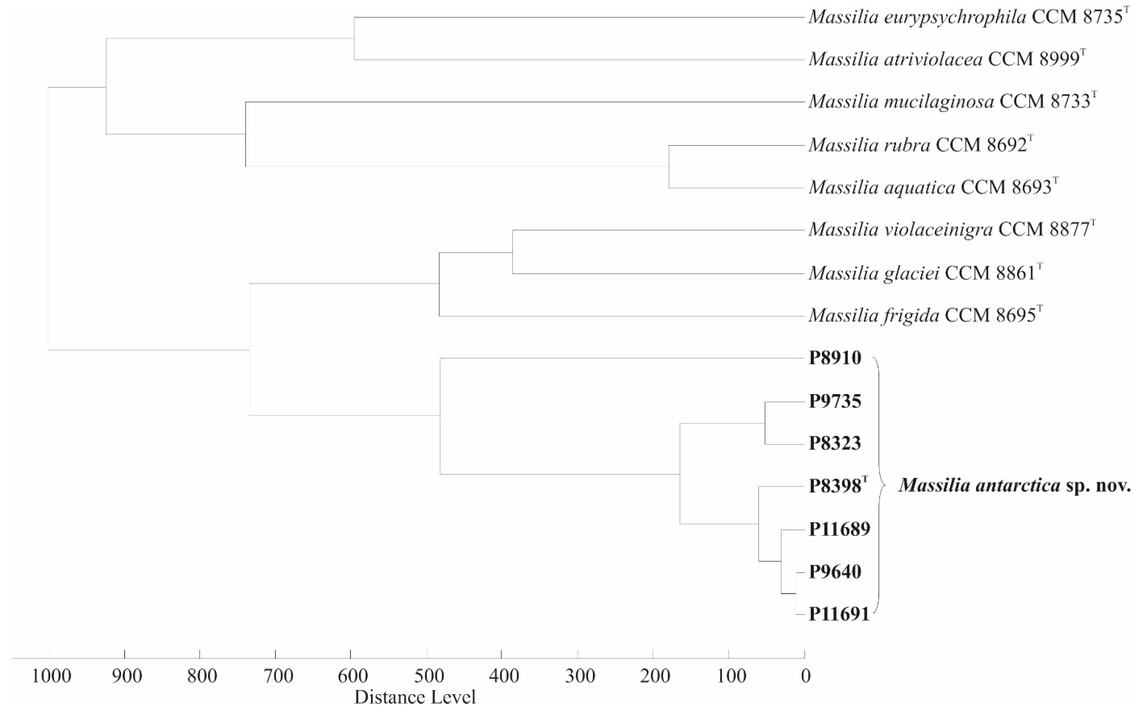
3.1. 16S rRNA, gyrB and lepA Gene Sequencing and Phylogenetic Relationship

The group of seven isolated strains studied (Table 1) was first classified by sequencing the 16S rRNA gene. The similarities between the isolates examined ranged from 98.76% to 100% and assigned the isolates to the Massilia group. The most closely related species based on pairwise sequence alignment were M. frigida (99.9–100% similarity), M. rubra (99.8–99.9%), M. violaceinigra (99.7–99.9%), M. mucilaginosa (99.5–99.7%), M. aquatica (99.7–99.9%), M. atriviolacea (99.4–99.5%), M. eurypsychrophila (98.6–98.8%) and M. glaciei (98.8–99%) (Table S1). Phylogenetic reconstruction based on the 16S rRNA gene sequence, carried out using the ML and NJ methods, divided all the studied isolates into a clade containing the species M. frigida, M. rubra and M. violaceinigra, with 81% bootstrap support (Figure S2). The results obtained prove that the isolates studied belong to the genus Massilia. Nevertheless, the 16S rRNA gene sequence similarities among the isolates and the Massilia spp. types were above the threshold of 98.7% and did not permit species discrimination. Thus, the partial gyrB and lepA gene sequences were additionally performed on all isolates and types. The thresholds for Massilia species differentiation were previously calculated to be 98% similarity for the gyrB gene and 99% similarity for the lepA gene [19]. In our study, the isolates made a distinct clade separating the new isolates from the known Massilia species (Figure 1). However, the sequences analysed had similarity values below the published threshold for the gyrB and lepA gene sequences, namely 100–97.2% for the gyrB gene and 100–97.8% for the lepA gene (Table 2). The sequencing similarity data indicate that the isolates belong to a new species with the representative P8398T (CCM 8941T) as the type strain. Therefore, the ANI and dDDH values between P8398T and P8910 were calculated based on whole-genome sequencing (Figure S3) to confirm that both isolates belong to a single species. These data were well above the threshold (Table 2) and confirmed the assignment of strains P8398T and P8910 to a single taxon.
The ANI values were resolved and the intergenomic distances between the genome sequences of P8398T and the representative types of Massilia spp. were analysed. The ANI values among P8398T and M. atriviolacea CCM 8999T, M. violaceinigra CCM 8877T, M. rubra CCM 8692T, M. mucilaginosa CCM 8733T, M. aquatica CCM 8693T, M. frigida CCM 8695T, M. glaciei CCM 8861T and M. eurypsychrophila CCM 8735T ranged from 79.5% to 92.1%, and the dDDH values between the above strains ranged from 23.1% to 46.4, respectively (Table 2). The determined ANI and dDDH data between the studied strain P8398T and Massilia spp. type strains differed from the threshold values of 95–96% (ANI) and 70% (dDDH) for bacterial species discrimination [32,39,40]. The obtained results clearly confirmed that strain P8398T represents a distinct Massilia species.

3.2. Genome-Based Phylogeny and Basic Genome Characterization

The complete chromosomal sequence of strain P8398T has been deposited at GenBank/EMBL/DDBJ under accession number CP065053. The version characterised in this study is CP065053.1. The complete genome has a size of 7.43 Mbp. The annotation predicted a total of 6509 genes. The majority of them (6392) were predicted to be protein-coding genes (CDSs), including 127 pseudogenes, 117 corresponded to RNA genes, including 22 genes for rRNAs (eight 5S, seven 16S and seven 23S rRNA), 91 genes for tRNAs and 4 genes for ncRNAs. The genomic G + C content was 63.8 mol%. Functional annotation of the CDSs in the form of division into clusters of orthologous genes (COG) assigned a specific function to the majority of CDSs (3956 genes, almost 62% of all CDSs), while the remaining genes belonged to the group without assignment or to the category (S) “Unknown Function”, representing 1337 and 1099 genes, respectively, suggesting that future research with the culture might reveal new, previously unknown traits (Table S2). As for the next functional groups, most of the genes were related to groups responsible for housekeeping functions, such as (K) “Transcription”, (E) “Amino Acid transport and metabolism” and (M) “Cell wall/membrane/envelope biogenesis”, all containing at least 5% of the total number of genes. This threshold was only reached by another functional group (T) “Signal transduction mechanisms”, indicating the presence of a sophisticated system involved in responses to various intra- and extracellular stimuli. For this task, the cells can use 244 genes belonging to the (N) “Cell motility” group. Their further investigation revealed 86 KEGG orthologues belonging to the KEGG BRITE group ko02035 “Bacterial Motility Proteins”, which contains both flagellar and pilus systems.
Although no gene was associated with the class (X) “Mobilome: prophages, transposons”, two prophage sequences were identified in the P8398T genome. While the first phage was marked as questionable, the second was intact (Table S3). Nevertheless, 21 of its 54 genes coded hypothetical proteins, suggesting that the phage is relatively inactive or that it is a new phage that has not been reported before. Neither a CRISPR arrangement nor coupled cas or cas-like genes to create an adaptive immune system have been predicted in the genome [69]. However, this does not mean that P8398T is completely devoid of systems that protect it from foreign DNA, as four restriction–modification (R-M) systems have been found in its genome whose function is, among other things, to defend against genome invasion [70] (Table S4). While two type II systems are probably inactive, as predicted methylases lacked coupled restriction endonucleases, the remaining type I and type IV systems could be active. The type IV systems are formed by only one restriction enzyme identified in the P8398T genome. Although the type I systems are more complex, this R-M system also appeared to be complete, containing one restriction, one modification and two specificity enzymes.
In the search for genes that cause antibiotic resistance, there was no perfect hit among the known genes. However, two copies of a putative catB2 gene (locus tags IV454_00155 and IV454_32525) coding for chloramphenicol acetyltransferases were found among the loose hits, both with 38.1% sequence identity to the best hit. Since experimental work has demonstrated the strain’s resistance to chloramphenicol, it can be assumed that at least one of these genes is active in the genome of P8398T.
The psychrophilic nature of P8398T and its ability to adapt to a hostile cold environment with freezing, thawing and strong UV irradiation is supported by a set of genes specialised in cold adaptation and several more versatile genes involved in the response to extreme conditions. In the first group, two copies of the cspA genes (IV454_17145 and IV454_22840) were found to encode cold shock proteins that play an important role in the immediate response to a decrease in temperature. We also identified some putative genes coding for anti-freeze proteins (IV454_32165, IV454_29925), which are likely to be involved in the synthesis or accumulation of cryoprotective substances. The main function of anti-freeze proteins is to reduce the freezing point of cell fluids in microorganisms to avoid freezing and to influence the shape and size of ice crystals [71,72]. Another important role is played by the genes for the composition of membrane fatty acids (unsaturated, polyunsaturated, methyl-branched, cis-form), which influence the fluidity of the membrane and the transition from the liquid crystalline to the gel phase [73]. The genome of P8398T contained 16 genes coding for proteins of the OmpA (membrane transport) family. However, it is not possible to predict which of these genes are directly related to cold adaptation. Finally, we also found genes responsible for the synthesis of polyhydroxyalkanoates (PHAs), phaC (IV454_22210) and phaR (IV454_22215). PHAs play an important role in the stress resistance of bacteria, and also have the potential for industrial use, but are more commonly studied in other extremophiles, halophiles and thermophiles [74].
One of the most striking manifestations of the phenotype of P8398T was the production of the purple pigment violacein. The genome of P8398T contains all the genes necessary for its production. In addition, the genes vioA (IV454_26305), vioB (IV454_26310), vioC (IV454_26315), vioD (IV454_26320) and vioE (IV454_26325) were found in a single predicted vioABCDE operon. Additional genes coding for transcription factors for violacein biosynthesis were also found. The genes jqsS/cqsS (IV454_01070), coding for the sensor kinase, and jqsR/cqsS (IV454_01075), coding for the response regulator, formed one operon, while jqsA/cqsA (IV454_01065), coding for a quorum-sensing autoinducer synthase, was a separate gene. Therefore, we are certain that the expression of the violacein biosynthetic operon in P8398T depends on quorum sensing, as recently reported for Rugamonas violacea, another psychrophilic species from Antarctica [30].
The strain P8398T exhibited strong gelatinase activity corresponding to the presence of the colA (IV454_08230) gene, which codes for a microbial collagenase. The hydrolytic activity of the strain is even broader. DNase activity is caused by a series of genes coding for exodeoxyribonucleases that produce 5′-phosphomonoesters: sbcB (IV454_15565) coding for exodeoxyribonuclease I, three copies of xthA (IV454_12835, IV454_17225, IV454_27120) coding for exodeoxyribonuclease III, recD (IV454_27425), recB (IV454_27430), recC (IV454_27435) coding for alpha, beta and gamma subunits of exodeoxyribonuclease V located in a single operon on the antisense strand and xseA (IV454_16715) and xseB (IV454_13465) coding for large and small subunits of exodeoxyribonuclease VII. On the other hand, no exodeoxyribonucleases producing 3′-phosphomonoesters were found in the genome. Lipase activity is caused by three copies of putative lip genes (IV454_06595, IV454_31055 and IV454_31865) coding for triacylglycerol lipases, pldA (IV454_06250) coding for phospholipase A and tesA (IV454_24625) coding for acyl-CoA thioesterase I. The ability to hydrolyse lecithin corresponded to the presence of three copies of the gene plc (IV454_12025, IV454_20950 and IV454_24185), which codes for phospholipase C. The strain also exhibited glycolic activity. The genome contained, among others, the gene amyA (IV454_01825, IV454_06345), coding for alpha-amylase, in two copies and the genes bglX (IV454_05025, IV454_11505, IV454_17410) and bglB (IV454_13665, IV454_19850, IV454_27750), coding for beta-glucosidase, in three copies each. In contrast, we could not find a gene responsible for the tyrosinase activity observed at the phenotype level.
The whole-genome sequence of strain P8910 (= CCM 9189) has been deposited with DDBJ/ENA/GenBank under accession number JAJTSQ000000000, and the version described in our study is JAJTSQ00000000.1. The resulting draft genome was 7.4 Mb in size with 190 contigs (N50 = 115,275; L50 = 20). Annotation revealed 6687 genes with 6474 protein-coding genes (CDS), 87 RNA genes including 8 genes for rRNAs (16S and 23S rRNA) and 79 genes for tRNAs. The genomic G + C content was 63.5 mol%, which is fully consistent with the percentages known for other Massilia spp. [6,7].

3.3. MALDI-TOF MS

The MALDI-TOF MS protein fingerprints of the seven examined isolates showed no significant similarity to any of the entries in the Bruker Biotyper database (version 10.0, 9607 entries). After expanding this database with protein signals from eight type strains of Massilia spp. including strain P8398T, all remaining six isolates could be correctly assigned to the corresponding species, resulting in log (scores) greater than 2.0. The mutual similarity between the examined isolates is confirmed by a dendrogram showing their cluster separated from the rest of Massilia spp. (Figure 2). The slightly divergent branch of P8910 was assigned to a common cluster according to the whole-genome sequencing results from ANI and the dDDH data. Visual inspection of the mass spectra revealed that all seven isolated strains shared 24 signals in the m/z range from 2500 to 10,000, while only two of these peaks were specific to the seven isolates (m/z = 3227 and 6453, which most likely corresponds to different ionisation forms of the same compound), allowing their differentiation from the rest of the Massilia spp. tested.

3.4. Chemotaxonomic Characterization

The polar lipid profile (Figure S4) of culture P8398T consisted of the main lipids diphosphatidylglycerol, phosphatidylglycerol and phosphatidylethanolamine. In addition, small quantities of an ornithine lipid (OL) and several unidentified lipids were detected, including an aminolipid (AL1) and nine lipids with no detectable functional group (L1-L9), as well as a yellow pigment spot. The polar lipid profile of strain P8398T did not show the presence of two unidentified phospholipids reported to be present in M. violaceinigra and M. atriviolacea [10,28]. The quinone system contained only ubiquinone Q-8, which is a feature of other Massilia species. The polyamine pattern consisted of 4.8 µmol (g dry weight)−1 putrescine, 0.5 µmol (g dry weight)−1 2-hydroxyputrescine, 0.3 µmol (g dry weight)−1 spermidine and 0.1 µmol (g dry weight)−1 cadaverine. A comparable polyamine pattern was described for Massilia norwichensis [19].
Analysis of the fatty acid methyl esters showed that the major fatty acids of all strains were uniformly iso-C16:0 and inseparably C16:1 ω7c/C16:1 ω6c (included in Summed Feature 3) via the Sherlock MIDI system, with an average abundance in the total fatty acid profiles (FA) of 24.8% and 55.4%, respectively (Table S5). The presence of these major FAs corresponded to the FA profiles of the most closely related Massilia spp. All strains belonging to the newly described species could be distinguished from their closest phylogenetic neighbours by the presence of capric acid (C10:0). Apart from the presence of capric acid, all Massilia spp. analysed in this study had similar FA profiles with insignificant quantitative differences, confirming their relatedness.

3.5. Repetitive PCR-Based Fingerprinting

The cluster analysis of the rep-PCR fingerprint profiles of the strains studied is shown in Figure S5. The four strains P8323, P9735, P8910 and P11689 had visually distinct fingerprints that enabled their clear differentiation. The fingerprints of the remaining three strains P8398T, P9640 and P11691 were visually similar, but minor differences due to the absence or presence of certain bands also allowed them to be distinguished. Interestingly, these three strains came from different, distant localities. These results showed that the rep-PCR band patterns obtained enabled fine differentiation of the isolates at the subgroup level and proved that they are not clonally related.

3.6. Phenotypic Characteristics and Description of Novel Species

Description of Massilia antarctica sp. nov. (M. antarctica etymology: ant.arc´ti.ca. L. fem. adj. antarctica southern, pertaining to Antarctica, the geographical origin of the isolated strains).
The cells are Gram-stain-negative rods, occurring separately or in clusters, motile with a long polar flagellum (Figure S6A). The width of the cells ranged from 880 nm to 1.14 µm and the length from 1.68 µm to 2.93 µm. The colonies on the R2A medium are circular, with entire margins, convex, smooth, glistening with blue-purple endopigment violacein, 2 mm in diameter after 72 h of cultivation at 20 °C and moderately slimy (Figure S6B). The results of the biochemical and physiological tests, which enabled the phenotypic distinction of the investigated strains from closely related species, are described in Table 3. The strains were resistant to chloramphenicol, and all exhibited strong hydrolytic activities. The detailed results of the phenotypic characterization of the new species M. antarctica sp. nov. are given in the Protologue species description (Box 1) and the variable results are given in Table S6. The type strain is P8398T (= CCM 8941T = LMG 32108T).
Box 1. Protologue species description—biochemical and physiological properties of Massilia antarctica sp. nov. All presented data are uniform for all seven isolates, the strain dependent data are shown in Table S6.
Phenotype characterisation
Aerobic growth only occurs on R2A agar and PCA agar. No growth was observed on TSA, BHI, Mac Conkey agar, Mueller–Hinton agar or Nutrient agar at 20 °C. Growth is observed between 1 °C and 25 °C, but not at −2 °C, nor at 30 °C. Cells grow well in the pH range 6.0–9.0, and pH 5.0 and pH 10.0 inhibits growth. Good growth on the R2A medium occurs without NaCl, while the presence of 0.5% NaCl (w/v) inhibits growth.
Acid production (aerobic) from glucose, fructose, xylose and maltose occurred, while the fermentation of glucose in the OF test medium is negative. Catalase, alkaline phosphatase, esterase lipase and leucine arylamidase were positive by API ZYM. Gelatine, casein, esculin, Tween 80, ONPG, starch and tyrosine hydrolysis positive.
Lipase (C14), cystine arylamidase, α-chymotrypsin, α-galactosidase, β-galactosidase, β-glucuronidase, N-acetyl-β-glucosaminidase, α-mannosidase and α-fucosidase were negative by API ZYM. Oxidase, nitrite reduction, fluorescein (King B medium), urease, lysine and ornithine decarboxylase, arginine dihydrolase, Simmons citrate, malonate utilisation and acetamide utilization were negative.
All strains were negative for the utilization (Biolog) of D-maltose, D-trehalose, gentiobiose, sucrose, turanose, stachyose, D-raffinose, α-D-lactose, D-melibiose, β-methyl-D-glucoside, D-salicin, α-D-glucose, D-mannose, N-acetyl-D-glucosamine, N-acetyl-β-D-mannosamine, N-Acetyl-D-galactosamine, N-acetyl neuraminic acid, D-fructose, D, galactose, 3-methyl glucose, D-fucose, L-fucose, L-rhamnose, inosine, D-sorbitol, D-mannitol, D-arabitol, myo-inositol, glycerol, D-aspartic acid, D-serine, gelatine, glycyl-L-proline, L-alanine, L-arginine, L-aspartic acid, L-glutamic acid, L-histidine, L-pyroglutamic acid, L-serine, pectin, D-galacturonic acid, D-galactonic acid lactone, D-gluconic acid, D-glucuronic acid, mucic acid, D-saccharic acid, p-hydroxy phenylacetic acid, methyl pyruvate, D-lactic acid methyl ester, L-lactic acid, citric acid, α-keto glutaric acid, D-malic acid, bromo-succinic acid, Tween 40, γ-amino-butyric acid, α-hydroxy-butyric acid, propionic acid, acetic acid and formic acid as carbon sources.
They were resistant to chloramphenicol but sensitive to ciprofloxacin, gentamicin, imipenem, kanamycin, cotrimoxazole, piperacillin, streptomycin and tetracycline.

Supplementary Materials

The following supporting information can be downloaded at: https://www.mdpi.com/article/10.3390/microorganisms10040704/s1, Figure S1: Sampling sites of Massilia antarctica sp. nov. isolates, Figure S2: Maximum Likelihood phylogenetic tree based on 16S rRNA gene analysis of Massilia spp., Figure S3: Heatmap generated with OrthoANI values between the whole-genome sequences of Massilia antarctica sp. nov. and related Massilia spp. type strains, Figure S4: Polar lipid profile of strain P8398T, Figure S5: Dendrogram based on cluster analysis of rep-PCR fingerprints from Massilia antarctica sp. nov. strains, Figure S6A: Cell morphology of strain P8398T observed using transmission electron microscopy, Figure S6B: Colony morphology of P8398T on R2A agar, Table S1: Pairwise 16S rRNA, gyrB and lepA genes sequence similarities, Table S2: Clusters of orthologous groups of P8398T strain, Table S3: Phage DNA within the P8398T genome, Table S4: R-M systems in the P8398T genome, Table S5: Cellular fatty acid contents of Massilia antarctica sp. nov. strains and the closest phylogenetic relatives, Table S6: Variable reactions of Massilia antarctica sp. nov. strains.

Author Contributions

Conceptualization, I.S. and P.Š.; methodology, I.S. and P.H.; software, P.Š., V.K. and K.S.; formal analysis, I.S., H.-J.B., P.H., S.K., R.S., E.S., J.P. and O.Š.; investigation, I.S., P.H. and K.S.; resources, I.S. and J.S.; data curation, I.S.; writing—original draft preparation, I.S., P.H. and K.S.; writing—review and editing, I.S. and P.Š.; visualization, I.S.; supervision, I.S.; project administration, I.S.; funding acquisition, I.S. and O.Š. All authors have read and agreed to the published version of the manuscript.

Funding

This study was supported by projects LM2015078 and CZ.02.1.01/0.0/0.0/18_046/0015974. This study was also partly funded by the Czech Collection of Microorganisms. CIISB, Instruct-CZ Centre of Instruct-ERIC EU consortium, funded by MEYS CR infrastructure project LM2018127, is gratefully acknowledged for the financial support of the measurements at the CEITEC Proteomics Core Facility.

Institutional Review Board Statement

Not applicable.

Informed Consent Statement

Not applicable.

Data Availability Statement

The genomes of two isolates (P8398T and P8910) were submitted to the GenBank database under accession numbers CP065053 and JAJTSQ000000000, respectively. The GenBank accession numbers (16S rRNA, gyrB and lepA genes) of M. antarctica sp. nov. strains are OM243916–OM 243922, OM296116-296122 and OM296123-OM296129, respectively.

Acknowledgments

The authors would like to thank the Czech Antarctic Research Programme and its crew for providing support. Daniel Krsek is thanked for transmission electron microscopy. The GCM 10K type strain sequencing project is gratefully acknowledged. We also wish to thank Jana Bajerová and Renée Šubrtová for excellent technical support.

Conflicts of Interest

The authors declare no conflict of interest.

References

  1. Barnes, D.K.A. Polar marine ecosystems. In Life at Extremes: Environments, Organisms, and Strategies for Survival; Bell, E.M., Ed.; Cabi: London, UK, 2012; pp. 1–9. [Google Scholar]
  2. Peeters, K.; Verleyen, E.; Hodgson, D.A.; Convey, P.; Ertz, D.; Vyverman, W.; Willems, A. Heterotrophic bacterial diversity in aquatic microbial mat communities from Antarctica. Polar Biol. 2012, 35, 543–554. [Google Scholar] [CrossRef] [Green Version]
  3. Sanyal, A.; Antony, R.; Samui, G.; Thamban, M. Microbial communities and their potential for degradation of dissolved organic carbon in cryoconite hole environments of Himalaya and Antarctica. Microbiol. Res. 2018, 208, 32–42. [Google Scholar] [CrossRef] [PubMed]
  4. La Scola, B.; Birtles, R.J.; Mallet, M.-N.; Raoult, D. Massilia timonae gen. nov., sp. nov., isolated from blood of an immunocompromised patient with cerebellar lesions. J. Clin. Microbiol. 1998, 36, 2847–2852. [Google Scholar] [CrossRef] [PubMed] [Green Version]
  5. Garrity, G.M.; Bell, J.A.; Lilburn, T. Family II. Oxalobacteraceae fam. nov. In Bergey’s Manual of Systematic Bacteriology, the Proteobacteria, Part C (The Alpha-, Beta-, Delta-, and Epsilonproteobacteria), 2nd ed.; Brenner, D.J., Krieg, N.R., Staley, J.T., Garrity, G.M., Eds.; Springer: New York, NY, USA, 2005; Volume 2, p. 623. [Google Scholar]
  6. Kämpfer, P.; Lodders, N.; Martin, K.; Falsen, E. Revision of the genus Massilia La Scola et al. 2000, with an emended description of the genus and inclusion of all species of the genus Naxibacter as new combinations, and proposal of Massilia consociata sp. nov. Int. J. Syst. Evol. Microbiol. 2011, 61, 1528–1533. [Google Scholar] [CrossRef] [PubMed]
  7. Singh, H.; Du, J.; Won, K.; Yang, J.-E.; Yin, C.; Kook, M.; Yi, T.-H. Massilia arvi sp. nov., isolated from fallow-land soil previously cultivated with Brassica oleracea, and emended description of the genus Massilia. Int. J. Syst. Evol. Microbiol. 2015, 65, 3690–3696. [Google Scholar] [CrossRef] [PubMed]
  8. Parte, A.C.; Carbasse, J.S.; Meier-Kolthoff, J.P.; Reimer, L.C.; Göker, M. List of Prokaryotic names with standing in nomenclature (LPSN) moves to the DSMZ. Int. J. Syst. Evol. Microbiol. 2020, 70, 5607–5612. [Google Scholar] [CrossRef]
  9. Zhang, B.; Yang, R.; Zhang, G.; Zhang, D.; Zhang, W.; Chen, T.; Liu, G. Massilia arenae sp. nov., isolated from sand soil in the Qinghai-Tibetan Plateau. Int. J. Syst. Evol. Microbiol. 2020, 70, 2435–2439. [Google Scholar] [CrossRef]
  10. Yang, E.; Zhao, M.; Li, S.; Wang, Y.; Sun, L.; Liu, J.; Wang, W. Massilia atriviolacea sp. nov., a dark purple-pigmented bacterium isolated from soil. Int. J. Syst. Evol. Microbiol. 2019, 69, 2135–2141. [Google Scholar] [CrossRef]
  11. Ren, M.; Li, X.; Zhang, Y.; Jin, Y.; Li, S.; Huang, H. Massilia armeniaca sp. nov., isolated from desert soil. Int. J. Syst. Evol. Microbiol. 2018, 68, 2319–2324. [Google Scholar] [CrossRef]
  12. Sun, L.-N.; Yang, E.-D.; Cui, D.-X.; Ni, Y.-W.; Wang, Y.-B.; Sun, D.-D.; Wang, W.-Y. Massilia buxea sp. nov., isolated from a rock surface. Int. J. Syst. Evol. Microbiol. 2017, 67, 4390–4396. [Google Scholar] [CrossRef]
  13. Feng, G.-D.; Yang, S.-Z.; Li, H.-P.; Zhu, H. Massilia putida sp. nov., a dimethyl disulfide-producing bacterium isolated from wolfram mine tailing. Int. J. Syst. Evol. Microbiol. 2016, 66, 50–55. [Google Scholar] [CrossRef] [PubMed]
  14. Shen, L.; Liu, Y.; Gu, Z.; Xu, B.; Wang, N.; Jiao, N.; Liu, H.; Zhou, Y. Massilia eurypsychrophila sp. nov. a facultatively psychrophilic bacteria isolated from ice core. Int. J. Syst. Evol. Microbiol. 2015, 65, 2124–2129. [Google Scholar] [CrossRef] [PubMed] [Green Version]
  15. Guo, B.; Liu, Y.; Gu, Z.; Shen, L.; Liu, K.; Wang, N.; Xing, T.; Liu, H.; Zhou, Y.; Li, J. Massilia psychrophila sp. nov., isolated from an ice core. Int. J. Syst. Evol. Microbiol. 2016, 66, 4088–4093. [Google Scholar] [CrossRef] [PubMed]
  16. Gu, Z.; Liu, Y.; Xu, B.; Wang, N.; Jiao, N.; Shen, L.; Liu, H.; Zhou, Y.; Liu, X.; Li, J.; et al. Massilia glaciei sp. nov., isolated from the Muztagh Glacier. Int. J. Syst. Evol. Microbiol. 2017, 67, 4075–4079. [Google Scholar] [CrossRef]
  17. Gallego, V.; Sanchez-Porro, C.; Garcia, M.T.; Ventosa, A. Massilia aurea sp. nov., isolated from drinking water. Int. J. Syst. Evol. Microbiol. 2006, 56, 2449–2453. [Google Scholar] [CrossRef]
  18. Lu, H.; Deng, T.; Liu, F.; Wang, Y.; Yang, X.; Xu, M. Duganella lactea sp. nov., Duganella guangzhouensis sp. nov., Duganella flavida sp. nov. and Massilia rivuli sp. nov., isolated from a subtropical stream in PR China and proposal to reclassify Duganella ginsengisoli as Massilia ginsengisoli comb. nov. Int. J. Syst. Evol. Microbiol. 2020, 70, 4822–4830. [Google Scholar] [CrossRef] [PubMed]
  19. Orthová, I.; Kampfer, P.; Glaeser, S.P.; Kaden, R.; Busse, H.-J. Massilia norwichensis sp. nov., isolated from an air sample. Int. J. Syst. Evol. Microbiol. 2015, 65, 56–64. [Google Scholar] [CrossRef] [PubMed]
  20. Kämpfer, P.; Lodders, N.; Martin, K.; Falsen, E. Massilia oculi sp. nov., isolated from a human clinical specimen. Int. J. Syst. Evol. Microbiol. 2012, 62, 364–369. [Google Scholar] [CrossRef] [PubMed] [Green Version]
  21. Wery, N.; Gerike, U.; Sharman, A.; Chaudhuri, J.B.; Hough, D.W.; Danson, M.J. Use of a packed-column bioreactor for isolation of diverse protease-producing bacteria from Antarctic soil. Appl. Environ. Microbiol. 2003, 69, 1457–1464. [Google Scholar] [CrossRef] [Green Version]
  22. Cong, B.; Yin, X.; Deng, A.; Shen, J.; Tian, Y.; Wang, S.; Yang, H. Diversity of cultivable microbes from soil of the Fildes Peninsula, Antarctica, and their potential application. Front. Microbiol. 2020, 11, 570836. [Google Scholar] [CrossRef]
  23. Holochová, P.; Mašlaňová, I.; Sedláček, I.; Švec, P.; Králová, S.; Kovařovic, V.; Busse, H.-J.; Staňková, E.; Barták, M.; Pantůček, R. Description of Massilia rubra sp. nov., Massilia aquatica sp. nov., Massilia mucilaginosa sp. nov., Massilia frigida sp. nov., and one Massilia genomospecies isolated from Antarctic streams, lakes and regoliths. Syst. Appl. Microbiol. 2020, 43, 126112. [Google Scholar] [CrossRef]
  24. Sajjad, W.; Din, G.; Rafiq, M.; Iqbal, A.; Khan, S.; Zada, S.; Ali, B.; Kang, S. Pigment production by cold-adapted bacteria and fungi: Colorful tale of cryosphere with wide range applications. Extremophiles 2020, 24, 447–473. [Google Scholar] [CrossRef] [PubMed]
  25. Dieser, M.; Greenwood, M.; Foreman, C. Carotenoid pigmentation in Antarctic heterotrophic bacteria as a strategy to withstand environmental stresses. Arct. Antarct. Alp. Res. 2010, 42, 396–405. [Google Scholar] [CrossRef] [Green Version]
  26. Zhang, Y.-Q.; Li, W.-J.; Zhang, K.-Y.; Tian, X.-P.; Jiang, Y.; Xu, L.-H.; Jiang, C.-L.; Lai, R. Massilia dura sp. nov., Massilia albidiflava sp. nov., Massilia plicata sp. nov. and Massilia lutea sp. nov., isolated from soils in China. Int. J. Syst. Evol. Microbiol. 2006, 56, 459–463. [Google Scholar] [CrossRef] [PubMed]
  27. Wang, J.; Zhang, J.; Pang, H.; Zhang, Y.; Li, Y.; Fan, J. Massilia flava sp. nov., isolated from soil. Int. J. Syst. Evol. Microbiol. 2012, 62, 580–585. [Google Scholar] [CrossRef] [Green Version]
  28. Wang, H.; Zhang, X.; Wang, S.; Zhao, B.; Lou, K.; Xing, X.-H. Massilia violaceinigra sp. nov., a novel purple-pigmented bacterium isolated from glacier permafrost. Int. J. Syst. Evol. Microbiol. 2018, 68, 2271–2278. [Google Scholar] [CrossRef]
  29. Myeong, N.R.; Seong, H.J.; Kim, H.-J.; Sul, W.J. Complete genome sequence of antibiotic and anticancer agent violacein producing Massilia sp. strain NR 4-1. J. Biotech. 2016, 223, 36–37. [Google Scholar] [CrossRef]
  30. Sedláček, I.; Holochová, P.; Sobotka, R.; Busse, H.-J.; Švec, P.; Králová, S.; Šedo, O.; Pilný, J.; Staňková, E.; Koublová, V.; et al. Classification of violacein-producing psychrophilic group of isolates associated with freshwater in Antarctica and description of Rugamonas violacea sp. nov. Microbiol. Spectr. 2021, 9, e00452-21. [Google Scholar] [CrossRef] [PubMed]
  31. Kýrová, K.; Sedláček, I.; Pantůček, R.; Králová, S.; Holochová, P.; Mašlaňová, I.; Staňková, E.; Kleinhagauer, T.; Gelbíčová, T.; Sobotka, R.; et al. Rufibacter ruber sp. nov., isolated from fragmentary rock. Int. J. Syst. Evol. Microbiol. 2016, 66, 4401–4405. [Google Scholar] [CrossRef] [PubMed]
  32. Chun, J.; Oren, A.; Ventosa, A.; Christensen, H.; Arahal, D.R.; Da Costa, M.S.; Rooney, A.P.; Yi, H.; Xu, X.-W.; De Meyer, S.; et al. Proposed minimal standards for the use of genome data for the taxonomy of prokaryotes. Int. J. Syst. Evol. Microbiol. 2018, 68, 461–466. [Google Scholar] [CrossRef] [PubMed]
  33. Yoon, S.-H.; Ha, S.-M.; Kwon, S.; Lim, J.; Kim, Y.; Seo, H.; Chun, J. Introducing EzBioCloud: A taxonomically united database of 16S rRNA gene sequences and whole-genome assemblies. Int. J. Syst. Evol. Microbiol. 2017, 67, 1613–1617. [Google Scholar] [CrossRef] [PubMed]
  34. Lee, I.; Chalita, M.; Ha, S.-M.; Na, S.-I.; Yoon, S.-H.; Chun, J. ContEst16S: An algorithm that identifies contaminated prokaryotic genomes using 16S RNA gene sequences. Int. J. Syst. Evol. Microbiol. 2017, 67, 2053–2057. [Google Scholar] [CrossRef] [PubMed]
  35. Tamura, K.; Stecher, G.; Kumar, S. MEGA11: Molecular evolutionary genetics analysis version 11. Mol. Biol. Evol. 2021, 38, 3022–3027. [Google Scholar] [CrossRef] [PubMed]
  36. Kimura, M. A simple method for estimating evolutionary rates of base substitutions through comparative studies of nucleotide sequences. J. Mol. Evol. 1980, 16, 111–120. [Google Scholar] [CrossRef] [PubMed]
  37. Wu, L.; Ma, J. The Global Catalogue of Microorganisms (GCM) 10K type strain sequencing project: Providing services to taxonomists for standard genome sequencing and annotation. Int. J. Syst. Evol. Microbiol. 2019, 69, 895–898. [Google Scholar] [CrossRef]
  38. Shi, W.; Sun, Q.; Fan, G.; Hideaki, S.; Moriya, O.; Itoh, T.; Zhou, Y.; Cai, M.; Kim, S.-G.; Lee, J.-S.; et al. gcType: A high-quality type strain genome database for microbial phylogenetic and functional research. Nucleic Acids Res. 2021, 49, D694–D705. [Google Scholar] [CrossRef]
  39. Yoon, S.-H.; Ha, S.-M.; Lim, J.; Kwon, S.; Chun, J. A large-scale evaluation of algorithms to calculate average nucleotide identity. Antonie Leeuwenhoek 2017, 110, 1281–1286. [Google Scholar] [CrossRef]
  40. Meier-Kolthoff, J.P.; Auch, A.F.; Klenk, H.-P.; Göker, M. Genome sequence-based species delimitation with confidence intervals and improved distance functions. BMC Bioinform. 2013, 14, 60. [Google Scholar] [CrossRef] [Green Version]
  41. Tatusova, T.; DiCuccio, M.; Badretdin, A.; Chetvernin, V.; Nawrocki, E.P.; Zaslavsky, L.; Lomsadze, A.; Pruitt, K.D.; Borodovsky, M.; Ostell, J. NCBI prokaryotic genome annotation pipeline. Nucleic Acids Res. 2016, 44, 6614–6624. [Google Scholar] [CrossRef]
  42. Taboada, B.; Estrada, K.; Ciria, R.; Merino, E. Operon-mapper: A web server for precise operon identification in bacterial and archaeal genomes. Bioinformatics 2018, 34, 4118–4120. [Google Scholar] [CrossRef]
  43. Cantalapiedra, C.P.; Hernández-Plaza, A.; Letunic, I.; Bork, P.; Huerta-Cepas, J. eggNOG-mapper v2: Functional annotation, orthology assignments, and domain prediction at the metagenomic scale. Mol. Biol. Evol. 2021, 38, 5825–5829. [Google Scholar] [CrossRef] [PubMed]
  44. Arndt, D.; Grant, J.R.; Marcu, A.; Sajed, T.; Pon, A.; Liang, Y.; Wishart, D.S. PHASTER: A better, faster version of the PHAST phage search tool. Nucleic Acids Res. 2016, 44, W16–W21. [Google Scholar] [CrossRef] [PubMed] [Green Version]
  45. Biswas, A.; Staals, R.; Morales, S.; Fineran, P.; Brown, C.M. CRISPRDetect: A flexible algorithm to define CRISPR arrays. BMC Genom. 2016, 17, 356. [Google Scholar] [CrossRef] [PubMed] [Green Version]
  46. Roberts, R.J.; Vincze, T.; Posfai, J.; Macelis, D. REBASE—A database for DNA restriction and modification: Enzymes, genes and genomes. Nucleic Acids Res. 2015, 43, D298–D299. [Google Scholar] [CrossRef] [PubMed]
  47. Kanehisa, M.; Sato, Y.; Kawashima, M.; Furumichi, M.; Tanabe, M. KEGG as a reference resource for gene and protein annotation. Nucleic Acids Res. 2016, 44, D457–D462. [Google Scholar] [CrossRef] [PubMed] [Green Version]
  48. Kanehisa, M.; Sato, Y.; Morishima, K. BlastKOALA and GhostKOALA: KEGG tools for functional characterization of genome and metagenome sequences. J. Mol. Biol. 2016, 428, 726–731. [Google Scholar] [CrossRef] [PubMed] [Green Version]
  49. Altschul, S.F.; Gish, W.; Miller, W.; Myers, E.W.; Lipman, D.J. Basic local alignment search tool. J. Mol. Biol. 1990, 215, 403–410. [Google Scholar] [CrossRef]
  50. Alcock, B.P.; Raphenya, A.R.; Lau, T.T.Y.; Tsang, K.K.; Bouchard, M.; Edalatmand, A.; Huynh, W.; Nguyen, A.-L.V.; Cheng, A.A.; Liu, S.; et al. CARD 2020: Antibiotic resistome surveillance with the comprehensive antibiotic resistance database. Nucleic Acids Res. 2020, 48, D517–D525. [Google Scholar] [CrossRef]
  51. Freiwald, A.; Sauer, S. Phylogenetic classification and identification of bacteria by mass spectrometry. Nat. Protoc. 2009, 4, 732–742. [Google Scholar] [CrossRef]
  52. Busse, H.-J.; Auling, G. Polyamine pattern as a chemotaxonomic marker within the Proteobacteria. Syst. Appl. Microbiol. 1988, 11, 1–8. [Google Scholar] [CrossRef]
  53. Busse, H.-J.; Bunka, S.; Hensel, A.; Lubitz, W. Discrimination of members of the family Pasteurellaceae based on polyamine patterns. Int. J. Syst. Evol. Microbiol. 1997, 47, 698–708. [Google Scholar] [CrossRef] [Green Version]
  54. Tindall, B.J. Lipid composition of Halobacterium lacusprofundi. FEMS Microbiol. Lett. 1990, 66, 199–202. [Google Scholar] [CrossRef]
  55. Tindall, B. A comparative study of the lipid composition of Halobacterium saccharovorum from various sources. Syst. Appl. Microbiol. 1990, 13, 128–130. [Google Scholar] [CrossRef]
  56. Altenburger, P.; Kämpfer, P.; Makristathis, A.; Lubitz, W.; Busse, H.-J. Classification of bacteria isolated from a medieval wall painting. J. Biotechnol. 1996, 47, 39–52. [Google Scholar] [CrossRef]
  57. Stolz, A.; Busse, H.-J.; Kampfer, P. Pseudomonas knackmussii sp. nov. Int. J. Syst. Evol. Microbiol. 2007, 57, 572–576. [Google Scholar] [CrossRef] [PubMed] [Green Version]
  58. Sasser, M. Identification of Bacteria by Gas Chromatography of Cellular Fatty Acids; MIDI Technical Note 101; Microbial ID, Inc.: Newark, DE, USA, 1990. [Google Scholar]
  59. Švec, P.; Pantůček, R.; Petráš, P.; Sedláček, I.; Nováková, D. Identification of Staphylococcus spp. using (GTG)5-PCR fingerprinting. Syst. Appl. Microbiol. 2010, 33, 451–456. [Google Scholar] [CrossRef] [PubMed]
  60. Carlone, G.M.; Valadez, M.J.; Pickett, M.J. Methods for distinguishing gram-positive from gram-negative bacteria. J. Clin. Microbiol. 1982, 16, 1157–1159. [Google Scholar] [CrossRef] [Green Version]
  61. Sedláček, I.; Králová, S.; Kýrová, K.; Mašlaňová, I.; Busse, H.-J.; Staňková, E.; Vrbovská, V.; Němec, M.; Barták, M.; Holochová, P.; et al. Red-pink pigmented Hymenobacter coccineus sp. nov., Hymenobacter lapidarius sp. nov. and Hymenobacter glacialis sp. nov., isolated from rocks in Antarctica. Int. J. Syst. Evol. Microbiol. 2017, 67, 1975–1983. [Google Scholar] [CrossRef]
  62. Da, X.; Jiang, F.; Chang, X.; Ren, L.; Qiu, X.; Kan, W.; Zhang, Y.; Deng, S.; Fang, C.; Peng, F. Pedobacter ardleyensis sp. nov., isolated from soil in Antarctica. Int. J. Syst. Evol. Microbiol. 2015, 65, 3841–3846. [Google Scholar] [CrossRef]
  63. Atlas, R.M. Handbook of Microbiological Media, 4th ed.; ASM Press: Washington, DC, USA, 2010. [Google Scholar]
  64. Barrow, G.I.; Feltham, R.K.A. Cowan and Steel’s Manual for the Identification of Medical Bacteria, 3rd ed.; Cambridge University Press: Cambridge, UK, 1993. [Google Scholar]
  65. Kosina, M.; Barták, M.; Mašlaňová, I.; Pascutti, A.V.; Šedo, O.; Lexa, M.; Sedláček, I. Pseudomonas prosekii sp. nov., a novel psychrotrophic bacterium from Antarctica. Curr. Microbiol. 2013, 67, 637–646. [Google Scholar] [CrossRef]
  66. Margesin, R.; Gander, S.; Zacke, G.; Gounot, A.M.; Schinner, F. Hydrocarbon degradation and enzyme activities of cold-adapted bacteria and yeasts. Extremophiles 2003, 7, 451–458. [Google Scholar] [CrossRef]
  67. CLSI. Performance Standards for Antimicrobial Susceptibility Testing. Twenty-Fifth Informational Supplement (M100-S25); Clinical and Laboratory Standards Institute: Wayne, PA, USA, 2015; Volume 35, No. 3. [Google Scholar]
  68. EUCAST. Breakpoint Tables for Interpretation of Mics and Zone Diameters, Version 12.0; The European Committee on Antimicrobial Susceptibility Testing: Växjö, Sweden, 2022. [Google Scholar]
  69. Sorek, R.; Lawrence, C.M.; Wiedenheft, B. CRISPR-mediated adaptive immune systems in bacteria and archaea. Annu. Rev. Biochem. 2013, 82, 237–266. [Google Scholar] [CrossRef] [PubMed] [Green Version]
  70. Vasu, K.; Nagaraja, V. Diverse functions of restriction-modification systems in addition to cellular defense. Microbiol. Mol. Biol. Rev. 2013, 77, 53–72. [Google Scholar] [CrossRef] [PubMed] [Green Version]
  71. Białkowska, A.; Majewska, E.; Olczak, A.; Twarda-Clapa, A. Ice binding proteins: Diverse biological roles and applications in different types of industry. Biomolecules 2020, 10, 274. [Google Scholar] [CrossRef] [Green Version]
  72. Casanueva, A.; Tuffin, M.; Cary, C.; Cowan, D.A. Molecular adaptations to psychrophily: The impact of ´omic´ technologies. Trends Microbiol. 2010, 18, 374–381. [Google Scholar] [CrossRef] [PubMed]
  73. Los, D.A.; Murata, N. Membrane fluidity and its roles in the perception of environmental signals. Biochim. Biophys. Acta Biomembr. 2004, 1666, 142–157. [Google Scholar] [CrossRef] [PubMed] [Green Version]
  74. Obruča, S.; Dvořák, P.; Sedláček, P.; Koller, M.; Sedlář, K.; Pernicová, I.; Šafránek, D. Polyhydroxyalkanoates synthesis by halophiles and thermophiles: Towards sustainable production of microbial bioplastics. Biotechnol. Adv. 2022, 107906. [Google Scholar] [CrossRef] [PubMed]
Figure 1. Phylogenetic maximum likelihood trees based on (A) gyrB and (B) lepA gene analyses showing phylogenetic positions of M. antarctica sp. nov. isolates and closely related Massilia spp. type strains. Bootstrap probability values (percentages of 500 tree replications) greater than 50%, in which the allied species are clustered, is shown next to the branches. The tree is drawn to scale, with the length of branches measured in the number of substitutions per site. Rhodoferax saidenbachensis D16T sequences were used as an outgroup. GenBank accession numbers of the sequences are indicated in parentheses. Bar, 0.1 substitutions per nucleotide position.
Microorganisms 10 00704 g001
Figure 2. Dendrogram obtained by cluster analysis of MALDI-TOF mass spectra of M. antarctica sp. nov. isolates and the closest related Massilia spp. types, generated with Biotyper 3.0 software using Pearson’s product-moment coefficient as a measure of similarity and the unweighted pair group average linked method (UPGMA) as a grouping method. The distance is shown in relative units.
Microorganisms 10 00704 g002
Table 1. Source of Massilia antarctica sp. nov. strains.
Strain NumberYear of IsolationLocality (GPS)
P8323 (CCM 8944)2017Seals stream, nearby Monolith lake, James Ross Island (63°53′10″ S, 57°56′50″ W)
P8398T (CCM 8941T)2017Glacial stream, Komárek valley, James Ross Island (63°49′51″ S, 57°49′48″ W)
P8910 (CCM 9189)2017Small stream nearby Whiskey glacier, James Ross Island (63°55′03″ S, 57°56′25″ W)
P96402018Lakelet nearby sea coast, Eagle Island (63°37′29″ S, 57°25′52″ W)
P97352018Lakelet below moraine Triangular, James Ross Island (63°51′42″ S, 57°51′07″ W)
P116892019Small lake, approx. 200 m SW from Rožmberk lake, James Ross Island (63°49′17″ S, 57°50′28″ W)
P116912019Small lake, approx. 200 m SW from Rožmberk lake, James Ross Island (63°49′17″ S, 57°50′28″ W)
Table 2. Sequence similarity values of analysed genes, average nucleotide identities and the dDDH values (%) of Massilia antarctica sp. nov. reference strains P8398T (= CCM 8941T) and P8910 (= CCM 9189) to related Massilia spp. All data were taken from this study.
SpeciesStrain Gene Sequence
Similarities of P8398T
ANI and dDDH Values of P8398TGene Sequence
Similarities of P8910
ANI and dDDH Values of P8910
16S rRNAgyrBlepAANIdDDH16S rRNAgyrBlepAANIdDDH
M. antarctica sp. nov.P8398T 10010010010010099.997.097.897.073.9
M. antarctica sp. nov.P891099.997.097.897.073.9100100100100100
M. aquaticaCCM 8693T99.795.895.087.633.799.896.095.087.733.7
M. atriviolaceaCCM 8999T 99.594.495.187.934.099.594.594.987.733.8
M. eurypsychrophilaCCM 8735T98.889.188.279.323.198.789.187.479.223.0
M. frigidaCCM 8695T10096.096.492.146.499.996.496.492.246.3
M. glacieiCCM 8861T98.990.988.979.023.499.090.788.579.023.1
M. mucilaginosaCCM 8733T99.796.595.588.034.799.696.695.188.034.6
M. rubraCCM 8692T99.995.494.988.034.499.995.594.588.134.4
M. violaceinigraCCM 8877T99.995.495.591.042.499.995.795.391.042.1
Table 3. Distinguishing properties of Massilia antarctica sp. nov. with types of nearest phylogenetically related species. 1. Massilia antarctica sp. nov., 2. M. rubra CCM 8692T, 3. M. mucilaginosa CCM 8733T, 4. M. aquatica CCM 8693T, 5. M. frigida CCM 8695T, 6. M. violaceinigra CCM 8877T, 7. M. atriviolacea CCM 8999T, 8. M. glaciei CCM 8861T, 9. M. eurypsychrophila CCM 8735T. +, positive; w, weak; -, negative; S, sensitive; R, resistant; all data were taken from this study.
Test1 *23456789
Violet pigment+----++--
Pink-red pigment-++++----
Oxidase-------++
Growth at 5 °C on R2A+++++-+++
Growth in 0.5% NaCl--++--w--
Acid from xylose++w---+--
API ZYM: Lipase (C14)-----+w-+
Cystine arylamidase-----+--+
BIOLOG GEN III: Arabitol------+--
Pectin------+--
Glucuronamide-++w++--+
Chloramphenicol (30 µg)RSRSSSRSS
Hydrolysis of: Gelatine++++++++-
Starch+-+-++++-
ONPG++--++-+-
Tween 80++++-++-+
Tyrosine+++++++--
* data are uniform for all isolates of M. antarctica sp. nov.
Publisher’s Note: MDPI stays neutral with regard to jurisdictional claims in published maps and institutional affiliations.

