Abstract
The lowland tapir (Tapirus terrestris) is the largest land mammal in Brazil and classified as a vulnerable species, according to the assessment of the risk of extinction. The present study aimed at investigating the occurrence and genetic diversity of hemoplasmas in free-ranging T. terrestris from the Brazilian Pantanal and Cerrado biomes. Blood samples were collected from 94 living and eight road-killed tapirs, totalizing 125 samples Conventional PCR targeting four different genes (16S rRNA, 23S rRNA, RNAse P, and dnaK) were performed, and the obtained sequences were submitted for phylogenetic, genotype diversity, and distance analyses. The association between hemoplasma positivity and possible risk variables (age, gender, and origin) was assessed. Out of 122 analyzed samples, 41 (41/122; 33.61% CI: 25.84–42.38%) were positive in the 16S rRNA-based PCR assay for hemoplasmas. Positivity for hemoplasmas did not differ between tapirs’ gender and age. Tapirs from Pantanal were 5.64 times more likely to present positive results for hemoplasmas when compared to tapirs sampled in Cerrado. BLASTn, phylogenetic, genotype diversity, and distance analyses performed herein showed that the sampled lowland tapirs might be infected by two genetically distinct hemoplasmas, namely ‘Candidatus Mycoplasma haematoterrestris’ and ‘Candidatus Mycoplasma haematotapirus’. While the former was positioned into “Mycoplasma haemofelis group” and closely related to ‘Candidatus Mycoplasma haematoparvum, the latter was positioned into “Mycoplasma suis group” and closely related to ‘Candidatus Mycoplasma haematobos’. The impact of both putative novel species on tapir health status should be investigated.
1. Introduction
The lowland tapir (Tapirus terrestris) is the largest land mammal in Brazil and the species is widely distributed throughout the country. Although the lowland tapir can still be found in four Brazilian biomes (Atlantic Forest, Pantanal wetlands, Amazon, and Cerrado), this species is classified as vulnerable according to extinction risk assessments [1,2].
The emergence of infectious diseases in tapir populations may occur as a consequence of habitat loss and increased contact between animals. In this instance, preventive medicine should be applied for both in situ and ex situ conservation attempts [3]. In the last decades, studies focusing on direct detection of different pathogens, such as Trypanosoma terrestris [4,5], Theileria sp. [6,7], and Mycobacterium sp. [8] and also serological evidence for bacterial, viral [9,10], and rickettsial [11] exposure have been reported in tapirs from Brazil.
The Mycoplasma genus (Mollicutes: Mycoplasmataceae) comprises bacteria lacking a cell wall and presenting a small genome (under 1 Mbp) [12,13,14]. Belonging to the Mycoplasma genus, hemoplasmas (hemotropic mycoplasmas) encompass species that attach to erythrocytes’ cell membranes of different mammalian hosts [15]. Even though the consequences of hemoplasma infection are still unrevealed for many vertebrate species [16,17,18] it has been associated with anemia in cats infected by Mycoplasma haemofelis [19,20] and sheep infected by Mycoplasma ovis [21].
Cultivation in vitro has not been achieved for hemoplasmas up to now. For this reason, the diagnosis mainly relies on the molecular detection of selected gene fragments. Recently, newly described genotypes and Candidatus species have been reported in wild animals from Brazil based on PCR amplification of selected molecular markers, namely 16S rRNA and 23S rRNA genes. Potentially novel species have been identified in capybaras (Hydrochoerus hydrochaeris) [22] hairy dwarf porcupines (Sphiggurus villosus) [23], opossums (Didelphis albiventris) [24], and coatis (Nasua nasua) [25] from Brazil. Moreover, putative new genotypes have also been reported in free-ranging animals from different regions of the country, such as non-human primates from the Brazilian Amazon [26] rodents from several Brazilian biomes [27] and bats [28,29].
To the best of the authors’ knowledge, this is the first report of hemoplasma infection in lowland tapirs. The present study aimed to investigate the occurrence and genetic diversity of hemotropic Mycoplasma in free-ranging T. terrestris from the Pantanal and Cerrado biomes in Brazil.
2. Materials and Methods
2.1. Study Areas
The Cerrado biome is the second largest of the Brazilian biomes, after the Amazon. This biome spreads across 2 million km2 of the central Brazilian plateau and accounts for 21% of the country’s land area, representing the most extensive savannah region in South America and the one harboring the highest biodiversity worldwide. It consists largely of a mosaic of different ecosystems and vegetation types, including tree and scrub savannah, grassland with scattered trees, and occasional patches of a dry, closed-canopy forest called ‘Cerradão’. Gallery forests are found throughout the region along rivers and streams [30].
The Pantanal is the largest continuous freshwater wetland on the planet, encompassing 179,300 km2 of low elevation floodplains of the upper Paraguay River, in the center of South America (Brazil, 78%; Bolivia, 18%; Paraguay, 4%). Dry and wet seasons are well defined and rainfall concentrates between November to March, during the summer, favoring the occurrence of a seasonal flood pulse. The landscape is known to include open woodlands, forests, floodable, and nonfloodable grasslands, and also temporary or permanent aquatic habitats [31]. The Pantanal wetland is one of the most important strongholds for tapirs in South America as it holds a large, continuous, healthy tapir population [32].
Most of the Pantanal wetland is held in private lands (93% of the land on the Brazilian side) [31]. Baía das Pedras Ranch is a private property of 145 km2 in the Nhecolândia subregion of the Southern Pantanal, Municipality of Aquidauana, Mato Grosso do Sul State. The main activity is extensive cattle raising over native grasses, the traditional method. This ranch includes a mosaic of seasonally inundated grasslands, lakes, gallery forests, scrub, and deciduous forests that supports an abundance of wildlife which is also exploited through ecotourism by the owners.
Currently, Brazil is facing a historic scenario of natural areas destruction. In 2020, 23% of Pantanal were estimated to be burned out by wildfires [33]. In 2021, the Cerrado presented an increase of 7.9% in the suppression of natural vegetation when compared to the previous year, totaling ~8531.44 km2 [34]. It is estimated that wildfires may have affected at least 65 million native vertebrates and 4 billion invertebrates, including endangered species such as the jaguar (Panthera onca), giant anteater (Myrmecophaga tridactyla), marsh deer (Blastocerus dichotomus), crowned solitary eagle (Buteogallus coronatus), and hyacinth macaw (Anodorhynchus hyacinthinus). The impacts caused by wildfires are related as direct injuries and death, or indirect injuries caused by habitat loss and resource depletion [35].
2.2. Sampling
This study was approved by the Ethics Committee for Animal Experimentation of FCAV/UNESP (Faculty of Agricultural and Veterinary Sciences of the São Paulo State University) under protocol number 4558/20. The “Instituto Chico Mendes de Conservação da Biodiversidade (ICMBIO)” provided the required annual permits for the capture and immobilization of tapirs and collection of biological samples (SISBIO# 14,603). All protocols for the capture, anesthesia, handling, and sampling of tapirs have been reviewed and approved by the Veterinary Advisors of the Association of Zoos and Aquariums (AZA)—Tapir Taxon Advisory Group (TAG), and the Veterinary Committee of the IUCN SSC Tapir Specialist Group (TSG).
From 2013 to 2018, blood samples from free-ranging T. terrestris were collected from 94 living and 8 road-killed individuals, totalizing 125 samples. From this total, 78 (78/125; 62.40% CI: 53.66–70.40%) samples were collected from 61 tapirs (61/94; 64.89%; CI: 54.83–73.78%) in the Pantanal wetland and 39 samples (39/125; 31.20% CI: 23.74–39.78%) were collected from 33 tapirs (33/94; 35.11% 26.22–45.17%) from the Cerrado. Both study areas are located in Mato Grosso do Sul State, central-western Brazil. Sampling was performed during tapir anesthesia for the installation of GPS collars by professionals from the “Iniciativa Nacional para a Conservação da Anta Brasileira (INCAB-IPÊ)” (Lowland Tapir Conservation Initiative (LTCI-IPÊ)). Some living animals (n = 20) were sampled more than once. Moreover, eight samples (8/125; 6.40% CI: 3.28–12.12%) belonging to eight road-killed animals on highways BR-267 and MS-040 in the Cerrado biome were collected during necropsy procedures.
The sampled living animals included females (46/94; 48.94% CI: 39.07–58.88%), males (48/94; 51.06% CI: 41.12–60.93%), adults (>48 months old) (50/94; 53.19% CI: 43.18–62.95%), and sub-adults (<48 months old) (44/94; 46.81% CI: 37.05–56.82%). The road killed animals included females (3/8; 37.50% CI: 13.68–69.43%), males (5/8; 62.50% CI: 30.57–86.32%), adults (>48 months old) (5/8; 62.50% CI: 30.57–86.32%), and sub-adults (<48 months old) (3/8; 37.50% CI: 13.68–69.43%). In total, 125 biological samples were obtained from 102 individual tapirs (living or road killed) identified as females (49/102; 48.04% CI: 38.59–57.63), males (53/102; 51.96% CI: 42.37–61.41), adults (55/102; 53.92% CI: 44.28–63.28%), and sub-adults (47/102; 46.08% CI: 36.72–55.72%).
2.3. DNA Extraction and PCR Protocols for Mammals’ Endogenous Genes
DNA extraction was performed using a commercial kit (InstaGene™ Matrix, Biorad®, Hercules, CA, USA) and following the manufacturers’ instructions. In order to ensure successful DNA extraction, a conventional PCR for the mammal-endogenous gene glyceraldehyde-3-phosphate dehydrogenase (gapdh) targeting a 450 bp fragment [36] was performed in all samples. Samples that were not successfully amplified by the gapdh-based PCR protocol were submitted to a second protocol targeting a 227 bp fragment of the irpb (interphotoreceptor retinoid-binding protein) gene [37]. Samples that did not yield amplicons in either of the used PCR protocols were excluded from the subsequent analysis.
2.4. Conventional Polymerase Chain Reaction (cPCR) Assays for Hemoplasmas Based on the 16S rRNA, 23S rRNA, RNAse P and dnaK Gene Fragments
Samples were screened for the presence of hemoplasmas DNA using a semi-nested PCR protocol targeting a fragment of approximately 1107 bp of the 16S rRNA gene [38,39]. Positive samples were submitted to PCR protocols targeting the 23S rRNA (approximately 800 bp) [18], RNAse P (approximately 165 bp) [16], and dnaK (approximately 544 bp) genes [40] for additional molecular characterization. Thermal conditions and PCR reagent concentrations were slightly modified from the originally published protocols for RNAse P and dnaK genes (Table 1). DNA obtained from a naturally infected sheep with Mycoplasma ovis [18] and ultrapure RNAse and DNAse-free water (Promega, Madison, WI, USA) were used as positive and negative controls, respectively, in all PCR assays for hemoplasmas.

Table 1.
Target genes, primers, thermal conditions, and reagent protocol used in the PCR assays for hemoplasmas based on the 16S rRNA, 23S rRNA, RNAse P, and dnaK genes.
The products obtained in PCR assays were separated by electrophoresis on a 1% agarose gel stained with ethidium bromide (Life Technologies™, Carlsbad, CA, USA) at 100 V/150 mA for 50 min. The gels were imaged under ultraviolet light (ChemiDoc MP Imaging System, Bio Rad™, Hercules, CA, USA) using the Image Lab Software v4.1 (Biorad, Hercules, CA, USA).
2.5. Sequencing
Amplified products were purified using a commercial kit (Wizard® SV Gel and PCR Clean-Up System, Promega, Madison, WI, USA) and sequenced using the BigDye™ Terminator v3.1 Cycle Sequencing Kit (Thermo Fisher Scientific™, Waltham, MA, USA) and ABI PRISM 310DNA Analyzer (Applied Biosystems™, Foster City, CA, USA) [41].
2.6. Sequence Analysis and Phylogeny
The obtained sequences were first submitted to a screening test using Bioedit v7.0.5.3 (http://www.mbio.ncsu.edu/BioEdit/bioedit.html, accessed on 8 December 2021) [42] to evaluate the electropherogram quality and generate the consensus sequences. The BLASTn program (National Center for Biotechnology Information, Bethesda, MD, USA) [43] was used to analyze the nucleotide sequences (BLASTn), aiming to browse and compare with sequences from an international database (GenBank) [44]. The consensus sequences obtained in the current study and those retrieved from GenBank were aligned using the ClustalW software [45] via Bioedit v7.0.5.3 (http://www.mbio.ncsu.edu/BioEdit/bioedit.html, accessed on 8 December 2021) [42] and also improved by an MAFFT alignment performed using GUIDANCE2 online server (http://ww.guidance.tau.ac.il, accessed on 8 December 2021) [46]. Phylogenetic inferences were based on Bayesian analysis via CIPRES online server (https://www.phylo.org/index.php/, accessed on 8 December 2021). The best-fit model was determined using jModeltest v2.1.6 via CIPRES online server (https://www.phylo.org/index.php/, accessed on 8 December 2021) [47]. The phylogenetic analyses were performed using the obtained sequences from the 16S rRNA, 23S rRNA, RNAse P, and dnaK-based PCR protocols. All the sequences obtained from the present work were submitted to the GenBank database.
2.7. Genetic Diversity Assessment
The genetic diversity assessment was performed using only the obtained 16S rRNA sequences. In order to calculate nucleotide diversity (π), polymorphism level (haplotype diversity—[dh]), number of haplotypes (h), and the average number of nucleotide differences (K) among the sequences obtained, the DnaSP program v5 (http://www.ub.edu/dnasp/, accessed on 21 December 2021) [48] was used. The Genotype network was constructed in PopART (http://popart.otago.ac.nz, accessed on 11 December 2021), using the TCS inference method [49]. GPS coordinates collected for each sampling location were used to develop a map representing the genotype distribution using PopART software. Additionally, a distance-based analysis was performed using SplitsTree v4.14.6 (University of Tubingen, Tubingen, Germany) [50] to investigate the genetic relationship among hemoplasma genotypes detected in the present study and those previously deposited in GenBank. The pairwise distance matrix from the alignment of the 16S rRNA sequences detected in tapirs in the current study was calculated using the p-distance model and included Transitions + Transversions substitutions with uniform rates. Analysis was performed using MEGAX software (https://www.megasoftware.net, accessed on 17 December 2021) [51,52]. Data were transferred to a Microsoft Excel 2016 sheet to construct the heat map according to the rates obtained on the pairwise distance matrix.
2.8. Statistical Analysis
The chi-square test was used to determine associations between variables (gender, location, and age) and the outcomes (positive or negative PCR results for hemoplasmas). Odds ratio (OR), 95% confidence interval, and p-values were calculated for each variable. Results considered significantly different when p < 0.05. Data were compiled and analyzed in Epi Info™ software (v7.1.5, Centers for Disease Control and Prevention, Atlanta, GA, USA).
3. Results
3.1. PCR for Mammals’ Endogenous Genes
Out of 125 blood samples tested for the presence of the gapdh gene fragment, 9 (9/125; 07.20% CI: 03.83–13.12%) did not amplify fragments of the expected size and were submitted to the irpb-based PCR protocol. Out of 9 analyzed samples, 3 (3/125; 02.40%; CI: 00.82–06.82%) did not successfully amplify fragments of the expected size and were excluded from the following analyses. All excluded samples were obtained from road-killed tapirs.
3.2. PCR Assays for Hemoplasmas
From 122 analyzed samples, 41 (41/122; 33.61% CI: 25.84–42.38%) presented bands of expected size on electrophoresis after being processed by the partial 16S rRNA-based PCR protocol. The positive samples were obtained from 34 living animals and two road-killed animals. Out of the 34 positive living animals, 20 were identified as males (20/34; 58.82% CI: 42.22–73.63%) and 14 were identified as females (14/34; 41.18% CI: 26.37–57.78%). Moreover, 30 animals were from the Pantanal (30/34; 88.24% CI: 73.38–95.33%) and 4 from the Cerrado (04/34; 11.76% CI: 04.67–26.62%). Fifteen (15/43; 44.12% CI: 28.88–60.55%) were adults and 19 (19/34; 55.88% CI: 39.45–71.12%) sub-adults. Some animals (n = 3) presented positive samples in more than one sampling, totalizing seven positive samples (mean value: 2.3 positive samples/tapir) from the same animals. The two samples from road-killed animals that yielded positive results for the partial 16S rRNA gene were obtained from a sub-adult female and an adult male. In total, 35.29% (36/102; CI: 26.71–44.95%) tapirs (living or road-killed) presented positive results for hemotropic Mycoplasma sp. Results regarding positive samples, sampling dates, age/gender/location from tapirs, and GenBank accession numbers were summarized in Table 2.

Table 2.
Identification, sampling dates, locations, gender/age, and Genbank accession number for each partial 16S rRNA-positive animals.
In BLASTn analysis, 16S rRNA sequences obtained from 28 samples presented identity rates ranging from 97.20–98.20% with sequences of ‘Candidatus Mycoplasma haematoparvum’ from dogs from the USA and Italy (MH094850, AY383241), with query cover values of 99–100% and an E-value of 0.0. Meanwhile, two partial 16S rRNA gene sequences presented identity rates of 94.99% with ‘Candidatus Mycoplasma haematobos’ from cattle from Cuba (MG948628) with 100% of query cover and an E-value of 0.0. Regarding partial 23S rRNA analysis, 18 samples (18/41; 43.90% CI: 29.89–58.96%) presented bands of the expected size. From these, seven samples that presented high intensity and unique bands on agarose gel electrophoresis were submitted to sequencing and deposited in the GenBank database (OM022254-OM022260). When amplifying a fragment of approximately 165 bp from the RNAse P gene, 12 samples (12/41; 29.27%; 17.61–44.48%) presented bands of the expected size. From this total, two samples were successfully sequenced and deposited in the GenBank database (OM317758-OM317759). Finally, two samples (2/41; 4.88%; CI: 1.35–16.14%) submitted to dnaK-based PCR protocol presented expected band sizes in agarose gel electrophoresis and one was successfully sequenced and deposited in the GenBank database (OM339521) (Table 2). Only one sample (ID: MIA-P) presented sequences for all four targeted genes. The BLASTn analysis results for each sequence obtained by amplifying 23S rRNA, RNAse P, and dnaK genes in the present study are shown in Table 3, Table 4 and Table 5.

Table 3.
BLASTn analysis of each partial hemoplasma 23S rRNA sequence obtained from free-ranging tapirs from the Brazilian biomes of Cerrado and Pantanal.

Table 4.
BLASTn analysis of each partial hemoplasma RNAse P sequence obtained from free-ranging tapirs from the Brazilian biomes of Cerrado and Pantanal.

Table 5.
BLASTn analysis of each partial hemoplasma dnaK sequence obtained from free-ranging tapirs from the Brazilian biomes of Cerrado and Pantanal.
3.3. Phylogenetic Inference
Phylogenetic trees were constructed for four partial gene fragments by Bayesian inference using 107 generations of MCMC (Monte Carlo Markov Chains) with two independent runs and 10% of burn-in (Figure 1, Figure 2, Figure 3 and Figure 4). For the partial 16S rRNA gene analysis (Figure 1), a total size alignment of 909 bp was constructed using 99 homolog sequences and one outgroup (AB042061).
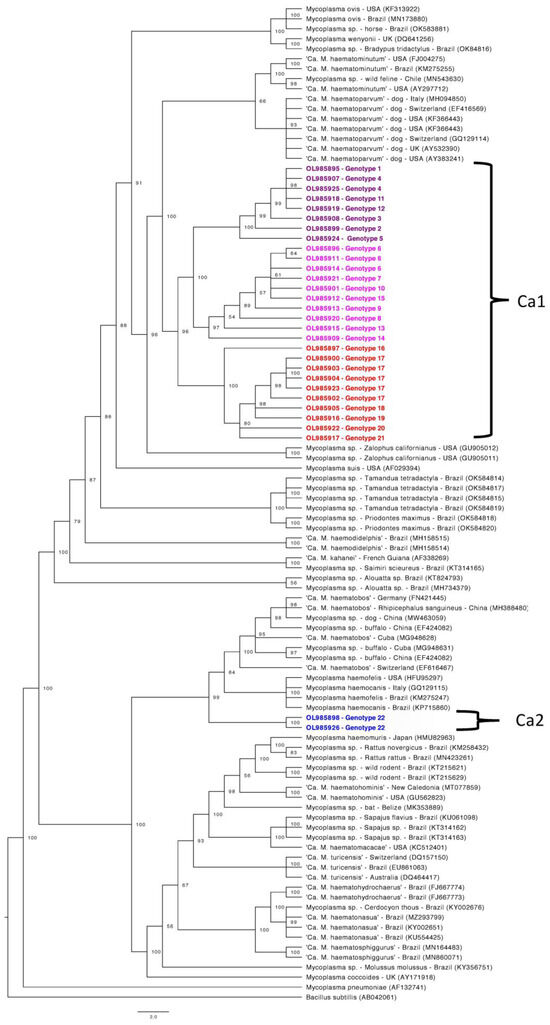
Figure 1.
Phylogenetic tree based on partial 16S rRNA gene fragments of Mycoplasma sp. Tree was constructed by Bayesian Inference and a sequence from Bacillus subtillis (AB042061) was used as outgroup. Sequences obtained in the present study (Ca1 and Ca2) are highlighted in colors: Ca1 are highlighted in purple, pink and red colors, differentiating each subclade formed. Ca2 is highlighted in blue.
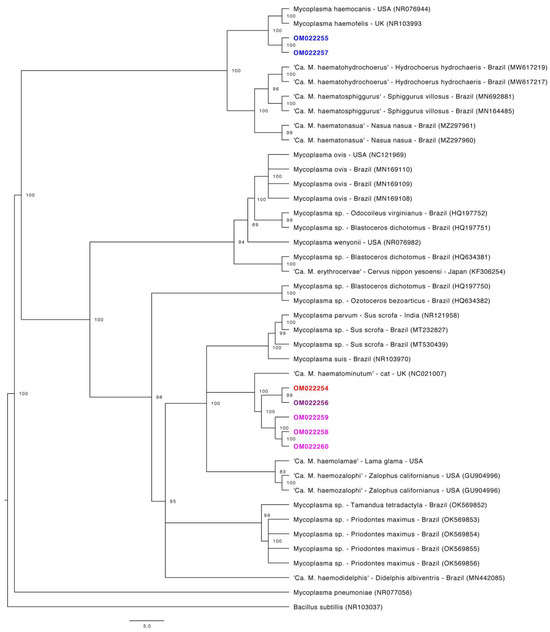
Figure 2.
Phylogenetic tree based on partial 23S rRNA gene fragments of Mycoplasma sp. Tree was constructed by Bayesian Inference and a sequence from Bacillus subtillis (NR103037) was used as outgroup. Sequences obtained in the present study are highlighted in colors: sequences that fit in Ca1 by the 16S rRNa phylogeny are highlighted in purple, pink and red colors. Sequences that fit in Ca2 by the 16S rRNA phylogeny are highlighted in blue.
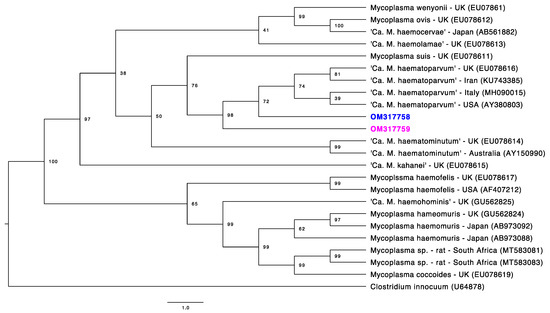
Figure 3.
Phylogenetic tree based on partial RNAse P gene fragments of Mycoplasma sp. Tree was constructed by Bayesian Inference and a sequence from Clostridium innocuum (U64878) was used as outgroup. Sequences obtained in the present study are highlighted in colors: the sequence that fit in Ca1 by the 16S rRNA phylogeny is highlighted in pink. The sequence that fit in Ca2 by the 16S rRNA phylogeny is highlighted in blue.
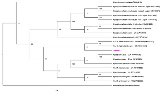
Figure 4.
Phylogenetic tree based on partial dnaK gene fragments of Mycoplasma sp. Tree was constructed by Bayesian Inference and a sequence from Klebisiella pneumonia (KJ690086) was used as outgroup. Sequences obtained in the present study is highlighted in pink, once it fits in Ca1 by the 16S rRNA phylogeny.
The best-fit model for this gene analysis was determined as F81+G. For the partial 23S rRNA gene analysis (Figure 2), an alignment with a total size of 720 bp was constructed using 41 homolog sequences and one outgroup (NR103037). The best-fit model for this gene analysis was determined as F81+G. For the partial RNAse P gene analysis (Figure 3), a total size alignment of 230 bp was constructed using 23 homolog sequences and one outgroup (U64878). The best-fit model for this gene analysis was determined as F81+I+G. For the partial dnaK gene analysis (Figure 4) a total size alignment of 622 bp was constructed using 19 homolog sequences and one outgroup (KJ690086). The best-fit model for this gene analysis was determined as F81+G.
Regarding the 16S rRNA phylogenetic analysis, our sequences were divided into two different major clades. The first clade was also sub-divided into three minor clades and positioned in the “Mycoplasma suis group”, whereas the second clade comprised of only three sequences and was distantly positioned in the “Mycoplasma haemofelis group”. These results suggest the occurrence of two distinct species occurring in sampled tapirs. In order to facilitate the understanding from now on these two clades will be identified as Ca1 and Ca2, respectively. Sequences that fitted in Ca1 on partial 16S rRNA-based phylogeny followed a similar pattern in other genes phylogenies, except for the RNAse P gene. In the phylogenetic tree based on the RNAse P gene, both obtained sequences were positioned in the “M. suis group”, albeit each one was positioned in a different group on 16S rRNA phylogeny. Moreover, one sample (ID: AA-P) that was positioned in Ca1 on 16S rRNA-based phylogeny was found in Ca2 in the phylogenetic inference based on the partial 23S rRNA.
3.4. Genetic Diversity Assessment
Regarding the 16S rRNA genotype analysis, 22 different genotypes were identified among 30 sequences (Figure 5). Values of nucleotide diversity (π), polymorphism level (haplotype diversity—[dh]), number of haplotypes (h), average number of nucleotide differences (K) among the sequences obtained, and the number of mutations between sequences from Ca1 and Ca2 are described in Table 6. 16S rRNA genotypes and sampling location of each obtained sequence are shown in Supplementary Table S1. The map representing genotype distributions along the sampling area reinforced the geographical distance between genotypes identified in Pantanal and Cerrado areas (Figure 6, Figure 7 and Figure 8).
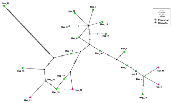
Figure 5.
Genotype diversity among 16S rRNA gene sequences detected herein. Analysis was made using DnaSP6. Inference and graphic representation were made by TCS network method on PopART software. Genotypes in blue were obtained from samples from tapirs in Pantanal regions meanwhile genotypes in green were obtained from samples from tapirs in Cerrado regions.

Table 6.
Values obtained regarding genotype diversity by DnaSP software and based on partial hemoplasma 16S rRNA sequences detected in tapirs from the present study.
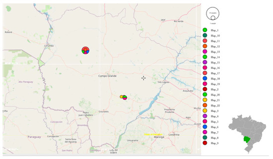
Figure 6.
Representation of the genotype distribution along all sampling areas. Map was constructed using PopART software based on GPS coordinates data of each sampling.
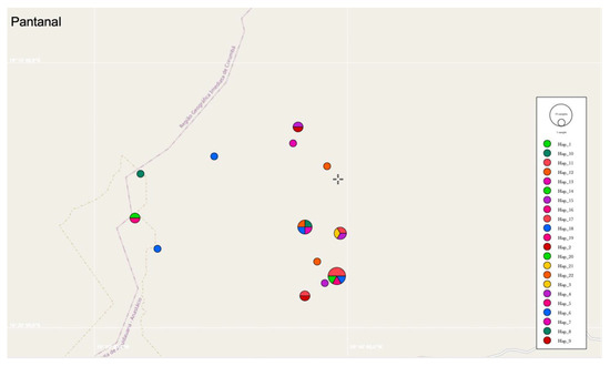
Figure 7.
Representation of the genotype distribution of Pantanal biome areas. Map was constructed using PopART software based on GPS coordinates data of each sampling.
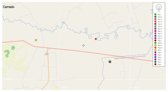
Figure 8.
Representation of the genotype distribution of Cerrado biome areas. Map was constructed using PopART software based on GPS coordinates data of each sampling.
3.5. Distance Analysis by SplitsTree
The distance analysis performed by the neighbor-joining method and displayed by SplitsTree v4.14.6 demonstrated that Ca1 and Ca2 sequences were distinctly disposed among other hemotropic Mycoplasma species and Candidatus (Figure 9).
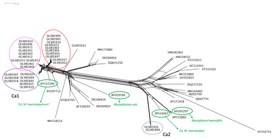
Figure 9.
Distance analysis of 16S rRNA fragments from different Mycoplasma species was made using SplitsTree v4.14.6 software. Sequences from Ca1 and Ca2 are indicated in the tree. Regarding Ca1 sequences, colors from the Splitstree graph match with the subclades highlighted in 16S rRNA phylogeny (purple, pink and red). The Ca2 sequences are highlighted in blue. The species M. suis and M. haemofelis are highlighted in green. The species ‘Ca. M. haematoparvum’ and ‘Ca. M. haematobos’ were also highlighted in green to demonstrate their position compared to Ca1 and Ca2.
3.6. Distance Matrix Analysis
The heat map with the distance matrix from all partial 16S rRNA sequences detected herein corroborated with other achieved results from the present study and demonstrated a marked difference between sequences from Ca1 and Ca2 (Figure 10). The minimum and maximum divergence percentage rates found for all the sequences that fit in Ca1 were 0.0% and 2.92%, respectively. When comparing to the phylogenetic closest sequences (FJ004275, KM275255, MN543630, AY297712, MH094850, EF416569, GQ129112, KF366443, GQ129114, AY532390, AY383241), the minimum and maximum divergence found between Ca1 members and those sequences was 1.52% and 3.27%, respectively. When comparing the Ca1 members and Ca2 members, the minimum and maximum divergence found was 15.23% and 16.22%, respectively. The minimum and maximum divergence percentage rates found for all members from Ca2 were 0.0%. When comparing to the phylogenetic closest sequences (FN421445, MG948628, MG948631, MH388480, MW463059, EF424082, EF616467, EF460765, HFU95297, GQ129115, KM275247, KP715860), the minimum and maximum divergence found between Ca2 members and those sequences was 5.12% and 7.23%, respectively. The distance matrix is available in Supplementary Table S2.

Figure 10.
A heatmap constructed using the distance matrix based on p-value between all sequences obtained herein. The scale on the right demonstrates the color shade transition between obtained values.
3.7. Statistical Analysis
Statistical analysis results including OR (odds ratio) and p-value are summarized in Table 7. Regarding the analysis between location and positivity for hemoplasmas, the number of positive tapirs sampled in Pantanal wetlands was statistically higher when compared to those sampled in the Cerrado region (p-value = 0.0001). The other analyzed variables (gender and age) did not present statistically significant differences for this outcome (p-value > 0.05). The OR value demonstrated that tapirs from Pantanal were 5.64 times more likely to present positive results for the PCR protocol targeting the partial Mycoplasma spp. 16S rRNA gene used in the present study when compared to tapirs sampled in Cerrado.

Table 7.
Statistical analysis comparing the occurrence of hemotropic Mycoplasma sp. in sampled tapirs and outcomes (gender, sampling location, and age).
4. Discussion
We described herein the occurrence of at least two genetically distinct hemoplasma species occurring in free-ranging tapirs. Reports of Candidatus and novel species of these bacteria in Brazilian wild fauna are becoming common due to the growing use of molecular and genetic analysis tools. Based mainly on the partial 16S rRNA and 23S rRNA genes amplification, novel species of hemotropic Mycoplasma spp. infecting capybaras [22], hairy dwarf porcupines [23], opossums [24], and coatis [25] have been proposed. When analyzing the topologies found by partial 16S rRNA gene phylogeny from these reports, sequences from each novel Candidatus species were positioned separately in clades inside the “Mycoplasma haemofelis group”, with satisfactory bootstrap values for species separation. In the present work, we detected different sequences from tapirs that fit in both the “Mycoplasma haemofelis group” (Ca2) and “Mycoplasma suis group” (Ca1) by partial 16S rRNA, 23S rRNA, and dnaK genes-based phylogeny, which was unprecedented for putative novel species of hemoplasmas reported in wild hosts from Brazil.
The 16S rRNA is considered the “gold standard” target gene for PCR assays aiming at detecting and identifying hemoplasmas and different pairs of primers have been used for its purpose [15]. Although this gene may be considered highly conserved, the 16S rRNA gene from bacterial species may exhibit considerable variations even on supposed conserved regions [53], which allow species differentiation. In the present study, all partial 16S rRNA sequences obtained for the Ca1 genotypes presented a similarity range of 97.20–98.20% with ‘Candidatus Mycoplasma haematoparvum’ detected in dogs from Italy and the USA (MH094850, AY383241) by BLASTn analysis. Meanwhile, the Ca2 genotypes demonstrated a similarity range of 94.99% with ‘Candidatus Mycoplasma haematobos’ detected in cattle from Cuba (MG948631). The p-distance values obtained by the distance matrix analysis showed that Ca1 members presented divergence rates of 0.0–2.92%, whereas the same comparison using the closest phylogenetic group and members of Ca1 presented rates of 1.52–3.27%. When using the 16S rRNA gene, a similarity rate of at least <97% is expected for bacteria belonging to different species. However, divergence rates <3% do not necessarily indicate that sequences belong to the same species [54]. Once phylogenetic analyses based on four molecular markers (16S rRNA, 23S rRNA, RNAse P, and dnaK) and 16S-based distance analyses strongly supported the differentiation between Ca1 and the closest hemoplasma species (‘Ca. M. haematoparvum’), it is likely that Ca1 represents a genetically distinct group of hemoplasmas.
The use of different genes for the molecular characterization of hemoplasmas is reported. Amplification of partial 23S rRNA gene was reported in phylogenetic studies of hemoplasmas identified in captive cervids from southern Brazil [55] and for the phylogenetic study of Mycoplasma ovis infecting sheep from the same region [18]. Recently, this gene has also been used for the characterization of novel Candidatus species of hemoplasmas from wild animals in Brazil [22,23,24,25]. In the present study, partial 23S rRNA sequences that fit in Ca1 presented similarity percentages ranging from 89.59–91.50% with a sequence of ‘Candidatus Mycoplasma haematominutum’ from the UK (HE613254). Meanwhile, 23S rRNA sequences that fit in Ca2 presented similarity ranges of 90.20–90.57% with a sequence of Mycoplasma haemofelis from the USA (NR103993). In fact, putative novel hemoplasma species may present lower similarity rates in BLASTn analysis for 23S rRNA sequences when compared to 16S rRNA sequences [22,55,56]. These differences may be explained by the fact that, although the 23S rRNA is considered as phylogenetically conserved as 16S rRNA, it presents a higher degree of sequence variability [57].
The RNAse P (rnpB) gene codifies the RNA subunit of endoribonuclease P with a length of approximately 400 bp [58]. Although only a few numbers of studies used phylogenetic trees of hemoplasmas using fragments of the RNAse P gene, our phylogeny analysis using this gene agreed with these studies, showing a separation of species among the “Mycoplasma haemofelis group” and “Mycoplasma suis/‘Ca. M. haematominutum’ group” [16,58]. However, both sequences obtained in the present study (OM317758-OM317759) were positioned in the “Mycoplasma haemofelis group”, albeit these same two samples were positioned separately in the “Mycoplasma haemofelis group” and “Mycoplasma suis group” in all other analyzed targeted genes. Although it is expected that phylogenies using RNAse P fragments of hemoplasmas may present similar topologies to those using 16S rRNA [58], the fragments obtained herein are very short (~100 bp), precluding robust species differentiation.
Amplification and subsequent phylogenetic analysis of the dnaK gene have been demonstrated to be a useful tool for the separation of hemoplasmas from other Mollicute species [59] and also for the separation of subspecies of hemoplasmas [60]. This gene is responsible for coding a chaperon protein (heat shock protein 70) and is considered a great genetic marker for species differentiation, since it may present more variable regions within the sequences when compared to ribosomal fragments [59]. Unfortunately, only one sequence from the present study was successfully sequenced using the dnaK (OM339521) PCR protocol, and comparison with other sequences obtained from tapirs was not possible. However, this single sequence followed a similar topology pattern of 16S rRNA and 23S rRNA phylogenies, since it fitted on Ca1 in the “Mycoplasma suis group”, reinforcing the phylogenetic position found for this putative novel hemoplasma species.
A high diversity of genotypes (n = 22) was found in the present work by analysis of 30 partial 16S rRNA sequences of hemoplasmas obtained from tapirs’ samples. Richness on genotype diversity was also reported for hemoplasmas detected in bats from Brazil [56]. Although the genotypes found in tapirs from the Pantanal diverged from those found in tapirs from the Cerrado, a genetic proximity between these genotypes was demonstrated on the genotype network analysis. The occurrence of more than one genotype in the same region infecting wild hosts has already been reported in bats from Brazil [56]. Interestingly, one animal that was sampled at three different times (IDs: WE-P-1, WE-P-2, and WE-P-3) presented three different genotypes according to the time of sampling. Moreover, one animal (ID: AA-P), that was sampled only once, was positioned in Ca1 on 16S rRNA-based phylogeny and in Ca2 in the phylogenetic inference based on the partial 23S rRNA. These data suggest that tapirs may be susceptible to co-infections with different hemoplasma genotypes or species, with these co-infections occurring simultaneously or at different times.
Some animals that were sampled more than once presented positive samples in more than one sampling (IDs: MA-P-1, MA-P-2, JE-P-1, JE-P-2, WEP-1, WEP-2, WE-P3). Meanwhile, some animals that also were sampled more than once presented positive results for one sample only (IDs: TD-P-1, VA-P-1, ANO-C-2, CNA-C-2, SO-C-2, FFO-P-2, JO-P-2, DO-P-2, TD-P-3, SAO-P-2). These results may suggest either that tapirs may be maintained as chronically infected hosts or that hemoplasma bacteremia may be too low, precluding molecular detection. Chronic infection by hemoplasmas has been commonly reported in domestic species, such as pigs [61], cattle [62], and cats [63,64]. Although it is not possible to extend these findings for wildlife, chronic infection by hemoplasmas is appointed as a cause of unthriftiness in newborn piglets [65] and lower calf birth weight in cattle [62] which may raise a red flag in the context of species conservation.
Herein, gender and age were not associated with hemoplasma infection in the sampled tapirs. Although tapirs from Cerrado presented more health abnormalities when compared to tapirs from other biomes [65], we found that animals sampled in the Pantanal were 5.64 times more prone to be infected by hemoplasmas when compared to tapirs sampled in the Cerrado. In cats, population density may be associated as a risk factor for hemoplasma infection [66]. Some Pantanal sites and habitats were already reported as able to sustain high densities of tapir populations when these animals are not exposed to anthropic actions [67].
Tapirs from both Cerrado and Pantanal regions were found parasitized by Amblyomma spp. and Rhipicephalus microplus ticks. Although information regarding the transmission of hemoplasmas by ticks and other vectors is still lacking, R. microplus ticks were appointed as capable of transmitting ‘Ca. M. haematobos’ for egg and larval stages and also to transmit this hemoplasma species for mice [68]. Future studies aiming at investigating the role of ticks in the transmission of hemoplasmas among tapirs are needed.
Mycoplasma haemofelis and ‘Ca. M. haematominutum’ DNA was detected in saliva and salivary glands of infected cats, suggesting that social interactions, such as aggression, may be related to hemoplasma infections [69]. Lowland tapirs were considered to be as solitary and show tolerance to other individuals not influenced by kinship [70]. More studies are necessary to elucidate the transmission routes for hemoplasmas among free-ranging lowland tapirs in Brazil.
The occurrence of anemia due to hemoplasma infection has been common in cats [15,19,71,72], pigs [61], cattle [73], sheep [74,75], and splenectomized dogs [76]. Regarding hemoplasma infection in wild animals, the occurrence of anemia has already been reported in reindeers (Rangifer tarandus) [77], non-human primates (Sapajus flavius) [17], and guignas (Leopardus guigna) [78]. Considering the occurrence of 35.29% (36/102; CI: 26.71–44.95%) infected individuals found in the present study, health assessment of infected animals may be useful to understand if hemoplasma-induced anemia is a threat for the largest land mammal species from Brazil.
Besides the assessment, for the first time, of the occurrence of hemotropic Mycoplasma among tapirs in two distinct Brazilian biomes, the present work showed high genetic diversity, and, at least, two genetically distinct hemoplasma species infecting these mammals. Accordingly, the obtained results reinforce the need for multi-locus and large-scale sequencing aiming at unraveling accurately the genetic diversity of hemoplasmas in wild animals.
5. Conclusions
At least two genetically distinct species of hemotropic Mycoplasma spp. occurs in free-ranging T. terrestris from the Pantanal and Cerrado regions in Brazil. The occurrence of hemoplasmas did not differ according to the gender or age of the sampled tapirs. Animals sampled in the Pantanal may be at a higher risk of becoming infected by hemoplasma when compared to those in the Cerrado. We propose that the two genetically divergent species found infecting tapirs from the present study represent putative novel Candidatus species and the names ‘Candidatus Mycoplasma haematoterrestris’ and ‘Candidatus Mycoplasma haematotapirus’ are proposed for the species found in Ca1 and Ca2, respectively.
Supplementary Materials
The following supporting information can be downloaded at: https://www.mdpi.com/2076-2607/10/3/614/s1, Table S1: Hemoplasma-16S rRNA genotypes found in tapirs sampled in the present study according to sampling location.; Table S2: Distance matrix for partial 16S rRNA.
Author Contributions
Conceptualization, A.C.B.M. and M.R.A.; methodology, A.C.B.M., E.P.M., A.d.C.C., A.C.C. and M.R.A.; formal analysis, A.C.B.M.; investigation, A.C.B.M. and M.R.A.; resources, E.P.M., M.R.A. and R.Z.M.; data curation A.C.B.M., E.P.M. and M.R.A.; writing—original draft preparation, A.C.B.M. and M.R.A.; writing—review and editing, E.P.M. and M.R.A.; visualization, M.R.A.; supervision, M.R.A.; project administration, M.R.A.; funding acquisition, E.P.M., M.R.A. and R.Z.M. All authors have read and agreed to the published version of the manuscript.
Funding
This work was supported by FAPESP (Foundation for Research Support of the State of São Paulo: #2018/02753-0; #2020/12037-0; #2019/26403-0) and CNPq (National Council for Scientific and Technological Development; Productivity Grant to MRA [CNPq Process #303701/2021-8]). LTCI’s financial support comes from national and international agencies, including zoological institutions, foundations, private businesses, and private individuals.
Institutional Review Board Statement
This study was approved by the Ethics Committee for Animal Experimentation of FCAV/UNESP (Faculty of Agricultural and Veterinary Sciences of the São Paulo State University) under protocol number 4558/20. The “Instituto Chico Mendes de Conservação da Biodiversidade (ICMBIO)” provided the required annual permits for the capture and immobilization of tapirs and collection of biological samples (SISBIO# 14,603).
Informed Consent Statement
Not applicable.
Data Availability Statement
Not applicable.
Acknowledgments
This study is part of Anna Mongruel’s PhD. She is carrying out her research at Programa de Pós-Graduação em Ciências Veterinárias—Universidade Estadual Júlio de Mesquita Filho (UNESP/Jaboticabal) and is sponsored by a fellowship from Foundation for Research Support of the State of São Paulo (FAPESP—Process number #2019/26403-0) at the the time of this study. Authors would like to thank the Lowland Tapir Conservation Initiative (LTCI)—Instituto de Pesquisas Ecológicas (IPÊ) for kindly providing the biological samples needed for the present study. The study of tapir health has been an important component of the long-term activities of the LTCI-IPÊ in Brazil. The LTCI has the institutional support from the International Union for Conservation of Nature (IUCN) Species Survival Commission (SSC) Tapir Specialist Group (TSG), Association of Zoos and Aquariums (AZA) Tapir Taxon Advisory Group (TAG), and European Association of Zoos and Aquariums (EAZA) Tapir Taxon Advisory Group (TAG).
Conflicts of Interest
The authors declare no conflict of interest.
References
- Medici, E.P.; Desbiez, A.L.J. Population Viability Analysis (PVA): Using a modeling tool to assess the viability of tapir populations in fragmented landscapes. Integr. Zool. 2012, 7, 356–372. [Google Scholar] [CrossRef]
- Medici, E.P.; Flesher, K.; Beisiegel, B.M.; Keuroghlian, A.; Desbiez, A.L.J.; Gatti, A.; Pontes, A.R.M.; Campos, C.B.; Tófoli, C.F.; Moraes, E.A., Jr.; et al. Tapirus terrestris (Linnaeus, 1758). In Livro Vermelho da Fauna Brasileira Ameaçada de Extinção, 1st ed.; Ministério do Meio Ambiente: Brasilia, Brazil, 2018; Volume II, pp. 59–68. [Google Scholar]
- Mangini, P.R.; Medici, E.P.; Fernandes-Santos, R.C. Tapir Health and Conservation Medicine. Integr. Zool. 2012, 7, 331–345. [Google Scholar] [CrossRef] [PubMed]
- Acosta, I.C.; da Costa, A.P.; Nunes, P.H.; Gondim, M.F.; Gatti, A.; Rossi, J.L., Jr.; Gennari, S.M.; Marcili, A. Morphological and molecular characterization and phylogenetic relation- ships of a new species of trypanosome in Tapirus terrestris (lowland tapir), Trypanosoma terrestris sp. nov., from Atlantic Rainforest of southeastern Brazil. Parasit. Vectors 2013, 6, 394. [Google Scholar] [CrossRef] [PubMed]
- Pérez, S.D.; Grummer, J.A.; Fernandes-Santos, R.C.; Testa-José, C.; Medici, E.P.; Marcili, A. Phylogenetics, patterns of genetic variation and population dynamics of Trypanosoma terrestris support both coevolution and ecological host-fitting as processes driving trypanosome evolution. Parasit. Vectors 2019, 12, 473–488. [Google Scholar] [CrossRef] [PubMed]
- Silveira, A.W.; Oliveira, G.G.; Santos, L.M.; Azuaga, L.B.S.; Coutinho, C.R.M.; Echeverria, J.T.; Antunes, T.R.; Ramos, C.A.N.; Souza, A.I. Natural infection of the South America Tapir (Tapirus terrestris) by Theileria equi. J. Wildl. Dis. 2017, 53, 411–413. [Google Scholar] [CrossRef] [PubMed]
- Gonçalves, T.; Barros, F.N.L.; Inoue, L.S.; Farias, D.M.; Lima, J.S.; Nobre, A.V.; Aidar, E.S.A.; Diniz, R.R.F.; Gering, A.P.; Scofield, A. Natural Theileria equi infection in captive Tapirus terrestris (Perissodactyla: Tapiridae) in the Brazilian Amazon. Ticks Tick-Borne Dis. 2020, 11, 101452. [Google Scholar] [CrossRef]
- Marcordes, S.; Lueders, I.; Grund, L.; Sliwa, A.; Maurer, F.P.; Hillemann, D.; Möbius, P.; Barth, S.A. Clinical outcome and diagnostic methods of atypical mycobacteriosis due to Mycobacterium avium ssp. hominissuis in a group of captive lowland tapirs (Tapirus terrestris). Transb. Emerg. Dis. 2021, 68, 1305–1313. [Google Scholar] [CrossRef] [PubMed]
- Medici, E.P.; Mangini, P.R.; Fernandes-Santos, R.C. Health assessment of wild lowland tapir populations in Brazil. J. Wildl. Dis. 2014, 50, 817–828. [Google Scholar] [CrossRef] [PubMed]
- Fernandes-Santos, R.C.; Fernandes, E.R.; Luiz, F.G.; Chaves, L.B.; Silva, S.R.; Medici, E.P. Rabies virus exposure in wild lowland tapirs (Tapirus terrestris) from three Brazilian biomes. J. Wildl. Dis. 2021, 57, 443–446. [Google Scholar] [CrossRef] [PubMed]
- Labruna, M.B.; Martins, T.F.; Acosta, I.; Serpa, M.; Soares, H.S.; Teixeira, R.; Fernandes-Santos, R.C.; Medici, E.P. Ticks and rickettsial exposure in lowland tapirs (Tapirus terrestris) of three Brazilian biomes. Ticks Tick-Borne Dis. 2021, 12, 101648. [Google Scholar] [CrossRef] [PubMed]
- Razin, S.; Yogev, D.; Naot, Y. Molecular Biology and Pathogenicity of Mycoplasmas. Microbiol. Mol. Biol. Rev. 1998, 62, 1094–1156. [Google Scholar] [CrossRef] [PubMed]
- Tratchenberg, S. Quick Guide: Mollicutes. Curr. Biol. 2005, 15, PR483–PR484. [Google Scholar] [CrossRef] [PubMed]
- Messick, J.B. Hemotrophic mycoplasmas (hemoplasmas): A review and new insights into pathogenic potential. Vet. Clin. Path. 2004, 33, 20–22. [Google Scholar] [CrossRef] [PubMed]
- Citti, C.; Baranowski, E.; Dordet-Frisoni, E.; Faucher, M.; Nouvel, L.X. Genomic Islands in Mycoplasmas. Genes 2020, 11, 836. [Google Scholar] [CrossRef] [PubMed]
- Maggi, R.G.; Chitwood, M.C.; Kennedy-Stoskopf, S.; Deperno, C.S. Novel hemotropic Mycoplasma species in white-tailed deer (Odocoileus virginianus). Comp. Immunol. Microbiol. Infect. Dis. 2013, 36, 607–611. [Google Scholar] [CrossRef] [PubMed]
- Ramalho, A.C.; Guerra, R.R.; Mongruel, A.C.B.; Vidotto, O.; Lucena, R.B.; Guerra, M.V.S.F.; Vieira, T.S.W.J.; Vieira, R.F.C. Mycoplasma sp. infection in captive Marcgrave’s capuchin monkeys (Sapajus flavius). Comp. Immunol. Microbiol. Infect. Dis. 2017, 5, 34–36. [Google Scholar] [CrossRef]
- Mongruel, A.C.B.; Spanhol, V.C.; Valente, J.D.M.; Porto, P.P.; Ogawa, L.; Otomura, F.H.; de Souza Marquez, E.; André, M.R.; Vieira, T.S.W.J.; da Costa Vieira, R.F.; et al. Survey of vector-borne and nematode parasites involved in the etiology of anemic syndrome in sheep from Southern Brazil. Rev. Bras. Parasitol. Vet. 2020, 29, e007320. [Google Scholar] [CrossRef] [PubMed]
- Nibblett, B.M.D.; Snead, E.C.; Waldner, C.; Taylor, S.M.; Jackson, M.L.; Knorr, L.M.A. Anemia in cats with hemotropic mycoplasma infection: Retrospective evaluation of 23 cases (1996–2005). Can. Vet. J. 2009, 50, 1181–1185. [Google Scholar] [PubMed]
- Tasker, S.; Peters, I.R.; Papasouliotis, K.; Cue, S.M.; Willi, B.; Hofmann-Lehmann, R.; Gruffydd-Jones, T.J.; Knowles, T.G.; Day, M.J.; Helps, C.R. Description of outcomes of experimental infection with feline haemoplasmas: Copy numbers, haematology, Coombs’ testing and blood glucose concentrations. Vet. Microbiol. 2009, 139, 323–332. [Google Scholar] [CrossRef]
- Neimark, H.; Hoff, B.; Ganter, M. Mycoplasma ovis comb. (formely Eperythrozoon ovis), an epierythrocytic agent of haemolytic anaemia in sheep and goats. Int. J. Syst. Evol. Microbiol. 2004, 54, 365–371. [Google Scholar] [CrossRef]
- Vieira, R.; Santos, N.; Valente, J.; Santos, L.P.; Lange, R.R.; Duque, J.; Ferrari, M.V.; Barros Filho, I.R.; Collere, F.; Ferrari, L.; et al. ‘Candidatus Mycoplasma haematohydrochoerus’, a novel hemoplasma species in capybaras (Hydrochoerus hydrochaeris) from Brazil. Infect. Genet. Evol. 2021, 93, 104988. [Google Scholar] [CrossRef] [PubMed]
- Valente, J.; Saldanha, A.; Martini, R.; Lange, R.R.; Baggio, R.A.; Martins, T.F.; Dos Santos, L.P.; de Sousa, R.S.; Vieira, T.; Vieira, R. ‘Candidatus Mycoplasma haemosphiggurus’ a novel haemoplasma species in orange-spined hairy dwarf porcupines (Sphiggurus villosus) from Southern Brazil. Transb. Emerg. Dis. 2021, 68, 1054–1061. [Google Scholar] [CrossRef]
- Pontarolo, G.H.; Kuhl, L.F.; Pedrassini, D.; Campos, M.; Figueiredo, F.B.; Valente, J.D.M.; Gonçalves, L.R.; André, M.R.; Vieira, T.S.W.J.; Vieira, F.R.C.; et al. ‘Candidatus Mycoplasma haemoalbiventris’, a novel hemoplasma species in white-eared opossums (Didelphis albiventris) from Brazil. Transb. Emerg. Dis. 2020, 68, 562–572. [Google Scholar] [CrossRef] [PubMed]
- Collere, F.; Delai, R.M.; Ferrari, L.; da Silva, L.H.; Fogaça, P.; Rodrigues, A.N.; Gonçalves, D.D.; Baggio, R.A.; Moraes, M.; Lux Hoppe, E.G.; et al. ‘Candidatus Mycoplasma haematonasua’ and tick-borne pathogens in ring-tailed coatis (Nasua nasua Linnaeus, 1976) from the Iguaçu National Park, Paraná State, southern Brazil. Transb. Emerg. Dis. 2021, 68, 3222–3229. [Google Scholar] [CrossRef]
- Bonato, L.; Figueiredo, M.A.P.; Gonçalves, L.R.; Machado, R.Z.; André, M.R. Occurrence and molecular characterization of Bartonella sp. and hemoplasmas in neotropical primates from Brazilian Amazon. Comp. Immunol. Microbiol. Infect. Dis. 2015, 42, 15–20. [Google Scholar] [CrossRef] [PubMed]
- Gonçalves, L.R.; Roque, A.L.; Matos, C.A.; Fernandes, S.J.; Olmos, I.D.; Machado, R.Z.; André, M.R. Diversity and molecular characterization of novel hemoplasmas infecting wild rodents from different Brazilian biomes. Comp. Immunol. Microbiol. Infect. Dis. 2015, 43, 50–56. [Google Scholar] [CrossRef] [PubMed]
- Ikeda, P.; Seki, M.C.; Carrasco, A.O.T.; Rudiak, L.V.; Miranda, J.M.D.; Gonçalves, S.M.M.; Hoppe, E.G.L.; Albuquerque, A.C.A.; Teixeira, M.M.G.; Passos, C.E.; et al. Evidence and molecular characterization of Bartonella sp. and hemoplasmas in neotropical bats in Brazil. Epidemiol. Infect. 2017, 145, 2038–2052. [Google Scholar] [CrossRef] [PubMed]
- Ikeda, P.; Menezes, T.R.; Torres, J.M.; de Oliveira, C.E.; Lourenço, E.C.; Herrera, H.M.; Machado, R.Z.; André, M.R. First molecular detection of piroplasmids in non-hematophagous bats from Brazil, with evidence of putative novel species. Parasitol. Res. 2021, 120, 301–310. [Google Scholar] [CrossRef] [PubMed]
- Delitti, W.B.C.; Meguro, M.; Pausas, J.G. Biomass and mineralmass estimates in “Cerrado” ecosystem. Braz. J. Bot. 2006, 4, 531–540. [Google Scholar] [CrossRef]
- Tomas, W.M.; de Oliveira Roque, F.; Morato, R.G.; Medici, P.E.; Chiaravalloti, R.M.; Tortato, F.R.; Penha, J.M.F.; Izzo, T.J.; Garcia, L.C.; Lourival, R.F.F.; et al. Sustainability Agenda for the Pantanal Wetland: Perspectives on a Collaborative Interface for Science, Policy, and Decision-Making. Trop. Conserv. Sci. 2019, 12, 1940082919872634. [Google Scholar] [CrossRef]
- Medici, E.P. Family Tapiridae (Tapirs). In Handbook of the Mammals of the World, 1st ed.; Wilson, D.E., Mittermeier, R.A., Eds.; Lynx Edicions: Barcelon, Spain, 2011; Volume 2, p. 886. [Google Scholar]
- LASA/UFRJ-Laboratório de Aplicações de Satélites Ambientais do Departamento de Meteorologia da Universidade Federal do Rio de Janeiro (UFRJ). Área Queimada–Pantanal 2020. Available online: https://lasa.ufrj.br/noticias/area-queimada-pantanal-2020/ (accessed on 29 December 2021).
- PRODES/INPE–Nota Técnica PRODES Cerrado 2021. Available online: https://www.gov.br/inpe/pt-br/assuntos/ultimas-noticias/nota-tecnica-prodes-cerrado-2021 (accessed on 29 December 2021).
- Berlinck, C.N.; Lima, L.H.A.; Pereira, A.M.M.; Carvalho, E.A.R., Jr.; Paula, R.C.; Thomas, W.M.; Morato, R.G. The Pantanal is on fire and only a sustainable agenda can save the largest wetland in the world. Braz. J. Biol. 2022, 82, e244200. [Google Scholar] [CrossRef]
- Birkenheuer, A.J.; Levy, M.G.; Breitschewerdt, E.B. Development and evaluation of a seminested PCR for detection and differentiation of Babesia gibsoni (Asian genotype) and B. canis DNA in canine blood samples. J. Clin. Microbiol. 2003, 41, 4172–4177. [Google Scholar] [CrossRef] [PubMed]
- Ferreira, E.C.; Contijo, C.M.; Cruz, I.; Melo, M.N.; Silva, A.M. Alternative PCR protocol using a single primer set for assessing DNA quality in several tissues from a large variety of mammalian species living in areas endemic for leishmaniasis. Mem. Inst. Oswaldo Cruz 2010, 105, 895–898. [Google Scholar] [CrossRef] [PubMed]
- Harasawa, R.; Orusa, R.; Giangaspero, M. Molecular evidence for hemotropic Mycoplasma infection in a Japanese badger (Meles meles anakuma) and a raccoon dog (Nyctereutes procyonoides viverrinus). J. Wildl. Dis. 2014, 50, 412–415. [Google Scholar] [CrossRef] [PubMed]
- Di Cataldo, S.; Kamani, J.; Cevidanes, A.; Msheliza, E.G.; Millán, J. Hemotropic mycoplasmas in bats captured near human settlements in Nigeria. Comp. Immunol. Microbiol. Infect. Dis. 2020, 70, 101448. [Google Scholar] [CrossRef]
- Descloux, E.; Mediannikov, O.; Gourinat, A.C.; Colot, J.; Chauvet, M.; Mermoud, I.; Desoutter, D.; Cazorla, C.; Klement-Frutos, E.; Antonini, L.; et al. Flying Fox Hemolytic Fever, Description of a New Zoonosis Caused by Candidatus Mycoplasma haemohominis. Infect. Dis. Soc. Am. 2021, 73, e1445–e1453. [Google Scholar] [CrossRef]
- Sanger, F.; Nicklen, S.; Coulson, A.R. DNA sequencing with chain-terminating inhibitors. Proc. Natl. Acad. Sci. USA 1977, 74, 5463–5467. [Google Scholar] [CrossRef]
- Hall, T.A. BioEdit: A User-Friendly Biological Sequence Alignment Editor and Analysis Program for Windows 95/98/NT. Nucl. Acid. Symp. Ser. 1999, 41, 95–98. [Google Scholar] [CrossRef]
- Altschul, S.F.; Gish, W.; Miller, W.; Myers, E.W.; Lipman, D.J. Basic local alignment search tool. J. Mol. Biol. 1999, 215, 403–410. [Google Scholar] [CrossRef]
- Benson, D.A.; Cavanaugh, M.; Clark, K.; Karsch-Mizrachi, I.; Ostell, J.; Pruitt, K.D.; Sayers, E.W. GenBank. Nucleic Acids Res. 2018, 46, D41–D47. [Google Scholar] [CrossRef] [PubMed]
- Thompson, J.D.; Higgins, D.G.; Gibson, T.J. CLUSTAL W: Improving the sensitivity of progressive multiple sequence alignment through sequence weighting, position-specific gap penalties and weight matrix choice. Nucl. Acids Res. 1994, 22, 4673–4680. [Google Scholar] [CrossRef] [PubMed]
- Sela, I.; Ashkenazy, H.; Katoh, K.; Pupko, T. GUIDANCE2: Accurate detection of unreliable alignment regions accounting for the uncertainty of multiple parameters. Nucl. Acids Res. 2015, 43, W7–W14. [Google Scholar] [CrossRef]
- Darriba, D.; Taboada, G.L.; Doallo, R.; Posada, D. jModelTest 2: More models, new heuristics and parallel computing. Nat. Methods 2012, 9, 772. [Google Scholar] [CrossRef] [PubMed]
- Librado, P.; Rozas, J. DnaSP v5: A software for comprehensive analysis of DNA polymorphism data. Bioinformatics 2009, 25, 1451–1452. [Google Scholar] [CrossRef] [PubMed]
- Clement, M.; Posada, D.; Crandall, K.A. TCS: A computer program to estimate gene genealogies. Mol. Ecol. 2000, 9, 1657–1659. [Google Scholar] [CrossRef] [PubMed]
- Huson, D.H.; Bryant, D. Application of phylogenetic networks in evolutionary studies. Mol. Biol. Evol. 2006, 23, 254–267. [Google Scholar] [CrossRef] [PubMed]
- Kumar, S.; Stecher, G.; Li, M.; Knyaz, C.; Tamura, K. MEGA X: Molecular Evolutionary Genetics Analysis across computing platforms. Mol. Biol. Evol. 2018, 35, 1547–1549. [Google Scholar] [CrossRef] [PubMed]
- Stecher, G.; Tamura, K.; Kumar, S. Molecular Evolutionary Genetics Analysis (MEGA) for macOS. Mol. Biol. Evol. 2020, 37, 1237–1239. [Google Scholar] [CrossRef]
- Martinez-Porchas, M.; Villalpando-Canchola, E.; Ortiz Suarez, L.E.; Vargas-Albores, F. How conserved are the conserved 16S-rRNA regions? PeerJ 2017, 5, e3036. [Google Scholar] [CrossRef] [PubMed]
- Drancourt, M.; Raoult, D. Sequence-based identification of new bacteria: A proposition for creation of an orphan bacterium repository. J. Clin. Microbiol. 2005, 43, 4311–4315. [Google Scholar] [CrossRef] [PubMed]
- Grazziotin, A.L.; Santos, A.P.; Guimaraes, A.M.; Mohamed, A.; Cubas, Z.S.; de Oliveira, M.J.; dos Santos, L.C.; de Moraes, W.; Vieira, R.F.; Donatti, L.; et al. Mycoplasma ovis in captive cervids: Prevalence, molecular characterization and phylogeny. Vet. Microbiol. 2011, 152, 415–4191. [Google Scholar] [CrossRef]
- Ikeda, P.; Torres, J.M.; Lourenço, E.C.; Albery, G.F.; Herrera, H.M.; de Oliveira, C.E.; Machado, R.Z.; André, M.R. Molecular detection and genotype diversity of hemoplasmas in non-hematophagous bats and associated ectoparasites sampled in peri-urban areas from Brazil. Acta Trop. 2022, 225, 106203. [Google Scholar] [CrossRef]
- Ludwig, W.; Kirchhof, G.; Klugbauer, N.; Weizenegger, M.; Betzel, D.; Ehrmann, M.; Hertel, C.; Jilg, S.; Tatzel, R.; Zitzelsberger, H.; et al. Complete 23S Ribosomal RNA Sequences of Gram-positive Bacteria with a Low DNA G+C Content. Syst. Appl. Microbiol. 1992, 15, 487–501. [Google Scholar] [CrossRef]
- Peters, I.R.; Helps, C.R.; McAuliffe, L.; Neimark, H.; Lappin, M.R.; Gruffydd-Jones, T.J.; Day, M.J.; Hoelzle, L.E.; Willi, B.; Meli, M.; et al. RNase P RNA gene (rnpB) phylogeny of Hemoplasmas and other Mycoplasma species. J. Clin. Microbiol. 2008, 46, 1873–1877. [Google Scholar] [CrossRef] [PubMed]
- Hicks, C.A.; Barker, E.N.; Brady, C.; Stokes, C.R.; Helps, C.R.; Tasker, S. Non-ribosomal phylogenetic exploration of Mollicutes species: New insights into haemoplasma taxonomy. Infect. Genet. Evol. 2014, 23, 99–105. [Google Scholar] [CrossRef] [PubMed]
- Harasawa, R.; Fujita, H.; Kadosaka, T.; Ando, S.; Rikihisa, Y. Proposal for ‘Candidatus Mycoplasma haemomuris subsp. Musculi’ in mice, and ‘Candidatus Mycoplasma haemomuris subsp. Ratti’ in rats. Intern. J. Syst. Evol. Microbiol. 2015, 65, 734–737. [Google Scholar] [CrossRef]
- Hoelzle, L.E. Haemotrophic mycoplasmas: Recent advances in Mycoplasma suis. Vet. Microbiol. 2008, 130, 215–226. [Google Scholar] [CrossRef]
- Tagawa, M.; Yamakawa, K.; Aoki, T.; Matsumoto, K.; Ishii, M.; Inokuma, H. Effect of chronic hemoplasma infection on cattle productivity. J. Vet. Med. Sci. 2013, 75, 1271–1275. [Google Scholar] [CrossRef]
- Novacco, M.; Boretti, F.S.; Wolf-Jäckel, G.A.; Riond, B.; Meli, M.L.; Willi, B.; Lutz, H.; Hofmann-Lehmann, R. Chronic “Candidatus Mycoplasma turicensis” infection. Vet. Res. 2011, 42, 59. [Google Scholar] [CrossRef]
- Barker, E.N. Update on Feline Hemoplasmosis. Veterinary Clinics of North America. Small Anim. Pract. 2019, 49, 733–743. [Google Scholar] [CrossRef] [PubMed]
- Fernandes-Santos, R.C.; Medici, E.P.; José, C.T.; Micheletti, T. Health assessment of wild lowland tapirs (Tapirus terrestris) in the highly threatened Cerrado biome, Brazil. J. Wildl. Dis. 2020, 56, 34–46. [Google Scholar] [CrossRef]
- Bergmann, M.; Englert, T.; Stuetzer, B.; Hawley, J.R.; Lappin, M.R.; Hartmann, K. Risk factors of different hemoplasma species infections in cats. BMC Vet. Res. 2017, 13, 52. [Google Scholar] [CrossRef] [PubMed]
- Trolle, M.; Noss, A.J.; Cordeiro, J.L.P.; Oliveira, J.F.B. Brazilian Tapir Density in the Pantanal: A Comparison of Systematic Camera-Trapping and Line-Transect Surveys. Biotropica 2008, 40, 211–217. [Google Scholar] [CrossRef]
- Shi, H.; Duan, L.; Liu, F.; Hu, Y.; Shi, Z.; Chen, X.; Yang, H.; Yan, B.; Yao, L. Rhipicephalus (Boophilus) microplus ticks as reservoir and vector of ‘Candidatus Mycoplasma haemobos’ in China. Vet. Parasitol. 2019, 274, 108929. [Google Scholar] [CrossRef] [PubMed]
- Dean, R.S.; Helps, C.R.; Gruffydd Jones, T.J.; Tasker, S. Use of real-time PCR to detect Mycoplasma haemofelis and ‘Candidatus Mycoplasma haemominutum’ in the saliva and salivary glands of haemoplasma-infected cats. J. Feline Med. Surg. 2008, 10, 413–417. [Google Scholar] [CrossRef] [PubMed]
- Pinho, G.M.; Gonçalves da Silva, A.; Hrbek, T.; Venticinque, E.M.; Farias, I.P. Kinship and social behavior of lowland tapirs (Tapirus terrestris) in a central Amazon landscape. PLoS ONE 2014, 9, e92507. [Google Scholar] [CrossRef] [PubMed]
- Santos, A.P.; Conrado, F.; Messick, J.B.; Biondo, A.W.; Oliveira, S.T.; Guimaraes, A.M.; Nascimento, N.C.; Pedralli, V.; Lasta, C.S.; González, F.H. Hemoplasma prevalence and hematological abnormalities associated with infection in three different cat populations from Southern Brazil. Braz. J. Vet. Parasitol. 2014, 23, 428–434. [Google Scholar] [CrossRef] [PubMed]
- Firmino, F.P.; Aquino, L.C.; Marçola, T.G.; Bittencourt, M.V.; McManus, C.M.; Paludo, G.R. Frequency and hematological alterations of different hemoplasma infections with retrovirusis co-infections in domestic cats from Brazil. Small Anim. Dis. 2016, 36, 731–736. [Google Scholar] [CrossRef]
- Genova, S.G.; Streeter, R.N.; Velguth, K.E.; Snider, T.A.; Kocan, K.M.; Simpson, K.M. Severe anemia associated with Mycoplasma wenyonii infection in a mature cow. Can. Vet. J. 2011, 52, 1018–1021. [Google Scholar]
- Tagawa, M.; Takeuchi, T.; Fujisawa, T.; Konno, Y.; Yamamoto, S.; Matsumoto, K.; Yokoyama, N.; Inokuma, H. A clinical case of severe anemia in a sheep coinfected with Mycoplasma ovis and ‘Candidatus Mycoplasma haemovis’ in Hokkaido, Japan. J. Vet. Med. Sci. 2012, 74, 99–102. [Google Scholar] [CrossRef]
- Martínez-Hernández, J.M.; Ballados-González, G.G.; Fernández-Bandala, D.; Martínez-Soto, S.; Velázquez-Osorio, V.; Martínez-Rodríguez, P.B.; Cruz-Romero, A.; Grostieta, E.; Lozano-Sardaneta, Y.; Colunga Salas, P.; et al. Molecular detection of Mycoplasma ovis in an outbreak of hemolytic anemia in sheep from Veracruz, Mexico. Trop. Anim. Health Prod. 2019, 51, 243–248. [Google Scholar] [CrossRef] [PubMed]
- Kemming, G.; Messick, J.B.; Mueller, W.; Enders, G.; Meisner, F.; Muenzing, S.; Kisch-Wedel, H.; Schropp, A.; Wojtczyk, C.; Packert, K.; et al. Can we continue research in splenectomized dogs? Mycoplasma haemocanis: Old problem-new insight. Eur. Surg. Res. 2004, 36, 198–205. [Google Scholar] [CrossRef]
- Stoffregen, W.C.; Alt, D.P.; Palmer, M.V.; Olsen, S.C.; Waters, W.R.; Stasko, J.A. Identification of a haemomycoplasma species in anemic reindeer (Rangifer tarandus). J. Wildl. Dis. 2006, 42, 249–258. [Google Scholar] [CrossRef] [PubMed]
- Sacristán, I.; Acuña, F.; Aguilar, E.; García, S.; López, M.J.; Cevinades, A.; Cabello, J.; Hidalgo-Hermoso, E.; Johnson, W.E.; Poulin, E.; et al. Assessing cross-species transmission of hemoplasmas at the wild-domestic felid interface in Chile using genetic and landscape variables analysis. Sci. Rep. 2019, 9, 16819. [Google Scholar] [CrossRef]
Publisher’s Note: MDPI stays neutral with regard to jurisdictional claims in published maps and institutional affiliations. |
© 2022 by the authors. Licensee MDPI, Basel, Switzerland. This article is an open access article distributed under the terms and conditions of the Creative Commons Attribution (CC BY) license (https://creativecommons.org/licenses/by/4.0/).