Article

Discrimination between the Two Closely Related Species of the Operational Group B. amyloliquefaciens Based on Whole-Cell Fatty Acid Profiling

1
Department of Microbiology, Faculty of Science and Informatics, University of Szeged, Közép Fasor 52, H-6726 Szeged, Hungary
2
Department of Biotechnology, Faculty of Chemical Engineering, Ho Chi Minh University of Technology (HCMUT), 268 Ly Thuong Kiet Street, District 10, Ho Chi Minh City 72607, Vietnam
3
Vietnam National University Ho Chi Minh City, Linh Trung Ward, Thu Duc District, Ho Chi Minh City 71351, Vietnam
4
Functional Genomics and Bioinformatics Group, Research Center for Forestry and Wood Industry, University of Sopron, Bajcsy-Zsilinszky Str. 4, H-9400 Sopron, Hungary
5
Institute of Biophysics, Biological Research Centre, Eötvös Loránd Research Network, Temesvári Krt. 62, H-6726 Szeged, Hungary
*
Author to whom correspondence should be addressed.
Microorganisms 2022, 10(2), 418; https://doi.org/10.3390/microorganisms10020418
Submission received: 31 December 2021 / Revised: 3 February 2022 / Accepted: 8 February 2022 / Published: 11 February 2022
(This article belongs to the Section Microbial Biotechnology)

Abstract

:
(1) Background: Bacillus velezensis and Bacillus amyloliquefaciens are closely related members of the “operational group B. amyloliquefaciens”, a taxonomical unit above species level within the ”Bacillus subtilis species complex”. They have similar morphological, physiological, biochemical, phenotypic, and phylogenetic characteristics. Thus, separating these two taxa from each another has proven to be difficult to implement and could not be pushed easily into the line of routine analyses. (2) Methods: The aim of this study was to determine whether whole FAME profiling could be used to distinguish between these two species, using both type strains and environmental isolates. Initially, the classification was determined by partial sequences of the gyrA and rpoB genes and the classified isolates and type strains were considered as samples to develop the identification method, based on FAME profiles. (3) Results: The dissimilarities in 16:0, 17:0 iso, and 17:0 FA components have drawn a distinction between the two species and minor differences in FA 14:0, 15:0 iso, and 16:0 iso were also visible. The statistical analysis of the FA profiles confirmed that the two taxa can be distinguished into two separate groups, where the isolates are identified without misreading. (4) Conclusions: Our study proposes that the developed easy and fast-automated identification tool based on cellular FA profiles can be routinely applied to distinguish B. velezensis and B. amyloliquefaciens.

1. Introduction

Bacillus velezensis and B. amyloliquefaciens, together with the B. siamensis and a black-pigment-producing strain, B. nakamurai, are members of the operational group B. amyloliquefaciens. This operational group belongs to the B. subtilis species complex with an eventful taxonomic history [1]. Both taxa are beneficial species, they have played increasingly important roles in applied microbiology [2,3,4,5]. Furthermore, several plant growth-promoting and biocontrol products from B. amyloliquefaciens and B. velezensis are now commercially available, including RhizoVital® (B. velezensis DSM 23117T; ABiTEP, GmbH, Berlin, Germany), Amylo-X® WG (B. amyloliquefaciens subsp. plantarum D747; Certis Europe BV, Utrecht, The Netherlands), RhizoPlus® (B. amyloliquefaciens FZB24; ABiTEP), and Taegro® (B. subtilis var. amyloliquefaciens FZB24; Novozymes Biologicals, Inc., Salem, VA, USA) [6].
B. amyloliquefaciens was first described by Fukumoto [7,8] and later revised by Priest et al. [7,8] as an industrial producer of amylase, while B. velezensis, first isolated from the river Vélez in Málaga (southern Spain), was initially described as a distinct ecotype of B. amyloliquefaciens by Ruiz-García et al. [9]. Morphological, physiological, chemotaxonomic, and phylogenetic interrelations have indicated that the two taxa are highly similar [2,7,10,11]. Furthermore, detailed examinations of the members of these two species, including phenotype analysis, phylogenetics, fatty acid methyl ester (FAME) analysis, DNA–DNA hybridization, microarray-based comparative genomic hybridization, genomic analysis, HPLC-electrospray ionization MS, and MALDI-TOF MS have revealed B. velezensis as plant-associated B. amyloliquefaciens subsp. plantarum subsp. nov., and B. amyloliquefaciens as non-plant-associated B. amyloliquefaciens subsp. amyloliquefaciens subsp. nov., respectively [12]. Subsequently, B. methylotrophicus, B. amyloliquefaciens subsp. plantarum, and B. oryzicola were reclassified later as heterotypic synonyms of B. velezensis, while B. amyloliquefaciens subsp. amyloliquefaciens was considered as B. amyloliquefaciens [13].
The earliest description differentiated B. velezensis and B. amyloliquefaciens, as well as other closely related taxa based on phenotypic and genetic differences [9], but in many cases, these taxonomical descriptions were later revised according to the current state of Bacillus taxonomy. A typical example for this is the identification history of strain DSM 23117T, which was first identified as B. amyloliquefaciens in 2008 [14], later revised as B. amyloliquefaciens subsp. plantarum in 2011 [12], and finally reclassified as B. velezensis in 2016 based on DNA–DNA hybridization, as well as phenotypic and phylogenetic analyses [13]. Furthermore, this statement was strongly confirmed for this strain using molecular methods [1]. Therefore, this well-defined strain was used as the type strain of B. velezensis in our study, although it is still named as B. amyloliquefaciens in several recent publications and GenBank sequences [15,16].
Over the last 15 years, interest in understanding the genetic relationship of the two taxa has led to many studies being published [1,3,10,11,12,13,14,17,18,19,20]. The two taxa share similar morphological, physiological, and phenotypic traits (Table 1) as well as 16S rRNA gene sequences, tetranucleotide frequency distribution, and DNA G+C contents [1,13]. Their average nucleotide identity and average amino acid identity is approximately 93.6–94.5% and 97.8% similarity, respectively [1,17], and their high DNA–DNA relatedness values showed 20 [9], 55 [1,13,17], or 80% similarity [14] in various studies. Strains B. velezensis DSM 23117T and B. amyloliquefaciens DSM 7T share 3345 genes in their core genomes, which have 97.89% similarity at the amino acid level [12]. Furthermore, the phylogenomic tree based on the core genome (799 genes) also indicates their close genetic relationship [13]. Both taxa are characterized by substantial production of secondary metabolites via non-ribosomal synthesis. However, only B. velezensis contains gene clusters synthesizing macrolactin and difficidin, which are lacking in B. amyloliquefaciens [1]. On the other hand, only B. amyloliquefaciens contains the amyA gene for industrial starch-liquefying α-amylase [12].
Previously, the whole-cell FAME profiles of B. velezensis and B. amyloliquefaciens have been studied only to a limited extent, therefore FAs were not considered as biomarkers distinguishing between them [2,10,11]. FAs are part of the bacterial cell membrane structure, and specific FAs and their ratios in cellular membranes have usually been revealed as biomarkers to distinguish closely related species of bacteria [2,4]. This study considered the possibility of using the cellular FAs with the application of a Sherlock chromatographic analysis system (CAS) as a taxonomic and diagnostic tool. The method, using FAs of 9–20 carbons in length and automated GC analysis, qualitatively and quantitatively analyzes bacterial whole-cell FAME [21]. Since the Sherlock CAS has developed, it has become capable of performing cost-effective, sensitive, reliable, and rapid analyses with a small amount of cell mass.
This study included DNA sequencing, as well as phylogenetic and FAME analyses with the aim of providing a complementary tool to distinguish B. velezensis and B. amyloliquefaciens based on their cellular FAs.

2. Materials and Methods

2.1. Bacillus Strains and Growth Conditions

Bacillus type strains including DSM 7T, DSM 1061T, and DSM 23117T were obtained from the DSMZ (German Collection of Microorganisms and Cell Cultures, Braunschweig, Germany), while the Bacillus field strains were isolated according to Vágvölgyi et al. [22]. Briefly, soil samples (5 g) were collected from agricultural fields and suspended in 50 mL of 1% NaCl solution with intensive mixing with a glass rod, then the suspensions were allowed to pellet for a minute. The supernatants were used to make a dilution series. Fifty µL of each diluted sample was spread onto the surface of yeast extract–glucose (YEG) medium (glucose 0.2%, yeast extract 0.2%, bacto agar 2%) supplemented with 50 μg mL−1 nystatin to suppress fungi. After 7 days of incubation, the dominant bacterial colony morphotypes were picked and cleaned until homogeneity on YEG medium. The isolated strains were deposited in the Szeged Microbiology Collection (SZMC, http://szmc.hu, accessed on 29 December 2021) of the Department of Microbiology, University of Szeged, Hungary.
For molecular taxonomical investigations, Bacillus strains were cultured in YEG medium and incubated at 37 °C overnight. Before FA profiling, bacteria were inoculated on trypticase soy broth agar (TSBA, Becton, Dickinson and Company, Sparks, NV, USA) with the quadrant streaking method and incubated at 28 °C for 24 ± 2 h.

2.2. PCR Amplification of the gyrA and rpoB Genes

Total cellular DNA was extracted by the E.Z.N.A.® Bacterial DNA Kit (Omega Bio-tek, Inc., Norcross, GA, USA) according to the manufacturer’s instructions. Amplification of the gyrA gene [22] was performed in 50 µL reaction mixtures containing 10 pmol of each primer (gyrAF: CAGTCAGGAAATGCGTACGTCCTT; gyrAR: CAAGGTAATGCTCCAGGCATTGCT), 10 nmol dNTP mix, 2 µL template DNA, 5 µL 10× PCR buffer, 6 µL of 25 mM MgCl2, and 1 U of Taq DNA polymerase (Thermo Fisher Scientific, Waltham, MA, USA). The PCR thermocycler (Doppio, VWR International GmbH, Darmstadt, Germany) was set to an initial denaturation step at 94 °C for 2 min, 30 cycles of denaturation at 94 °C for 30 s, annealing at 53 °C for 45 s and extension at 72 °C for 60 s, and a final extension at 72 °C for 5 min. The amplification of the rpoB gene [23] was conducted in 50 µL reaction mixtures containing 20 pmol of each primer (rpoBF: AGGTCAACTAGTTCAGTATGGACG; rpoBRO: GTCCTACATTGGCAAGATCGTATC), 10 nmol dNTP mix, 2 µL template DNA, 5 µL 10× PCR buffer, 6 µL of 25 mM MgCl2, and 0.4 U of Taq DNA polymerase. The PCR cycling parameters included an initial denaturation step at 94 °C for 2 min, 30 cycles of denaturation at 94 °C for 30 s, annealing at 57 °C for 30 s, extension at 72 °C for 50 s, and a final extension at 72 °C for 5 min. Sequencing of the amplified DNA fragments was performed on an ABI 3730XL sequencer (Thermo Fisher Scientific, Waltham, MA, USA) using Sanger sequencing.

2.3. Phylogenetic Analysis

Sequences were analyzed using the Mega X software [24]. The NCBI Nucleotide BLAST similarity search was carried out at https://blast.ncbi.nlm.nih.gov/Blast.cgi, accessed on 29 December 2021. Alignments were performed using the MUSCLE algorithm. The phylogenetic tree was inferred using the Neighbor-Joining method [24] with 1000 bootstrap replicates. The evolutionary distances were computed using the Tamura-Nei method and the units of the number of base substitutions per site [25]. The rate variation among sites was modeled with a gamma distribution (shape parameter = 0.5).

2.4. The Fatty Acid Methyl Ester (FAME) Analysis

The MIDI Sherlock® Microbial Identification System (MIS, MIDI Inc., Newark, NJ, USA) was applied for the data acquisition [21]. The composition of whole-cell FAs was determined by the Sherlock CAS Software ver. 6.4 (Microbial ID Inc., Newark, DE, USA) operating through the LabSolution ver. 5.97 software on a Nexera GC-2030 gas chromatograph equipped with an AOC-20i Plus autoinjector (Shimadzu, Kyoto, Japan). For the separation of the FAs, the RTSBA6 method provided by the manufacturer was applied on a HP-Ultra 2, 25 m × 0.2 mm × 0.33 µm film thickness, fused, silica capillary column (Agilent, Santa Clara, CA, USA). Injector and detector temperatures were 250 °C and 300 °C, respectively. Carrier gas was hydrogen at a flow rate of 1.48 mL min−1, while the detector gases were nitrogen (make up), oxygen and hydrogen with the follow flows of 30, 30, and 350 mL min−1, respectively. Samples were introduced in an injection volume of 2 µL in split mode with a 40:1 split ratio. The oven program started at 168.1 °C, which ramped up to 291 °C with 28 °C min−1, and then up to 300 °C with 60 °C min−1, holding at this temperature for 1.50 min. The total column oven program time was 6.04 min. The 1300-C rapid calibration standard mix (Microbial ID Inc., Newark, DE, USA) was used for retention time calibration and system suitability purposes. The B. subtilis strain ATCC 6633T and pure hexane were considered as the quality control and the negative control, respectively. Whole-cell FAME profiles were analyzed by the library RTSBA6.21 (Microbial ID Inc., Newark, DE, USA).

2.5. Sample Pretreatment

Sample processing was carried out according to the SherlockTM Operating CAS Manual [21]. Briefly, 20–40 mg of cells was harvested and placed in a clean glass tube. Then, 1 mL of reagent 1 (45 g NaOH, 150 mL of methanol and 150 mL of distilled water) was added to the sample and heated at 95–100 °C in a water bath (Precision water bath NB-301, HandyLAB®System, N-BIOTEK, Bucheon-si, Korea). After 5 min, the sample was removed from the water bath, vortexed and heated for an additional 25 min. The sample was mixed with 2 mL of reagent 2 (325 mL of 6.0 N HCl, 275 mL of methanol) and incubated at 80 °C in a water bath for 10 min. Subsequently, 1.25 mL of reagent 3 (200 mL of hexane, 200 mL of methyl tert-butyl ether) was added and the derivatized FAs were extracted for 10 min in a laboratory rotator (Rotator drive STR4 Stuart, Cole-ParmerTM, Vernon Hills, IL, USA). The organic (upper) phase was recovered and washed with 3 mL of reagent 4 (10.8 g NaOH, 900 mL distilled water) for 5 min in a laboratory rotator. The resulting organic (upper) phase from the tube was transferred to a clean vial for GC analysis.

2.6. Statistical Analysis

The library generation function of MIS Sherlock Command Centre ver. 6.4 was applied to install a new library of Bacillus named RTSBA7. New entries of Bacillus species were added by a statistical summary of a set of related samples. The MIS Sherlock Command Centre had been applied also for data analysis. The Dendrogram cluster analysis technique, using Euclidian distance (ED) metric, was applied for determining the distance between individual FAs, producing unweighted pair matchings based on FA compositions. The results were displayed graphically to depict the relatedness between organisms. The 2D plot cluster analysis technique using a principal component (PC) analysis was used to separate groups of samples in an n-dimensional space.

3. Results

3.1. The Classification of B. velezensis and B. amyloliquefaciens Based on Molecular Markers

The application of the type strains is necessary for the development of a reliable FA profiling method capable of differentiating between the closely related B. velezensis and B. amyloliquefaciens species, and the use of a confirmatory method is also essential for the classification of unknown Bacillus isolates. Therefore, as a confirmatory analysis, the partial sequences of the genes encoding the subunit A protein of DNA gyrase (gyrA) and the RNA polymerase beta-subunit (rpoB) were determined to identify the isolated strains. The BLASTN comparison showed high similarities between the examined strains (Table 2) and corresponding records of B. velezensis and B. amyloliquefaciens strains in the GenBank database. Accordingly, the gyrA and rpoB sequences of B. velezensis DSM 23117T and the isolated strains displayed approximately 100% similarity with both B. velezensis and B. amyloliquefaciens records in GenBank. Furthermore, the gyrA and rpoB genes of B. amyloliquefaciens DSM 7T and DSM 1061T strains shared also approximately 100% similarities with numerous B. amyloliquefaciens and B. velezensis strains in GenBank, respectively. Thus, the classification using both gyrA and rpoB genes revealed high relatedness values between B. velezensis and B. amyloliquefaciens, and the BLAST results also showed the presence of sequences from possibly misidentified strains deposited in the GenBank.
To investigate the relatedness of strains, a phylogenetic tree was also built using the Neighbor-Joining method with 1000 bootstrap replicates. The B. subtilis ATCC 6633 strain was considered as the outgroup. Analyses with the gyrA sequences (Figure S1), rpoB sequences (Figure S2), and their concatenation (Figure 1) generated similar phylogenetic trees without notable differences. The analysis separated B. velezensis and B. amyloliquefaciens strains into two corresponding clades of the phylogenetic tree in the case of the type strains; other databases collected strains and the field isolates (Table S1) with the bootstrap values of 83 and 98%, respectively (Figure 1). The phylogenetic tree, based on gyrA and rpoB sequences, shows the existence of two tightly related monophyletic groups: (1) B. velezensis, containing our field Bacillus isolates, type strain DSM 23117T together with the other reference strains At1, AS43.3, BIM B-439D, AP183, KKLW, SQR9, S141, QST713, BvL03, LF01, WRN014, and SGAir0473; (2) B. amyloliquefaciens, containing type strain DSM 7T and DSM 1061T together with the other reference strains LL3, TA208, and XH7.

3.2. FAME Profiles of B. velezensis and B. amyloliquefaciens Strains

The content of FAs was revealed (Table 3, Figure S3) and the features were constructed from the MIS analysis of sixteen B. velezensis strains (n = 3) and two B. amyloliquefaciens strains (n = 25). The 15:0 iso (13-methyltetradecanoic), 15:0 anteiso (12-methyltetradecanoic), 16:0 (n-hexadecanoic), 17:0 iso (15-methylhexadecanoic), and 17:0 anteiso (14-methylhexadecanoic) have been primary FA components in both taxa. The predominant content of 15:0 iso and 15:0 anteiso are 30.39 ± 2.53 and 32.13 ± 2.33 (%) in B. velezensis and 27.85 ± 1.67 and 31.88 ± 1.98 (%) in B. amyloliquefaciens, respectively. Besides, the minor content of FA 14:0 iso (12-methyltridecanoic), 14:0 (n-tetradecanoic), 16:0 iso (14-methylpentadecanoic), 16:1 ω11c (cis-5-hexadecenoic), and 17:1 iso ω10c ((6Z)-15-methyl-6-hexadecenoic) are approximately from 1.0 to 3.5% in both taxa (Table 3). Especially, the FA 16:0, 17:0 iso, and 17:0 have drawn a distinction between B. velezensis and B. amyloliquefaciens. In the case of B. velezensis, the proportions of 16:0, 17:0 iso, and 17:0 anteiso were 12.53 ± 1.82, 8.52 ± 0.96, and 5.50 ± 0.85 (%), respectively, which were compared with those for the B. amyloliquefaciens strains, which were 4.55 ± 0.54, 15.98 ± 1.95, and 8.97 ± 0.73 (%), respectively.

3.3. Differentiation of FAME Profiles between B. velezensis and B. amyloliquefaciens

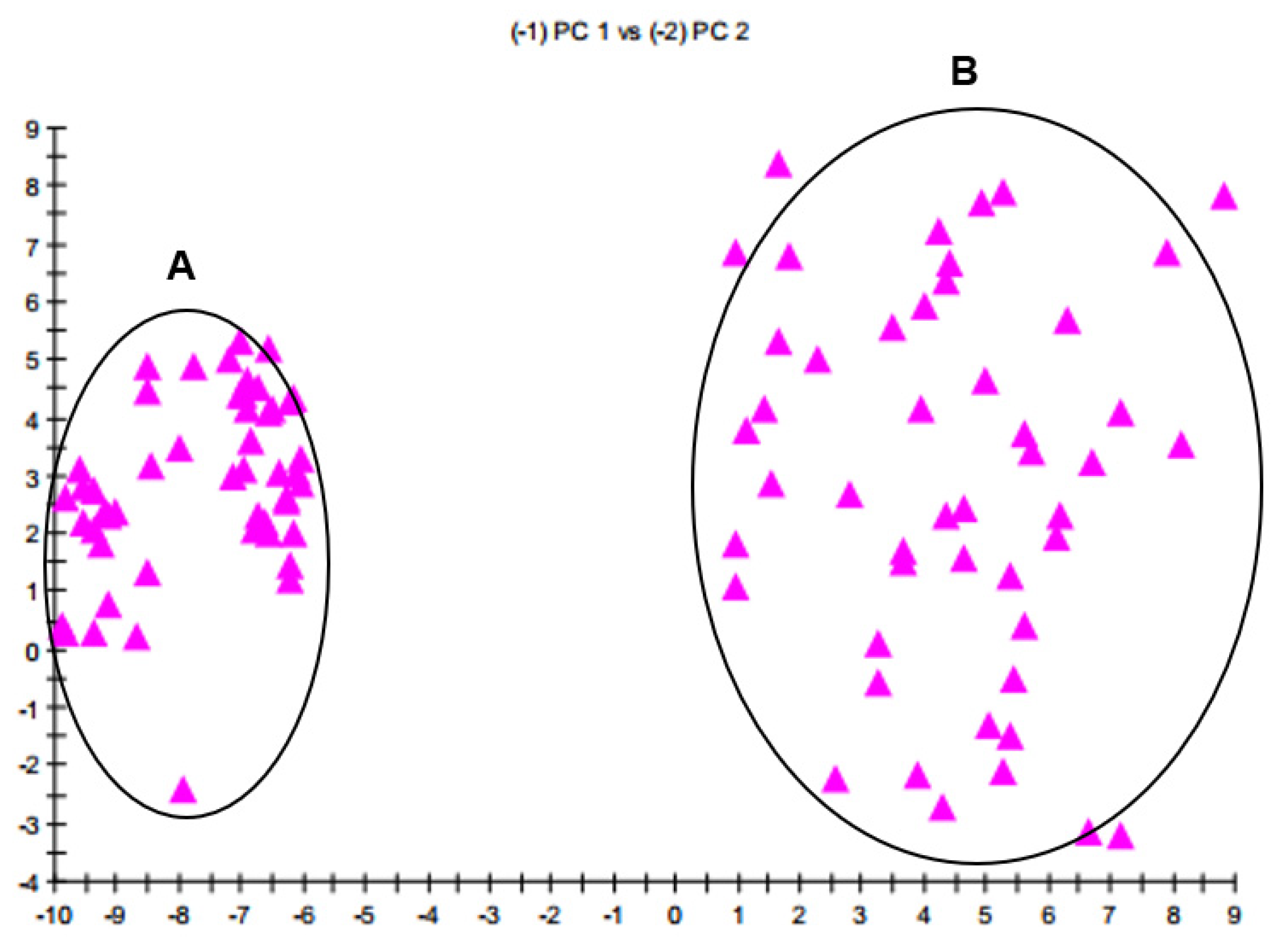
The FA profiles were consistently typical and distinguishable between B. velezensis and B. amyloliquefaciens. Principal component analysis enabled us to look at data with high dimensionality and observe the most critical aspects of the data in two or three dimensions. The 2D plot built from PC 1 and PC 2 (Figure 2) showed a separation of the two taxa in n-dimensional space. The group A represented FA components of B. amyloliquefaciens with ED2 (Euclidian distance squared) ~ 36, and the group B represented FAs of B. velezensis with ED2 ~ 100. A group with calculated ED2 ≤ 100 was considered as the same species. The FA profiles of the two taxa could be distinguished into two separate groups of strains.
Additionally, according to the criterion established by MIS, when the similarity index (SI) is larger than 0.5 and separated from other organisms from the library by at least 0.100, the sample is considered identified. The SI value generated from calculations of distance in multi-dimensional space illustrated the relation between analyzing FA profiles and the mean FAs of library’s database as its match. In our case, all identified samples exhibited high matches with SI > 0.5 and well-separated SI (>0.1) confirming that the method is reliable with high confidence.
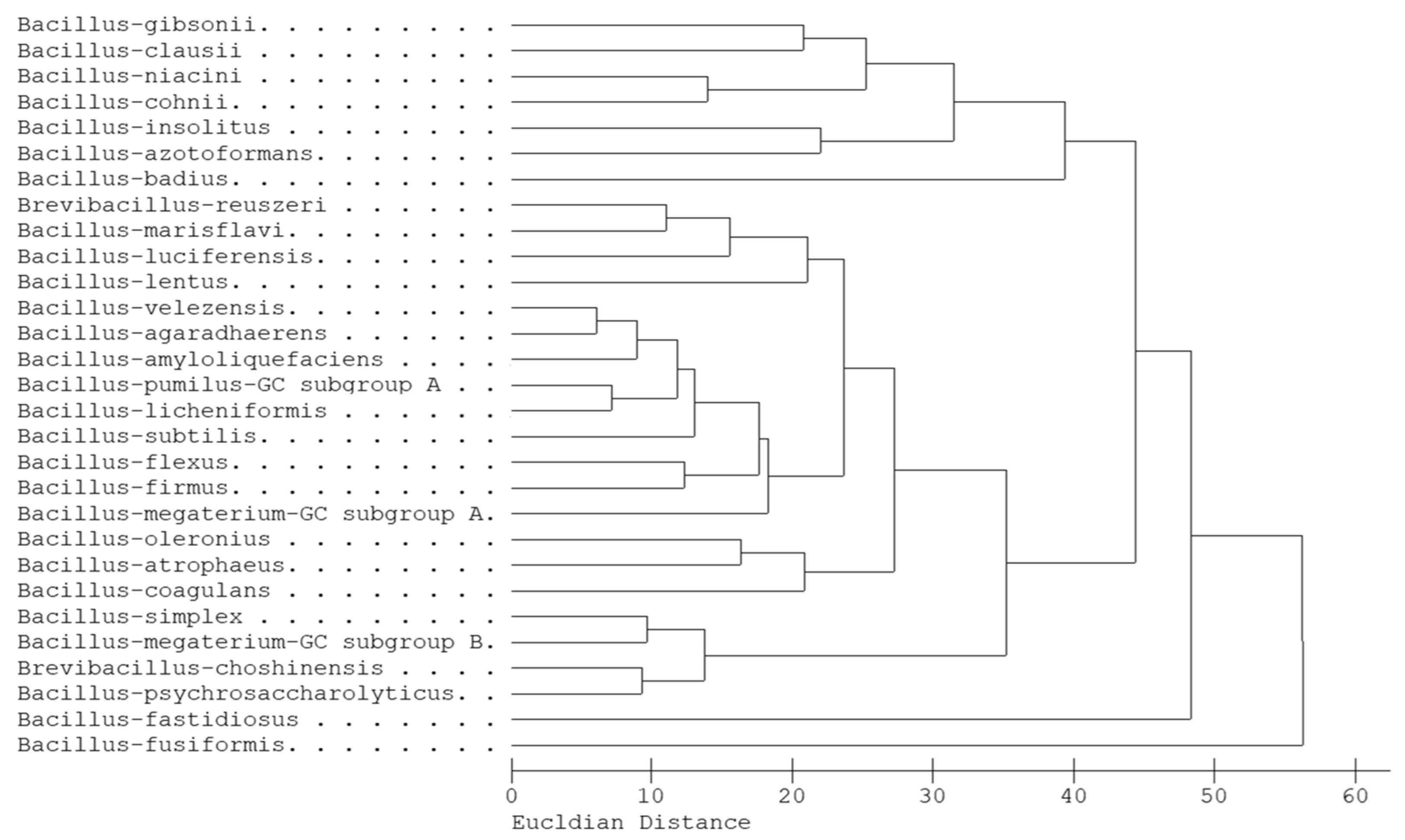
The dendrogram analysis drawn from diverse FA profiles of Bacillus species showed a relationship between them (Figure 3) via the ED index. These profiles were obtained from the Sherlock library that had been built from diverse strains (more than 20 strains) within each species. The samples were collected from across the world to avoid potential geographic bias and carefully analyzed with many replications to make the entries of the library [21]. The FA profiles of B. velezensis and B. amyloliquefaciens—together with B. agaradhaerens, B. pumilus, B. licheniformis, and B. subtilis—formed one cluster, which could be distinguished from other clusters. Furthermore, FAs of B. velezensis, B. agaradhaerens, and B. amyloliquefaciens formed a tight phylogenetic branch, which showed the highest phenotypic similarity. The FA profiles of group-related species were highly similar and were also determined previously [26,27,28].

4. Discussion

The classification of B. velezensis and B. amyloliquefaciens has usually been a particularly confounding taxonomic problem. Moreover, it was also concluded in previous reports that the whole-cell FAME profiles had not yielded satisfying results for discriminating these two species [2,10,11]. However, our research efforts, aimed at developing a whole-cell FAME profile-based method for distinguishing both taxa, led to other conclusions.
The gyrA and rpoB sequences proved to be effective for resolving these closely related species of the B. subtilis group [1,19,26]. The previous use of gyrA [12,19] and rpoB [1,11] as phylogenetic markers had drawn clear distinction between the two taxa. Accordingly, their highly conserved rpoB sequences shared approximately 98% similarity [1], but the NJ tree obtained from their gyrA sequences distinguished them with bootstrap values of 100 and 52% [12].
In the present study, gyrA and rpoB sequences were amplified and aligned to determine Bacillus strains to the species level. The result of BLAST alignments showed high relatedness between the sequences of the studied strains and records of both B. velezensis and B. amyloliquefaciens from the GenBank database. Thus, the discrimination of the two taxa were confused, because, unfortunately, several B. velezensis strains are still named as B. amyloliquefaciens in the GenBank and vice versa, which makes the identifications difficult. Therefore, for the detailed analysis, the partial sequences of gyrA and rpoB genes were concatenated and included in the phylogenetic analysis for comparative purposes. The selection of reference sequences was carefully considered according to previous classifications to avoid misinterpretations [2,7,12,16,29]. The studied strains clustered into two separate clades on the phylogenetic tree (Figure 1) differentiating B. velezensis strains from the cluster of strains related to B. amyloliquefaciens DSM 7T and DSM 1061T. Then the result of this classification was used for developing the identification method based on FA profiles.
In the current work, FAs were identified based on their estimated carbon lengths determined relative to the calibration standard and by comparing with the peak table. It could be concluded that certain deviations can be found between our results and previously published FA profiles [2,10,11]. In general, both taxa possess a higher content of branched-odd FAs, including 15:0 iso, 15:0 anteiso, 17:0 iso, and 17:0 anteiso than other FAs. The presence of branched-chain FAs is expected to increase the membrane’s fluidity because of their low melting point temperatures, and are already remarkable biomarkers used in Bacillus taxonomy [5]. The 15:0 iso and 15:0 anteiso FAs have shared a prominent proportion, similar to other species within the “B. subtilis species complex” [9] and their high ratio has been indicated as a common feature in Bacillus species [5]. These FAs had been considered as being distinguishable features between many other Bacillus species reported previously [25]. Discriminating biomarkers useful for distinguishing between the two taxa were 14:0, 16:0, 16:0 iso, 17:0 iso, and 17:0 anteiso. The FA profiles of B. velezensis could be characterized by higher 14:0 and 16:0 contents and lower 16:0 iso, 17:0 iso and 17:0 anteiso contents in comparison to B. amyloliquefaciens (Figure 4).
Our comprehensive study proved that these features are valuable taxonomical biomarkers with high discriminatory power, even though previous studies reported on the insufficiency of FA components in the discrimination of the two taxa. In the previous studies, certain involved isolates were misidentified and the novel classification of these strains, proven by many recent scientific contributions, helped us in giving a better conclusion for this issue. As shown in Table 4, the present investigation was similar to the report of Wang et al. [14]. However, it was a misapprehension that the publication considered Bacillus strain BCRC 14193 as B. amyloliquefaciens, which was later reclassified as B. velezensis by Dunlap et al. [13]. Currently, considering strain BCRC 14193 as B. velezensis, a distinguishable FA comparison was obviously drawn between the two taxa (Table 4). In 2011, a comparison among FAs from six strains of B. velezensis and five strains of B. amyloliquefaciens had been reported, with varied cellular FA compositions showing differences between the two taxa in the case of FA 14:0, 16:0, and 16:0 iso [12]. However, B. velezensis DSM 23117T and B. amyloliquefaciens DSM 7T contained a high content of FA 17:0, but lacked 17:0 anteiso [12], which made a difference with other Bacillus strains, and it is difficult to find a comparison, as well as the relatedness with our present study, due to insufficient data. The FA profiles in our study also shared high similarity to those of B. velezensis sp. nov. CR-502T and B. amyloliquefaciens DSM 7T with some minor differences [9]. It is interesting that only B. amyloliquefaciens DSM 7T, from the publication of Ruiz-García et al. [9], contained FA 16:1 ω5c, 16:1 ω9c, and 17:1 iso ω7c, which had not been detected by other authors.
A high-quality library plays an important role in the classification. This study carefully constructed the library RTSBA7 from 16 strains of B. velezensis (n = 3) and 2 strains of B. amyloliquefaciens (n = 25) and from the data available in the RTSBA6, containing B. amyloliquefaciens using various strains constructed by MIDI (Table 2).
Altogether a total of 31 FAs were detected in B. velezensis (Table S2) and 38 FAs were determined in B. amyloliquefaciens (Table S3) during the analyses. The calculations were interfered with by occasionally detected peaks irregularly observed on the chromatograms with small peak areas; however the MIS analysis, with many replicates of samples, can detect and remove these variations, creating precise whole-cell FA features (Figure 4). For example, in the case of B. velezensis, the MIS analysis did not consider FA 9:0, because there were only 3 out of 48 samples containing it, with a low mean (=0.06) and high SD/mean (=4.15). Otherwise, FA 14:0 (2.87 ± 0.70 (%)) detected from 1.69 to 4.89% in all 48 samples was considered as a valuable parameter. Accordingly, characterizations of both taxa have contained 13 FAs as features of the analyses (Table 2), which are reported in the Supplementary Tables (Tables S2 and S3).
Once a sample has been analyzed by Sherlock, its FA composition can be matched with those of known organisms that are stored in the library. The Sherlock Library search lists the most likely matches to the query composition, and provides an SI for each match, which is a numerical value expressing how closely the FA composition of the query compares with the mean FA composition of the strains used to create the library entry listed as its match. The database search presents the best matches and associated SI. This value is a software-generated calculation of the distance in multi-dimensional space between the profile of the query and the mean profile of the closest library entry. Our results showed good matches between the experimental samples and the FA composition in the library, with SI > 0.5 and well-separated SI (>0.1). In addition, the FA compositions could be separated between the two taxa and among other Bacillus species.

5. Conclusions

In our study, a method using FAs of 9–20 carbons in length and automated GC analysis were developed to qualitatively and quantitatively analyze the bacterial whole-cell FAs as taxonomical markers. To the best of our knowledge, this is the first time that the method based on whole-cell FA profiles operated by MIS has been applied to distinguish between B. velezensis and B. amyloliquefaciens with comprehensive evidence. By taking advantage of the current knowledge regarding biomarkers, the FA-based identification proved to be applicable for the differentiation between these closely related species. Our experiments provided a cost-effective, reliable, and fast-automated solution for discrimination between these taxa.

Supplementary Materials

The following supporting information can be downloaded at: https://www.mdpi.com/article/10.3390/microorganisms10020418/s1, Table S1: Reference Bacillus strains and their sequence data used in phylogenetic construction; Table S2: Parameters of the whole-cell FA features of B. velezensis; Table S3: Parameters of the whole-cell FA features of B. amyloliquefaciens; Figure S1: The Neighbor-Joining phylogenetic tree based on gyrA gene sequences; Figure S2: The Neighbor-Joining phylogenetic tree based on rpoB gene sequences; Figure S3: Chromatograms of the FAs of B. velezensis (A) and B. amyloliquefaciens (B).

Author Contributions

Conceptualization, A.S. and G.S.; methodology, A.T. and T.H.; software, T.H.; validation, B.L. and T.H.; formal analysis, A.S. and T.H.; investigation, B.L., M.V., O.K. and T.H.; resources, A.S., C.V. and L.K.; data curation, G.S.; writing—original draft preparation, A.S. and T.H.; writing—review and editing, C.V., G.S. and L.K.; visualization, B.L.; supervision, A.S. and G.S.; project administration, A.S. and T.H.; funding acquisition, A.S. All authors have read and agreed to the published version of the manuscript.

Funding

This work was supported by the grant OTKA K-128659 from the Hungarian Scientific Research Fund.

Institutional Review Board Statement

Not applicable.

Informed Consent Statement

Not applicable.

Data Availability Statement

DNA sequence data supporting the reported results can be found in the NCBI GenBank database (https://www.ncbi.nlm.nih.gov/genbank, 29 December 2021) under the accession numbers listed in Table 1.

Acknowledgments

We thank Craig Kunitsky, Gary Jackoway, Mike Alexander and Supporting MIDI Team (MIDI Inc.) and the whole company for technical help and continuous support to adopt the Sherlock MIDI system at our site.

Conflicts of Interest

The authors declare no conflict of interest.

References

  1. Fan, B.; Blom, J.; Klenk, H.P.; Borriss, R. Bacillus amyloliquefaciens, Bacillus velezensis, and Bacillus siamensis form an “Operational Group B. amyloliquefaciens” within the B. subtilis species complex. Front. Microbiol. 2017, 8, 22. [Google Scholar] [CrossRef] [Green Version]
  2. De Carvalho, C.C.C.R.; Caramujo, M.J. The various roles of fatty acids. Molecules 2018, 23, 2583. [Google Scholar] [CrossRef] [PubMed] [Green Version]
  3. Choi, J.; Nam, J.; Seo, M.-H. Complete genome sequence of Bacillus velezensis NST6 and comparison with the species belonging to operational group B. amyloliquefaciens. Genomics 2021, 113, 380–386. [Google Scholar] [CrossRef] [PubMed]
  4. da Costa, M.S.; Albuquerque, L.; Nobre, M.F.; Wait, R. The identification of fatty acids in bacteria. In Methods in Microbiology; Elsevier Ltd.: Amsterdam, The Netherlands, 2011; Volume 38, pp. 183–196. [Google Scholar]
  5. Diomandé, S.E.; Nguyen-The, C.; Guinebretière, M.H.; Broussolle, V.; Brillard, J. Role of fatty acids in Bacillus environmental adaptation. Front. Microbiol. 2015, 6, 813. [Google Scholar] [CrossRef] [PubMed] [Green Version]
  6. Rabbee, M.; Ali, M.S.; Choi, J.; Hwang, B.S.; Jeong, S.C.; Baek, K. Bacillus velezensis: A valuable member of bioactive molecules within plant microbiomes. Molecules 2019, 24, 1046. [Google Scholar] [CrossRef] [PubMed] [Green Version]
  7. Fukumoto, J. Studies on the production of bacterial amylase. I. Isolation of bacteria secreting potent amylases and their distribution. J. Agric. Chem. Soc. Jpn. 1943, 19, 487–503. [Google Scholar] [CrossRef]
  8. Priest, F.G.; Goodfellow, M.; Shute, L.A.; Berkeley, W. Bacillus amyloliquefaciens sp. nov., nom. rev. Int. J. Syst. Bacteriol. 1987, 37, 69–71. [Google Scholar] [CrossRef]
  9. Ruiz-García, C.; Béjar, V.; Martínez-Checa, F.; Llamas, I.; Quesada, E. Bacillus velezensis sp. nov., a surfactant-producing bacterium isolated from the river Vélez in Málaga, southern Spain. Int. J. Syst. Evol. Microbiol. 2005, 55, 191–195. [Google Scholar] [CrossRef] [Green Version]
  10. Fan, B.; Wang, C.; Song, X.; Ding, X.; Wu, L.; Wu, H.; Gao, X.; Borriss, R. Bacillus velezensis FZB42 in 2018: The Gram-positive model strain for plant growth promotion and biocontrol. Front. Microbiol. 2018, 9, 2491. [Google Scholar] [CrossRef] [Green Version]
  11. Grady, E.N.; MacDonald, J.; Ho, M.T.; Weselowski, B.; McDowell, T.; Solomon, O.; Renaud, J.; Yuan, Z.C. Characterization and complete genome analysis of the surfactin-producing, plant-protecting bacterium Bacillus velezensis 9D-6. BMC Microbiol. 2019, 19, 5. [Google Scholar] [CrossRef]
  12. Borriss, R.; Chen, X.-H.; Rückert, C.; Blom, J.; Becker, A.; Baumgarth, B.; Fan, B.; Pukall, R.; Schumann, P.; Spröer, C.; et al. Relationship of Bacillus amyloliquefaciens clades associated with strains DSM 7T and FZB42T: A proposal for Bacillus amyloliquefaciens subsp. amyloliquefaciens subsp. nov. and Bacillus amyloliquefaciens subsp. plantarum subsp. nov. based on complete genome sequence comparisons. Int. J. Syst. Evol. Microbiol. 2011, 61, 1786–1801. [Google Scholar] [CrossRef] [PubMed]
  13. Dunlap, C.A.; Kim, S.J.; Kwon, S.W.; Rooney, A.P. Bacillus velezensis is not a later heterotypic synonym of Bacillus amyloliquefaciens; Bacillus methylotrophicus, Bacillus amyloliquefaciens subsp. Plantarum and ‘Bacillus oryzicola’ are later heterotypic synonyms of Bacillus velezensis based on phylogenom. Int. J. Syst. Evol. Microbiol. 2016, 66, 1212–1217. [Google Scholar] [CrossRef]
  14. Wang, L.T.; Lee, F.L.; Tai, C.J.; Kuo, H.P. Bacillus velezensis is a later heterotypic synonym of Bacillus amyloliquefaciens. Int. J. Syst. Evol. Microbiol. 2008, 58, 671–675. [Google Scholar] [CrossRef] [Green Version]
  15. Liu, S.; Tian, Y.; Jia, M.; Lu, X.; Yue, L.; Zhao, X.; Jin, W.; Wang, Y.; Zhang, Y.; Xie, Z.; et al. Induction of salt tolerance in Arabidopsis thaliana by volatiles from Bacillus amyloliquefaciens FZB42 via the jasmonic acid signaling pathway. Front. Microbiol. 2020, 11, 562934. [Google Scholar] [CrossRef]
  16. Molina-Santiago, C.; Vela-Corcía, D.; Petras, D.; Díaz-Martínez, L.; Pérez-Lorente, A.I.; Sopeña-Torres, S.; Pearson, J.; Caraballo-Rodríguez, A.M.; Dorrestein, P.C.; de Vicente, A.; et al. Chemical interplay and complementary adaptative strategies toggle bacterial antagonism and co-existence. Cell Rep. 2021, 36, 109449. [Google Scholar] [CrossRef]
  17. Dunlap, C.A.; Kim, S.J.; Kwon, S.W.; Rooney, A.P. Phylogenomic analysis shows that Bacillus amyloliquefaciens subsp. plantarum is a later heterotypic synonym of Bacillus methylotrophicus. Int. J. Syst. Evol. Microbiol. 2015, 65 Pt 7, 2104–2109. [Google Scholar] [CrossRef]
  18. Liu, G.; Kong, Y.; Fan, Y.; Geng, C.; Peng, D.; Sun, M. Data on genome analysis of Bacillus velezensis LS69. Data Br. 2017, 13, 1–5. [Google Scholar] [CrossRef] [PubMed]
  19. Reva, O.N.; Dixelius, C.; Meijer, J.; Priest, F.G. Taxonomic characterization and plant colonizing abilities of some bacteria related to Bacillus amyloliquefaciens and Bacillus subtilis. FEMS Microbiol. Ecol. 2004, 48, 249–259. [Google Scholar] [CrossRef]
  20. Reva, O.N.; Swanevelder, D.Z.H.; Mwita, L.A.; Mwakilili, A.D.; Muzondiwa, D.; Joubert, M.; Chan, W.Y.; Lutz, S.; Ahrens, C.H.; Avdeeva, L.V.; et al. Genetic, epigenetic and phenotypic diversity of four Bacillus velezensis strains used for plant protection or as probiotics. Front. Microbiol. 2019, 10, 2610. [Google Scholar] [CrossRef] [Green Version]
  21. MIDI Inc. The Sherlock Chromatographic Analysis System Operating Manual, 6th ed.; MIDI, Inc.: Newark, DE, USA, 2018. [Google Scholar]
  22. Vágvölgyi, C.; Sajben-Nagy, E.; Bóka, B.; Vörös, M.; Berki, A.; Palágyi, A.; Krisch, J.; Škrbić, B.; Đurišić-Mladenović, N.; Manczinger, L. Isolation and characterization of antagonistic Bacillus strains capable to degrade ethylenethiourea. Curr. Microbiol. 2013, 66, 243–250. [Google Scholar] [CrossRef]
  23. Stefanic, P.; Mandic-Mulec, I. Social interactions and distribution of Bacillus subtilis pherotypes at microscale. J. Bacteriol. 2009, 191, 1756–1764. [Google Scholar] [CrossRef] [PubMed] [Green Version]
  24. Saitou, N.; Nei, M. The neighbor-joining method: A new method for reconstructing phylogenetic trees. Mol. Biol. Evol. 1987, 4, 406–425. [Google Scholar] [CrossRef] [PubMed]
  25. Tamura, K.; Nei, M. Estimation of the number of nucleotide substitutions in the control region of mitochondrial DNA in humans and chimpanzees. Mol. Biol. Evol. 1993, 10, 512–526. [Google Scholar] [CrossRef] [PubMed]
  26. Roberts, M.S.; Nakamura, L.K.; Cohan, F.M. Bacillus mojavensis sp. nov., distinguishable from Bacillus subtilis by sexual isolation, divergence in DNA sequence, and differences in fatty acid composition. Int. J. Syst. Bacteriol. 1994, 44, 256–264. [Google Scholar] [CrossRef] [Green Version]
  27. Slabbinck, B.; De Baets, B.; Dawyndt, P.; De Vos, P. Genus-wide Bacillus species identification through proper artificial neural network experiments on fatty acid profiles. Antonie Van Leeuwenhoek 2008, 94, 187–198. [Google Scholar] [CrossRef]
  28. Truong, T.V.; Nackos, A.N.; Williams, J.R.; Vanderwerken, D.N.; Kimball, J.A.; Murray, J.A.; Hawkes, J.E.; Harvey, D.J.; Tolley, H.D.; Robison, R.A.; et al. Differentiation of Bacillus endospore species from fatty acid methyl ester biomarkers. Anal. Methods 2010, 2, 638–644. [Google Scholar] [CrossRef]
  29. Perez, K.J.; dos Viana, J.S.; Lopes, F.C.; Pereira, J.Q.; dos Santos, D.M.; Oliveira, J.S.; Velho, R.V.; Crispim, S.M.; Nicoli, J.R.; Brandelli, A.; et al. Bacillus spp. isolated from puba as a source of biosurfactants and antimicrobial lipopeptides. Front. Microbiol. 2017, 8, 61. [Google Scholar] [CrossRef] [Green Version]
Figure 1. Neighbor-Joining phylogenetic tree based on the concatenation of gyrA and rpoB gene sequences. Evolutionary distances were computed by the Tamura-Nei method. Bars, 0.010 substitutions per nucleotide position.
Microorganisms 10 00418 g001
Figure 2. The 2D plot between FA components of B. velezensis and B. amyloliquefaciens. (A) FA components from the group of B. amyloliquefaciens, (B) FA components from the group of B. velezensis.
Microorganisms 10 00418 g002
Figure 3. The relationship of FA profiles among the Bacillus species in the MIS library.
Microorganisms 10 00418 g003
Figure 4. Comparison charts of (A) B. velezensis and (B) B. amiloliqefaciens based on FA profiles created in the MIS library.
Microorganisms 10 00418 g004
Table 1. Characteristics of B. velezensis and B. amyloliquefaciens determined by different techniques.
Characteristics aB. velezensisbB. amyloliquefaciensbReferences
PigmentationCreamy whiteCreamy white[9]
Oxidase++
Acid in API system from:
- Glycogen+nd
- Lactose++
- Melibiose+
- Methyl α-ᴅ-glycoside++
- ᴅ-Raffinose++
- ᴅ-Turanose+
Hydrolysis of
- Tween 20+
- Tween 80nd
- DNA
Arginine dihydrolase
ONPG+
Non-ribosomally synthesized secondary metabolites [1,10,12]
- Surfactin++
- Macrolactin+
- Bacillaene++
- Fengycin+
- Difficidin+
- Bacillibactin++
- Bacilysin++
Ribosomally synthesized antimicrobial compounds
- Sublancin
- Subtilosin
- Amylocyclicin++
- Plantazolicin+
Plant colonization+[12]
AZCL-HE cellulose liquefaction+
Growth in lactose minimal medium++
Amylase AmyA+
Amylase AmyE+
Cellulase BglC+
Xylanase XynA+
16S rRNA gene sequence similarity (%)99.7%[14]
rpoB gene sequence similarity (%)>98%[1]
gyrB gene sequence similarity (%)>95.5%[14]
DNA relatedness value (%)74–84%[14]
a ONPG: O-nitrophenyl β-ᴅ-galactopyranoside, AZCL-HE: endocellulase activity determined by using insoluble azurine cross-linked (AZCL)-HE-cellulose. b +: detected, −: not detected, nd: not determined.
Table 2. Bacillus strains used in the FA profiling study.
Strains aGenbank Accession NumberOrigin
gyrArpoB
B. velezensis
SZMC 24980OK256097OK256115soil sample from pepper field, Totovo selo, Serbia
SZMC 24981OK256098OK256116soil sample from pepper field, Totovo selo, Serbia
SZMC 24982OK256099OK256117soil sample from pepper field, Totovo selo, Serbia
SZMC 24983OK256100OK256118soil sample from pepper field, Totovo selo, Serbia
SZMC 24984OK256101OK256119soil sample from pepper field, Cantavir, Serbia
SZMC 24985OK256102OK256120soil sample from pepper field, Cantavir, Serbia
SZMC 24986OK256103OK256121soil sample from tomato field, Cantavir, Serbia
SZMC 24995OK256104OK256122soil sample from tomato field, Cantavir, Serbia
SZMC 25020OK256105OK256123soil sample from tomato field, Cenej, Serbia
SZMC 25646OK256106OK256124pea rhizosphere, Madaras, Hungary
SZMC 25647OK256107OK256125pea rhizosphere, Madaras, Hungary
SZMC 25610OK256108OK256126maize rhizosphere, Vaszar, Hungary
SZMC 6046OK256109OK256127tomato rhizosphere, Hungary
SZMC 16093BOK256110OK256128tomato rhizosphere, Hungary
SZMC 6387JOK256111OK256129tomato rhizosphere, Hungary
DSM 23117T (=BGSC 10A6 = FZB42 = LMG 26770 = SZMC 27497)OK256112OK256130plant pathogen-infested soil of a sugar beet field, Brandenburg, Germany
B. amyloliquefaciens
DSM 7T (=ATCC 23350 = SZMC 6027)OK256113OK256131soil and industrial amylase fermentations, Japan
DSM 1061T (=IAM 1523 = SZMC 6222)OK256114OK256132unknown origin
B. subtilis
ATCC 6633TCP039755.1CP039755.1Japan
a ATCC—American Type Culture Collection, BGSC—Bacillus Genetic Stock Center, DSM—German Collection of Microorganisms and Cell Cultures, FZB—Research Center Borstel, IAM—Institute of Applied Microbiology, University of Tokyo, LMG—Belgian Coordinated Collections of Microorganisms/LMG Bacteria Collection, SZMC—Szeged Microbiology Collection.
Table 3. Cellular fatty acid compositions (mean (%) ± SD).
Feature/FAB. velezensisB. amyloliquefaciens
12:00.48 ± 0.230.54 ± 0.17
13:0 iso0.89 ± 0.220.50 ± 0.19
14:0 iso1.18 ± 0.581.44 ± 0.11
14:02.87 ± 0.700.61 ± 0.14
15:0 iso30.39 ± 2.5327.84 ± 1.65
15:0 anteiso32.13 ± 2.3331.92 ± 1.98
16:0 iso1.70 ± 0.773.51 ± 0.19
16:1 ω11c1.65 ± 0.421.09 ± 0.28
16:012.53 ± 1.824.57 ± 0.55
17:1 iso ω10c0.85 ± 0.471.07 ± 0.37
17:0 iso8.52 ± 0.9615.92 ± 1.96
17:0 anteiso5.50 ± 0.858.99 ± 0.73
18:00.60 ± 0.140.59 ± 0.23
Table 4. Comparison of FA components of B. velezensis and B. amyloliquefaciens reported in the literature.
FeatureThis StudyRuiz-García et al. [9]Wang et al. [14]Borriss et al. [12]
B. v.aB. a.bB. v.B. a.B. v.B. v.B. a.B. a.B. v.B. a.
CR-502TDSM 7TBCRC 17467TBCRC 14193BCRC 11601TBCRC 17038DSM 23117TDSM 7T
12:00.480.54--------
13:0 iso0.890.500.87-----0.310.38
14:0 iso1.181.441.082.46-1.31.51.70.430.99
14:02.870.612.96-3.83.1--1.210.36
15:0 iso30.3927.8429.8630.5030.424.026.323.731.0040.29
15:0 anteiso32.1331.9232.7036.4827.628.732.333.831.7328.32
16:0 iso1.703.511.314.521.02.23.84.31.012.13
16:1 ω5c---2.14------
16:1 ω7c--------0.190.42
16:1 ω9c---0.62------
16:1 ω11c1.651.094.42-3.52.71.7-2.591.23
16:012.534.5713.414.5218.319.05.87.07.603.02
17:1 iso ω7c---1.67------
17:1 iso ω10c0.851.071.44-1.31.31.7-2.702.59
17:0 iso8.5215.927.679.017.810.316.317.612.1113.14
17:0 anteiso5.508.994.277.063.45.49.010.0--
17:00.170.22------7.706.46
18:00.600.59---1.1----
aB. v.: B. velezensis; bB. a.: B. amyloliquefaciens.
Publisher’s Note: MDPI stays neutral with regard to jurisdictional claims in published maps and institutional affiliations.