Review

Immunomodulatory Effects of Probiotics: A Novel Preventive Approach for the Control of Bovine Mastitis

by
A. K. M. Humayun Kober
1,2,3,*,†,
Sudeb Saha
1,2,4,†,
Md. Aminul Islam
1,5,†,
Muhammad Shahid Riaz Rajoka
1,2,†,
Kohtaro Fukuyama
1,2,
Hisashi Aso
2,6,7,
Julio Villena
1,8,* and
Haruki Kitazawa
1,2,*
1
Laboratory of Animal Food Function, Graduate School of Agricultural Science, Tohoku University, Sendai 980-8572, Japan
2
Livestock Immunology Unit, International Education and Research Centre for Food and Agricultural Immunology, Graduate School of Agricultural Science, Tohoku University, Sendai 980-8572, Japan
3
Department of Dairy and Poultry Science, Chittagong Veterinary and Animal Sciences University, Khulshi, Chittagong 4225, Bangladesh
4
Department of Dairy Science, Sylhet Agricultural University, Sylhet 3100, Bangladesh
5
Department of Medicine, Faculty of Veterinary Science, Bangladesh Agricultural University, Mymensingh 2202, Bangladesh
6
Laboratory of Animal Health Science, Graduate School of Agricultural Science, Tohoku University, Sendai 980-8572, Japan
7
The Cattle Museum, Maesawa, Oshu 029-4205, Japan
8
Laboratory of Immunobiotechnology, Reference Centre for Lactobacilli (CERELA-CONICET), San Miguel de Tucuman 4000, Argentina
*
Authors to whom correspondence should be addressed.
JSPS Research Fellow.
Microorganisms 2022, 10(11), 2255; https://doi.org/10.3390/microorganisms10112255
Submission received: 22 September 2022 / Revised: 3 November 2022 / Accepted: 9 November 2022 / Published: 14 November 2022
(This article belongs to the Special Issue Probiotics for Next Generations)

Abstract

:
Bovine mastitis (BM) is one of the most common diseases of dairy cattle, causing economic and welfare problems in dairy farming worldwide. Because of the predominant bacterial etiology, the treatment of BM is mostly based on antibiotics. However, the antimicrobial resistance (AMR), treatment effectiveness, and the cost of mastitis at farm level are linked to limitations in the antibiotic therapy. These scenarios have prompted the quest for new preventive options, probiotics being one interesting alternative. This review article sought to provide an overview of the recent advances in the use of probiotics for the prevention and treatment of BM. The cellular and molecular interactions of beneficial microbes with mammary gland (MG) cells and the impact of these interactions in the immune responses to infections are revised. While most research has demonstrated that some probiotics strains can suppress mammary pathogens by competitive exclusion or the production of antimicrobial compounds, recent evidence suggest that other probiotic strains have a remarkable ability to modulate the response of MG to Toll-like receptor (TLR)-mediated inflammation. Immunomodulatory probiotics or immunobiotics can modulate the expression of negative regulators of TLR signaling in the MG epithelium, regulating the expression of pro-inflammatory cytokines and chemokines induced upon pathogen challenge. The scientific evidence revised here indicates that immunobiotics can have a beneficial role in MG immunobiology and therefore they can be used as a preventive strategy for the management of BM and AMR, the enhancement of animal and human health, and the improvement of dairy cow milk production.

1. Introduction

Bovine mastitis (BM) is one of the most prevalent and costly diseases of dairy cattle worldwide. BM refers to inflammation of the mammary gland (MG), which greatly affects milk production and impacts product quality, animal health and welfare, the economic profit of the dairy industry, and public health [1,2,3]. Numerous pathogens, including bacteria, viruses, and fungi can induce a variety of clinical symptoms, from asymptomatic sub-clinical infection to severe acute MG inflammation [4,5]. Acute inflammation brought on by the intense activation of immune cells by cytokines and chemokines production is one of the major clinical symptoms of MG infection [6,7]. The cure rate of mastitis depends on the species of mastitis-causing pathogens and the efficacy of antibiotics used, as well as the host immune status [8,9,10]. However, the irrational use of antibiotic medications frequently results in the emergence of antimicrobial resistance (AMR), which poses a serious threat to the wellbeing of dairy cattle. Additionally, AMR due to BM might pose serious risks to public health through the spread of AMR bacterial infections and drug residues through the consumption of raw milk from cows that have received antibiotic treatment [11]. Another major issue for the dairy sector is that the presence of antibiotic residues in milk can interfere with the fermentation process, particularly in cheese and yogurt production [1]. Due to both the low frequency of mastitis during lactation being cured and the high probability of AMR genes spread to pathogens by antibiotic therapy [12,13], it is necessary to review the traditional antibiotic-based treatment strategy and look for innovative and sustainable therapeutic alternatives. Herbal remedies, homeopathy, probiotics, genetic selection, and vaccination have been investigated as potential alternatives to antibiotics for the treatment or prevention of mastitis [14,15,16,17]. Compared to herbal therapy, homeopathy, and vaccination, it was found that probiotics may have a superior efficacy in terms of cost and effectiveness. In addition, the use of probiotics may reduce AMR, and it is environment friendly [18]. Thus, probiotics, particularly those with immunomodulatory capacities or immunobiotics, could be an interesting alternative to control or treat BM [3,19,20,21,22,23,24,25]. Immunobiotics have beneficial effects on human and animal health by modulating the host immune responses and are considered safe for human and healthy livestock production [19]. While most research has demonstrated that some probiotic strains can suppress mammary pathogens by competitive exclusion or the production of antimicrobial compounds, recent evidence suggest that other probiotic strains have a remarkable ability to modulate the response of MG to Toll-like receptor (TLR)-mediated inflammation. In this regard, using an originally established bovine mammary epithelial (BME) cell line, our group demonstrated that immunobiotic lactic acid bacteria (LAB) strains could be effectively used in the prevention of BM through the modulation of mammary epithelium immune responses [20]. The application of immunobiotics in the field of MG immunobiology is a novel concept. According to several research works, probiotics in dairy cows or breast-feeding women have reportedly inspired claims of usefulness for preventing or treating mastitis [26,27,28,29,30]. However, despite the amount of research being carried out on this topic, research is still needed to conclusively demonstrate the efficacy of immunobiotics and to clarify in detail their mechanisms of action. Considering the current understanding of host-bacterial interaction in MG mucosa, this review sought to explore whether the probiotic concept fits the MG biology, and if mastitis prevention or treatment is amenable to probiotic/immunobiotic intervention. A summary of scientific literature on the use of putative probiotics in mastitis trials is only taken into consideration here because the small number of studies did not allow meta-analysis. The review also explores the cellular and molecular interactions of immunobiotic LAB with bovine mammary epithelial cells and the transcriptional markers of mastitis, providing a novel preventive approach for the effective control of mastitis in dairy cows.

2. Global Economic Impact of Bovine Mastitis

BM causes enormous economic losses to the dairy industry. These economic losses are due to the treatment, production loss, changes in product quality, discarded milk, extra labor, culling, poor animal welfare, and the risk of other diseases [4]. Mastitis causes one of the biggest expenses for the dairy industry worldwide, including India [31], Bangladesh [32], Japan [33,34], Canada [35], the USA [36], Colombia [37], Netherlands [38], the United Kingdom [5], and Australia [39,40] for treatment and control. The cost or impact of mastitis on the economy varies by country, and even by region within a country, depending on market condition and milk quality regulations. Ultimately, BM contributes to potential financial losses to the farmer and the dairy industry. The anticipated losses to BM are related to 31% in milk production, 24% treatment costs, 18% wasted milk, 4% excess labor demand, and 23% premature culling [41,42]. A total annual cost due to BM has been estimated as USD 19.7 to USD 32 billion in the global dairy industries [36]. Annual economic losses due to BM are projected to be USD 2 billion in the United States [36], USD 0.31 billion in Canada [35], USD 0.8 billion in Colombia [37], USD 1.3 billion in Australia [39,40], USD ~1.55 billion in the EU [39], USD ~0.77 billion in Japan [33,34], USD ~0.002 billion in Bangladesh [32], and USD ~0.8 billion in India [31]. A single case of clinical mastitis may lead to an economic loss that ranges between USD 128 and USD 444 [41,42]. According to these data, mastitis has a significant economic impact on dairy cattle production.

3. Prevention and Control Strategies of Bovine Mastitis

Currently, most BM management strategies focus on prevention during milking by following hygienic milking practices and a good management system. Other alternative approaches for the control of mastitis include vaccination, antibiotics, immunotherapy, bacteriocins, bacteriophages, antimicrobial peptides, stem cell therapy, native secretory factors, diet, dry cow and lactation therapy, genetic selection, botanicals, nanoparticle technology-based therapy, and probiotics [18]. The most widely utilized techniques to treat mastitis are antibiotic therapy and vaccination, which are not always effective in the control of BM. Extensive and uncontrolled use of antibiotics for the treatment along with the induction and spread of AMR in mastitis has led to reduced responsiveness to antibiotic therapy [43,44]. On the other hand, vaccination has limited effectiveness against BM because of the variety of microorganisms involved in its development [24,45,46]. Even though there are several commercial vaccines available, the majority do not provide complete protection, and are also expensive, limiting their extensive application [18,47]. Thus, alternative tactics are required to efficiently manage mastitis.
The high-throughput sequencing of bacterial 16S rRNA genes performed in the past few years has demonstrated the existence of bovine milk and MG microbiota [48]. In fact, a diverse range of opportunistic and commensal bacteria can inhabit the intramammary ecosystem [49,50]. Metagenomic investigations have demonstrated diverse bacterial groups in MG and milk samples obtained from non-mastitis quarters of dairy cows. Furthermore, there is growing evidence that the reduction of the diversity or the alteration of MG significantly augment the susceptibility to BM [49,50]. It is postulated that MG commensal bacteria help the bovine host to resist the pathogens’ colonization by several mechanisms, including competition for nutrients, conditioning the microecosystem to disfavor pathogen growth through organic acid production, the synthesis of antimicrobial compounds, the inhibition of adhesion to epithelial surfaces, and the regulation of MG epithelium immunity [49,50] (Figure 1). Recent studies revealed that probiotics may be used as a novel tool to treat or prevent BM while also addressing the issue of emerging AMR [3,20,27]. One innovative preventive strategy for the management of BM is the use of probiotics with immunomodulatory capacities or immunobiotics.

4. In Vivo Studies of Probiotics for the Prevention or Treatment of Bovine Mastitis

The microorganisms that have been used as probiotics for the prevention or treatment of BM are presented in Table 1. Some studies have evaluated the ability of probiotics to protect against BM using mice models. L. lactis LMG 7930 was shown to possess antagonistic effects against ruminant mastitis-causing pathogens [51]. Campeiro et al., 2017 [51] conducted an in-depth investigation of the mammary gland immune response induced by intramammary inoculum of a live culture of the LMG 7930 strain using the mouse mastitis model. The work reported that L. lactis LMG 7930 injected into the mouse inguinal glands before the injection of Staphylococcus chromogenes, decreased pathogen loads by improving the innate immune response. More recently, it was shown that Enterococcus mundtii H81, originally isolated from the milk of healthy dairy cows, injected to the nipple duct before S. aureus infection reduced neutrophil infiltration and TNF-α and IL-1β production, as well as the phosphorylation of p65 nuclear transcription factor kappa B (NF-κB) and IκB [52].
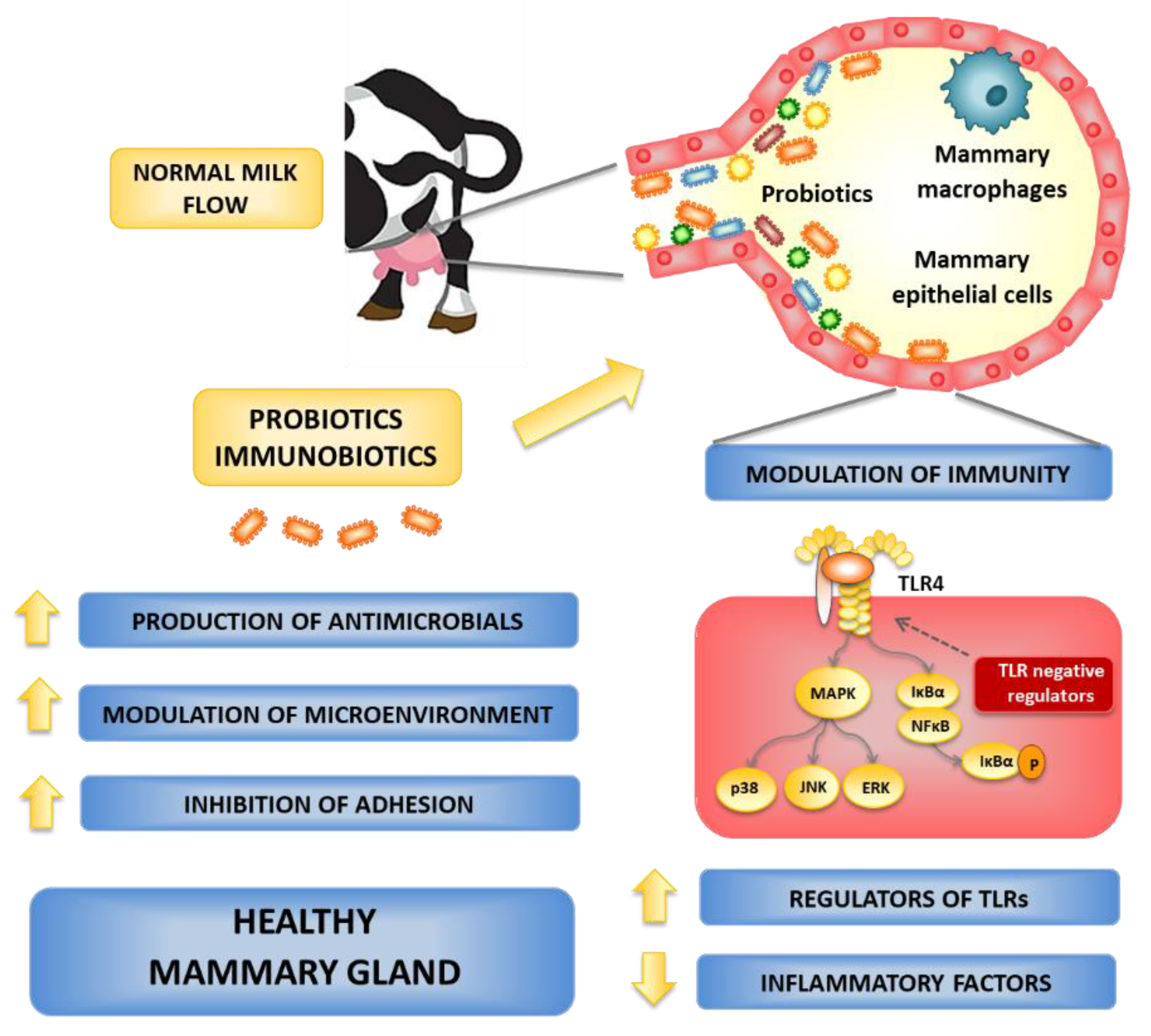
To combat mastitis, probiotics have also been administered to cows by the oral route or through an intramammary infusion. When LAB such as L. lactis are infused into the MG quarters, they induce significant increases in the recruitment of neutrophils and lymphocytes to the udder [17]. The administration of live culture of L. lactis DPC3147 into the MG enhance the innate immune response [17]. The expression of IL- and IL-8 genes were markedly increased in DPC3147-treated animals, with the highest expression corresponding to high somatic cell count in bovine mammary secretion [17,53]. Moreover, live cultures of L. lactis DPC3147 showed the same potential as antibiotic therapy for the eradication of persistent subclinical mastitis [17]. In addition, different strains of Lactobacillus, Bacillus, Bifidobacteria, and Enterococcus could reduce inflammation of the MG and control the milk microorganisms in dairy cows with mastitis [48]. Peng et al. (2013) [7] found that ultrasonicated Enterococcus faecium SF68 is compatible with cow MG and is an enhancer of innate immunity by accelerating neutrophil chemotaxis immediately following milking and lowering the somatic cell count (SCC) in milk. The treatment with Bifidobacterium breve and L. lactis subsp. lactis LMG 7930 reduces the infection rate of subclinical mastitis and SCC in milk [54,55]. Furthermore, the B. breve and L. lactis DPC3147 used in intramammary infusion lessens chronic subclinical mastitis in cows [51,54]. Another study observed that the intramammary infusion of L. lactis subsp. lactis CRL1655 and Lactobacillus perolens CRL1724 can mitigate the mastitis and lower SCC in milk by inducing a rise in IgG levels in blood and milk [29]. Animals that had received L. lactis CRL1655 and L. perolens CRL1724 infusions did not show any clinical symptoms related to MG infection. Of note, the blood serum of lactobacilli-treated animals had markedly enhanced opsonophagocytic activity against Staphylococcus spp. and improved expression of the proinflammatory cytokines IL-1β, IL-8, and TNF-α [29]. Intramammary treatment with inactivated cultures of Lacticaseibacillus rhamnosus GG to quarters naturally affected by subclinical mastitis has a transient pro-inflammatory activity assessed by low SCC and is capable of modifying the microbiota of milk [55]. These findings revealed that different probiotic strains have the potential to prevent mastitis by antagonizing pathogens, relieving MG inflammation, and regulating milk microorganisms (Figure 2).
On the other hand, there are fewer studies that have evaluated the effect of orally administered probiotics for improving mastitis in animals. It was observed that the addition in the feed of the marine-derived Bacillus amyloliquefaciens GB-9 enhanced immunity and protected against mastitis in Saanen dairy goats [56]. It was also demonstrated that the feeding of Holstein cows with supplemental Saccharomyces cerevisiae and L. lactis reduced MG inflammation and the proportion of microorganisms with the ability to cause BM, including Enterococcus and Streptococcus [53]. Similarly, the addition to the diet of a symbiotic formulated with medicinal plants (Taraxacum officinalis and Zingiber officinalis, and Lactobacillus and S. cerevisiae) significantly reduced the prevalence of subclinical mastitis [57]. In addition, supplements of S. cerevisiae improved the calcium level in blood [58] and tended to reduce mastitis. We evaluated the capacity of Bacillus subtilis C-3102, which has been extensively used as a probiotic feed additive for livestock, to impact on dairy cows with a previous history of mastitis [59]. The prophylactic administration of the C-3102 strain significantly reduced the mean SCC in milk. When blood immune cells were analyzed, no differences were found in the levels of granulocytes, monocytes, and B cells between control and C-3102-treated animals. However, cows receiving B. subtilis C-3102 had an increased proportion of CD4+ T cells and lower levels of the inflammatory WC1+γδ+ T cells and CD8+γδ+ T cells. In addition, bovine peripheral blood dendritic cells were analyzed, and it was found that the C-3102 treatment increased CD11c+CD172ahigh populations, while it did not affect the levels of CD11c+CD172adim dendritic cells [59]. Therefore, the oral ingestion of probiotics has the potential to offer beneficial effects beyond intestinal health, but the mechanism underlying these effects in distal mucosal sites such as the MG in the bovine host remains largely unknown.
Table 1. Summary of in vivo studies of probiotics against bovine mastitis.
Species Administration RouteIn-Vivo ModelMechanism of Probiotics against Mastitis References
L. lactis LMG 7930Injection through inguinal glandsCD-1 miceStaphylococcus chromogenes
IL-1β
TNF-α ↑
[51]
E. mundtii H81Intramammary infusionBALB/c micePathogens ↓
NF-κB
Inflammatory response ↓
[52]
L. lactis DPC3147Infusion via streak canalHolstein–Friesian cowsLymphocytes ↑
Neutrophils ↑
Acute phase protein haptaglobin ↑
Milk amyloid A ↑
[17]
L. lactis DPC3147Intramammary infusion Holstein–Friesian, New Zealand Friesian, Norwegian Red, Normande and Montebelliards cows Pathogens ↓
Intramammary response ↑
[29]
L. lactis DPC 3147Intramammary infusion Holstein Friesian cowsIL-8
Somatic cell count ↓
[60]
L. rhamnosus GGIntramammary inoculation Multiparous water buffaloes (Bubalus bubalis)Leukocytes ↑
Pseudomonas
Somatic cell count ↓
[55]
E. faecium SF68Intramammary infusionHolstein cowsInnate immunity ↑
Metalloproteinase 9 ↑
Neutrophil infiltration ↓
[7]
B. breveIntramammary infusionHolstein cowsPathogens ↓
Innate immune response ↑
Somatic cell counts ↓
[61]
B. breveIntramammary InfusionHolstein cowsPathogens ↓
Innate immune response ↑
[54]
L. lactis subsp. lactis CRL1655; L. perolens CRL1724Intramammary infusionHolstein cowsPathogens ↓
Innate immune response ↑
Somatic cell counts ↓
[30]
Saccharomyces cerevisiae and LactobacillusOral feedFleckvieh cowsPrevalence of subclinical mastitis ↓[58]
Saccharomyces cerevisiae and L. lactisOral feedHolstein cowsMammary gland inflammation ↓
Enterococcus
Streptococcus
Lactococcus
[48]
Bacillus subtilis C-3102Oral feedHolstein cowsInflammation ↓
Blood CD4+ T cells ↑
Blood CD11c+CD172ahigh dendritic cells ↑
Blood WC1+γδ+ T ↓
Blood CD8+γδ+ T cells ↓
[59]
Abbreviations: ↑, increased; ↓, decreased.

5. In Vitro Studies of Probiotics for the Prevention or Treatment of Bovine Mastitis

The use of probiotics in animal trials has been shown to improve udder health, reduce udder inflammation by lowering the pathogen load, and lower the SCC in milk, according to the in vivo research described above (Table 1). However, there is still no clarity on the precise underlying mechanisms of the function of probiotics in the prevention of BM in cattle due to the limitation of adequate mammary experimental systems related to the bovine host. Several research groups have performed in vitro studies to evaluate the effect of probiotics against predominant mastitis-causing pathogens [62,63,64,65,66,67,68,69]. It was shown that several microorganisms can beneficially modulate parameters related to the control or prevention of BM (Table 2). Studies have reported the ability of potential probiotic strains to induce inhibition and antagonistic activity against relevant BM pathogens, including S. aureus, S. agalactiae, S. haemolyticus, S. simulans, S. vitulinus, Listeria innocua, and L. monocytogenes.
Considering that the mammary epithelium acts as a first line of defense against pathogens causing BM, several studies focused on the in vitro evaluation of potential probiotic strains using bovine mammary epithelial cells (Table 2). The use of primary cells for in vitro investigation has the benefits of reflecting appropriate mitogenic responses as well as preserved physiological functions, including those related to the generation of immune responses [70]. However, the isolation of epithelial cells from MG tissue is costly, difficult, and allows only single and short-term experiments. For this reason, the use of cell lines has advantages over primary cultures because of their replicate capability by numerous passages and the retention of reasonably constant cellular characteristics [3]. Different types of cell lines, including bovine mammary epithelial cells and bovine teat canal epithelial cells, have been used to evaluate the efficacy of potential probiotic strains against mastitis pathogens (Table 2). The teat canal of a cow is a longitudinally folded cylinder-shaped body covered with an epithelial cell layer, which has contact with the teat lumen and the environment [12,30]. Thus, some studies concentrated in BTCEC cells as in vitro models. It was shown that LAB strains including L. lactis subsp. lactis CRL 1655 can adhere to teat canal epithelial cells and inhibit the growth of S. aureus, S. epidermidis, S. xylosus, S. uberis, S. agalactiae, and E. coli [30,71]. Similarly, it was demonstrated that LAB strains may interfere with the adhesion of S. aureus to the MDA-MB-231 [59] and bMEC [72] cell lines as well as decrease the internalization of S. agalactiae, S. intermedius, and S. aureus in BME-UV1 cells [56].
Table 2. Summary of in vitro studies of probiotics against bovine mastitis.
Species/StrainsExperimental ModeProbiotics EffectsReferences
L. bulgaricusIn vitro (SDS-PAGE)Inhibition S. aureus and S. agalactiae by bacteriocin production[62]
L. acidophilus DSM 20079, L. plantarum ATCC 8014, L. casei ATCC 39392, L. reuteri ATCC 23272In vitro (Well diffusion and Co-culture)Inhibition and antagonistic activity against S. aureus[63]
L. helveticusIn vitro (Well diffusion and Co-culture) Antimicrobial activity against S. aureus, S. haemolyticus, S. simulans, S. vitulinus[64]
B. amyloliquefaciens, B. cereus, B. licheniformis, B. subtilis ATCC 21332In vitro (Agar diffusion)Inhibition of Gram-positive bacteria[65]
L. sakei EIR/BG-1 In vitro (Well diffusion)Antimicrobial and antibiofilm activity against S. agalactiae, S. dysgalactiae subsp. dysgalactiae, and methicillin resistant S. aureus [66]
L. rhamnosus ATCC 7469, L. plantarum 2/37In vitro (Culture based method)Antimicrobial activity against S. aureus, S. xylosus, and S. epidermidis by biofilm production[67]
L. plantarumIn vitro (Phage mixture)Antimicrobial activity against S. aureus [68]
Enterococcus hirae CRL 1842, E. hirae B6.1B, Enterococcus hirae CRL 1846, Enterococcus hirae CRL 1847, Enterococcus hirae CRL 1848, Enterococcus hirae CRL 1837, Enterococcus hirae CRL 1834, Enterococcus hirae CRL 1835In vitro (Diffusion plate technique)Inhibition of L. innocua, L. monocytogenes, and S. dysgalactiae by bacteriocin production[69]
L. paracasei, L. plantarum, L. lactis, L. rhamnosusBTCECAdhesion to teat canal epithelial cells and inhibition of the growth of S. aureus, S. epidermidis, S. xylosus, S. uberis, S. agalactiae, and E. coli[30]
L. perolens, L. lactis subsp. Lactis CRL 1655BTCECAdhesion to teat canal epithelial cells and inhibition of the growth of S. dysgalactiae and S. aureus[71]
L. casei BL23bMECsAnti-inflammatory properties and inhibition of the internalization of S. aureus[73]
L. lactis V7 bMECs Modulation of CXCL8 production and inhibition of cell invasion by S. aureus and E. coli[74]
L. casei CIRM-BIA 667bMECs Adhesion to teat canal epithelial cells and inhibition of the growth of S. aureus[72]
L. brevis 1595, L. brevis 1597, L. plantarum 1610, L. casei 1542, L. lactis 1596, L. garvieae 1605bMECsAnti-inflammatory properties (reduction of IL-8) against E. coli[9]
L. gasseri LA806bMECsReduction of proinflammatory cytokines (IL-8, IL-6, IL-1α, TNF-α) and prevention of S. aureus colonization[13]
P. stilesii HOL36L1, L. lactis GIRO4S8, W. paramesenteroides GIR46L4, W. confusa GIR48L1, W. cibaria GIRO27L2, L. plantarum GUZ3L2, L. paracasei GIR53L1, S. lutetiensis HOL36L2MDA-MB-231Interference with adhesion and inhibition of S. aureus[75]
L. lactis LMG 7930BME-UV1Reduction of the internalization of S. aureus, S. agalactiae, S. aureus, S. chromogenes, S. epidermidis, S. intermedius, and L. cremoris[51]
L. lactis LL11BME-UV1Reduction of TNF-α against mastitis pathogen[76]
L. rhamnosus GR-1bMECsAmeliorates E. coli induced Inflammation[77]
L. acidophilus CRL2074BME Reduction of proinflammatory cytokines and chemokines (IL-1α, IL-1β, MCP-1, IL-8, CXCL3) induced by E. coli derived LPS[3]
Abbreviations: bovine mammary epithelial cells (BME, BME-UV1, bMEC, bMECs); bovine teat canal epithelial cells (BTCEC).
When pathogenic bacteria enter the udder lumen via the teat canal, they interact with bovine mammary epithelial cells to establish colonization. This microbe invasion is sensed by the host through pattern recognition receptors (PRRs), such as the Toll-like receptors (TLRs) expressed in bovine mammary epithelial cells that detect microbial associated molecular patterns (MAMPs). This pathogen–epithelial cell crosstalk induces the generation of a local inflammatory response [78]. Some studies have also focused on the ability of potential probiotic strains to modulate the immune responses of bovine mammary epithelial cells, particularly in their capacity to beneficially regulate the production of inflammatory cytokines and chemokines (Table 2). It was observed that the treatment of bMEC cells with L. lactis V7 [57], L. gasseri LA806 [13], and other LAB strains [9] differentially modulated the production of inflammatory factors in response to S. aureus or E. coli challenges. Similarly, L. lactis LL11 was shown to reduce TNF-α production in BME-UV1 cells after challenge with BM pathogens [32].
Considering that E. coli infection in the bovine mammary gland often results in clinical mastitis, which is characterized by acute symptoms of inflammation [79], and that Gram-negative bacteria induce inflammatory responses through lipopolysaccharide (LPS) [80], some studies focused in the TLR4 signaling pathway in the MG epithelium. It was reported that the challenge of primary bovine mammary epithelial cells with E. coli induce a remarkable inflammatory response characterized by the increased expression of TLR4, NOD1, NOD2, pyrin domain-containing protein 3 (NLRP3), and the serine protease caspase 1 [77]. These changes were associated with the up regulation of TNF-α, IL-1β, IL-6, IL-8, and IL-18. Of note, the treatment of MG cells with L. rhamnosus GR-1 significantly reduced the expression of all inflammatory genes. Furthermore, the authors reported an improved expression of IL-10 in bMECs treated with the GR-1 strain. Our group developed a bovine mammary epithelial cell line (BME cells) for the evaluation of probiotics/immunobiotics in terms of their capacity to modulate pro- and anti-inflammatory responses against pathogen invasion (2002). We demonstrated that TLR4 is strongly expressed in BME cells and that LPS stimulation induces a remarkable transcriptomic response characterized by the up regulation of inflammatory genes [27]. In our experience, TLR4 activation in BME cells resulted in the expression of IL-1α, IL-1β, MCP-1, CXCL2, CXCL3, CXCL5, CXCL8, CXCL9, and C6, inflammatory genes that were not significantly modified after the activation of the TLR2 signaling pathway [27]. Our results showed that the in vitro BME cell system could be of value for the evaluation of immunomodulatory treatments aimed at beneficially modulating inflammation in the bovine mammary gland. Also, the findings of Rose et al. (2017) [81] corroborate to our study. In this regard, using several LAB strains isolated from the feedlot cattle environment [82], we studied the capacity of potential immunobiotic strains to modulate TLR4-mediated inflammation in BME cells. We found a strain dependent capacity of LAB to modulate the innate immune response in bovine mammary epithelial cells when BME cells were treated with lactobacilli before challenge with LPS. L. acidophilus CRL2074 decreased the expression of IL-1α, IL-1β, MCP-1, IL-8, and CXCL3, while L. rhamnosus CRL2084 diminished the expression of IL-1β, MCP-1, and IL-8 in BME cells challenged with E. coli derived LPS [3]. Of note, our study was the first in demonstrating that immunomodulatory lactobacilli may exert their beneficial effects on LPS-induced inflammation by differentially modulating the expression of negative regulators of the TLR4-signaling pathway in bovine mammary epithelial cells. In fact, L. acidophilus CRL2074 was able to upregulate A20, SIGIRR, and Tollip expression, whereas L. rhamnosus CRL2084 only upregulated the expression of Tollip, which was consistent with its lower capacity to modulate inflammatory genes in BME cells [3]. The information obtained in our in vitro studies allows us to propose a potential mechanism of action to explain the immunomodulatory effect of LAB strains such as L. acidophilus CRL2074 (Figure 2). Immunobiotic lactobacilli would interact with BME cells and be recognized by immune receptors such as TLR2, inducing the up regulation of negative regulators of TLR-signaling. The subsequent challenge of BME cells with LPS or Gram-negative pathogens such as E. coli would occur in the presence of the transcriptomic changes already induced by lactobacilli, which would differentially regulate TLR4-MyD88-NF-kB signaling cascade, reducing the secretion of inflammatory cytokines and chemokines. Further studies are necessary to find the precise bacterial molecules and immune receptors involved in the induction of TLR negative regulators in the MG epithelium by immunobiotic bacteria, which would also explain the differences between the strains.

6. Possible Routes for Probiotics to Control the Bovine Mastitis

In practical terms, probiotics can be applied to bovine hosts through different routes, including intramammary, topical, or orally.

6.1. Local Administration of Probiotics

The intramammary administration of probiotics would have the benefit to act at different levels, as follows: (a) induce the colonization of probiotic strains that would locally produce substances with the capacity to inhibit the growth of pathogens as well as interfere with their adhesion, and/or (b) modulate the local mucosal immune response, impacting both innate and adaptive immunity (Figure 2). Innate immune mechanisms predominate in the early stage of infection in the MG, and they are mediated not only by epithelial cells but also by local immune cells such as macrophages. These phagocytes are in the alveoli and can rapidly recognize and phagocytize bacteria that enter the MG cistern. In response, activated macrophages release pro-inflammatory cytokines and chemokines that induce the recruitment and activation of immune cells, including neutrophils and natural killer cells (NK) that help to control the infection [3,20,27]. In vitro models based on macrophages from bovine MG are not available and therefore it is not possible to assess the impact of intramammary administered probiotics on the ability of these immune cells to respond to pathogens. The development of in vitro models based on macrophages from the bovine mammary mucosa would be of great value, not only to predict the impact of probiotics on the local innate immune responses but also on adaptive immunity, considering that the activated macrophages can modulate the responses mediated by B and T cells through their membrane molecules and the production of biological mediators.
Another interesting line of research that has been raised in recent years is the topical administration of probiotics and the use of bacterial colonizers at the teat apex of MG. As mentioned earlier, the port of entry for most MG pathogens is the teat canal. The function of the teat sphincter muscle is to keep the orifice closed and thereby isolate the interior of the MG. This means that any damage to this structure is related to an increase in the incidence of mastitis. The teat canal is lined with keratin, which provides an additional physical barrier, preventing bacterial migration towards the MG cistern [83,84]. Esterified and non-esterified fatty acids (myristic, palmitoleic, and linoleic) function as bacteriostatics and are associated with keratin. Moreover, certain cationic proteins associated with keratin can bind to pathogens, increasing their susceptibility to osmolarity changes [83,85].
Of note, the teat skin, particularly the teat apex proximal to the entrance of the teat canal, and the teat canal itself are colonized by microbiota. Recent research has begun to determine the composition of this microbial population and evaluate its impact on the resistance to infections. It was shown that one member of the microbiota is Corynebacterium bovis. This species of bacterium was considered a minor mastitis pathogen with limited clinical significance [86], while more recent studies have suggested that it is part of the udder core microbiota, with a potential protective role against dysbiosis [87]. C. bovis has become of interest because quarters harboring this bacterium are less likely to become infected with other more severe pathogens. Compared to uninfected mammary quarters, those that had shed C. bovis were more resistant to an experimental intramammary challenge with S. aureus. However, they were more sensitive to a challenge with S. agalactiae [88]. It was also shown that bacteriocins produced by certain Corynebacterium species colonizing the teat canals may inhibit the growth of major mastitis pathogens [89]. Thus, the colonization of cattle teats with the members of teat skin microbiota might be a potential tool for the prevention of bovine mastitis. A similar approach is being explored with members of the respiratory commensal microbiota, including strains of the species Corynebacterium pseudodiphtheriticum [90,91] and Dolosigranulum pigrum [92], which are used to effectively improve resistance against the bacterial and viral pathogens of the respiratory tract. Further studies are needed to step forward in the positioning of certain strains of the local microbiota as next-generation probiotics for MG.

6.2. Oral Administration of Probiotics

As mentioned before, the oral ingestion of probiotics has the potential to offer beneficial effects beyond intestinal health, positively impacting bovine MG. Few studies have evaluated the effect of orally administered probiotics on the BM (Table 1). In addition to those studies, it was shown that the oral administration of L. casei Zhang and L. plantarum P-8 significantly reduced SCC and augmented the production of milk as well as the concentrations of milk IgG, lactoferrin, and lysozyme [93]. Although pathogens associated with mastitis or parameters that evaluate the local inflammatory response were not studied in this work, the results gave indirect evidence of the beneficial effect of orally administered probiotics on MG. The mechanisms involved in the beneficial effects induced by orally administered probiotics/immunobiotics in the bovine host remains largely unknown. Some studies carried out in humans and mice could give clues to these mechanisms. Two hypotheses, not mutually exclusive, have been proposed to explain the effect in MG induced by orally administered probiotics: (a) entero-mammary bacterial migration, and (b) the modulation of the common mucosal immune system.
The concept of entero-mammary bacterial translocation involves the transport of live bacteria from the gut to the MG [94], and studies have suggested that antigen presenting cells (dendritic cells and macrophages) play an important role in this process [95]. It was demonstrated that the intestinal microbiota obtained from cows with mastitis transferred to Germ-free mice via the fecal microbiome transplantation method induced the development of mastitis in recipient mice [96]. Microbiota from cows with mastitis enhanced the expression of several inflammatory markers in murine mammary glands, including NF-κB, MAPK-ERK, MAPK-p38, MAPK-JNK, and STAT3, and the production of IL-6 and TNF-α. Of note, this effect was not observed when the intestinal microbiota were obtained from healthy cows. Interestingly, when L. casei Zhang was used as a supplement in the intestinal microbiota from cows with mastitis, the induction of mastitis in mice and the inflammatory response were significantly reduced [96]. Similarly, other recent works demonstrated the capacity of the fecal transfer of the intestinal microbiota from cows with mastitis to induce MG disease in Germ-free mice [97]. The studies suggest that intestinal microorganisms, including orally administered probiotics, could be transported to the MG. However, whether or not the two ecological niches are inter-related to undergo constant microbial exchanges still need to experimentally proven.
On the other hand, it was demonstrated that intestinal microbiota and orally administered probiotics can regulate the so called “common mucosal immune system”, exerting beneficial effects not only in the intestinal mucosa, but also in distal mucosal sites [98]. Of note, most scientific works investigating the capacity of orally administered probiotics to modulate immune responses in distal mucosal sites have focused mainly on the respiratory tract [98,99]. Research work demonstrated that probiotics could induce the mobilization of B and T cells from the intestinal mucosa to the respiratory tract. These immune cells homed in the respiratory mucosa produce immune factors such as IgA and cytokines, including IFN-γ and IL-10, which regulate the local population of immune and non-immune cells, enhancing their ability to respond to pathogens and protecting against inflammatory damage [100]. It is speculated that probiotics could exert a similar effect on MG immunity. It was demonstrated that B cells from the maternal gut can migrate to the MG during late pregnancy and lactation [101]. The mobilized B cells produce IgA in the MG mucosa, helping to improve the protection not only locally but in addition to the nursing child. Pioneer studies by Ahlstedt et al. (1975) [102] suggested that the IgA-secreting B cells homed in the MG originated in the gastrointestinal tract. Furthermore, it was shown that orally administered non-pathogenic E. coli to pregnant women resulted in a significant enhancement of IgA-secreting B cells specific against E. coli antigens in the colostrum [103]. The treatment of rats with L. fermentum CECT5716 during pregnancy and lactation improved the milk composition and the levels of IgA [104]. Similarly, it was found that the oral administration of the probiotic strain L. casei CRL431 to mice increased the levels of IgA-producing B cells in MG, but it did not affect the numbers of CD4+ or CD8+ T cells [105]. It has also been postulated that the modulation of systemic immune responses by orally administered probiotics could impact MG immunity. The oral administration of the CRL431 strain to mice differentially modulate the production of inflammatory and regulatory cytokines, improving the immune response to breast cancer cells [106]. In line with these studies, we postulated that orally administered B. subtilis C-3102 to cows activates blood dendritic cells, which then migrate into the MG where they can help to regulate inflammation via the induction of Treg cells [59]. These studies indicate that the modulation of the common mucosal immune system by probiotics has the potential to beneficially modulate immunity in MG, although further research should explore the effects and mechanisms in details in the bovine host.
The few studies conducted so far with orally administered probiotics postulate two mechanisms to explain the beneficial effects as mentioned above. Both the stimulation of the common mucosal immune system and the transport of bacteria from the intestine to the MG involve an interaction of microorganisms with cells of the immune system, giving the latter a relevant role in the effect of probiotics. Further cellular and molecular studies focused on the interaction between probiotics and bovine intestinal immune cells, and the impact of this interaction on the immunobiology of the MG, are of fundamental importance, not only to elucidate the mechanisms of action but also to improve the selection and implementation of probiotic feeds intended to increase resistance to BM.

7. Conclusions

In the present review, we provided a general overview of the research works demonstrating the capacity of probiotics to improve the resistance to mastitis in the bovine host. Probiotic microorganisms may influence susceptibility to MG infection through different mechanisms, including microbe–microbe interactions and modulation of immune responses. We highlight the potential of immunobiotic strains with the capacity to modulate the immune responses in bovine mammary epithelial cells as a safe and effective alternative to combat BM. Research papers provide strong evidence for the benefits of local application (intramammary or topical) of probiotics, while effects mediated by orally administered beneficial microorganisms need further investigation. Immunobiotic LAB can be used as an efficient immunobiotechnological tool to help in controlling AMR, which ultimately will lead to the development of the drug-independent healthy production of dairy cows.

Author Contributions

Conceptualization A.K.M.H.K., J.V., S.S. and H.K.; writing—original draft preparation, A.K.M.H.K., S.S., M.S.R.R. and K.F.; writing—review and editing, supervision, M.A.I., H.A. and J.V.; funding acquisition, H.A. and H.K. All authors have read and agreed to the published version of the manuscript.

Funding

This study was supported by Grant-in-Aid for Scientific Research (A) (19H00965) and Grant-in-Aid for JSPS fellows (15F15401, 18F18081, 21F50394, 22F22080) from the Japan Society for the Promotion of Science (JSPS) to H.K., and by JSPS Core-to-Core Program, A. Advanced Research Networks entitled Establishment of international agricultural immunology research-core for a quantum improvement in food safety. This study was also supported by the project NARO Bio-oriented Technology Research Advancement Institute (research program on development of innovative technology, no. 29006A), by Japan Racing Association, and by a Grant-in-Aid for Scientific Research (B) (15H04586, 18H02326, 21H02340) and Challenging Exploratory Research (24658224, 26660217, 16K15021) to H.A. K.F. was supported by JST, the establishment of university fellowships towards the creation of science technology innovation, Grant Number JPMJFS2102.

Institutional Review Board Statement

Not applicable.

Informed Consent Statement

Not applicable.

Data Availability Statement

Not applicable.

Conflicts of Interest

The authors declare that there are no conflict of interest.

References

  1. Hasan, M.S.; Kober, A.K.M.H.; Rana, E.A.; Bari, M.S. Association of udder lesions with subclinical mastitis in dairy cows of Chattogram, Bangladesh. Adv. Anim. Vet. Sci. 2022, 10, 226–235. [Google Scholar]
  2. Islam, M.A.; Rony, S.A.; Kitazawa, H.; Rahman, A.K.M.A. Bayesian latent class evaluation of three tests for the screening of subclinical caprine mastitis in Bangladesh. Trop. Anim. Health Prod. 2020, 52, 2873–2881. [Google Scholar] [CrossRef] [PubMed]
  3. Fukuyama, K.; Islam, M.A.; Takagi, M.; Ikeda-Ohtsubo, W.; Kurata, S.; Aso, H.; Vignolo, G.; Villena, J.; Kitazawa, H. Evaluation of the immunomodulatory ability of lactic acid bacteria Isolated from feedlot cattle against mastitis Using a Bovine Mammary Epithelial Cells In Vitro Assay. Pathogens 2020, 9, 410. [Google Scholar] [CrossRef]
  4. Halasa, T.; Huijps, K.; Østerås, O.; Hogeveen, H. Economic effects of bovine mastitis and mastitis management: A review. Vet. Q. 2007, 29, 18–31. [Google Scholar] [CrossRef]
  5. Jensen, K.; Günther, J.; Talbot, R.; Petzl, W.; Zerbe, H.; Schuberth, H.J.; Seyfert, H.M.; Glass, E.J. Escherichia coli- and Staphylococcus aureus-induced mastitis differentially modulate transcriptional responses in neighbouring uninfected bovine mammary gland quarters. BMC Genom. 2013, 14, 36. [Google Scholar] [CrossRef] [PubMed] [Green Version]
  6. Schukken, Y.H.; Günther, J.; Fitzpatrick, J.; Fontaine, M.C.; Goetze, L.; Holst, O.; Leigh, J.; Petzl, W.; Schuberth, H.-J.; Sipka, A.; et al. Host-response patterns of intramammary infections in dairy cows. Vet. Immunol. Immunopathol. 2011, 144, 270–289. [Google Scholar] [CrossRef]
  7. Peng, H.Y.; Tiantong, A.; Chen, S.E.; Piamya, P.; Liu, W.B.; Peh, H.C.; Lee, J.W.; Chen, M.T.; Nagahata, H.; Chang, C.J. Ultrasonicated Enterococcus faecium SF68 enhances neutrophil free radical production and udder innate immunity of drying-off dairy cows. J. Dairy Res. 2013, 80, 349–359. [Google Scholar] [CrossRef]
  8. Kalmus, P.; Simojoki, H.; Orro, T.; Taponen, S.; Mustonen, K.; Halopainen, J.; Payörälä, S. Efficacy of 5-day parenteral versus intramammary benzylpenicillin for treatment of clinical mastitis caused by gram-positive bacteria susceptible to penicillin in vitro. J. Dairy Sci. 2014, 97, 2155–2164. [Google Scholar] [CrossRef] [Green Version]
  9. Bouchard, D.S.; Seridan, B.; Saraoui, T.; Rault, L.; Germon, P.; Gonzalez-Moreno, C.; Nader-Macias, F.M.; Baud, D.; François, P.; Chuat, V.; et al. Lactic acid bacteria isolated from bovine mammary microbiota: Potential allies against bovine mastitis. PLoS ONE 2015, 10, e0144831. [Google Scholar] [CrossRef]
  10. Bradley, A.J.; Breen, J.E.; Payne, B.; Green, M.J. A comparison of broad-spectrum and narrow-spectrum dry cow therapy used alone and in combination with a teat sealant. J. Dairy Sci. 2011, 94, 692–704. [Google Scholar] [CrossRef]
  11. Dalton, J.C. Antibiotic residue prevention in milk and dairy beef. West. Dairy News 2006, 6, 79. [Google Scholar]
  12. Saini, V.; McClure, J.T.; Léger, D.; Keefe, G.C.; Scholl, D.T.; Morck, D.W.; Barkema, H.W. Antimicrobial resistance profiles of common mastitis pathogens on Canadian dairy farms. J. Dairy Sci. 2012, 95, 4319–4332. [Google Scholar] [CrossRef] [PubMed]
  13. Blanchet, F.; Rault, L.; Peton, V.; Le Loir, Y.; Blondeau, C.; Lenoir, L.; Dubourdeaux, M.; Even, S. Heat inactivation partially preserved barrier and immunomodulatory effects of Lactobacillus gasseri LA806 in an in vitro model of bovine mastitis. Benef. Microbes 2021, 12, 95–106. [Google Scholar] [CrossRef] [PubMed]
  14. Chaimanee, V.; Sakulsingharoj, C.; Deejing, S.; Seetakoses, P.; Niamsup, P. Screening and characterization of bacteriocin-producing bacteria capable of inhibiting the growth of bovine mastitis. Maejo Int. J. Sci. Technol. 2009, 3, 43–52. [Google Scholar]
  15. Espeche, M.C.; Otero, M.C.; Sesma, F.; Nader-Macias, M.E.F. Screening of surface properties and antagonistic substances production by lactic acid bacteria isolated from the mammary gland of healthy and mastitic cows. Vet. Microbiol. 2009, 135, 346–357. [Google Scholar] [CrossRef]
  16. Crispie, F.; Alonso-Gomez, M.; O’Loughlin, C.; Klostermann, K.; Flynn, J.; Arkins, S.; Meaney, W.; Ross, R.P.; Hill, C. Intramammary infusion of a live culture for treatment of bovine mastitis: Effect of live lactococci on the mammary immune response. J. Dairy Res. 2008, 75, 374–384. [Google Scholar] [CrossRef]
  17. Beecher, C.; Daly, M.; Berry, D.P.; Klostermann, K.; Flynn, J.; Meaney, W.; Hill, C.; McCarthy, T.V.; Ross, R.P.; Giblin, L. Administration of a live culture of Lactococcus lactis DPC 3147 into the bovine mammary gland stimulates the local host immune response, particularly IL-1β and IL-8 gene expression. J. Dairy Res. 2009, 76, 340–348. [Google Scholar] [CrossRef] [PubMed] [Green Version]
  18. Sharun, K.; Dhama, K.; Tiwari, R.; Gugjoo, M.B.; Yatoo, M.I.; Patel, S.K.; Pathak, M.; Karthik, K.; Khurana, S.K.; Singh, R.; et al. Advances in therapeutic and management approaches of bovine mastitis: A comprehensive review. Vet. Q. 2021, 41, 107–136. [Google Scholar] [CrossRef]
  19. Villena, J.; Aso, H.; Rutten, V.P.M.G.; Takahashi, H.; van Eden, W.; Kitazawa, H. Immunobiotics for the Bovine Host: Their Interaction with Intestinal Epithelial Cells and Their Effect on Antiviral Immunity. Front. Immunol. 2018, 9, 326. [Google Scholar] [CrossRef] [Green Version]
  20. Mansilla, F.; Takagi, M.; Garcia-Castillo, V.; Aso, H.; Nader-Macias, M.E.; Vignolo, G.; Kitazawa, H.; Villena, J. Modulation of Toll-like receptor-mediated innate immunity in bovine intestinal epithelial cells by lactic acid bacteria isolated from feedlot cattle. Benef. Microbes 2020, 11, 269–282. [Google Scholar] [CrossRef]
  21. Francoz, D.; Wellemans, V.; Dupre, J.P.; Roy, J.P.; Labelle, F.; Lacasse, P.; Dufour, S. Invited review: A systematic review and qualitative analysis of treatments other than conventional antimicrobials for clinical mastitis in dairy cows. J. Dairy Sci. 2017, 100, 7751–7770. [Google Scholar] [CrossRef] [PubMed] [Green Version]
  22. Martin, P.; Barkema, H.W.; Brito, L.F.; Narayana, S.G.; Miglior, F. Symposium review: Novel strategies to genetically improve mastitis resistance in dairy cattle. J. Dairy Sci. 2018, 101, 2724–2736. [Google Scholar] [CrossRef] [PubMed]
  23. Deb, R.; Kumar, A.; Chakraborty, S.; Verma, A.K.; Tiwari, R.; Dhama, K.; Singh, U.; Kumar, S. Trends in diagnosis and control of bovine mastitis: A review. Pak. J. Biol. Sci. 2013, 16, 1653–1661. [Google Scholar] [CrossRef]
  24. Bradley, A.J.; Breen, J.E.; Payne, B.; White, V.; Green, M.J. An investigation of the efficacy of a polyvalent mastitis vaccine using different vaccination regimens under field conditions in the United Kingdom. J. Dairy Sci. 2015, 98, 1706–1720. [Google Scholar] [CrossRef] [PubMed] [Green Version]
  25. Arroyo, R.; Martin, V.; Maldonado, A.; Jimenez, E.; Fernandez, L.; Rodriguez, J.M. Treatment of infectious mastitis during lactation: Antibiotics versus oral administration of Lactobacilli isolated from breast milk. Clin. Infect. Dis. 2010, 50, 1551–1558. [Google Scholar] [CrossRef] [Green Version]
  26. Klostermann, K.; Crispie, F.; Flynn, J.; Ross, R.P.; Hill, C.; Meaney, W.J. Intramammary infusion of a live culture of Lactococcus lactis for treatment of bovine mastitis: Comparison of antibiotic treatment in field trials. J. Dairy Res. 2008, 75, 365–373. [Google Scholar] [CrossRef] [Green Version]
  27. Islam, M.A.; Takagi, M.; Fukuyama, K.; Komatsu, R.; Albarracin, L.; Nochi, T.; Suda, Y.; Ikeda-Ohtsubo, W.; Rutten, V.; Eden, W.V.; et al. Transcriptome analysis of the inflammatory responses of bovine mammary epithelial cells: Exploring immunomodulatory target genes for bovine mastitis. Pathogens 2020, 9, 200. [Google Scholar] [CrossRef]
  28. Kober, A.K.M.H.; Riaz Rajoka, M.S.; Mehwish, H.M.; Villena, J.; Kitazawa, H. Immunomodulation potential of probiotics: A novel strategy for improving livestock health, immunity, and productivity. Microorganisms 2022, 10, 388. [Google Scholar] [CrossRef]
  29. Pellegrino, M.; Berardo, N.; Giraudo, J.; Nader-Macias, M.E.F.; Bogni, C. Bovine mastitis prevention: Humoral and cellular response of dairy cows inoculated with lactic acid bacteria at the dry-off period. Benef. Microbes 2017, 8, 589–596. [Google Scholar] [CrossRef]
  30. Diepers, A.; Krömker, V.; Zinke, C.; Wente, N.; Pan, L.; Paulsen, K.; Paduch, J.H. In vitro ability of lactic acid bacteria to inhibit mastitis-causing pathogens. Sustain. Chem. Pharm. 2017, 5, 84–92. [Google Scholar] [CrossRef]
  31. Bardhan, D. Estimates of economic losses due to clinical mastitis in organized dairy farms. Indian J. Dairy Sci. 2013, 66, 168–172. [Google Scholar]
  32. Bari, M.; Alam, M.; Uddin, M.; Rahman, M. Prevalence and associated risk factors of bovine clinical mastitis in Patiyaupazila under Chittagong district of Bangladesh. Int. J. Nat. Sci. 2016, 4, 5–9. [Google Scholar] [CrossRef]
  33. Iweka, P.; Kawamura, S.; Mitani, T.; Kawaguchi, T.; Koseki, S. Online milk quality assessment during milking using near-infrared spectroscopic sensing system. Environ. Control Biol. 2020, 58, 1–6. [Google Scholar] [CrossRef] [Green Version]
  34. Takahashi, H. Early diagnosis and cytokine therapy of subclinical mastitis in dairy cows. Proc. Jpn. Soc. Anim. Nutr. Met. 2005, 49, 59–70. [Google Scholar]
  35. Aghamohammadi, M.; Haine, D.; Kelton, D.F.; Barkema, H.W.; Hogeveen, H.; Keefe, G.P.; Dufour, S. Herd-level mastitis-associated costs on Canadian dairy farms. Front. Vet. Sci. 2018, 5, 100. [Google Scholar] [CrossRef]
  36. Liang, D.; Arnold, L.M.; Stowe, C.J.; Harmon, R.J.; Bewley, J.M. Estimating US dairy clinical disease costs with stochastic simulation model. J. Dairy Sci. 2017, 100, 1472–1486. [Google Scholar] [CrossRef] [Green Version]
  37. Romero, J.; Benavides, E.; Meza, C. Assessing financial impacts of subclinical mastitis on Colombian dairy farms. Front. Vet. Sci. 2018, 5, 273. [Google Scholar] [CrossRef] [Green Version]
  38. Hogeveen, H.; Huijps, K.; Lam, T.J. Economic aspects of mastitis: New developments. N. Z. Vet. J. 2011, 59, 16–23. [Google Scholar] [CrossRef]
  39. Hillerton, J.E.; Berry, E.A. Treating mastitis in the cow—A tradition or an archaism. J. Appl. Microbiol. 2005, 98, 1250–1255. [Google Scholar] [CrossRef]
  40. Dego, O.K. Bovine Mastitis: Part I. In Animal Reproduction in Veterinary Medicine; Aral, F., Payan-Carreira, R., Quaresma, M., Eds.; IntechOpen: London, UK, 2020. [Google Scholar]
  41. Rollin, E.; Dhuyvetter, K.C.; Overton, M.W. The cost of clinical mastitis in the first 30 days of lactation: An economic modeling tool. Prev. Vet. Med. 2015, 122, 257–264. [Google Scholar] [CrossRef] [Green Version]
  42. Huijps, K.; Lam, T.J.; Hogeveen, H. Costs of mastitis: Facts and perception. J. Dairy Res. 2008, 75, 113–120. [Google Scholar] [CrossRef] [PubMed] [Green Version]
  43. Babra, C.; Tiwari, J.G.; Pier, G.; Thein, T.H.; Sunagar, R.; Sundareshan, S.; Isloor, S.; Hegde, N.R.; de Wet, S.; Deighton, M.; et al. The persistence of biofilm-associated antibiotic resistance of Staphylococcus aureus isolated from clinical bovine mastitis cases in Australia. Folia Microbiol. 2013, 58, 469–474. [Google Scholar] [CrossRef] [PubMed]
  44. Park, Y.K.; Fox, L.K.; Hancock, D.D.; McMahan, W.; Park, Y.H. Prevalence and antibiotic resistance of mastitis pathogens isolated from dairy herds transitioning to organic management. J. Vet. Sci. 2012, 13, 103–105. [Google Scholar] [CrossRef] [Green Version]
  45. Wilson, D.J.; Mallard, B.A.; Burton, J.L.; Schukken, Y.H.; Grohn, Y.T. Association of Escherichia coli J5-specific serum antibody responses with clinical mastitis outcome for J5 vaccinate and control dairy cattle. Clin. Vaccine Immunol. 2009, 16, 209–217. [Google Scholar] [CrossRef] [PubMed] [Green Version]
  46. Collado, R.; Prenafeta, A.; González-González, L.; Pérez-Pons, J.A.; Sitjà, M. Probing vaccine antigens against bovine mastitis caused by Streptococcus uberis. Vaccine 2016, 34, 3848–3854. [Google Scholar] [CrossRef]
  47. Côté-Gravel, J.; Malouin, F. Symposium review: Features of Staphylococcus aureus mastitis pathogenesis that guide vaccine development strategies. J. Dairy Sci. 2019, 102, 4727–4740. [Google Scholar] [CrossRef]
  48. Maity, S.; Ambatipudi, K. Mammary microbial dysbiosis leads to the zoonosis of bovine mastitis: A One-Health perspective. FEMS Microbiol Ecol. 2020, 97, fiaa241. [Google Scholar] [CrossRef]
  49. Derakhshani, H.; Fehr, K.B.; Sepehri, S.; Francoz, D.; De Buck, J.; Barkema, H.W.; Plaizier, J.C.; Khafipour, E. Invited review: Microbiota of the bovine udder: Contributing factors and potential implications for udder health and mastitis susceptibility. J. Dairy Sci. 2018, 101, 10605–10625. [Google Scholar] [CrossRef] [Green Version]
  50. Bronzo, V.; Lopreiato, V.; Riva, F.; Amadori, M.; Curone, G.; Addis, M.F.; Cremonesi, P.; Moroni, P.; Trevisi, E.; Castiglioni, B. The Role of Innate Immune Response and Microbiome in Resilience of Dairy Cattle to Disease: The Mastitis Model. Animals 2020, 10, 1397. [Google Scholar] [CrossRef]
  51. Camperio, C.; Armas, F.; Biasibetti, E.; Frassanito, P.; Giovannelli, C.; Spuria, L.; D’Agostino, C.; Tait, S.; Capucchio, M.T.; Marianelli, C. A mouse mastitis model to study the effects of the intramammary infusion of a food-grade Lactococcus lactis strain. PLoS ONE 2017, 12, e0184218. [Google Scholar] [CrossRef] [Green Version]
  52. Qiu, M.; Feng, L.; Yu, Z.; Zhao, C.; Gao, S.; Bao, L.; Zhang, N.; Fu, Y.; Hu, X. Probiotic Enterococcus mundtii H81inhibits the NF-κB signaling pathway to ameliorate Staphylococcus aureus-induced mastitis in mice. Microb. Pathog. 2022, 164, 105414. [Google Scholar] [CrossRef] [PubMed]
  53. Gao, J.; Liu, Y.C.; Wang, Y.; Li, H.; Wang, X.M.; Wu, Y.; Zhang, D.R.; Gao, S.; Qi, Z.L. Impact of yeast and lactic acid bacteria on mastitis and milk microbiota composition of dairy cows. AMB Express 2020, 10, 22. [Google Scholar] [CrossRef] [PubMed] [Green Version]
  54. Nagahata, H.; Mukai, T.; Natsume, Y.; Okuda, M.; Ando, T.; Hisaeda, K.; Gondaira, S.; Higuchi, H. Effects of intramammary infusion of Bifidobacterium breve on mastitis pathogens and somatic cell response in quarters from dairy cows with chronic subclinical mastitis. Anim. Sci. J. 2020, 91, e13406. [Google Scholar] [CrossRef]
  55. Catozzi, C.; Cuscó, A.; Lecchi, C.; De Carlo, E.; Vecchio, D.; Martucciello, A.; D’Angelo, L.; Francino, O.; Sanchez Bonastre, A.; Ceciliani, F. Impact of intramammary inoculation of inactivated Lactobacillus rhamnosus and antibiotics on the milk microbiota of water buffalo with subclinical mastitis. PLoS ONE 2019, 14, e0210204. [Google Scholar] [CrossRef] [PubMed]
  56. Li, Y.; Jiang, N.; Zhang, W.; Lv, Z.; Liu, J.; Shi, H. Bacillus amyloliquefaciens-9 Reduces Somatic Cell Count and Modifies Fecal Microbiota in Lactating Goats. Mar. Drugs 2021, 19, 404. [Google Scholar] [CrossRef] [PubMed]
  57. Lamari, I.; Mimoune, N.; Khelef, D. Učinak dodatka prehrani na subklinički mastitis krava. Vet. Stanica 2021, 52, 445–460. [Google Scholar] [CrossRef]
  58. Saha, S.; Singha, S.; Ahmed, S.S.; Toledo-Alvarado, H.; Khan, M.M.H. Effects of yeast (Saccharomyces cerevisiae type boulardii CNCM I-1079) supplementation on growth performance and blood metabolites in Black Bengal goat kids. Vet. Arh. 2018, 88, 661–672. [Google Scholar] [CrossRef]
  59. Urakawa, M.; Zhuang, T.; Sato, H.; Takanashi, S.; Yoshimura, K.; Endo, Y.; Katsura, T.; Umino, T.; Tanaka, K.; Watanabe, H.; et al. Prevention of mastitis in multiparous dairy cows with a previous history of mastitis by oral feeding with probiotic Bacillus subtilis. Anim. Sci. J. 2022, 93, e13764. [Google Scholar] [CrossRef]
  60. Kitching, M.; Mathur, H.; Flynn, J.; Byrne, N.; Dillon, P.; Sayers, R.; Rea, M.C.; Hill, C.; Ross, R.P. A Live Bio-Therapeutic for Mastitis, Containing Lactococcus lactis DPC3147 with Comparable Efficacy to Antibiotic Treatment. Front. Microbiol. 2019, 10, 2220. [Google Scholar] [CrossRef] [Green Version]
  61. Nagahata, H.; Kine, M.; Watanabe, H.; Tanaka, A.; Takahashi, A.; Gondaira, S.; Higuchi, H. Somatic cell and innate immune responses in mammary glands of lactating cows to intramammary infusion of Bifidobacterium breve at pre-drying off period. J. Vet. Med. Sci. 2021, 83, 1845–1851. [Google Scholar] [CrossRef]
  62. Kim, H.J.; Kim, J.H.; Son, J.H.; Seo, H.J.; Park, S.J.; Paek, N.S.; Kim, S.K. Characterization of bacteriocin produced by Lactobacillus bulgaricus. J. Microbiol. Biotechol. 2004, 14, 503–508. [Google Scholar]
  63. Nazila, A.S.; Rooha, K.K.; Bagher, Y.; Taher, N.S. Antagonistic activity of probiotic lactobacilli against Staphylococcus aureus isolated from bovine mastitis. Afr. J. Microbiol. Res. 2010, 4, 2169–2173. [Google Scholar]
  64. Gulbe, G.; Valdovska, A.; Saulite, V.; Jermolajevs, J. In vitro assessment for antimicrobial activity of Lactobacillus helveticus and its natural glycopeptides against mastitis causing pathogens in dairy cattle. Open Biotechnol. J. 2015, 9, 61–66. [Google Scholar] [CrossRef] [Green Version]
  65. Al-Qumber, M.; Tagg, J.R. Commensal bacilli inhibitory to mastitis pathogens isolated from the udder microbiota of healthy cows. J. Appl. Microbiol. 2006, 101, 1152–1160. [Google Scholar] [CrossRef] [PubMed]
  66. Sevin, S.; Karaca, B.; Haliscelik, O.; Kibar, H.; OmerOglou, E.; Kiran, F. Postbiotics secreted by Lactobacillus sakei EIR/CM-1 isolated from cow milk microbiota, display antibacterial and antibiofilm activity against ruminant mastitis-causing pathogens. Ital. J. Anim. Sci. 2021, 20, 1302–1316. [Google Scholar] [CrossRef]
  67. Wallis, J.K.; Krömker, V.; Paduch, J.H. Biofilm challenge: Lactic acid bacteria isolated from bovine udders versus Staphylococci. Foods 2019, 8, 79. [Google Scholar] [CrossRef] [Green Version]
  68. Titze, I.; Krömker, V. Antimicrobial activity of a phage mixture and a lactic acid bacterium against Staphylococcus aureus from bovine mastitis. Vet. Sci. 2020, 7, 31. [Google Scholar] [CrossRef]
  69. Espeche, M.C.; Pellegrino, M.; Frola, I.; Larriestra, A.; Bogni, C.; Nader-Macías, M.F. Lactic acid bacteria from raw milk as potentially beneficial strains to prevent bovine mastitis. Anaerobe 2012, 18, 103–109. [Google Scholar] [CrossRef]
  70. Jiao, X.; Li, K.; Geng, M.; Li, K.; Liang, W.; Zhang, J.; Zhang, Q.; Gao, H.; Wei, X.; Yang, J. Activated T cells are the cellular source of IL-22 that enhances proliferation and survival of lymphocytes in Nile tilapia. Fish Shellfish Immunol. 2022, 128, 216–227. [Google Scholar] [CrossRef]
  71. Pellegrino, M.S.; Frola, I.D.; Natanael, B.; Gobelli, D.; Nader-Macias, M.E.; Bogni, C.I. In vitro characterization of lactic acid bacteria isolated from bovine milk as potential probiotic strains to prevent bovine mastitis. Probiotics Antimicob. Proteins 2019, 11, 74–84. [Google Scholar] [CrossRef]
  72. Bouchard, D.S.; Rault, L.; Berkova, N.; Le Loir, Y.; Even, S. Inhibition of Staphylococcus aureus invasion into bovine mammary epithelial cells by contact with live Lactobacillus casei. Appl. Environ. Microbiol. 2013, 79, 877–885. [Google Scholar] [CrossRef] [PubMed] [Green Version]
  73. Souza, R.F.S.; Rault, L.; Seyffert, N.; Azevedo, V.; Le Loir, Y.; Even, S. Lactobacillus casei BL23 modulates the innate immune response in Staphylococcus aureus-stimulated bovine mammary epithelial cells. Benef. Microbes 2018, 9, 985–995. [Google Scholar] [CrossRef] [PubMed]
  74. Assis, B.S.; Germon, P.; Silva, A.M.; Even, S.; Nicoli, J.R.; Loir, Y.L. Lactococcus lactis V7 inhibits the cell invasion of bovine mammary epithelial cells by Escherichia coli and Staphylococcus aureus. Benef. Microbes 2015, 6, 879–886. [Google Scholar] [CrossRef] [PubMed]
  75. Steinberg, R.S.; Silva, L.C.; de Souza, M.R.; Reis, R.B.; Bicalho, A.F.; Nunes, J.P.; Dias, A.A.; Nicoli, J.R.; Neumann, E.; Nunes, Á.C. Prospecting of potentially probiotic lactic acid bacteria from bovine mammary ecosystem: Imminent partners from bacteriotherapy against bovine mastitis. Int. Microbiol. 2022, 25, 189–206. [Google Scholar] [CrossRef] [PubMed]
  76. Malvisi, M.; Stuknytė, M.; Magro, G.; Minozzi, G.; Giardini, A.; De Noni, I.; Piccinini, R. Antibacterial activity and immunomodulatory effects on a bovine mammary epithelial cell line exerted by nisin A-producing Lactococcus lactis strains. J. Dairy Sci. 2016, 99, 2288–2296. [Google Scholar] [CrossRef] [Green Version]
  77. Wu, Q.; Liu, M.C.; Yang, J.; Wang, J.F.; Zhu, Y.H. Lactobacillus rhamnosus GR-1 Ameliorates Escherichia coli-Induced Inflammation and Cell Damage via Attenuation of ASC-Independent NLRP3 Inflammasome Activation. Appl. Environ. Microbiol. 2015, 82, 1173–1182. [Google Scholar] [CrossRef] [Green Version]
  78. Paape, M.; Mehrzad, J.; Zhao, X.; Detilleux, J.; Burvenich, C. Defense of the bovine mammary gland by polymorphonuclear neutrophil leukocytes. J. Mammary Gland Biol. Neoplasia 2002, 7, 109–121. [Google Scholar] [CrossRef]
  79. Burvenich, C.; Van Merris, V.; Mehrzad, J.; Diez-Fraile, A.; Duchateau, L. Severity of E. coli mastitis is mainly determined by cow factors. Vet. Res. 2003, 34, 521–564. [Google Scholar] [CrossRef] [Green Version]
  80. Mitterhuemer, S.; Petzl, W.; Krebs, S.; Mehne, D.; Klanner, A.; Wolf, E.; Zerbe, H.; Blum, H. Escherichia coli infection induces distinct local and systemic transcriptome responses in the mammary gland. BMC Genom. 2010, 11, 138. [Google Scholar] [CrossRef] [Green Version]
  81. Rose, M.T.; Aso, H.; Yonekura, S.; Komatsu, T.; Hagino, A.; Ozutsumi, K.; Obara, Y. In vitro differentiation of a cloned bovine mammary epithelial cell. J. Dairy Res. 2002, 69, 345–355. [Google Scholar] [CrossRef]
  82. Maldonado, N.C.; Ficoseco, C.A.; Mansilla, F.A.; Melian, C.; Hebert, E.M.; Vignolo, G.M.; Nader-Macias, M.E.F. Identification, characterization and selection of autochthonous lactic acid bacteria as probiotic for feedlot cattle. Livest. Sci. 2018, 212, 99–110. [Google Scholar] [CrossRef]
  83. Oviedo-Boyso, J.; Valdez-Alarcón, J.J.; Cajero-Juárez, M.; Ochoa-Zarzosa, A.; López-Meza, J.E.; Bravo-Patiño, A.B.; Baizabal-Aguirre, V.M. Innate immune response of bovine mammary gland to pathogenic bacteria responsible for mastitis. J. Infect. 2007, 54, 399–409. [Google Scholar] [CrossRef] [PubMed]
  84. Oviedo-Boyso, J.; Valdez-Alarcón, J.J.; Cajero-Juárez, M.; Ochoa-Zarzosa, A.; López-Meza, J.E.; Bravo-Patiño, A.B.; Baizabal-Aguirre, V.M. The capacity of bovine endothelial cells to eliminate intracellular Staphylococcus aureus and Staphylococcus epidermidis is increased by the proinflammatory cytokines TNF-α and IL-1β. FEMS Immunol. Med. Microbiol. 2008, 54, 53–59. [Google Scholar] [CrossRef] [Green Version]
  85. Paulrud, C.O. Basic concepts of the bovine teat canal. Vet. Res. Commun. 2005, 29, 215–245. [Google Scholar] [CrossRef] [PubMed]
  86. Huxley, J.N.; Helps, C.R.; Bradley, A.J. Identification of Corynebacterium bovis by endonuclease restriction analysis of the 16S rRNA gene sequence. J. Dairy Sci. 2004, 87, 38–45. [Google Scholar] [CrossRef] [Green Version]
  87. Rault, L.; Lévêque, P.A.; Barbey, S.; Launay, F.; Larroque, H.; Le Loir, Y.; Germon, P.; Guinard-Flament, J.; Even, S. Bovine teat cistern microbiota composition and richness are associated with the immune and microbial responses during transition to once-daily milking. Front. Microbiol. 2020, 11, 602404. [Google Scholar] [CrossRef] [PubMed]
  88. Silva, V.M.; Souza, M.T.; Blagitz, M.G.; Souza, F.N.; Batista, C.F.; Alves, A.J.; Fernandes, A.C.C.; Sanchez, E.M.R.; Ordinola-Ramirez, C.M.; da Costa, L.; et al. Milk lymphocyte profile and macrophage functions: New insights into the immunity of the mammary gland in quarters infected with Corynebacterium bovis. BMC Vet. Res. 2021, 17, 282. [Google Scholar] [CrossRef] [PubMed]
  89. Braem, S.; Abrahamse, E.L.; Duthoo, W.; Notebaert, W. What determines the specificity of conflict adaptation? A review, critical analysis, and proposed synthesis. Front. Psychol. 2014, 5, 1134. [Google Scholar] [CrossRef]
  90. Kanmani, P.; Clua, P.; Vizoso-Pinto, M.G.; Rodriguez, C.; Alvarez, S.; Melnikov, V.; Takahashi, H.; Kitazawa, H.; Villena, J. Respiratory commensal bacteria Corynebacterium pseudodiphtheriticum improves resistance of infant mice to respiratory syncytial virus and Streptococcus pneumoniae superinfection. Front. Microbiol. 2017, 8, 1613. [Google Scholar] [CrossRef] [Green Version]
  91. Dentice Maidana, S.; Ortiz Moyano, R.; Vargas, J.M.; Fukuyama, K.; Kurata, S.; Melnikov, V.; Jure, M.Á.; Kitazawa, H.; Villena, J. Respiratory commensal bacteria increase protection against hypermucoviscous carbapenem-resistant Klebsiella pneumoniae ST25 infection. Pathogens 2022, 11, 1063. [Google Scholar] [CrossRef]
  92. Ortiz Moyano, R.; Raya Tonetti, F.; Tomokiyo, M.; Kanmani, P.; Vizoso-Pinto, M.G.; Kim, H.; Quilodrán-Vega, S.; Melnikov, V.; Alvarez, S.; Takahashi, H.; et al. The ability of respiratory commensal bacteria to beneficially modulate the lung innate immune response is a strain dependent characteristic. Microorganisms 2020, 8, 727. [Google Scholar] [CrossRef] [PubMed]
  93. Xu, H.; Huang, W.; Hou, Q.; Kwok, L.; Sun, Z.; Ma, H.; Zhao, F.; Lee, Y.K.; Zhang, H. The effects of probiotics administration on the milk production, milk components and fecal bacteria microbiota of dairy cows. Sci. Bull. 2017, 62, 767–774. [Google Scholar] [CrossRef] [Green Version]
  94. Luo, S.; Wang, Y.; Kang, X.; Liu, P.; Wang, G. Research progress on the association between mastitis and gastrointestinal microbes in dairy cows and the effect of probiotics. Microb. Pathog. 2022, 173, 105809. [Google Scholar] [CrossRef] [PubMed]
  95. Young, W.; Hine, B.C.; Wallace, O.A.; Callaghan, M.; Bibiloni, R. Transfer of intestinal bacterial components to mammary secretions in the cow. PeerJ 2015, 3, e888. [Google Scholar] [CrossRef]
  96. Ma, C.; Sun, Z.; Zeng, B.; Huang, S.; Zhao, J.; Zhang, Y.; Su, X.; Xu, J.; Wei, H.; Zhang, H. Cow-to-mouse fecal transplantations suggest intestinal microbiome as one cause of mastitis. Microbiome 2018, 6, 200. [Google Scholar] [CrossRef]
  97. Hoque, M.N.; Rahman, M.S.; Islam, T.; Sultana, M.; Crandall, K.A.; Hossain, M.A. Induction of mastitis by cow-to-mouse fecal and milk microbiota transplantation causes microbiome dysbiosis and genomic functional perturbation in mice. Anim. Microbiome 2022, 4, 43. [Google Scholar] [CrossRef]
  98. Villena, J.; Kitazawa, H. The Modulation of mucosal antiviral immunity by immunobiotics: Could they offer any benefit in the SARS-CoV-2 pandemic? Front. Physiol. 2020, 11, 699. [Google Scholar] [CrossRef]
  99. Villena, J.; Li, C.; Vizoso-Pinto, M.G.; Sacur, J.; Ren, L.; Kitazawa, H. Lactiplantibacillus plantarum as a potential adjuvant and delivery system for the development of SARS-CoV-2 oral vaccines. Microorganisms 2021, 9, 683. [Google Scholar] [CrossRef]
  100. Zelaya, H.; Alvarez, S.; Kitazawa, H.; Villena, J. Respiratory antiviral immunity and immunobiotics: Beneficial effects on inflammation-coagulation interaction during influenza virus infection. Front. Immunol. 2016, 7, 633. [Google Scholar] [CrossRef] [Green Version]
  101. Rodríguez, J.M.; Fernández, L.; Verhasselt, V. The gut-breast axis: Programming health for life. Nutrients 2021, 13, 606. [Google Scholar] [CrossRef]
  102. Ahlstedt, S.; Carlsson, B.; Hanson, L.A.; Goldblum, R.M. Antibody production by human colostral cells. I. Immunoglobulin class, specificity, and quantity. Scand. J. Immunol. 1975, 4, 535–539. [Google Scholar] [CrossRef] [PubMed]
  103. Goldblum, R.M.; Ahlstedt, S.; Carlsson, B.; Hanson, L.A.; Jodal, U.; Lidin-Janson, G.; Sohl-Akerlund, A. Antibody-forming cells in human colostrum after oral immunisation. Nature 1975, 257, 97–98. [Google Scholar] [CrossRef] [PubMed]
  104. Azagra-Boronat, I.; Tres, A.; Massot-Cladera, M.; Franch, À.; Castell, M.; Guardiola, F.; Pérez-Cano, F.J.; Rodríguez-Lagunas, M.J. Lactobacillus fermentum CECT5716 supplementation in rats during pregnancy and lactation affects mammary milk composition. J. Dairy Sci. 2020, 103, 2982–2992. [Google Scholar] [CrossRef] [PubMed]
  105. De Moreno de LeBlanc, A.; Galdeano, C.M.; Chaves, S.; Perdigón, G. Oral administration of L. casei CRL 431 increases immunity in bronchus and mammary glands. Eur. J. Inflamm. 2005, 3, 23–28. [Google Scholar] [CrossRef]
  106. Utz, V.E.M.; Perdigón, G.; de Moreno de LeBlanc, A. Milk fermented by Lactobacillus casei CRL431 modifies cytokine profiles associated to different stages of breast cancer development in mice. Benef. Microbes 2019, 10, 689–697. [Google Scholar] [CrossRef] [PubMed]
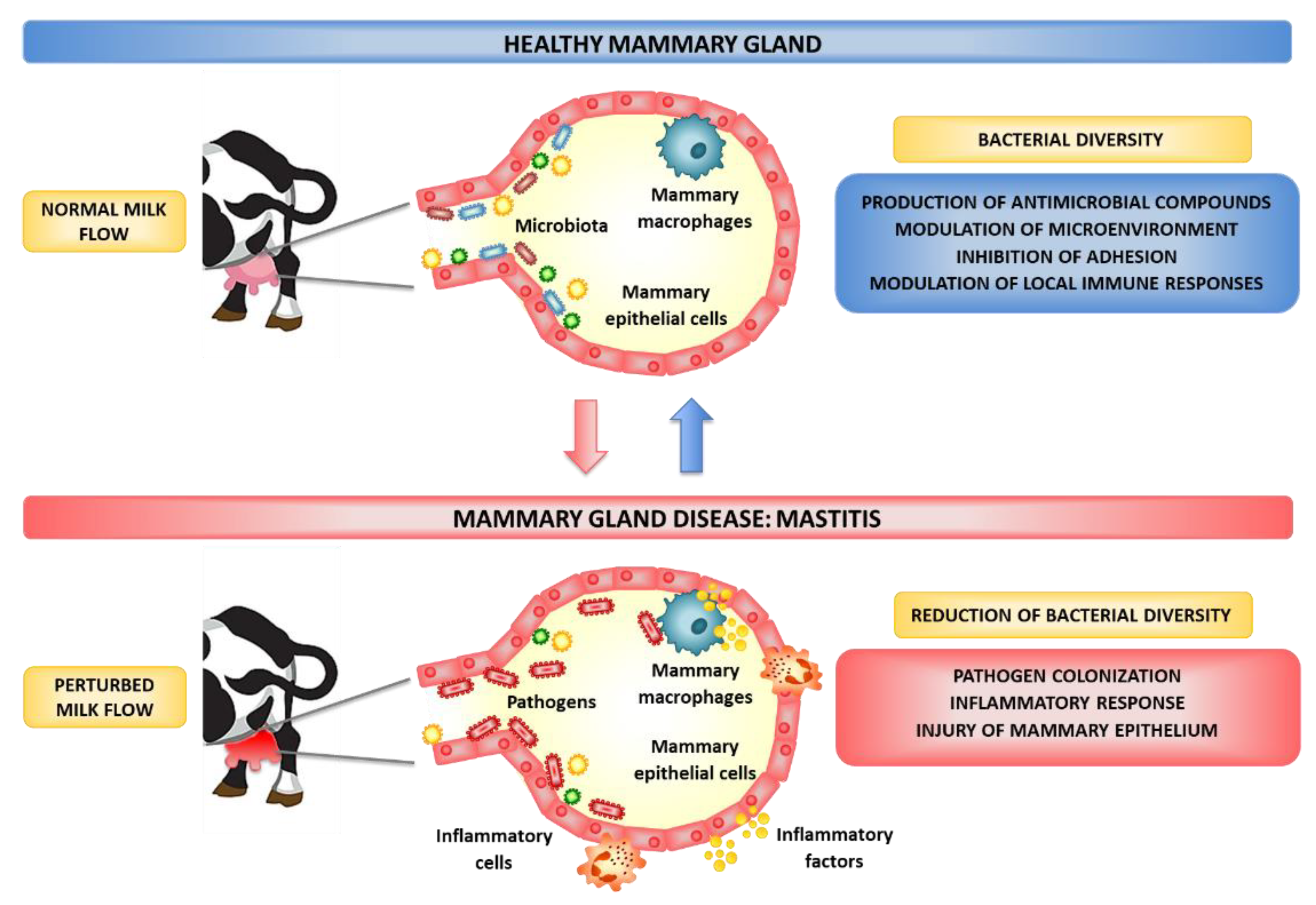
Figure 1. Effect of bovine mammary gland microbiota on the susceptibility to infections.
Microorganisms 10 02255 g001
Figure 2. Effect of probiotic and immunobiotic microorganisms on the susceptibility to bovine mammary gland infections.
Microorganisms 10 02255 g002
Publisher’s Note: MDPI stays neutral with regard to jurisdictional claims in published maps and institutional affiliations.