Article

Genetic Diversity and Geographical Distribution of the Red Tide Species Coscinodiscus granii Revealed Using a High-Resolution Molecular Marker

1
School of Marine Sciences, Ningbo University, Ningbo 315211, China
2
CAS Key Laboratory of Marine Ecology and Environmental Sciences, Institute of Oceanology, Chinese Academy of Sciences, Qingdao 266071, China
3
Laboratory of Marine Ecology and Environmental Science, Qingdao National Laboratory for Marine Science and Technology, Qingdao 266200, China
4
Center for Ocean Mega-Science, Chinese Academy of Sciences, Qingdao 266071, China
5
Department of Molecular Biology and Biochemistry, Simon Fraser University, 8888 University Drive, Burnaby, BC V5A 1S6, Canada
*
Author to whom correspondence should be addressed.
Microorganisms 2022, 10(10), 2028; https://doi.org/10.3390/microorganisms10102028
Submission received: 4 September 2022 / Revised: 28 September 2022 / Accepted: 5 October 2022 / Published: 14 October 2022
(This article belongs to the Special Issue Microbial Communities on the Surface of Algae)

Abstract

:
Diatoms are responsible for approximately 40% of the global primary photosynthetic production and account for up to 20% of global carbon fixation. Coscinodiscus granii is a red tide forming species of the phylum Bacillariophyta that has been detected in a wide range of coastal regions, suggesting the possibility of the existence of high genetic diversity with differential adaptation. Common molecular markers including 18S rDNA, 16S rDNA, ITS, cox1, and rbcL do not provide sufficient resolution for distinguishing intra-species genetic diversity, hindering in-depth research on intra-species genetic diversity and their spatial and temporal dynamics. In this project, we aimed to develop molecular markers with high resolution and specificity for C. granii, attempting to identify different taxa of this species, which will set up a stage for subsequent functional assays. Comparative genomics analysis of the mtDNAs of C. granii strains identified a genomic region with high genomic variations, which was used to guide the development of a molecular marker with high resolution and high specificity. This new molecular marker, which was named cgmt1 (C. granii mitochondrial 1), was 376 bp in size and differentiated C. granii samples collected in coastal regions of China into three different clades. Preliminary analysis of field samples collected in various coastal regions in China revealed that C. granii clades were almost exclusively found in the Bohai Sea and the north Yellow Sea. This newly developed molecular marker cgmt1 could be used for tracking intra-species genetic diversity and biogeographic distribution of C. granii in different ecosystems.

1. Introduction

Diatoms (Bacillariophyta) are single-celled eukaryotic algae that serve as primary source of photosynthesis (and oxygen production) in the ocean. Diatoms are the most species-rich group of algae, with more than 200,000 estimated species, contributing up to 20% of global primary productivity [1,2,3]. Coscinodiscus (Coscinodiscophyceae, Bacillariophyta) is a species-rich genus with 174 taxonomically accepted species (Guiry and Guiry 2022), among which 49 species have been identified in various coastal regions in China [4]. Some Coscinodiscus species can form red tides that may cause serious damage to aquaculture through competitive utilization of nutrients or by causing extensive clogging of fishing nets and other equipment [5,6] and can cause hypoxia and bring substantial losses to fishery by generating a large amount of sediments [7]. As some Coscinodiscus species cannot tolerate cold temperature, their distribution indicates warming ocean temperatures and have been used to track global warming (https://sites.evergreen.edu/vms-spring/coscinodiscus/accessed on 1 June 2016).
Coscinodiscus granii L.F. Gough 1905 is a cosmopolitan Coscinodiscus species [8]. Coscinodiscus granii plays an important role in the marine ecosystem and has an important impact on the carbon pool because of its relatively large cell sizes and high carbon content [9]. Coscinodiscus granii has been found to form characteristic autumn red tides in the Baltic Sea and exist at high densities in spring and summer in the North European Seas [10,11]. In China, C. granii is commonly found in coastal waters and has been listed as the dominant phytoplankton species in the Bohai Sea [12,13].
Despite the wide existence of C. granii, few studies have been conducted on C. granii [14], little is known about the biogeographic distribution characteristics of C. granii and about its genetic diversity. Early research on diatoms including C. granii mainly focused on morphological characteristics observed using microscopy. The application of molecular biology methods and common molecular markers including 18S rDNA, 28S rDNA, ITS, cox1, and rbcL enabled quantitative analysis of phytoplankton [15,16,17]. While these molecular markers can be effectively used to distinguish species, they have limited power in resolving intra-species variations. In addition, accumulating evidence suggested the existence of high genetic diversity among red tide strains isolated from different geographical regions. For example, Phaeocystis globosa strains isolated from Hong Kong and Shantou, China were shown to have different toxicity during red tides, optimum growth temperature, and colony size [18]. As such, we expect that similar studies are needed to address C. granii intra-species diversity, which will be valuable for tracking ecosystemic functions and the formation red tides. We hypothesize that C. granii genetic diversity plays a role in its adaptation and red tide development. The goal of this study was to develop a molecular marker that has low intra-genome variation and high resolution for distinguishing different strains.
Mitochondrial genomes (mtDNAs), which carry many important genes related to cell respiration and metabolism, have been analyzed in diverse studies, including evolutionary studies, species identification, and speciation analysis [19,20]. Multiple mtDNA-based molecular markers including cox1 and cytochrome b (cob), 16S rDNA have been commonly used for species identification. Molecular markers with higher resolution and specificity have been developed based on the comparative analysis of mtDNAs for many eukaryotic algae [18,21,22].
In this project, we aimed to develop a molecular marker with both high resolution and high specificity for distinguishing C. granii strains through comparative analysis of C. granii mtDNAs. Taking advantage of the recently constructed mtDNA [9] as the reference sequence, we identified intra-species genome variations (GVs) among six C. granii strains. We also tested the applicability of this new molecular marker in probing C. granii genetic diversity in the coastal waters of China.

2. Materials and Methods

2.1. Strain Isolation, Culturing, and Preservation

Six candidate C. granii strains were isolated in water samples collected during expeditions in coastal waters of Jiaozhou Bay, China (Figure 1 and Table 1). The expeditions, which were carried out during August and November in 2020 on research vehicle “Innovation”, were supported by the Jiaozhou Bay National Marine Ecosystem Research Station, Institute of Oceanology, the Chinese Academy of Sciences. These strains were first identified using light microscopy (Zeiss, Oberkochen, Germany). Morphological characteristics of these strains accorded with the description of C. granii [23]. Single cells were identified by morphology and picked up by pipetting individual cells using a micropipette under an inverted light microscope, followed by multiple washes with sterile culture medium before transferring each single cell to 24-well polystyrene cell culture plate. The strains were cultured in L1 medium (including 1‰ volume fraction Na2SiO3·9H2O and a mixture of 1% streptomycin liquid 100×, Solarbio, Beijing, China) with autoclaved and sterile filtered seawater. The cultures were maintained at 19–20 °C, with an irradiance of about 68 μmol photons m−2 s−1 in a photoperiod cycle of 12 h L/12 h D. Vegetative cells of six cultures were harvested for sequencing. Identification of these C. granii strains were based on both morphological features and molecular characterization using full-length 18S rDNA and ribulose-1, 5-bisphosphate carboxylase (rbcL) genes [21]. Phylogenetic tree was based on maximum likelihood (ML), using by MEGA7 [24].

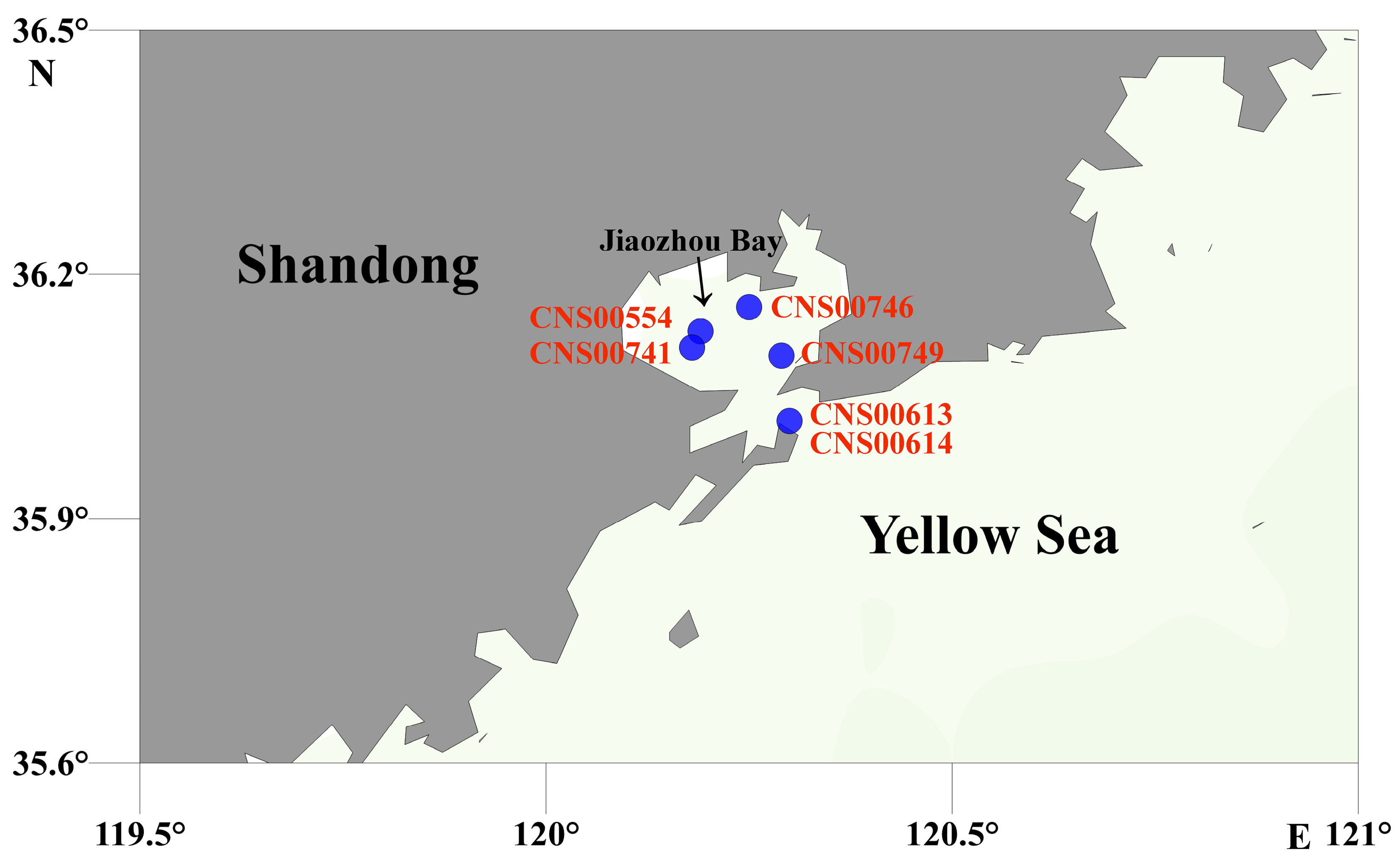
2.2. Field Sampling and Initial Preparation

Field samples were collected from 66 sampling sites from coastal waters of China, including the Bohai Sea (October 2019), the North Yellow Sea (October 2019), the Changjiang Estuary (March 2019), the East China Sea (September 2019), the Pearl River Estuary (June 2019), the Beibu Gulf (January 2019), and the Jiaozhou Bay (March 2021). Details of the sampling sites were shown in Figure 2 and Table S1. At each sampling site, 1000 mL surface water was filtered using mesh (Hebei Anping Wire Mesh Co., Ltd., Shijiazhuang, China) to remove large, suspended solids, zooplankton, and phytoplankton, followed by a second filtration through a 0.2 μm polycarbonate membranes (Millipore Corporation, Billerica, MA, USA) using a vacuum filtration pump. The filter membranes were transferred in tubes and were then snap-frozen in liquid nitrogen for brief storage for a few weeks.

2.3. DNA Preparation and Genome Sequencing

For cultures of C. granii strains, algal cells at exponential growth phase were transferred to a 50 mL centrifuge tube. Then, the algal cells were obtained by centrifugation (12,000× g, 6 min) and stored in a 1.5 mL EP tube for storage in liquid nitrogen for subsequent DNA extraction. Total genomic DNA from cultures and field samples were extracted with DNAsecure Plant Kit (Tiangen Biotech, Beijing, China) according to manufacturer’s instructions and quantified using a NanoDrop One spectrophotometer (Labtech International Ltd., Uckfield, UK). Genomic DNA samples of six C. granii strains were prepared for genome sequencing. The sequencing libraries were prepared by using the NEB Next® Ultra™ DNA Library Prep Kit for Illumina (NEB, Ipswich, MA, USA). The PCR products were purified using AMPure XP system (Beckman Coulter, Beverly, MA, USA), and libraries were analyzed for size distribution by NGS3K/Caliper and quantified using real-time PCR (Qubit®3.0 Flurometer, Thermo Scientific, Waltham, MA, USA). After qualification, the libraries were sequenced using a 2 × 150 bp Illumina NovaSeq 6000 platform (Illumina, San Diego, CA, USA) at Novogene (Beijing, China), yielding about 5 Gb sequencing data of paired-end reads with 150 bp in length.

2.4. Filtering and Assembly of Sequencing Data

Raw data were filtered into clean data with Fastq following a series of quality control (QC) procedures as previously described [25]. Here, briefly, the raw data processing steps included: (1) removing reads with >10 nt aligned to the adapters; (2) removing reads with ≥10% unidentified nucleotides (N); (3) removing reads with >50% bases having Phred quality < 5; and (4) removing putative PCR duplicates generated by PCR amplification in the library construction process.
The filtered reads were assembled into scaffolds with SPAdes (v3.14) [26], Platanus-allee (2.0.2) [27], and ABySS (2.1.5) [28] with default parameters. With the mtDNA of Thalassiosira pseudonana (NC_007405) serving as references, scaffolds corresponding to mtDNAs of C. granii were identified using BLAST with default parameters. Analysis of the resulting scaffold using MEGA 7.0 (v7.0) and DOTTER (v4.44.1) to estimate whether sequences at the ends achieved overlap. Subsequently, the obtained draft mtDNA sequences were checked by aligning sequencing reads against the mtDNA using the MEM algorithm of BWA v0.7.17. The results of alignments were extracted with SAMtools (1.9) and visualized with IGV v2.8.12 [18]. According to alignments, assembly errors were corrected and N regions were replaced in the draft mtDNA. Molecular markers including full-length 18S rDNA, 16S rDNA, ITS, and rbcL were assembled with SPAdes. Finally, the assembly results of the mtDNA and molecular markers of the C. granii strain (CNS00554) were all validated with BWA and IGV. The circular mtDNA (CNS00554) of C. granii was 34,970 bp in size (GenBank accession number: MW435847). The annotation of protein coding genes (PCGs), transfer RNA (tRNA) genes, ribosomal RNA (rRNA) genes, and open reading frames (orfs) was conducted using Open Reading Frame Finder (ORF finder) with SmartBLAST and BLASTP, tRNAscan-SE, and MFannot [9].

2.5. Phylogenetic Analysis and Synteny Analysis

A total of the 27 shared protein-coding genes (PCGs), including atp6, 8, 9; cob; cox1, 2, 3; nad1-7, 4L, 9; rpl2, 13, 14, 19; rps3, 4, 8; and tatC, from 34 publicly available diatom mtDNAs and two species of Ochrophyta, were first individually aligned using MAFFT [29] and then trimmed using trimAL [30] with default parameters: gt = 1, and all amino acid sequences were concatenated using Phyutility v1.2.2 [31]. Mitochondrial genes of two species Sargassum fusiforme (KJ946428) and Sargassum muticum (KJ938301) in Ochrophyta were selected as out-group taxa. The maximum likelihood (ML) phylogenetic tree was constructed using IQ-TREE (v1.6.12) [32]. Bootstrap analysis was performed using the ultrafast bootstrap approximation with 1000 replicates. In this study, p value was 0.01 after the ILD test, which indicated that sequence concatenation did not affect phylogenetic accuracy [19]. Comparison of mtDNAs of C. granii and C. wailesii was shown in the CIRCOS (v0.69) [21]. The order and content of genes in the mtDNAs of C. granii and C. wailesii were also displayed by Microsoft Excel 2019.

2.6. Developing Molecular Markers with High Resolution and High Specificity Based on C. granii mtDNAs

To search for GVs, we aligned Illumina reads of the six C. granii strains against the mtDNA of the reference strain CNS00554 using BWA (v0.7.17) with default parameters. Alignment results were screened using SAMtools with default parameters, and single-nucleotide variants (SNVs) with homozygous support >85% were called using VarScan (v2.4.4) [33]. The nucleotide diversity (Pi) values of C. granii mtDNAs were evaluated. The 400 bp (the length was appropriate for metabarcoding projects using Illumina DNA sequencing platform) sliding windows in the mtDNA of CNS00554 for SNVs were visualized using the R package circlize (v0.4.11) [34]. Primer Premier 5.0 was used to design the primers (cgmt1-F: TGGTGGGGAGGTTCTGTT; cgmt1-R: TTAAGCCTAAGTAGAGTTGA) of the novel high-resolution and high-specificity molecular markers cgmt1 (C. granii mitochondrial 1). The primers were synthesized by Sangon Biotech Co., Ltd., Shanghai, China. In order to verify the specificity and resolution of the designed primers, PCR amplification experiments were carried out. The PCR mixture contained 2 μL of each primer cgmt1-F and cgmt1-R (final concentration 200 nM each), 25 μL 2 × Taq Mastermix (Tiangen, China), 50 ng of template DNA and added ddH2O water to 50 μL volume. The PCR protocol was 94 °C for 3 min, 32 cycles of 30 s at 94 °C, 30 s at 55 °C, and 50 s at 72 °C, and a final extension at 72 °C for 10 min.

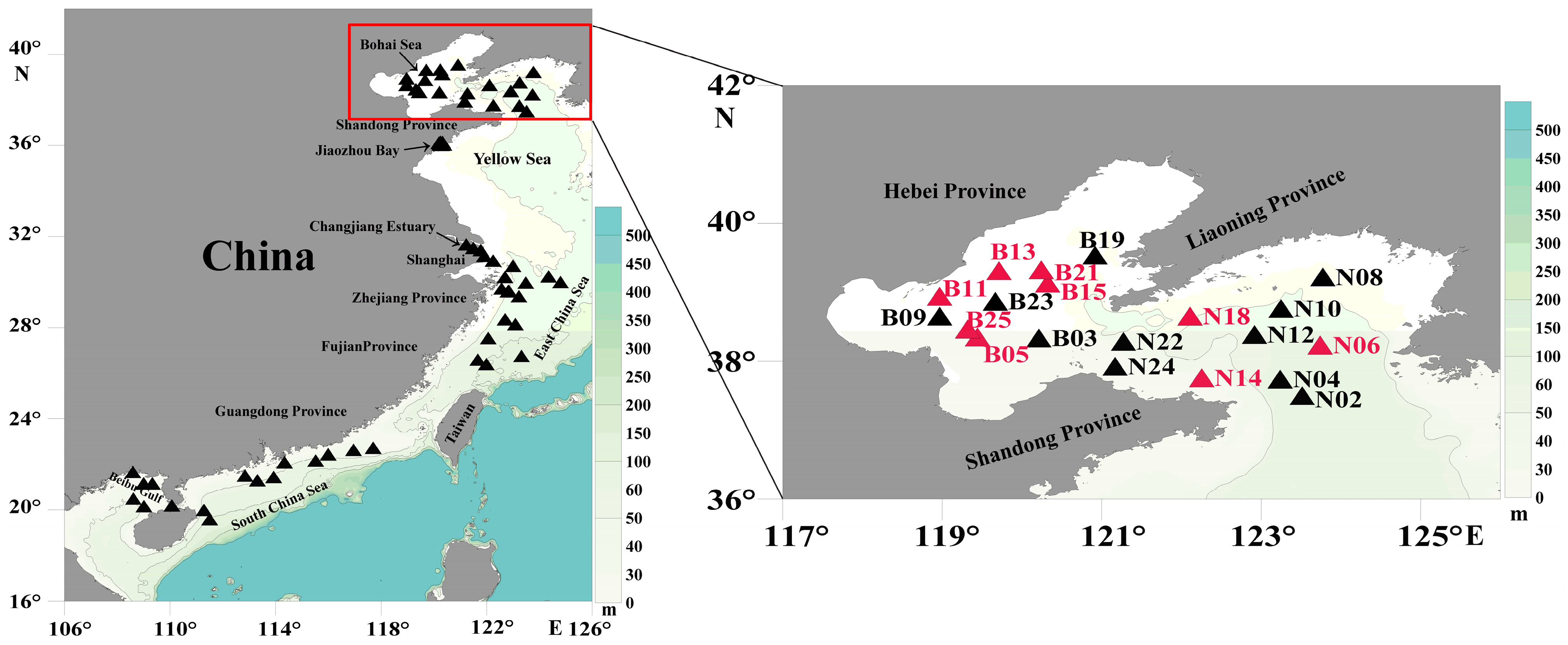
2.7. Genetic Diversity and Biogeographic Distribution Analyses Based on the Molecular Marker

With the high-resolution and high-specificity molecular marker developed in this project, we explored C. granii genetic diversity by examining amplification results of the molecular marker in 66 environmental samples collected from expeditions to the Bohai Sea, the North Yellow Sea, the Jiaozhou Bay, the Changjiang Estuary, the East China Sea, the Pearl River Estuary, and the Beibu Gulf (Figure 2 and Table S1). The environmental samples were PCR amplified and cloned before Sanger sequencing. The obtained forward and reversed fragments were assembled by ContigExpress (Vector NTI Suite 6.0, Invitrogen). Sanger sequencing results were aligned using MAFFT followed by editing using MEGA 7.0. The ML phylogenetic tree was constructed by the method mentioned above.

3. Results

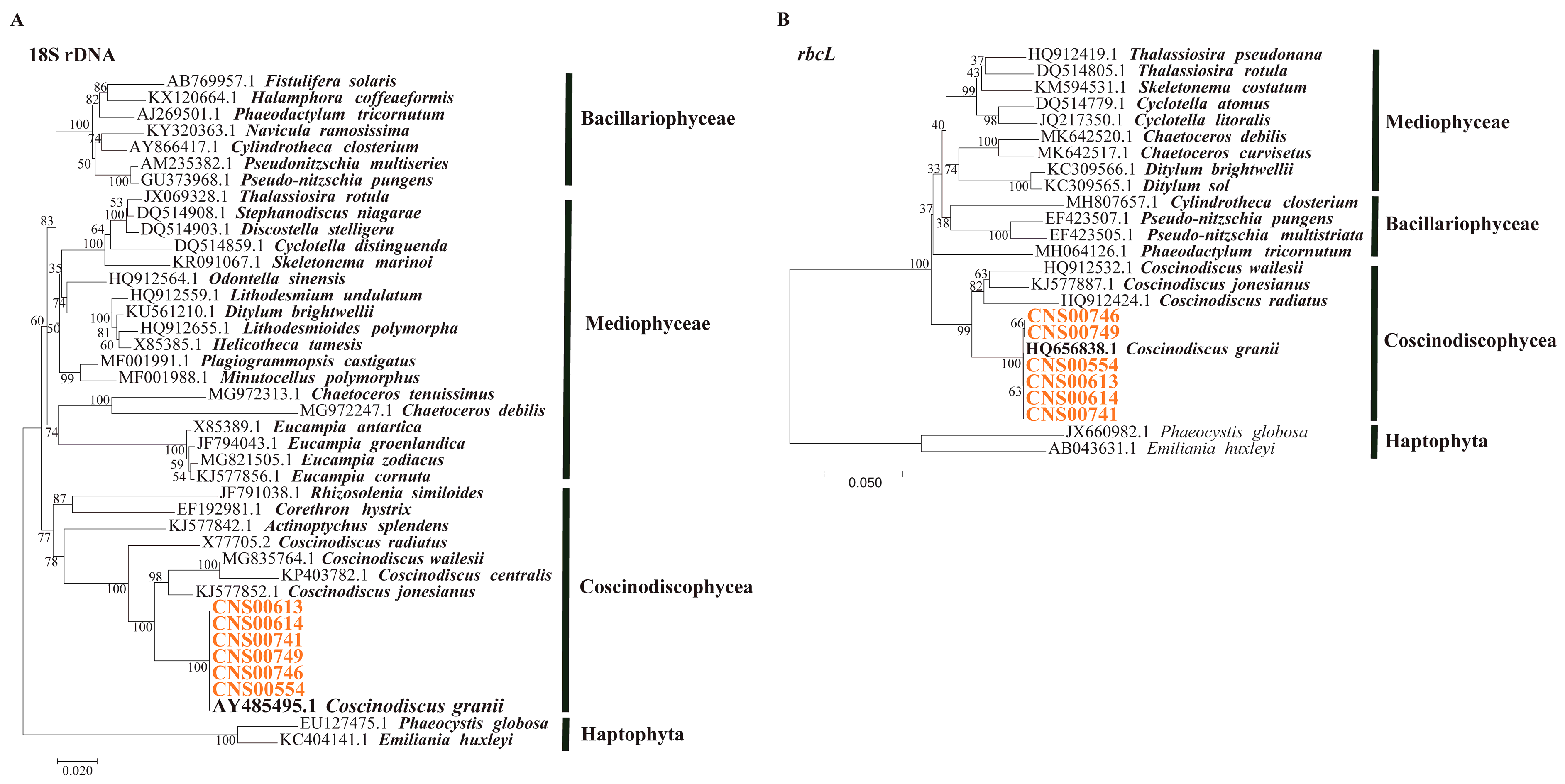
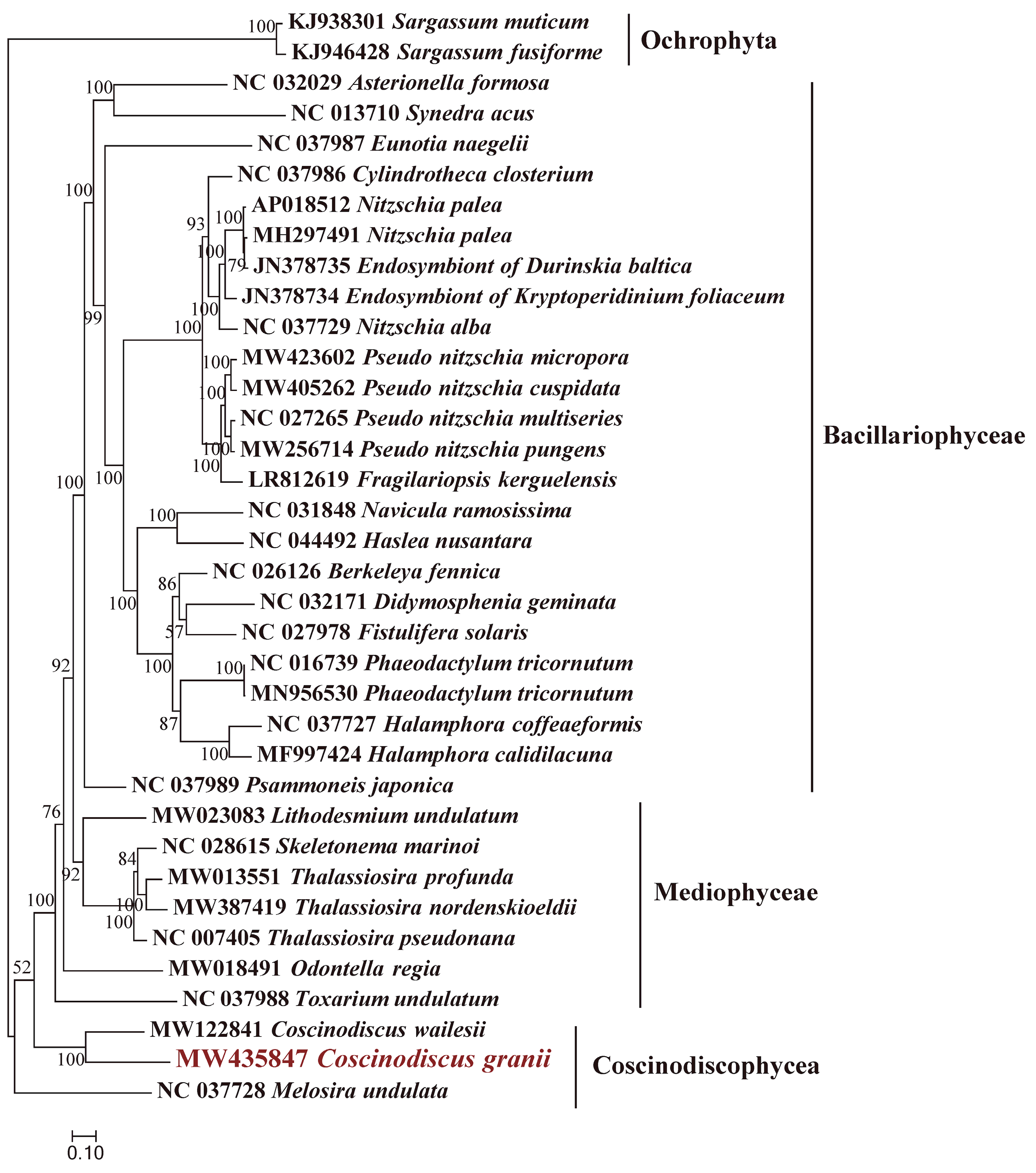
3.1. Morphological and Molecular Identification of C. granii Strains

Six candidate C. granii strains (CNS00554, CNS00613, CNS00614, CNS00741, CNS00746, and CNS00749) were collected in the Jiaozhou Bay. The cells of C. granii were easily identified in girdle view due to its wedged shape. The cell lengths of C. granii were about 60–300 μm and height was about 30–180 μm. In addition to morphological identification, this study used two common molecular markers (full-length 18S rDNA and rbcL) for further identification. The six strains were clustered well with C. granii sequences reported previously in the maximum likelihood (ML) phylogenetic trees based on the marker full-length 18S and rbcL (Figure 3). The results of phylogenetic analysis indicated that these six strains were all C. granii.

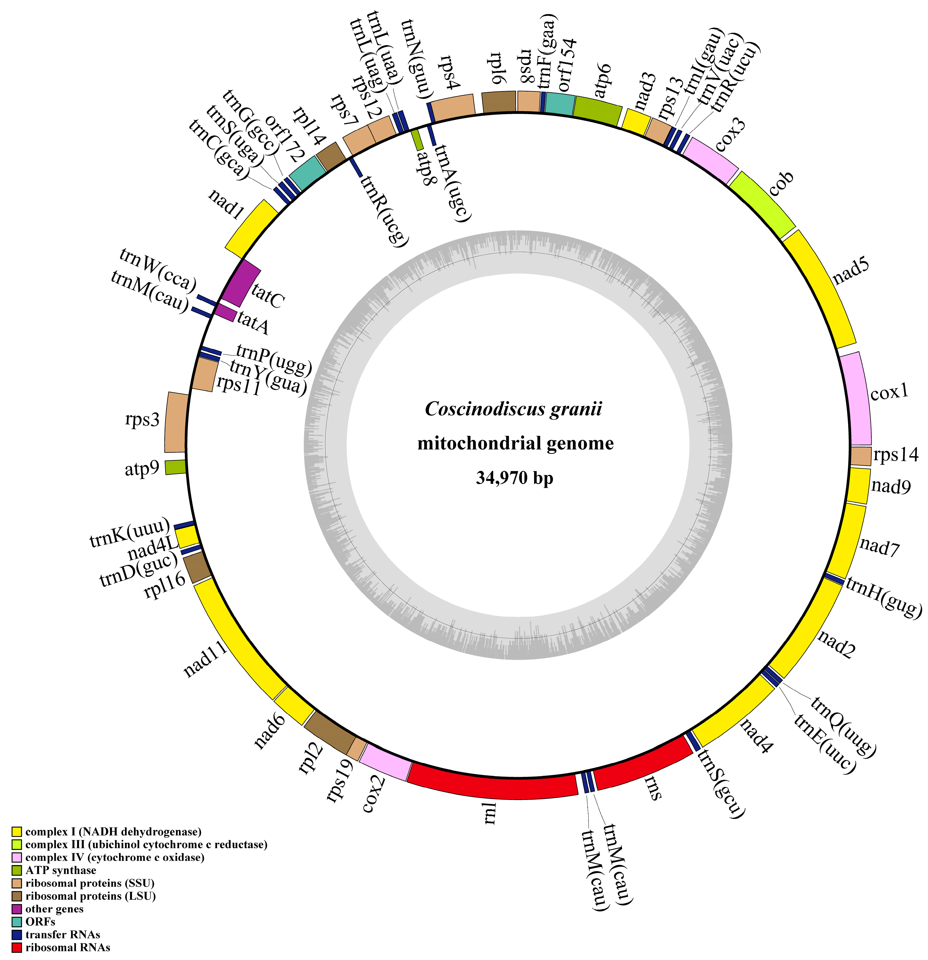
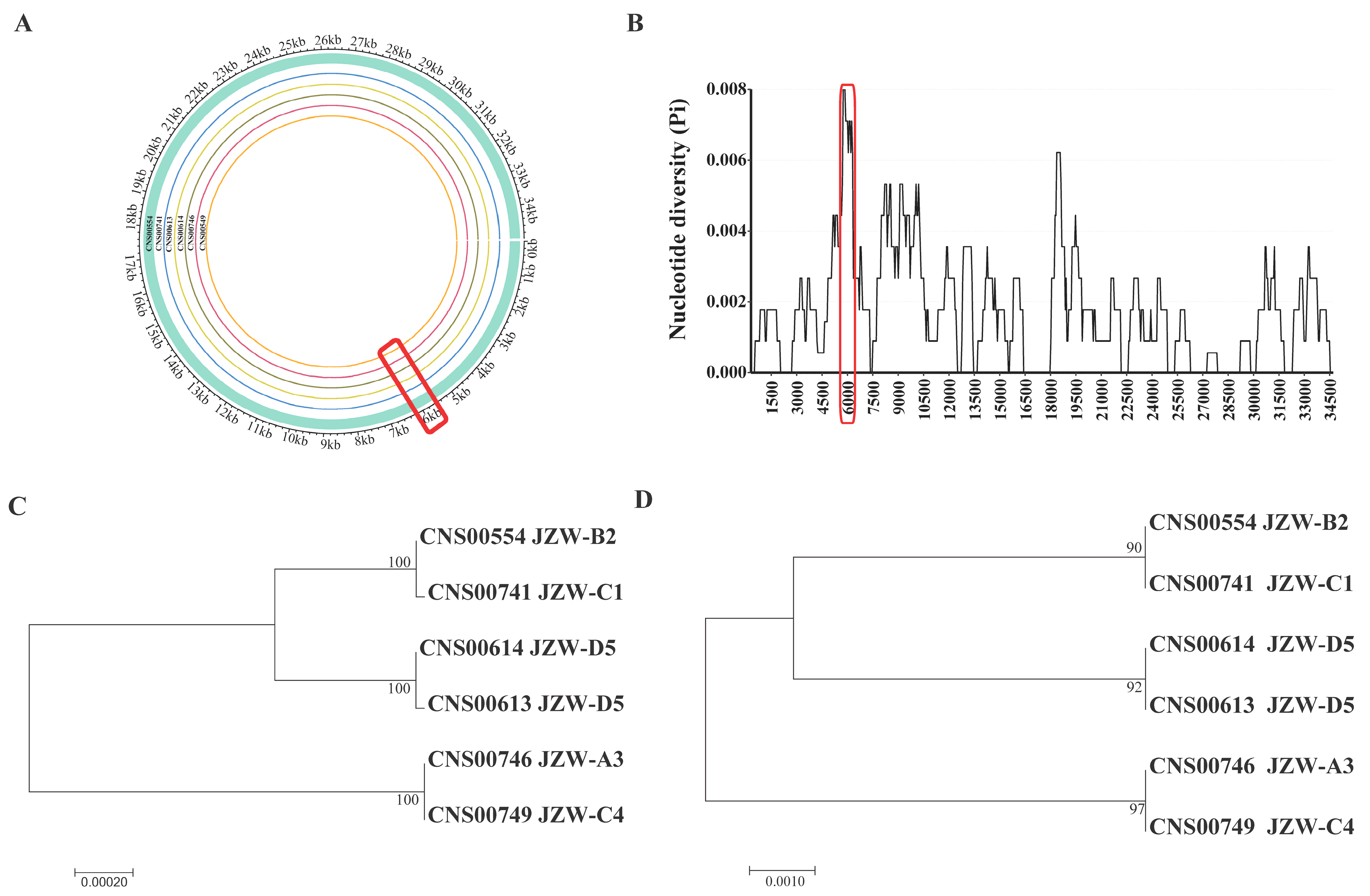
3.2. Phylogenetic Analysis and Synteny Analysis of C. granii mtDNAs

The complete mtDNA (GenBank accession number: MW435847) of C. granii (strain CNS00554) has a circular genome of 34,970 bp in size, encoding 60 genes (Figure 4). The ML phylogenetic trees constructed using 27 common PCGs shared by mtDNAs of 34 Bacillariophyta and two Ochrophyta species as outgroup taxa indicated that 34 species in Bacillariophyta formed a single clade-the Bacillariophyceae. The Mediophyceae and Coscinodiscophyceae were both paraphyletic assemblages. The phylogenetic tree also showed that the C. granii (MW435847) clustered with C. wailesii (MW122841) with robust support (Figure 5). Synteny comparison of mtDNAs of C. granii and C. wailesii revealed the order of genes in the mtDNAs of the two species were generally similar, except for the rearrangements (translocation and inversion) of a few gene blocks, including trnP(ugg)-trnY(gua)-rps11 and atp8-trnA(ugc) (Figure 6).

3.3. Defining a Molecular Marker with High Resolution and High Specificity for Distinguishing C. granii Strains

To develop molecular markers for tracking genetic diversity of C. granii using Illumina DNA sequencing, the length of the molecular markers should ideally be between 350 and 400 bp. Comparative analysis of the six C. granii mtDNAs identified a 400 bp-window with dense variations (Figure 7A). Furthermore, sliding window analysis of nucleotide variability (Pi) was used by complete mtDNAs of six C. granii strains (Figure 7B). In the end, we identified a genomic region containing 6 SNVs in mtDNA of C. granii strain CNS00554 (position: 5649–6048 bp). According to the optimization principle of primer software design, the amplification primers were designed in the region of 5749–6248 bp. Phylogenetic analysis using this small region suggested that it could be used to effectively distinguish these strains as molecular marker. Six strains of C. granii were divided into three clades based on this region (Figure 7D), which achieved similar resolution as the complete mtDNA (Figure 7C). The high resolution of molecular marker, which we named cgmt1, was 397 bp in size.
To test the specificity of newly developed molecular marker cgmt1, two analyses were carried out as the follows. First, we carried out BLAST searches of cgmt1 in NCBI NT database, which showed low similarity (less than 29%) to sequences of other species, suggesting high specificity to C. granii. Second, we carried out PCR assays using primers designed against cgmt1 and DNA preparations extracted from different 14 representative eukaryotic algae species, including Amphidinium carterae, Alexandrium tamarense, Karenia mikimotoi, and Prorocentrum donghaiense of the phylum Dinoflagellata, Chattonella marina, Aureococcus anophagefferens, and Heterosigma akashiwo of the phylum Ochrophyta, Skeletonema costatum, Thalassiosira weissflogii, Chaetoceros curvisetus, C. wailesii, and Coscinodiscus sp. of the phylum Bacillariophyta, Phaeocystis globosa and Isochrysis galbana of the phylum Haptophyta. We also included DNA preparations extracted from six C. granii strains as positive control. While most of these 14 representative eukaryotic algae species showed completely negative results, some showed some but greatly reduced signals. (Figure S1).

3.4. Probing C. granii Genetic Diversity and Geographical Distribution Using cgmt1

To evaluate genetic diversity and explore the biogeographical distribution of C. granii strains, we PCR amplified cgmt1 from 66 environmental samples collected from various coastal areas of China. Among these 66 PCR amplicons, nine showed strong signals, which were further analyzed by cloning and sequencing. Sequencing results revealed that C. granii was mainly detected in the Bohai Sea and the North Yellow Sea (Figure 2). Phylogenetic analysis based on cgmt1 sequence from 6 different strains of C. granii and 55 sequencing results from 9 positive environmental samples showed high genetic diversity (Figure 8). The phylogenetic analysis revealed at least three clades. Clade 1 included sequences of environmental samples from the Yellow Sea and the Bohai Sea, as well as sequences of strains from the Jiaozhou Bay. Clade 2 contained only sequences from the Jiaozhou Bay. Clade 3 contained sequences of environmental samples collected from the Bohai Sea and strains from the Jiaozhou Bay. The results indicated that the molecular marker cgmt1 had high resolution, and that it could be used for tracking genetic diversity of C. granii in the field studies.

4. Discussion

Coscinodiscus granii has a worldwide distribution, suggesting adaptation to a wide range of temperatures [11] and salinities and possibly high genetic diversity. Coscinodiscus granii has been reported to form dense red tides, usually present great abundance in numerous ocean regions around the world, including the Bohai Sea and the Yellow Sea of China. However, current methods including morphology-based methods and common molecular marker-based methods have been inadequate for uncovering intra-species genetic differences. Common molecular markers including 18S rDNA, 28S rDNA, rbcL, and ITS have been demonstrated to be ineffective in resolving Phaeocystis globosa intra-species genetic diversity [18]. While cox1 gene showed high resolution for resolving some algal species, the presence of introns in cox1 in many species limits its application [16,35]. Molecular markers with high resolution and high specificity for intra-species genetic diversity analysis have been developed through comparative analysis of mtDNAs [19,21,22].
Our analysis revealed the mtDNA of C. granii was 34,970 bp in size, which was shorter than the mtDNA (36,071 bp) of C. wailesii and most diatom mtDNAs that were generally compact with few repeats and a small number of introns [36]. In this study, the mtDNA of C. granii included small intergenic regions, the absence of introns, and the low repeat content, which made it smaller size of mtDNA than others [22,37,38,39]. Rearrangements of DNA sequence containing four main events: translocation, inversion, duplication, and deletion [40]. Comparative analysis of C. granii and C. wailesii mtDNAs showed that most gene arrangements were conserved, except for the rearrangements of a few gene blocks, including translocation and inversion events. Notably, ribosomal protein coding genes rps2, rps10, and rpl5 that are found in mtDNAs of many other diatom species [21,22] were absent from the mtDNA of C. granii. Furthermore, the two orfs identified in C. granii shared no similarity with these three genes. The reason for this may be that the three genes were lost or transferred to the nucleus during evolution [41]. The AT content of the mtDNA of C. granii was 75.70%, which was essentially the same as that of C. wailesii (75.00%), and substantially higher than that of M. undulata (78.40%) in the class Coscinodiscophyceae [40]. Comparative analysis of mtDNAs can facilitate exploring synteny relationships among closely related species and ascertaining gene gains or losses in evolution [42].
Mitochondrial genomes evolve quickly and thus provide a rich source of molecular variations at the nucleotide level, which made it more suitable for the development of high-resolution molecular markers [43,44]. In this study, we collected and isolated six C. granii strains from Jiaozhou Bay in China, covering two seasons (summer and autumn). We successfully developed a new molecular marker named cgmt1 based on comparative analysis of C. granii mtDNAs, which showed high resolution and specificity in distinguishing different C. granii strains. Furthermore, we used cgmt1 as the molecular marker to track and probe C. granii geographical distribution and genetic diversity in environmental samples collected in the Bohai Sea, the North Yellow Sea, the Jiaozhou Bay, the Changjiang Estuary, the East China Sea, the Pearl River Estuary, and the Beibu Gulf. In this study, phylogenetic analysis of cgmt1 amplified from different samples showed that C. granii was mainly distributed in the Bohai Sea and the North Yellow Sea of China, which was consistent with the results of previous studies [12]. The results also highlighted the reliability and practicability of the newly developed molecular markers and indicated that C. granii had high genetic diversity. Indeed, such effective marker cgmt1 could facilitate studies on C. granii. On the one hand, it could be used in metabarcoding analyses for tracking the geographical distribution patterns of C. granii genotypes not only in Chinese coastal waters but also other ocean regions of the world. On the other hand, we could distinguish different strains of C. granii from different geographical spans and seasons, especially during red tides period, to evaluate strain-specific differential contribution to red tides and to trace the possible origin of C. granii red tides.

5. Conclusions

Through comparative analysis of mtDNAs, we successfully developed a new molecular marker cgmt1 (C. granii mitochondrial 1) with high resolution and specificity for C. granii intra-species genetic differences. We demonstrated that it could be used effectively to investigate the biogeographical distribution and track genetic diversity of C. granii. This molecular marker holds great potential applications for the studies on biogeographic distribution and intra-species genetic diversity of C. granii in coastal waters of China and other countries.

Supplementary Materials

The following supporting information can be downloaded at: https://www.mdpi.com/article/10.3390/microorganisms10102028/s1, Figure S1: Specificity of molecular marker cgmt1; Table S1: The sampling station information of environmental samples.

Author Contributions

N.C. conceived and designed the experiments. H.H. collected the samples. H.H. performed the experiments. H.H. wrote the manuscript with input from other authors. H.H., Q.X. and H.S. analyzed the data. Q.X., H.S. and N.C. read and approved the manuscript. All authors have read and agreed to the published version of the manuscript.

Funding

This research was supported by the Natural Science Foundation of China (42176162), the Strategic Priority Research Program of Chinese Academy of Sciences (Grant No. XDB42000000), the Chinese Academy of Sciences Pioneer Hundred Talents Program (to Nansheng Chen), the Taishan Scholar Project Special Fund (to Nansheng Chen), the Qingdao Innovation and Creation Plan (Talent Development Program—5th Annual Pioneer and Innovator Leadership Award to Nansheng Chen, 19-3-2-16-zhc), the Post-doctoral Program of Qingdao 2020 (Grant No. E0KY061), and the Natural Science Foundation of Shandong Province, China (Grant No. ZR2020QD095).

Institutional Review Board Statement

Not applicable.

Informed Consent Statement

Not applicable.

Data Availability Statement

The original contributions presented in the study are publicly available. The data can be found here: https://www.ncbi.nlm.nih.gov/nuccore/MW435847, MW122841, MZ571469-MZ57147, MZ561055-MZ561066, MZ544474-MZ544479, MZ544480-MZ544485 (accessed on 2 November 2021); Illumina raw sequences are available on NCBI’s sequence read archive (SRA) at BioProject PRJNA689860, PRJNA686182, PRJNA777367.

Acknowledgments

We are thankful to all staff of marine ecological environment genomics research group in Institute of Oceanology, Chinese Academy of Sciences. We are grateful to colleagues from the Jiaozhou Bay Marine Ecosystem Research Station for their help in field sampling.

Conflicts of Interest

The authors declare that they have no known personal relationships or competing financial interests that could have appeared to influence the work reported in this paper.

References

  1. Malviya, S.; Scalco, E.; Audic, S.; Vincent, F.; Veluchamy, A.; Poulain, J.; Wincker, P.; Iudicone, D.; de Vargas, C.; Bittner, L.; et al. Insights into global diatom distribution and diversity in the world’s ocean. Proc. Natl. Acad. Sci. USA 2016, 113, E1516–E1525. [Google Scholar] [CrossRef] [Green Version]
  2. Angela, F.; Marianne, J.; Jean-Pierre, B.; Benjamin, B.; Thomas, M. Diatom Molecular Research Comes of Age: Model Species for Studying Phytoplankton Biology and Diversity. Plant Cell 2019, 32, 547–572. [Google Scholar] [CrossRef] [Green Version]
  3. Mann, D.G.; Vanormelingen, P. An inordinate fondness? The number, distributions, and origins of diatom species. J. Eukaryot. Microbiol. 2013, 60, 414–420. [Google Scholar] [CrossRef] [PubMed] [Green Version]
  4. Huang, H.; Wang, Y.; Song, H.; Wang, J.; Chen, Y.; Zhao, Y.; Liu, F.; Chen, N. The complete mitochondrial genome and phylogenetic analysis of Coscinodiscus wailesii (Coscinodiscophyceae, Bacillariophyta). Mitochondrial DNA B Res. 2021, 6, 1849–1851. [Google Scholar] [CrossRef] [PubMed]
  5. Tetsuya, N.; Kazutaka, M.; Satoshi, N. Effects of temperature and salinity on the growth of the giant diatom Coscinodiscus wailesii isolated from Harima-Nada, Seto Inland Sea, Japan. Nippon Suisan Gakkaishi 2000, 66, 993–998. [Google Scholar] [CrossRef] [Green Version]
  6. Nishikawa, T.; Tarutani, K.; Yamamoto, T. Nitrate and phosphate uptake kinetics of the harmful diatom Coscinodiscus wailesii, a causative organism in the bleaching of aquacultured Porphyra thalli. Harmful Algae 2010, 9, 563–567. [Google Scholar] [CrossRef]
  7. Laing, I.; Gollasch, S. Coscinodiscus wailesii—A Nuisance Diatom in European Waters. In Invasive Aquatic Species of Europe. Distribution, Impacts and Management; Springer: Amsterdam, The Netherlands, 2002. [Google Scholar]
  8. Yan, D.; Beardall, J.; Gao, K. Variation in cell size of the diatom Coscinodiscus granii influences photosynthetic performance and growth. Photosynth. Res. 2018, 137, 41–52. [Google Scholar] [CrossRef]
  9. Huang, H.; Song, H.; Zhao, Z.; Liu, F.; Chen, N. Complete mitochondrial genome of Coscinodiscus granii (Coscinodiscophyceae, Bacillariophyta). Mitochondrial DNA Part B 2021, 6, 2332–2334. [Google Scholar] [CrossRef] [PubMed]
  10. Wasmund, N.; Pollehne, F.; Postel, L.; Siegel, H.B.; Zettler, M.L. Assessment of the biological state of the Baltic Sea in 2007. Proc. Natl. Acad. Sci. USA 2008, 89, 8842–8846. [Google Scholar] [CrossRef] [Green Version]
  11. Kraberg, A.C.; Baumann, M.; Dürselen, C. Coastal Phytoplankton; Verlag Dr. Friedrich Pfei: München, Germany, 2010. [Google Scholar]
  12. Zhang, Q.; Yin, C.; Xu, Y.; Shi, H. The phytoplankton community sampled by Nets in the dominant area monitoring red tide in Bohai Bay in summer, 2006. J. Tianjing Univ. Sci. Technol. 2007, 22, 20–23. [Google Scholar] [CrossRef]
  13. Zhang, X.; Xu, X.; Fang, E.J.; Hou, C.; Chen, W.; Zhang, B. The characteristic of phytoplankton community and its variation at artificial reef area of the coastal sea of Tianjin. Mar. Sci. Bull. 2019, 21, 41–55. [Google Scholar]
  14. Theriot, E.C.; Ashworth, M.; Ruck, E.; Jansen, N. A preliminary multigene phylogeny of the diatoms. Plant Ecol. Evol. 2010, 143, 278–296. [Google Scholar] [CrossRef] [Green Version]
  15. Hu, X.; Zhang, Q.; Chen, Z.; Kong, F.; Wang, J.; Yu, R. Genetic diversity of Phaeocystis globosa strains isolated from the beibu gulf, the south China sea. Oceanol. Limnol. Sin. 2019, 50, 601–610. [Google Scholar]
  16. Mao, Q.; Yang, S.; Jin, W.; Yan, Z.; Yan, Y.; Jiao, L.; Xu, B.; Jiao, J. Research status and prospects for DNA barcode technology in algal taxonomy. J. Hydroecol. 2020, 41, 9–18. [Google Scholar] [CrossRef]
  17. Chen, Y.; Xu, Q.; Gibson, K.; Chen, N. Metabarcoding dissection of harmful algal bloom species in the East China Sea off Southern Zhejiang Province in late spring. Mar. Pollut. Bull. 2021, 169, 112586. [Google Scholar] [CrossRef]
  18. Song, H.; Chen, Y.; Gibson, K.; Liu, S.; Yu, Z.; Chen, N. High genetic diversity of the harmful algal bloom species Phaeocystis globosa revealed using the molecular marker COX1. Harmful Algae 2021, 107, 102065. [Google Scholar] [CrossRef] [PubMed]
  19. Wang, Y.; Chen, Y.; Wang, J.; Liu, F.; Chen, N. Mitochondrial genome of the harmful algal bloom species Odontella regia (Mediophyceae, Bacillariophyta). J. Appl. Phycol. 2021, 33, 855–868. [Google Scholar] [CrossRef]
  20. Plese, B.; Kenny, N.J.; Rossi, M.E.; Cardenas, P.; Schuster, A.; Taboada, S.; Koutsouveli, V.; Riesgo, A. Mitochondrial evolution in the Demospongiae (Porifera): Phylogeny, divergence time, and genome biology. Mol. Phylogenet. Evol. 2021, 155, 107011. [Google Scholar] [CrossRef] [PubMed]
  21. Chen, Y.; Wang, Y.; Liu, K.; Liu, F.; Chen, N. Development of a high-resolution molecular marker for tracking Pseudo-nitzschia pungens genetic diversity through comparative analysis of mitochondrial genomes. J. Appl. Phycol. 2021, 33, 2283–2298. [Google Scholar] [CrossRef]
  22. Zhang, M.; Cui, Z.; Liu, F.; Chen, N. Definition of a High-Resolution Molecular Marker for Tracking the Genetic Diversity of the Harmful Algal Species Eucampia zodiacus Through Comparative Analysis of Mitochondrial Genomes. Front. Microbiol. 2021, 12, 631144. [Google Scholar] [CrossRef] [PubMed]
  23. Goessling, J.W.; Paulo, C.; Michael, K. Photo-Protection in the Centric Diatom Coscinodiscus granii is Not Controlled by Chloroplast High-Light Avoidance Movement. Front. Mar. Sci. 2016, 2, 115. [Google Scholar] [CrossRef] [Green Version]
  24. Kumar, S.; Stecher, G.; Tamura, K. MEGA7: Molecular Evolutionary Genetics Analysis Version 7.0 for Bigger Datasets. Mol. Biol. Evol. 2016, 33, 1870–1874. [Google Scholar] [CrossRef] [PubMed]
  25. Song, H.; Liu, F.; Li, Z.; Xu, Q.; Chen, Y.; Yu, Z.; Chen, N. Development of a high-resolution molecular marker for tracking Phaeocystis globosa genetic diversity through comparative analysis of chloroplast genomes. Harmful Algae 2020, 99, 101911. [Google Scholar] [CrossRef] [PubMed]
  26. Bankevich, A.; Nurk, S.; Antipov, D.; Gurevich, A.A.; Dvorkin, M.; Kulikov, A.S.; Lesin, V.M.; Nikolenko, S.I.; Pham, S.; Prjibelski, A.D.; et al. SPAdes: A new genome assembly algorithm and its applications to single-cell sequencing. J. Comput. Biol. 2012, 19, 455–477. [Google Scholar] [CrossRef] [PubMed] [Green Version]
  27. Kajitani, R.; Yoshimura, D.; Okuno, M.; Minakuchi, Y.; Kagoshima, H.; Fujiyama, A.; Kubokawa, K.; Kohara, Y.; Toyoda, A.; Itoh, T. Platanus-allee is a de novo haplotype assembler enabling a comprehensive access to divergent heterozygous regions. Nat. Commun. 2019, 10, 1702. [Google Scholar] [CrossRef] [PubMed] [Green Version]
  28. Jackman, S.D.; Vandervalk, B.P.; Mohamadi, H.; Chu, J.; Yeo, S.; Hammond, S.A.; Jahesh, G.; Khan, H.; Coombe, L.; Warren, R.L.; et al. ABySS 2.0: Resource-efficient assembly of large genomes using a Bloom filter. Genome Res. 2017, 27, 768–777. [Google Scholar] [CrossRef] [Green Version]
  29. Nakamura, T.; Yamada, K.D.; Tomii, K.; Katoh, K. Parallelization of MAFFT for large-scale multiple sequence alignments. Bioinformatics 2018, 34, 2490–2492. [Google Scholar] [CrossRef] [PubMed] [Green Version]
  30. Capella-Gutierrez, S.; Silla-Martinez, J.M.; Gabaldon, T. trimAl: A tool for automated alignment trimming in large-scale phylogenetic analyses. Bioinformatics 2009, 25, 1972–1973. [Google Scholar] [CrossRef] [PubMed] [Green Version]
  31. He, Z.; Chen, Y.; Wang, Y.; Liu, K.; Xu, Q.; Li, Y.; Chen, N. Comparative analysis of Pseudo-nitzschia chloroplast genomes revealed extensive inverted region variation and Pseudo-nitzschia speciation. Front. Mar. Sci. 2022, 9, 784579. [Google Scholar] [CrossRef]
  32. Liu, K.; Liu, S.; Chen, Y.; Liu, F.; Chen, N. Complete mitochondrial genome of Thalassiosira profunda (Mediophyceae, Bacillariophyta). Mitochondrial DNA Part B 2021, 6, 1560–1562. [Google Scholar] [CrossRef] [PubMed]
  33. Koboldt, D.C.; Zhang, Q.; Larson, D.E.; Shen, D.; McLellan, M.D.; Lin, L.; Miller, C.A.; Mardis, E.R.; Ding, L.; Wilson, R.K. VarScan 2: Somatic mutation and copy number alteration discovery in cancer by exome sequencing. Genome Res. 2022, 22, 568–576. [Google Scholar] [CrossRef] [Green Version]
  34. Gu, Z.; Gu, L.; Eils, R.; Schlesner, M.; Brors, B. circlize Implements and enhances circular visualization in R. Bioinformatics 2014, 30, 2811–2812. [Google Scholar] [CrossRef]
  35. Evans, K.M.; Wortley, A.H.; Mann, D.G. An assessment of potential diatom “barcode” genes (cox1, rbcL, 18S and ITS rDNA) and their effectiveness in determining relationships in Sellaphora (Bacillariophyta). Protist 2007, 158, 349–364. [Google Scholar] [CrossRef]
  36. Secq, O.L.; Green, B.R. Complex repeat structures and novel features in the mitochondrial genomes of the diatoms Phaeodactylum tricornutum and Thalassiosira pseudonana. Gene 2011, 476, 20–26. [Google Scholar] [CrossRef]
  37. Kamikawa, R.; Azuma, T.; Ishii, K.I.; Matsuno, Y.; Miyashita, H. Diversity of Organellar Genomes in Non-photosynthetic Diatoms. Protist 2018, 169, 351–361. [Google Scholar] [CrossRef]
  38. Wynn, E.L.; Christensen, A.C. Repeats of Unusual Size in Plant Mitochondrial Genomes: Identification, Incidence and Evolution. G3 2019, 9, 549–559. [Google Scholar] [CrossRef] [Green Version]
  39. Liu, F.; Liu, S.; Huang, T.; Chen, N. Construction and comparative analysis of mitochondrial genome in the brown tide forming alga Aureococcus anophagefferens (Pelagophyceae, Ochrophyta). J. Appl. Phycol. 2020, 32, 441–450. [Google Scholar] [CrossRef]
  40. Pogoda, C.S.; Keepers, K.G.; Hamsher, S.E.; Stepanek, J.G.; Kane, N.C.; Kociolek, J.P. Comparative analysis of the mitochondrial genomes of six newly sequenced diatoms reveals group II introns in the barcoding region of cox1. Mitochondrial DNA Part A 2019, 30, 43–51. [Google Scholar] [CrossRef]
  41. Ravin, N.V.; Galachyants, Y.P.; Mardanov, A.V.; Beletsky, A.V.; Petrova, D.P.; Sherbakova, T.A.; Zakharova, Y.R.; Likhoshway, Y.V.; Skryabin, K.G.; Grachev, M.A. Complete sequence of the mitochondrial genome of a diatom alga Synedraacus and comparative analysis of diatom mitochondrial genomes. Curr. Genet. 2010, 56, 215–223. [Google Scholar] [CrossRef]
  42. Lei, B.; Li, S.; Liu, G.; Chen, Z.; Su, A.; Li, P.; Li, Z.; Hua, J. Evolution of mitochondrial gene content: Loss of genes, tRNAs and introns between Gossypium harknessii and other plants. Plant Syst. Evol. 2013, 299, 1889–1897. [Google Scholar] [CrossRef]
  43. Chen, S.; Wei, D.; Shao, R.; Shi, J.; Dou, W.; Wang, J. Evolution of multipartite mitochondrial genomes in the booklice of the genus Liposcelis (Psocoptera). BMC Genom. 2014, 15, 861. [Google Scholar] [CrossRef] [PubMed] [Green Version]
  44. Kikuchi, T.; Afrin, T.; Yoshida, M. Complete mitochondrial genomes of four entomopathogenic nematode species of the genus Steinernema. Parasit. Vectors 2016, 9, 430. [Google Scholar] [CrossRef] [PubMed]
Figure 1. Sampling sites of the six strains of Coscinodiscus granii. Sampling sites are marked by blue shapes.
Microorganisms 10 02028 g001
Figure 2. Geographical distribution of environmental samples used in this study. The environmental samples are marked by triangles. The results of positive amplified samples are marked by red shapes.
Microorganisms 10 02028 g002
Figure 3. Identification of six strains of C. granii. (A,B): ML Phylogenetic trees based on the marker 18S rDNA and rbcL, respectively.
Microorganisms 10 02028 g003
Figure 4. Circular maps of the mtDNA of Coscinodiscus granii (Strain CNS00554). Circular map of the mtDNA of C. granii. The transcriptional direction inside the ring is clockwise and the transcriptional direction outside the ring is counterclockwise. The protein-coding genes and rRNAs and tRNAs genes are labeled inside or outside the circle. The color of the gene boxes indicates the different functional groups of genes. The ring of bar graphs on the inner circle shows the GC content in dark gray.
Microorganisms 10 02028 g004
Figure 5. Phylogenetic analysis of C. granii (CNS00554). Maximum likelihood (ML) phylogenetic tree using concatenated amino acid sequences of 27 common protein-coding genes from 34 publicly diatom mtDNAs, and Sargassum fusiforme (KJ946428) and Sargassum muticum (KJ938301) in Ochrophyta were used as out-group taxa. The numbers beside branch nodes are the percentage of 1000 bootstrap values.
Microorganisms 10 02028 g005
Figure 6. Synteny comparison of C. granii and C. wailesii. (A): Block diagram of gene content and gene rearrangement, tRNAs are indicated by capital single letter. Solid line boxes indicate the same conserved block of genes, while dotted boxes indicate translocation event and gene block connected by a red line indicates inversion event. Blocks with the same color represent the same type of genes. (B): The assignment of genes into different functional groups is indicated by colors.
Microorganisms 10 02028 g006
Figure 7. Genomic variations and phylogenetic trees based on maximum likelihood (ML) analysis of six C. granii strains. (A): Genomic variations density in six C. granii strains. The cyan band represented the reference genome CNS00554. The range of cgmt1 region was marked by red box. From inside to outside, circles represent six C. granii strains isolated from the Jiaozhou Bay. (B): Sliding window analysis of nucleotide variability (Pi) across complete mtDNAs of six C. granii strains. (C): Phylogenetic analysis using the whole mtDNAs of six C. granii strains. (D): Phylogenetic analysis using the newly developed marker cgmt1. The sea area label marked with abbreviations: Jiaozhou Bay (JZW).
Microorganisms 10 02028 g007
Figure 8. Phylogenetic analysis based on the cgmt1 sequences of C. granii obtained from strains and environmental samples. The sea areas were marked with abbreviations: Jiaozhou Bay (JZW), Bohai Sea (BH), and Yellow Sea (HH).
Microorganisms 10 02028 g008
Table 1. The sampling station information of the six Coscinodiscus granii strains.
Strain NumberStationsSampling TimeLongitude (°E)Latitude (°N)
CNS00554JZW-B22020.08120.1936.13
CNS00741JZW-C12020.08120.1836.11
CNS00613JZW-D52020.09120.3036.02
CNS00614JZW-D52020.09120.3036.02
CNS00746JZW-A32020.11120.2536.16
CNS00749JZW-A42020.11120.2936.10
Publisher’s Note: MDPI stays neutral with regard to jurisdictional claims in published maps and institutional affiliations.

