Article

Inhibitory Properties of Aldehydes and Related Compounds against Phytophthora infestans—Identification of a New Lead

by
John J. Mackrill
1,*,
Roberta A. Kehoe
2,
Limian Zheng
1,
Mary L. McKee
2,
Elaine C. O’Sullivan
2,
Barbara M. Doyle Prestwich
3 and
Florence O. McCarthy
2,*
1
Department of Physiology, School of Medicine, University College Cork, T12 K8AF Cork, Ireland
2
School of Chemistry and Analytical and Biological Chemistry Research Facility, University College Cork, Western Road, T12 K8AF Cork, Ireland
3
School of Biological, Earth and Environmental Sciences, University College Cork, T12 K8AF Cork, Ireland
*
Authors to whom correspondence should be addressed.
Pathogens 2020, 9(7), 542; https://doi.org/10.3390/pathogens9070542
Submission received: 5 June 2020 / Revised: 1 July 2020 / Accepted: 3 July 2020 / Published: 7 July 2020
(This article belongs to the Special Issue Biology and Pathology of Phytophthora infestans)

Abstract

:
The pathogen Phytophthora infestans is responsible for catastrophic crop damage on a global scale which totals billions of euros annually. The discovery of new inhibitors of this organism is of paramount agricultural importance and of critical relevance to food security. Current strategies for crop treatment are inadequate with the emergence of resistant strains and problematic toxicity. Natural products such as cinnamaldehyde have been reported to have fungicidal properties and are the seed for many new discovery research programmes. We report a probe of the cinnamaldehyde framework to investigate the aldehyde subunit and its role in a subset of aromatic aldehydes in order to identify new lead compounds to act against P. infestans. An ellipticine derivative which incorporates an aldehyde (9-formyl-6-methyl ellipticine, 34) has been identified with exceptional activity versus P. infestans with limited toxicity and potential for use as a fungicide.

1. Introduction

One of the most significant threats to global food production and agriculture is the pathogenic oomycete Phytophthora infestans (P. infestans) [1]. It was first described by Berkeley in 1846 and subsequently named Phytophthora infestans by deBary in 1876, which aptly translates as “plant destroyer” [2,3]. Most infamously known for the Irish potato famine in 1845–1849, the pathogen caused incessant, devastating outbreaks of late potato blight resulting in the deaths of one million people and a further one million emigrating [4]. The current global impact of P. infestans is in excess of $6.2 billion per annum due to crop loss and the use of fungicides [5]. The necessity of fungicides to combat the spread of P. infestans is evident and with resistant strains continually developing, designing novel fungicides to target this pathogen is of high importance.
The Irish Famine of the 1840s spurred the development of the first-ever pesticide to be adopted worldwide. Developed by French scientists, Pierre Millardet and Ulysse Gayon, the Bordeaux mixture was used to combat the P. infestans blight. This fungicide is a mixture of copper sulphate and lime, which forms the active compound copper(II) hydroxide, stabilised by calcium sulphate. The emergence of this fungicidal treatment was imperative to the conservation of crops and averting hunger, therefore saving lives [6]. The use of the Bordeaux mixture diminished through the emergence of less toxic and more targeted fungicides [7].
The Bordeaux mixture was broadly used from the 1890s until it was superseded by the dithiocarbamate class of fungicides in the 1960s. The broad-spectrum fungicides were used due to their high biological and chemical activity, with the benefit of low production cost [8]. Mancozeb (1) and Propineb (3) were two of the most widely used dithiocarbamate fungicides [9,10]. Both of these organosulfur fungicides have been found to break down into toxic metabolites as degradation begins during storage to form the respective toxic metabolites (Figure 1) [11,12]. Ethylene thiourea 2 (R = H), one of the main metabolites of Mancozeb, has been shown to have many toxic effects in rats such as teratogenicity, developmental toxicity and promoting thyroid neoplasms [11]. Propylene thiourea 4 (R = CH3) from Proprineb has also been shown to affect the thyroid and the nervous system [8]. Propineb (2) has been banned by the European Commission and Mancozeb (1) is now under scrutiny for its negative effects.
Following dithiocarbamates, phenylamide fungicides were developed in the 1970s. A significant advance in the control of P. infestans is attributed to the phenylamides. Metalaxyl (5) (Figure 2) is one of the most popular phenylamide fungicides, however, seven years after the product was commercialised, the first resistant strains of P. infestans were identified in Israel [13]. By 1980, resistant isolates of P. infestans were discovered outside of Israel, in Ireland, Switzerland and The Netherlands, demonstrating a need for further fungicidal development. To combat the growing resistance of P. infestans for phenylamides, a mixture was formulated with other fungicides (e.g., Mancozeb, 1) which resulted in the reduction of resistant strains emerging [14].
Approved by the EU in 2017, oxathiapiprolin (6) has been used in more recent years as a targeted antioomycete fungicide (Figure 2). It was designed and synthesised by Pasteris et al. in 2016 [15] basing the structure around a piperidine-thiazole-carbonyl core. After numerous derivatives were synthesised and tested, the chemical structure of oxathiapiprolin (6) exhibited the greatest control over P. infestans with low toxicity in humans, birds and fish. The authors also investigated the mechanism of action of oxathiapiprolin and uncovered a novel target for oomycete inhibition. The compound binds to an oxysterol binding protein, although the role for this family is not very well understood. Oxathiapiprolin (6) has been approved in many countries around the world and requires a smaller quantity to be used on crops [16]. However, similarly to the phenylamides, oxathiapiprolin has already been found to be ineffective against emerging resistant strains of Phytophthora capsici [15,17]. The current focus has turned towards the use of natural products as fungicides to replace the previous synthetic alternatives. Although the first report of antimicrobial natural products was in 1676, development has been slow for these compounds [18]. Of prime interest for natural source fungicides is the essential oil, cinnamaldehyde.
Cinnamaldehyde (7, Figure 3) is a natural product extracted from the bark of the plant genus Cinnamona. This conjugated aromatic compound has shown to inhibit the growth of bacteria, filamentous moulds and yeast [19]. There are over 250 species of cinnamon plants and trees and are native to Sri Lanka and South India [20]. Cinnamaldehyde was first isolated from cinnamon oil in 1834 by Dumas and Peligot [21] and was first synthesised in 1854 by Chiozza [22]. The essential oil is generally recognised as safe by the United States Food and Drug Administration and has been used in foods and medicines as flavourings for many years [23]. It is also reported to possess many pharmacological activities such as antidiabetic and anti-inflammatory effects [24,25]. Extensive research has also been carried out on the antibacterial properties of cinnamaldehyde. The compound has demonstrated strong activity against many food-borne bacteria such as Bacillus, Staphylococcus and Enterobacter spp. and can therefore be used as a food preservative [26,27,28].
Studies by Quintanilla and Soylu [29,30] were some of the first to demonstrate the effect essential oils have on P. infestans and a follow up study by Ferhout et al. found that cinnamaldehyde (7) expressed a greater inhibitory effect on fungal strains than thyme oil [31]. Research by Raut determined cinnamaldehyde to be the most effective phenylpropanoid from plant origin against Candida albicans biofilm formation, being effective at completely inhibiting mature biofilms and identifying its potential [32]. Jantan et al. tested 14 essential oils against six dermatophytes, one filamentous fungus and five strains of yeasts, finding again that cinnamaldehyde possessed the strongest activity against all the different strains of fungi studied [33].
From experimental evidence, it appears cinnamaldehyde (7) inhibits cell wall biosynthesis, membrane function and some specific enzyme activities. However, specific cinnamaldehyde targets are still not well established. It is known that at lethal concentrations, the cell membrane is disrupted by cinnamaldehyde whereas at below lethal concentrations, it can inhibit ATPase and at low concentrations, it disrupts enzymes involved in cytokine action. It has been demonstrated in many studies that cinnamaldehyde (7) interacts with microbial cell membranes and that it is capable of altering the membrane lipid profile [34]. It is thought that the lipophilicity of cinnamaldehyde allows the molecule to partition through the lipid membrane of the cells and thus, disrupts bilayer structure. This renders the cell more permeable and prone to leakage of intracellular material causing cell death [26]. At lethal concentrations, cinnamaldehyde acts as a noncompetitive inhibitor of cell wall synthesising enzymes leading to the prevention of cell wall formation [35].
At sub-lethal concentrations, cinnamaldehyde inhibits transmembrane ATPase activity in E. coli and Listeria monocytogenes by entering into the cellular periplasm [36]. This enzyme is essential for maintaining transmembrane electrochemical proton gradient, necessary for the maintenance of cellular pH and uptake of nutrients [37]. At low concentration, cinnamaldehyde has been demonstrated to inhibit GDP dependent polymerisation, preventing cell division [38]. In P.parasitica var. nicotianae, Lu et al. found that cinnamaldehyde completely inhibited mycelial growth, sporangial formation, zoospore production and germination at concentrations ranging from about 10 μM to 50 μM [39].
In a study of resistance of Capsicum annuum (pepper plant) against root rot caused by P.capsici, Li et al. discovered that resistant plants displayed increased transcription of genes involved in the phenylpropanoid biosynthesis pathway, especially those involved in cinnamaldehyde production [40]. This suggests that cinnamaldehyde is a naturally occurring protective compound, whose production is enhanced in plant strains that are resistant to oomycete infections.
Transient Receptor Potential (TRP) ion channels play an important role in sensing damage to cells. Members of this channel superfamily, most closely related to the polycystic kidney disease (PKD) family, are encoded within the genomes of all oomycetes examined [41]. Compounds that are susceptible to nucleophilic attack from cystine can cause TRP channel activation, disrupting Ca2+ levels in the cell. Hu et al. demonstrated that the α, β-unsaturated bond in cinnamaldehyde is susceptible to nucleophilic attack from cystine, and therefore, modulates Ca2+ homeostasis in Phytophthora capsici (P. capsici) [42]. Intracellular free Ca2+ is a universal second messenger in eukaryotic cells, regulates cellular processes such as sporulation, germination and growth and is important in every life-cycle stage [41,43]. The covalently bound cinnamaldehyde to cystine was postulated to activate the TRP channel leading to an efflux of Ca2+ out of the cell and subsequently leads to cell death. To test their theory that the α, β-unsaturated bond in cinnamaldehyde is important, Hu monitored Ca2+ levels using a cinnamaldehyde derivative without the α, β-unsaturated bond, hydrocinnamaldehyde (8, Figure 3). Treatment of P. capsici with hydrocinnamaldehyde (8) demonstrated no inhibitory effects of zoospore growth indicating that the Michael addition through the α, β-unsaturated bond of cinnamaldehyde was crucial for stimulating Ca2+ efflux and growth inhibition of P. capsici.
Thus, although many studies exist on antimicrobial activity against several different groups of microorganisms, there is much to be learned about the mode of action of cinnamaldehyde 7 and the scope of its effects on P. infestans. As we set out to identify new agents against P. infestans, we initiated a broad study on the importance of the aldehyde aspect of cinnamaldehyde.
Previous cinnamaldehyde analogues that have been subjected to P. infestans testing have primarily retained their aldehyde functionality and are substituted on the aromatic ring, examples of which are represented in Figure 4 (913) [44]. Separately, a recent study by Watamoto found a diverse set of compounds to demonstrate inhibitory effects on fungal strains, amongst which some of the structural features of our targets align [45]. Two hit compounds from this screen (14 and 15, Figure 4) have shown strong fungicidal effects and to inhibit the metabolic activity of C. albicans. Of these, Bay 11-7082 (14) possesses an α, β-unsaturated bond similar to cinnamaldehyde and may act through a similar mechanism. Ellipticine (15) has known antimicrobial and especially anticancer properties: testing at bacterial planktonic mode showed (15) had averaged a minimal inhibitory concentration (MIC) of 8.45 µM across all six Candida strains and hence is worthy of further investigation.
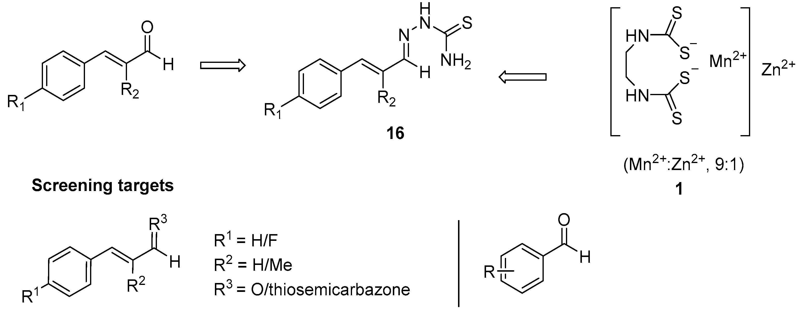
The aims of this study are to synthesise and evaluate cinnamaldehyde and aromatic aldehyde derivatives in assays of P. infestans growth and zoospore production. Cinnamaldehyde derivatives will be accessed by specific synthetic modification at positions R1, R2 and R3 represented in Figure 5. 4-Fluorocinnamaldehyde (R1 = F, 17) was chosen to probe the effect on electron density withdrawal on the Michael acceptor. At the R2 position α-methylcinnamaldehyde (R2 = CH3, 18) will test steric effects as the methyl group can influence attack on this reactive element. Finally, the rationale is provided by the structure of Mancozeb (1, Figure 5) for the generation of a hybrid structure 16 and the related thiosemicarbazone at the R3 position. Extension of the conjugated aldehyde will also be assessed to identify optimal chain length.
In order to probe the aldehyde functionality, a series of aromatic aldehydes and ellipticine aldehydes will also be assessed in the P. infestans assay to screen for potential lead compounds for future development (Figure 5). Given the experience of the group, of particular interest are the ellipticines which would offer a new framework for the inhibition of P. infestans growth and the potential for overcoming resistance.

2. Results

Synthesis of novel cinnamaldehyde and ellipticine derivatives was initially undertaken and in addition to some commercial aldehydes were assessed in a parallel approach, via the determination of inhibitory effects on the mycelial growth and zoosporogenesis of Phytophthora infestans and cytotoxicity against a human cell line.

2.1. Effects of Mancozeb, Cinnamaldehyde and Hydrocinnamaldehyde on P. infestans Mycelial Growth

There is substantial published evidence indicating that Mancozeb (1) [9] and cinnamaldehyde (7) [39,42] inhibit the mycelial growth of Phytophthora and of other oomycetes. In contrast, at concentrations up to 2 mM, hydrocinnamaldehyde (8) is reported to have no detectable effect on the growth of mycelia from the zoospores of P.capsici [42]. We sought to test the potency of these molecules in inhibiting the mycelial growth in our experimental system (Figure 6). After five days of incubation, cinnamaldehyde displayed a minimal inhibitory concentration (MIC, the lowest concentration resulting in no visible growth) for P. infestans mycelial growth of approximately 1 mM and a half-maximal inhibitory concentration (IC50) of about 0.5 mM (see Supplementary Information). In contrast to the results of Hu et al. [42], we found that hydrocinnamaldehyde inhibited Phytophthora mycelial growth with an MIC of 2 mM and an IC50 of between 0.25 and 0.5 mM. However, it should be noted that the data reported by Hu et al. are from a different species (P.capsici versus P. infestans) and mycelia were generated from a different source (germinated from zoospores versus subcultured from growing mycelia). Mancozeb inhibited P. infestans growth most effectively, with an MIC of 100 μM and an IC50 of between 25 and 50 μM. These MIC and IC50 values for Mancozeb are similar to those reported previously for P. infestans, other oomycetes and in the fungus Cylindrocarpon destructans [10,46]. Based on these findings, we selected a fixed concentration of 25 μM to screen compounds for their ability to inhibit P. infestans growth (in order to identify molecules that are more effective than Mancozeb), with 1 mM cinnamaldehyde and 100 μM Mancozeb in positive control experiments.

2.2. Cinnamaldehyde and Aryl Aldehyde Screen

Due to the commercial availability of cinnamaldehyde based starting materials, trans-cinnamaldehyde (7), 4-fluorocinnamaldehyde 17 and α-methylcinnamaldehyde 18 were chosen as starting points for analogue formation and for testing against P. infestans (Figure 7). In addition, a series of aromatic aldehydes were identified for screening to probe the relevance of the conjugated aldehyde as a key functional group responsible for the effects (2024). Indole-3-aldehydes 20 and 21 are structurally related to the tryptophan-derived plant metabolite parent (19) which has been identified to confer immunity in plants to microbial pathogen infection [47].

2.2.1. Synthesis of Extended Cinnamaldehyde Derivatives

To probe the effect of the aldehyde group of cinnamaldehyde on the inhibition of P. infestans growth, trans-cinnamaldehyde (7) was treated with (carbethoxymethylene)triphenylphosphorane 25 (Scheme 1) yielding product as a colourless oil 26. The same procedure was followed for the 4-fluorocinnamaldehyde 17 starting material however after 2 h there was still evidence of starting material. An additional 0.1 equivalent of the ylide 25 was added and stirred at room temperature for an additional 16 h. The crude material was isolated with the product 27 isolated as a colourless oil which solidified to colourless crystals overnight.
α-Methylcinnamaldehyde 18 was also used in this reaction however it proved more problematic. Treatment with 25 and stirring in dichloromethane for 48 h showed no consumption of starting material by TLC analysis. A second equivalent of the ylide 25 was added and the reaction was let stir at 35 °C for a further 48 h. NMR analysis of the crude reaction mixture showed evidence of product formation but also the characteristic aldehyde starting material peak at 9.59 ppm with isolation of the pure product proving problematic.

2.2.2. Synthesis of Cinnamaldehyde Thiosemicarbazone Derivatives

Given the structural distinction between cinnamaldehyde (7) and Mancozeb (1), it was envisaged that synthesis of a hybrid thiosemicarbazone derivative of the cinnamaldehyde could proffer improved activity based on the individual components. trans-Cinnamaldehyde (7) was dissolved in absolute ethanol and treated with thiosemicarbazide and a catalytic amount of acetic acid (Scheme 2). The starting material was consumed within 45 min and after isolation resulted in pure product 28 in 34% yield. The 4-fluorocinnamaldehyde reaction proceeded to completion after 16 h at 80 °C and the α-methyl derivative 30 was formed after 48 h at 80 °C. Both 28 and 29 crystallised as yellow needles whereas 30 was an amorphous white solid.

2.3. Synthesis of Ellipticines and 9-Formylellipticines

The Gribble and Saulnier route to ellipticine (15) was chosen to probe the potential of ellipticines as it is a versatile and high yielding synthesis [48,49]. The amenable nature of the ellipticine scaffold allows for derivatisation at numerous positions (Scheme 3). The N-6 position of ellipticine was substituted by the addition of sodium hydride to deprotonate the indole nitrogen (Scheme 3). Due to different alkyl iodides possessing different reactivities, the time taken for the reaction to go to completion varied. For the methyl derivative 31, 16 h was sufficient to achieve full conversion, whereas the ethyl derivative 32 took 72 h to go to completion and the isopropyl derivative 33 48 h albeit with significantly reduced yield. The quantity of alkylating agent could only be used in slight excess as the addition of excess or extended reaction times can lead to quaternary salt formation on the pyridine nitrogen.
Ellipticine was then formylated at the nine-position to form the aldehyde by a method described by Plug et al. [50] and as first described by Duff (Scheme 3) [51,52]. This reaction was carried out on 6-substituted ellipticine derivatives to form 9-formylellipticine derivatives 34, 35 and 36. The formylation step was generally high yielding with good purity.

2.4. Screening of Cinnamaldehydes, Aromatic Aldehydes and Ellipticines against P. infestans

Compounds were tested for their ability to inhibit the mycelial growth and the production of zoospores by P. infestans [42]. The cytotoxicity of compounds against human cells was also evaluated, in the human embryonic kidney 293T (HEK-293T) cell line, using a sodium 3’-[1-(phenylaminocarbonyl)-3,4-tetrazolium]-bis (4-methoxy-6-nitro) benzene sulfonic acid hydrate (XTT) reduction assay [53,54]. For all assays, a fixed concentration of 25 μM was selected for screening purposes. As described in Section 2.1, Mancozeb (1) and cinnamaldehyde (7) were used in positive control experiments at their MIC values, of 100 μM and 1 mM, respectively. The vehicle for these compounds, DMSO, was used at a final concentration of 0.1% in negative control experiments.

2.4.1. Phytophthora infestans Mycelial Growth Assay

In total, 17 compounds were evaluated for relative inhibitory effects on P. infestans mycelial growth at a concentration 25 µM after 5, 9 and 13 days.

Day 5

Cinnamaldehyde (7) and 9-formyl-6-methylellipticine 34 exhibit the best inhibition of P. infestans with no detectable growth for both compounds (Figure 8). All cinnamaldehyde derivatives 17, 18, 2630 prove not as potent as the parent cinnamaldehyde (7), although this is tested at a much higher concentration. It is interesting that thiosemicarbazone cinnamaldehyde derivatives 2830 show the slowest mycelial growth rate of all the test cinnamaldehyde series. As an example, the α-methyl thiosemicarbazone derivative 30 demonstrates only 38% growth which suggests the cinnamaldehyde/Mancozeb hybrid approach warrants further exploration. Unexpectedly, derivative 26 (ethyl-(2E)-5-phenylpenta-2,4-dienoate) caused a small but significant increase in P. infestans mycelial growth, of 113% relative to the vehicle control.
As expected, cinnamaldehyde (7) shows good inhibition of P. infestans which given its simple structure has been postulated to arise from the presence of an α,β-unsaturated aldehyde group and its potential to interact with cysteines in target proteins. Compounds (7), 17, 18, 2024 and 3436 arise from different chemical classes but all contain an aldehyde and in comparing their activity, there is no clear relationship between this functional group and mycelial growth.
The functionalised cinnamaldehydes 17 and 18 which contain the α,β-unsaturated aldehyde are identified with relatively low mycelial growth inhibition within this screen. Each of the proprietary aromatic aldehydes tested (2024) show moderate to low inhibition of P. infestans with 5-bromoindole-3-carbaldehyde (21) seen as the most promising.
The most effective compounds in the screen are cinnamaldehyde (7) and 9-formyl-6-methylellipticine 34 and structurally these comprise an α,β-unsaturated aldehyde and an aromatic aldehyde. While it is evidently important for potency, this suggests that the aldehyde group is not solely responsible for inhibition of mycelial growth and that other factors must be explored.
Of the ellipticines, all six-alkylated compounds 31, 34, 35 and 36 exerted a significant growth inhibitory effect, with 34 having a C-9 aldehyde installed on the A-ring which may contribute to the activity of the compound. Although Mancozeb (1) is four times the concentration of the remaining test compounds, it causes a relatively low inhibition of growth, with six other test compounds demonstrating more potent effects.

Day 9

After nine days, it can be seen once again that cinnamaldehyde (7) and ellipticine aldehyde derivative 34 still demonstrate 100% inhibition of mycelial growth (Figure 9). Mancozeb (1) shows a decrease of 13% in the inhibition of mycelial growth since Day 5. The growth-promoting effect of compound 26 is maintained, at 6% greater than the vehicle control. Again, of particular note are the ellipticines 31, 34 and 35 which show promising potency at this extended stage.

Day 13

The data in Figure 10 demonstrate that there is a high rate of mycelial growth in P. infestans incubated with many of the test compounds after 13 days. However, there is still a consistent undetectable growth for cinnamaldehyde (7) and only 6% growth for 34. Mancozeb (1) has an increased rate of mycelium growth of 14% since Day 9. Aside from the ellipticines 31, 3436 the consistent activity of cinnamaldehyde/Mancozeb hybrid 30 is of note. The growth-promoting effect of 26 was not statistically significant after 13 days (the same as V, 100% growth).

2.4.2. P. infestans Zoosporogenesis and Sporangiogenesis

The test compounds were investigated for their effects on zoosporogenesis. The number of zoospores produced after two weeks’ culture under each condition was counted using a haemocytometer. All compounds with the exception of the aldehydes 20, 21, 23 and 24, and ellipticine (15), significantly reduced the production of zoospores. Several compounds demonstrated complete inhibition of zoospore formation with the ellipticines 31, 34 and 35 identified as comparable with cinnamaldehyde (Figure 11).
Decreased zoosporogenesis could result from decreased production of the sporangia, or from the defective generation of zoospores within these fruiting bodies. To test this, sporangiogenesis was measured in P. infestans cultured in the presence of aldehyde or cinnamaldehyde test compounds for a period of two weeks (Figure 12). Both Mancozeb, aldehyde 22 and ellipticine derivative 36 all caused significant reductions in sporangiogenesis. These fruiting structures were undetectable in P. infestans cultured in the presence of 1 mM cinnamaldehyde or 25 μM of ellipticines 31, 34 and 35, despite the latter three compounds permitting some mycelial growth after 13 days (see Figure 10). The parent compound ellipticine (15) caused a significant increase in sporangiogenesis, despite having no significant effects on mycelial growth or zoosporogenesis (Figure 10 and Figure 11).

2.4.3. Human Embryonic Kidney Cell XTT Assay

To determine the toxicity of our screening compounds towards mammalian cells, an XTT reduction assay was performed. This assay is an improved form of the MTT (3-(4,5-dimethylthiazol-2-yl)-2,5-diphenyltetrazolium bromide) method, in which XTT is reduced to an orange formazan dye by mitochondrial dehydrogenases [53,54]. The levels of this product in the extracellular medium can be measured using spectrophotometry. The activity of mitochondrial dehydrogenases is partially determined by the number of metabolically active cells in the assay, and so XTT reduction can be used as an indirect indicator of cell viability. This XTT cytotoxicity assay was applied to human embryonic kidney 293T cells (HEK-293T) to determine the toxicity of the compounds against a cell type of mammalian origin. In total, seven cinnamaldehyde, five ellipticines in addition to five stock aldehydes were tested in triplicate at 25 µM (Figure 13) and incubated for 24 h. Cinnamaldehyde and Mancozeb were tested at concentrations of 1 mM and 100 μM, respectively.

Results from XTT Bioassay

There is a large variance in toxicity values throughout the library of compounds tested in this study (Figure 13). Given the electrophilic nature of the screening panel of compounds and potential for bioadduct formation, the establishment of relative toxicity is an important step in the development towards lead compound choice and potential for field trials. The most toxic compound with 5% XTT reduction relative to the vehicle control, is cinnamaldehyde (7). Mancozeb (1) shows high toxicity at 12% of control. There is a variance between the cinnamaldehyde derivatives with both α, β-unsaturated esters 26 and 27 demonstrating the lowest toxicity. Of the aldehydes, compound 23 displays the greatest inhibition of XTT reduction. All of the ellipticines, with the exception of compound 36, display significant toxicity to this human cell line.

3. Discussion

Considering data from Day 5 mycelial growth inhibition and the XTT assay, relationships can be drawn from the data relating the compound structure to the inhibitory effects against P. infestans. From the outset, cinnamaldehyde (7) proved very effective at limiting P. infestans growth albeit at elevated concentrations while interestingly the standard Mancozeb (1) was much less effective across the evaluations. The synthetic modifications of cinnamaldehyde (1718, 2630) proved instructive with overall less inhibitory ability than the parent (7). In light of the relative concentrations used (1 mM versus 25 μM), the promising data for 28 and 30 should be explored further especially given their limited cytotoxicity against human cells. Both compounds arise from the thiosemicarbazone cinnamaldehyde Mancozeb hybrid and have significant growth inhibition of P. infestans (56% and 38%, respectively) so it is evident that aldehyde conversion can have an effect on growth. Substitution on the aromatic ring, at the two-position in the chain or extension to a conjugated ester, appears to have little beneficial effect. In relation to the toxicities of the cinnamaldehyde thiosemicarbazone derivatives, 4-fluoro 29 proves to be the most toxic at 65% cell viability. In addition, 4-fluorocinnamaldehyde 17 is also more toxic than α-methylcinnamaldehyde 18 with 47% viability in comparison to 96%. Conversion of 4-fluorocinnamaldehyde 17 to the extended unsaturated ester 27 increases the cell viability by 53% indicating that the aldehyde induces a greater cytotoxic effect. It is possible that the 4-fluoro derivatives have higher cytotoxic effects due to the electron-withdrawing influence the fluorine atom has on the Michael acceptor.
The commercial aromatic aldehydes had little effect at limiting P. infestans growth although the 5-bromoindole-3-carbaldehyde 21 stands out at 52% growth versus control. This needs to be balanced against the 15% decrease in XTT reduction which limits the further use of this specific framework. Indole derivatives and indole-3-carbaldehydes are natural products derived from the amino acid tryptophan and have been shown to provide innate plant resistance against pathogens so this deserves consideration for future investigations [47]. It is also of interest to note that the indole is a component of the subsequent ellipticines screened.
On Day 5, the parent compound ellipticine (15) had little effect on mycelial growth, 106% relative to the control. When this compound is methylated at the N-6 position to generate 31, the growth rate drops to 21%, which decreases to no detectable growth when 31 is formylated at the nine-position to form 34. It appears that not only is substituting the N-6 site important, the alkyl group that is substituted also plays a role. 9-Formyl-6-methylellipticine 34 shows 0% growth of P. infestans whereas increasing to 6-ethyl 35 gives 69% growth and 6-isopropyl 36 demonstrates a 56% growth. Evidently, the size of the substituent at the N-6 position impacts the potency of ellipticine derivatives [52,55]. It is postulated that protecting the N-6 position plays a vital role in lowering the growth rate of P. infestans and the addition of an aldehyde group at the C-9 position results in complete inhibition of growth. Compound 34 is as effective as cinnamaldehyde (7) at a dosage 40 times less and suggests that there is real merit in further exploration of this structure. On extension of the assay to Day 9 and Day 13, a consistent pattern emerges and only after Day 13 do we record any growth with the ellipticine 34 (6%).
Evaluation of zoosporogenesis identified that compounds 31, 34, 35 and cinnamaldehyde (7) completely eradicated the formation of zoospores which is an essential mechanism of growth. This is the first time an ellipticine has recorded such activity.
There is no strong structural correlation between toxicity toward a human cell line and inhibition of P. infestans mycelial derived from this study (Figure 14). The commercial pesticide Mancozeb (1) expressed a low cell viability value of 12% and a midrange level of P. infestans inhibition at 50% even at four times the concentration of the other test compounds. From the data collected, Mancozeb (1) does not represent the strongest candidate in this study. In addition, cinnamaldehyde (7) is at a greater concentration than the test compounds and has also been shown by Hu et al. [42] to have an effective concentration of 2 mM and therefore, also appears to have limitations. The viability percentage for cinnamaldehyde of 5% represents a serious concern for its potential use as a fungicidal agent. With respect to the cinnamaldehyde derivatives, all are less cytotoxic than cinnamaldehyde (7) (albeit at lower concentrations) and of special note is compound 30 (methyl-substituted cinnamaldehyde thiosemicarbazone) with excellent cytotoxicity to mycelial growth inhibition profile worthy of further investigation.
The parent compound ellipticine (15) has a low cell viability of 21%. Substitution at the N-6 position, for example, 6-methylellipticine 31 lowers the toxicity to 37% which lowers further to 40% upon addition of a formyl group at the nine-position to form 9-formyl-6-methylellipticine 34. In addition to substitution playing a vital role in toxicity, the size of the substituted group also has an effect. In comparison to 9-formyl-6-methylellipticine 34, 9-formyl-6-ethylellipticine 35 and 9-formyl-6-isopropylellipticine 36 show cell viabilities of 14% and 82%, respectively. It is evident that the isopropyl substituent expresses the highest cell viability for these compounds. Of the ellipticines, 34 provides the best cytotoxicity to P. infestans growth inhibition profile (Figure 14) and merits further exploration of its activity. It is significant to note that the screening concentration of 25 μM is most likely not the optimal concentration for Phytophthora growth inhibition and hence this profile has the potential to be significantly improved.

4. Materials and Methods

Solvents were distilled prior to use by the following methods: ethyl acetate was distilled from potassium carbonate; THF was freshly distilled from sodium and benzophenone and hexane was distilled prior to use. Organic phases were dried using anhydrous magnesium sulphate. Where room temperature is quoted in a method, this is within the temperature range 15–20 °C.
All commercial reagents were used without further purification unless otherwise stated. Alkyllithium reagents were titrated prior to use using the Gilman double titration procedure as follows; Two 100 mL conical flasks were prepared, the first one containing water (25 mL) and the second one containing dibromoethane (2 mL, stoppered with a SubaSeal under nitrogen with a provision for pressure release). The alkyllithium reagent (1 mL) was added via syringe to each flask [56,57]. The reaction with dibromoethane was more vigorous than that with water, and the flask was swirled during addition. Two drops of phenolphthalein were added to both flasks. The first flask was titrated against 0.1 M HCl until the purple colour disappeared. The SubaSeal was removed from the second flask and water (25 mL) was added, forming a biphasic mixture and was titrated as before. (Note: constant stirring was maintained to ensure mixing of both phases during titration). Titre value 1 represents the total base (alkyllithium and inorganic base) while titre 2 represents the free base (inorganic base not resulting from the alkyllithium). Thus, the molarity of the alkyllithium reagent was calculated using the following equation: molarity of alkyllithium (M) = [(titre 1−titre 2) × 0.1].
In reactions utilising alkyllithium and sodium hydride, all glassware was flame dried under nitrogen prior to use. All low-temperature reactions were carried out in a three-necked round-bottomed flask equipped with a low-temperature thermometer and the internal temperatures are quoted. Syringes were used to transfer small volumes of alkyllithium reagents while cannulation from the reagent bottle into a precalibrated addition funnel was used for larger volumes (>15 mL). Low-temperature reactions used the following cooling mixture: −100 °C, absolute ethanol and liquid nitrogen.
1H (300 MHz) and 13C (75.5 MHz) NMR spectra were recorded on a Bruker AVANCE 300 NMR spectrometer. 1H (600 MHz) and 13C (150.9 MHz) NMR spectra were recorded on a Bruker AVANCE III 600 NMR spectrometer equipped with a Bruker Dual C/H cryoprobe or a Bruker Broadband Observe H&F cryoprobe. All spectra were recorded at 300 K (26.9 °C) in deuterated dimethylsulfoxide (DMSO-d6) using DMSO-d6 as the reference peak or in deuterated chloroform (CDCl3) using trimethylsilane as an internal standard unless otherwise specified. Chemical shifts (δH and δC) are reported in parts per million (ppm) relative to the reference peak. Coupling constants (J) are expressed in Hertz (Hz). Splitting patterns in 1H spectra are designated as s (singlet), br s (broad singlet), d (doublet), t (triplet), br t (broad triplet), q (quartet), sept (septet), dd (doublet of doublets), ddd (doublet of doublet of doublets) and m (multiplet). Signal assignments were supported by COSY (correlation spectroscopy) or HMBC (Heteronuclear Multiple-Bond Correlation spectroscopy) experiments where necessary. 13C NMR spectra were assigned (aromatic C, CH, CH2, CH3) with the aid of DEPT (Distortionless Enhancement by Polarisation Transfer) experiments run in DEPT-90, DEPT-135 and DEPT-q modes. Specific assignments were made using HSQC (Heteronuclear Single Quantum Correlation) and HMBC (Heteronuclear Multiple-Bond Correlation) experiments. All spectroscopic data for known compounds were in agreement with those previously reported unless otherwise stated. Samples used for comparison (stacked plots) were run at equal concentrations (6–8 mg per 0.65 mL solvent). Carbon analysis is given for compounds that are novel or where full analysis has not been published in the literature.
Infrared spectra were recorded on a Bruker Tensor 37 FT-IR spectrophotometer interfaced with Opus version 7.2.139.1294 over a range of 400–4000 cm−1. An average of 16 scans was taken for each spectrum obtained with a resolution of 4 cm−1. Melting points were measured on a Uni-Melt Thomas Hoover capillary melting point apparatus and are uncorrected. Melting points or boiling points were not obtained for semi-solids or oils.
Thin Layer Chromatography (TLC) was carried out on precoated silica gel plates (Merck 60 F254). Visualisation was achieved by UV light detection (254 nm or 366 nm), Wet flash chromatography was carried out using Kieselgel silica gel 60, 0.040–0.063 mm (Merck).
Low-resolution mass spectra were recorded on a Waters Quattro Micro triple quadrupole spectrometer (QAA1202) in electron spray ionisation mode (ESI) using acetonitrile/water (1:1) containing 0.1% Formic acid as eluent. High-resolution mass spectrometry (HRMS) spectra were recorded on Waters Vion IMS (model no. SAA055K) in electron spray ionisation mode (ESI) using acetonitrile/water (1:1) containing 0.1% Formic acid as eluent.
Ethyl-(2E)-5-phenylpenta-2,4-dienoate 26
trans-Cinnamaldehyde 7 (0.100 g, 0.76 mmol) in dichloromethane (5 mL) was treated with (carbethoxymethylene)triphenylphosphorane 25 (0.267 g, 0.76 mmol) and allowed stir at room temperature for 1 h. The solvent was removed under reduced pressure and hexane was added. The triphenylphosphine oxide byproduct precipitated out as a pale pink solid and was isolated by vacuum filtration. The mother liquor was transferred into a round-bottomed flask and solvent was removed under reduced pressure. The crude product was purified by column chromatography eluting in hexane: ethyl acetate (100:0–99:1). The product eluted as a colourless oil (0.128 g, 83.5%). IR νmax/cm−1: 3062, 3019, 2982, 1723, 1178; δH (300 MHz, CDCl3): 1.31 (3H, t, J 7.1, CH2CH3), 4.22 (2H, q, J 7.1, CH2CH3), 5.98 (1H, d, J 15.2, H-2), 6.80-6.94 (2H, m, H-5, H-4), 7.24–7.50 (6H, m, H-3, ArH); m/z (ESI+) 202 [(M + H)+, 50%].
Ethyl-(2E,4E)-5-(4-fluorophenyl)penta-2,4-dienoate 27
4-Fuorocinnamaldehyde 17 (0.100 g, 0.33 mmol) in dichloromethane (5 mL) was treated with (carbethoxymethylene)triphenylphosphorane 25 (0.115 g, 0.33 mmol) and allowed stir at room temperature for 2 h. Analysis by TLC showed residual starting material so an additional portion of (carbethoxymethylene)triphenylphosphorane (0.010 mg, 0.03 mmol) was added and allowed stir overnight. The solvent was removed under reduced pressure and hexane was added. The white triphenylphosphine oxide byproduct precipitated out and was isolated by vacuum filtration. The mother liquor was transferred into a round-bottomed flask and solvent was removed under reduced pressure. The crude product was purified by column chromatography eluting in hexane: ethyl acetate (100:0–97:3). The product eluted as a colourless oil but solidified overnight to a white crystalline solid. (0.43 g, 64.5%). m.p. 41-43 °C (Lit. m.p. 44–46 °C) [58]; IR νmax/cm−1: 2986, 1696, 1238, 1176; δH (300 MHz, CDCl3): 1.31 (3H, t, J 6.9, CH2CH3), 4.23 (2H, q, J 6.9, CH2CH3), 5.98 (1H, d, J 15.3, H-2), 6.77 (1H, dd, J 15.3, 9.9, H-4), 6.86 (1H, d, J 15.3, H-5), 7.04 (2H, t, J 8.6, H-6′, H-2′), 7.36-7.47 (3H, m, H-5′, H-3′, H-3); m/z (ESI+) 221 [(M + H)+, 30%].
2-((E)-3-Phenylallylidene)hydrazine-1-carbothioamide 28
To a solution of trans-cinnamaldehyde 7 (0.100 g, 0.76 mmol) in absolute ethanol (5 mL) was added thiosemicarbazide (0.069 mg, 0.76 mmol) and acetic acid (2 drops). The reaction mixture was heated to 80 °C and allowed stir for 45 min. The reaction mixture was cooled to 0 °C for 3 h without agitation. A precipitate formed and was isolated by vacuum filtration. The crude product was recrystallised from ethyl acetate: hexane to form pale yellow crystals (54 mg, 34.3%) m.p. 115–117 °C (Lit. m.p. 110–113 °C) [59]; IR νmax/cm−1: 3413, 3255, 3155, 3028, 2981, 1610, 1590, 1536, 1372, 1291, 818; δH (300 MHz, CDCl3): 6.47 (1H, br s, NH2) 6.78 (1H, dd, J 16.0, 9.0, H-2), 6.95 (1H, d, J 16.0, H-3), 7.15 (1H, br s, NH2), 7.30–7.40 (3H, m, H-2′, H-3′, H-4′), 7.45 (2H, dd, J 8.1, 2.0, H-1′, H-5′), 7.75 (1H, d, J 9.0, H-1), 10.08 (1H, s, CNNH); m/z (ESI+) 206 ((M + H)+, 100%].
2-((E)-3-(4-Fluorophenyl)allylidene)hydrazine-1-carbothioamide 29
To a solution of 4-fluorocinnamaldehyde 17 (0.100 g, 0.67 mmol) in absolute ethanol (5 mL) was added thiosemicarbazide (0.060 g, 0.67 mmol) and acetic acid (2 drops). The reaction mixture was heated to 80 °C and allowed stir for 16 h. The reaction mixture was cooled to 0 °C for 3 h without agitation. A precipitate formed and was isolated by vacuum filtration. The crude product was recrystallised from ethyl acetate: hexane to form pure, pale yellow crystals (0.89 g, 59.7%) m.p. 144–146 °C; IR νmax/cm−1: 3386, 3249, 3149, 2985, 1610, 1600, 1528, 1505, 1475, 1286, 1156, 1082, 814; δH (300 MHz, CDCl3): 6.40 (1H, br s, NH2), 6.72 (1H, dd, J 16.0, 9.0, H-3), 6.90 (1H, d, J 16.0, H-2), 7.05 (2H, t, J 8.6, H-2′, H-6′), 7.13 (1H, br s, NH2), 7.43 (2H, dd, J 8.6, 5.4, H-5′, H-3′), 7.72 (1H, d, J 9.0, H-1), 9.95 (1H, br s, CNNH); δC (75.5 MHz, DMSO-d6): 79.63 (CH), 116.27 (CH, JC-F 21.7, aromatic CH), 125.41 (CH, aromatic CH), 125.44 (CH, aromatic CH), 129.47 (d, CH JC-F 8.6, aromatic CH), 133.02 (d, C, JC-F 3.3, aromatic C), 138.09 (CH), 145.11 (CH), 162.75 (d, C, JC-F 247.1, CF), 178.19 (C, CS); m/z (ESI+) 224 [(M + H)+, 100%]; HRMS (ESI+): Exact mass calculated for [C10H11FN3S]+ 224.0652. Found 224.0651.
2-Methyl-3-phenylallylidene hydrazine-1-carbothioamide 30
To a solution of α-methylcinnamaldehyde 18 (0.100 g, 0.68 mmol) in absolute ethanol (5 mL) was added thiosemicarbazide (0.062 g, 0.68 mmol) and acetic acid (2 drops). The reaction mixture was heated to 80 °C and allowed stir for 48 h. The reaction mixture was cooled to 0 °C for 3 h without agitation. A precipitate formed and was isolated by vacuum filtration. The crude product was recrystallised from ethyl acetate: hexane to form a paper-like white solid (0.76 g, 51.0%) m.p. 173–175 °C (Lit. m.p. 170–172 °C) [60]; IR νmax/cm−1: 3423, 3241, 3149, 2965, 1602, 1508, 819; δH (300 MHz, CDCl3): 2.12 (3H, s, CH3-2), 6.39 (1H, br s, NH2), 6.80 (1H, s, H-3), 7.14 (1H, br s, NH2), 7.27-7.41 (5H, m, ArH), 7.69 (1H, s, H-1), 9.73 (1H, br s, CNNH); m/z (ESI+) 220 [(M + H)+, 100%].
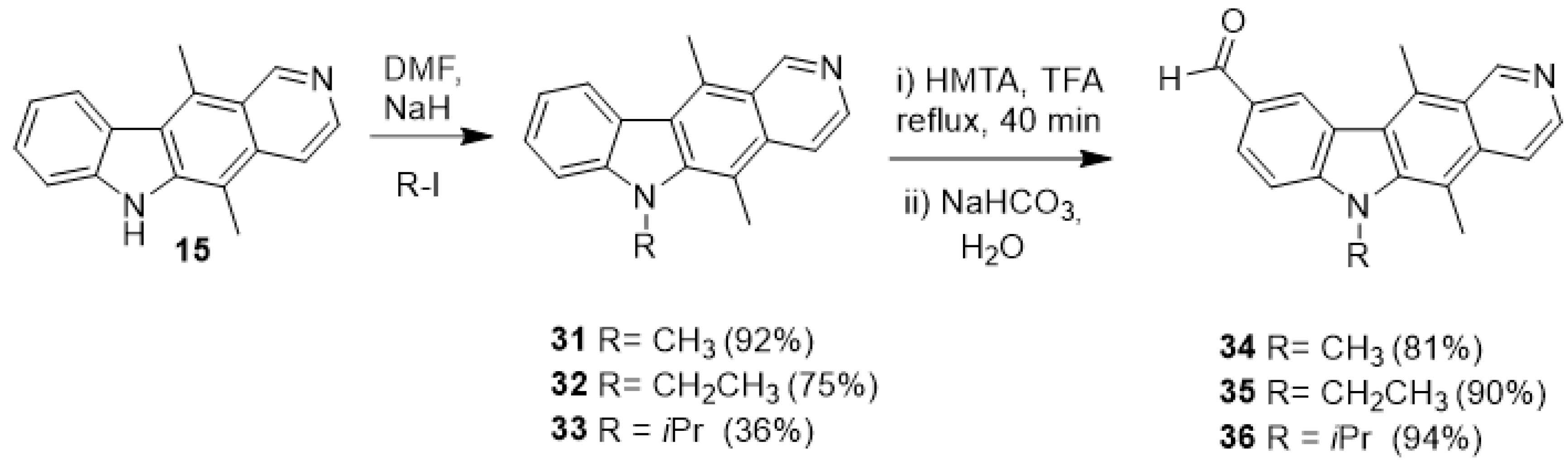
5,6,11-Trimethyl-6H-pyrido [4,3-b]carbazole 31
5,11-Dimethyl-6H-pyrido[4,3b]carbazole 15 (1.50 g, 6.10 mmol) in anhydrous DMF (20 mL) was treated with NaH (0.375 g, 60% dispersion in mineral oil 9.4 mmol,) at 0 °C under nitrogen and allowed stir for 30 min, forming a deep red suspension. Iodomethane (0.39 mL, 6.20 mmol) was added and the reaction was allowed warm to room temperature and stir overnight. The reaction mixture was cooled to 0 °C and water (100 mL) was added producing a pale orange precipitate. The yellow product was isolated by vacuum filtration and washed with water (150 mL) and did not require further purification (1.46 g, 91.8%). m.p 205–206 °C (Lit. m.p 202–204 °C) [61]; IR νmax/cm−1: 2921, 2852, 1588, 1476, 1449, 1241, 1102, 806; δH (300 MHz, DMSO-d6): 3.03 (3H, s, CH3-5), 3.19 (3H, s, CH3-11), 4.13 (3H, s, NCH3), 7.31 (1H, overlapping ddd, J 8.0, 6.4, 1.5, H-9), 7.58 (1H, dd, J 8.0, 1.5, H-8), 7.62 (1H, d, J 1.5, H-7), 7.99 (1H, d, J 6.0, H-4), 8.37 (1H, d, J 8.0, H-10), 8.45 (1H, d, J 6.0, H-3), 9.68 (1H, s, H-1); m/z (ESI+) 261 [(M + H)+, 100%].
6-Ethyl-5,11-dimethyl-6H-pyrido[4,3-b]carbazole 32
5,11-Dimethyl-6H-pyrido[4,3-b]carbazole 15 (3.33 g, 13.5 mmol) in anhydrous DMF (60 mL), at 0 °C under nitrogen, was treated with sodium hydride (0.812 g, 20.3 mmol, 60% dispersion in mineral oil) and left to stir for 30 min forming a red suspension. Iodoethane (1.14 mL, 2.22 g, 14.2 mmol) was added and the reaction was allowed to warm to room temperature and was left to stir for a total of 72 h. The reaction mixture was cooled on ice and water (200 mL) was added. The aqueous layer was extracted with dichloromethane-methanol 90:10 (5 × 50 mL). The combined organic extracts were washed with water (5 × 50 mL) and brine (1 × 50 mL), dried over magnesium sulphate, filtered and the solvent removed under reduced pressure. Purification by column chromatography eluting with dichloromethane-methanol (99:1) gave the product as a yellow solid (2.35 g, 63.4%). m.p. 164–165 °C; νmax/cm−1 (KBr): 3020, 2966, 2926, 1596, 1588, 1475, 1395, 1382, 1346, 1232, 741; δH (600 MHz, DMSO-d6): 1.33 [3H, t, J 7.1, N(6)CH2CH3], 2.84 [3H, s, C(5)CH3], 3.03 [3H, s, C(11)CH3], 4.50 [2H, q, J 7.1, N(6)CH2CH3], 7.26 [1H, t, J 7.4, C(9)H], 7.53–7.58 [2H, m, C(7)H, C(8)H], 7.92 [1H, d, J 6.1, C(4)H], 8.26 [1H, d, J 7.8, C(10)H], 8.41 [1H, d, J 6.0, C(3)H], 9.58 [1H, s, C(1)H]; δC (150.9 MHz, DMSO-d6): 13.4 [CH3, C(5)CH3], 14.7 [CH3, C(11)CH3], 15.4 [CH3, N(6)CH2CH3], 40.2 [CH2, N(6)CH2CH3], 108.7 (C, aromatic C), 109.4 (CH, aromatic CH), 116.3 (CH, aromatic CH), 120.1 (CH, aromatic CH), 122.4 (C, aromatic C), 123.3 (C, aromatic C), 124.2 (CH, aromatic CH), 124.5 (C, aromatic C), 127.7 (CH, aromatic CH), 128.8 (C, aromatic C), 134.0 (C, aromatic C), 140.3 (C, aromatic C), 141.2 (CH, aromatic CH), 144.1 (C, aromatic C), 149.9 (CH, aromatic CH); m/z (ESI+): 275 [(M + H)+ 100%]; HRMS (ESI+): Exact mass calculated for C19H19N2+ 275.1548. Found 275.1547.
6-Isopropyl-5,11-dimethyl-6H-pyrido[4,3-b]carbazole 33
5,11-Dimethyl-6H-pyrido[4,3-b]carbazole 15 (2.31 g, 9.37 mmol) was added portion-wise to a suspension of sodium hydride (0.75 g, 60% dispersion in mineral oil, equivalent to 0.45 g sodium hydride, 18.8 mmol) in dimethylformamide (10 mL) forming a deep red suspension which was stirred at room temperature for 45 min under nitrogen. 2-Iodopropane (1.10 mL, 1.90 g, 11.2 mmol) in dimethylformamide (5 mL) was added dropwise and the reaction was stirred for 48 h. The reaction mixture was cooled on ice and water (50 mL) added. The aqueous layer was extracted with dichloromethane—Methanol 90:10 (3 × 50 mL). A precipitate in the aqueous layer which would not extract was filtered and found to be unreacted ellipticine (402 mg). Combined organic layers were washed with water (5 × 20 mL) and brine (1 × 30 mL), dried over magnesium sulphate and solvent removed under reduced pressure. Purification by column chromatography eluting with dichloromethane – methanol 99:1 gave the product as a yellow solid (0.96 g, 35.5%). m.p. 168–170 °C; νmax/cm−1 (KBr): 3435, 3079, 2970, 2933, 1598, 1587, 1469, 1390, 1367, 1345, 1288, 1236, 1104, 804, 741; δH (600 MHz, DMSO-d6): 1.59 [6H, d, J 6.9, N(6)CH(CH3)2], 2.87 [3H, s, C(5)CH3], 3.15 [3H, s, C(11)CH3], 5.32 [1H, sept, J 7.0, N(6)CH(CH3)2], 7.28 [1H, overlapping ddd, J 7.8, 7.2, 0.5, C(9)H], 7.51 [1H, overlapping ddd, J 8.2, 7.2, 1.0, C(8)H], 7.77 [1H, d, J 8.2, C(7)H], 7.92 [1H, d, J 6.0, C(4)H], 8.34 [1H, d, J 7.8, C(10)H], 8.43 [1H, d, J 5.8, C(3)H], 9.66 [1H, s, C(1)H]; δH (600 MHz, DMSO-d6): 14.8 [CH3, C(11)CH3], 15.3 [CH3, C(5)CH3], 21.4 [2 × CH3, N(6)CH(CH3)2], 49.6 [CH, N(6)CH(CH3)2], 109.6 (C, aromatic C), 113.6 (CH, aromatic CH), 116.6 (CH, aromatic CH), 120.2 (CH, aromatic CH), 122.9 (C, aromatic C), 124.5 (CH, aromatic CH), 124.9 (C, aromatic C), 125.3 (C, aromatic C), 127.0 (CH, aromatic CH), 128.3 (C, aromatic C), 134.6 (C, aromatic C), 141.4 (CH, aromatic CH), 143.1 (C, aromatic C), 143.2 (C, aromatic C), 149.9 (CH, aromatic CH); m/z (ESI+): 289 [(M + H)+, 100%]; HRMS (ESI+): Exact mass calculated for C20H21N2+ 289.1705. Found 289.1707.
5,6,11-Trimethyl-6H-pyrido[4,3-b]carbazole-9-carbaldehyde 34
5,6,11-Trimethyl-6H-pyrido[4,3-b]carbazole 31 (0.600 g, 2.33 mmol) was dissolved in trifluoroacetic acid (30 mL) and hexamethylenetetramine (3.24 g, 23.41 mmol) added portion-wise. The reaction was heated to reflux for 1 h. Upon cooling to room temperature, the mixture was cooled to 0 °C and poured into water (200 mL). Solid sodium bicarbonate was added until neutralised and the mixture was extracted with dichloromethane-methanol (150 mL × 4, 9:1). The combined organic layers were washed with water (2 × 100 mL) and brine (2 × 100 mL), dried, filtered and solvent removed under reduced pressure to yield an orange solid (0.53 g, 81.0%). m.p. 219–222 °C (Lit. m.p. 222-223 °C) [55,62]; IR νmax/cm−1: 3214, 2922, 2852, 1674, 1586, 1461, 1380, 1356, 1099, 983; δH (300 MHz, DMSO-d6): 3.01 (3H, s, CH3-5), 3.21 (3H, s, CH3-11), 4.16 (3H, s, NCH3), 7.75 (1H, d, J 8.7, H-7), 8.01 (1H, d, J 5.8, H-4), 8.07 (1H, dd, J 8.7, 1.5, H-8), 8.49 (1H, d, J 5.8, H-3), 8.78 (1H, d, J 1.5, H-10), 9.70 (1H, s, H-1), 10.10 (1H, s, CHO); m/z (ESI+) 289 [(M + H)+, 100%].
6-Ethyl-5,11-dimethyl-6H-pyrido[4,3-b]carbazole-9-carbaldehyde 35
6-Ethyl-5,11-dimethyl-6H-pyrido[4,3-b]carbazole 32 (1.00 g, 3.65 mmol) was stirred in trifluoroacetic acid (40 mL) and hexamethylenetetramine (5.11 g, 36.5 mmol) was added portion-wise. The reaction was heated to reflux for 40 min. Upon cooling the reaction mixture was cooled on ice and poured into water (250 mL). The reaction mixture was neutralised using solid sodium bicarbonate [Caution: vigorous reaction] and extracted with dichloromethane-methanol 90:10 (3 × 100 mL). The combined organic extracts were washed with water (2 × 100 mL) and brine (1 × 100 mL), dried over magnesium sulphate, filtered and the solvent removed under reduced pressure to give the product as an orange solid which was used without further purification (1.00 g, 91.0%). m.p. 192–196 °C; νmax/cm−1 (KBr): 2970, 2929, 2720, 1682, 1589, 1452, 1381, 1315, 1235, 1100, 809; δH (500 MHz, DMSO-d6): 1.42 [3H, t, J 6.9, N(6)CH2CH3], 2.95 [3H, s, C(5)CH3], 3.19 [3H, s, C(11)CH3], 4.66 [2H, q, J 6.8, N(6)CH2CH3], 7.78 [1H, d, J 8.5, C(7)H], 8.03 [1H, d, J 5.9, C(4)H], 8.08 [1H, d, J 8.4, C(8)H], 8.49 [1H, d, J 5.6, C(3)H], 8.77 [1H, s, C(10)H], 9.79 [1H, s, C(1)H], 10.10 [1H, s, C(9)CHO]; δC (125.8 MHz, DMSO-d6): 13.4 [CH3, C(5)CH3], 14.9 [CH3, C(11)CH3], 15.5 [CH3, N(6)CH2CH3], 40.7 [CH2, N(6)CH2CH3], 109.9 (CH, aromatic CH), 110.3 (C, aromatic C), 116.6 (CH, aromatic CH), 122.9 (C, aromatic C), 123.3 (C, aromatic C), 123.9 (C, aromatic C), 127.1 (CH, aromatic CH), 128.9 (CH, aromatic CH), 129.3 (C, aromatic C), 129.7 (C, aromatic C), 134.5 (C, aromatic C), 140.6 (C, aromatic C), 141.9 (CH, aromatic CH), 147.9 (C, aromatic C), 150.2 (CH, aromatic CH), 192.4 [C, C(9)CHO]; m/z (ESI+): 303 [(M + H)+, 100%]; HRMS (ESI+): Exact mass calculated for C20H19N2O+ 303.1497. Found 303.1485.
6-Isopropyl-5,11-dimethyl-6H-pyrido[4,3-b]carbazole-9-carbaldehyde 36
6-Isopropyl-5,11-dimethyl-6H-pyrido[4,3-b]carbazole 33 (0.81 g, 2.82 mmol) was stirred in trifluoroacetic acid (40 mL) and hexamethylenetetramine (3.96 g, 28.2 mmol) was added portion-wise. The reaction was heated to reflux for 40 min. Upon cooling the reaction mixture was cooled on ice and poured into water (250 mL). The reaction mixture was neutralised using solid sodium bicarbonate [Caution: vigorous reaction] and extracted with dichloromethane-methanol 90:10 (3 × 100 mL). The combined organic extracts were washed with water (2 × 100 mL) and brine (1 × 100 mL), dried over magnesium sulphate, filtered and the solvent removed under reduced pressure to give the product as an orange solid which was used without further purification (0.836 g, 93.7%). m.p. 200–205 °C; νmax/cm−1 (KBr): 3412, 2969, 2929, 1683, 1588, 1574, 1386, 1321, 1286, 1240, 1192, 1140, 1104, 812; δH (600 MHz, DMSO-d6): 1.67 [6H, d, J 7.0, N(6)CH(CH3)2], 2.94 [3H, s, C(5)CH3], 3.24 [3H, s, C(11)CH3], 5.45 [1H, sept, J 6.9, N(6)CH(CH3)2], 7.97 [1H, d, J 8.6, C(7)H], 8.01 [1H, d, J 6.1, C(4)H], 8.04 [1H, dd, J 8.6, 1.4, C(8)H], 8.50 [1H, d, J 6.0, C(3)H], 8.84 [1H, d, J 1.1, C(10)H], 9.73 [1H, s, C(1)H], 10.11 [1H, s, C(9)CHO]; δC (150.9 MHz, DMSO-d6): 14.9 [CH3, C(11)CH3], 15.3 [CH3, C(5)CH3], 21.3 [2 × CH3, N(6)CH(CH3)2], 50.0 [CH, N(6)CH(CH3)2], 110.8 (C, aromatic C), 113.6 (CH, aromatic CH), 116.9 (CH, aromatic CH), 123.2 (C, aromatic C), 124.1 (C, aromatic C), 125.2 (C, aromatic C), 127.2 (CH, aromatic CH), 128.0 (CH, aromatic CH), 129.1 (C, aromatic C), 129.3 (C, aromatic C), 135.0 (C, aromatic C), 141.9 (CH, aromatic CH), 143.0 (C, aromatic C), 147.1 (C, aromatic C), 150.1 (CH, aromatic CH), 192.6 [C, C(9)CHO]; m/z (ESI+): 317 [(M + H)+, 100%]; HRMS (ESI+): Exact mass calculated for C21H21N2O+ 317.1654. Found 317.1651.
Phytophthora infestans Mycelial Growth Assays
Phytophthora infestans strain 88,069 (mating type A1) was originally obtained from Professor Sophien Kamoun (The Sainsbury Laboratory, Norwich, UK) and maintained on RyeA agar at 20 °C in the dark [63]. For mycelial growth assay, compounds were dissolved in DMSO in a vial and 15 mL Rye B agar added, at a temperature of approximately 50 °C (the final concentration of DMSO was 0.1%). The vial was shaken to distribute the contents and the mixture was then poured into a clean, sterile 10 cm Petri dish. A 10 mm plug of P. infestans mycelium was placed in the centre of the dish, which was then sealed with parafilm. The experiments were carried out in triplicate at 25 µM (unless otherwise stated), stored at 20 °C in the dark and checked periodically for mycelial growth.
Phytophthora infestans Zoosporogenesis and Sporangiogenesis Assays
These were performed essentially as described by Lu et al. [39]. Following two weeks of culture on Rye B agar, sporangia were collected from plates in 5 mL of modified Petri’s solution (MPS, 5 mM CaCl2, 1 mM MgSO4, 1 mM KH2PO4 and 0.8 mM KCl), by scraping with a scalpel blade and were transferred to a 50 mL sterile tube. Plates were washed with another 5 mL of MPS and this was combined with the first 5 mL. To initiate zoospore release, sporangia were place on ice, in the dark, for 3h. Sporangia or zoospores were counted by brightfield microscopy using a 10× objective and a haemocytometer, in duplicate.
XTT reduction assay
Human embryonic kidney-293T (HEK-293T) cells were obtained from LGC Standards (Middlesex, UK) and were maintained in Dulbecco’s Modified Eagle’s Medium containing 10% foetal bovine serum, 100 units/mL penicillin and 100 μg/mL streptomycin, at 37 °C in a humidified atmosphere of 5% CO2/95% air. For assays, cells were subcultured on 96-well microtitre plates at a density of 5 × 10⁴ cells/well, in a volume of 50 mL. Following overnight culture, 50 mL of each test compound at twice their final concentration (50 mM for all apart from cinnamaldehyde (2 mM) and Mancozeb (200 μM)) was added and incubated for another 24 h. Cells were incubated with 100 mL XTT reagents (Sigma-Aldrich, Ireland) for an additional 4 h, after which, the absorbance of the extracellular formazan reduction product was measured at a wavelength of of 490 nm, with a reference wavelength of 675 nm [53,54], using a microtitre plate spectrophotometer.
Statistical Methods
Numerical data were statistically compared using one-way analysis of variance (ANOVA) and Tukey’s post hoc test (using ezANOVA software, available at: https://people.cas.sc.edu/rorden/ezanova/index.html.) A probability threshold of p < 0.05 was taken as statistically significant. Generation of histograms and linear regression analyses were performed using Microsoft Excel.

5. Conclusions

It is evident from the synthesis and biological evaluation that both cell viability and inhibitory effects are impacted by discreet modifications of the cinnamaldehyde and ellipticine scaffolds. The mycelium growth assay provided information at Days 5, 9 and 13 of P. infestans incubation. From an initial focus on the cinnamaldehyde derivatives, the cinnamaldehyde Mancozeb hybrid proved fruitful and is a template for further discovery given its excellent P. infestans growth inhibition/toxicity profile. While the effects of cinnamaldehyde derivatives were somewhat predicted, uniquely in the screen of aldehydes the ellipticine aldehydes have emerged with excellent Phytophthora growth inhibitory properties. The aldehyde functionality on ellipticine has been shown to increase significantly the inhibitory effects of the compound and limit the toxicity to an extent though this needs to be tempered further before progressing. In contrast, the stock aldehydes did not demonstrate strong inhibition which suggests that there are additional factors affecting the oomycete growth.
The effects of ellipticine substitution have identified that alkylation of the six-position considerably improves mycelial growth inhibition. Ellipticines 31 and 34 have significant growth inhibitory properties on mycelia and inhibit zoosporogenesis and sporangiogenesis in comparison to ellipticine (15). It is evident from the biological data that 34 demonstrates the greatest inhibitory effects on P. infestans with a mycelium growth of just 6% after 13 days of incubation and the comparison with cinnamaldehyde is favourable given the respective dosages. The XTT cytotoxicity assay has highlighted that cinnamaldehyde and ellipticine compounds were amongst the most highly toxic of all the compounds tested and currently limits their potential use as fungicides. However, the discovery of 9-formyl-6-methylellipticine 34 as a potent growth inhibitor is the subject of our investigation into the influence of 2- and 6-alkylated 9-formylellipticines on P. infestans growth [64]. This study has resulted in promising new cinnamaldehyde and ellipticine candidates for P. infestans control that warrant further study to examine the role of substituents and mechanism of action.

Supplementary Materials

The Supplementary Information is available online at https://www.mdpi.com/2076-0817/9/7/542/s1. Experimental: Synthesis of Ellipticine Framework, NMR data: NMR data for compounds 2633 and 3536, Figure S1: Mycelial Growth Inhibition of cinnamaldehyde (7), Figure S2: Mycelial Growth Inhibition of hydrocinnamaldehyde 6, Figure S3: Mycelial Growth Inhibition of Mancozeb 1.

Author Contributions

B.M.D.P., F.O.M. and J.J.M. conceived and designed the experiments. F.O.M. and J.J.M. wrote the manuscript. E.C.O., L.Z., M.L.M. and R.A.K. performed the experiments, characterised the synthetic products and analysed the data. All authors have read and agreed to the published version of the manuscript.

Funding

This work was funded by the Department of Agriculture, Food and the Marine (Government of Ireland), Research Stimulus Fund grant number 15S669, awarded to J.J.M., and funded L.Z. and R.A.K. E.C.O. and M.L.M. were both funded under the Irish Research Council Postgraduate Scholarship Scheme.

Acknowledgments

The authors thank Don Kelleher and Eileen Dillane for valuable technical assistance.

Conflicts of Interest

The authors declare no conflicts of interest.

References

  1. Judelson, H.S.; Blanco, F.A. The spores of Phytophthora: Weapons of the plant destroyer. Nat. Rev. Microbiol. 2005, 3, 47. [Google Scholar] [CrossRef] [PubMed]
  2. Berkeley, M.J. Observations, botanical and physiological on the potato murrain. J. Horticult. Soc. Lond. 1846, 1, 9–34. [Google Scholar]
  3. de Bary, A. Researches into the nature of the potato-fungus-Phytophthora infestans. J. R. Agric. Soc. 1876, 2, 239–268. [Google Scholar]
  4. Kelly, B.D. The Great Irish Famine (1845–52) and the Irish asylum system: Remembering, forgetting, and remembering again. Ir. J. Med. Sci. 2019, 188, 953–956. [Google Scholar] [CrossRef]
  5. Martin, M.D.; Cappellini, E.; Samaniego, J.A.; Zepeda, M.L.; Campos, P.F.; Seguin-Orlando, A.; Wales, N.; Orlando, L.; Ho, S.Y.W.; Dietrich, F.S.; et al. Reconstructing genome evolution in historic samples of the Irish potato famine pathogen. Nat. Commun. 2013, 4, 2172. [Google Scholar] [CrossRef] [PubMed]
  6. Dixon, B. Pushing Bordeaux mixture. Lancet Infect. Dis. 2004, 4, 594. [Google Scholar] [CrossRef]
  7. Lamichhane, J.R.; Osdaghi, E.; Behlau, F.; Köhl, J.; Jones, J.B.; Aubertot, J.-N. Thirteen decades of antimicrobial copper compounds applied in agriculture. A review. Agron. Sustain. Dev. 2018, 38, 28. [Google Scholar] [CrossRef] [Green Version]
  8. Kazos, E.A.; Stalikas, C.D.; Nanos, C.G.; Konidari, C.N. Determination of dithiocarbamate fungicide propineb and its main metabolite propylenethiourea in airborne samples. Chemosphere 2007, 68, 2104–2110. [Google Scholar] [CrossRef]
  9. Thind, T.S.; Hollomon, D.W. Thiocarbamate fungicides: Reliable tools in resistance management and future outlook. Pest Manag. Sci. 2018, 74, 1547–1551. [Google Scholar] [CrossRef]
  10. Gullino, M.L.; Tinivella, F.; Garibaldi, A.; Kemmitt, G.M.; Bacci, L.; Sheppard, B. Mancozeb: Past, Present, and Future. Plant Dis. 2010, 94, 1076–1087. [Google Scholar] [CrossRef] [Green Version]
  11. Axelstad, M.; Boberg, J.; Nellemann, C.; Kiersgaard, M.; Jacobsen, P.R.; Christiansen, S.; Hougaard, K.S.; Hass, U. Exposure to the Widely Used Fungicide Mancozeb Causes Thyroid Hormone Disruption in Rat Dams but No Behavioral Effects in the Offspring. Toxicol. Sci. 2011, 120, 439–446. [Google Scholar] [CrossRef]
  12. Lo, C.-C.; Ho, M.-H. Determination of imidazolidine-2-thione (ethylenethiourea) in ethylenebisdithiocarbamate formulations. Pestic. Sci. 1993, 37, 247–251. [Google Scholar] [CrossRef]
  13. Reuveni, M.; Eyal, H.; Cohen, Y. Development of resistance to metalaxyl in Pseudoperonospora cubensis. Plant Dis. 1980, 64, 1108–1109. [Google Scholar] [CrossRef]
  14. Staub, T. Fungicide resistance: A continuing challenge. Plant Dis. 1984, 1026–1031. [Google Scholar] [CrossRef]
  15. Pasteris, R.J.; Hanagan, M.A.; Bisaha, J.J.; Finkelstein, B.L.; Hoffman, L.E.; Gregory, V.; Andreassi, J.L.; Sweigard, J.A.; Klyashchitsky, B.A.; Henry, Y.T.; et al. Discovery of oxathiapiprolin, a new oomycete fungicide that targets an oxysterol binding protein. Bioorg. Med. Chem. 2016, 24, 354–361. [Google Scholar] [CrossRef] [PubMed]
  16. Miao, J.; Chi, Y.; Lin, D.; Tyler, B.M.; Liu, X. Mutations in ORP1 Conferring Oxathiapiprolin Resistance Confirmed by Genome Editing using CRISPR/Cas9 in Phytophthora capsici and P. sojae. Phytopathology 2018, 108, 1412–1419. [Google Scholar] [CrossRef] [PubMed] [Green Version]
  17. Miao, J.; Cai, M.; Dong, X.; Liu, L.; Lin, D.; Zhang, C.; Pang, Z.; Liu, X. Resistance Assessment for Oxathiapiprolin in Phytophthora capsici and the Detection of a Point Mutation (G769W) in PcORP1 that Confers Resistance. Front. Microbiol. 2016, 7, 615. [Google Scholar] [CrossRef] [Green Version]
  18. Dobell, C. Antony van Leeuwenhoek and his “Little Animals”: Being some Account of the Father of Protozoology and Bacteriology and his Multifarious Discoveries in these Disciplines. Nature 1932, 130, 679–680. [Google Scholar]
  19. Shreaz, S.; Sheikh, R.A.; Rimple, B.; Hashmi, A.A.; Nikhat, M.; Khan, L.A. Anticandidal activity of cinnamaldehyde, its ligand and Ni(II) complex: Effect of increase in ring and side chain. Microb. Pathog. 2010, 49, 75–82. [Google Scholar] [CrossRef]
  20. Sangal, A. Role of cinnamon as beneficial antidiabetic food adjunct: A review. Adv. Appl. Sci. Res. 2011, 2, 440–450. [Google Scholar]
  21. Dumas, J.; Peligot, H. Sur l’Huile de Cannelle, l’Acide Hippurique, et l’Acide Sébacique. Ann. Chim. Phys. 1834, 57, 305–334. [Google Scholar]
  22. Chiozza, L. Surla production artificielle de l’essence de cannelle. C. R. Hebd. Séances l’Acad. Sci. 1856, 1, 222. [Google Scholar]
  23. Hooth, M.J.; Sills, R.C.; Burka, L.T.; Haseman, J.K.; Witt, K.L.; Orzech, D.P.; Fuciarelli, A.F.; Graves, S.W.; Johnson, J.D.; Bucher, J.R. Toxicology and carcinogenesis studies of microencapsulated trans-cinnamaldehyde in rats and mice. Food Chem. Toxicol. 2004, 42, 1757–1768. [Google Scholar] [CrossRef] [PubMed]
  24. Kim, S.H.; Hyun, S.H.; Choung, S.Y. Anti-diabetic effect of cinnamon extract on blood glucose in db/db mice. J. Ethnopharmacol. 2006, 104, 119–123. [Google Scholar] [CrossRef] [PubMed]
  25. Chao, L.K.; Hua, K.-F.; Hsu, H.-Y.; Cheng, S.-S.; Liu, J.-Y.; Chang, S.-T. Study on the Antiinflammatory Activity of Essential Oil from Leaves of Cinnamomum osmophloeum. J. Agric. Food Chem. 2005, 53, 7274–7278. [Google Scholar] [CrossRef] [PubMed]
  26. Burt, S. Essential oils: Their antibacterial properties and potential applications in foods—A review. Int. J. Food Microbiol. 2004, 94, 223–253. [Google Scholar] [CrossRef] [PubMed]
  27. Gyawali, R.; Ibrahim, S.A. Natural products as antimicrobial agents. Food Control 2014, 46, 412–429. [Google Scholar] [CrossRef]
  28. Masuda, S.; Hara-Kudo, Y.; Kumagai, S. Reduction of Escherichia coli O157:H7 Populations in Soy Sauce, a Fermented Seasoning. J. Food Prot. 1998, 61, 657–661. [Google Scholar] [CrossRef] [PubMed]
  29. Quintanilla, P.; Rohloff, J.; Iversen, T.H. Influence of essential oils on Phytophthora infestans. Potato Res. 2002, 45, 225–235. [Google Scholar] [CrossRef]
  30. Soylu, E.M.; Soylu, S.; Kurt, S. Antimicrobial activities of the essential oils of various plants against tomato late blight disease agent Phytophthora infestans. Mycopathologia 2006, 161, 119–128. [Google Scholar] [CrossRef]
  31. Ferhout, H.; Bohatier, J.; Guillot, J.; Chalchat, J.C. Antifungal Activity of Selected Essential Oils, Cinnamaldehyde and Carvacrol against Malassezia furfur and Candida albicans. J. Essent. Oil Res. 1999, 11, 119–129. [Google Scholar] [CrossRef]
  32. Raut, J.S. Phenylpropanoids of Plant Origin as Inhibitors of Biofilm Formation by Candida albicans. J. Microbiol. Biotechnol. 2014, 1216–1225. [Google Scholar] [CrossRef] [PubMed] [Green Version]
  33. Jantan, I.B.; Moharam, B.A.K.; Santhanam, J.; Jamal, J.A. Correlation between Chemical Composition and Antifungal Activity of the Essential Oils of Eight Cinnamomum. Species. Pharm. Biol. 2008, 46, 406–412. [Google Scholar] [CrossRef] [Green Version]
  34. Wendakoon, C.N.; Sakaguchi, M. Inhibition of Amino Acid Decarboxylase Activity of Enterobacter aerogenes by Active Components in Spices. J. Food Prot. 1995, 58, 280–283. [Google Scholar] [CrossRef]
  35. Bang, K.-H.; Lee, D.-W.; Park, H.-M.; Rhee, Y.-H. Inhibition of Fungal Cell Wall Synthesizing Enzymes by trans-Cinnamaldehyde. Biosci. Biotechnol. Biochem. 2000, 64, 1061–1063. [Google Scholar] [CrossRef] [PubMed]
  36. Gill, A.O.; Holley, R.A. Inhibition of membrane bound ATPases of Escherichia coli and Listeria monocytogenes by plant oil aromatics. Int. J. Food Microbiol. 2006, 111, 170–174. [Google Scholar] [CrossRef] [PubMed]
  37. Serrano, R. Structure and function of proton translocating ATPase in plasma membranes of plants and fungi. Biochim. Biophys. Acta (BBA) Rev. Biomembr. 1988, 947, 1–28. [Google Scholar] [CrossRef]
  38. Domadia, P.; Swarup, S.; Bhunia, A.; Sivaraman, J.; Dasgupta, D. Inhibition of bacterial cell division protein FtsZ by cinnamaldehyde. Biochem. Pharmacol. 2007, 74, 831–840. [Google Scholar] [CrossRef] [PubMed]
  39. Lu, M.; Han, Z.; Yao, L. In vitro and in vivo antimicrobial efficacy of essential oils and individual compounds against Phytophthora parasitica var. nicotianae. J. Appl. Microbiol. 2013, 115, 187–198. [Google Scholar] [CrossRef]
  40. Li, Y.; Yu, T.; Wu, T.; Wang, R.; Wang, H.; Du, H.; Xu, X.; Xie, D.; Xu, X. The dynamic transcriptome of pepper (Capsicum annuum) whole roots reveals an important role for the phenylpropanoid biosynthesis pathway in root resistance to Phytophthora capsici. Gene 2020, 728, 144288. [Google Scholar] [CrossRef]
  41. Zheng, L.; Mackrill, J.J. Calcium Signaling in Oomycetes: An Evolutionary Perspective. Front. Physiol. 2016, 7, 123. [Google Scholar] [CrossRef] [PubMed] [Green Version]
  42. Hu, L.; Wang, D.; Liu, L.; Chen, J.; Xue, Y.; Shi, Z. Ca(2+) efflux is involved in cinnamaldehyde-induced growth inhibition of Phytophthora capsici. PLoS ONE 2013, 8, e76264. [Google Scholar] [CrossRef] [PubMed] [Green Version]
  43. Courchesne, W.E.; Tunc, M.; Liao, S. Amiodarone induces stress responses and calcium flux mediated by the cell wall in Saccharomyces cerevisiae. Can. J. Microbiol. 2009, 55, 288–303. [Google Scholar] [CrossRef]
  44. Shreaz, S.; Wani, W.A.; Behbehani, J.M.; Raja, V.; Irshad, M.; Karched, M.; Ali, I.; Siddiqi, W.A.; Hun, L.T. Cinnamaldehyde and its derivatives, a novel class of antifungal agents. Fitoterapia 2016, 112, 116–131. [Google Scholar] [CrossRef] [PubMed]
  45. Watamoto, T.; Egusa, H.; Sawase, T.; Yatani, H. Screening of Pharmacologically Active Small Molecule Compounds Identifies Antifungal Agents against Candida Biofilms. Front. Microbiol. 2015, 6, 1453. [Google Scholar] [CrossRef] [Green Version]
  46. Shin, J.-H.; Fu, T.; Park, K.H.; Kim, K.S. The Effect of Fungicides on Mycelial Growth and Conidial Germination of the Ginseng Root Rot Fungus, Cylindrocarpon destructans. Mycobiology 2017, 45, 220–225. [Google Scholar] [CrossRef] [Green Version]
  47. Stahl, E.; Bellwon, P.; Huber, S.; Schlaeppi, K.; Bernsdorff, F.; Vallat-Michel, A.; Mauch, F.; Zeier, J. Regulatory and Functional Aspects of Indolic Metabolism in Plant Systemic Acquired Resistance. Mol. Plant 2016, 9, 662–681. [Google Scholar] [CrossRef] [Green Version]
  48. Saulnier, M.G.; Gribble, G.W. An efficient synthesis of ellipticine. J. Org. Chem. 1982, 47, 2810–2812. [Google Scholar] [CrossRef]
  49. Gribble, G.W.; Saulnier, M.G.; Obaza-Nutaitis, J.A.; Ketcha, D.M. A versatile and efficient construction of the 6H-pyrido[4,3-b]carbazole ring system. Syntheses of the antitumor alkaloids ellipticine, 9-methoxyellipticine, and olivacine, and their analogs. J. Org. Chem. 1992, 57, 5891–5899. [Google Scholar] [CrossRef]
  50. Sullivan, E.C.O.; Miller, C.M.; Deane, F.M.; McCarthy, F.O. Emerging Targets in the Bioactivity of Ellipticines and Derivatives. Studies in Natural Products Chemistry; Elsevier Science Publishers: Amsterdam, The Netherlands, 2012. [Google Scholar]
  51. Plug, P.M.J.; Koomen, G.-J.; Pandit, K.U. An Expedient Synthesis of 9-Hydroxyellipticine. Synthesis 1992, 1221–1222. [Google Scholar] [CrossRef]
  52. Miller, C.M.; O’Sullivan, E.C.; Devine, K.J.; McCarthy, F.O. Synthesis and Biological Evaluation of Novel Isoellipticine Derivatives and Salts. Org. Biomol. Chem. 2012, 10, 7912–7921. [Google Scholar] [CrossRef] [PubMed]
  53. Roehm, N.W.; Rodgers, G.H.; Hatfield, S.M.; Glasebrook, A.L. An improved colorimetric assay for cell proliferation and viability utilizing the tetrazolium salt XTT. J. Immunol. Methods 1991, 142, 257–265. [Google Scholar] [CrossRef]
  54. Scudiero, D.A.; Shoemaker, R.H.; Paull, K.D.; Monks, A.; Tierney, S.; Nofziger, T.H.; Currens, M.J.; Seniff, D.; Boyd, M.R. Evaluation of a soluble tetrazolium/formazan assay for cell growth and drug sensitivity in culture using human and other tumor cell lines. Cancer Res. 1988, 48, 4827–4833. [Google Scholar] [PubMed]
  55. Deane, F.M.; O’Sullivan, E.C.; Maguire, A.R.; Gilbert, J.; Sakoff, J.A.; McCluskey, A.; McCarthy, F.O. Synthesis and evaluation of novel Ellipticines as potential anti-cancer agents. Org. Biomol. Chem. 2013, 11, 1334–1344. [Google Scholar] [CrossRef] [PubMed]
  56. Gilman, H.; Cartledge, F.K.; Sim, S.-Y. The analysis of organic-substituted group IVB lithium compounds. J. Organomet. Chem. 1963, 1, 8–14. [Google Scholar] [CrossRef]
  57. Whitesides, G.M.; Casey, C.P.; Krieger, J.K. Thermal decomposition of vinylic copper(I) and silver(I) organometallic compounds. J. Am. Chem. Soc. 1971, 93, 1379–1389. [Google Scholar] [CrossRef]
  58. Magrioti, V.; Nikolaou, A.; Smyrniotou, A.; Shah, I.; Constantinou-Kokotou, V.; Dennis, E.A.; Kokotos, G. New potent and selective polyfluoroalkyl ketone inhibitors of GVIA calcium-independent phospholipase A2. Bioorg. Med. Chem. 2013, 21, 5823–5829. [Google Scholar] [CrossRef] [Green Version]
  59. da Silva, J.B.P.; Navarro, D.M.d.A.F.; da Silva, A.G.; Santos, G.K.N.; Dutra, K.A.; Moreira, D.R.; Ramos, M.N.; Espíndola, J.W.P.; de Oliveira, A.D.T.; Brondani, D.J.; et al. Thiosemicarbazones as Aedes aegypti larvicidal. Eur. J. Med. Chem. 2015, 100, 162–175. [Google Scholar] [CrossRef]
  60. Anbazhagan, R.; Sankaran, K.R. Syntheses, spectral characterization, single crystal X-ray diffraction and DFT computational studies of novel thiazole derivatives. J. Mol. Struct. 2013, 1050, 73–80. [Google Scholar] [CrossRef]
  61. Deane, F.M.; Miller, C.M.; Maguire, A.R.; McCarthy, F.O. Modifications to the Vilsmeier-Haack formylation of 1,4-dimethylcarbazole and its application to the synthesis of ellipticines. J. Heterocycl. Chem. 2011, 48, 814–823. [Google Scholar] [CrossRef]
  62. Mori, R.; Kato, A.; Komenoi, K.; Kurasaki, H.; Iijima, T.; Kawagoshi, M.; Kiran, Y.B.; Takeda, S.; Sakai, N.; Konakahara, T. Synthesis and in vitro antitumor activity of novel 2-alkyl-5-methoxycarbonyl-11-methyl-6H-pyrido[4,3-b]carbazol-2-ium and 2-alkylellipticin-2-ium chloride derivatives. Eur. J. Med. Chem. 2014, 82, 16–35. [Google Scholar] [CrossRef] [PubMed]
  63. Caten, C.E.; Jinks, J.L. Spontaneous variability of single isolates of Phytophthora infestans. I. Cultural variation. Can. J. Bot. 1968, 46, 329–348. [Google Scholar] [CrossRef]
  64. McKee, M.L.; Zheng, L.; O’Sullivan, E.C.; Kehoe, R.A.; Doyle, B.M.; Mackrill, J.J.; McCarthy, F.O. Synthesis and evaluation of novel ellipticines and derivatives as inhibitors of Phytophthora infestans. Pathogens 2020. under review. [Google Scholar]
Figure 1. Mancozeb (1) (and Proprineb 3) metabolising to toxic thioureas 2 and 4.
Pathogens 09 00542 g001
Figure 2. Chemical structure of fungicide Metalaxyl (5) and Oxathiapiprolin (6).
Pathogens 09 00542 g002
Figure 3. Chemical structure of cinnamaldehyde (7) and hydrocinnamaldehyde (8).
Pathogens 09 00542 g003
Figure 4. Cinnamaldehyde analogues previously tested against fungal strains and ellipticine (15).
Pathogens 09 00542 g004
Figure 5. Structure of Mancozeb (1), a cinnamaldehyde thiosemicarbazone hybrid 16 and screening targets.
Pathogens 09 00542 g005
Figure 6. Concentration dependency of the effects of cinnamaldehyde, hydrocinnamaldehyde and Mancozeb on P. infestans mycelial growth. Different concentrations of (A) cinnamaldehyde, (B) hydrocinnamaldehyde or (C) Mancozeb were incorporated into RyeB agar plates. These were inoculated with plugs of P. infestans cultures and the effects on mycelial growth were assayed after 5 days. Values are expressed relative to vehicle (solvent)-only controls (V) and represent the mean values of three independent experiments conducted in duplicate. Error bars represent the standard error of the mean; n.d., no growth detected; and levels of statistical significance from V, determined using ANOVA are: * = p < 0.05; ** = p < 0.01; and *** = p < 0.001.
Pathogens 09 00542 g006
Figure 7. trans-Cinnamaldehyde (7), 4-fluorocinnamaldehyde 17 and α-methylcinnamaldehyde 18 and structures of test Ar-CHO compounds 1924 for screening.
Pathogens 09 00542 g007
Scheme 1. Synthesis of cinnamaldehyde extended conjugated esters.
Pathogens 09 00542 sch001
Scheme 2. Synthesis of cinnamaldehyde thiosemicarbazone derivatives.
Pathogens 09 00542 sch002
Scheme 3. Synthesis of 6-substituted ellipticines and corresponding aldehyde derivatives.
Pathogens 09 00542 sch003
Figure 8. Screen of the effects of compounds on P. infestans mycelial growth after 5 days of incubation. Compounds were incorporated into RyeB agar at a concentration of 25 μM and their effects were measured after 5 days. Mancozeb was used at 100 μM and cinnamaldehyde at 1 mM. Untreated cultures were grown on RyeB only. Values are the mean of three independent experiments, expressed relative to vehicle-treated growth (V, 0.02% ethanol). Error bars indicate the standard error in the mean; n.d., no growth detected; and levels of statistical significance from V, determined using ANOVA are: * = p < 0.05; ** = p < 0.01; and *** = p < 0.001.
Pathogens 09 00542 g008
Figure 9. Screen of the effects of compounds on P. infestans mycelial growth after 9 days of incubation. For details, please refer to the legend of Figure 8. Levels of statistical significance from V, determined using ANOVA are: * = p < 0.05; ** = p < 0.01; and *** = p < 0.001.
Pathogens 09 00542 g009
Figure 10. Screen of the effects of compounds on P. infestans mycelial growth after 13 days of incubation. For details, please refer to the legend of Figure 8. Levels of statistical significance from V, determined using ANOVA are: * = p < 0.05; ** = p < 0.01; and *** = p < 0.001.
Pathogens 09 00542 g010
Figure 11. Effect of test compounds on P. infestans zoosporogenesis. P. infestans was cultured for 14 days on RyeB containing 25 μM of test compounds, 100 μM Mancozeb, 1 mM cinnamaldehyde, or vehicle (0.02% ethanol). Zoospores were then released from sporangia and counted using a haemocytometer. Data represent the absolute number of zoospores produced from each plate and are the mean of three independent experiments performed in duplicate. Error bars show the standard error in the mean values; n.d., no growth detected; and levels of statistical significance relative to V, determined using ANOVA are: * = p < 0.05; and ** = p < 0.01.
Pathogens 09 00542 g011
Figure 12. Effect of aldehyde and ellipticine test compounds on P. infestans sporangiogenesis. P. infestans was cultured for 14 days on RyeB containing 25 μM of test compounds, 100 μM Mancozeb, 1 mM cinnamaldehyde, or vehicle (0.02% ethanol). Sporangia were then detached from mycelia and counted using a haemocytometer. Data represent the absolute number of sporangia produced from each plate and are the mean of three independent experiments performed in duplicate. Error bars indicate the standard error in the mean values; n.d., no growth detected; and levels of statistical significance relative to V, determined using ANOVA are: * = p < 0.05; and ** = p < 0.01.
Pathogens 09 00542 g012
Figure 13. Effect of test compounds on reduction of XTT by a human cell line, HEK-293T. Cells were cultured in 96-well plates for 24 h in the presence of test compounds, then with XTT reagent for an additional 4 h. The absorbance of the orange formazan product was measured at a wavelength of 490 nm, with a reference wavelength of 675 nm. Data are from three independent experiments, performed in triplicate and normalised to the vehicle control (V, 0.02% ethanol). The exceptions to this are the experiments from “cinnamaldehyde derivatives”, which were performed once in triplicate. Error bars indicate the standard error in the mean; n.d., no growth detected; and levels of statistical significance from V, determined using ANOVA are: ** = p <0.01; and *** = p < 0.001.
Pathogens 09 00542 g013
Figure 14. Comparison of the effects of test compounds on XTT reduction by human cells versus mycelial growth inhibition in P. infestans. Graph showing XTT reduction by HEK-293T cells (a proxy for viability) versus inhibition of P. infestans mycelial growth after 5 days. In both cases, values are normalised to the vehicle-treated control (V = 100%). The dashed line indicates the best-fit for a linear relationship between these parameters and the R2 the correlation coefficient of this fit.
Pathogens 09 00542 g014

