Article

Description of Virulent Factors and Horizontal Gene Transfers of Keratitis-Associated Amoeba Acanthamoeba Triangularis by Genome Analysis

1
Institut de Recherche pour le Développement IRD 198, Aix-Marseille Université UM63, Assistance Publique – Hôpitaux de Marseille (AP-HM), Microbes, Evolution, Phylogeny and Infection (MEΦI), Institut Hospitalo-Universitaire (IHU) - Méditerranée Infection, 13005 Marseille, France
2
R&D Department, Amoéba, 38 Avenue des Frères Montgolfier, 69680 Chassieu, France
*
Author to whom correspondence should be addressed.
Pathogens 2020, 9(3), 217; https://doi.org/10.3390/pathogens9030217
Submission received: 12 February 2020 / Revised: 10 March 2020 / Accepted: 13 March 2020 / Published: 16 March 2020

Abstract

:
Acanthamoeba triangularis strain SH 621 is a free-living amoeba belonging to Acanthamoeba ribo-genotype T4. This ubiquitous protist is among the free-living amoebas responsible for Acanthamoeba keratitis, a severe infection of human cornea. Genome sequencing and genomic comparison were carried out to explore the biological functions and to better understand the virulence mechanism related to the pathogenicity of Acanthamoeba keratitis. The genome assembly harbored a length of 66.43 Mb encompassing 13,849 scaffolds. The analysis of predicted proteins reported the presence of 37,062 ORFs. A complete annotation revealed 33,168 and 16,605 genes that matched with NCBI non-redundant protein sequence (nr) and Cluster of Orthologous Group of proteins (COG) databases, respectively. The Kyoto Encyclopedia of Genes and Genomes Pathway (KEGG) annotation reported a great number of genes related to carbohydrate, amino acid and lipid metabolic pathways. The pangenome performed with 8 available amoeba genomes belonging to genus Acanthamoeba revealed a core genome containing 843 clusters of orthologous genes with a ratio core genome/pangenome of less than 0.02. We detected 48 genes related to virulent factors of Acanthamoeba keratitis. Best hit analyses in nr database identified 99 homologous genes shared with amoeba-resisting microorganisms. This study allows the deciphering the genome of a free-living amoeba with medical interest and provides genomic data to better understand virulence-related Acanthamoeba keratitis.

1. Introduction

Acanthamoeba species are ubiquitous free-living amoebas (FLA) widely distributed in diverse environments, including fresh water, ocean sediment, dust, soil, and air [1]. These protists are causative agents of Acanthamoeba keratitis (AK), a serious infection of the cornea associated with trauma, exposure to contaminated water, and contact lens use [2,3,4,5]. This infection is frequently encountered around the world and causes vision loss in case of misdiagnosis, inadequate use of lens-contact carrier, or inappropriate treatment [3,4,5]. The pathogenicity mechanisms of AK have been reported and divided into direct and indirect factors [2]. The direct factors characterize the ability to adhere to epithelial cells of the host cornea by the expression of acanthopodia and adhesin proteins [6,7]. Once the host tissue is penetrated, amoebas phagocyte the epithelial cornea and secrete toxins, leading to inducible cell apoptosis [6]. The indirect factors are related to different characteristics, including the ability to encyst, morphology of the protist, or drug resistance [2]. Based on the 18S rRNA gene, the genus Acanthamoeba is defined and distributed into 22 different ribo-genotypes (T1–T22) [5,8]. Most of the human infections due to Acanthamoeba are related to ribo-genotype T4 [9]. Indeed, several studies have estimated that 90% of AK amoebic keratitis cases have been linked to this genotype [9]. Acanthamoeba triangularis strain SH 621 is a ubiquitous FLA belonging to genotype T4, the most pathogenic group of Acanthamoeba species [10]. A. triangularis has a two-stage life, switching between a trophozoite form and a cyst form. The A. triangularis trophozoite is an active stage in which the amoeba moves, feeds, and divides. Under harsh unfavorable conditions, such as changes in temperature, pH, lack of nutrients, treatment with disinfectant or therapeutic agents, the amoebas have the ability to adopt a resistant double-walled cyst stage [11]. Free-living amoeba (FLA) food microorganisms, by selective grazing to grow, contribute to the regulation of the environmental microbial population [1,12]. Despite that, some microorganisms named amoeba-resisting microorganisms (ARMs), such as bacteria, fungi, or giant viruses, have the capacity to bypass the internalization in order to survive and multiply in amoebas [13,14,15,16]. The close contact between FLA and ARM promotes horizontal gene transfers, leading to the evolution of microorganisms [17,18].
In this study, we explore the A. triangularis genome sequenced with NextSeq Illumina technology. The gene content was analyzed by a comparison of the set of protein sequences with the main public databases. Then, we performed pan-genomic and core genomic analyses within the Acanthamoeba species. Through comparative genomic analysis, we gain insight into the potential pathogenicity pathways in the setting of Acanthamoeba keratitis. Finally, we study horizontal gene transfers with ARMs.

2. Results

2.1. General Genomic Features

The trimmed reads resulted in 8,540,421 reads used for the assembly of the A. triangularis genome. The draft genome size of A. triangularis obtained represents 66,434,030 nucleotides (nt). The genome was assembled into 13,849 scaffolds. For comparison with other Acanthamoeba available in public databases, the genome length of A. triangularis is larger than the genome of A. castellanii Neff (42.02 Mb) and Acanthamoeba polyphaga Linc-AP1(49.35Mb), that was assembled in 2013 [19] and 2019 [20], respectively. However, the A. triangularis genome size is smaller than the genome size of Acanthamoeba castellanii ATCC 50370 (120.6Mb) and Acanthamoeba polyphaga ATCC 30872 (115.3Mb), assembled in 2015 [21].
The genome harbored a guanine–cytosine (GC) content of 58.6%, which is similar to the GC content of A. polyphaga (58.1%) and A. castellanii (57.8%) [20]. The main A. triangularis characteristics are presented in Table 1.
We detected one complete 18S rRNA gene of A. triangularis with a length of 1748 nucleotides. Moreover, several other partial sequences related to 18S rRNA of A. triangularis were found in the genome in different or similar scaffolds (Figure S1).
Phylogenetic analysis based on the 18S rRNA gene showed that A. triangularis is most closely related to the Acanthamoeba spp. belonging to genotype T4, including Acanthamoeba castellanii strain CDC:0786:V042 (U07403.1), Acanthamoeba polyphaga strain HC-2 (AF019056.1), and Acanthamoeba genotype T4 isolate Aud6 (KF733230.1) (Figure S2). A total of 37,062 putative genes were predicted (Table 2).
We found a larger number of predicted proteins for A. triangularis than for A. castellanii Neff (20,681 ORFs) [19]. In contrast, we found a smaller number of genes predicted for A. triangularis than the genome of A. castellanii ATCC 50370 (82,310 ORF) [21], which is explained by the difference in genome length.

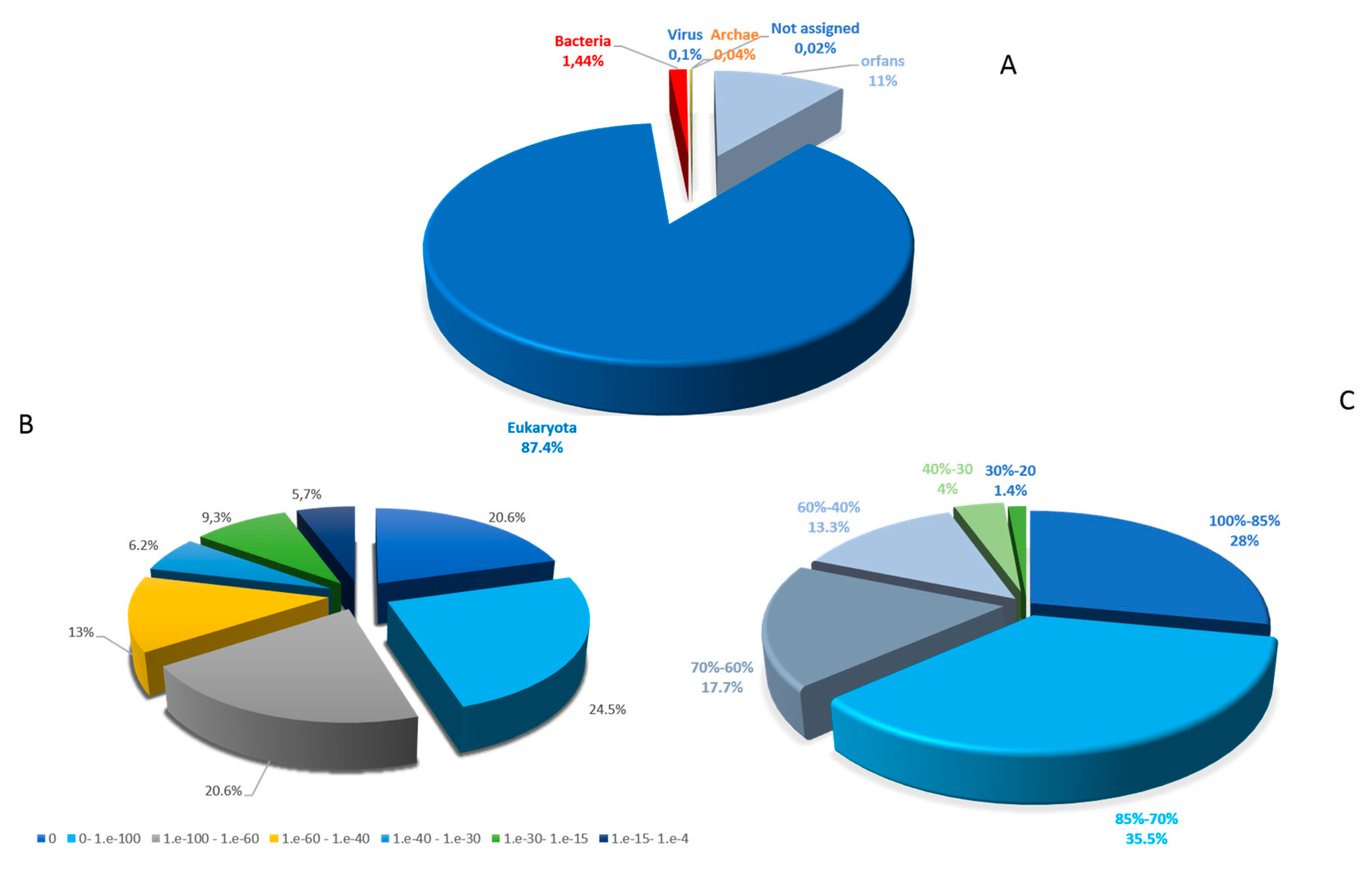
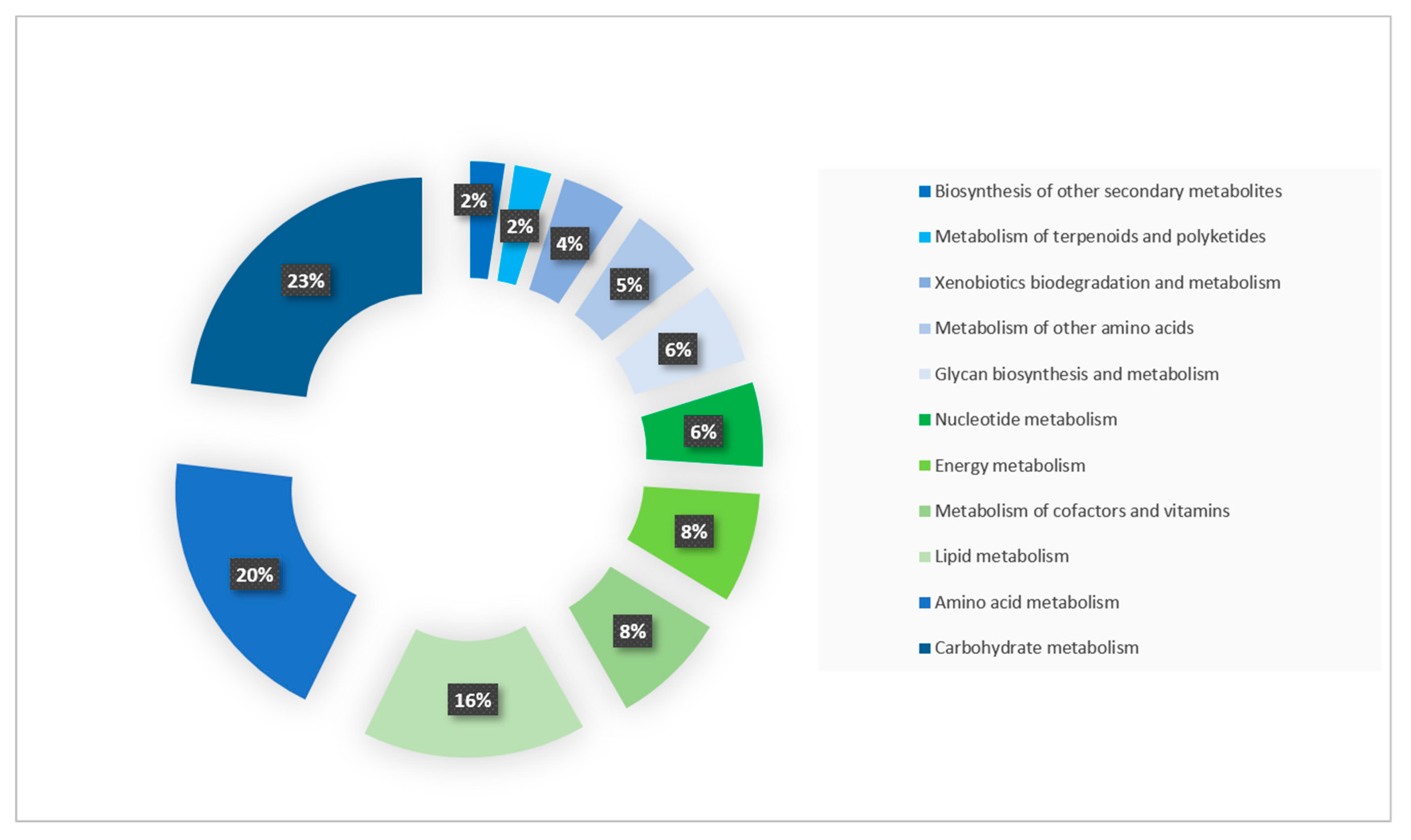
2.2. Functional Annotation

In order to conduct an in-depth analysis of the genetic content of A. triangularis, the putative proteins were compared with different databases (Table S1).
The BLASTp analysis revealed that 33,168 (89%) protein sequences have a hit in the nr database and 4094 (11%) are classified as ORFans. The taxonomical distribution of the sequences matched against the nr database revealed that a large proportion of the predicted protein sequences were shared with eukaryotes (32,567: 98.2%), followed by bacteria (537: 1.6%), viruses (35: 0.11%), archaea (15: 0.05%) and unassigned organisms (14: 0.04%) (Figure 1A). Among the best BLASTp hits with eukaryotes, 30,548 sequences (83.5%) are shared with A. castellanii strain Neff (ATCC 30010), an amoeba that can cause human keratitis and which is phylogenetically close to A. triangularis. The e-value distribution analysis of the genes in the nr database revealed that 65.8% of sequences had a very high homology, as indicated by an E-value <10− 60, whereas only 5.7% of the sequence had a best BLAST with an E-value ranging from 1e-15 and 1e-4 (Figure 1B). The similarity distribution of the hits in the nr dataset indicated that 81% of the sequences had a similarity higher than 60%, while only 19% of sequences had a similarity less than 5.5% (Figure 1C). The mean length of protein sequences was at 374 amino acids.
To obtain more information on gene functions, we searched with BLAST and the predicted proteins against the Cluster of Orthologous database (COG). The COG investigation had assigned 16,605 genes (44.8%) to the COG classification and distributed in 23 COG groups. The cluster for “unknown function” (3402: 20.5%) was the largest represented followed by “signal transduction mechanisms” (2040: 12.3%), “post-translational modification, protein turnover and chaperones” (1675: 10.1%), “intracellular trafficking secretion, and vesicular transport” (970: 5.8%) and “transcription” (922: 5.6%). Among the COG functional classes, the categories “cell motility” (50: 0.3%) and “extracellular” (23: 0.1%) were less represented (Figure 2).
To better understand the biological functions and metabolic pathways of protein sequences, we performed an enrichment analysis with KEGG pathway database. Among the 37,062 protein sequences of A. triangularis, 10,101 (27%) were assigned to a function in KEGG database and were matched in 348 KEGG pathways. A large part of proteins (3228: 32%) were involved in metabolic pathways and were classified into 11 subcategories. The genes involved in carbohydrate metabolism (747) were the most abundant entries followed by genes implicated in amino acid metabolism (632) and lipid metabolism (502). Furthermore, we found 1628, 1864, and 1943 proteins sequences mapped with genetic information processing, environmental information processing, and cellular process, respectively (Figure 3).

2.3. Gene Related to Keratitis Pathogenicity

The identification of genes possibly related to keratitis virulence was performed by a genomic comparative study between Acanthamoeba spp. and different amoebas not involved in keratitis pathogenesis. The analysis reported 314 clusters containing 1004 orthologs within the genus Acanthamoeba. Among these 314 clusters, 48 genes are related to the pathology of Acanthamoeba keratitis (Table S2). The virulent factors causing keratitis are divided into two classes, including the factors contributing directly and indirectly to Acanthamoeba pathogenicity [6] (Table S2 and Table 3; Figure 4).
Among the primary factors, we found 45 genes related to virulence. We detected the presence of mannose-inducted protein (MIP), a gene encoding an important transmembrane protein involved in the adhesion to the surface of the cornea [27]. We found 17 genes related to cytoskeleton, especially 3 genes encoding for actin-binding protein. The analysis reported the presence of 8 lipases, especially 3 phospholipases playing a potential role in membrane disruption and host cell lysis [6]. The survey showed the presence of various genes encoding peptidases (n = 11), which are enzymes that facilitate host invasion [6]. Furthermore, we reported the presence of 1 glycosidase. The study exhibited the presence of a peroxidase and a glutathione peroxidase that are antioxidant enzymes involved in the amoeba defense against reactive oxygen species. Among the factors indirectly related to pathogenicity, we found numerous heat shock protein genes (3 genes) involved in high-temperature survival [6] (Table 3). All these genes potentially related to the pathogenesis of keratitis were not clustered in a common region of the A. triangularis genome.

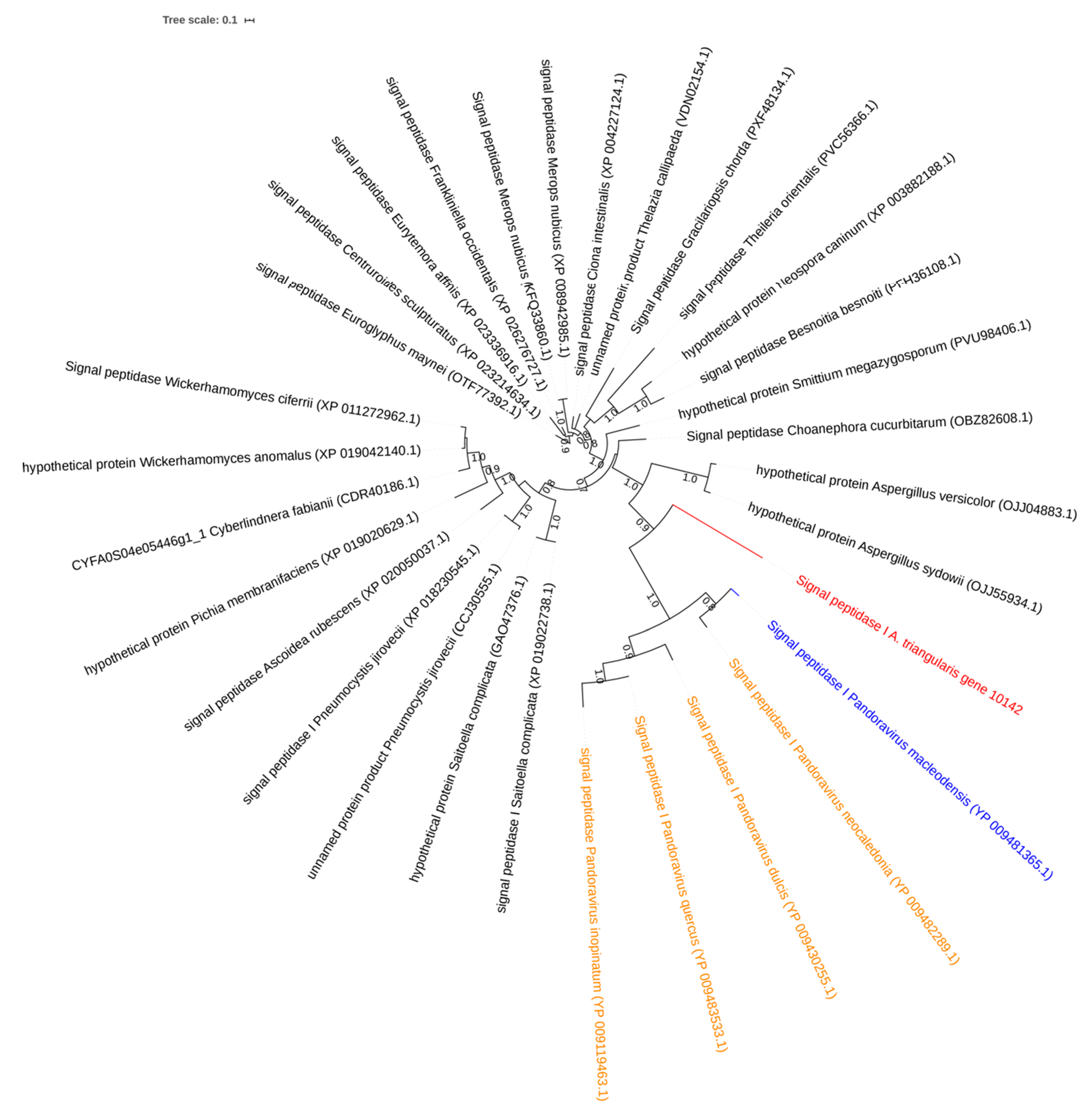
2.4. Investigation on Sequences Inherited from Potential Horizontal Gene Gransfers

The analysis of the best BLASTp hits for A. triangularis predicted proteins revealed the presence of 99 hits in amoeba-resistant microorganisms (ARMs), including amoeba-resistant bacteria (ARBs), fungi, amoeba endosymbionts, and giant viruses (Table S3). Among the 99 ARM best matches with A. triangularis, we identified 62 genes shared with bacteria that could multiply or survive within amoebae. The majority of ARB sequences belonged to the obligate intracellular bacteria of the Chlamydiae phylum. Indeed, we reported 44 best hits (involving 44.4% of ARM genes shared with A. triangularis) belonging to Chlamydia members that lysis Acanthamoeba species or live in endosymbiosis within amoebas. We found sixteen genes (16.2%) best matching with bacteria isolated in amoebae from environmental samples or able to survive in-vitro within amoebae such as Acinetobacter spp. and Pseudomonas spp. The analysis revealed the presence of two hypothetical sequences with best hits belonging to L. pneumophila, a pathogenic human bacterium leading to respiratory illness. Finally, two sequences (cleavage stimulation factor subunit 2 and mitochondrial import inner membrane translocase subunit TIM9) of A. triangularis are shared with Cryptococcus neoformans, a human pathogenic fungi in immunocompromised patients that can invade and multiply within A. castellanii [28].
Of the 99 ARM homologs identified in the genome of A. triangularis, 37 have best matched with viruses, including 35 with giant viruses (35.4%). Most of these viral sequences are shared with Pandoraviridae members. Indeed, we identified 19 genes belonging to 5 different Pandoravirus strains (P. quercus, P. inopinatum, P. macleodensis, P. neocaledonia, and P. salinus) with A. triangularis best match. These 19 genes encode 9 hypothetical proteins, 1 F-box domain-containing protein, 1 methyltransferase, 1 ribonuclease BN, 2 ribonucleoside-diphosphate reductase small chain, 1 signal peptidase I, 1 metallophosphatase, 1 morn repeat protein, 1 transp Tc5 C and 1 serine/threonine protein kinase. Furthermore, we identified 5 A. triangularis homologous sequences shared with Medusavirus, a giant virus that replicates on Acanthamoeba castellanii, which was isolated from hot spring water [29]. Five of the thirty-five genes with best match to the giant viruses belonged to members of the Mimiviridae, including Mimivirus, Tupanvirus, and Catovirus. The identification of one A. triangularis gene best matching with Marseillevirus sequence encoding hypothetical protein was reported (Table S3). Other homologs of A. triangularis, shared with giant viruses, belonged to Pithovirus sibericum and Mollivirus sibericum, which were isolated from Siberian permafrost [30,31]. A further functional enrichment by COG revealed that most of the A. triangularis genes shared with ARM organisms were assigned an unknown function (11 genes), followed by amino acid transport and metabolism (4 genes), carbohydrate and metabolism (3 genes), and coenzyme transport and metabolism (3 genes) (Figure S3).
The study of putative horizontal transfers was evaluated by phylogenetic reconstructions based on the A. triangularis homologs with ARMs. Among the 99 potential genes shared with ARMs, we were able to obtain 82 phylogenetic trees. The protein sequences with insufficient numbers of hits did not allow us to examine the potential horizontal transfers, as this was the case for 17 genes. The phylogenetic analyses showed that lateral gene transfer were confirmed for 62 (62.6%) protein sequences, including 34 and 28, which had a best hit with organisms belonging to ARBs and giant viruses, respectively.
A hypothetical protein of A. triangularis (gene 4683) shared homologs with Candidatus Protochlamydia amoebophila and A. castellanii, an amoeba phylogenetically related to A. triangularis. Furthermore, the phylogeny tree based on this hypothetical protein of A. triangularis and these ARM homologs showed that the closest homolog to the A. triangularis gene identified so far was found in Candidatus Protochlamydia and suggests that the gene was transferred from A. triangularis to Candidatus Protochlamydia (Figure 5). The BLASTp analysis revealed that the signal peptidase I gene of A. triangularis (gene 10142) was homologous with 4 Pandoravirus strains. Moreover, the phylogeny tree showed clustering of signal; these peptidase I homologs suggest a putative horizontal gene transfer from Pandoraviruses to A. triangularis (Figure 6).

2.5. Pan-Genome and Core Genome Analyses of Acanthamoeba spp.

The genome of 8 different Acanthamoeba species was compared in order to analyze the genetic diversity between the different Acanthamoeba species. Acanthamoeba pangenome size reaches 59,450 genes encompassing clusters or unique genes (Figure 7).
A total of 843 clusters composed the core genome, which represented 1.5% of the pangenome. Among the core genome, A. triangularis exhibited 399 clusters (0.7%) with two representative sequences and 227 clusters (0.4%) composed of three representative sequences (Table S4). Moreover, A. triangularis genes were part of 99 clusters of unique genes (Table S4). The COG analysis showed that the “unknown function”, “post-translational modification, protein turnover, and chaperones” function, and “signal transduction mechanisms” function (63) are among the categories the most represented for core genome and A. triangularis unique sequences (Figure S4 and Figure S5). Surprisingly, we found a large number of unique genes for Acanthamoeba castellanii (n = 8,236 genes) and Acanthamoeba lugdunensis (n = 2544). These unique genes play a role in signal transduction, metabolism pathway, and DNA biosynthesis (Figure S6). However, a large part of these genes is categorized as unknown function (233 and 137 for A. castellanii and A. lugdunensis, respectively). This high number of unique genes could be explained by the larger predicted protein number of A. castellanii (73,447) and A. lugdonensis (65,171) compared to the other amoebae used in the analysis (Acanthamoeba culberstoni, 22,241; Acanthamoeba lenticulate, 29,468; Acanthamoeba polyphaga, 32,524; Acanthamoeba quina, 49,881; Acanthamoeba rhysodes, 47,088 and A. triangularis, 39,411). Finally, a phylogenetic tree based on the presence and absence of homologous genes within the Acanthamoeba pangenome showed that A. triangularis was clustered with A. rhysodes (Figure S7).

3. Discussion

Members of the genus Acanthamoeba are amoebae in which we have a particular interest because they are involved in human infections and have a potential role as vectors for pathogenic microorganisms [3,5,13]. Currently, the characterizing of Acanthamoeba spp. is based on ribosomal sequences and only two Acanthamoeba genomes have been deep investigated, especially A. castellanii and A. polyphaga [19,20,32,33].
In our study, we explored the genomic content of A. triangularis strain SH 621, an amoeba belonging to Acanthamoeba ribo-genotype T4, related to human keratitis. The genomic approach provided the main characteristics of this protist with a significant genome of more than 66.43 Mb encompassing 13,849 scaffolds. A. triangularis harbored a genome size approximatively 2-fold smaller than A. castellanii ATCC 50370 (120.6 M) and A. polyphaga ATCC 30872 (115.3 Mb). The size of A. castellanii ATCC 50370 and A. polyphaga ATCC 30872 is composed of large number of contigs (A. polyphaga; 224,482 scaffolds, A. castellanii 221,748 scaffolds) with very short sequences, suggesting a potential overestimation of the size genome and an assembly mistake.
Moreover, A. castellanii Neff (42.02 Mb) and A. polyphaga Linc-AP1(49.35Mb) [20] have a genome size of 1.7- and 1.3-fold smaller than the A. triangularis, respectively. These differences in sizes can be explained by a sequencing technology and assembly tools used, which were different between the sequenced Acanthamoeba genome. Currently, the characterizing of Acanthamoeba is based on ribosomal sequences. However, we observed that Acanthamoeba organism can contain multiple copies of 18S rRNA genes, including one complete and 3 incompletes. This aspect could be a source of heterogeneity and represents a potential limit of ribo-typing. So, genomic analysis could provide more information in order to classify the Acanthamoeba species.
The comparison of 37,062 predicted protein sequences against the three main public databases allowed us to obtain a detailed annotation and better knowledge on the biologic function of A. triangularis genes. The annotation revealed a composition from diverse putative origins of the draft genome sequences. These results show a great proportion with eukaryotic organisms, especially with A. castellanii strain Neff ATCC 30010. The analysis based on A. triangularis virulence reported 48 genes related to keratitis mechanisms. We found gene encoding for mannose-binding proteins (MBP), important proteins that mediate the amoeba adhesion to the corneal epithelial cells [34,35]. The adhesion of amoebas to the host cells is an essential step in the pathogenesis of Acanthamoeba keratitis. Several studies reported the central pathogenic property of the MPB and its involvement in the destruction of target cells [27,34,35,36]. MBP had been potential targets to develop therapeutic antibodies in order to inhibit the host–parasites interaction [37]. We also identified various proteases directly related to the pathogenicity of Acanthamoeba spp., especially serine protease and metalloproteases that contribute to the evasion of the host. The analysis revealed the presence of 3 phospholipases shared with potentially pathogenic Acanthamoeba species. The role of phospholipase in the membrane disruption, entrance in the host cells, and cell lysis was suspected in several studies [6,38]. Furthermore, phospholipase involvement has been reported in the induction of inflammatory responses facilitating the Acanthamoeba virulence [6]. The genomic study showed the presence of heat shock protein (Hsps) specific to pathogenic Acanthamoeba species. Hsps are essential to survive and adapt organisms at higher temperatures in order to maintain their metabolic activities within the host [39,40]. So, the genomic analysis is a useful strategy to identify and characterize potential targets to develop a new therapeutical approach.
BLASTp and phylogenetic analyses revealed horizontal gene transfers between A. triangularis and some pathogenic bacteria, such as L. pneumophila, a human respiratory pathogen that multiplies and lysis Acanthamoeba spp. [41]. Among the ARM organisms, it was the “Chlamydia endosymbionts” organisms that had the highest number of homologs with A. triangularis, including Protochlamydia amoebophila and Neochlamydia organisms. These homolog genes could be exchanged during horizontal endosymbiont transmission. Out of 99 putative genes exchanged with ARMs, 49 genes were virus-infecting amoebas. The proportion of giant viral sequences within A. triangularis (0.13%) was smaller compared to A. castellani Neff (1.2%) and W. magna c2c maky (0.3%) [33,42]. However, the proportion was higher compared to the giant virus sequences contained in the genome of A. polyphaga (0.1%) [21]. The presence of viral sequences within A. triangularis genome is consistent with the history of giant viruses which is related to the genus Acanthamoeba. Indeed, since the isolation of the first giant virus named “Acanthamoeba polyphaga mimivirus” from A. polyphaga, the Acanthamoebas species have been used as support to isolate giant viruses by co-culture [43,44,45,46,47]. These amoebae seemed to be more permissive compared to other families of amoebae since a large part of the giant viruses have been isolated from A. castellanii and A. polyphaga, including Mimivirus, Marseillevirus, Pandoravirus, and Tupanvirus [43,45,46,48]. The majority of viral sequence genes best matching with A. triangularis were viruses invading amoebas from the Acanthamoeba genus. Furthermore, we observed that the large part of exchanged genes is assigned a function, which could bring a benefit to the organism that receives the sequences.
These sequence exchanges have shown that Acanthamoeba organisms represent a biological niche that contributes to the genome heterogeneity of FLA. Moreover, the low part of the core genome (0.02) compared to the pangenome demonstrated the plasticity of Acanthamoeba genomes.
Therefore, the study provides better knowledge on the FLA of Amoebozoa clade and insight into the unexplored world of FLA. The proximity between FLA and microorganisms favor horizontal gene transfers. In addition, pathogenicity analysis has revealed that some virulence genes related to keratitis are specific to the genus Acanthamoeba.

4. Materials and Methods

4.1. Culture of Acanthamoeba Triangularis Strain SH 621

The culture of A. triangularis (ATCC 50254TM) was performed at 30 °C using 175 cm² culture flasks in PYG medium (Thermo Fisher Scientific, Illkirch, France) [49]. When the trophozoites formed a monolayer, the amoebas were detached from the flask and harvested by centrifugation at 700× g for 10 min, followed by three washing steps using Page’s modified Neff’s Amoeba Saline medium (2 mM NaCl, 16 μM MgSO4, 27.2 μM CaCl2, 1 mM Na2HPO4, 1 mM KH2PO4). Amoeba quantification was performed using a KOVA® slide cell counting chamber.

4.2. Extraction and Sequencing of DNA

DNA of A. triangularis was extracted with 1 volume of phenol/chloroform/isoamyl alcohol (50:49:1) (Merck KGaA). The mix was centrifugated 5 min at 10,000× g and we recovered the aqueous phase. We repeated twice this DNA extraction step. Then, we precipitated the DNA with the addition of 2 volumes of ethanol (Merck KGaA) and gently stirred by hand. The suspension was incubated overnight at −20 °C, centrifuged at 10,000× g for 30 min at 4 °C, the supernatant was removed and the DNA solution dried at room temperature. Finally, DNA was dissolved in 0.5 mL in TE buffer (10 mM Tris, 0.1 mM EDTA). Genomic DNA (gDNA) of Acanthamoeba triangularis was quantified by a Qubit assay with the high sensitivity kit (Life Technologies, Carlsbad, CA, USA) to 7.1 ng/µL. Genomic DNA was next sequenced on the MiSeq Technology (Illumina Inc, San Diego, CA, USA) with the paired-end strategy. To prepare the paired-end library, a dilution was performed to require 1 ng of genome. The tagmentation step fragmented and tagged the DNA. Then a limited number of cycle PCR cycles (12) completed the tag adapters. After purification on AMPure XP beads (Beckman Coulter Inc, Fullerton, CA, USA), the libraries were then normalized on specific beads according to the Nextera XT protocol (Illumina). Normalized libraries were pooled into a single library for sequencing on the MiSeq. The pooled single strand library was loaded onto the reagent cartridge and then onto the instrument along with the flow cell. Automated cluster generation and paired-end sequencing with dual index reads were performed in a single 39-h run in 2 × 250-bp. Total information of 3.4 Gb was obtained with a cluster density of 360,000 per mm2 and, finally, with a cluster passing quality control filters of 96.8%. Within this run, the index representation for A. triangularis was determined at 63.84%. The 4,302,070 paired-end reads were filtered according to the read qualities.

4.3. Genome Assembly

The quality of raw data from DNA sequencing was controlled using FastQC software (https://www.bioinformatics.babraham.ac.uk/projects/fastqc/). Raw data were trimmed with the Trimmomatic software [50]. Indeed, the reads with low quality were removed and only the reads with average quality above 28 were selected. All trimmed reads of DNA were assembled de novo using CLC Genomics Workbench v7.51 (https://www.qiagenbioinformatics.com/products/clc-genomics-workbench/). The 64-word size and 100 bubble size parameters were used. The contigs with size under 980 bp were removed. To identify and remove scaffolds that were likely to have originated from bacterial or viral contaminants, we conducted BLASTn searches against the local databases with the megablast option [51]. The assembly was improved with GapFiller [52]. The quality assessment of the genome assembly was analyzed using QUAST software [53]. The genome was deposited in Genbank under the number accession CACVKS010000000. Then, a phylogenetic analysis based on the 18S rRNA gene was performed. The 18S rRNA gene of A. triangularis strain SH 621 was identified by BLASTn comparison between the amoebal genome assembly and the 18S rRNA sequences of A. triangularis (AF316547.2) available in the NCBI GenBank nucleotide sequence database (nt). The 18S rRNA gene of A. triangularis strain SH 621 was deposited in Genbank (LR757994). Homologs were searched for using BLASTn against the nt database. Multiple sequence alignment was carried out using MUSCLE software [54]. Finally, a phylogenetic analysis of these nucleotide sequences was performed using MEGA version 7 and the maximum likelihood (ML) algorithm, with 1000 bootstrap replicates [55].

4.4. Functional Annotation

The open reading frame prediction was performed using AUGUSTUS, a software optimized for the eukaryotic genome [56]. The biological function of A. triangularis predicted proteins was analyzed by a comparison of their sequences with those from public databases, including the NCBI non-redundant protein sequence database (nr), Cluster of Orthologous Group of proteins (COG) database and against Kyoto Encyclopedia of Genes and Genomes Pathway (KEGG) database. Firstly, the function of protein sequences was identified by a BLASTp search against the nr database with E-value cutoff at 1e-03 [51]. The COG annotation was performed using EggNOG [57,58] with diamond as mapping mode. To analyze the metabolic pathway and the biological function of the genes, we have mapped the A. triangularis sequences against the Kyoto Encyclopedia of Genes and Genomes Pathway [59].

4.5. Analysis of Virulence Related Genes

To identify genes possibly related to Acanthamoeba keratitis, we performed comparative genomic analysis with Acanthamoeba species (Acanthamoeba culberstoni: CDFF01000001.1, Acanthamoeba lenticulata: NAVB01000001.1, Acanthamoeba polyphaga: LQHA01000001.1, Acanthamoeba lugdunensis: CDFB01000001.1, Acanthamoeba quina: CDFN01000001.1, Acanthamoeba rhysodes: CDFC01000001.1, Acanthamoeba castellanii: CDFL01000001.1, and Acanthamoeba triangularis: CACVKS010000000) and other amoebas, including non-pathogenic amoebas (Willaertia magna: PRJEB30797, Naegleria gruberi: GCA_000004985.1, N. lovaniensis: GCA_003324165.1, Dictyostelium discoideum: PRJNA13925, and Acytostelium subglobosum: PRJNA280978) and pathogenic amoebas (Entamoeba histolitica: PRJDB4673 and Naegleria fowleri: GCA_000499105.1) that are not involved in keratitis. All Acanthamoeba spp. selected for the analysis belong to the species and ribo-genotypes related to keratitis in humans [2,10,60,61,62,63]. In order to identify only the genes related to virulence among these belonging to Acanthamoeba spp., we included other amoebas that are not related to keratitis pathogenesis. The genes possibly related to the pathogenicity of keratitis were identified according to a single criterion; they are shared only among the opportunistic amoebae of the genus Acanthamoeba used in the study that have been described as being associated with keratitis in the literature [2,3,6,10]. First, we recovered protein sequences of Dictyostelium discoideum (PRJNA13925), Acytostelium subglobosum (PRJNA280978), and Entamoeba histolitica (PRJDB4673) in the Genbank database. For the other amoebas, we performed a protein prediction using the AUGUSTUS program [56]. Then, Proteinortho v5 software was used with these 15 amoeba genomes using as parameters, 60% coverage and 50% amino acid identity, and an e-value of 1e-4 as significance thresholds. To identify the genes related to keratitis mechanisms, we recovered only the homologous genes belonging to 8 Acanthamoeba species (A. culberstoni, A. lenticulata, A. polyphaga, A. lugdunensis, A. quina, A. rhysodes, A. castellanii, and A. triangularis) that were not considered as orthologous genes with others FLA protein sequences used in the analysis. Then, we analyzed the function of these genes shared between Acanthamoeba spp.

4.6. Study of Horizontal Gene Transfers Between A. triangularis and ARMs

We identified the A. triangularis predicted proteins best matching with proteins from amoeba-resistant microorganisms. For each of these protein sequences, we performed a BLASTp search against the NCBI non-redundant (nr) protein sequence database, with an e-value cutoff 1e-03. To confirm the potential horizontal transfers for the genes revealing a best match with amoeba-resistant microorganism homologs, we carried out for a phylogenetic reconstruction all these genes. The alignment of protein sequences were performed using MUSCLE [54]. Phylogenetic trees were obtained using FastTree software [64] and by maximum likelihood method with Jones–Taylor–Thornton (JTT) model with MEGA 7.0.25 software. Phylogenetic trees were visualized using iTOL v3 online [65].

4.7. Comparative Genomic Analyses

To identify the best reciprocal hits between different amoebas genomes of the genus Acanthamoeba, we used Proteinortho v5 with 60% coverage and 50% amino acid identity, and an e-value of 1e-4 as significance threshold [66]. The clustering was obtained from the predicted proteins using the AUGUSTUS program for eight Acanthamoeba species (Acanthamoeba culberstoni: CDFF01000001.1, Acanthamoeba lenticulata: NAVB01000001.1, Acanthamoeba polyphaga: LQHA01000001.1, Acanthamoeba lugdunensis: CDFB01000001.1, Acanthamoeba quina: CDFN01000001.1, Acanthamoeba rhysodes: CDFC01000001.1, Acanthamoeba castellanii: CDFL01000001.1 and Acanthamoeba triangularis: CACVKS010000000). The functional enrichment was carried out by comparison of protein sequences against COG databases. Finally, pangenome tree based on genus Acanthamoeba was generated using the GET_HOMOLOGUES package with the standard parameters [67]. The phylogenetic tree was visualized using iTOL v3 online [65].

Supplementary Materials

The following are available online at https://www.mdpi.com/2076-0817/9/3/217/s1. Table S1: Functional annotation analysis of Acanthamoeba triangularis protein sequences; Table S2: List of Acanthamoeba triangularis protein sequences possibly related to keratitis pathogenicity; Table S3: List of Acanthamoeba triangularis protein sequences shared with amoeba-resisting microorganisms; Table S4: List of specific genes and genes of core genome of Acanthamoeba triangularis. Figure S1: Identification of 18S rRNA gene of A. triangularis by comparison against nt database on NCBI. Figure S2: Phylogenetic analysis of amoebas of A. triangularis strain SH 621. Figure S3: Representation of COG functional categories of the A. triangularis sequences shared with ARMs. Figure S4: Representation of COG functional categories of the core genome. Figure S5: Representation of COG functional categories of the unique genes of A. triangularis. Figure S6: Representation of COG functional categories of the unique genes of Acanthamoeba castellanii and Acanthamoeba lugdunensis. Figure S7: Representation of pangenomic tree based on genus Acanthamoeba.

Author Contributions

Conceptualization, P.C. and B.L.S.; Formal analysis, I.H. and J.A.; Supervision, P.C. and B.L.S.; Writing – original draft, I.H.; Writing – review and editing, J.A., P.C., and B.L.S. All authors have read and agree to the published version of the manuscript.

Funding

This research was funded by the French Government under the “Investissements d’avenir” (Investments for the Future) program managed by the Agence Nationale de la Recherche (ANR, French National Agency for Research), (reference: Méditerranée Infection 10-IAHU-03), by Région Provence-Alpes-Côte d’Azur and European funding FEDER PRIMI.

Conflicts of Interest

The authors declare no conflict of interest.

References

  1. Rodríguez-Zaragoza, S. Ecology of free-living amoebae. Crit. Rev. Microbiol. 1994, 20, 225–241. [Google Scholar] [CrossRef]
  2. Khan, N.A. Acanthamoeba: Biology and increasing importance in human health. FEMS Microbiol. Rev. 2006, 30, 564–595. [Google Scholar] [CrossRef] [Green Version]
  3. Siddiqui, R.; Khan, N.A. Biology and pathogenesis of Acanthamoeba. Parasites Vectors 2012, 5, 6. [Google Scholar] [CrossRef] [Green Version]
  4. Clarke, D.W.; Niederkorn, J.Y. The pathophysiology of Acanthamoeba keratitis. Trends Parasitol. 2006, 22, 175–180. [Google Scholar] [CrossRef]
  5. Kot, K.; Łanocha-Arendarczyk, N.A.; Kosik-Bogacka, D.I. Amoebas from the genus Acanthamoeba and their pathogenic properties. Ann. Parasitol. 2018, 64, 299–308. [Google Scholar]
  6. Lorenzo-Morales, J.; Khan, N.A.; Walochnik, J. An update on Acanthamoeba keratitis: Diagnosis, pathogenesis and treatment. Parasite 2015, 22. [Google Scholar] [CrossRef] [Green Version]
  7. Trabelsi, H.; Dendana, F.; Sellami, A.; Sellami, H.; Cheikhrouhou, F.; Neji, S.; Makni, F.; Ayadi, A. Pathogenic free-living amoebae: Epidemiology and clinical review. Pathol. Biol. 2012, 60, 399–405. [Google Scholar] [CrossRef] [PubMed]
  8. Taher, E.E.; Méabed, E.M.H.; Abdallah, I.; Abdel Wahed, W.Y. Acanthamoeba keratitis in noncompliant soft contact lenses users: Genotyping and risk factors, a study from Cairo, Egypt. J. Infect. Public Health 2018, 11, 377–383. [Google Scholar] [CrossRef] [PubMed]
  9. Castro-Artavia, E.; Retana-Moreira, L.; Lorenzo-Morales, J.; Abrahams-Sandí, E. Potentially pathogenic Acanthamoeba genotype T4 isolated from dental units and emergency combination showers. Mem. Inst. Oswaldo Cruz 2017, 112, 817–821. [Google Scholar] [CrossRef] [PubMed] [Green Version]
  10. Xuan, Y.-H.; Chung, B.-S.; Hong, Y.-C.; Kong, H.-H.; Hahn, T.-W.; Chung, D.-I. Keratitis by acanthamoeba triangularis: Report of cases and characterization of isolates. Korean J. Parasitol. 2008, 46, 157–164. [Google Scholar] [CrossRef] [PubMed]
  11. Anwar, A.; Khan, N.A.; Siddiqui, R. Combating Acanthamoeba spp. cysts: What are the options? Parasit Vectors 2018, 11, 26. [Google Scholar] [CrossRef] [PubMed] [Green Version]
  12. Clarholm, M. Protozoan grazing of bacteria in soil—Impact and importance. Microb. Ecol. 1981, 7, 343–350. [Google Scholar] [CrossRef] [PubMed]
  13. Greub, G.; Raoult, D. Microorganisms resistant to free-living amoebae. Clin. Microbiol. Rev. 2004, 17, 413–433. [Google Scholar] [CrossRef] [PubMed] [Green Version]
  14. Beye, M.; Hasni, I.; Seng, P.; Michelle, C.; La Scola, B.; Raoult, D.; Fournier, P.-E. Genomic analysis of a Raoultella ornithinolytica strain causing prosthetic joint infection in an immunocompetent patient. Sci. Rep. 2018, 8, 9462. [Google Scholar] [CrossRef]
  15. Hasni, I.; Jarry, A.; Quelard, B.; Carlino, A.; Eberst, J.-B.; Abbe, O.; Demanèche, S. Intracellular behaviour of three legionella pneumophila strains within three amoeba strains, including willaertia magna C2c maky. Pathogens 2020, 9, 105. [Google Scholar] [CrossRef] [Green Version]
  16. Andreani, J.; Khalil, J.Y.B.; Baptiste, E.; Hasni, I.; Michelle, C.; Raoult, D.; Levasseur, A.; La Scola, B. Orpheovirus IHUMI-LCC2: A New Virus among the Giant Viruses. Front. Microbiol. 2018, 8, 2643. [Google Scholar] [CrossRef] [Green Version]
  17. Moliner, C.; Raoult, D.; Fournier, P.-E. Evidence of horizontal gene transfer between amoeba and bacteria. Clin. Microbiol. Infect. 2009, 15, 178–180. [Google Scholar] [CrossRef] [Green Version]
  18. Bertelli, C.; Greub, G. Lateral gene exchanges shape the genomes of amoeba-resisting microorganisms. Front. Cell Infect. Microbiol. 2012, 2, 110. [Google Scholar] [CrossRef] [Green Version]
  19. Clarke, M.; Lohan, A.J.; Liu, B.; Lagkouvardos, I.; Roy, S.; Zafar, N.; Bertelli, C.; Schilde, C.; Kianianmomeni, A.; Bürglin, T.R.; et al. Genome of Acanthamoeba castellanii highlights extensive lateral gene transfer and early evolution of tyrosine kinase signaling. Genome Biol. 2013, 14, R11. [Google Scholar] [CrossRef] [Green Version]
  20. Karlyshev, A.V. Remarkable features of mitochondrial dna of acanthamoeba polyphaga linc ap-1, revealed by whole-genome sequencing. Microbiol. Resour. Announc. 2019, 8, e00430-19. [Google Scholar] [CrossRef] [Green Version]
  21. Chelkha, N.; Levasseur, A.; Pontarotti, P.; Raoult, D.; Scola, B.L.; Colson, P. A phylogenomic study of acanthamoeba polyphaga draft genome sequences suggests genetic exchanges with giant viruses. Front. Microbiol. 2018, 9, 2098. [Google Scholar] [CrossRef] [PubMed] [Green Version]
  22. Zysset-Burri, D.C.; Müller, N.; Beuret, C.; Heller, M.; Schürch, N.; Gottstein, B.; Wittwer, M. Genome-wide identification of pathogenicity factors of the free-living amoeba Naegleria fowleri. BMC Genom. 2014, 15, 496. [Google Scholar] [CrossRef] [PubMed] [Green Version]
  23. Fritz-Laylin, L.K.; Prochnik, S.E.; Ginger, M.L.; Dacks, J.B.; Carpenter, M.L.; Field, M.C.; Kuo, A.; Paredez, A.; Chapman, J.; Pham, J.; et al. The genome of naegleria gruberi illuminates early eukaryotic versatility. Cell 2010, 140, 631–642. [Google Scholar] [CrossRef] [PubMed] [Green Version]
  24. Liechti, N.; Schürch, N.; Bruggmann, R.; Wittwer, M. The genome of Naegleria lovaniensis, the basis for a comparative approach to unravel pathogenicity factors of the human pathogenic amoeba N. fowleri. BMC Genom. 2018, 19, 654. [Google Scholar] [CrossRef] [Green Version]
  25. Sucgang, R.; Kuo, A.; Tian, X.; Salerno, W.; Parikh, A.; Feasley, C.L.; Dalin, E.; Tu, H.; Huang, E.; Barry, K.; et al. Comparative genomics of the social amoebae Dictyostelium discoideum and Dictyostelium purpureum. Genome Biol. 2011, 12, R20. [Google Scholar] [CrossRef] [Green Version]
  26. Lorenzi, H.A.; Puiu, D.; Miller, J.R.; Brinkac, L.M.; Amedeo, P.; Hall, N.; Caler, E.V. New assembly, reannotation and analysis of the entamoeba histolytica genome reveal new genomic features and protein content information. PLoS Negl. Trop. Dis. 2010, 4, e716. [Google Scholar] [CrossRef] [Green Version]
  27. Huth, S.; Reverey, J.F.; Leippe, M.; Selhuber-Unkel, C. Adhesion forces and mechanics in mannose-mediated acanthamoeba interactions. PLoS ONE 2017, 12, e0176207. [Google Scholar] [CrossRef] [Green Version]
  28. Guimaraes, A.J.; Gomes, K.X.; Cortines, J.R.; Peralta, J.M.; Peralta, R.H.S. Acanthamoeba spp. as a universal host for pathogenic microorganisms: One bridge from environment to host virulence. Microbiol. Res. 2016, 193, 30–38. [Google Scholar] [CrossRef]
  29. Yoshikawa, G.; Blanc-Mathieu, R.; Song, C.; Kayama, Y.; Mochizuki, T.; Murata, K.; Ogata, H.; Takemura, M. Medusavirus, a novel large DNA virus discovered from hot spring water. J. Virol. 2019, 93, e02130-18. [Google Scholar] [CrossRef] [Green Version]
  30. Legendre, M.; Lartigue, A.; Bertaux, L.; Jeudy, S.; Bartoli, J.; Lescot, M.; Alempic, J.-M.; Ramus, C.; Bruley, C.; Labadie, K.; et al. In-depth study of Mollivirus sibericum, a new 30,000-y-old giant virus infecting Acanthamoeba. Proc. Natl. Acad. Sci. USA 2015, 112, E5327–E5335. [Google Scholar] [CrossRef] [Green Version]
  31. Legendre, M.; Bartoli, J.; Shmakova, L.; Jeudy, S.; Labadie, K.; Adrait, A.; Lescot, M.; Poirot, O.; Bertaux, L.; Bruley, C.; et al. Thirty-thousand-year-old distant relative of giant icosahedral DNA viruses with a pandoravirus morphology. Proc. Natl. Acad. Sci. USA 2014, 111, 4274–4279. [Google Scholar] [CrossRef] [PubMed] [Green Version]
  32. Risler, A.; Coupat-Goutaland, B.; Pélandakis, M. Genotyping and phylogenetic analysis of Acanthamoeba isolates associated with keratitis. Parasitol. Res. 2013, 112, 3807–3816. [Google Scholar] [CrossRef] [PubMed]
  33. Maumus, F.; Blanc, G. Study of gene trafficking between acanthamoeba and giant viruses suggests an undiscovered family of amoeba-infecting viruses. Genome Biol. Evol. 2016, 8, 3351–3363. [Google Scholar] [CrossRef] [PubMed] [Green Version]
  34. Garate, M.; Cubillos, I.; Marchant, J.; Panjwani, N. Biochemical characterization and functional studies of acanthamoeba mannose-binding protein. Infect. Immun. 2005, 73, 5775–5781. [Google Scholar] [CrossRef] [PubMed] [Green Version]
  35. Panjwani, N. Pathogenesis of acanthamoeba keratitis. Ocul. Surf. 2010, 8, 70–79. [Google Scholar] [CrossRef] [Green Version]
  36. Hurt, M.; Niederkorn, J.; Alizadeh, H. Effects of mannose on Acanthamoeba castellanii proliferation and cytolytic ability to corneal epithelial cells. Investig. Ophthalmol. Vis. Sci. 2003, 44, 3424–3431. [Google Scholar] [CrossRef] [PubMed] [Green Version]
  37. Garate, M.; Alizadeh, H.; Neelam, S.; Niederkorn, J.Y.; Panjwani, N. Oral immunization with acanthamoeba castellanii mannose-binding protein ameliorates amoebic keratitis. Infect. Immun. 2006, 74, 7032–7034. [Google Scholar] [CrossRef] [Green Version]
  38. Mortazavi, P.N.; Keisary, E.; Loh, L.N.; Jung, S.-Y.; Khan, N.A. Possible roles of phospholipase A2 in the biological activities of acanthamoeba castellanii (T4 genotype). Protist 2011, 162, 168–176. [Google Scholar] [CrossRef]
  39. Pérez-Serrano, J.; Martínez, J.; Pérez, B.; Bernadina, W.E.; Rodríguez-Caabeiro, F. In vitro shock response to different stressors in free living and pathogenic Acanthamoeba. Int. J. Parasitol. 2000, 30, 829–835. [Google Scholar] [CrossRef]
  40. Podlipaeva, I.I.; Shmakov, L.A.; Gilichinskiĭ, D.A.; Gudkov, A.V. Heat shock protein of HSP70 family revealed in some contemporary freshwater Amoebae and in Acanthamoeba sp. from cysts isolated from permafrost samples. Tsitologiia 2006, 48, 691–694. [Google Scholar]
  41. Cirillo, J.D.; Falkow, S.; Tompkins, L.S. Growth of legionella pneumophila in acanthamoeba castellanii enhances invasion. Infect. Immun. 1994, 62, 3254–3261. [Google Scholar] [CrossRef] [PubMed] [Green Version]
  42. Hasni, I.; Chelkha, N.; Baptiste, E.; Mameri, M.R.; Lachuer, J.; Plasson, F.; Colson, P.; Scola, B.L. Investigation of potential pathogenicity of Willaertia magna by investigating the transfer of bacteria pathogenicity genes into its genome. Sci. Rep. 2019, 9, 1–12. [Google Scholar] [CrossRef]
  43. La Scola, B.; Audic, S.; Robert, C.; Jungang, L.; de Lamballerie, X.; Drancourt, M.; Birtles, R.; Claverie, J.-M.; Raoult, D. A giant virus in amoebae. Science 2003, 299, 2033. [Google Scholar] [CrossRef]
  44. Andreani, J.; Khalil, J.Y.B.; Sevvana, M.; Benamar, S.; Di Pinto, F.; Bitam, I.; Colson, P.; Klose, T.; Rossmann, M.G.; Raoult, D.; et al. Pacmanvirus, a new giant icosahedral virus at the crossroads between asfarviridae and faustoviruses. J. Virol. 2017, 91, e00212-17. [Google Scholar] [CrossRef] [PubMed] [Green Version]
  45. Colson, P.; Pagnier, I.; Yoosuf, N.; Fournous, G.; La Scola, B.; Raoult, D. “Marseilleviridae”, a new family of giant viruses infecting amoebae. Arch. Virol. 2013, 158, 915–920. [Google Scholar] [CrossRef] [PubMed] [Green Version]
  46. Philippe, N.; Legendre, M.; Doutre, G.; Couté, Y.; Poirot, O.; Lescot, M.; Arslan, D.; Seltzer, V.; Bertaux, L.; Bruley, C.; et al. Pandoraviruses: Amoeba viruses with genomes up to 2.5 mb reaching that of parasitic eukaryotes. Science 2013, 341, 281–286. [Google Scholar] [CrossRef] [PubMed] [Green Version]
  47. Khalil, J.Y.B.; Andreani, J.; La Scola, B. Updating strategies for isolating and discovering giant viruses. Curr. Opin. Microbiol. 2016, 31, 80–87. [Google Scholar] [CrossRef]
  48. Abrahão, J.; Silva, L.; Silva, L.S.; Khalil, J.Y.B.; Rodrigues, R.; Arantes, T.; Assis, F.; Boratto, P.; Andrade, M.; Kroon, E.G.; et al. Tailed giant Tupanvirus possesses the most complete translational apparatus of the known virosphere. Nat. Commun. 2018, 9, 1–12. [Google Scholar] [CrossRef] [Green Version]
  49. Niyyati, M.; Abedkhojasteh, H.; Salehi, M.; Farnia, S.; Rezaeian, M. Axenic cultivation and pathogenic assays of acanthamoeba strains using physical parameters. Iran. J. Parasitol. 2013, 8, 186–189. [Google Scholar]
  50. Bolger, A.M.; Lohse, M.; Usadel, B. Trimmomatic: A flexible trimmer for Illumina sequence data. Bioinformatics 2014, 30, 2114–2120. [Google Scholar] [CrossRef] [Green Version]
  51. Altschul, S.F. BLAST Algorithm. In eLS.; American Cancer Society: Atlanta, GA, USA, 2014; ISBN 978-0-470-01590-2. [Google Scholar]
  52. Nadalin, F.; Vezzi, F.; Policriti, A. GapFiller: A de novo assembly approach to fill the gap within paired reads. BMC Bioinform. 2012, 13, S8. [Google Scholar] [CrossRef] [Green Version]
  53. Gurevich, A.; Saveliev, V.; Vyahhi, N.; Tesler, G. QUAST: Quality assessment tool for genome assemblies. Bioinformatics 2013, 29, 1072–1075. [Google Scholar] [CrossRef]
  54. Edgar, R.C. MUSCLE: Multiple sequence alignment with high accuracy and high throughput. Nucleic Acids Res. 2004, 32, 1792–1797. [Google Scholar] [CrossRef] [Green Version]
  55. Kumar, S.; Stecher, G.; Tamura, K. MEGA7: Molecular evolutionary genetics analysis version 7.0 for bigger datasets. Mol. Biol. Evol. 2016, 33, 1870–1874. [Google Scholar] [CrossRef] [Green Version]
  56. Stanke, M.; Morgenstern, B. AUGUSTUS: A web server for gene prediction in eukaryotes that allows user-defined constraints. Nucleic Acids Res. 2005, 33, W465–W467. [Google Scholar] [CrossRef] [Green Version]
  57. Tatusov, R.L.; Galperin, M.Y.; Natale, D.A.; Koonin, E.V. The COG database: A tool for genome-scale analysis of protein functions and evolution. Nucleic Acids Res. 2000, 28, 33–36. [Google Scholar] [CrossRef] [Green Version]
  58. Jensen, L.J.; Julien, P.; Kuhn, M.; von Mering, C.; Muller, J.; Doerks, T.; Bork, P. eggNOG: Automated construction and annotation of orthologous groups of genes. Nucleic Acids Res. 2008, 36, D250–D254. [Google Scholar] [CrossRef]
  59. Kanehisa, M.; Goto, S. KEGG: Kyoto encyclopedia of genes and genomes. Nucleic Acids Res. 2000, 28, 27–30. [Google Scholar] [CrossRef]
  60. Hong, J.; Ji, J.; Xu, J.; Cao, W.; Liu, Z.; Sun, X. An unusual case of Acanthamoeba Polyphaga and Pseudomonas Aeruginosa keratitis. Diagn. Pathol. 2014, 9, 105. [Google Scholar] [CrossRef] [Green Version]
  61. González-Robles, A.; Omaña-Molina, M.; Salazar-Villatoro, L.; Flores-Maldonado, C.; Lorenzo-Morales, J.; Reyes-Batlle, M.; Arnalich-Montiel, F.; Martínez-Palomo, A. Acanthamoeba culbertsoni isolated from a clinical case with intraocular dissemination: Structure and in vitro analysis of the interaction with hamster cornea and MDCK epithelial cell monolayers. Exp. Parasitol. 2017, 183, 245–253. [Google Scholar] [CrossRef]
  62. Ledee, D.R.; Hay, J.; Byers, T.J.; Seal, D.V.; Kirkness, C.M. Acanthamoeba griffini. Molecular characterization of a new corneal pathogen. Investig. Ophthalmol. Vis. Sci. 1996, 37, 544–550. [Google Scholar]
  63. Arnalich-Montiel, F.; Lumbreras-Fernández, B.; Martín-Navarro, C.M.; Valladares, B.; Lopez-Velez, R.; Morcillo-Laiz, R.; Lorenzo-Morales, J. Influence of acanthamoeba genotype on clinical course and outcomes for patients with acanthamoeba keratitis in Spain. J. Clin. Microbiol. 2014, 52, 1213–1216. [Google Scholar] [CrossRef] [PubMed] [Green Version]
  64. Price, M.N.; Dehal, P.S.; Arkin, A.P. FastTree: Computing Large Minimum Evolution Trees with Profiles instead of a Distance Matrix. Mol. Biol. Evol. 2009, 26, 1641–1650. [Google Scholar] [CrossRef]
  65. Letunic, I.; Bork, P. Interactive tree of life (iTOL) v3: An online tool for the display and annotation of phylogenetic and other trees. Nucleic Acids Res. 2016, 44, W242–W245. [Google Scholar] [CrossRef]
  66. Lechner, M.; Findeiß, S.; Steiner, L.; Marz, M.; Stadler, P.F.; Prohaska, S.J. Proteinortho: Detection of (Co-)orthologs in large-scale analysis. BMC Bioinform. 2011, 12, 124. [Google Scholar] [CrossRef] [Green Version]
  67. Contreras-Moreira, B.; Vinuesa, P. GET_HOMOLOGUES, a versatile software package for scalable and robust microbial pangenome analysis. Appl. Environ. Microbiol. 2013, 79, 7696–7701. [Google Scholar] [CrossRef] [Green Version]
Figure 1. Characteristics of similarity search of each Acanthamoeba triangularis protein sequences assigned to a function in NCBI non-redundant protein sequence (nr) database. (A) taxonomical distribution. (B) E-value distribution. (C) Similarity distribution.
Pathogens 09 00217 g001
Figure 2. Representation of A. triangularis genes related to different clusters of gene categories.
Pathogens 09 00217 g002
Figure 3. Distribution of A. triangularis protein sequences involved in the metabolic pathway. The protein of A. triangularis was compared to the Kyoto Encyclopedia of Genes and Genomes Pathway (KEGG) database and the repartition of these protein sequences in the metabolic pathway was visualized.
Pathogens 09 00217 g003
Figure 4. Infection of human cornea by Acanthamoeba species. (a) Acanthamoeba detached from contaminated lens. (b) Acanthamoeba spp. attached and adhered to epithelial cells by receptor (mannose receptors and glycoproteins) and secreted metalloproteases to digest external environment. (c) Acanthamoeba spp. destroyed epithelium and Brownan’s membrane in order to penetrate within stroma environment (secretion of proteases, glycosidases, and hydrolytic enzymes). (d) Destruction of stromae. (e) Radial keratoneuritis (an infiltrate along the corneal nerves).
Pathogens 09 00217 g004
Figure 5. Representation of horizontal transfer analysis. Phylogenetic tree for A. triangularis protein of putative ARM origin. The tree was constructed using maximum-likelihood method based on hypothetical protein sequences of A. triangularis. The tree was performed with 21 homologous sequences of A. triangularis retrieved by BLASTp on NCBI. In red: hypothetical protein of A. triangularis SH621; in blue: the closest homolog from Candidatus Protochlamydia amoebophila; in orange: other homologs from chlamydia; in green: homologs from other amoebas; in black: homologs from other organisms.
Pathogens 09 00217 g005
Figure 6. Representation of horizontal transfer analysis. Phylogenetic tree for A. triangularis protein of putative ARM origin. The tree was constructed using maximum-likelihood method based on the signal peptidase I sequences of A. triangularis. The tree was performed with 30 homologous sequences of A. triangularis retrieved by BLASTp on NCBI. In red: signal peptidase I of A. triangularis; in orange: homologs from ARM (Pandoravirus strains); in black: homologs from other organisms.
Pathogens 09 00217 g006
Figure 7. Flower plot showing the core, dispensable, and strain-specific genes of the 8 Acanthamoeba species. The flower plot displays the core gene number (in the center), the dispensable gene number (in the annulus), and the strain-specific gene number (in the petals) for the 8 Acanthamoeba species. The numbers under the strain name denote the total number of related genes. Different colors indicate different Acanthamoeba T-genotype groups: T4 genotype in blue; T5 strains in yellow; T10 strains in red; T11 strains in green.
Pathogens 09 00217 g007
Table 1. Summary of the A. triangularis genome.
ParameterNumber
Haploid genome size (bp)66,434,030
Sequence contigs (n)13,849
GC-content (%)58.6
Maximal scaffold size (bp)80,033
Minimal scaffold size (bp)980
Average scaffold size5322
N508852
N754376
bp, base pairs; N50, 50% of the genome assembly is, as contigs, larger than this size; N75, 75% of the genome assembly is, as contigs, larger than this size.”
Table 2. Comparison of the main genomic features of several amoebas.
OrganismsGenome Size (Mb)Predicted ProteinsAnnotated ProteinsG+C %
Acanthamoeba triangularis ATCC 502546637,06233,16858.6
Acanthamoeba castellanii ATCC 503704220,68115,455 57.8
Willaertia magna C2c Maky 3718,51913,57125
Naegleria fowleri ATCC 308633017,25216,02135
Naegleria gruberi NEG-M 4115,727909033
Naegleria lovaniensis ATCC 305693115,19513,00537
Dictyostelium discoideum AX43413,541842222
Entamoeba histolytica strain HM-1: IMSS218201407624
Source of data: Acanthamoeba castellanii ATCC 50370 [19], Naegleria fowleri [22], Naegleria gruberi [23], Naegleria lovaniensis [24], Dictyostelium discoideum [25], and Entamoeba histolytica [26].
Table 3. List of genes possibly related to the pathogenesis in Acanthamoeba keratitis.
Gene IdentificationFunction
Adhesion
gene 34934mannose binding
Metalloproteases
gene 12288Aminopeptidase I zinc metalloprotease (M18)
gene 1047metalloenzyme superfamily
gene 9969metallocarboxypeptidase
gene 3258metalloenzyme
Proteases
gene 19397peptidase S8 and S53 subtilisin kexin sedolisin
gene 3757PFAM peptidase T2 asparaginase 2
gene 8789peptidase C19 family
gene 9969metallocarboxypeptidase
gene 11390peptidase S8 and S53, subtilisin, kexin, sedolisin
gene 12288Aminopeptidase I zinc metalloprotease (M18)
gene 12995peptidase M17 family
gene 25924Serine aminopeptidase, S33
gene 26649peptidase C19 family
gene 27916peptidase C19 family
gene 9026peptidase C19 family protein
Temperature tolerance
gene 6737Hsp20/alpha crystallin family
gene 1586Hsp70 protein
gene 8645Hsp20/alpha crystallin family protein
Phospholipases
gene 8337phospholipase A2 activator activity
gene 27752phospholipase D
gene 34693phospholipase D
Antioxidant defense
gene 15075glutathione peroxidase
gene 31764peroxidase
gene 27487oxidoreductase

