Article

Gut Symbiotic Microbial Communities in the IUCN Critically Endangered Pinna nobilis Suffering from Mass Mortalities, Revealed by 16S rRNA Amplicon NGS

by
Athanasios Lattos
1,
Ioannis A. Giantsis
2,*,
Dimitrios Karagiannis
3,
John A. Theodorou
4 and
Basile Michaelidis
1
1
Laboratory of Animal Physiology, Department of Zoology, Faculty of Science, School of Biology, Aristotle University of Thessaloniki, 54124 Thessaloniki, Greece
2
Department of Animal Science, Faculty of Agricultural Sciences, University of Western Macedonia, 53100 Florina, Greece
3
National Reference Laboratory for Mollusc Diseases, Ministry of Rural Development and Food, 54627 Thessaloniki, Greece
4
Department of Animal Production Fisheries & Aquaculture, University of Patras, 30200 Mesolonghi, Greece
*
Author to whom correspondence should be addressed.
Pathogens 2020, 9(12), 1002; https://doi.org/10.3390/pathogens9121002
Submission received: 29 October 2020 / Revised: 24 November 2020 / Accepted: 27 November 2020 / Published: 29 November 2020
(This article belongs to the Special Issue Spontaneous Diseases of Mollusks)

Abstract

:
Mass mortality events due to disease outbreaks have recently affected almost every healthy population of fan mussel, Pinna nobilis in Mediterranean Sea. The devastating mortality of the species has turned the interest of the research towards the causes of these events. After the haplosporidan infestation and the infection by Mycobacterium sp., new emerging pathogens have arisen based on the latest research. In the present study, a metagenomic approach of 16S rRNA next generation sequencing (NGS) was applied in order to assess the bacterial diversity within the digestive gland of diseased individuals as well as to carry out geographical correlations among the biodiversity of microbiome in the endangered species Pinna nobilis. The specimens originated from the mortalities occurred in 2019 in the region of Greece. Together with other bacterial genera, the results confirmed the presence of Vibrio spp., assuming synergistic effects in the mortality events of the species. Alongside with the presence of Vibrio spp., numerous bacterial genera were detected as well, including Aliivibrio spp., Photobacterium spp., Pseudoalteromonas spp., Psychrilyobacter spp. and Mycoplasma spp. Bacteria of the genus Mycoplasma were in high abundance particularly in the sample originated from Limnos island representing the first time recorded in Pinna nobilis. In conclusion, apart from exclusively the Haplosporidan and the Mycobacterium parasites, the presence of potentially pathogenic bacterial taxa detected, such as Vibrio spp., Photobactrium spp. and Alivibrio spp. lead us to assume that mortality events in the endangered Fan mussel, Pinna nobilis, may be attributed to synergistic effects of more pathogens.

1. Introduction

Microbial communities constitute the subject of numerous studies for many years, which have been mostly relied however, on culture dependent methods for detection, identification and characterization of virulence traits of various pathogens [1]. With the recent advances in molecular biology and bioinformatics, the theory of Holobionts arose to describe the interactions between the host and its symbiotic organisms as one entirety [2]. Bacterial diversity in bivalves is highly influenced by their filter feeding behavior with the ingestion of bacteria as well as by stressful conditions in their environment [3,4]. Research on the microbiota of aquatic animals has indicated its important role in many key functions such as immune responses-disease resistance, proper assimilation of nutrients through good intestine function and stimulation of biological processes [5,6]. This relationship is reciprocal for the microbiome and the host, due to the provision of a stable and nutritious environment provided by the host [7,8]. On the other hand, global warming because of climate change may result to lower immune responses and physiological performance in aquatic organisms [9]. As a result of the decline in immune responses, opportunistic pathogens, that may already belong to microbiome of the host, take advantage of that opportunity and multiply, creating colonies uncontrollably, to the detriment of the host [10,11].
Modern molecular tools, particularly Next Generation Sequencing (NGS) of the 16s rRNA gene, contributed to a rapid increase in the knowledge for microbial ecology. Symbiotic microbiota in invertebrates are of high importance as health indicators in ecosystems [12]. Using NGS, the interactions between a host and its microbiome, as well as the effects of environmental variables on these interactions can be also evaluated [4]. Considering that common microbiota, especially in the gut, are known to promote resistance against potential opportunist pathogens [13], the study and the comparison of the microbiota of mortality events with the microbiota of healthy individuals is of great interest. However, there are only few studies related with the microbiome within invertebrates and even fewer are associated with mortality events. In 2013, Bayer et al. [14], investigated the microbial assemblage of an ecosystem engineer gorgonian coral species of Mediteranean Sea, Eunicella cavolini, in different depths of a French Mediteranean coast. As a result, no distinct difference emerged in the structure of the microbiome of the corals of different water levels. However, bacteria from the genus Endozoicomonas were in abundance, which is frequently found to have association with several marine invertebrate species. In the context of climate change, Blanquer et al. [15], investigated the possible change in the diversity of the microbiome during the early detection of a disease in the sponge species Ircinia fasciculata, comparing microbiome of healthy individuals with microbiome of diseased individuals. The results showed a small shift, particularly in Gammaproteobacteria and Acidobacteria concentrations, while Deltaproteobacteria declined in diseased tissues versus healthy ones. Additionally, they observed an increase in bacterial diversity significantly in diseased tissues.
Concerning the microbiome of bivalves, Meisterhans et al. [16], studied the microbiota in each organ of the Manila clam, Ruditapes philippinarum and they tried to correlate the microbiome of each organ separately and also comparing with the environmental conditions of differentiated habitats. Another aim of this study was to evaluate the responses of Ruditapes philippinarum in a different habitat quality. Whereas there were differences in the microbiota from each organ, this study revealed no correlation with the particularities of environmental conditions of different habitats. Moreover, reciprocal transplantation experiments indicated that only in the degradation of the quality of the habitat had negative effects on the welfare and on microbiome structure, but not inversely. On the other hand, Vezzulli et al. [4], revealed that Crassostrea gigas is more susceptible in infections than the Mediterranean mussel Mytilus galloprovinciallis. Specifically, by using 16s rDNA gene based bacterial microbiota profile they showed that C. gigas is a better host for the growth of opportunist bacteria Vibrio spp. than M. galloprovincialis. Additionally, they reported that the microbiome of the Mediterranean mussel acts as an inhibitor for the growth of those bacteria. In 2018, Li et al. [17], investigated the impacts of diet and rise of temperature on the gut microbiota of the Mytilus coruscus. Microbial community in their project was investigated through the use of 16s rRNA gene sequencing. The results obtained from their study supported their hypothesis that increasing the temperature may react with the microbial diversity of the gut and favor the proliferation of opportunistic bacteria. No significant difference was observed in the gut microbiota with the use of microalgal diets. Later in 2019, Li et al. [18], investigated the microbial diversity in the hemolymph of the mussel Mytilus coruscus with the presence of Vibrio cyclitrophicus in different temperatures determined by Illumina Hiseq sequencing of 16s rRNA gene. Specimens in higher temperature (27 °C) suffered from greater mortality losses with the presence of Vibrio cyclitrophicus in the ninth day that was associated also with the decline in abundance in the bacterial diversity in the hemolymph. Vibrio presence promoted the proliferation of other opportunist bacteria at the low temperature (21 °C). They finally indicated that temperature is the key factor in shaping microbial communities. Milan et al. [19], investigated the microbial diversity of a mass mortality event occurred in the bivalve Chamelea gallina in association with the gene expression profile. Overexpression of several genes associated with xenobiotic metabolism and down regulation of genes associated with immune and stress responses were observed. In conclusion, they indicated that the great abundance of opportunistic bacteria is directly related with the low immune responses they observed from the gene profile. In 2019, Green et al. [20], simulated a mortality event episode in pacific oyster, Crassostrea gigas, raising the temperature rapidly (20 to 25 °C). They investigated the microbial diversity of this heat shock with and without the presence of antibiotics in the water. NGS of the 16s rRNA gene revealed the difference in microbial diversity between the examined temperatures, with a notable increase in Vibrio spp. abundance in parallel with the increase in temperature. The presence of the antibiotics showed the importance of the temperature in the proliferation of bacteria, showing differences up to 70% in mortality rates with the inhibitor factor i.e., the presence of antibiotics in bacterial growth.
Pinna nobilis is the largest marine bivalve in Mediterranean Sea and one of the largest worldwide, inhabiting depths between 0.5 m and 60 m, reaching maximum length up to 120 cm with lifespan that can overdraw 45 years [21,22,23]. Population diminishing of P. nobilis begun in 1980 as a consequence of anthropogenic activities. Illegal fishing for consumption or decorative purposes, natural habitat destruction and marine pollution were the causes of collapse of many populations in the Mediterranean Sea. Thus, the species went through a serious threat during the last 40 years on account of anthropogenic activities [24,25,26,27]. As a consequence of these threats, P. nobilis was included in a protection regime (Barcelona Convention and EU Habitats Directive) which led the species to a recovery of its populations, as already shown also by genetic analyses [25,26]. Unfortunately, recently, it was included in the IUCN Red List of threatened species, characterized as critically endangered [28]. P. nobilis populations suffer from mass mortality events, and risk of extinction in many of their habitats. This phenomenon has been investigated since the first mass mortality event occurred in the south-western Mediterranean coast of Spain, which caused mortalities in the local populations of fan mussel, whereas populations in the northern coasts seemed to be unaffected [29,30]. As causative agent for those mortalities is considered the protozoan parasite Haplosporidium pinnae which continued to devastate populations of fan mussel, P. nobilis in France, Italy, Tunisia, Turkey, Greece, reaching out one of the last safe shelters for the fan mussel in Croatia [22,31,32,33,34,35,36]. Strong inflammatory responses were observed in all infestation cases caused by H. pinnae, leading to infiltrations in the digestive gland. Nevertheless, H. pinnae is not the only pathogen recognized to infect fan mussels. In particular, bacteria belonging to the genus Mycobacterium were firstly recorded in fan mussel populations from Italy and Greece, also considered responsible for mortalities, as well as the opportunistic Vibrio mediterranei detected in stabled animals with association of stress observed in captivity [34,37,38,39].
It is still questionable, how the geographical patterns and the human activities of the sampling areas can affect the microbial diversity of the fan mussels. The fan mussel population of Thermaikos gulf was the last population collapsed, although there were no mortalities in the Spring and in early Summer months of 2019. In contrast, the populations in Lesvos island and in Limnos island, suffered from mass mortality events and their reproductive rate provides infected animals. Thus, the primary aims of this study were to investigate the variability and abundance of microbial communities in infected individuals of the endangered fan mussel, P. nobilis, applying 16s rRNA NGS technology and correlate them to each other. It is also crucial to investigate the bacterial communities and make correlations with the human activity in regional level as well as evaluate the consequences to the natural habitat of the endangered P. nobilis.

2. Materials and Methods

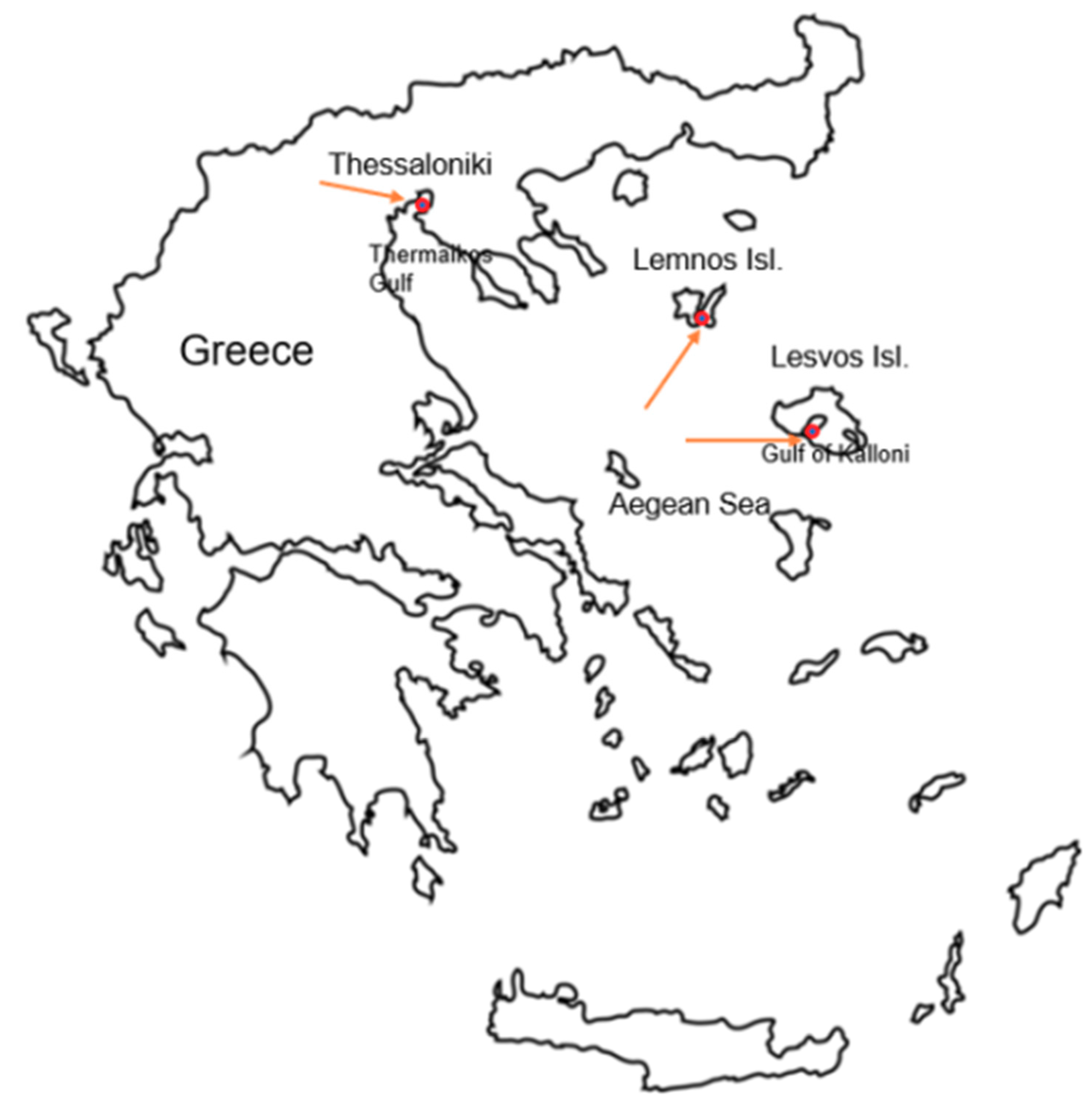
Assessment of Gut Microbiome in P. nobilis from Greek Mass Mortality Events

Digestive gland tissues from P. nobilis specimens, originating from mass mortality events as described in Lattos et al. [34], (Table 1), were obtained to identify the microbial diversity in the gut of infected fan mussels. Three specimens originated from Thermaikos Gulf, Thessaloniki (Ther01, Ther02, Ther03), two specimens from Kalloni Gulf, Lesvos island (Kal10, Kal12) and specimen originated from Limnos island, North Aegean, were analyzed (Figure 1).
Among the analyzed samples as examined previously in Lattos et al. [34], Ther01 was infected only by Mycobacterium sp. while in Ther02 and Ther03 H. pinnae and Mycobacterium sp. were detected. Samples from Lesvos were infected either by both parasites, or solely by the Mycobacterium sp., whereas the sample Lim01 was the only representative from Limnos island, North Aegean, hosting both pathogens (Table 1). After the first investigation published in 2020, which indicated the presence of H. pinnae and Mycobacterium sp. [34], a monitoring study started in the end of 2019 and continues nowadays, indicating the presence of both parasites in every alive population existing in Greece. Hence, no healthy individual was included in the analysis.
Approximately 20 mg of the digestive gland were placed in a 1.5 mL sterile tube and proceeded for DNA extraction. QIAamp®DNA mini kit (Qiagen, Hilden, Germany) was used for isolation and purification of total microbial DNA, according to the protocol provided by the manufacturer company. The quality and the quantity of the extracted DNA was checked using a NanoDrop (Shimadzu, Kyoto, Japan). Library preparation was performed on the V4 region of 16s rRNA gene. Primers used for 16s rRNA gene amplification were those described by Caporaso et al. [40], and the sequencing procedure was implemented using Illumina Miseq technology. Sequence data were deposited to the NCBI database under the BioProject accession number PRJNA661537. Heatmaps demonstrating relative bacterial abundance were generated using Multiple Experiment Viewer (MEV) online version, whereas bacterial abundances where also depicted in bar graphs created in Microsoft excel.

3. Results

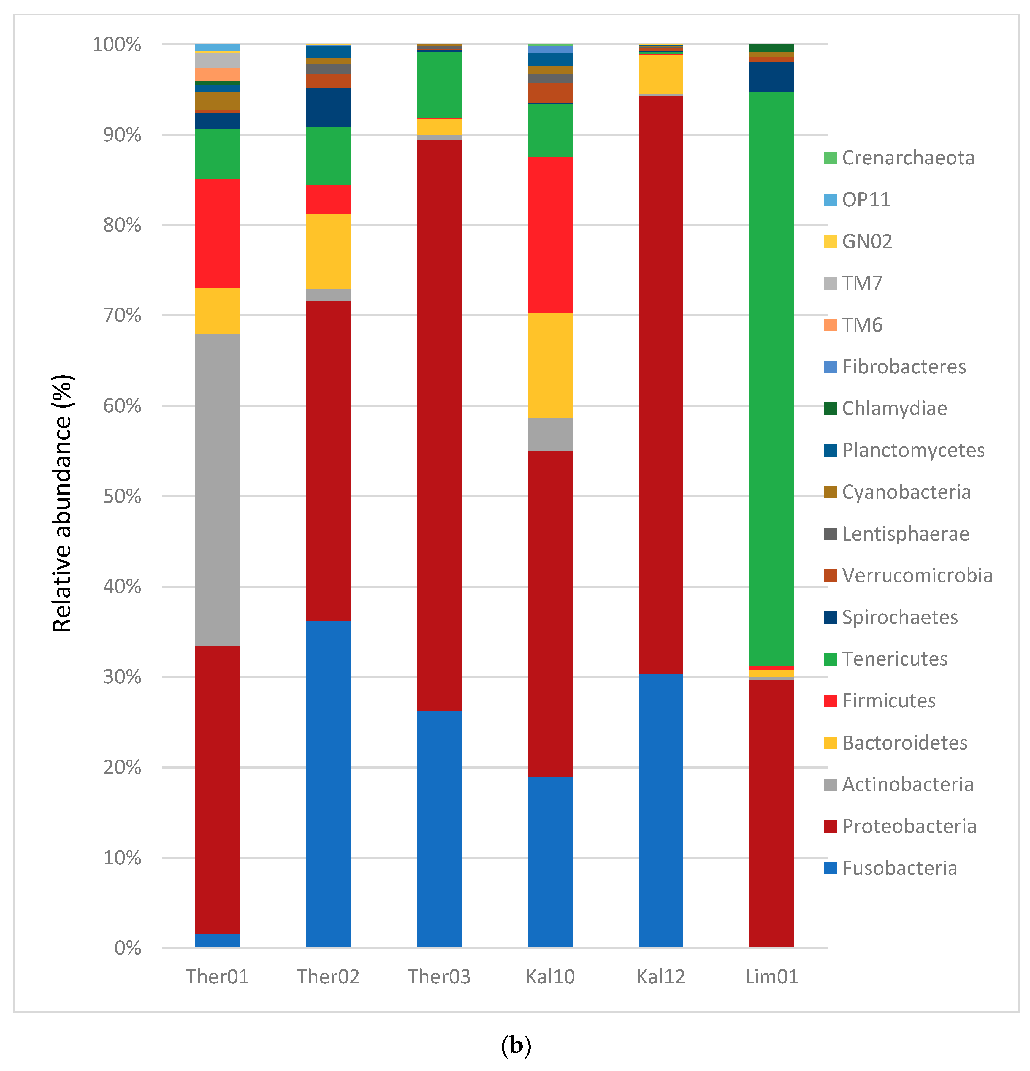
3.1. Gut Microbiota Phylum Analysis in Pinna nobilis

A total number of 1220 Operational Taxonomic Units (OTUs) were identified from the gastrointestinal gland of the fan mussel P. nobilis examined specimens. Seventeen different bacterial phyla were detected with 14 of them having >0.5% abundance in at least one sample analyzed (Figure 2). The bacteria of the phylum Proteobacteria, exhibited the greatest average abundance with 30.59% of the total microbiota identified, followed by the bacterial phyla Tenericutes and Fusobacteria with average abundances 13.18% and 12.16%, respectively. Moreover, the existence of Archaea, in particular of the phylum Crenarchaeota was also revealed. Concerning the specimens originated from Thermaikos Gulf, Thessaloniki, the dominant phyla in Ther02 and Ther03 were Fusobacteria and Proteobacteria, while in Ther01 the dominant phyla were Proteobacteria and Actinobacteria. Specifically, in the Ther01 Actinobacteria had the greatest abundance with 24.98% of the total microbiota and Proteobacteria had 22.99% respectively. In Ther03 Proteobacteria showed the greatest abundance of 41.74%, followed by Fusobacteria phylum with 17.34% of the total microbiome detected. In the sample Ther02, Fusobacteria was the dominant phylum (20.78%) with Proteobacteria slightly lower (20.38%). In both samples originated from Kalloni Gulf, Lesvos Island, the dominant bacterial phyla were Proteobacteria with abundance ratio 18.26% and 50.48%, for samples Kal10 and Kal12, respectively, followed by Fusobacteria, with 9.65% and 23.96%, respectively. Interestingly, in the sample Kal10, the phylum Firmicutes presented a differentiation in comparison with Kal12, with relative abundance ratio 8.72% compared to the 0.11% respectively. The gut microbiota in the sample of Limnos island followed a different pattern with dominant bacterial phyla Tenericutes and Proteobacteria, of 63.59% and 29.68% abundance, respectively.

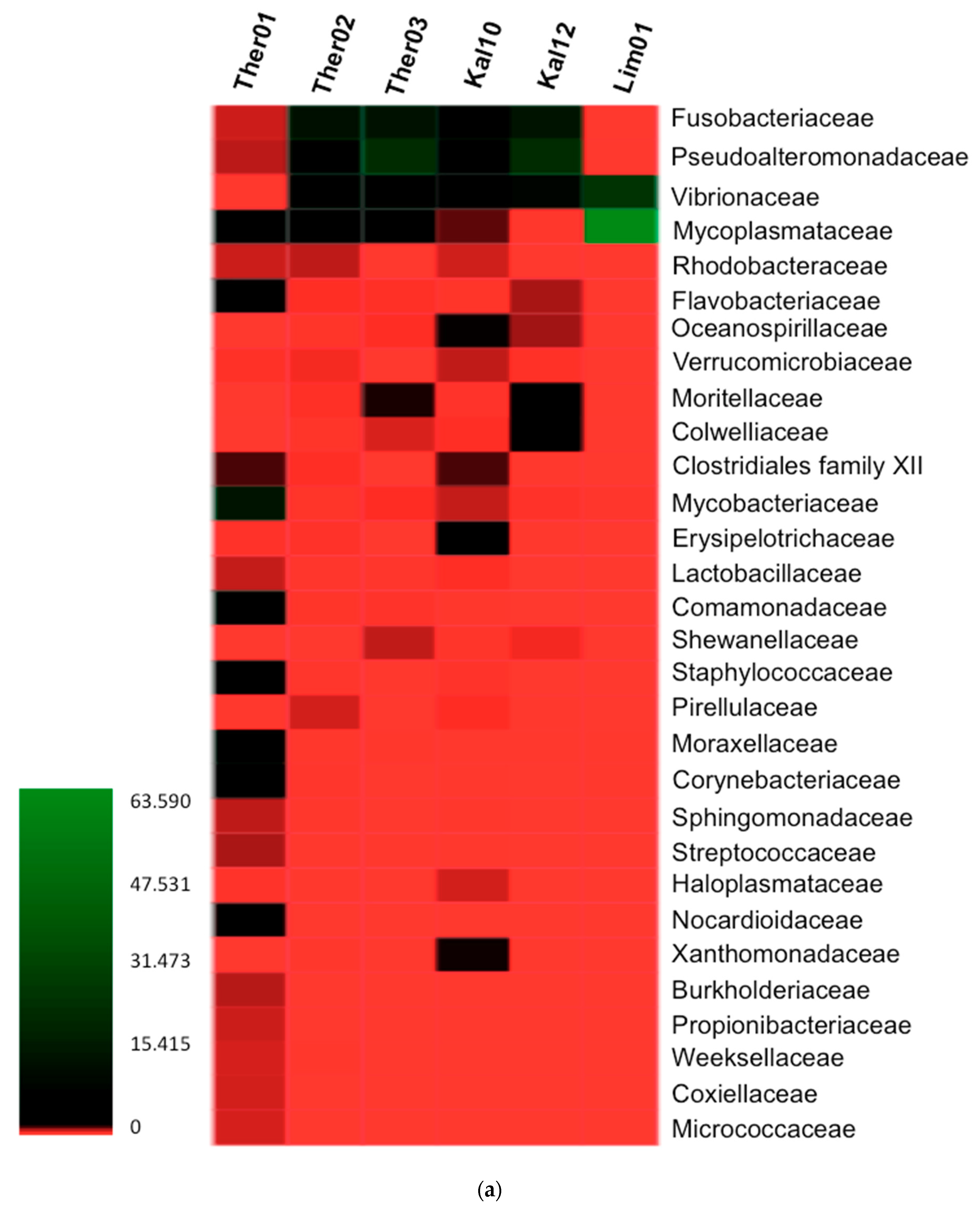
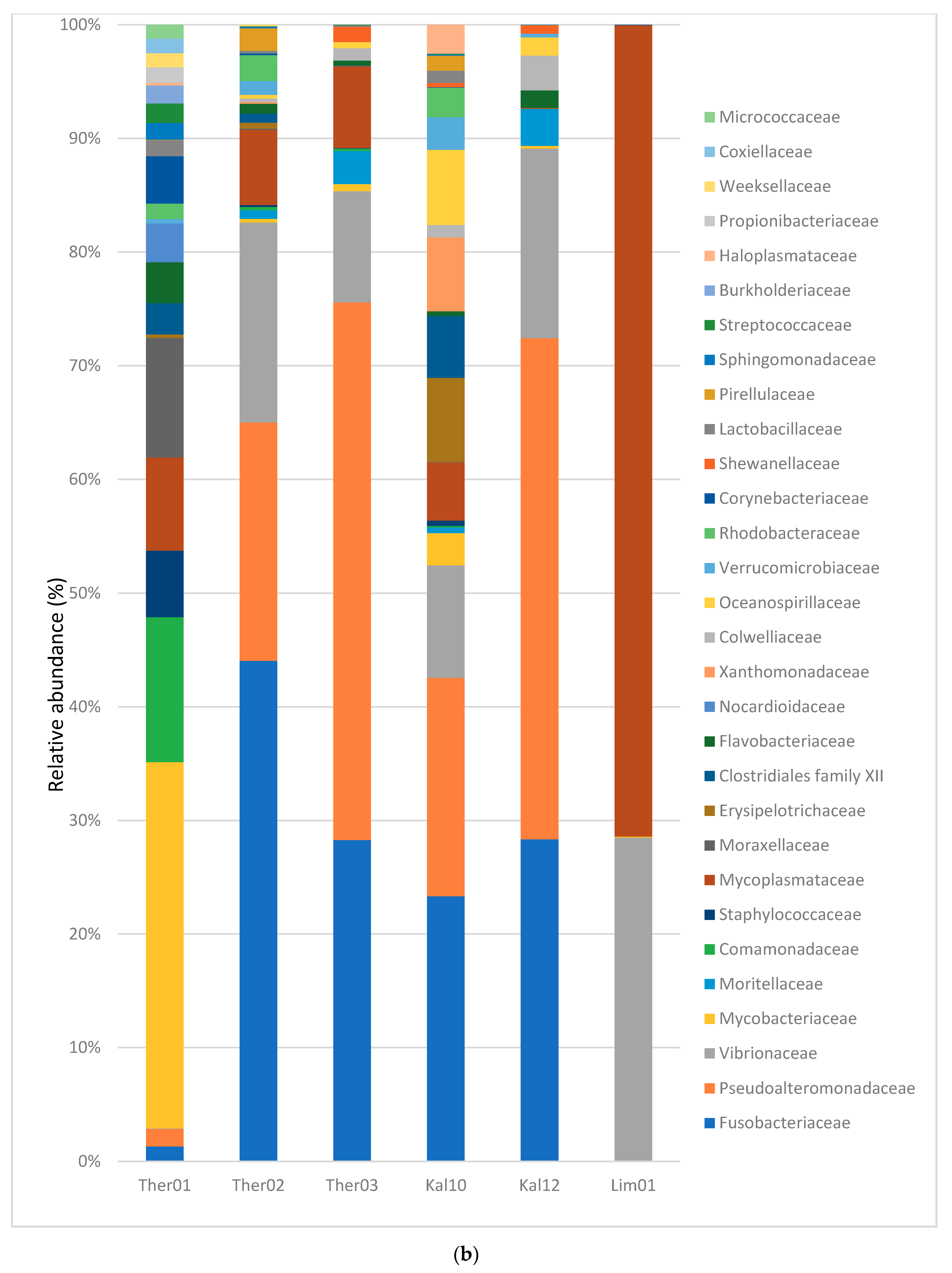
3.2. Gut Microbiota Families Analysis in Pinna nobilis

The detection of 30 bacterial families was the result of the analysis of the OTUs with the highest relative abundance. Additionally, the analysis using the OTUs with relative abundance >0.5% and presence in at least one sample indicated 123 OTUs. A heatmap and a bar graph was created to demonstrate the relative abundance of the bacterial families in the gastrointestinal samples of Fan mussel (Figure 3). Mycoplasmataceae, Alteromonadaceae, Fusobacteriaceae, Vibrionaceae and Mycobacteriaceae were the five families with the most dominant average presence in the samples. In the sample coded Ther01, Mycobacteriaceae was the most dominant family with relative abundance at 14.24%. Comamonadaceae, Moraxellaceae, Mycoplasmataceae, Staphylococcaceae families followed with lower relative abundances i.e., 5.64%, 4.65%, 3.62% and 2.59% respectively. In Ther02, Fusobacteriaceae, Alteromonadaceae, Vibrionaceae, Mycoplasmataceae and Rhodobacteriaceae were the families with abundance rates 12.78%, 6.08%, 5.11%, 1.92%, 0.66% respectively. In the third sample from Thermaikos Gulf (Ther03) bacterial families with the highest abundance percentages were Alteromonadaceae, Fusobacteriaceae, Vibrionaceae, Mycoplasmataceae and Moritellaceae with 22.66%, 13.54%, 4.69%, 3.44% and 1.40% correspondingly. In Kal10, most abundant families were Fusobacteriaceae, Alteromonadaceae, Vibrionaceae, Erysipelotrichaceae and Clostridiales Family XII with rates 5.19%, 4.28%, 2.20%, 1.65 and 1.21% respectively. In the second sample from Lesvos island (Kal12) the most abundant microbiota familes detected were Alteromonadaceae, Fusobacteriaceae, Vibrionaceae, Moritellaceae and Colwelliaceae (22.33%, 14.35%, 8.45%, 1.64% and 1.56%). In the sample originated from Limnos island the diversity in the microbiota was scarce, with most abundant families to be Mycoplasmataceae, Vibrionaceae, Mycobacteriaceae, Moraxellaceae and Corynebacteriaceae with rates 63.59%, 25.38%, 0.07%, 0.02% and 0.02% respectively.

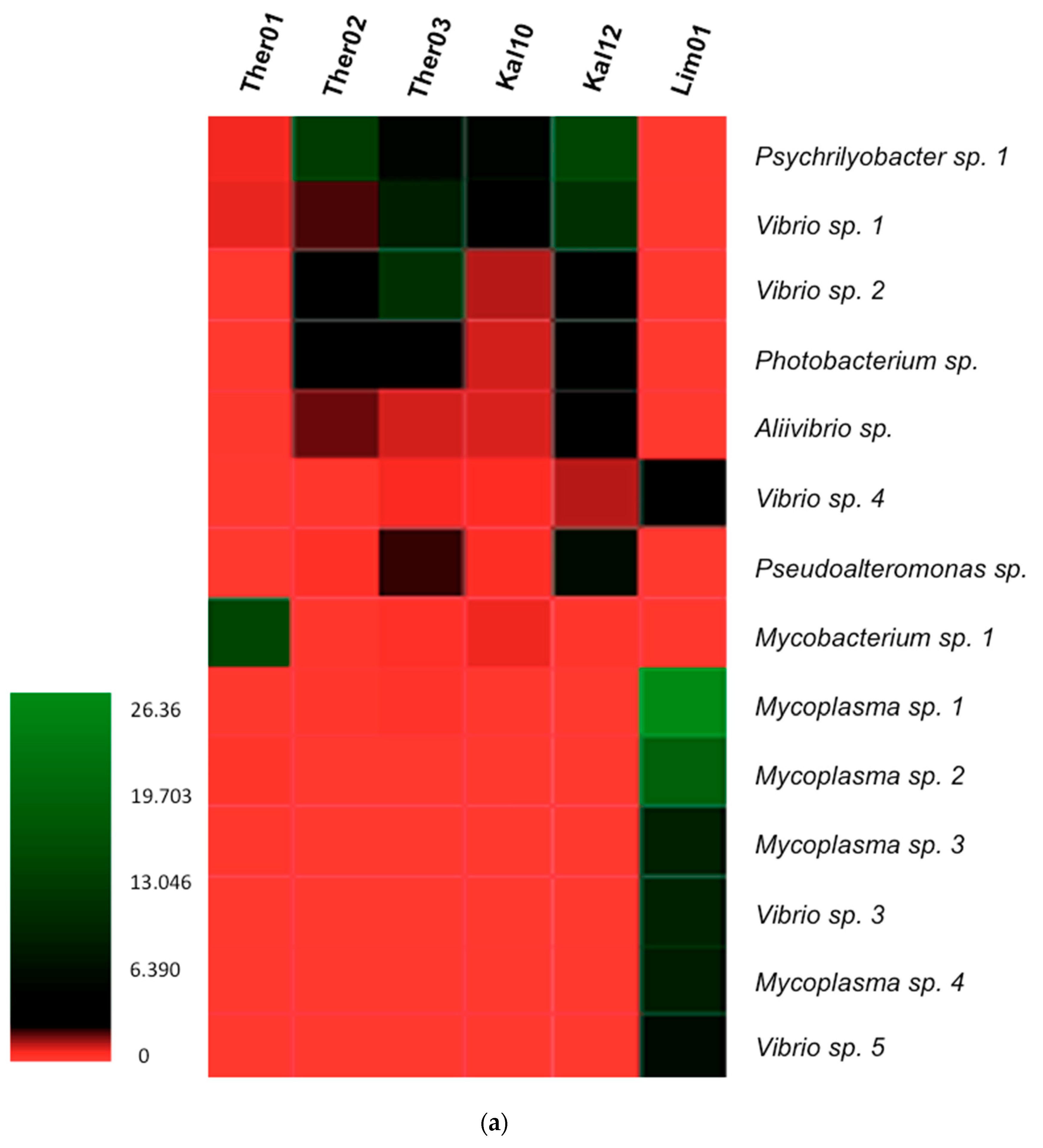
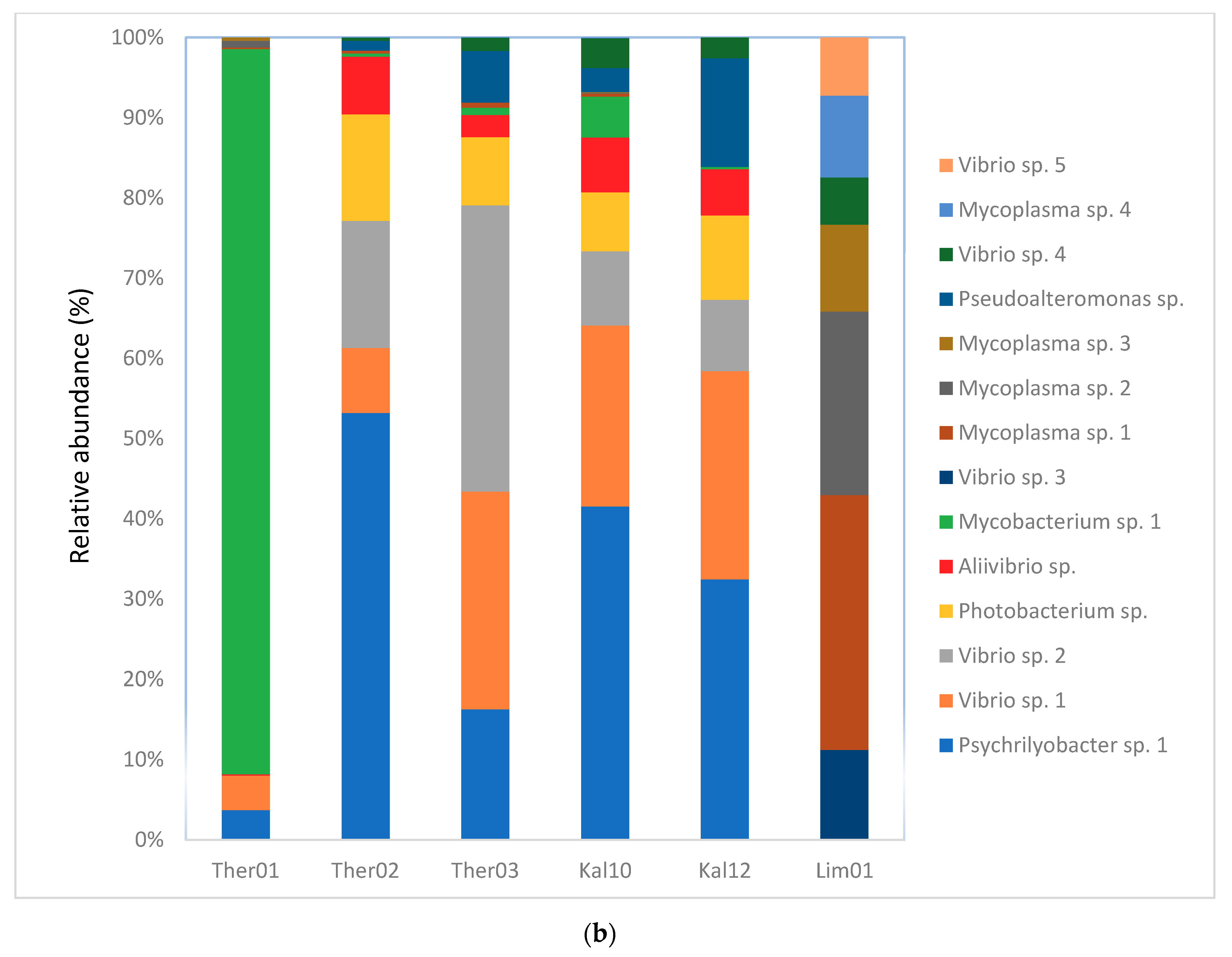
3.3. Gut Microbiota Genus Analysis in Pinna nobilis

A heatmap and a bar graph were constructed to demonstrate the most abundant bacterial genera within the Fan mussel, P. nobilis (Figure 4). Fourteen bacterial genera were selected among 1220 OTUs, having average abundance in the samples >1% and presence at least in one of the samples. In samples originated from Thermaikos Gulf the most abundant genera were Mycobacterium sp. 1, Psychrilyobacter sp. 1 and Vibrio sp. 2, in Ther01, Ther02 and Ther03 respectively. Vibrio sp. 1, Psychrilyobacter sp. 1 and Photobacterium sp. were also representatives in the specimens of Thermaikos gulf, in higher abundance than the total average presence of bacteria in the samples (2.52%). In samples from Lesvos island, Psychrilyobacter sp. 1, Vibrio sp. 1 and Vibrio sp. 2 were the most abundant genera in Kal10, while Psychrilyobacter sp. 1, Vibrio sp. 1, Vibrio sp. 2, Photobacterium sp., Aliivibrio sp. and Pseudoalteromonas sp. were the most abundant in Kal12. In the unique specimen from Limnos isl. the dominant genera were Mycoplasma sp.1, Mycoplasma sp.2, Mycoplasma sp.3, Mycoplasma sp.4, Vibrio sp. 3, Vibrio sp. 4 and Vibrio sp. 5.

3.4. Blastn Search of Bacterial Genera

Blast search against NCBI database revealed 100% similarity of Vibrio sp. 1 with two microbial species i.e., Vibrio splendidus and Vibrio atlanticus. Searches in the same database revealed 100% similarity of Vibrio sp. 2 with many Vibrio species such as V. toranzoniae, V. crassostreae, V. kanaloae, V. gallacicus, V. gigantis, V. chagasii, V. coralliirubri and V. pomeroyi. Blastn search for Vibrio sp. 3 revealed 100% similarity with V. astriarenae, 99.58% similarity with V. agarivorans, 99.58% similarity with V. diabolicus and the same again similarity with V. mediterranei. As a result of searching for Vibrio sp. 4, 100% similarity was revealed with V. hippocampi, alongside with lower similarities with a variety of Vibrio spp. such as V. aestuarianus, V. alginolyticus, V. thalassae and V. mediterranei with 98.74% similarity and V. sinensis with 98.73% similarity. Finally, Blast for Vibrio sp. 5 demonstrated 98.5% similarities with V. mediterranei and V. shilonii. Mycoplasma sp. 3 was the only species in which Blast search revealed 84.57% similarities with Candidatus Mycoplasma girerdii, while other Mycoplasma spp. search leaded only in higher taxa classification.

4. Discussion

The present study demonstrated a great variability of symbiotic bacteria within the infected Fan mussel, P. nobilis specimens originating from mass mortality events in Greece. Among these bacterial species, several potential pathogens were determined. Generally, investigating the gut microbiome using 16s rRNA sequencing in bivalves, revealed a great bacterial diversity, many of which may have symbiotic or pathogenic role in the host. Carella et al. [39], confirmed the co-occurrence of other potential pathogens, alongside with H. pinnae and Mycobacterium sp. and in particular the existence of a new Vibrio sp. that infects P. nobilis populations and constitutes an opportunistic pathogen for this bivalve. V. mediterranei has been proved to be an opportunistic pathogen even for stabled individuals of P. nobilis and has been associated with mortalities in captured conditions [38]. As a result, in this study five different OTUs, genetically related with Vibrio sp. were confirmed in the specimens of all Greek regions investigated. Blastn search against NCBI database revealed high percentage similarities with many Vibrio species, which actually constitute pathogenic organisms in many cases for bivalves and are in relation with many mass mortality events in all life stages of marine bivalves.
The aquatic environment harbors a great abundance of bacteria. Bivalves, with their efficient filter feeding mechanism, may ingest a plethora of microorganisms mostly depending on the habitat. While most of the bacteria in the marine environment are not pathogenic to bivalves, unless their presence is characterized by a large number of colonies, some can be pathogenic or can utilize bivalves as an intermediate host in order to infect and become pathogenic to the final host. Within this diversity of microorganisms in the marine environment, the genus Vibrio seems to play a key role in the pathogenicity in marine bivalves. The genus Vibrio comprises more than 130 bacterial species which can be grouped in 17 monophyletic clades [41,42]. The vast majority of bacterial diseases of the bivalves have as causative agent the Vibrio spp. [43]. V. aestuarianus is an important pathogen responsible for massive mortalities in oyster, Crassostrea gigas in Europe and responsible for a greatly negative impact in farms associate with the culture of this species [10,44,45,46]. The first case of bivalves infected by V. alginolyticus was described in 1965 [47]. They investigated the virulence of V. alginolyticus in four different species of bivalve mollusks in different life stages. Later on, Luna-González et al. [48], challenged four bivalve species with V. alginolyticus and indicated that scallop species were more susceptible to the pathogen than the other bivalve species tested. In 2005, Gomez-Leon et al. [49], investigated two mortality events happened in 2001 and 2002 in cultured carpet shell clam, Ruditapes decussatus. V. alginolyticus was the major causative agent isolated from moribund clam larvae in the first mortality while in the second mortality the same causative agent was isolated alongside with V. splendidus Biovar II, causing mortalities up to 62% and 73% of the clam population, respectively. Further, V. splendidus strains are mainly linked with mortalities in mussel larvae, although there are many described cases causing mortalities in clams, scallops and in oysters [50,51,52,53,54,55]. V. tubiashii was isolated, for the first time, causing mortalities in juvenile clams (M. mercenariae) and oyster (C. virginica) larvae, in hatcheries located in North America but its first official description was in 1984 [56]. More recently, Prado et al. [57], isolated a new strain of V. tubiashii, from flat oyster (Ostrea edulis) and Manila clam (Ruditapes philippinarum) cultures during an outbreak of the disease in two shellfish hatcheries. Vibrio tapetis was responsible for the mass mortalities caused in juveniles and adults in the spring and summer of 1987, reported in the northwest culture beds of France [58]. The disease caused by V. tapetis, was named BRD (Brown-Ring-Disease) because of the characteristic brown deposit in the inner part of the valves and affects mostly cultured clams, but has lower impact on wild populations [59]. Brown ring disease is temperature dependent and can cause high mortalities in R. philippinarum, but not in R. decussatus which has proved to be resistant to BRD [60,61]. V. crassostreae can be found in diseased animals and can be characterized pathogenic for pacific oysters, only with the acquisition of a specific plasmid, which is essentially for killing [62]. Vibrio corallilyticus was initially described as a coral pathogen and was associated as bivalve pathogen infecting oyster larvae (Crassostrea virginica and Crassostrea gigas) after challenging oysters in bacterial strains of this Vibrio spp. [63,64]. In addition, M. galloprovinciallis challenged with V. corrallilyticus strains were unable to activate immune responses, although it is considered to be resistant to Vibrio infections [65]. Kim et al. [66], investigated the mortality event occurred in a Pacific oyster hatchery and described V. corallilyticus as the main etiological agent of this event. V. mediterranei was isolated for the first time from plankton, sediments and seawater in two coastal areas in south Valencia, Spain [67]. It is considered to be an emerging pathogen, with a world-wide distribution, having infected corals, cultivated seaweeds and the endangered bivalve P. nobilis causing mortalities alongside with other pathogens [38,68,69].It should be emphasized that not all Vibrio species act as pathogens for bivalves. Apart from the species considered to be pathogens for the bivalves, there are Vibrio spp. that have been detected in the bivalve microbiota in diseased or healthy animals. Vibrio species, not associated with mortalities in bivalves, have been detected in bivalve microbiota and they have been initially described living in seawater, in marine crustaceans, in sponges, in corals, in sea horses, in eels and in fish [42,70,71,72,73]. Interestingly, some Vibrio species are also of public health importance. Particularly V. vulnificus, V. parahaemolyticus and V. cholerae, are considered to be hazardous in bivalve seafood [3]. In our study, Vibrio spp. was identified in all examined specimens (Figure 3). Although, it was not applicable to be unambiguously identified at species level, taking into consideration that Vibrio sp. 3 exhibited 99.58% similarity with V. mediterranei, the possibility of representing a pathogen for P. nobilis contributing in mortalities cannot be ruled out. Additionally, despite forbidden by the local authorities for conservation purposes [21], fishing and consumption of raw fan mussels still occurs (personal communication) and in such a case detected Vibrio sp. may be harmful for public health.
Concerning the rest identified bacterial genera, Photobacterium species, is the genus expected the most, that can be commonly found in the marine environment and in the intestinal content of marine animals [19]. They constitute one of the oldest genera in the Vibrionaceae and they have been detected in high abundance in the microbiota of Vibrio-infected C. gigas [11]. Although, most Photobacterium spp. have not been described as pathogenic for marine bivalves, isolated from marine bivalves and crustaceans and are common inhabitants of marine environment, subspecies of Photobacterium damselae are pathogenic for aquatic animals, especially for fish [74,75,76]. In a recent research, Eggermont et al. [77], isolated bacteria belonging to the family Vibrionaceae from wild blue mussel (Mytilus edulis) and assessed their impacts on mussel larviculture. They concluded that most isolates belonged to the genera Vibrio and Photobacterium and also that Photobacterium species with the highest virulence factor in blue mussel was Photobacterium sanguinicancri. P. sanguinicancri has been isolated from marine animals previously alongside with other Vibrionaceae [78]. Interestingly, the OTU identified in this research, which is related with Photobacterium genera presents similarities 100% with P. sanguinicancri as well as with other Photobacterium spp. which have been characterized for their symbiotic traits in bivalves [79].
Among the remaining bacterial genera Aliivibrio, Pseudoalteromonas and Psychrilyobacter constitute probably the most important ones that further discussed.
The genus Aliivibrio was first established by Urbanczyk et al. [80], to reclassify Vibrio spp. and specifically reconstructed the taxonomy and nomenclature of V. fischeri, V. logei, V. salmonicida and V. wodanis, which based on 16s rRNA gene sequencing and after of concatenation of several genes (recA, rpoA, pyrH, gyrB) were renamed as Aliivibrio spp. The genus Aliivibrio includes species with both symbiotic and pathogenic traits. A. fischeri has a worldwide distribution and usually establishes positive relationships with aquatic animals such as cephalopods and fish [81]. A. logei has been reported to have symbiotic effects on squids in the genus Sepiola, although it is characterized as pathogen of the eels [82,83]. A. salmonicida is of great interest on account of its ability to infect fish in low temperature and cause mortality in various marine organisms including bivalves [76,80,84]. OTU identified and related with Aliivibrio genus in this research have similarities 100% with all the aforementioned Aliivibrio species and therefore ranked as a potential collaborator to rest of the pathogens caused the mortalities in P. nobilis.
The genus Pseudoalteromonas hosts 41 bacterial species, among which 16 are antimicrobial metabolite producers [85]. Antibacterial activities of Pseudoalteromonas spp. utilized in shellfish larviculture as probiotics and achieved higher growth rate, survival rate and higher resistances against infections with Vibrio spp. [86,87,88]. Although there are many reports of benefits from the utilization of Pseudoalteromonas spp. in shellfish culture, there are also reports of opportunistic behavior of this bacterial genus in mortalities caused by Vibrio spp. [89]. We can conclude the dual nature of this genera that act beneficially under the favorite physiological condition of the host and turns into a pathogen when they find the opportunity in a stressful condition.
Similarly, Psychrilyobacter spp. was detected in all specimens examined, let alone in high relative abundance, except from the sample originated from the island of Limnos. To our knowledge, there is no report of mortality or even pathogenesis of this genus in bivalves. On the other hand, this specific genus has been detected in the microbiome of bivalves in many cases indicating a beneficial association in disease resistance of the Pacific oysters, C. gigas because of the detection of this genus in oysters that were not infected from OsHV-1 virus [90,91,92,93].
Mycoplasmas are widespread in nature and are characterized for their parasite behavior in humans, mammals, reptiles, fish arthropods and plants [94]. Bacteria belonging to the genus Mycoplasma infect a wide range of bivalves and have been reported generally as potential bivalve pathogens [59]. First report of this bacterial genus in marine bivalves was by Harshbarger et al. [95], whereas, later, according to Azevedo [96], an unusual branchial infection by a mycoplasma-like microorganism in C. edule was reported and associated with high mortalities of this bivalve in Portugal in 1991 and 1992. According to the same study a high prevalence rate (65–70%) of mycoplasma-like microorganisms occurred in gaping cockles and lower abundance in living cockles. He concluded that infection with Mycoplasma sp. and high temperatures were the etiological agents of the mortalities. Since then, to our knowledge, no other mortalities have been attributed to Mycoplasma spp. as the primary causative agent. King et al. [97], reported the presence of Mycoplasma spp. in the stomach microbiome of oysters while in the gut the abundance of the genus wasn’t interestingly high. Lokmer et al. [98], similarly detected high abundance of Mollicutes class, especially bacteria of the genus Mycoplasma in the gut of oysters after transplantation of the species in a new environment. There are many other studies investigating the microbiome which have detected the genus Mycoplasma as symbiotic bacteria in bivalves [99], while Mycoplasma spp. detected in mortalities of C. gallina alongside with other genera [19].
In the P. nobilis specimens analyzed, relative abundance of Bacteria using a metagenomic approach, demonstrated 14 different OTUs (Average Relative abundance at least 1%) from the examined specimens. Except for Psychrilyobacter spp., which according to the literature has not been assigned as a causative agent or as opportunistic genus of bacteria, all other OTUs detected can be potential pathogen for the species. According to our results, four OTUs were detected in relation with Vibrio spp. in all specimens in various quantities, which mostly play the primary role in bacterial infections, in agreement with the several cases of mortality events [44]. OTUs with the criteria mentioned above, related with Mycoplasma spp. were detected in all samples, with the sample originated from Limnos island showing the highest relative abundance, adding all the OTUs relating with Mycoplasma spp. Kal12 was the only sample resulting OTUs in relation with Mycoplasma spp. with average relative abundance slightly reaching 0.5%, that was therefore excluded from the generated graphs. It’s worth noting that Lim01 which had the greatest abundance in Mycoplasma spp. and the second highest in Vibrio spp. exhibited the lowest total bacterial diversity. OTUs in relation with Photobacterium spp. were detected in almost all samples except from Ther01 and Lim01 with average abundance degree 1.91%. These results are in line with Milan et al. [19], concerning the detection of Photobacterium spp. in mortality events in different bivalve species examined. On the other hand, no great change in abundance of this genus reported in mortality of oysters [20]. OTUs related with Pseudoalteromonas spp., were detected in the same specimens as Photobacterium spp., in accordance to Li et al. [18], in hemolymph of mussels infected by V. cyclitrophicus. Pseudoalteromonas spp. were also present but in lower abundance in the control samples without V. cyclitrophicus. Finally, Aliivibrio spp. had the lowest average relative abundance in the samples.
Based on our analyses and in line with previous data [34], all specimens, in all status collected, exhibited high abundances of potential pathogens. Bacteria of the genus Vibrio were present in all three areas examined showing no correlation with the habitat change. Alongside with the Vibrio spp., most of the potential pathogenic bacteria species followed the same pattern and were detected in all three habitats. Additionally, there were no differences in Vibrio spp. abundances between the samples collected in the winter (Kal10, Kal12) and the samples collected during warmer months (Ther01, Ther02, Ther03 and Lim01), leading to the conclusion of the synergistical effect of those bacterial species in mortality events. Interestingly, the only bacterial species with high correlation to the habitat status were bacteria belonging to the genus Mycoplasma spp. that appeared to play a key role in the microbiota of P. nobilis originated from Limnos island, decreasing also the gut microbial diversity. Our results are in accordance with bacteriological study in P. nobilis implemented in three separate locations located in Eastern Adriatic coast [100], also demonstrating the presence of V. splendidus clade bacteria hosting P. nobilis.
However, to our knowledge, this is the first study approaching P. nobilis microbiome using a 16s-rRNA Metagenomic tool. Overall, in the present study many potential pathogens were determined, which, may be considered to act as potential pathogens or may have a secondary role in mortality events.Although the use of 16s-rRNA may have some limitations in the distinction of bacteria at species level and despite the fact that the examination of 6 gut microbiomes is only relatively sufficient for a general conclusion, our study demonstrated that the microbial diversity in diseased P. nobilis specimens contains specific genera of bacteria of high pathogenic importance. These bacterial genera occur in all geographical areas where mortalities took place, having similar abundance levels. Excluding the unique individual originated from the last collapsed population of Limnos island, the remaining investigated populations demonstrate the same bacteria genera with slight differences in abundances, although all of these populations have collapsed. We therefore assume that mortalities may be due to several bacterial genera and not only on account of the genera already reported. Devastating mortality events in P. nobilis populations are threatening fan mussel towards extinction. At the beginning, the H. pinnae was detected and the Mycobacterium sp., almost at the same time, were targeted as possible causes for the mortalities in the Mediterranean Sea. Mycobacteriosis is a chronic disease, probably influenced by the climate change, infecting both marine and terrestrial animal species with underestimated impacts on infection of aquatic animals [101]. Mycobacterial infections in marine bivalves are in scarce, although in 2019 and 2020 two cases of infections were reported, playing a significant role in mortality events of the endangered bivalve P. nobilis in the Mediterranean Sea [34,37]. Previously, Grimm et al. [102], reported the presence of a Mycobacterium sp. in the Atlantic sea scallop Placopecten magellanicus, as causative agent of orange nodular lesions. However, following the recent results of the studies concerning the mortalities, new pathogens have arisen alongside and seemed to play a key role in mortalities of P. nobilis. Prado et al. [38], exhibited a new pathogen in their effort to rescue and relocate P. nobilis individuals in close water systems. Peak of mortalities occurred when the temperatures reached above 25 °C and the causative agent was V. mediterranei, a newly unknown pathogen until that time in fan mussel, P. nobilis. In addition, Carella et al. [39], illustrated all the aforementioned pathogens plus a Perkinsus sp. in two specimens examined which indicated one more potential threat for the fan mussel. In order to unravel the puzzling situation of the collapse of P. nobilis populations, the occurrence of Rhodococcus erythropolis arose as a new bacteria species having a potential role in mortality events [103]. Fan mussel populations are collapsing one after the other in Mediterranean Sea. Recently, an epidemiological study showed the rapid progression even in the last safe places in Croatia [104]. In Greece, the population of the species suffered enormous damage, limiting them in few areas but with the presence of Mycobacterium sp. and H. pinnae. These last population hot spots are suffering from mortalities mostly on warmer months. In conclusion, according to our results and in line the aforementioned recent studies, probably P. nobilis mortalities are not caused solely by the infection with H. pinnae and Mycobacterium sp. as previously assumed, but also, other pathogenic bacterial genera may act synergistically with these parasites, resulting in the observed mortalities.

Author Contributions

Conceptualization, A.L. and I.A.G.; methodology, A.L. and I.A.G.; software, A.L.; validation, B.M.; investigation, A.L., D.K. and J.A.T.; resources, I.A.G. and B.M.; writing—original draft preparation, A.L.; writing—review and editing, I.A.G., J.A.T. and B.M.; supervision, B.M.; project administration, J.A.T. and B.M.; All authors have read and agreed to the published version of the manuscript.

Funding

Τhis research work was supported by the Hellenic Foundation for Research and Innovation (HFRI) and the General Secretariat for Research and Technology (GSRT), under the HFRI PhD Fellowship grant (GA. no. 1654).

Conflicts of Interest

The authors declare no conflict of interest.

References

  1. Sibley, C.D.; Peirano, G.; Church, D.L. Molecular methods for pathogen and microbial community detection and characterization: Current and potential application in diagnostic microbiology. Infect. Genet. Evol. 2012, 12, 505–521. [Google Scholar] [CrossRef]
  2. Bordenstein, S.R.; Theis, K.R. Host biology in light of the microbiome: Ten principles of holobionts and hologenomes. PLoS Biol. 2015, 13, 1–23. [Google Scholar] [CrossRef] [Green Version]
  3. Oliveira, J.; Cunha, A.; Castilho, F.; Romalde, J.L.; Pereira, M.J. Microbial contamination and purification of bivalve shellfish: Crucial aspects in monitoring and future perspectives—A mini-review. Food Control. 2011, 22, 805–816. [Google Scholar] [CrossRef]
  4. Vezzulli, L.; Stagnaro, L.; Grande, C.; Tassistro, G.; Canesi, L.; Pruzzo, C. Comparative 16SrDNA Gene-Based Microbiota Profiles of the Pacific Oyster (Crassostrea gigas) and the Mediterranean Mussel (Mytilus galloprovincialis) from a Shellfish Farm (Ligurian Sea, Italy). Microb. Ecol. 2018, 75, 495–504. [Google Scholar] [CrossRef]
  5. Prado, S.; Romalde, J.L.; Barja, J.L. Review of probiotics for use in bivalve hatcheries. Vet. Microbiol. 2010, 145, 187–197. [Google Scholar] [CrossRef] [Green Version]
  6. Ringø, E.; Zhou, Z.; Vecino, J.L.G.; Wadsworth, S.; Romero, J.; Krogdahl, A.; Olsen, R.E.; Dimitroglou, A.; Foey, A.; Davies, S.; et al. Effect of dietary components on the gut microbiota of aquatic animals. A never-ending story? Aquac. Nutr. 2016, 22, 219–282. [Google Scholar] [CrossRef] [Green Version]
  7. Flint, H.J. The impact of nutrition on the human microbiome. Nutr. Rev. 2012, 70. [Google Scholar] [CrossRef]
  8. Biesalski, H.K. Nutrition meets the microbiome: Micronutrients and the microbiota. Ann. N. Y. Acad. Sci. 2016, 1372, 53–64. [Google Scholar] [CrossRef]
  9. Matozzo, V.; Marin, M.G. Bivalve immune responses and climate changes: Is there a relationship? Invertebr. Surviv. J. 2011, 8, 70–77. [Google Scholar]
  10. Garnier, M.; Labreuche, Y.; Garcia, C.; Robert, M.; Nicolas, J.L. Evidence for the involvement of pathogenic bacteria in summer mortalities of the pacific oyster Crassostrea gigas. Microb. Ecol. 2007, 53, 187–196. [Google Scholar] [CrossRef] [Green Version]
  11. Lokmer, A.; Mathias Wegner, K. Hemolymph microbiome of Pacific oysters in response to temperature, temperature stress and infection. ISME J. 2015, 9, 670–682. [Google Scholar] [CrossRef] [PubMed] [Green Version]
  12. Pierce, M.L.; Ward, J.E. Microbial Ecology of the Bivalvia, with an Emphasis on the Family Ostreidae. J. Shellfish Res. 2018, 37, 793–806. [Google Scholar] [CrossRef]
  13. Llewellyn, M.S.; Leadbeater, S.; Garcia, C.; Sylvain, F.E.; Custodio, M.; Ang, K.P.; Powell, F.; Carvalho, G.R.; Creer, S.; Elliot, J.; et al. Parasitism perturbs the mucosal microbiome of Atlantic Salmon. Sci. Rep. 2017, 7, 1–10. [Google Scholar] [CrossRef] [Green Version]
  14. Bayer, T.; Arif, C.; Ferrier-Pagès, C.; Zoccola, D.; Aranda, M.; Voolstra, C.R. Bacteria of the genus Endozoicomonas dominate the microbiome of the Mediterranean gorgonian coral Eunicella cavolini. Mar. Ecol. Prog. Ser. 2013, 479, 75–84. [Google Scholar] [CrossRef] [Green Version]
  15. Blanquer, A.; Uriz, M.J.; Cebrian, E.; Galand, P.E. Snapshot of a bacterial microbiome shift during the early symptoms of a massive sponge die-off in the western Mediterranean. Front. Microbiol. 2016, 7, 1–10. [Google Scholar] [CrossRef] [PubMed] [Green Version]
  16. Meisterhans, G.; Raymond, N.; Girault, E.; Lambert, C.; Bourrasseau, L.; de Montaudouin, X.; Garabetian, F.; Jude-Lemeilleur, F. Structure of Manila Clam (Ruditapes philippinarum) Microbiota at the Organ Scale in Contrasting Sets of Individuals. Microb. Ecol. 2016, 71, 194–206. [Google Scholar] [CrossRef]
  17. Li, Y.F.; Yang, N.; Liang, X.; Yoshida, A.; Osatomi, K.; Power, D.; Batista, F.M.; Yang, J.L. Elevated seawater temperatures decrease microbial diversity in the gut of Mytilus coruscus. Front. Physiol. 2018, 9, 1–9. [Google Scholar] [CrossRef] [PubMed] [Green Version]
  18. Li, Y.F.; Chen, Y.W.; Xu, J.K.; Ding, W.Y.; Shao, A.Q.; Zhu, Y.T.; Wang, C.; Liang, X.; Yang, J.L. Temperature elevation and Vibrio cyclitrophicus infection reduce the diversity of haemolymph microbiome of the mussel Mytilus coruscus. Sci. Rep. 2019, 9, 1–10. [Google Scholar] [CrossRef]
  19. Milan, M.; Smits, M.; Dalla Rovere, G.; Iori, S.; Zampieri, A.; Carraro, L.; Martino, C.; Papetti, C.; Ianni, A.; Ferri, N.; et al. Host-microbiota interactions shed light on mortality events in the striped venus clam Chamelea gallina. Mol. Ecol. 2019, 28, 4486–4499. [Google Scholar] [CrossRef]
  20. Green, T.J.; Siboni, N.; King, W.L.; Labbate, M.; Seymour, J.R.; Raftos, D. Simulated Marine Heat Wave Alters Abundance and Structure of Vibrio Populations Associated with the Pacific Oyster Resulting in a Mass Mortality Event. Microb. Ecol. 2019, 77, 736–747. [Google Scholar] [CrossRef]
  21. Theodorou, J.A.; James, R.; Tagalis, D.; Tzovenis, I.; Hellio, C.; Katselis, G. Density and size structure of the endangered fan mussel Pinna nobilis (Linnaeus 1758), in the shallow water zone of Maliakos Gulf, Greece. Acta Adriat. 2017, 58, 63–76. [Google Scholar] [CrossRef]
  22. Katsanevakis, S.; Tsirintanis, K.; Tsaparis, D.; Doukas, D.; Sini, M.; Athanassopoulou, F.; Κolygas, M.; Tontis, D.; Koutsoubas, D.; Bakopoulos, V. The cryptogenic parasite Haplosporidium pinnae invades the Aegean Sea and causes the collapse of Pinna nobilis populations. Aquat. Invasions 2019, 14, 150–164. [Google Scholar] [CrossRef]
  23. Zotou, M.; Gkrantounis, P.; Karadimou, E.; Tsirintanis, K.; Sini, M.; Poursanidis, D.; Azzolin, M.; Dailianis, T.; Kytinou, E.; Issaris, Y.; et al. Pinna nobilis in the Greek seas (NE Mediterranean): On the brink of extinction? Mediterr. Mar. Sci. 2020, 21, 575–591. [Google Scholar] [CrossRef]
  24. Rabaoui, L.; Tlig-Zouari, S.; Katsanevakis, S.; Ben Hassine, O.K. Modelling population density of Pinna nobilis (Bivalvia) on the eastern and southeastern coast of Tunisia. J. Molluscan Stud. 2010, 76, 340–347. [Google Scholar] [CrossRef] [Green Version]
  25. Sanna, D.; Cossu, P.; Dedola, G.L.; Scarpa, F.; Maltagliati, F.; Castelli, A.; Franzoi, P.; Lai, T.; Cristo, B.; Curini-Galletti, M.; et al. Mitochondrial DNA Reveals Genetic Structuring of Pinna nobilis across the Mediterranean Sea. PLoS ONE 2013, 8, 1–16. [Google Scholar] [CrossRef] [PubMed] [Green Version]
  26. Sanna, D.; Debola, G.L.; Scarpa, F.; Lai, T.; Cossu, P.; Curini-Galletti, M.; Francalacci, P.; Casu, M. New mitochondrial and nuclear primers for the Mediterranean marine bivalve Pinna nobilis. Mediterr. Mar. Sci. 2014, 15, 416. [Google Scholar] [CrossRef] [Green Version]
  27. Vázquez-Luis, M.; Borg, J.A.; Morell, C.; Banach-Esteve, G.; Deudero, S. Influence of boat anchoring on Pinna nobilis: A field experiment using mimic units. Mar. Freshw. Res. 2015, 66, 786–794. [Google Scholar] [CrossRef] [Green Version]
  28. Kersting, D.; Benabdi, M.; Čižmek, H.; Grau, A.; Jimenez, C.; Katsanevakis, S.; Öztürk, B.; Tuncer, S.; Tunesi, L.; Vázquez-Luis, M.; et al. Pinna nobilis. The IUCN Red List of Threatened Species 2019: e.T160075998A160081499. 2019. Available online: https://dx.doi.org/10.2305/IUCN.UK.2019-3.RLTS.T160075998A160081499.en (accessed on 20 October 2020).
  29. Darriba, S. First haplosporidan parasite reported infecting a member of the Superfamily Pinnoidea (Pinna nobilis) during a mortality event in Alicante (Spain, Western Mediterranean). J. Invertebr. Pathol. 2017, 148, 14–19. [Google Scholar] [CrossRef]
  30. Vázquez-Luis, M.; Álvarez, E.; Barrajón, A.; García-March, J.R.; Grau, A.; Hendriks, I.E.; Jiménez, S.; Kersting, D.; Moreno, D.; Pérez, M.; et al. S.O.S. Pinna nobilis: A mass mortality event in western Mediterranean Sea. Front. Mar. Sci. 2017, 4, 1–6. [Google Scholar] [CrossRef] [Green Version]
  31. Catanese, G.; Grau, A.; Valencia, J.M.; Garcia-March, J.R.; Vázquez-Luis, M.; Alvarez, E.; Deudero, S.; Darriba, S.; Carballal, M.J.; Villalba, A. Haplosporidium pinnae sp. nov., a haplosporidan parasite associated with mass mortalities of the fan mussel, Pinna nobilis, in the Western Mediterranean Sea. J. Invertebr. Pathol. 2018, 157, 9–24. [Google Scholar] [CrossRef]
  32. Panarese, R.; Tedesco, P.; Chimienti, G.; Latrofa, M.S.; Quaglio, F.; Passantino, G.; Buonavoglia, C.; Gustinelli, A.; Tursi, A.; Otranto, D. Haplosporidium pinnae associated with mass mortality in endangered Pinna nobilis (Linnaeus 1758) fan mussels. J. Invertebr. Pathol. 2019, 164, 32–37. [Google Scholar] [CrossRef] [PubMed]
  33. Cabanellas-Reboredo, M.; Vázquez-Luis, M.; Mourre, B.; Álvarez, E.; Deudero, S.; Amores, Á.; Addis, P.; Ballesteros, E.; Barrajón, A.; Coppa, S.; et al. Tracking a mass mortality outbreak of pen shell Pinna nobilis populations: A collaborative effort of scientists and citizens. Sci. Rep. 2019, 9, 1–11. [Google Scholar] [CrossRef] [PubMed] [Green Version]
  34. Lattos, A.; Giantsis, I.A.; Karagiannis, D.; Michaelidis, B. First detection of the invasive Haplosporidian and Mycobacteria parasites hosting the endangered bivalve Pinna nobilis in Thermaikos Gulf, North Greece. Mar. Environ. Res. 2020, 155, 104889. [Google Scholar] [CrossRef] [PubMed]
  35. García-March, J.R.; Tena, J.; Henandis, S.; Vázquez-Luis, M.; López, D.; Téllez, C.; Prado, P.; Navas, J.I.; Bernal, J.; Catanese, G.; et al. Can we save a marine species affected by a highly infective, highly lethal, waterborne disease from extinction? Biol. Conserv. 2020, 243, 108498. [Google Scholar] [CrossRef]
  36. Čižmek, H.; Čolić, B.; Gračan, R.; Grau, A.; Catanese, G. An emergency situation for pen shells in the Mediterranean: The Adriatic Sea, one of the last Pinna nobilis shelters, is now affected by a mass mortality event. J. Invertebr. Pathol. 2020, 173, 107388. [Google Scholar] [CrossRef]
  37. Carella, F.; Aceto, S.; Pollaro, F.; Miccio, A.; Iaria, C.; Carrasco, N.; Prado, P.; De Vico, G. A mycobacterial disease is associated with the silent mass mortality of the pen shell Pinna nobilis along the Tyrrhenian coastline of Italy. Sci. Rep. 2019, 9, 1–12. [Google Scholar] [CrossRef]
  38. Prado, P.; Carrasco, N.; Catanese, G.; Grau, A.; Cabanes, P.; Carella, F.; García-March, J.R.; Tena, J.; Roque, A.; Bertomeu, E.; et al. Presence of Vibrio mediterranei associated to major mortality in stabled individuals of Pinna nobilis L. Aquaculture 2020, 519, 734899. [Google Scholar] [CrossRef]
  39. Carella, F.; Elisabetta, A.; Simone, F.; Fulvio, S.; Daniela, M.; Prado, P.; Rossella, P.; Marino, F.; Eleonora, F.; Tobia, P.; et al. In the Wake of the Ongoing Mass Mortality Events: Co-occurrence of Mycobacterium, Haplosporidium and Other Pathogens in Pinna nobilis Collected in Italy and Spain (Mediterranean Sea). Front. Mar. Sci. 2020, 7, 1–14. [Google Scholar] [CrossRef] [Green Version]
  40. Caporaso, J.G.; Lauber, C.L.; Walters, W.A.; Berg-Lyons, D.; Huntley, J.; Fierer, N.; Owens, S.M.; Betley, J.; Fraser, L.; Bauer, M.; et al. Ultra-high-throughput microbial community analysis on the Illumina HiSeq and MiSeq platforms. ISME J. 2012, 6, 1621–1624. [Google Scholar] [CrossRef] [Green Version]
  41. Sawabe, T.; Ogura, Y.; Matsumura, Y.; Feng, G.; Rohul Amin, A.K.M.; Mino, S.; Nakagawa, S.; Sawabe, T.; Kumar, R.; Fukui, Y.; et al. Updating the Vibrio clades defined by multilocus sequence phylogeny: Proposal of eight new clades, and the description of Vibrio tritonius sp. nov. Front. Microbiol. 2013, 4, 1–14. [Google Scholar] [CrossRef] [Green Version]
  42. Poli, A.; Romano, I.; Mastascusa, V.; Buono, L.; Orlando, P.; Nicolaus, B.; Leone, L.; Hong, K.W.; Chan, K.G.; Goh, K.M.; et al. Vibrio coralliirubri sp. nov., a new species isolated from mucus of red coral (Corallium rubrum) collected at Procida island, Italy. Antonie van Leeuwenhoek 2018, 111, 1105–1115. [Google Scholar] [CrossRef] [PubMed]
  43. Cheng, W.; Hsiao, I.S.; Hsu, C.H.; Chen, J.C. Change in water temperature on the immune response of Taiwan abalone Haliotis diversicolor supertexta and its susceptibility to Vibrio parahaemolyticus. Fish Shellfish Immunol. 2004, 17, 235–243. [Google Scholar] [CrossRef] [PubMed]
  44. Saulnier, D.; de Decker, S.; Haffner, P.; Cobret, L.; Robert, M.; Garcia, C. A large-scale epidemiological study to identify bacteria pathogenic to Pacific Oyster Crassostrea gigas and correlation between virulence and metalloprotease-like activity. Microb. Ecol. 2010, 59, 787–798. [Google Scholar] [CrossRef] [PubMed] [Green Version]
  45. Barbosa Solomieu, V.; Renault, T.; Travers, M.A. Mass mortality in bivalves and the intricate case of the Pacific oyster, Crassostrea gigas. J. Invertebr. Pathol. 2015, 131, 2–10. [Google Scholar] [CrossRef]
  46. Mandas, D.; Salati, F.; Polinas, M.; Sanna, M.A.; Zobba, R.; Burrai, G.P.; Alberti, A.; Antuofermo, E. Histopathological and Molecular Study of Pacific Oyster Tissues Provides Insights into V. aestuarianus Infection Related to Oyster Mortality. Pathogens 2020, 9, 492. [Google Scholar] [CrossRef]
  47. Tubiash, H.S.; Chanley, P.E.; Leifson, E. Bacillary necrosis, a disease of larval and juvenile bivalve mollusks. I. Etiology and epizootiology. J. Bacteriol. 1965, 90, 1036–1044. [Google Scholar] [CrossRef] [Green Version]
  48. Luna-González, A.; Maeda-Martínez, A.N.; Sainz, J.C.; Ascencio-Valle, F. Comparative susceptibility of veliger larvae of four bivalve mollusks to a Vibrio alginolyticus strain. Dis. Aquat. Organ. 2002, 49, 221–226. [Google Scholar] [CrossRef]
  49. Gomez-Leon, J.; Villamil, L.; Lemos, M.L.; Novoa, B.; Figueras, A. Isolation of Vibrio alginolyticus and Vibrio splendidus from Aquacultured Carpet Shell Clam (Ruditapes decussatus) Larvae Associated with Mass Mortalities. Appl. Environ. Microbiol. 2005, 71, 98–104. [Google Scholar] [CrossRef] [Green Version]
  50. Sugumar, G.; Nakai, T.; Hirata, Y.; Matsubara, D.; Muroga, K. Vibrio splendidus biovar II as the causative agent of bacillary necrosis of Japanese oyster Crassostrea gigas larvae. Dis. Aquat. Organ. 1998, 33, 111–118. [Google Scholar] [CrossRef]
  51. Le Roux, F.; Gay, M.; Lambert, C.; Waechter, M.; Poubalanne, S.; Chollet, B.; Nicolas, J.L.; Berthe, F. Comparative analysis of Vibrio splendidus-related strains isolated during Crassostrea gigas mortality events. Aquat. Living Resour. 2002, 15, 251–258. [Google Scholar] [CrossRef] [Green Version]
  52. Liu, R.; Qiu, L.; Yu, Z.; Zi, J.; Yue, F.; Wang, L.; Zhang, H.; Teng, W.; Liu, X.; Song, L. Identification and characterisation of pathogenic Vibrio splendidus from Yesso scallop (Patinopecten yessoensis) cultured in a low temperature environment. J. Invertebr. Pathol. 2013, 114, 144–150. [Google Scholar] [CrossRef] [PubMed]
  53. Rojas, R.; Miranda, C.D.; Opazo, R.; Romero, J. Characterization and pathogenicity of Vibrio splendidus strains associated with massive mortalities of commercial hatchery-reared larvae of scallop Argopecten purpuratus (Lamarck, 1819). J. Invertebr. Pathol. 2015, 124, 61–69. [Google Scholar] [CrossRef] [PubMed]
  54. Domeneghetti, S.; Varotto, L.; Civettini, M.; Rosani, U.; Stauder, M.; Pretto, T.; Pezzati, E.; Arcangeli, G.; Turolla, E.; Pallavicini, A.; et al. Mortality occurrence and pathogen detection in Crassostrea gigas and Mytilus galloprovincialis close-growing in shallow waters (Goro lagoon, Italy). Fish Shellfish Immunol. 2014, 41, 37–44. [Google Scholar] [CrossRef] [PubMed]
  55. Oden, E.; Burioli, E.A.V.; Trancart, S.; Pitel, P.H.; Houssin, M. Multilocus sequence analysis of Vibrio splendidus related-strains isolated from blue mussel Mytilus sp. during mortality events. Aquaculture 2016, 464, 420–427. [Google Scholar] [CrossRef]
  56. Hada, H.S.; West, P.A.; Lee, J.V. Vibrio tubiashii sp. nov., a pathogen of bivalve mollusks. Int. J. Syst. Bacteriol. 1984, 34, 1–4. [Google Scholar] [CrossRef]
  57. Prado, S.; Dubert, J.; Barja, J.L. Characterization of pathogenic vibrios isolated from bivalve hatcheries in Galicia, NW Atlantic coast of Spain. Description of Vibrio tubiashii subsp. europaensis subsp. nov. Syst. Appl. Microbiol. 2015, 38, 26–29. [Google Scholar] [CrossRef]
  58. Paillard, C.; Maes, P.; Oubella, R. Brown ring disease in clams. Annu. Rev. Fish Dis. 1994, 4, 219–240. [Google Scholar] [CrossRef]
  59. Paillard, C.; Le Roux, F.; Borrego, J.J. Bacterial disease in marine bivalves, a review of recent studies: Trends and evolution. Aquat. Living Resour. 2004, 17, 477–498. [Google Scholar] [CrossRef] [Green Version]
  60. Allam, B.; Paillard, C.; Ford, S.E. Pathogenicity of Vibrio tapetis, the etiological agent of brown ring disease in clams. Dis. Aquat. Organ. 2002, 48, 221–231. [Google Scholar] [CrossRef] [Green Version]
  61. Paillard, C.; Allam, B.; Oubella, R. Effect of temperature on defense parameters in Manila clam Ruditapes philippinarum challenged with Vibrio tapetis. Dis. Aquat. Organ. 2004, 59, 249–262. [Google Scholar] [CrossRef]
  62. Bruto, M.; James, A.; Petton, B.; Labreuche, Y.; Chenivesse, S.; Alunno-Bruscia, M.; Polz, M.F.; Le Roux, F. Vibrio crassostreae, a benign oyster colonizer turned into a pathogen after plasmid acquisition. ISME J. 2017, 11, 1043–1052. [Google Scholar] [CrossRef] [PubMed] [Green Version]
  63. Ben-Haim, Y.; Thompson, F.L.; Thompson, C.C.; Cnockaert, M.C.; Hoste, B.; Swings, J.; Rosenberg, E. Vibrio coralliilyticus sp. nov., a temperature-dependent pathogen of the coral Pocillopora damicornis. Int. J. Syst. Evol. Microbiol. 2003, 53, 309–315. [Google Scholar] [CrossRef] [PubMed]
  64. Richards, G.P.; Watson, M.A.; Needleman, D.S.; Church, K.M.; Häse, C.C. Mortalities of Eastern And Pacific oyster larvae caused by the pathogens Vibrio coralliilyticus and Vibrio tubiashii. Appl. Environ. Microbiol. 2015, 81, 292–297. [Google Scholar] [CrossRef] [PubMed] [Green Version]
  65. Balbi, T.; Auguste, M.; Cortese, K.; Montagna, M.; Borello, A.; Pruzzo, C.; Vezzulli, L.; Canesi, L. Responses of Mytilus galloprovincialis to challenge with the emerging marine pathogen Vibrio coralliilyticus. Fish Shellfish Immunol. 2019, 84, 352–360. [Google Scholar] [CrossRef] [PubMed] [Green Version]
  66. Kim, H.J.; Jun, J.W.; Giri, S.S.; Chi, C.; Yun, S.; Kim, S.G.; Kim, S.W.; Han, S.J.; Kwon, J.; Oh, W.T.; et al. Identification and genome analysis of Vibrio coralliilyticus causing mortality of pacific oyster (Crassostrea gigas) larvae. Pathogens 2020, 9, 206. [Google Scholar] [CrossRef] [Green Version]
  67. Pujalte, M.J.; Garay, E. Proposal of Vibrio mediterranei sp. nov.: A New Marine Member of the Genus Vibrio. Int. J. Syst. Bact. 1986, 36, 278–281. [Google Scholar] [CrossRef]
  68. Thompson, F.L.; Hoste, B.; Thompson, C.C.; Huys, G.; Swings, J. The coral bleaching Vibrio shiloi Kushmaro et al. 2001 is a later synonym of Vibrio mediterranei Pujalte and Garay 1986. Syst. Appl. Microbiol. 2001, 24, 516–519. [Google Scholar] [CrossRef]
  69. Yang, R.; Liu, Q.; He, Y.; Tao, Z.; Xu, M.; Luo, Q.; Chen, J.; Chen, H. Isolation and identification of Vibrio mediterranei 117-T6 as a pathogen associated with yellow spot disease of Pyropia (Bangiales, Rhodophyta). Aquaculture 2020, 526, 735372. [Google Scholar] [CrossRef]
  70. Thompson, F.L.; Thompson, C.C.; Li, Y.; Gomez-Gil, B.; Vandenberghe, J.; Hoste, B.; Swings, J. Vibrio kanaloae sp. nov., Vibrio pomeroyi sp. nov. and Vibrio chagasii sp. nov., from sea water and marine animals. Int. J. Syst. Evol. Microbiol. 2003, 53, 753–759. [Google Scholar] [CrossRef]
  71. Le Roux, F.; Goubet, A.; Thompson, F.L.; Faury, N.; Gay, M.; Swings, J.; Saulnier, D. Vibrio gigantis sp. nov., isolated from the haemolymph of cultured oysters (Crassostrea gigas). Int. J. Syst. Evol. Microbiol. 2005, 55, 2251–2255. [Google Scholar] [CrossRef]
  72. Fabbro, C.; Celussi, M.; Russell, H.; del Negro, P. Phenotypic and genetic diversity of coexisting Listonella anguillarum, Vibrio harveyi and Vibrio chagassi recovered from skin haemorrhages of diseased sand smelt, Atherina boyeri, in the Gulf of Trieste (NE Adriatic Sea). Lett. Appl. Microbiol. 2012, 54, 153–159. [Google Scholar] [CrossRef] [PubMed]
  73. Lasa, A.; Avendaño-Herrera, R.; Estrada, J.M.; Romalde, J.L. Isolation and identification of Vibrio toranzoniae associated with diseased red conger eel (Genypterus chilensis) farmed in Chile. Vet. Microbiol. 2015, 179, 327–331. [Google Scholar] [CrossRef] [PubMed]
  74. Gomez-Gil, B.; Roque, A.; Rotllant, G.; Peinado, L.; Romalde, J.L.; Doce, A.; Cabanillas-Beltrán, H.; Chimetto, L.A.; Thompson, F.L. Photobacterium swingsii sp. nov., isolated from marine organisms. Int. J. Syst. Evol. Microbiol. 2011, 61, 315–319. [Google Scholar] [CrossRef] [PubMed]
  75. Rivas, A.J.; Lemos, M.L.; Osorio, C.R. Photobacterium damselae subsp. Damselae, a bacterium pathogenic for marine animals and humans. Front. Microbiol. 2013, 4, 1–6. [Google Scholar] [CrossRef] [Green Version]
  76. Romalde, J.L.; Diéguez, A.L.; Lasa, A.; Balboa, S. New Vibrio species associated to molluscan microbiota: A review. Front. Microbiol. 2014, 4, 1–11. [Google Scholar] [CrossRef]
  77. Eggermont, M.; Bossier, P.; Pande, G.S.J.; Delahaut, V.; Rayhan, A.M.; Gupta, N.; Islam, S.S.; Yumo, E.; Nevejan, N.; Sorgeloos, P.; et al. Isolation of Vibrionaceae from wild blue mussel (Mytilus edulis) adults and their impact on blue mussel larviculture. FEMS Microbiol. Ecol. 2017, 93, 1–11. [Google Scholar] [CrossRef] [Green Version]
  78. Gomez-Gil, B.; Roque, A.; Rotllant, G.; Romalde, J.L.; Doce, A.; Eggermont, M.; Defoirdt, T. Photobacterium sanguinicancri sp. nov. isolated from marine animals. Antonie van Leeuwenhoek 2016, 109, 817–825. [Google Scholar] [CrossRef]
  79. Urbanczyk, H.; Ast, J.C.; Dunlap, P.V. Phylogeny, genomics, and symbiosis of Photobacterium. FEMS Microbiol. Rev. 2011, 35, 324–342. [Google Scholar] [CrossRef]
  80. Urbanczyk, H.; Ast, J.C.; Higgins, M.J.; Carson, J.; Dunlap, P.V. Reclassification of Vibrio fischeri, Vibrio logei, Vibrio salmonicida and Vibrio wodanis as Aliivibrio fischeri gen. nov., comb. nov., Aliivibrio logei comb. nov., Aliivibrio salmonicida comb. nov. and Aliivibrio wodanis comb. nov. Int. J. Syst. Evol. Microbiol. 2007, 57, 2823–2829. [Google Scholar] [CrossRef] [Green Version]
  81. Stabili, L.; Giangrande, A.; Pizzolante, G.; Caruso, G.; Alifano, P. Characterization of Vibrios Diversity in the Mucus of the Polychaete Myxicola infundibulum (Annellida, Polichaeta). Microb. Ecol. 2014, 67, 186–194. [Google Scholar] [CrossRef]
  82. Fidopiastis, P.M.; Von Boletzky, S.; Ruby, E.G. A new niche for Vibrio logei, the predominant light organ symbiont of squids in the genus Sepiola. J. Bacteriol. 1998, 180, 59–64. [Google Scholar] [CrossRef] [PubMed] [Green Version]
  83. Ebob, T.J.; Mboto, C.I.; Asikong, B.E.E.; Ukwuoma, I.C. Vibrio cholerae Incursion in Africa, the Journey So Far. J. Sci. Res. Rep. 2019, 25, 1–12. [Google Scholar] [CrossRef]
  84. Olafsen, J.A.; Mikkelsen, H.V.; Glaever, H.M.; Hansen, G.H. Indigenous bacteria in hemolymph and tissues of marine bivalves at low temperatures. Appl. Environ. Microbiol. 1993, 59, 1848–1854. [Google Scholar] [CrossRef] [Green Version]
  85. Offret, C.; Desriac, F.; Le Chevalier, P.; Mounier, J.; Jégou, C.; Fleury, Y. Spotlight on antimicrobial metabolites from the marine bacteria Pseudoalteromonas: Chemodiversity and ecological significance. Mar. Drugs 2016, 14, 129. [Google Scholar] [CrossRef] [PubMed] [Green Version]
  86. Longeon, A.; Peduzzi, J.; Barthélemy, M.; Corre, S.; Nicolas, J.L.; Guyot, M. Purification and partial identification of novel antimicrobial protein from marine bacterium Pseudoalteromonas species strain X153. Mar. Biotechnol. 2004, 6, 633–641. [Google Scholar] [CrossRef]
  87. Kesarcodi-Watson, A.; Miner, P.; Nicolas, J.L.; Robert, R. Protective effect of four potential probiotics against pathogen-challenge of the larvae of three bivalves: Pacific oyster (Crassostrea gigas), flat oyster (Ostrea edulis) and scallop (Pecten maximus). Aquaculture 2012, 344–349, 29–34. [Google Scholar] [CrossRef]
  88. Ma, Y.X.; Liu, J.C.; Li, M.; Tao, W.; Yu, Z.C.; Liu, Y. Bin The use of Pseudoalteromonas sp. F15 in larviculture of the Yesso scallop, Patinopecten yessoensis. Aquac. Res. 2019, 50, 1844–1850. [Google Scholar] [CrossRef]
  89. Sandaa, R.A.; Brunvold, L.; Magnesen, T.; Bergh, Ø. Monitoring the opportunistic bacteria Pseudoalteromonas sp. LT-13 in a great scallop, Pecten maximus hatchery. Aquaculture 2008, 276, 14–21. [Google Scholar] [CrossRef]
  90. Chen, H.; Liu, Z.; Shi, Y.; Ding, H.H. Microbiological analysis and microbiota in oyster: A review. Invertebr. Surviv. J. 2016, 13, 374–388. [Google Scholar] [CrossRef]
  91. King, W.L.; Siboni, N.; Williams, N.L.R.; Kahlke, T.; Nguyen, K.V.; Jenkins, C.; Dove, M.; O’Connor, W.; Seymour, J.R.; Labbate, M. Variability in the composition of pacific oyster microbiomes across oyster families exhibiting different levels of susceptibility to OsHV-1 μvar disease. Front. Microbiol. 2019, 10, 1–12. [Google Scholar] [CrossRef]
  92. Clerissi, C.; de Lorgeril, J.; Petton, B.; Lucasson, A.; Escoubas, J.M.; Gueguen, Y.; Dégremont, L.; Mitta, G.; Toulza, E. Microbiota Composition and Evenness Predict Survival Rate of Oysters Confronted to Pacific Oyster Mortality Syndrome. Front. Microbiol. 2020, 11, 1–11. [Google Scholar] [CrossRef] [PubMed] [Green Version]
  93. Griffin, T.W.; Baer, J.G.; Ward, J.E. Direct Comparison of Fecal and Gut Microbiota in the Blue Mussel (Mytilus edulis) Discourages Fecal Sampling as a Proxy for Resident Gut Community. Microb. Ecol. 2020, 1–13. [Google Scholar] [CrossRef]
  94. Razin, S.; Yogev, D.; Naot, Y. Molecular biology and pathogenicity of mycoplasmas. Microbiol. Mol. Biol. Rev. 1998, 62, 1094–1156. [Google Scholar] [CrossRef] [PubMed] [Green Version]
  95. Harshbarger, J.C.; Chang, S.C.; Otto, S.V. Chlamydiae (with Phages), Mycoplasmas, and Rickettsiae in Chesapeake Bay Bivalves. Science 1977, 196, 666–668. [Google Scholar] [CrossRef]
  96. Azevedo, C. Occurrence of an unusual branchial mycoplasma-like infection in cockle Cerastoderma edule (Moliusca, Bivalvia). Dis. Aquat. Organ. 1993, 16, 55–59. [Google Scholar] [CrossRef]
  97. King, G.M.; Judd, C.; Kuske, C.R.; Smith, C. Analysis of Stomach and Gut Microbiomes of the Eastern Oyster (Crassostrea virginica) from Coastal Louisiana, USA. PLoS ONE 2012, 7. [Google Scholar] [CrossRef] [Green Version]
  98. Lokmer, A.; Kuenzel, S.; Baines, J.F.; Wegner, K.M. The role of tissue-specific microbiota in initial establishment success of Pacific oysters. Environ. Microbiol. 2016, 18, 970–987. [Google Scholar] [CrossRef] [Green Version]
  99. Cleary, D.F.R.; Becking, L.E.; Polónia, A.R.M.; Freitas, R.M.; Gomes, N.C.M. Composition and predicted functional ecology of mussel-associated bacteria in Indonesian marine lakes. Antonie van Leeuwenhoek 2015, 107, 821–834. [Google Scholar] [CrossRef]
  100. Pavlinec, Ž.; Zupičić, I.G.; Oraić, D.; Petani, B.; Mustać, B.; Mihaljević, Ž.; Beck, R.; Zrnčić, S. Assessment of predominant bacteria in noble pen shell (Pinna nobilis) collected in the Eastern Adriatic Sea. Environ. Monit. Assess. 2020, 192. [Google Scholar] [CrossRef]
  101. Davidovich, N.; Morick, D.; Carella, F. Mycobacteriosis in aquatic invertebrates: A review of its emergence. Microorganisms 2020, 8, 1249. [Google Scholar] [CrossRef]
  102. Grimm, C.; Huntsberger, C.; Markey, K.; Inglis, S.; Smolowitz, R. Identification of a Mycobacterium sp. as the causative agent of orange nodular lesions in the Atlantic sea scallop Placopecten magellanicus. Dis. Aquat. Organ. 2016, 118, 247–258. [Google Scholar] [CrossRef] [PubMed]
  103. Scarpa, F.; Sanna, D.; Azzena, I.; Mugetti, D.; Cerruti, F.; Hosseini, S.; Cossu, P.; Pinna, S.; Grech, D.; Cabana, D.; et al. Multiple non-species-specific pathogens possibly triggered the mass mortality in pinna nobilis. Life 2020, 10, 238. [Google Scholar] [CrossRef] [PubMed]
  104. Šarić, T.; Župan, I.; Aceto, S.; Villari, G.; Palić, D.; De Vico, G.; Carella, F. Epidemiology of noble pen shell (Pinna nobilis l. 1758) mass mortality events in adriatic sea is characterised with rapid spreading and acute disease progression. Pathogens 2020, 9, 776. [Google Scholar] [CrossRef] [PubMed]
Figure 1. Sampling areas of Pinna nobilis specimens, collected for the needs of the microbial investigation [34].
Pathogens 09 01002 g001
Figure 2. (a) Relative abundance of bacterial communities of infected P. nobilis digestive gland at phylum level, including the archaea phylum Crenarchaeota, presented in a heatmap. (b) Relative abundance of bacterial phyla in infected P. nobilis specimens presented in bar graph.
Pathogens 09 01002 g002aPathogens 09 01002 g002b
Figure 3. (a) Relative abundance of bacterial communities of infected P. nobilis digestive gland at family level, represented in a heatmap. (b) Relative abundance of bacterial families in infected P. nobilis specimens.
Pathogens 09 01002 g003aPathogens 09 01002 g003b
Figure 4. (a) Bacterial diversity of infected P. nobilis specimens in genus level, documented in a heatmap. (b) Relative abundance of bacterial genera in infected P. nobilis specimens.
Pathogens 09 01002 g004aPathogens 09 01002 g004b
Table 1. Identities of the samples originated from Aegean Sea. [34].
Specimen idH. pinnaeMycobacterium spp.
Kal10 +
Kal12++
Lim01++
Ther01 +
Ther02++
Ther03++
Publisher’s Note: MDPI stays neutral with regard to jurisdictional claims in published maps and institutional affiliations.

