Review

Invasive Saprochaete Infections: An Emerging Threat to Immunocompromised Patients

1
Internal Medicine Department, Wayne State University/Detroit Medical Center, Detroit, MI 48201, USA
2
Division of Infectious Diseases, Internal Medicine Department, American University of Beirut Medical Center, Beirut 1107 2020, Lebanon
*
Author to whom correspondence should be addressed.
Pathogens 2020, 9(11), 922; https://doi.org/10.3390/pathogens9110922
Submission received: 3 October 2020 / Revised: 5 November 2020 / Accepted: 6 November 2020 / Published: 7 November 2020

Abstract

:
Saprochaete clavata and Saprochaete capitata are emerging fungal pathogens that are responsible for life threatening infections in immunocompromised patients, particularly in the setting of profound neutropenia. They have been associated with multiple hospital outbreaks mainly in Europe. In this article, we present a comprehensive review of the epidemiology, clinical presentation, diagnosis, antifungal susceptibility and treatment of these organisms. The diagnosis of invasive Saprochaete disease is challenging and relies primarily on the isolation of the fungi from blood or tissue samples. Both species are frequently misidentified as they are identical macroscopically and microscopically. Internal transcribed spacer sequencing and matrix-assisted laser desorption ionization-time of flight mass spectrometry are useful tools for the differentiation of these fungi to a species level. Saprochaete spp. are intrinsically resistant to echinocandins and highly resistant to fluconazole. Current literature suggests the use of an amphotericin B formulation with or without flucytosine for the initial treatment of these infections. Treatment with extended spectrum azoles might be promising based on in vitro minimum inhibitory concentration values and results from case reports and case series. Source control and recovery of the immune system are crucial for successful therapy.

1. Introduction

Invasive fungal infections (IFIs) constitute a major cause of mortality and morbidity especially in severely immunocompromised patients. These infections are being increasingly recognized worldwide, partly due to advancements in diagnostic testing, implementation of aggressive chemotherapeutic protocols and use of antifungal prophylaxis in immunocompromised patients. In one report, the incidence of IFIs increased from 23.2 cases / 100,000 patients in 2008 to 31.8 cases / 100,000 patients in 2015 [1]. To date, Aspergillus, Candida, Cryptococcus and Pneumocystis remain the most predominant fungal pathogens affecting immunocompromised patients [2]; however, over the past decade, we witnessed the emergence of less common fungal species as causative agents for life-threatening IFIs such as Saprochaetes and Trichosporon spp. [3]. These fungal organisms can cause blood stream infections as well as invasive and disseminated multiorgan disease.
Saprochaete clavata (formerly Geotrichum clavatum) and Saprochaete capitata (formerly Geotrichum capitatum, Blastoschizomyces capitatus, Magnusiomyces capitatus) are closely related organisms that are often misidentified due to their close phenotypical resemblances [3,4]. They are arthroconidial yeast-like filamentous fungi whose taxonomy has undergone multiple revisions over the years, largely due to changes in the rules of fungal nomenclature [5]. They are microbiologically and phylogenetically related to ascomycetous yeasts and are classified in the family Dipodascaceae, order Saccharomycetales [4,6].
The diagnosis of Saprochaete invasive infections remains challenging and relies primarily on clinical suspicion and isolation of these organisms from blood, tissue samples or sterile body fluids. Despite advances in diagnostic microbiology, S. clavata and S. capitata can often be misidentified even in laboratories that are equipped with advanced diagnostic tools such as automated identification systems, internal transcribed spacer (ITS) sequencing or matrix-assisted laser desorption ionization-time of flight mass spectrometry (MALDI-TOF MS) [4,5]. Both organisms appear to be predominantly resistant to echinocandins and fluconazole [7]. Reporting mortality from IFIs can be challenging as infected patients tend to be critically ill and death can often be attributed to numerous factors. Nevertheless, forty-two-day all-cause mortality for both Mucorales and Aspergillus spp. has been estimated at 28% in one study, compared to 20.5% for Candida [1]. Despite early isolation and initiation of adequate antifungal therapy, all-cause mortality rates as high as 40%-80% for invasive Saprochaete spp. infections have been reported in some patient groups (Table 1) [8,9,10].
The epidemiology, clinical presentation, diagnosis, antifungal susceptibility and treatment of these organisms will be reviewed in this article.

2. Epidemiology

S. capitata and S. clavata have emerged as causative agents responsible for multiple hospital outbreaks predominantly in Europe (Table 1) [3,9,11], with Italy having the highest number of reported cases to date. It is worth noting that outbreaks appear to be more peculiar to S. clavata compared to S. capitata, with the latter being associated with a high number of sporadic cases reported in several countries around the world such as Brazil [12], India [13,14], Iraq [15], Japan [16], Tunisia [17] and Turkey [18,19]. A recent cohort study in patients with cancer reported a rate of Saprochaete spp. infection approaching 3.4 per 100,000 patient admissions [20].
Saprochaete spp. can be found in nature, especially in soil [14,21]. They have also been isolated from dairy products [22], as well as dishwashers in household and healthcare facilities [23,24,25,26], making these contaminated sources potential suspects for some of the reported outbreaks. The majority of patients who develop invasive S. capitata or S. clavata infection are initially colonized with the organism [11]. The site of entry of these pathogens into the human host remains unclear; however, invasion through the respiratory or gastrointestinal tract is likely [3,15]. Damage to the gastrointestinal mucosal barrier and ulcerations induced by chemotherapy might allow fungal translocation, increasing the risk of fungemia [3]. The mode of transmission is not defined; however, the fact that isolates from the same clade were responsible for hospital outbreaks in France suggests that human–human infection in hospitalized patients is possible and could be related to environmental contamination or device colonization [5,9,26]. For instance, a recent French S. clavata outbreak was linked to a dishwasher with a deficient heating system [26]. Fly-to-human transmission is unlikely; however, S. clavata has been isolated from Drosophila fly body surfaces [2,12,14].
The majority of patients with invasive Saprochaete infections have, at the time of fungemia, an impaired immunity due to profound neutropenia secondary to chemotherapy administration in the setting of hematological malignancy. There appears to be a particular association with cytarabine use and acute leukemia (Table 1) [3,10,27,28,29,30,31,32]. Although exceedingly rare, invasive Saprochaete infections have been reported in solid organ transplant recipients receiving immunosuppressive therapy [33,34,35], and critically ill patients with prolonged intensive care unit stay without underlying malignancy [36,37]. Other underlying risk factors in immunocompromised patients include dysbiosis due to the prophylactic use of antibiotics and antifungal agents in patients prior to the infection [11,16,38]. A case of S. capitata infection has also been reported in a patient with CARD9 deficiency—a genetic immune disorder characterized by susceptibility to fungal infections [15]. Saprochaete pulmonary infections have been reported in immunocompetent patients with underlying lung disease such as asthma, chronic obstructive lung disease, history of tuberculosis infection or bacterial pneumonia [39,40,41,42,43].

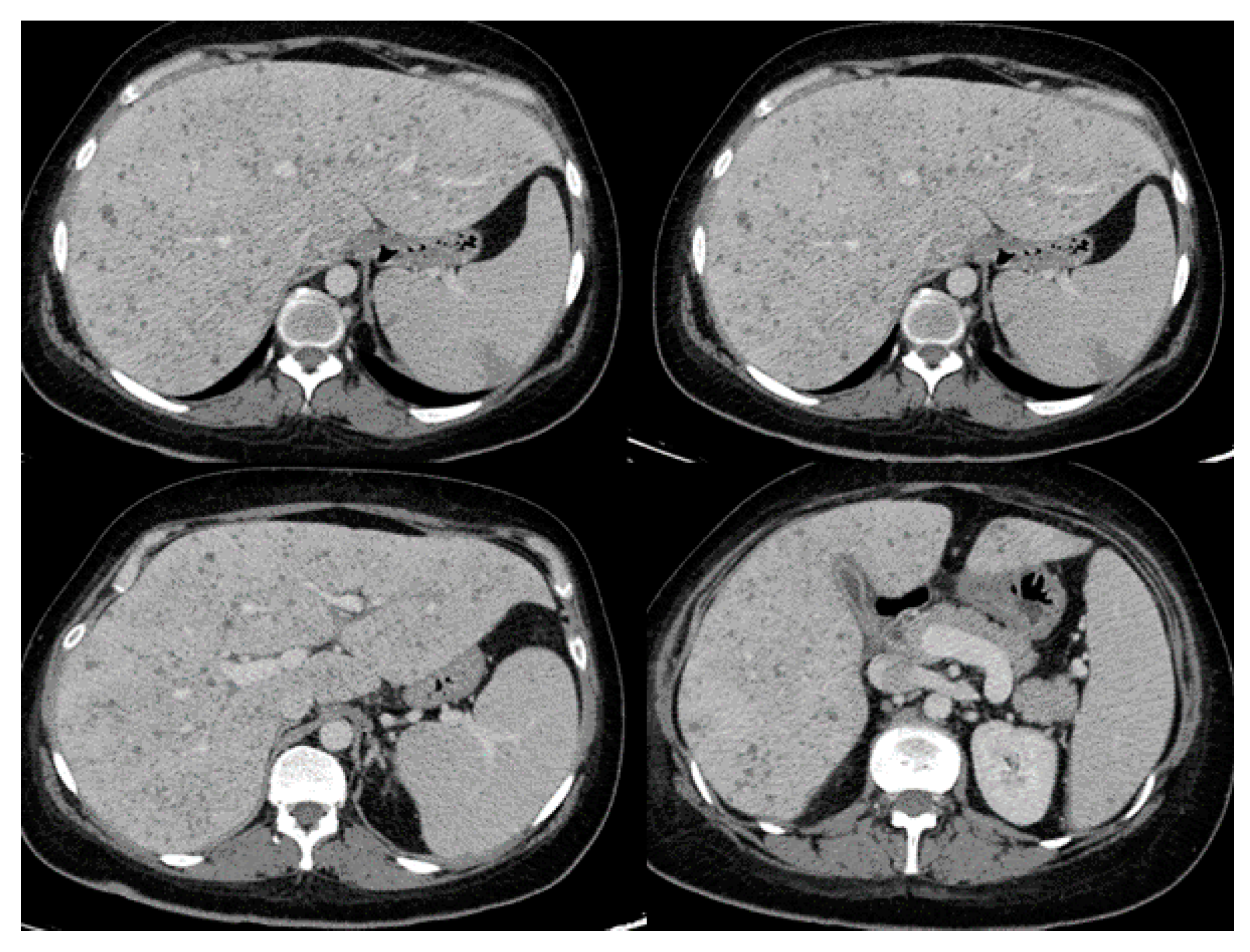
3. Clinical Presentation
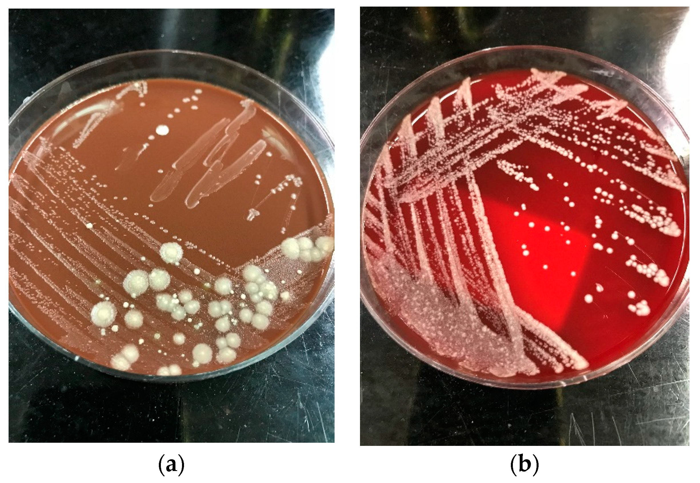
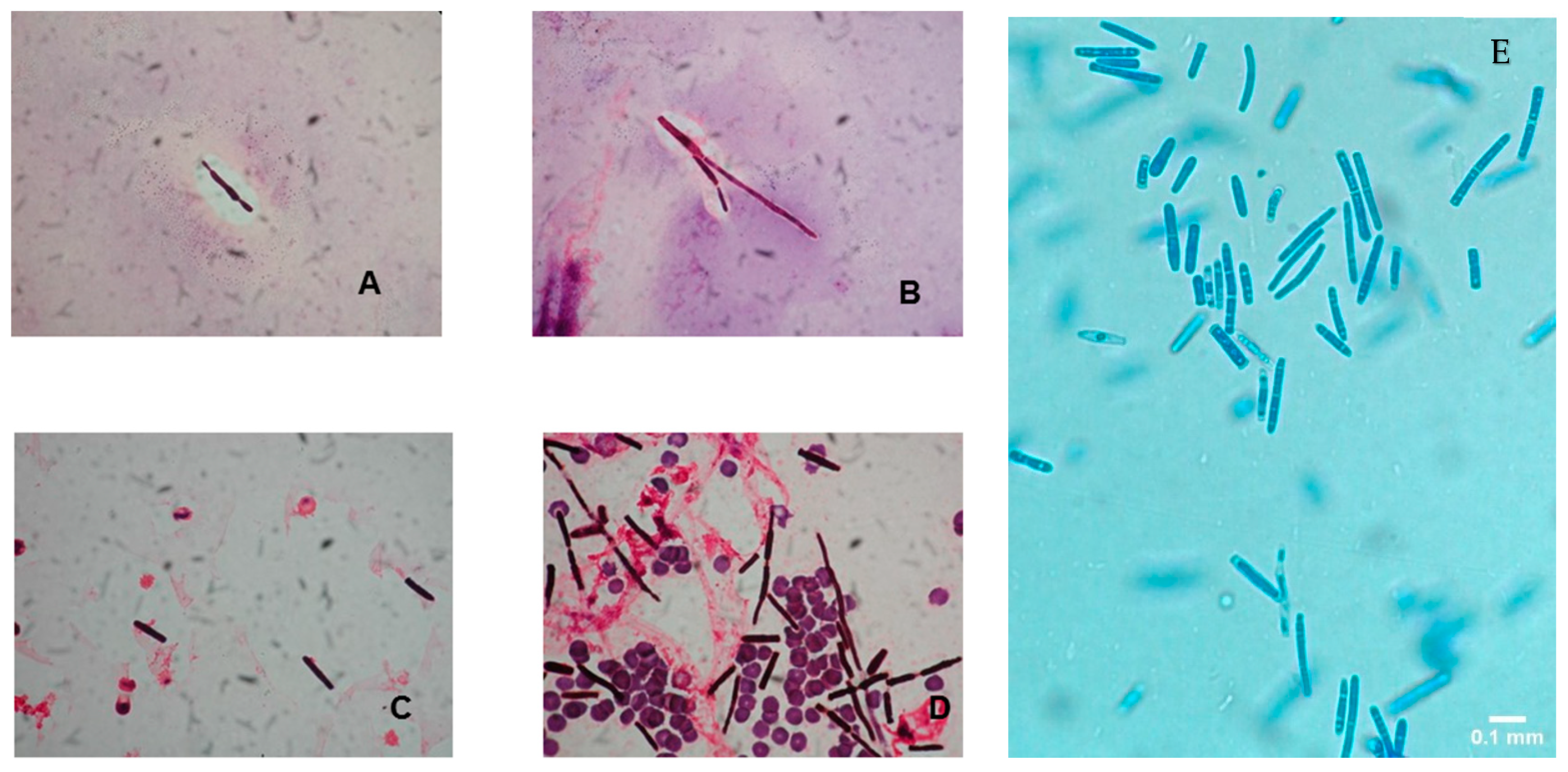
Disseminated disease is common with Saprochaetes especially in severely immunosuppressed patients. Clinical presentation mimics that of invasive candidiasis, and fungemia is common, although Saprochaetes tend to affect the lungs and deep organs more frequently than Candida spp. [54,60]. Patients often present with an acute febrile illness, which could be the only initial manifestation, or could be accompanied by various other symptoms depending on the sites of disease involvement. Symptoms usually progress rapidly to multiorgan failure, shock and death in the majority of patients despite adequate antifungal therapy [11]. Mortality approaches 60–80%; however, rapid molecular identification and prompt initiation of appropriate antifungal therapy have been shown to decrease the numbers of disease-related deaths [3,9,45,61]. The clinical presentation in immunocompetent patients is less severe, and in this population, S. capitata has been exclusively isolated from sputum samples [42].
Table 2 lists common presenting symptoms and corresponding radiographic findings as reported in patients with invasive Saprochaete spp. infections depending on the site of disease involvement. Patients with pulmonary disease present with shortness of breath and cough which could be productive of purulent or bloody sputum. Most common radiographic findings include diffuse bilateral infiltrates, ground glass opacities, pleural effusion and parenchymal nodules. S. Clavata empyema has also been reported, highlighting the importance of obtaining prompt diagnostic thoracentesis and source control [50]. Clinical progression to respiratory failure is common, often requiring intubation and mechanical ventilation. Patients with intraabdominal disease can present with diarrhea, abdominal pain and jaundice in the setting of biliary duct obstruction. Dysuria and hematuria can be present in cases of renal and bladder involvement. Rarely, Saprochaetes can cause peritonitis and abdominal compartment syndrome [45,52]. The most common radiographic findings include hepatosplenomegaly and hypodense parenchymal lesions or nodules involving deep organs (Figure 1). Central nervous system (CNS) involvement can manifest as high-grade fevers, mental status changes and seizures that can progress to status epilepticus. Even if the patients recover from their illness and respond to antifungal therapy, they are at risk of developing long term neurological sequalae [10]. A brain mass or multiple lesions with or without surrounding edema and hemorrhages are usually seen on imaging. Skin lesions are seen in some of the patients with disseminated disease. Reported cutaneous manifestations include erythematous nodules and papules on the lower extremities and back mimicking lesions seen in disseminated candidiasis [62]. Black necrotic plaques involving the perianal area, and wound infection at the site of a laparotomy scar have also been reported [33].

4. Diagnosis

The diagnosis of invasive Saprochaete spp. infection is proven by histopathologic or direct microscopic visualization of the fungi from a normally sterile body site [63]. The clinical significance of isolating these organisms from respiratory, urinary or stool cultures of immunosuppressed patients remains unclear; however, in the absence of other identifiable organisms and in the setting of a clinically documented infection, this represents probable invasive fungal disease [9,54]. In the absence of host factors such as immunosuppression or prolonged neutropenia, the isolation of Saprochaetes from non-sterile sites likely represents a contamination and care must be exerted when interpreting such microbiological results [14,42].
The colonies of Saprochaete spp. grow on a multitude of fungal media within 24–48 hours; however, cultures may require incubation for up to 5 days. S. clavata and S. capitata are often misidentified as they are identical in macroscopic and microscopic analysis [4,28]. This misidentification may have contributed to the significantly lower numbers of reported S. clavata infections prior to the availability of more advanced diagnostic modalities [3,11]. Distinguishing between these 2 organisms to the species level is important for epidemiological and outbreak investigation purposes, as well as for clinical reasons, as S. clavata and S. capitata can have different antifungal susceptibility profiles [7]. Macroscopically, Saprochaete spp. form yeast-like, farinose, dry cottony colonies with frosted glass appearance on the plate (Figure 2) [3,30]. Microscopically, they form true fragmented hyphae, pseudo-hyphae, arthroconidia, annelloconidia and blastoconidia [3,42,44] (Figure 3).
Saprochaete spp. are urease negative, a characteristic that helps distinguish them from Trichosporon spp. [28]. S. clavata, unlike S. capitata, has the ability to assimilate carbon sources such as cellobiose, salicin and arbutin [5,12]; however, many S. capitata strains could grow on all carbon sources [5], and as many as 15% of S. clavata strains do not assimilate cellobiose, making biochemical testing alone unreliable for accurate species identification (Table 1) [11]. Moreover, commercial test kits often lack salicin and arbutin [4,64]. Both species are thermotolerant with optimal growth observed at temperatures between 30 °C and 40 °C [4,5]. In one study, S. clavata, unlike S. capitata isolates, did not grow at temperatures above 45 °C, and isolates preincubated at that temperature became nonviable and demonstrated no regrowth [5]. Differentiation between both species based on incubation temperatures, however, is unreliable as other studies demonstrated that both species exhibited similar growth patterns at different temperatures (Table 1) [23,26].
Commercial systems for yeast identification such as VITEK-2 (ID-YEST card; BioMérieux) and API ID32C (BioMérieux) can successfully identify some of the S. capitata isolates; however, none of the systems cover S. clavata [3,11,30,35,48]. Nucleotide sequencing of the ITS and partial large subunit (LSU) loci can discriminate between Saprochaetes at the species level; however, misidentification is common as the diagnostic ability of these tests is dependent on the accuracy and public availability of the nucleotide sequences (e.g., GenBank) [4,5]. Multilocus sequencing of protein binding loci such as Rbp2, Act or Tef1α might allow for more accurate identification [5,11]. MALDI-TOF mass spectrometry is a useful and reliable modality for the identification and discrimination between Saprochaete spp. and other arthroconidial fungi; however, as with nucleotide sequencing, the accuracy of this test is highly limited by the quality and number of mass spectra available in commercial and in-house fungal libraries [11,65]. S. capitata and S. clavata are better distinguished by MALDI-TOF when newer databases such as Bruker BioTyper (3.0 or 3.1) (Bruker Daltonics, Bremen, Germany) or expanded in-house databases are used (Table 1) [8]. The development of polymerase chain reaction (PCR) assays that are highly species specific may be useful in early identification of the organisms from blood or tissue samples, allowing prompt initiation of adequate antifungal therapy; however, such assays are not yet available for commercial use [66]. In the setting of a suspected outbreak, whole-genome sequence (WGS) typing is the best method to determine clonality and to infer strain relatedness [3]. A single clone was identified by WGS typing as responsible for most cases of a French cluster of infections [9,67]. Recently, the investigation of an S. clavata outbreak in France demonstrated that clinical and environmental isolates were clustered within the same clade and the outbreak ended after discarding the contaminated dishwasher and kitchen utensils [26]
Arthroconidial fungi can cross react with Aspergillus galactomannan (GM) and serum 1,3-beta-D-glucan (BDG); however, these tests are not sensitive, nor specific for Saprochaetes [3,17,49] and therefore have little clinical utility for the diagnosis of invasive infections with these organisms. It is worth noting, however, that a positive GM test in a profoundly neutropenic patient with clinical findings suggestive of invasive Candida infection should prompt physicians to suspect invasive Saprochaete disease, especially that these organisms are resistant to echinocandins and fluconazole, which are the drugs of choice used for the treatment of invasive candidiasis [49].

5. Antifungal Susceptibility and Treatment

No clinical breakpoints are defined for Saprochaetes; therefore, antifungal susceptibility results should be interpreted with caution. Based on in vitro susceptibility results from numerous case reports and case series, these organisms appear to be intrinsically resistant to echinocandins and highly resistant to fluconazole [3,11]. A mutation in the FKS gene hot spot 1 (FKS HS1) which codes a subunit in the β-1,3-D -glucan synthase was detected at a position highly associated with echinocandin resistance in S. capitata isolates [68]. Prolonged treatment with echinocandins (particularly caspofungin) appears to be a risk factor for breakthrough infections with these organisms [32,68], although breakthrough IFIs while on fluconazole, posaconazole and amphotericin B have also been reported [17,38,45,48,49,52]. Minimum inhibitory concentration (MIC) values for voriconazole (0.03–1 mg/L), amphotericin B (0.03–2 mg/L), itraconazole (0.01–1 mg/L) and posaconazole (0.03–1 mg/L) are generally low for both fungi [7,11,29,34,36,44,45,47]. MIC values for flucytosine are higher for S. capitata (0.06–64 mg/L) compared to S. clavata isolates (0.25–0.5 mg/L) [7,12,48,50], while MIC values for isavuconazole appear to be high for both species (1– 4 mg/L) suggesting resistance to this antifungal agent [69]. Acquired resistance to amphotericin B and flucytosine during treatment has also been reported [3].
To date, there is no established therapeutic regimen for the treatment of invasive Saprochaete spp. infections, largely due to the rarity and challenging diagnosis of these organisms and lack of standardized antifungal breakpoints [3]. All treatment recommendations are based on expert opinion and extrapolated data from case reports and small case series. The list of antifungal regimens and duration of administration reported in the literature to treat Saprochaete infections is extensive. In general, echinocandins and fluconazole should be avoided given the high in vitro MIC values and the frequent numbers of breakthrough Saprochaete infections reported while patients are receiving these drugs [7]. High-dose fluconazole might be an appropriate treatment when isolates are susceptible [17,28]; however, avoidance of this drug for empiric therapy might be prudent given the high probability of resistance. Mixed results have been reported with the use of voriconazole monotherapy [17,27], a formulation of amphotericin B monotherapy [11,33] or a combination of amphotericin B and voriconazole [11,27,29,38,44] for the treatment of both S. clavata and S. capitata infections. Combination therapy with caspofungin and voriconazole has also been reported given the potential in vitro synergy, however, with similar mixed results [11,30,70,71]. The European Society of Clinical Microbiology and Infectious Diseases (ESCMID) guidelines published in 2014 recommend any formulation of amphotericin B with or without flucytosine as initial therapy [72]. Antifungal agents are often associated with significant side effects ranging from nephrotoxicity, electrolyte derangements, neurotoxicity, bone marrow suppression and hepatotoxicity among others; therefore, when using combination therapy, clinicians should balance the potential benefits versus risks of adding a second antifungal agent.
The presence of a central venous catheters (CVC) may be a risk factor for the development of catheter-related Saprochaete spp. infections. D’Antonio et al. investigated six S. capitata strains recovered from patients with CVC-related fungemia. Restriction enzyme DNA analysis of clinical isolates from blood and catheter tips were identical, and all isolates showed high ability to produce slime in glucose-containing solutions [56]. In a clinical setting, this characteristic may allow Saprochaete spp. to form biofilms on CVCs and other prosthetic medical devices and may contribute to the pathogenic potential of these organisms [56,73]. Removal of the CVC within 5 days after onset of infection appeared to positively influence outcomes in patients with S. capitata catheter-related infection in one study [54]; however, no firm evidence in the form of randomized controlled trials (RCTs), quasi-RCTs or large observational studies exists in the literature to support this practice. Timely administration of antifungal agents and source control (e.g., drainage of an empyema or retroperitoneal fluid collection) are a mainstay for the treatment of invasive Saprochaete spp. Infections [3,74,75]. Recovery from immunosuppression likely plays a crucial role in successful treatment [11,31]. The use of granulocyte stimulating factor (G-CSF) or Interferon-gamma in combination with antifungal therapy has been successful in eliminating the infection in some patients [29,76]; however, the impact of these regimens on clinical outcomes remains unclear.

6. Conclusions

A wide array of host and environmental factors may contribute to the increased risk for infection with Saprochaetes; however, definite associations are yet to be evaluated. The incidence of these infections may further increase with the development of novel chemotherapeutic and immunosuppressive therapies; therefore, physicians should vigilantly monitor the emergence of such rare pathogens in the hematologic malignancy population. Prophylactic and empirical antifungal protocols should be updated constantly to reflect the changing local epidemiology of IFIs.

Author Contributions

Conceptualization, S.S.K.; methodology, S.S.K.; software, S.E.Z.; validation, S.S.K., writing—original draft preparation, S.E.Z.; writing—review and editing, J.-R.H., S.E.Z. and S.S.K.; visualization S.S.K.; supervision, S.S.K. All authors have read and agreed to the published version of the manuscript.

Funding

This research received no external funding.

Acknowledgments

Figure 2a,b and Figure 3E are courtesy of Rima Asmar, Microbiology laboratory, American University of Beirut Medical Center. The authors would like to thank Maria Ilaria Del Principe, MD, for providing Figure 3A–D and the CT scan image showing hepatosplenic abscesses. Figure 1 and Figure 3A–D were reproduced from Del Principe, M.I et al. Mycoses 2016, 59, 594–601, doi:10.1111/myc.12508 with permissions from “John Wiley and Sons” under license number 4915460075071.

Conflicts of Interest

The authors declare no conflict of interest.

References

  1. Webb, B.J.; Ferraro, J.P.; Rea, S.; Kaufusi, S.; Goodman, B.E.; Spalding, J. Epidemiology and Clinical Features of Invasive Fungal Infection in a US Health Care Network. Open Forum Infect. Dis. 2018, 5, ofy187. [Google Scholar] [CrossRef] [PubMed] [Green Version]
  2. Schmiedel, Y.; Zimmerli, S. Common invasive fungal diseases: An overview of invasive candidiasis, aspergillosis, cryptococcosis, and Pneumocystis pneumonia. Swiss. Med. Wkly 2016, 146, w14281. [Google Scholar] [CrossRef] [Green Version]
  3. Lo Cascio, G.; Vincenzi, M.; Soldani, F.; De Carolis, E.; Maccacaro, L.; Sorrentino, A.; Nadali, G.; Cesaro, S.; Sommavilla, M.; Niero, V.; et al. Outbreak of Saprochaete clavata Sepsis in Hematology Patients: Combined Use of MALDI-TOF and Sequencing Strategy to Identify and Correlate the Episodes. Front. Microbiol. 2020, 11. [Google Scholar] [CrossRef] [PubMed] [Green Version]
  4. Desnos-Ollivier, M.; Blanc, C.; Garcia-Hermoso, D.; Hoinard, D.; Alanio, A.; Dromer, F. Misidentification of Saprochaete clavata as Magnusiomyces capitatus in clinical isolates: Utility of internal transcribed spacer sequencing and matrix-assisted laser desorption ionization-time of flight mass spectrometry and importance of reliable databases. J. Clin. Microbiol. 2014, 52, 2196–2198. [Google Scholar] [CrossRef] [PubMed] [Green Version]
  5. Kaplan, E.; Al-Hatmi, A.M.S.; Ilkit, M.; Gerrits van den Ende, A.H.G.; Hagen, F.; Meis, J.F.; de Hoog, G.S. Molecular Diagnostics of Arthroconidial Yeasts, Frequent Pulmonary Opportunists. J. Clin. Microbiol. 2018, 56. [Google Scholar] [CrossRef] [PubMed] [Green Version]
  6. Hoog, G.S.; Smith, M.; Guého, E. A Revision of the Genus Geotrichum and Its Teleomorphs; Centraalbureau voar Schimmelcultures: Utrecht, The Netherlands, 1986. [Google Scholar]
  7. Esposto, M.C.; Prigitano, A.; Lo Cascio, G.; Ossi, C.; Grancini, A.; Cavanna, C.; Lallitto, F.; Tejada, M.; Bandettini, R.; Mularoni, A.; et al. Yeast-like filamentous fungi: Molecular identification and in vitro susceptibility study. Med. Mycol. 2019, 57, 909–913. [Google Scholar] [CrossRef] [PubMed]
  8. Duran Graeff, L.; Seidel, D.; Vehreschild, M.J.; Hamprecht, A.; Kindo, A.; Racil, Z.; Demeter, J.; De Hoog, S.; Aurbach, U.; Ziegler, M.; et al. Invasive infections due to Saprochaete and Geotrichum species: Report of 23 cases from the FungiScope Registry. Mycoses 2017, 60, 273–279. [Google Scholar] [CrossRef]
  9. Vaux, S.; Criscuolo, A.; Desnos-Ollivier, M.; Diancourt, L.; Tarnaud, C.; Vandenbogaert, M.; Brisse, S.; Coignard, B.; Dromer, F. Multicenter outbreak of infections by Saprochaete clavata, an unrecognized opportunistic fungal pathogen. mBio 2014, 5. [Google Scholar] [CrossRef] [Green Version]
  10. Yilmaz Karapinar, D.; Karadas, N.; Onder Sivis, Z.; Yazici, P.; Duyu, M.; Metin, D.; Karapinar, B.; Aydinok, Y. Rare severe mycotic infections in children receiving empirical caspofungin treatment for febrile neutropenia. Braz. J. Infect. Dis. 2015, 19, 549–552. [Google Scholar] [CrossRef] [Green Version]
  11. Buchta, V.; Bolehovská, R.; Hovorková, E.; Cornely, O.A.; Seidel, D.; Žák, P. Saprochaete clavata Invasive Infections—A New Threat to Hematological-Oncological Patients. Front. Microbiol. 2019, 10. [Google Scholar] [CrossRef]
  12. Parahym, A.M.; Rolim Neto, P.J.; da Silva, C.M.; Domingos Ide, F.; Goncalves, S.S.; Leite, E.P.; de Morais, V.L.; Macedo, D.P.; de Lima Neto, R.G.; Neves, R.P. Invasive infection due to Saprochaete capitata in a young patient with hematological malignancies. Braz. J. Microbiol. 2015, 46, 527–530. [Google Scholar] [CrossRef] [Green Version]
  13. Bansal, N.; Devarajan, V.; Ghafur, K.A.; Sethuraman, N.; Sree, V.L. Breakthrough Saprochaete Capitata infections among patients with hematological malignancies. Leuk Lymphoma 2018, 59, 1762–1763. [Google Scholar] [CrossRef] [PubMed]
  14. Pamidimukkala, U.; Kancharla, A.; Sudhaharan, S.; Gundeti, S.; Mandarapu, S.; Nagalla, V.K.; Raju, S.B.; Karanam, S.D. Isolation of the Rare Opportunistic Yeast Saprochaete capitata from Clinical Samples-Experience from a Tertiary Care Hospital in Southern India and a Brief Review of the Literature. J. Clin. Diagn. Res. 2017, 11, Dc36–Dc42. [Google Scholar] [CrossRef]
  15. Erman, B.; Fırtına, S.; Aksoy, B.A.; Aydogdu, S.; Genç, G.E.; Doğan, Ö.; Bozkurt, C.; Fışgın, T.; Çipe, F.E. Invasive Saprochaete capitata Infection in a Patient with Autosomal Recessive CARD9 Deficiency and a Review of the Literature. J. Clin. Immunol. 2020, 40, 466–474. [Google Scholar] [CrossRef]
  16. Ikuta, K.; Torimoto, Y.; Yamamoto, M.; Okamura, N.; Hosoki, T.; Sato, K.; Fujiya, M.; Kohgo, Y. Successful treatment of systemic Geotrichum capitatum infection by liposomal amphotericin-B, itraconazole, and voriconazole in a Japanese man. Intern. Med. 2010, 49, 2499–2503. [Google Scholar] [CrossRef] [PubMed] [Green Version]
  17. Ben Neji, H.; Bchir, M.; Hamdoun, M.; Kallel, A.; Kallel, K.; Bahri, O.; Meddeb, B. Geotrichum capitatum fungemia in patients treated for acute leukemia. Med. Mal. Infect. 2019, 49, 284–286. [Google Scholar] [CrossRef] [PubMed]
  18. Ozdemir, Z.C.; Bozkurt Turhan, A.; Duzenli Kar, Y.; Dinleyici, C.E.; Bor, O. Fatal course of Saprochaete capitata fungemia in children with acute lymphoblastic leukemia. Pediatr. Hematol. Oncol. 2017, 34, 66–72. [Google Scholar] [CrossRef]
  19. Hazirolan, G.; Aypak, A.; Aksu, N. An unusual case of urinary tract infection caused by Saprochaete capitata under anidulafungin treatment. J. Mycol. Med. 2017, 27, 387–390. [Google Scholar] [CrossRef]
  20. Cornely, O.A.; Gachot, B.; Akan, H.; Bassetti, M.; Uzun, O.; Kibbler, C.; Marchetti, O.; de Burghgraeve, P.; Ramadan, S.; Pylkkanen, L.; et al. Epidemiology and Outcome of Fungemia in a Cancer Cohort of the Infectious Diseases Group (IDG) of the European Organization for Research and Treatment of Cancer (EORTC 65031). Clin. Infect. Dis. 2015, 61, 324–331. [Google Scholar] [CrossRef]
  21. Garcia-Ruiz, J.C.; Lopez-Soria, L.; Olazabal, I.; Amutio, E.; Arrieta-Aguirre, I.; Velasco-Benito, V.; Ponton, J.; Moragues, M.D. Invasive infections caused by Saprochaete capitata in patients with haematological malignancies: Report of five cases and review of the antifungal therapy. Rev. Iberoam. Micol. 2013, 30, 248–255. [Google Scholar] [CrossRef]
  22. Gurgui, M.; Sanchez, F.; March, F.; Lopez-Contreras, J.; Martino, R.; Cotura, A.; Galvez, M.L.; Roig, C.; Coll, P. Nosocomial outbreak of Blastoschizomyces capitatus associated with contaminated milk in a haematological unit. J. Hosp. Infect. 2011, 78, 274–278. [Google Scholar] [CrossRef]
  23. Döğen, A.; Kaplan, E.; Öksüz, Z.; Serin, M.S.; Ilkit, M.; de Hoog, G.S. Dishwashers are a major source of human opportunistic yeast-like fungi in indoor environments in Mersin, Turkey. Med. Mycol. 2013, 51, 493–498. [Google Scholar] [CrossRef] [Green Version]
  24. Gümral, R.; Özhak-Baysan, B.; Tümgör, A.; Saraçlı, M.A.; Yıldıran, Ş.T.; Ilkit, M.; Zupančič, J.; Novak-Babič, M.; Gunde-Cimerman, N.; Zalar, P.; et al. Dishwashers provide a selective extreme environment for human-opportunistic yeast-like fungi. Fungal. Divers. 2016, 76, 1–9. [Google Scholar] [CrossRef]
  25. Zalar, P.; Novak, M.; de Hoog, G.S.; Gunde-Cimerman, N. Dishwashers—A man-made ecological niche accommodating human opportunistic fungal pathogens. Fungal. Biol. 2011, 115, 997–1007. [Google Scholar] [CrossRef]
  26. Menu, E.; Criscuolo, A.; Desnos-Ollivier, M.; Cassagne, C.; D’Incan, E.; Furst, S.; Ranque, S.; Berger, P.; Dromer, F. Saprochaete clavata Outbreak Infecting Cancer Center through Dishwasher. Emerg. Infect. Dis. 2020, 26, 2031–2038. [Google Scholar] [CrossRef] [PubMed]
  27. Stanzani, M.; Cricca, M.; Sassi, C.; Sutto, E.; De Cicco, G.; Bonifazi, F.; Bertuzzi, C.; Bacci, F.; Paolini, S.; Cavo, M.; et al. Saprochaete clavata infections in patients undergoing treatment for haematological malignancies: A report of a monocentric outbreak and review of the literature. Mycoses 2019, 62, 1100–1107. [Google Scholar] [CrossRef]
  28. Girmenia, C.; Pagano, L.; Martino, B.; D’Antonio, D.; Fanci, R.; Specchia, G.; Melillo, L.; Buelli, M.; Pizzarelli, G.; Venditti, M.; et al. Invasive infections caused by Trichosporon species and Geotrichum capitatum in patients with hematological malignancies: A retrospective multicenter study from Italy and review of the literature. J. Clin. Microbiol. 2005, 43, 1818–1828. [Google Scholar] [CrossRef] [Green Version]
  29. Favre, S.; Rougeron, A.; Levoir, L.; Pérard, B.; Milpied, N.; Accoceberry, I.; Gabriel, F.; Vigouroux, S. Saprochaete clavata invasive infection in a patient with severe aplastic anemia: Efficacy of voriconazole and liposomal amphotericin B with adjuvant granulocyte transfusions before neutrophil recovery following allogeneic bone marrow transplantation. Med. Mycol. Case Rep. 2016, 11, 21–23. [Google Scholar] [CrossRef]
  30. Liu, X.; Zhou, W.; Jiang, Y.; Kuang, L. Invasive Fungal Infection Caused by Geotrichum clavatum in a Child with Acute Leukemia: First Documented Case from Mainland China. Jpn. J. Infect Dis. 2019, 72, 130–132. [Google Scholar] [CrossRef] [Green Version]
  31. Del Principe, M.I.; Sarmati, L.; Cefalo, M.; Fontana, C.; De Santis, G.; Buccisano, F.; Maurillo, L.; De Bellis, E.; Postorino, M.; Sconocchia, G.; et al. A cluster of Geotrichum clavatum (Saprochaete clavata) infection in haematological patients: A first Italian report and review of literature. Mycoses 2016, 59, 594–601. [Google Scholar] [CrossRef]
  32. Bretagne, S.; Renaudat, C.; Desnos-Ollivier, M.; Sitbon, K.; Lortholary, O.; Dromer, F.; Group, o.b.o.t.F.M.S. Predisposing factors and outcome of uncommon yeast species-related fungaemia based on an exhaustive surveillance programme (2002–14). J. Antimicrob. Chemother. 2017, 72, 1784–1793. [Google Scholar] [CrossRef]
  33. Hajar, Z.; Medawar, W.; Rizk, N. Saprochaete capitata (Geotrichum capitatum), an emerging fungal infection in kidney transplant recipients. J. Mycol. Med. 2018, 28, 387–389. [Google Scholar] [CrossRef]
  34. Pavone, P.; Oliva, A.; Raponi, G.; Pugliese, F.; Martelli, S.; Celli, P.; Sacco, F.; Vullo, V.; Mastroianni, C.M.; Russo, G. Disseminated fungal infection due to Saprochaete clavata in a kidney transplant recipient. J. Mycol. Med. 2019, 29, 278–281. [Google Scholar] [CrossRef]
  35. Mandarapu, S.; Krishna, V.; Raju, S.B.; Pamidimukkala, U.; Nimmagadda, S. Saprochaete capitata fungal infection in renal transplant recipient. Indian J. Nephrol. 2016, 26, 464–466. [Google Scholar] [CrossRef]
  36. Cavanna, C.; Lallitto, F.; Mangione, F.; Tamarozzi, F.; Marone, P.; Ceriana, P. Fungemia due to Saprochaete capitata in a non-neutropenic patient hospitalized in an intensive care unit after cardiac surgery. J. Mycol. Med. 2017, 27, 281–284. [Google Scholar] [CrossRef]
  37. Miglietta, F.; Palumbo, C.; Pizzolante, M.; Faneschi, M.L.; Cucurachi, M.; Velardi, L.; Lobreglio, G. A rare case of Saprochaete capitata fungemia in a critical ill patient without hematologic and oncological disorders. Rev. Esp. Quim. 2016, 29, 47–49. [Google Scholar]
  38. Chittick, P.; Palavecino, E.L.; Delashmitt, B.; Evans, J.; Peacock, J.E., Jr. Case of fatal Blastoschizomyces capitatus infection occurring in a patient receiving empiric micafungin therapy. Antimicrob. Agents Chemother. 2009, 53, 5306–5307. [Google Scholar] [CrossRef] [Green Version]
  39. Sreeja, S.; Banashankari, G.S.; Bhavana, M.V.; Devi, D.R. Blastoschizomyces capitatus pneumonia: A rare case. Indian J. Pathol. Microbiol. 2011, 54, 846–847. [Google Scholar] [CrossRef]
  40. Chauhan, S.; Tiwari, S.; Singh, P.; Goyal, R. Pulmonary Infection by Blastoschizomyces capitatus in An old Tuberculosis Patient. J. Clin. Diagn. Res. Jcdr. 2015, 9, DD01–DD02. [Google Scholar] [CrossRef]
  41. Wills, T.S.; Degryse, A.; Lavina, J.; Sinnott, J.T. Blastoschizomyces capitatus pneumonia in an immunocompetent male. South Med. J. 2004, 97, 702–704. [Google Scholar] [CrossRef]
  42. Tanabe, M.B.; Patel, S.A. Blastoschizomyces capitatus pulmonary infections in immunocompetent patients: Case report, case series and literature review. Epidemiol. Infect. 2018, 146, 58–64. [Google Scholar] [CrossRef]
  43. Subramanya Supram, H.; Gokhale, S.; Chakrabarti, A.; Rudramurthy, S.M.; Gupta, S.; Honnavar, P. Emergence of Magnusiomyces capitatus infections in Western Nepal. Med. Mycol. 2015, 54, 103–110. [Google Scholar] [CrossRef] [Green Version]
  44. Salguero Fernandez, I.; Najera Botello, L.; Orden Martinez, B.; Roustan Gullon, G. Disseminated fungemia by Saprochaete clavata. Enferm. Infecc. Microbiol. Clin. 2019, 37, 283–284. [Google Scholar] [CrossRef] [PubMed]
  45. Camus, V.; Thibault, M.L.; David, M.; Gargala, G.; Compagnon, P.; Lamoureux, F.; Girault, C.; Michot, J.B.; Stamatoullas, A.; Lanic, H.; et al. Invasive Geotrichum clavatum fungal infection in an acute myeloid leukaemia patient: A case report and review. Mycopathologia 2014, 177, 319–324. [Google Scholar] [CrossRef]
  46. Ulu-Kilic, A.; Atalay, M.A.; Metan, G.; Cevahir, F.; Koc, N.; Eser, B.; Cetin, M.; Kaynar, L.; Alp, E. Saprochaete capitata as an emerging fungus among patients with haematological malignencies. Mycoses 2015, 58, 491–497. [Google Scholar] [CrossRef]
  47. Miglietta, F.; Vella, A.; Faneschi, M.L.; Lobreglio, G.; Rizzo, A.; Palumbo, C.; Palumbo, C.; Di Renzo, N.; Pizzolante, M. Geotrichum capitatum septicaemia in a haematological patient after acute myeloid leukaemia relapse: Identification using MALDI-TOF mass spectrometry and review of the literature. Infez. Med. 2015, 23, 161–167. [Google Scholar]
  48. Savini, V.; Catavitello, C.; Balbinot, A.; Masciarelli, G.; Astolfi, D.; Pompilio, A.; Di Bonaventura, G.; D’Amario, C.; D’Antonio, D. Multidrug-resistant Geotrichum capitatum from a haematology ward. Mycoses 2011, 54, 542–543. [Google Scholar] [CrossRef]
  49. Bonini, A.; Capatti, C.; Parmeggiani, M.; Gugliotta, L.; Micozzi, A.; Gentile, G.; Capria, S.; Girmenia, C. Galactomannan detection in Geotrichum capitatum invasive infections: Report of 2 new cases and review of diagnostic options. Diagn. Microbiol. Infect. Dis. 2008, 62, 450–452. [Google Scholar] [CrossRef]
  50. Brunetti, G.; Visconti, V.; Ghezzi, M.C.; Mantovani, S.; Ferretti, G.; Raponi, G. Management and treatment of Magnusiomyces capitatus (Geotrichum capitatum) pleural infection in a non-neutropenic patient with posaconazole. A new therapeutic opportunity? New Microbiol. 2016, 39, 307–309. [Google Scholar]
  51. Martino, P.; Venditti, M.; Micozzi, A.; Morace, G.; Polonelli, L.; Mantovani, M.P.; Petti, M.C.; Burgio, V.L.; Santini, C.; Serra, P.; et al. Blastoschizomyces capitatus: An emerging cause of invasive fungal disease in leukemia patients. Rev. Infect Dis. 1990, 12, 570–582. [Google Scholar] [CrossRef]
  52. Schuermans, C.; van Bergen, M.; Coorevits, L.; Verhaegen, J.; Lagrou, K.; Surmont, I.; Jeurissen, A. Breakthrough Saprochaete capitata infections in patients receiving echinocandins: Case report and review of the literature. Med. Mycol. 2011, 49, 414–418. [Google Scholar] [CrossRef] [Green Version]
  53. Ersoz, G.; Otag, F.; Erturan, Z.; Aslan, G.; Kaya, A.; Emekdas, G.; Sugita, T. An outbreak of Dipodascus capitatus infection in the ICU: Three case reports and review of the literature. Jpn. J. Infect. Dis. 2004, 57, 248–252. [Google Scholar]
  54. Martino, R.; Salavert, M.; Parody, R.; Tomás, J.F.; de la Cámara, R.; Vázquez, L.; Jarque, I.; Prieto, E.; Sastre, J.L.; Gadea, I.; et al. Blastoschizomyces capitatus infection in patients with leukemia: Report of 26 cases. Clin. Infect. Dis. 2004, 38, 335–341. [Google Scholar] [CrossRef] [Green Version]
  55. Pérez-Sanchez, I.; Anguita, J.; Martín-Rabadan, P.; Muñoz, P.; Serrano, D.; Escudero, A.; Pintado, T. Blastoschizomyces capitatus infection in acute leukemia patients. Leuk Lymphoma 2000, 39, 209–212. [Google Scholar] [CrossRef]
  56. D’Antonio, D.; Parruti, G.; Pontieri, E.; Di Bonaventura, G.; Manzoli, L.; Sferra, R.; Vetuschi, A.; Piccolomini, R.; Romano, F.; Staniscia, T. Slime production by clinical isolates of Blastoschizomyces capitatus from patients with hematological malignancies and catheter-related fungemia. Eur. J. Clin. Microbiol. Infect. Dis. 2004, 23, 787–789. [Google Scholar] [CrossRef] [PubMed]
  57. Fraser, M.; Brown, Z.; Houldsworth, M.; Borman, A.M.; Johnson, E.M. Rapid identification of 6328 isolates of pathogenic yeasts using MALDI-ToF MS and a simplified, rapid extraction procedure that is compatible with the Bruker Biotyper platform and database. Med. Mycol. 2016, 54, 80–88. [Google Scholar] [CrossRef] [Green Version]
  58. Westblade, L.F.; Jennemann, R.; Branda, J.A.; Bythrow, M.; Ferraro, M.J.; Garner, O.B.; Ginocchio, C.C.; Lewinski, M.A.; Manji, R.; Mochon, A.B.; et al. Multicenter study evaluating the Vitek MS system for identification of medically important yeasts. J. Clin. Microbiol. 2013, 51, 2267–2272. [Google Scholar] [CrossRef] [Green Version]
  59. Lohmann, C.; Sabou, M.; Moussaoui, W.; Prévost, G.; Delarbre, J.M.; Candolfi, E.; Gravet, A.; Letscher-Bru, V. Comparison between the Biflex III-Biotyper and the Axima-SARAMIS systems for yeast identification by matrix-assisted laser desorption ionization-time of flight mass spectrometry. J. Clin. Microbiol. 2013, 51, 1231–1236. [Google Scholar] [CrossRef] [Green Version]
  60. Viscoli, C.; Girmenia, C.; Marinus, A.; Collette, L.; Martino, P.; Vandercam, B.; Doyen, C.; Lebeau, B.; Spence, D.; Krcmery, V.; et al. Candidemia in cancer patients: A prospective, multicenter surveillance study by the Invasive Fungal Infection Group (IFIG) of the European Organization for Research and Treatment of Cancer (EORTC). Clin. Infect. Dis. 1999, 28, 1071–1079. [Google Scholar] [CrossRef] [Green Version]
  61. Picard, M.; Cassaing, S.; Letocart, P.; Verdeil, X.; Protin, C.; Chauvin, P.; Iriart, X.; Cavalie, L.; Valentin, A.; Marchou, B.; et al. Concomitant cases of disseminated Geotrichum clavatum infections in patients with acute myeloid leukemia. Leuk Lymphoma 2014, 55, 1186–1188. [Google Scholar] [CrossRef] [PubMed]
  62. Bodey, G.P.; Luna, M. Skin Lesions Associated With Disseminated Candidiasis. JAMA 1974, 229, 1466–1468. [Google Scholar] [CrossRef]
  63. Donnelly, J.P.; Chen, S.C.; Kauffman, C.A.; Steinbach, W.J.; Baddley, J.W.; Verweij, P.E.; Clancy, C.J.; Wingard, J.R.; Lockhart, S.R.; Groll, A.H.; et al. Revision and Update of the Consensus Definitions of Invasive Fungal Disease From the European Organization for Research and Treatment of Cancer and the Mycoses Study Group Education and Research Consortium. Clin. Infect. Dis. 2019. [Google Scholar] [CrossRef] [PubMed] [Green Version]
  64. Guého, E.; de Hoog, G.S.; Smith, M.T.; Meyer, S.A. DNA relatedness, taxonomy, and medical significance of Geotrichum capitatum. J. Clin. Microbiol. 1987, 25, 1191. [Google Scholar] [CrossRef] [Green Version]
  65. Kolecka, A.; Khayhan, K.; Groenewald, M.; Theelen, B.; Arabatzis, M.; Velegraki, A.; Kostrzewa, M.; Mares, M.; Taj-Aldeen, S.J.; Boekhout, T. Identification of medically relevant species of arthroconidial yeasts by use of matrix-assisted laser desorption ionization-time of flight mass spectrometry. J. Clin. Microbiol. 2013, 51, 2491–2500. [Google Scholar] [CrossRef] [Green Version]
  66. Arrieta-Aguirre, I.; Menendez-Manjon, P.; Cuetara, M.S.; Lopez-Soria, L.; Garcia-Ruiz, J.C.; Moragues, M.D. Molecular Identification of Saprochaete capitata in Human Blood and Paraffinized Tissue Samples. J. Clin. Microbiol. 2017, 55, 2556–2559. [Google Scholar] [CrossRef] [Green Version]
  67. Bougnoux, M.E.; Brun, S.; Zahar, J.R. Healthcare-associated fungal outbreaks: New and uncommon species, New molecular tools for investigation and prevention. Antimicrob. Resist. Infect. Control. 2018, 7, 45. [Google Scholar] [CrossRef] [Green Version]
  68. Arrieta-Aguirre, I.; Menendez-Manjon, P.; Cuetara, M.S.; Fernandez de Larrinoa, I.; Garcia-Ruiz, J.C.; Moragues, M.D. Sequencing of FKS Hot Spot 1 from Saprochaete capitata To Search for a Relationship to Reduced Echinocandin Susceptibility. Antimicrob. Agents Chemother. 2018, 62. [Google Scholar] [CrossRef] [Green Version]
  69. Desnos-Ollivier, M.; Bretagne, S.; Boullie, A.; Gautier, C.; Dromer, F.; Lortholary, O. Isavuconazole MIC distribution of 29 yeast species responsible for invasive infections (2015-2017). Clin. Microbiol. Infect. 2019, 25, 634.e631–634.e634. [Google Scholar] [CrossRef]
  70. Etienne, A.; Datry, A.; Gaspar, N.; Morel, V.; Delabesse, E.; Lmimouni, B.; Vernant, J.P.; Dhédin, N. Successful treatment of disseminated Geotrichum capitatum infection with a combination of caspofungin and voriconazole in an immunocompromised patient. Mycoses 2008, 51, 270–272. [Google Scholar] [CrossRef]
  71. Fianchi, L.; Montini, L.; Caira, M.; Voso, M.T.; Maviglia, R.; Posteraro, B.; Pagano, L. Combined voriconazole plus caspofungin therapy for the treatment of probable Geotrichum pneumonia in a leukemia patient. Infection 2008, 36, 65–67. [Google Scholar] [CrossRef]
  72. Arendrup, M.C.; Boekhout, T.; Akova, M.; Meis, J.F.; Cornely, O.A.; Lortholary, O. ESCMID and ECMM joint clinical guidelines for the diagnosis and management of rare invasive yeast infections. Clin. Microbiol. Infect. 2014, 20 (Suppl. 3), 76–98. [Google Scholar] [CrossRef] [Green Version]
  73. Kernien, J.F.; Snarr, B.D.; Sheppard, D.C.; Nett, J.E. The Interface between Fungal Biofilms and Innate Immunity. Front. Immunol. 2018, 8, 1968. [Google Scholar] [CrossRef] [PubMed] [Green Version]
  74. Radich, R.J.; Alvarez, Z.C.; Valenzuela, R.A.; Castillo, H.F.; Moraga, V.F.; Castillo, A.J. Role of surgery in Saprochaete capitata (S. capitata) sepsis in a patient with acute myeloid leukemia. Rev. Med. Chil. 2017, 145, 1067–1071. [Google Scholar] [CrossRef]
  75. D’Assumpcao, C.; Lee, B.; Heidari, A. A Case of Magnusiomyces capitatus Peritonitis Without Underlying Malignancies. J. Investig. Med. High Impact Case Rep. 2018, 6, 2324709618795268. [Google Scholar] [CrossRef] [Green Version]
  76. DeMaio, J.; Colman, L. The use of adjuvant interferon-gamma therapy for hepatosplenic Blastoschizomyces capitatus infection in a patient with leukemia. Clin. Infect. Dis. 2000, 31, 822–824. [Google Scholar] [CrossRef]
Figure 1. Abdominal computed tomography showing hepatosplenic abscesses in a patient with invasive Saprochaete clavata infection (reproduced from Del Principe, M.I et al. Mycoses 2016, 59, 594–601, doi:10.1111/myc.12508 with permission from John Wiley and Sons under license number 4915460075071).
Pathogens 09 00922 g001
Figure 2. Dry cottony colonies with frosted glass appearance of Saprochaete spp. on (a) blood agar and (b) chocolate agar, respectively. The isolates were incubated at 26 °C for 72 h and later identified as S. capitata by MALDI-TOF.
Pathogens 09 00922 g002
Figure 3. (A–D) Gram stain preparation of blood cultures showing arthroconidia and hyphal elements of Saprochaete clavata. Magnification ×1000 (reproduced from Del Principe, M.I et al. Mycoses 2016, 59, 594–601, doi:10.1111/myc.12508 with permission from John Wiley and Sons under license number 4915460075071). (E) Lactophenol cotton blue preparation of peritoneal fluid cultures showing arthroconidia suggestive of Saprochaete spp. infection. The organism was identified as S. capitata by MALDI-TOF. Magnification ×40.
Pathogens 09 00922 g003
Table 1. Epidemiological and clinical features of S. clavata and S. capitata as reported in the literature.
S. clavataS. capitata
Country
Outbreaks
  • Multiple outbreaks in Italy [3,27]
  • Multicenter outbreak in France [9]
  • Outbreak from contaminated dishwasher and utensils in France [26]
  • Outbreak in Italy in 1984 [51]
  • Outbreak from contaminated milk in Spain [22]
  • Outbreak in the intensive care unit in Japan [53]
Mortality
  • 65–80% [8,9]
Risk Factors ϐ
  • Profound neutropenia [3,11,27,29,34]
  • Central venous catheter [3,11,29,45]
  • Chemotherapy (predominantly for acute leukemia) [3,8,27,30,32,45]
  • Prolonged use of broad-spectrum antibiotics [3,11,27,30]
  • Prior antifungal use for prophylaxis or treatment [27,34,45]
  • Hematopoietic stem cell transplant [44]
  • Profound neutropenia [10,12,14,18,49]
  • Central venous catheter [10,16,36,38,46,54,56]
  • Chemotherapy (predominantly for acute leukemia) [8,12,13,17,32,52]
  • Prolonged use of broad-spectrum antibiotics [12,17,33,52]
  • Prior antifungal use for prophylaxis or treatment [10,12,46,52]
  • Hematopoietic stem cell transplant [28,46]
  • Critical illness and prolonged intensive care unit stay [36,37]
  • Gastrointestinal disease (e.g., perforation or biliary stasis) [11,13]
  • Total parenteral nutrition [46]
  • CARD-9 deficiency [15]
  • Immunosuppressive therapy (tacrolimus, mycophenolate mofetil, prednisone) [33,35,54]
Source of Isolation ψ
Carbon Assimilation *
  • (+) cellobiose
  • (+) Salicin
  • (+) arbutin
  • (−) cellobiose
  • (−) Salicin
  • (−) arbutin
Growth Temperature
  • Optimal growth: 30–40 °C
  • Partial growth at 45 °C. Robust regrowth at lower temperatures ф
Optimal growth: 30–40 °C No growth at temperatures > 45 °C. Isolates become nonviable ω
MADLI-TOF Platforms UsedSuccessful identification
  • MALDI-TOF Biotyper system (Bruker Daltonics) [11,27,29]
  • MALDI-TOF MS-Vitek (bioMérieux, Marcy l’Etoile, France) [30]
  • MALDI-TOF SARAMIS (bioMérieux, Marcy l’Etoile, France) [3]
Failed identification
  • MALDI-TOF MS-Vitek (bioMérieux, Marcy l’Etoile, France) [3]
Successful identification
  • MALDI-TOF (Bruker Daltonics) [19,36,37,47,57]
  • MALDI-TOF MS-Vitek (bioMérieux, Marcy l’Etoile, France) [58]
Failed identification
  • MALDI-TOF Axima-SARAMIS (Shimadzu-AnagnosTec) and MALDI-TOF Biflex III-BioTyper (Bruker Daltonics) [59]
ϐ Acute leukemia and prior exposure to caspofungin have been shown to be independent risk factors for the development of Saprochaete infections [32]. To the best of our knowledge, the remainder of the listed risk factors have not been shown to be independently associated with increased risk of Saprochaete infections in a multivariate analysis. ψ S. capitata is more frequently isolated from sputum or bronchial samples compared to S. clavata. Both species are isolated from blood samples frequently. * Approximately 15% of S. clavata strains do not assimilate cellobiose; some S. capitata strains can assimilate all carbon sources. ф S. clavata and S. capitata isolates exhibited similar growth at a range of 30–48 °C. No growth seen at temperatures >48 °C [26]. ω S. capitata isolates grew at a range of 5–47 °C in one study [23].
Table 2. Common presenting symptoms and corresponding radiographic findings reported in patients with invasive Saprochaete infections.
Presenting Symptoms Radiographic Findings
Pulmonary Involvement [17,27,29,30,35,50]
  • Fever
  • Respiratory distress
  • Cough with purulent sputum
  • Pulmonary infiltrates
  • Parenchymal micronodules
  • Ground glass infiltrates
  • Pleural effusion
Intraabdominal and genitourinary Involvement [15,17,19,27,28,29,30,34,45,52]
  • Fever
  • Diarrhea
  • Jaundice
  • Dysuria
  • Hematuria
  • Abdominal compartment syndrome
  • Hypodense parenchymal lesions that could involve the liver, spleen and/or kidneys
  • Hepatosplenomegaly
  • Biliary duct obstruction
  • Abdominal ascites
  • Abdominal wall collection
Central Nervous System Involvement [10,27,44]
  • Fever
  • Mental status changes
  • Seizures or status epilepticus
  • Brain mass or lesion(s); Surrounding brain edema and hemorrhagic foci can be present
Skin Involvement [17,33,44]
  • Asymptomatic, blackish-brown necrotic plaques around peri-anal area
  • Erythematous nodules and papules on legs and back
  • Laparotomy wound SSTI
  • Not applicable
Publisher’s Note: MDPI stays neutral with regard to jurisdictional claims in published maps and institutional affiliations.

