Article

New Species of the Genus Curvularia: C. tamilnaduensis and C. coimbatorensis from Fungal Keratitis Cases in South India

1
Department of Microbiology, Faculty of Science and Informatics, University of Szeged, 6726 Szeged, Hungary
2
MTA-SZTE “Lendület” Fungal Pathogenicity Mechanisms Research Group, 6726 Szeged, Hungary
3
Department of Medical Laboratory Sciences, College of Applied Medical Sciences, Majmaah University, Al Majmaah 11952, Saudi Arabia
4
Greenlink Analytical and Research Laboratory India Private Ltd., Coimbatore, Tamil Nadu 641014, India
5
Department of Microbiology, Dr. G.R. Damodaran College of Science, Coimbatore, Tamil Nadu 641014, India
6
Synthetic and Systems Biology Unit, Institute of Biochemistry, Biological Research Centre, Hungarian Academy of Sciences, 6726 Szeged, Hungary
7
Aravind Eye Hospital and Postgraduate Institute of Ophthalmology, Coimbatore, Tamil Nadu 641014, India
*
Authors to whom correspondence should be addressed.
Pathogens 2020, 9(1), 9; https://doi.org/10.3390/pathogens9010009
Submission received: 6 December 2019 / Revised: 17 December 2019 / Accepted: 18 December 2019 / Published: 20 December 2019
(This article belongs to the Section Human Pathogens)

Abstract

:
Members of the genus Curvularia are melanin-producing dematiaceous fungi of increasing clinical importance as causal agents of both local and invasive infections. This study contributes to the taxonomical and clinical knowledge of this genus by describing two new Curvularia species based on isolates from corneal scrapings of South Indian fungal keratitis patients. The phylogeny of the genus was updated based on three phylogenetic markers: the internal transcribed spacer (ITS) region of the ribosomal RNA gene cluster as well as fragments of the glyceraldehyde-3-phosphate dehydrogenase (gpdh) and translation elongation factor 1-α (tef1α) genes. The maximum likelihood phylogenetic tree constructed from the alignment of the three concatenated loci revealed that the examined isolates are representing two new, yet undescribed, Curvularia species. Examination of colony and microscopic morphology revealed differences between the two species as well as between the new species and their close relatives. The new species were formally described as Curvularia tamilnaduensis N. Kiss & S. Kocsubé sp. nov. and Curvularia coimbatorensis N. Kiss & S. Kocsubé sp. nov. Antifungal susceptibility testing by the broth microdilution method of CLSI (Clinical & Laboratory Standards Institute) revealed that the type strain of C. coimbatorensis is less susceptible to a series of antifungals than the C. tamilnaduensis strains.

Graphical Abstract

1. Introduction

The fungal genus Curvularia (Ascomycota, Pleosporales, Pleosporaceae) comprises of dematiaceous, melanin-producing molds with various lifestyles including saprophytism, plant endophytism [1], plant parasitism [2], and human pathogenicity [3].
The genus-level identification of Curvularia was performed traditionally by the examination of pigmentation, as well as the morphology of the septate conidia and hyphae [3]. The first sequence-based species-level identification attempts targeted the internal transcribed spacer (ITS) region of the ribosomal RNA gene cluster, which alone, however, proved inappropriate, either for the purposes of exact diagnosis [4] or for the phylogenetic resolution of the genus and the clarification of its relationship to the closely related genera Bipolaris, Cochliobolus, and Drechslera [3]. Multilocus sequence typing (MLST) involving fragments of the nuclear ribosomal large subunit RNA (LSU) as well as the glyceraldehyde-3-phosphate dehydrogenase (gpdh) and translation elongation factor 1-α (tef1a) genes in addition to ITS had resulted in the recently accepted phylogenetic concept of the genus Curvularia [5], which was applied in more recent works [6,7,8]. Recently, the genus involves more than 100 described species, which can be divided into six clades (americana, eragrostidis, hominis, lunata, spicifera, and trifolii) according to Madrid et al. [7] based on MLST of four loci (ITS, LSU, gpdh, and the RNA polymerase II subunit rpb2).
Krizsán et al. [3] reviewed the clinical importance of the genus Curvularia, and identified Curvularia australiensis, Curvularia geniculata, Curvularia hawaiiensis, Curvularia lunata, Curvularia pallescens, and Curvularia spicifera as the species most frequently isolated from clinical samples. Further members of the genus with confirmed clinical relevance include Curvularia americana, Curvularia chlamydospora, Curvularia hominis, Curvularia muehlenbeckiae, Curvularia pseudolunata [7], Curvularia brachyspora [9], Curvularia senegalensis [10,11], Curvularia clavata [12], Curvularia tuberculata [13], and Curvularia inaequalis [14,15,16]. A Curvularia infection in humans is designated as curvulariosis, a subtype of phaeohyphomycoses (i.e., fungal infections caused by dematiaceous fungi) [3]. The resulting diseases include deep and disseminated infections [3,17,18,19], infections complicating peritoneal dialysis [14,20,21], respiratory infections including sinusitis and bronchopulmonary mycosis [3,10,22], urinary tract infections [23], as well as localized infections affecting the skin, nail [4,24,25], and the eye. Among eye infections, the involvement of Curvularia spp. is most frequent in keratitis—a suppurative, ulcerative disease of the cornea, but endophthalmitis and chronic dacryocystitis cases have also been reported [3,26].
In this study, we describe two new species of the genus Curvularia, the type strains of which were isolated from corneal scraping samples derived from South Indian patients diagnosed with fungal keratitis.

2. Results

2.1. Strain Selection and Case Details

About two thirds of the dematiaceous fungi isolated from corneal ulcers in the Aravind Eye Hospital, Coimbatore, Tamil Nadu, India belong to the genus Curvularia (unpublished data). The four strains involved in this study were selected retrospectively based on the inability of reliable species-level identification of some Curvularia isolates by ITS sequence analysis. Details available of the cases are presented in Table 1. All four patients were diagnosed with fungal corneal ulcer. The corneal scrapings from the ulcers were in all cases positive for fungal filaments in direct microscopy (both 10% KOH and Gram staining). None of the cases had a history of contact lens wear. History of falling dust (2) and mud (1) into the eye was recorded as predisposing factors. Based on the typical clinical picture and the KOH report, topical antifungal therapy was started with natamycin (5% suspension) and econazole drops (2%) every half an hour, along with homatropine (1%) administered three times a day. Unfortunately, the patients were lost to follow up after one or two visits.

2.2. Updated Phylogeny of the Genus Curvularia

Table 2 shows the strains and sequences involved in the phylogenetic analysis of the genus Curvularia, including four isolates derived from cases of fungal keratitis diagnosed and treated in the Aravind Eye Hospital, Coimbatore, Tamil Nadu, India. The tef1α dataset consisted of 902 characters of nucleotide alignment without binary characters. The gpdh dataset contained 684 characters with 601 characters of nucleotide alignment and 63 binary characters derived from indel coding. The length of the ITS alignment was 1193 characters long, containing 896 bp of nucleotide data and 297 binary characters.
On the phylograms obtained from each of the three loci, the four keratitis isolates of this study were resolved as two new species with over 80% of confidence values (data not shown), one of them represented by the single isolate SZMC 22225, while the other one by isolates SZMC 22226, SZMC 26758, and SZMC 26758. As the individual inferences were largely congruent, the three loci were concatenated and partitioned. The phylogenetic tree obtained from the concatenated dataset is shown in Figure 1.

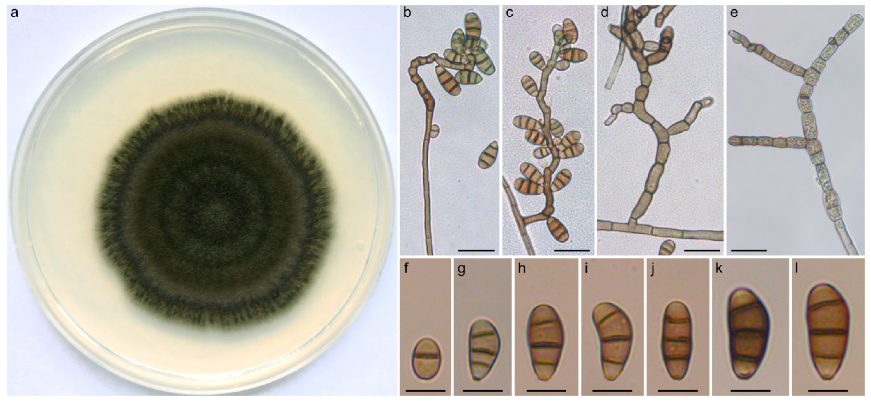
2.3. Taxonomy and Related Information

Curvularia coimbatorensis N. Kiss & S. Kocsubé sp. nov. (Figure 2). MycoBank accession number: MB 833656. The etymology is referring to the city in Tamil Nadu, South India where the type strain was isolated.
Vegetative hyphae septate, subhyaline to brown, branched, smooth, 3–4 µm in width. Colonies on PDA reaching approximately 4–6 cm in diameter after 7 days at 25 °C, surface funiculose, margin fimbriate, olivaceous black to olivaceous grey, velutinous with sparse aerial mycelium. Conidiophores erect, often branched, in most cases uniformly brown, sometimes pale brown at apex, seminematous, septate, flexuous, in most cases geniculate towards the apex, up to 210 µm long, 3–4 µm wide, basal cells sometimes swollen. Conidiogenous cells integrated, terminal, or intercalary with sympodial proliferation, smooth, brown, mono- or polytretic. Chlamydospores not observed. Conidia ellipsoidal to clavate to obovoid, asymmetrical with paler end cells, usually curved at the third cell from the base, (13-)16–18(-23) × (7-)8–9(-10) µm, 3-distoseptate, hila slightly protuberant, thickened and darkened.
Specimens examined: India, Coimbatore, human corneal scraping from corneal ulcer, 2012, (holotype: freeze dried culture specimen in the Szeged Microbiological Collection (SZMC) at the Department of Microbiology, Faculty of Science and Informatics, University of Szeged, Hungary, SZMC 22225, includes ex-type culture).
Curvularia tamilnaduensis N. Kiss & S. Kocsubé sp. nov. (Figure 3). MycoBank accession number: MB 833657. The etymology is referring to the state of South India where the type strain and the other two examined strains were isolated.
Vegetative hyphae septate, subhyaline to brown, branched, smooth walled, but often heavily asperulate, 2–3 µm in width. Colonies on PDA reaching approximately 6–7 cm in diameter after 7 days at 25 °C, surface lanose, aerial mycelium abundant, margin fimbriate, olivaceous green. Conidiophores erect, usually unbranched, in most cases uniformly brown, sometimes with paler tip, seminematous, septate, slightly flexuous, rarely geniculate towards the apex, up to 125 µm long, 2.5–4 µm wide. Conidiogenous cells integrated, terminal or intercalary, smooth, pale brown to brown, mono- or polytretic, proliferating sympodially. Chlamydospores present, subglobose, terminal and intercalary, 8–22 µm in diameter. Conidia ellipsoidal to clavate to obovoid, asymmetrical with paler basal and apical cells, usually curved at the third cell from the base which is darker than the other cells, (15-)20–23(-28) × (7-)8–10(-11) µm, (2-)3-distoseptate with non-protuberant, thickened, and darkened hila.
Specimens examined: India, Coimbatore, human corneal scraping from corneal ulcer, 2013, (holotype: freeze dried culture specimen in the Szeged Microbiological Collection (SZMC) at the Department of Microbiology, Faculty of Science and Informatics, University of Szeged, Hungary, SZMC 22226, includes ex-type culture); India, Coimbatore, human corneal scraping from corneal ulcer, 2011, (SZMC 26758); India, Coimbatore, human corneal scraping from corneal ulcer, 2011–2013, (SZMC 26759).

2.4. Antifungal Susceptibilities of Curvularia Strains Isolated from Fungal Keratitis

The minimum inhibitory concentrations (MIC) of nine antifungal agents towards C. coimbatorensis SZMC 22225, C. tamilnaduensis SZMC 22226, SZMC 26758, and SZMC 26759, as well as the type strains of C. australiensis (CBS 172.57), C. hawaiiensis (CBS 173.57), and C. spicifera (CBS 274.52) are shown in Table 3. The MIC of natamycin was 2 µg mL−1 for both new species and all other strains tested, while substantial differences between them could be observed in the case of clotrimazole, econazole, miconazole, and terbinafine, with the type strain of C. coimbatorensis having 4, 8, 4, and 4–8 times higher values, respectively. Among the tested isolates, the type strain of C. spicifera proved to be the less susceptible to clotrimazole, econazole, fluconazole, ketoconazole, and miconazole. Notable strain-to-strain variations between the C. tamilnaduensis strains could be observed only in the case of itraconazole and ketoconazole with detected MIC ranges of 0.03–0.25 and 0.06–0.25, respectively.

3. Discussion

The phylogenetic tree obtained from the concatenated dataset of three loci presents an update about the phylogeny of Curvularia, which is mostly in agreement with the recently published phylogenies of this genus (Figure 1). C. ischaemi formed a clade with C. coicis, which is in contradiction with the results of Tan et al. [8] and Tibpromma et al. [27], where C. ischaemi formed a sister clade to C. gladioli, but in agreement with the phylogram obtained by Madrid et al. [7] and Manamgoda et al. [28]. Our analysis placed C. perotidis as a sister clade to C. australiensis, however, other studies [7,8,27,29] suggested that this species is closer to C. spicifera. The placement of C. variabilis was also different from previously published articles [8,29]. According to the analyses of Tan et al. [8] and Marin-Felix et al. [29], C. variabilis forms a clade with C. hawaiiensis, C. nodosa, C. dactyloctenicola, and C. beasleyi, however, in this study we found C. variabilis as a sister clade of C. tsudae and C. mebaldsii. The same authors found C. tripogonis, C. pseudorobusta, C. robusta, C. alcornii, C. protuberata, and C. inaequalis as members of two distinct monophyletic clades, while our results indicate that these species are closely related and paraphyletic, however, none of the topologies have strong statistical supports. The observed slight differences between the previous inferences and our analyses did not affect the validity of any of the previously described species, and some of them might be the result of the slightly broader taxon sampling.
One of the newly described species, C. coimbatorensis is only known from the type specimen isolated from corneal ulcer. Phylogenetic analysis based on three loci placed C. coimbatorensis as a sister clade to the other newly described species C. tamilnaduensis. The two species are closely related, but can be distinguished by tef1a, gpdh, and ITS sequences, with percentage identities of 99%, 98%, and 99%, respectively. C. petersonii [8] is also closely related and can be distinguished by all three loci (98% in tef1a, 93% in gpdh and 96% in ITS). C. coimbatorensis differs from C. tamilnaduensis in colony morphology, the lack of chlamydospores, and the size of conidia. C. petersonii is very similar in colony morphology, however, has significantly shorter (up to 110 µm) and only slightly geniculate conidiophores bearing narrower (5-)5.5–6(-7) conidia [8]. C. coimbatorensis has longer conidiophores.
The phylogenetic analysis based on three loci placed the other newly described species, C. tamilnaduensis as a sister clade to the recently described species C. petersonii. C. tamilnaduensis can be reliably distinguished from the ex-type of C. petersonii by tef1a, gpdh and ITS sequences with percentage identities of 99%, 95%, and 96%, respectively. The two species also differ by morphology, as C. petersonii has not been reported to produce chlamydospores and has different conidial dimensions (17–19 × 5.5–6) [8]. C. americana [7] and C. verruculosa [30] are also related species with considerable amount of genetic distances and none of these species have been reported before to have chlamydopores. C. americana has 4(-5)-distoseptate and wider (7–15 µm) conidia, while C. verruculosa has mostly 3-distoseptate conidia, but also wider (12–17 µm) than those of C. tamilnaduensis.
The antifungal susceptibilities of the examined strains of C. coimbatorensis and C. tamilnaduensis to amphotericin B, clotrimazole, econazole, fluconazole, itraconazole, ketoconazole, miconazole, natamycin, and terbinafine were within the MIC ranges reported for other clinically relevant Curvularia species in the study of Guarro et al. [11] and the review of Krizsán et al. [3]. The type strain of C. coimbatorensis proved to be less susceptible than the strains of C. tamilnaduensis to all antifungals except for natamycin. For itraconazole and ketoconazole our results are in agreement with the study of Guarro et al. [11], who reported that amphotericin B, itraconazole, miconazole and ketoconazole are highly effective against a series of Curvularia species known from fungal keratitis (C. brachyspora, C. clavata, C. geniculata, C. lunata, C. pallescens, C. senegalensis, and C. verruculosa).

4. Materials and Methods

4.1. Curvularia Strains, Culture Conditions, and Morphological Examination

The Curvularia strains involved in this study derived from corneal scrapings from fungal corneal ulcers of keratitis patients attending the Aravind Eye Hospital and Postgraduate Institute of Ophthalmology, Coimbatore, India. All cases were initially screened by experienced ophthalmologists, and the corneal scrapings were collected following the clinical diagnosis of fungal keratitis. The samples were initially processed microbiologically for the isolation of the causative agents as described earlier [31]. The corneal scrapings of all patients were subjected to Gram stain, Giemsa stain, and 10% KOH wet mount. Culture methods involved direct inoculation of specimens onto 5% sheep blood agar, chocolate agar, non-nutrient agar, potato dextrose agar, thioglycolate broth, and brain–heart infusion broth. The microbial cultures were considered positive only if the growth of the same organism was demonstrated on two or more solid media, or there was confluent growth at the site of inoculation on one solid medium with consistent direct microscopic findings. The isolates were deposited in the Szeged Microbiology Collection (SZMC, Szeged, Hungary) under the accession numbers SZMC 22225, SZMC 22226, SZMC 26758, and SZMC 26759. Colony morphology of the isolates was examined on PDA (BioLab, Budapest, Hungary) medium after 7 days of incubation at 25 °C under normal day/night light conditions. Micromorphological characters were examined with a Leica DMI 4000B (Leica, Wetzlar, Germany) microscope equipped with a Leica DFC 295 camera. Microscopic features were examined in lactic acid (100% v/v) on glass slides. Conidiophores were studied in the same mounting fluid with the transparent tape method. Conidiophores and conidia were measured using the software ImageJ v2.52a (National Institute of Mental Health, Bethesda, MD, USA). Size ranges of the conidia were derived from 50 measurements. Lengths and widths are given as (minimum value) mean size minus SD-mean size plus SD (maximum value).

4.2. DNA Extraction, Amplification, Sequencing, and Phylogenetic Analysis

Genomic DNA was isolated from the examined Curvularia strains SZMC 22225, SZMC 22226, SZMC 26758, and SZMC 26759 with the Masterpure™ Yeast DNA Purification Kit (Epicentre Biotechnologies, Madison, WI, USA) according to the manufacturer’s instructions. Fragments of tef1a and gpdh were amplified as described previously [5,32,33]. The ITS region of the ribosomal RNA gene cluster was amplified according to White et al. [34]. Sequencing of the amplicons was carried out on a 3500 Genetic Analyzer (Thermo Fisher Scientific, Waltham, MA, USA) by the sequencing service of the Biological Research Centre, Szeged, Hungary. Resulting sequences were deposited in the GenBank Nucleotide database (www.ncbi.nlm.nih.gov) under the accession numbers shown in Table 2.
Sequences of the four clinical isolates were aligned with publicly available sequences of 108 previously described Curvularia species, as well as Bipolaris maydis as the outgroup (Table 2). Phylogenetic analyses were conducted using three loci (tef1α, gpdh and ITS). Sequences of all three loci were aligned with the phylogeny-aware sequence alignment tool Canopy v0.1.4 using RAxML as tree estimator and PRANK [35] with the -F option as the aligner with 10 iterations and seed decomposition strategy. Alignments of the three loci were concatenated and partitioned by region. The tef1α sequences formed one partition while in the case of gpdh sequences the dataset was partitioned to exons and introns. The ITS dataset was divided to rDNA and ITS1-ITS2 regions. Alignments of gpdh and ITS datasets contained high number of indels with important phylogenetic signal, therefore gaps were coded as absence/presence characters by SequenceMatrix v1.8 [36] using the simple indel coding algorithm [37]. The two indel matrices were concatenated and added as a single partition to the dataset. Maximum likelihood analysis was performed using RAxML-NG v0.9.0 [38] under the GTR model with gamma-distributed rate heterogeneity using empirical base frequencies. As indel-based datasets do not contain constant sites, the ascertainment bias correction described by Lewis [39] was used for this partition. Statistical support of the best ML tree was obtained with 1000 thorough bootstrap replicates.

4.3. Antifungal Susceptibility Testing

In vitro antifungal susceptibility tests were carried out according to the CLSI M38-A2 broth microdilution method [40]. Nine antifungal agents: amphotericin B, clotrimazole, econazole, fluconazole, itraconazole, ketoconazole, miconazole, natamycin and terbinafine (Sigma-Aldrich, Budapest, Hungary) were examined. Microtiter plates were incubated at 35 °C for 72 h. Plates were evaluated both spectrophotometrically with a Spectrostar Nano microplate reader (BMG Labtech, Ortenberg, Germany) and by visual examination.

5. Conclusions

The present study demonstrates, that although the phylogeny of the genus Curvularia is resolved and well established, further expansion can be expected both in the list of described Curvularia species and in the known spectrum of clinically relevant members of the genus. The collection of further keratitis isolates from the genus Curvularia and gaining data about their antifungal susceptibilities are therefore tasks of increasing importance. Furthermore, comparing the infectivity of various Curvularia species causing keratitis—including the recently described ones—in animal keratitis models would be an intriguing topic for future research.

Author Contributions

Conceptualization, S.K., L.K., T.P. and C.V.; methodology, N.K., A.M., M.H. and S.K.; software, S.K.; validation, R.R., P.M., M.H., C.V., M.V. and S.K.; formal analysis, K.K., T.P. and M.V.; investigation, N.K., A.M., P.M., K.K., M.H., M.V. and S.K.; resources, R.R., P.M., A.M., T.P. and C.V.; data curation, N.K., S.K., K.K., L.K. and M.H.; writing—original draft preparation, N.K., S.K., L.K.; writing—review and editing, N.K., S.K., L.K., P.M., T.P. and C.V.; visualization, N.K., M.H. and S.K.; supervision, S.K.; project administration, S.K., T.P., L.K., and P.M.; funding acquisition, S.K., T.P., L.K. and P.M. All authors have read and agreed to the published version of the manuscript.

Funding

This research was funded by grants NKFI PD-116609 (National Research, Development and Innovation Office, Hungary), GINOP-2.3.2-15-2016-00035 (Széchenyi 2020 Programme) and also supported by the COST action HUPLANTcontrol (Control of Human Pathogenic Micro-organisms in Plant Production Systems, CA16110). LK is grantee of the János Bolyai Research Scholarship (Hungarian Academy of Sciences) and the Bolyai Plus Scholarship (New National Excellence Programme). TP and MH are supported by the grants LP2016-8/2016 and by the FIKP program (TUDFO/4738-1/2019 ITM) of the Ministry of Human Capacities.

Acknowledgments

The authors wish to thank Venkatapathy Narendran (Aravind Eye Hospital and Postgraduate Institute of Ophthalmology, Coimbatore, Tamil Nadu, India), Coimbatore Subramanian Shobana (Department of Microbiology, PSG College of Arts and Science, Coimbatore, Tamil Nadu, India) and Kanesan Panneer Selvam (Department of Microbiology, M.R Government Arts College, Mannargudi, Tamil Nadu, India) for constantly supporting the research efforts on fungal keratitis within the frames of the Indo-Hungarian Fungal Keratitis Research Group.

Conflicts of Interest

The authors declare no conflict of interest.

References

  1. Bengyella, L.; Iftikhar, S.; Nawaz, K.; Fonmboh, D.J.; Yekwa, E.L.; Jones, R.C.; Njanu, Y.M.T.; Roy, P. Biotechnological application of endophytic filamentous Bipolaris and Curvularia: A review on bioeconomy impact. World J. Microbiol. Biotechnol. 2019, 35, 69. [Google Scholar] [CrossRef] [PubMed]
  2. Kusai, N.A.; Azmi, M.M.Z.; Zulkifly, S.; Yusof, M.T.; Zainudin, N.A.I.M. Morphological and molecular characterization of Curvularia and related species associated with leaf spot disease of rice in Peninsular Malaysia. Rend. Lincei Sci. Fis. Nat. 2016, 27, 205–214. [Google Scholar] [CrossRef]
  3. Krizsán, K.; Papp, T.; Manikandan, P.; Shobana, C.S.; Chandrasekaran, M.; Vágvölgyi, C.; Kredics, L. Clinical Importance of the Genus Curvularia. In Medical Mycology: Current Trends and Future Prospects; Razzaghi-Abyaneh, M., Shams-Ghahfarokhi, M., Rai, M., Eds.; CRC Press: Boca Raton, FL, USA, 2016; pp. 147–204. [Google Scholar] [CrossRef]
  4. Yanagihara, M.; Kawasaki, M.; Ishizaki, H.; Anzaw, K.; Udagawa, S.; Mochizuki, T.; Sato, Y.; Tachikawa, N.; Hanakawa, H. Tiny keratotic brown lesions on the interdigital web between the toes of a healthy man caused by Curvularia species infection and a review of cutaneous Curvularia infections. Mycoscience 2010, 51, 224–233. [Google Scholar] [CrossRef]
  5. Manamgoda, D.S.; Cai, L.; McKenzie, E.H.C.; Crous, P.W.; Madrid, H.; Chukeatirote, E.; Shivas, R.G.; Tan, Y.P.; Hyde, K.D. A phylogenetic and taxonomic re-evaluation of the Bipolaris-Cochliobolus-Curvularia complex. Fungal Divers. 2012, 56, 131–144. [Google Scholar] [CrossRef]
  6. Paredes, K.; Capilla, J.; Sutton, D.A.; Mayayo, E.; Fothergill, A.W.; Guarro, J. Virulence of Curvularia in a murine model. Mycoses 2013, 56, 512–515. [Google Scholar] [CrossRef] [PubMed]
  7. Madrid, H.; da Cunha, K.C.; Gené, J.; Dijksterhuis, J.; Cano, J.; Sutton, D.A.; Guarro, J.; Crous, P.W. Novel Curvularia species from clinical specimens. Persoonia 2014, 33, 48–60. [Google Scholar] [CrossRef] [PubMed] [Green Version]
  8. Tan, Y.P.; Crous, P.W.; Shivas, R.G. Cryptic species of Curvularia in the culture collection of the Queensland Plant Pathology Herbarium. MycoKeys 2018, 35, 1–25. [Google Scholar] [CrossRef] [PubMed] [Green Version]
  9. Marcus, L.; Vismer, H.F.; van der Hoven, H.J.; Gove, E.; Meewes, P. Mycotic keratitis caused by Curvularia brachyspora (Boedjin). A report of the first case. Mycopathologia 1992, 119, 29–33. [Google Scholar] [CrossRef] [PubMed]
  10. Travis, W.D.; Kwon-Chung, K.J.; Kleiner, D.E.; Geber, A.; Lawson, W.; Pass, H.I.; Henderson, D. Unusual aspects of allergic bronchopulmonary fungal disease: Report of two cases due to Curvularia organisms associated with allergic fungal sinusitis. Hum. Pathol. 1991, 22, 1240–1248. [Google Scholar] [CrossRef]
  11. Guarro, J.; Akiti, T.; Horta, R.A.; Morizot Leite-Filho, L.A.; Gené, J.; Ferreira-Gomes, S.; Aguilar, C.; Ortoneda, M. Mycotic keratitis due to Curvularia senegalensis and in vitro antifungal susceptibilities of Curvularia spp. J. Clin. Microbiol. 1999, 37, 4170–4173. [Google Scholar] [PubMed]
  12. Fan, Y.M.; Huang, W.M.; Li, S.F.; Wu, G.F.; Li, W.; Chen, R.Y. Cutaneous phaeohyphomycosis of foot caused by Curvularia clavata. Mycoses 2009, 52, 544–546. [Google Scholar] [CrossRef] [PubMed]
  13. Vasikasin, V.; Nasomsong, W.; Srisuttiyakorn, C.; Mitthamsiri, W.; Oer-Areemitr, N.; Changpradub, D. Disseminated phaeohyphomycosis caused by Curvularia tuberculata in a previously healthy man. Mycopathologia 2019, 184, 321–325. [Google Scholar] [CrossRef]
  14. Pimentel, J.D.; Mahadevan, K.; Woodgyer, A.; Sigler, L.; Gibas, C.; Harris, O.C.; Lupino, M.; Athan, E. Peritonitis due to Curvularia inaequalis in an elderly patient undergoing peritoneal dialysis and a review of six cases of peritonitis associated with other Curvularia spp. J. Clin. Microbiol. 2005, 43, 4288–4292. [Google Scholar] [CrossRef] [PubMed] [Green Version]
  15. Posteraro, B.; Scarano, E.; La Sorda, M.; Torelli, R.; De Corso, E.; Mulé, A.; Paludetti, G.; Fadda, G.; Sanguinetti, M. Eosinophilic fungal rhinosinusitis due to the unusual pathogen Curvularia inaequalis. Mycoses 2010, 53, 84–88. [Google Scholar] [CrossRef]
  16. Cruz, R.; Barthel, E.; Espinoza, J. Allergic rhinosinusitis by Curvularia inaequalis (Shear) Boedijn. Rev. Chil. Infectol. 2013, 30, 319–322. (In Spanish) [Google Scholar] [CrossRef] [PubMed]
  17. Flanagan, K.L.; Bryceson, A.D. Disseminated infection due to Bipolaris australiensis in a young immunocompetent man: Case report and review. Clin. Infect. Dis. 1997, 25, 311–313. [Google Scholar] [CrossRef] [PubMed] [Green Version]
  18. Filizzola, M.J.; Martinez, F.; Rauf, S.J. Phaeohyphomycosis of the central nervous system in immunocompetent hosts: Report of a case and review of the literature. Int. J. Infect. Dis. 2003, 7, 282–286. [Google Scholar] [CrossRef] [Green Version]
  19. Gadgil, N.; Kupferman, M.; Smitherman, S.; Fuller, G.N.; Rao, G. Curvularia brain abscess. J. Clin. Neurosci. 2013, 20, 173–175. [Google Scholar] [CrossRef]
  20. Vachharajani, T.J.; Zaman, F.; Latif, S.; Penn, R.; Abreo, K.D. Curvularia geniculata fungal peritonitis: A case report with review of literature. Int. Urol. Nephrol. 2005, 37, 781–784. [Google Scholar] [CrossRef]
  21. Diskin, C.J.; Stokes, T.J.; Dansby, L.M.; Radcliff, L.; Carter, T.B. Case report and review: Is the tendency for Curvularia tubular obstruction significant in pathogenesis? Perit. Dial. Int. 2008, 28, 678–679. [Google Scholar]
  22. Saenz, R.E.; Brown, W.D.; Sanders, C.V. Allergic bronchopulmonary disease caused by Bipolaris hawaiiensis presenting as a necrotizing pneumonia: Case report and review of literature. Am. J. Med. Sci. 2001, 321, 209–212. [Google Scholar] [CrossRef] [PubMed]
  23. Robson, A.M.; Craver, R.D. Curvularia urinary tract infection: A case report. Pediatr. Nephrol. 1994, 8, 83–84. [Google Scholar] [CrossRef] [PubMed]
  24. Safdar, A. Curvularia—Favorable response to oral itraconazole therapy in two patients with locally invasive phaeohyphomycosis. Clin. Microbiol. Infect. 2003, 9, 1219–1223. [Google Scholar] [CrossRef] [PubMed] [Green Version]
  25. Fernandez, M.; Noyola, D.E.; Rossmann, S.N.; Edwards, M.S. Cutaneous phaeohyphomycosis caused by Curvularia lunata and a review of Curvularia infections in pediatrics. Pediatr. Infect. Dis. J. 1999, 18, 727–731. [Google Scholar] [CrossRef] [PubMed]
  26. Dave, V.P.; Joseph, J.; Pathengay, A.; Pappuru, R.R.; Das, T. Clinical presentations, diagnosis, and management outcomes of Curvularia endophthalmitis and a review of literature. Retina 2018. [Google Scholar] [CrossRef] [PubMed]
  27. Tibpromma, S.; Hyde, K.D.; Bhat, J.D.; Mortimer, P.E.; Xu, J.; Promputtha, I.; Doilom, M.; Yang, J.B.; Tang, A.M.C.; Karunarathna, S.C. Identification of endophytic fungi from leaves of Pandanaceae based on their morphotypes and DNA sequence data from southern Thailand. MycoKeys 2018, 33, 25–67. [Google Scholar] [CrossRef] [PubMed]
  28. Manamgoda, D.S.; Rossman, A.Y.; Castlebury, L.A.; Crous, P.W.; Madrid, H.; Chukeatirote, E.; Hyde, K.D. The genus Bipolaris. Stud. Mycol. 2014, 79, 221–288. [Google Scholar] [CrossRef] [Green Version]
  29. Marin-Felix, Y.; Senwanna, C.; Cheewangkoon, R.; Crous, P.W. New species and records of Bipolaris and Curvularia from Thailand. Mycosphere 2017, 8, 1556–1574. [Google Scholar] [CrossRef]
  30. Sivanesan, A. Graminicolous species of Bipolaris, Curvularia, Drechslera, Exserohilum and their teleomorphs. Mycol. Pap. 1987, 158, 1–261. [Google Scholar]
  31. Mythili, A.; Babu Singh, Y.R.; Priya, R.; Shafeeq Hassan, A.; Manikandan, P.; Panneerselvam, K.; Narendran, V.; Shobana, C.S. In vitro and comparative study on the extracellular enzyme activity of molds isolated from keratomycosis and soil. Int. J. Ophthalmol. 2014, 7, 778–784. [Google Scholar] [CrossRef]
  32. Schoch, C.L.; Crous, P.W.; Groenewald, J.Z.; Boehm, E.W.A.; Burgess, T.I.; de Gruyter, J.; de Hoog, G.S.; Dixon, L.J.; Grube, M.; Gueidan, C.; et al. A class-wide phylogenetic assessment of Dothideomycetes. Stud. Mycol. 2009, 64, 1–15. [Google Scholar] [CrossRef] [PubMed]
  33. Berbee, M.; Pirseyedi, M.; Hubbard, S. Cochliobolus phylogenetics and the origin of known, highly virulent pathogens, inferred from ITS and glyceraldehyde-3-phosphate dehydrogenase gene sequences. Mycologia 1999, 91, 964–977. [Google Scholar] [CrossRef]
  34. White, T.J.; Bruns, T.D.; Lee, S.; Taylor, J.W. Amplification and direct sequencing of fungal ribosomal genes for phylogenetics. In PCR Protocols: A Guide to Methods and Applications; Innis, M.A., Gelfand, D.H., Sninsky, J.J., White, J.W., Eds.; Academic Press: San Diego, CA, USA, 1990; pp. 315–322. [Google Scholar]
  35. Löytynoja, A. Phylogeny-aware alignment with PRANK. Meth. Mol. Biol. 2014, 1079, 155–170. [Google Scholar] [CrossRef]
  36. Vaidya, G.; Lohman, D.J.; Meier, R. SequenceMatrix: Concatenation software for the fast assembly of multigene datasets with character set and codon information. Cladistics 2011, 27, 171–180. [Google Scholar] [CrossRef]
  37. Simmons, M.P.; Ochoterena, H. Gaps as characters in sequence-based phylogenetic analysis. Syst. Biol. 2000, 49, 369–381. [Google Scholar] [CrossRef] [Green Version]
  38. Kozlov, A.M.; Darriba, D.; Flouri, T.; Morel, B.; Stamatakis, A. RAxML-NG: A fast, scalable, and user-friendly tool for maximum likelihood phylogenetic inference. Bioinformatics 2019, 35, 4453–4455. [Google Scholar] [CrossRef] [Green Version]
  39. Lewis, P.O. A likelihood approach to estimating phylogeny from discrete morphological character data. Syst. Biol. 2001, 50, 913–925. [Google Scholar] [CrossRef] [Green Version]
  40. Clinical and Laboratory Standards Institute. Reference Method for Broth Dilution Antifungal Susceptibility Testing of Yeasts, 3rd ed.; Approved Standard, CLSI Document M27-A3; Clinical and Laboratory Standards Institute: Wayne, PA, USA, 2008. [Google Scholar]
Figure 1. Maximum likelihood phylogeny of the genus Curvularia inferred from the concatenated internal transcribed spacer (ITS), translation elongation factor 1-α (tef1a), and glyceraldehyde-3-phosphate dehydrogenase (gpdh) sequences. The isolates examined in this study are shown as the new species Curvularia tamilnaduensis and Curvularia coimbatorensis (highlighted in color). Sequences of the reference Curvularia strains were collected from the GenBank Nucleotide database (Table 1). Bootstrap support values greater than 60% are shown above the branches. Bipolaris maydis CBS 136.29 was used to root the tree. Abbreviations of culture collections: BRIP: Plant Pathology Herbarium, Queensland, Australia; CBS: Westerdijk Fungal Biodiversity Institute culture collection, The Netherlands; CPC: Cultures of Pedro Crous, housed at Westerdijk Fungal Biodiversity Institute; DAOM: Canadian National Mycological Herbarium, Ottawa, Canada; GUCC: Guizhou University Culture Collection, Guizhou, China; IMI: CABI Bioscience, Eggham, UK; IRAN: Iranian Fungal Culture Collection, Iranian Research Institute of Plant Protection, Tehran, Iran; KUMCC: Culture Collection of Kunming Institute of Botany, Kunming, China; MFLUCC: Mae Fah Luang Culture Collection, Chiang Rai, Thailand; SCUA: Collection of Fungal Cultures, Department of Plant Protection, Shahid Chamran University of Ahvaz, Iran; SZMC: Szeged Microbiology Collection, Szeged, Hungary; UTHSC: University of Tennessee Health Science Center, Memphis, USA. T: type strain.
Pathogens 09 00009 g001
Figure 2. Morphological features of Curvularia coimbatorensis SZMC 2225. (a) Colony morphology on PDA (potato dextrose agar) medium after 7 days at 25 °C; (b,c) conidiophores with septate conidia; (d) branching conidiophores; (e) swollen cells; (fl) septate conidia. Scale bars: (be) 20 µm; (fl) 10 µm.
Pathogens 09 00009 g002
Figure 3. Morphological features of Curvularia tamilnaduensis SZMC 2226. (a) Colony morphology on PDA medium after 7 days at 25 °C; (b) conidiophores with septate conidia; (c) subglobose intercalary chlamydospore; (df) septate conidia. Scale bars: (b,c) 20 µm; (di) 10 µm.
Pathogens 09 00009 g003
Table 1. Case details of the fungal keratitis infections.
StrainAgeSexClinical DiagnosisCorneal ScrapingTherapyOutcome
SZMC 2222580MaleFungal corneal ulcer11 July 2012NAT, ECZ, HTRLost to follow up after two visits
SZMC 2222666MaleFungal corneal ulcer2 March 2013NAT, ECZ, HTRLost to follow up after one visit
SZMC 2675840MaleFungal corneal ulcer21 March 2011NAT, ECZ, HTRLost to follow up after one visit
SZMC 26759NANAFungal corneal ulcerNANALost to follow up
NAT: natamycin (5%); ECZ: econazole (2%), HTR: homatropine (1%); NA: data not available.
Table 2. Sequences used for the phylogenetic analysis.
Curvularia SpeciesStrainGenBank Accession Number
ITStef1agpdh
Bipolaris maydisCBS 136.29 TKJ909780KM093794KM034846
Curvularia aeriaBRIP 61232bKX139029KU552155KU552162
Curvularia affinisCBS 154.34 TKJ909782KM196566KM230401
Curvularia ahvazensisSCUA-1bi TKJ415539MG428686MG428693
Curvularia akaiiCBS 317.86JX256420KM196569KM230402
Curvularia akaiiensisBRIP 16080 THE861833KJ415453KJ415407
Curvularia alcorniiMFLUCC 10-0703 TJX256424JX266589JX276433
Curvularia americanaUTHSC 08-3414 TKJ415540-HF565488
Curvularia asiaticaMFLUCC 10-0711 TKJ415541JX266593JX276436
Curvularia australiensisBRIP 12044 TKJ415542KJ415452KJ415406
Curvularia australisBRIP 12521 TMH414892KJ415451KJ415405
Curvularia bannoniiBRIP 16732 TMH414894KJ415450KJ415404
Curvularia beasleyiBRIP 10972 TMH414911MH433654MH433638
Curvularia beerburrumensisBRIP 12942 TKP400638MH433657MH433634
Curvularia boeremaeIMI 164633 TKJ415543-MH433641
Curvularia borreriaeMFLUCC 11-0422KJ922372KM196571KP419987
Curvularia bothriochloaeBRIP 12522 TKJ909765KJ415449KJ415403
Curvularia brachysporaCBS 186.50HG778984KM230405KM061784
Curvularia buchloësCBS 246.49 TMF490814KM196588KM061789
Curvularia carica-papayaeCBS 135941 THG779021-HG779146
Curvularia chiangmaiensisCPC 28829 TMH275055MF490857MF490836
Curvularia chlamydosporaUTHSC 07-2764 TKU552205-HG779151
Curvularia chonburiensisMFLUCC 16-0375 TMH414897-MH412747
Curvularia clavataBRIP 61680bAF081447KU552159KU552167
Curvularia coatesiaeBRIP 24261 TMH414898MH433659MH433636
Curvularia coicisCBS 192.29 TLT631357JN601006AF081410
Curvularia colbraniiBRIP 13066 TLT631310MH433660MH433642
Curvularia comoriensisCBS 110673KJ415544-LT715841
Curvularia crassiseptumCBS 503.90 THG778985-LT715882
Curvularia crustaceaBRIP 13524 TMF490815KJ415448KJ415402
Curvularia cymbopogonisCBS 419.78KJ415545-HG779129
Curvularia dactyloctenicolaCPC 28810 TLT631356MF490858MF490837
Curvularia dactylocteniiBRIP 12846 TJN192375KJ415447KJ415401
Curvularia deightoniiCBS 537.70MH414899-LT715839
Curvularia ellisiiCBS 193.62 THG778986JN601007JN600963
Curvularia eragrosticolaBRIP 12538 TKJ909781MH433661MH433643
Curvularia eragrostidisCBS 189.48HG778987-HG779154
Curvularia geniculataCBS 187.50JN192376KM230410KM083609
Curvularia gladioliCBS 210.79KJ415546-HG779123
Curvularia graminicolaBRIP 23186a TKJ415547JN601008JN600964
Curvularia harveyiBRIP 57412 TKJ415548KJ415446KJ415400
Curvularia hawaiiensisBRIP 11987 TKJ415549KJ415445KJ415399
Curvularia heteropogonicolaBRIP 14579 THG779011KJ415444KJ415398
Curvularia heteropogonisCBS 284.91 TJN192380JN601013JN600969
Curvularia hominisCBS 136985 TKJ922375-HG779106
Curvularia homomorphaCBS 156.60 THG778991JN601014JN600970
Curvularia inaequalisCBS 102.42 TMH861533KM196574KM061787
Curvularia intermediaCBS 334.64MH414900-HG779155
Curvularia ischaemiCBS 630.82 TMH855025-LT715790
Curvularia kenpeggiiBRIP 14530 TMH414901MH433662MH433644
Curvularia kusanoiCBS 137.29JX256429JN601016LT715862
Curvularia lamingtonensisBRIP 12259 TJF812154MH433663MH433645
Curvularia lunataCBS 730.96 TMH414902JX266596JX276441
Curvularia malinaCBS 131274 THE792934KR493095KP153179
Curvularia mebaldsiiBRIP 12900 TMF139088MH433664MH433647
Curvularia micropusCBS 127235KJ909770-LT715859
Curvularia microsporaGUCC6272 TMG846737MF139115MF139106
Curvularia miyakeiCBS 197.29 TKP400647KM196568KM083611
Curvularia mosaddeghiiIRAN 3131C TKJ415550MH392152MH392155
Curvularia muehlenbeckiaeCBS 144.63 TMH414910KM196578KP419996
Curvularia neergaardiiBRIP 12919 TKJ415551KJ415443KJ415397
Curvularia neoindicaIMI 129790 TMF490816MH433667MH433649
Curvularia nicotiaeBRIP 11983 TJN601033KJ415442KJ415396
Curvularia nodosaCPC 28800 TKP400650MF490859MF490838
Curvularia nodulosaCBS 160.58JN192384JN601019JN600975
Curvularia oryzaeCBS 169.53 TKJ922380KM196590KP645344
Curvularia ovariicolaCBS 470.90 TMH275056JN601020JN600976
Curvularia pallescensCBS 156.35 TKJ415552KM196570KM083606
Curvularia pandanicolaMFLUCC 15-0746 THG778995MH412763MH412748
Curvularia papendorfiiCBS 308.67 TMH414905KJ415441KJ415395
Curvularia perotidisCBS 350.90 TKY905678KM230407HG779138
Curvularia petersoniiBRIP 14642 TMH414906MH433668MH433650
Curvularia pisiCBS 190.48 TKJ415553KY905697KY905690
Curvularia platziiBRIP 27703b TKJ922373MH433669MH433651
Curvularia portulacaeBRIP 14541 TKJ922376KJ415440KJ415393
Curvularia prasadiiCBS 143.64 TMF490819KM230408KM061785
Curvularia protuberataCBS 376.65 THE861842KM196576KM083605
Curvularia pseudobrachysporaCPC 28808 THE861838MF490862MF490841
Curvularia pseudolunataUTHSC 09-2092 TJN192386-HF565459
Curvularia pseudorobustaUTHSC 08-3458MH414907-HF565476
Curvularia raveneliiBRIP 13165 TKJ415555JN601024JN600978
Curvularia reesiiBRIP 4358 TKJ909783MH433670MH433637
Curvularia richardiaeBRIP 4371 TKX139030KJ415438KJ415391
Curvularia robustaCBS 624.68 TKJ415556KM196577KM083613
Curvularia rouhaniiSCUA-2bi-2 THG779001MG428687MG428694
Curvularia ryleyiBRIP 12554 TKY905679KJ415437KJ415390
Curvularia senegalensisCBS 149.71KJ415558-HG779128
Curvularia soliCBS 222.96 TMH414904KY905698KY905691
Curvularia sorghinaBRIP 15900 TKP400655KJ415435KJ415388
Curvularia sp.BRIP 17068bKP400654MH433666MH433648
Curvularia sp.AR5117HE861826KP735698KP645349
Curvularia sp.MFLUCC 120177JN192387KP735697KP645348
Curvularia sp.UTHSC 8809MH414908-HF565477
Curvularia spiciferaCBS 274.52KJ909777JN601023JN600979
Curvularia sporobolicolaBRIP 23040b TMH275057MH433671MH433652
Curvularia subpapendorfiiCBS 656.74 THG779023KM196585KM061791
Curvularia thailandicumMFLUCC 15-0747 TJN192388MH412764MH412749
Curvularia trifoliiCBS 173.55KJ415559-HG779124
Curvularia tripogonisBRIP 12375 TKC424596JN601025JN600980
Curvularia tropicalisBRIP 14834 TJX256433KJ415434KJ415387
Curvularia tsudaeATCC 44764 THG779024KC503940KC747745
Curvularia tuberculateCBS 146.63 TMF490822JX266599JX276445
Curvularia uncinateCBS 221.52 THG779026-HG779134
Curvularia variabilisCPC 28815 TKP400652MF490865MF490844
Curvularia verruciformisCBS 537.75MH414909-HG779133
Curvularia verruculosaCBS 150.63MH275058KP735695KP645346
Curvularia warraberensisBRIP 14817 TAF071338MH433672MH433653
Curvularia xishuangbannaensisKUMCC 17-0185 TKJ909780MH412765MH412750
Curvularia gudauskasiiDAOM 165085KX139029KM093794AF081393
Curvularia tamilnaduensis sp. nov.SZMC 22226 T *MN628311MN628303MN628307
SZMC 26758 *MN628308MN628300MN628304
SZMC 26759 *MN628309MN628301MN628305
Curvularia coimbatorensis sp. nov.SZMC 22225 T *MN628310MN628302MN628306
T type strain; * Strains examined during the present study. Sequences derived from the present study are set in bold.
Table 3. Antifungal susceptibilities of the Curvularia coimbatorensis and Curvularia tamilnaduensis strains in comparison with the type strains of Curvularia australiensis, Curvularia hawaiiensis, and Curvularia spicifera determined by the CLSI (Clinical & Laboratory Standards Institute) broth microdilution method (minimum inhibitory concentrations (MIC) values in µg mL−1).
StrainAntifungal Agent
AMBCLTECNFLCITCKTCMCZNTMTRB
C. australiensis CBS 172.57 T0.250.250.125160.030.250.2520.25
C. hawaiiensis CBS 173.57 T0.250.060.0640.030.060.12520.25
C. spicifera CBS 274.52 T0.542>320.252221
C. coimbatorensis SZMC 22225 T0.50.51320.250.25121
C. tamilnaduensis SZMC 22226 T10.1250.12580.030.060.2520.25
C. tamilnaduensis SZMC 267580.50.1250.125160.030.250.2520.125
C. tamilnaduensis SZMC 2675910.1250.125160.250.250.2520.25
T: type strain; AMB: amphotericin B; CLT: clotrimazole; ECN: econazole; FLC: fluconazole; ITC: itraconazole; KTC: ketoconazole; MCZ: miconazole; NTM: natamycin; TRB: terbinafine.

