Article

Piper betel Compounds Piperidine, Eugenyl Acetate, and Chlorogenic Acid Are Broad-Spectrum Anti-Vibrio Compounds that Are Also Effective on MDR Strains of the Pathogen

by
Erika Acosta-Smith
1,2,
Nidia Leon-Sicairos
2,3,
Sandeep Tiwari
4,
Hector Flores-Villaseñor
2,
Adrian Canizalez-Roman
2,
Ranjith Kumavath
5,
Preetam Ghosh
6,
Vasco Azevedo
4 and
Debmalya Barh
7,*
1
Programa Regional del Noroeste para el Doctorado en Biotecnología-FCQB, Universidad Autónoma de Sinaloa, Avenida de las Américas y Josefa Ortiz (Ciudad Universitaria), Culiacán Sin. 80030, Mexico
2
CIASaP, Facultad de Medicina, Universidad Autónoma de Sinaloa. Cedros y Sauces, Fracc. Fresnos, Culiacán Sin. 80246, Mexico
3
Departamento de Investigación, Hospital Pediátrico de Sinaloa, Blvd. Constitución y Donato Guerra S/N, Almada, Culiacán Sin. 80200, Mexico
4
Laboratório de Genética Celular e Molecular, Programa de Pós-graduação em Bioinformática, Instituto de Ciências Biológicas (ICB), Universidade Federal de Minas Gerais, Av. Antonio Carlos 6627, Pampulha, Belo Horizonte, CEP 31270-901, Brazil
5
Department of Genomic Sciences, School of Biological Sciences, Central University of Kerala, Kasaragod 671121, India
6
Department of Computer Science, Virginia Commonwealth University, Richmond, VA 23284, USA
7
Centre for Genomics and Applied Gene Technology, Institute of Integrative Omics and Applied Biotechnology (IIOAB), Nonakuri, Purba Medinipur, West Bengal-721172, India
*
Author to whom correspondence should be addressed.
Pathogens 2019, 8(2), 64; https://doi.org/10.3390/pathogens8020064
Submission received: 17 March 2019 / Revised: 30 April 2019 / Accepted: 7 May 2019 / Published: 13 May 2019
(This article belongs to the Section Vaccines and Therapeutic Developments)

Abstract

:
The natural population of the aquatic environment supports a diverse aquatic biota and a robust seafood industry. However, this environment also provides an appropriate niche for the growth of pathogenic bacteria that cause problems for human health. For example, species of the genus Vibrio inhabit marine and estuarine environments. This genus includes species that are pathogenic to aquaculture, invertebrates, and humans. In humans, they can cause prominent diseases like gastroenteritis, wound infections, and septicemia. The increased number of multidrug resistant (MDR) Vibrio strains has drawn the attention of the scientific community to develop new broad-spectrum antibiotics. Hence, in this paper we report the bactericidal effects of compounds derived from Piper betel plants: piperidine, chlorogenic acid, and eugenyl acetate, against various strains of Vibrio species. The different MIC90 values were approximately in a range of 2–6 mg/mL, 5–16 mg/mL, 5–20 mg/mL, and 30–80 mg/mL, for piperidine, chlorogenic acid, and eugenyl acetate, respectively. Piperidine showed the best anti-Vibrio effect against the five Vibrio species tested. Interestingly, combinations of sub-inhibitory concentrations of piperidine, chlorogenic acid, and eugenyl acetate showed inhibitory effects in the Vibrio strains. Furthermore, these compounds showed synergism or partial synergism effects against MDR strains of the Vibrio species when they were incubated with antibiotics (ampicillin and chloramphenicol).

1. Introduction

Vibrio is a heterogeneous and polyphyletic genus with Gram-negative, curved-rod shaped, motile bacteria with high affinity for salinity and temperatures, fluctuating from 10 °C to 30 °C [1,2]. Several species of the genus are associated with infections like gastroenteritis, wound infection, and septicemia [3]. Vibrio cholerae O1 (classical O1 serotype strain), is the most important species responsible for cholera epidemics, and the species non-O1 serogroup V. cholerae O139 is the causative agent for gastroenteritis and extra-intestinal infections [4,5]. V. cholerae non-O1 also causes septicemia that leads to death [6,7]. V. cholerae serogroups Inaba and Ogawa belong to the classical and El Tor biotypes, and both serogroups were reported to be involved in cholera outbreaks [8,9,10]. Vibrio parahaemolyticus, a seawater bacterium, infects human through wounds or raw sea fish or seafood consumption, and causes inflammation of small intestine, diarrhea, cramping, and septicemia [11,12,13]. Vibrio alginolyticus and Vibrio furnissii are also seawater bacteria that cause superficial wound and ear infections (otitis media and otitis externa) [14] and diarrhea, respectively [15]. Vibrio fluvialis is uniquely associated with diarrhea outbreaks [16], and in rare cases, causes extra-intestinal infections such as hemorrhagic cellulitis with bacteremia, cerebritis, and peritonitis [17]. Although the infections caused by Vibrio species can be treated with various antibiotics, the multidrug resistant (MDR) strains emphasize the need to search for new broad-spectrum antibiotics to tackle the pathogens.
Historically, the shrubberies of Piper betel plant (family: Piperaceae) are used in Ayurvedic and folk medicine [18]. The crude extract is reported to be gastro-protective [19], with antimicrobial [20], anti-fungal [21], and anti-inflammatory [22] properties. However, the exact mechanism of the active compounds extracted from the betel leaf is still unclear.
Our group reported a set of seven compounds derived from the leaves of Piper betel plant (piperdardine, pinoresinol, guineensine, dehydropipernonaline, piperrolein B, eugenyl acetate, and chlorogenic acid), where some of these were previously proposed to be highly effective against a broad spectrum of Vibrio species. In a preliminary experimental work, 60 mM of piperdardine was shown to exhibit an equal growth inhibition effect to 100 µg/mL of chloramphenicol in V. cholerae O1 Inaba [23].
Here, we further report four Piper betel compounds (piperidine, eugenyl acetate, chlorogenic acid, and pinoresinol) that are effective against V. cholerae non-O1, V. cholerae O1 Ogawa, V. cholerae O1 Inaba, V. parahaemolyticus, V. alginolyticus, V. furnissii, and V. fluvialis. We also show that these Piper betel compounds are equally effective against MDR strains of the Vibrio species by acting in combination or in synergy with antibiotics [24].

2. Material and Methods

2.1. Bacterial Culture and Antibacterial Agents

Vibrio cholerae non-O1 Ogawa and Vibrio cholerae O1 Inaba were obtained from the National Institute of Diagnosis and Epidemiologic Reference (INDRE), Mexico. Vibrio parahaemolyticus TX2103 (CAIM 729) was obtained from the Collection of Aquatic Important Microorganisms (www.ciad.mx/caim), and had been isolated during the 1998 Texas (USA) outbreak [25,26]. Vibrio cholerae non-O1 (UIR22F), Vibrio alginolyticus (UIR22G1), Vibrio furnissii (UIR16A2), Vibrio fluvialis (UIR16A1), and Vibrio parahaemolyticus MDR (UIR10C4) strains were isolated by us [11,27,28,29,30]. The antimicrobial resistance (MDR) patterns of Vibrio spp. are given in Table 1A. Other authors have reported similar MDR patterns (Table 1B) [31,32,33,34,35,36,37]. Bacteria were cultured in Mueller–Hinton (MH) broth and maintained for 16–18 h in an incubating shaker at 37 °C to reach the logarithmic phase. The antibacterial compounds used in this study were purchased from Sigma Aldrich (St. Louis, MI, USA) and were dissolved in water (piperidine, 104094) or with 0.05% Tween 80 and 10% DMSO (chlorogenic acid, C3878; or eugenyl acetate, W246905; or pinoresinol, 40574). This solution (0.05% Tween 80 and 10% DMSO plus each compound) did not affect the bacterial cultures by itself during the experiments (not shown).

2.2. Evaluation of the Antibacterial Activity of Compounds on Vibrio spp.

Exponential phase bacteria were adjusted to an absorbance of 0.1 at 600 nm (approximately 107 CFU/mL). The bactericidal activity of compounds was tested following two well-established methods.
(1) Disk diffusion method; here 100 μL of the suspension of each Vibrio strain (containing 107 CFU/mL) prepared from an overnight culture were used to seed each prepared and dried Mueller–Hinton agar plate. Then, commercial Sensi-DisksTM (10 µg/mL ampicillin and 30 µg/mL chloramphenicol, purchased from BD) or sterile paper disc of 6 mm (filter paper mini Trans-Blot Bio-Rad Cat. No. 1703932) impregnated with the compound (piperidine in H2O, at concentrations of 1, 3, 7, and 10 mg/disk), were placed in MH agar plates, and then incubated at 37 °C for 24 h. Negative control was also prepared by impregnating paper disc with solvent (H2O) used to dissolve the piperidine. Finally, the antimicrobial activity was evaluated by measuring the inhibition diameter zone around the tested Vibrio strain [38]. The mean of the inhibition diameter zones for each antibacterial compound was determined as the average of three independent experiments.
(2) For colony-forming units (CFU/mL) assay [39], approximately 105 CFU/mL of bacterial suspensions were re-suspended in tubes containing MH broth either alone (control of bacterial growth) or with standard drug (30 µg/mL of chlorampenicol, control of bacterial inhibition), or with piperidine (1, 2, 3, 10 mg/mL dissolved in H2O), or with chlorogenic acid (5, 10, 15, 20, 25, or 30 mg/mL dissolved in 0.05% Tween 80 and 10% DMSO) or with eugenyl acetate (dissolved in 0.05% Tween 80 and 10% DMSO), or with pinoresinol (20, 30, 40, or 50 mg/mL dissolved in 0.05% Tween 80 and 10% DMSO), or with the solvents used (H2O or 0.05% Tween 80 and 10% DMSO). Tubes were maintained for 0, 20, 40, 60, and 80 min in an incubation shaker at 37 °C. The number of CFUs of viable bacteria was counted each time after inoculating the serial 10-fold dilutions from BHI broth onto BHI agar plates.

2.3. Determination of Compound Minimum Inhibitory Concentrations against Vibrio spp.

The minimum inhibitory concentration (MIC) of compounds was determined by agar dilution method as described by the National Committee for Clinical Laboratory Standards (NCCLS). Briefly, the solution of compounds in serial two-fold concentrations were added into agar as follows: piperidine (0.5–16 mg/mL), eugenyl acetate (0.5–30 mg/mL), chlorogenic acid (0.5–40 mg/mL), and pinoresinol (0.5–50 mg/mL). The MIC was defined and the bacterial inocula were prepared as previously described, except that the final inocula of approximately 104 CFU/spot of bacterial inoculum were applied to the plates and then incubated at 37 °C for 24 h. Quality control analyses of the methods were regularly performed for each test. The MIC for each compound also was calculated by the Disk diffusion method as has been described before. The MIC was recorded as the lowest concentration of antimicrobial agent with no visible growth.

2.4. Determination of the Inhibition Parameters of Piper betel Compounds in Combination with Compounds or Standard Drugs on Vibrio spp.

Various combinations of compounds plus Piper betel compounds, or compounds plus standard drugs were tested by the disk diffusion method and colony-forming unit assay (CFU/mL), as mentioned above. The concentrations of piperidine tested ranged from 0.5 to 16 mg/mL and for chlorogenic acid and eugenyl acetate from 0.5 to 40 mg/mL, and for the standard drugs chloramphenicol and ampicillin from 10, 15, and 30 µg/mL or 5, 10, and 50 µg/mL, respectively. The combinations for each strain were tested in triplicates.

2.5. Determination of the Synergistic Activity of Compounds plus Antibiotics

To examine the effects of combinations of the different compounds on bacterial survival, or the synergistic activity of the compounds in combination with antibiotics, we used the checkerboard broth dilution method to determine the fractional inhibitory concentration index (FIC) [40]. This index is calculated according to the following formula: FIC of drug A (FIC A) = (MIC of drug A in combination)/(MIC of A); FIC of drug B (FIC B) = (MIC of drug B in combination)/(MIC of B).

2.6. Statistical Analysis

All experiments were repeated at least twice in triplicate for confirmation of the results. Data were expressed as mean ± SEM, where SEM is the standard error of the mean. Data were compared using two-tailed Student’s t-test, and p < 0.05 was considered statistically significant.
Statistical analysis on synergy: The calculated FIC index was interpreted as synergistic (≤0.5), partial synergy (>0.5 but <1), indifferent (>1 but <4.0), or antagonistic (≥4.0) [40,41].

3. Results and Discussion

3.1. Bactericidal Activity of Piperidine on Vibrio spp.

Piperidine exhibited bactericidal activity against all Vibrio species tested (Figure 1). By visualizing the inhibition zones, the best effects were on V. cholerae O1 Ogawa (B), V. furnisii (F), V. cholerae non-O1 (A), V. parahaemolyticus TX2103 (D), and V. alginolyticus (E). The inhibition zone was moderate in V. parahaemolyticus MDR (H), V. fluvialis (G), and V. cholerae O1 Inaba (C). The bactericidal effect of piperidine appeared during the first 24 hours of incubation and was concentration-dependent (Figure 1 disks 1–3 that correspond to 3, 7, and 10 mg/mL of piperidine, respectively). The anti-Vibrio effect of piperidine was better than the antibiotic ampicillin for ampicillin non-resistant Vibrio strains V. cholerae O1 Ogawa (B), V. furnisii (Figure 1F), and V. fluvialis (G). Considering the MDR spectrum of the tested strains (Table 1A,B), we observed that piperidine was effective on MDR strains of V. cholerae O1 Ogawa (B), V. cholerae O1 Inaba (C), V. alginolyticus (E), and V. parahaemolyticus MDR (H).
Based on the disk diffusion method, we observed that 10 mg/mL of piperidine (disk 3) inhibited bacterial growth of V. parahaemolyticus TX2103, V. alginolyticus, V. furnissiii, and V. fluvialis strains, similar to a growth inhibitory effect of 30 µg/mL of chloramphenicol (Figure 1, A–H disk 6). Interestingly, these strains although susceptible to piperidine and chloramphenicol, were however resistant to 10–50 µg/mL of ampicillin, corroborating our previous determinations (Table 1). The compounds chlorogenic acid (10–30 mg/mL) and eugenyl acetate (10–40 mg/mL) also inhibited the growth of Vibrio spp. in similar conditions tested for the assays with piperidine (data not shown). However, intriguingly during the disk diffusion method the compounds chlorogenic and eugenyl acetate showed green and yellow pigmentation, respectively, on the inhibition zones (not shown). In this case, we decided to estimate the CFU/mL to corroborate the results explained above. By this method, we found that low concentration of the compounds were necessary to inhibit the growth of the Vibrio strains (Table 2), which was observed because this technique is more sensitive than the disk diffusion assay. The leaves of the Piper betel plant have long been in use in the local Indian system of medicine for its antioxidant and antimicrobial properties [42,43]. Some groups of researchers have reported the antimicrobial properties of Piper betel extracts [43,44]; however to the best of our knowledge, this is the first report of the antibacterial activity of Piper betel derivatives against Vibrio spp.

3.2. Determination of MICs against Vibrio spp.

All of the tested Piper betel compounds exhibited significant in vitro activity against approximately all Vibrio spp. In Table 2, the MICs of piperidine at which 90% of Vibrio spp. growth was inhibited (MICs90) were approximately in the range of 2–6 mg/mL, and those of chlorogenic acid were 5–16 mg/mL. The MICs90 of eugenyl acetate and pinoresinol were in the range of 5–20 mg/mL, and 30–80 mg/mL, respectively. Table 2 indicates results of only V. cholerae Inaba, V. parahaemolyticus TX 2103, V. parahaemolyticus O3:K6, V. furnisii (UIR16A2), and V. fluvialis (UIR16A1); however the compounds also affected other Vibrio spp. at the same concentrations (not shown).

3.3. Antibacterial Activity of Mixtures of Piper betel-Derived Compounds against Vibrio spp.

According to our observations, the compound piperidine exhibited the best antibacterial activity in all Vibrio spp. When V. cholera Inaba, V. parahaemolyticus TX 2103, V. parahaemolyticus O6:K6, V. furnisii, and V. fluvialis were incubated with piperidine, chlorogenic acid, and eugenyl acetate at their MICs (Table 3), the bacterial growth was inhibited during the initial 24 and 36 h. Interestingly, when sub-inhibitory concentrations of piperidine (0.5–2.0 mg/mL), chlorogenic acid (1.0–2.0 mg/mL), and eugenyl acetate (0.5–2.0 mg/mL) were combined, these three compounds (0.5–3.0 mg/mL) were able to inhibit the growth of all Vibrio spp. (Table 2). The inhibitory effect persisted for more than 24 h with no noticeable regrowth.

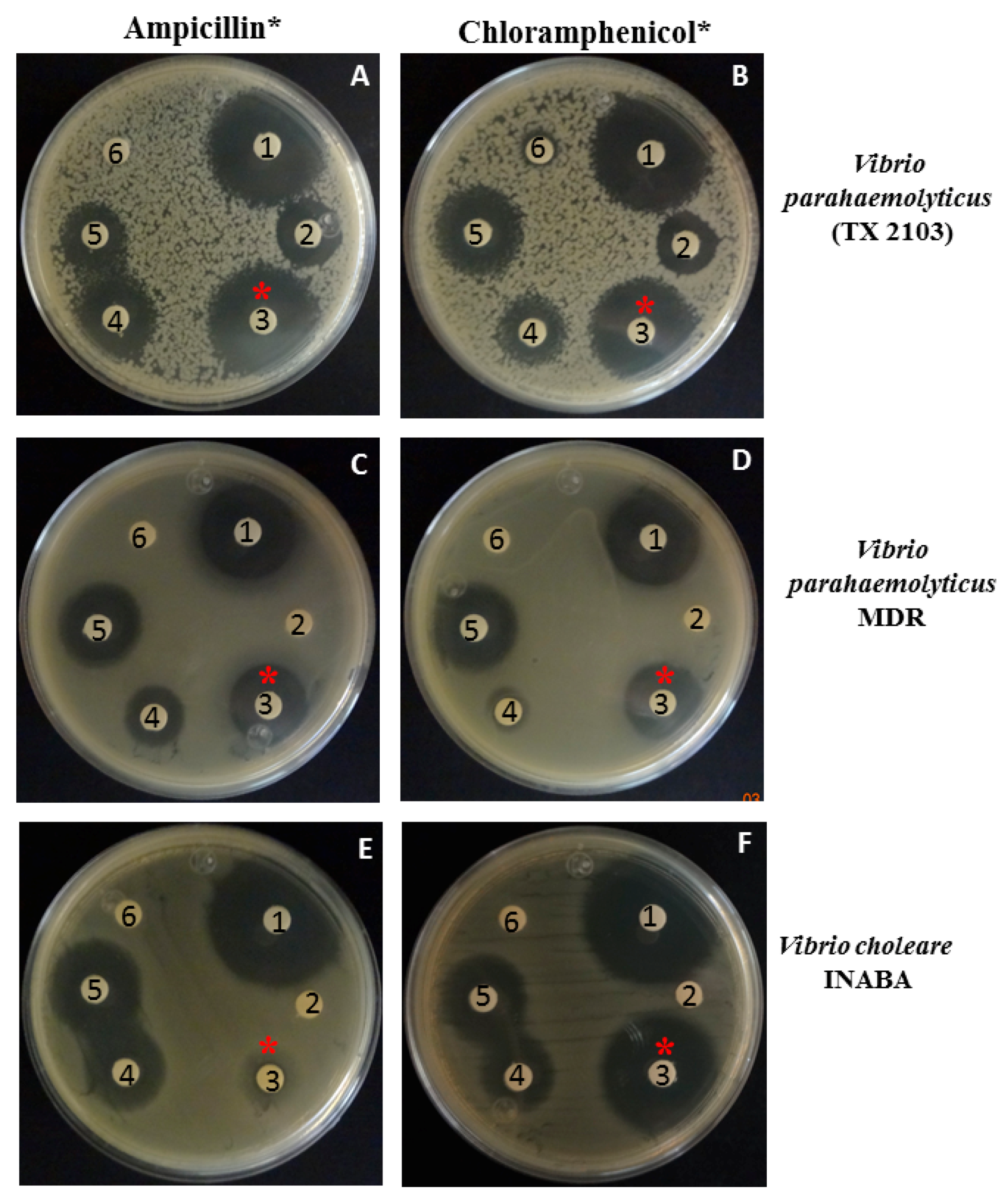
3.4. Antibacterial Activity of Piperidine in Combination with Ampicillin or Chloramphenicol against Vibrio spp.

The antibacterial effect of compounds mixed in combinations against Vibrio was also tested. In these experiments, we used combinations of compounds in Vibrio strains that were resistant to antibiotics, V. parahaemolyticus MDR, V. parahaemolyticus TX2103 and V. cholerae Inaba. In the results, (Figure 2), at concentrations of 1 and 3 mg/mL of piperidine (Panel A, C, and E: disks 4, and 5, respectively), an inhibition zone was observed (more visible at 4 mg/mL piperidine), but when 1 mg/mL of piperidine was added in the filter in combination with 2.5 µg/mL of ampicillin, a clear inhibition zone was observed in V. parahemolyticus MDR and V. parahaemolyticus (panels A and C, disk 3), indicating that in combination the antibacterial activity is better. In disk 2 approximately 2.5 µg/mL of ampicillin were added; however this was not effective in inhibiting the growth (Panel A, C, and E: disks 2).
Moreover, in these strains, the combination of 1 mg/mL of piperidine and 7 µg/mL of chloramphenicol (panels B, D, and F, respectively, filter 3) showed a inhibition zone, similar to those obtained in filters impregnated with 30 µg/mL of chloramphenicol (disk 1 in all panels) used as control of inhibition. Filters or disk 6 correspond to H2O (used to dissolve piperidine) and ampicillin, respectively. The present data were corroborated by CFU/mL counts where we observed similar effects.
In the case of chlorogenic acid, a range of 6–20 mg/mL had antibacterial activity; however if 3 mg/mL of this compound were combined with 15 µg/mL of chloramphenicol or 10 µg/mL of ampicillin, we observed antibacterial activity. Eugenyl acetate inhibited the bacterial growth of Vibrio spp. at concentrations ranging from 6 to 20 mg/mL. When sub-inhibitory concentrations of this compound (3 and 10 mg/mL) were added in combination with 10 µg/mL of chloramphenicol or ampicillin, the inhibition zones were similar to those obtained with 30 or 50 µg/mL of chloramphenicol or ampicillin, respectively (Table 3).
Additionally, we evaluated the possible synergistic effect of the compounds in the presence of antibiotics against three different Vibrio strains. In the test results, the FIC index of piperidine in combination with chloramphenicol or ampicillin ranged from 0.45 to 0.83 against the three different Vibrio spp. tested (Table 3). Piperidine induced an increase in the activity of both chloramphenicol and ampicillin and had partial synergistic effects with chloramphenicol and ampicillin in all the strains tested; however in combination with ampicillin it exhibited synergistic effects against V. parahaemolyticus MDR, and V. cholera O1 Inaba.
On the other hand, chlorogenic acid induced an increase in the activity of both antibiotics in all the Vibrio strains; it showed partial synergism ranging from 0.35 to 1 FIC index but in combination with ampicillin in V. parahaemolyticus MDR it demonstrated synergistic effect (Table 3). Similar results were observed with eugenyl acetate and antibiotics in the three Vibrio strains. In addition, the combination with ampicillin presented synergistic effects against V. cholera O1 Inaba (Table 3).
Nowadays, drug-resistant bacterial infections cause substantial mortality and morbidity in patients, and this is due to the spread of bacterial strain antibiotic resistance. This has become a significant global public health concern [45]. Original approaches to combat multidrug resistant microorganisms are currently lacking, and adversely affect various areas of clinical medicine such as the care of critically and chronically ill, transplantation medicine, and surgery etc. Hence, there is an urgent need for effective drugs to prevent and combat opportunistic pathogens. The World Health Organization has identified MDR bacteria as one of the top three threats to human health [45]. One approach to combating MDR infections is the combination of two or more antimicrobial compounds of natural or synthetic origin with different modes of action. This is an attractive alternative, leading to the search for new compounds which have potential against MDR pathogens; however, we must investigate their modes of action, efficacy, and safety in animal models and finally in clinical trials.
In the context of the mechanism of action of the compounds, our results lead to the speculation that the mechanism is based on an alteration in bacterial membrane permeabilization, as the different Vibrio species tested here showed different susceptibilities to the compounds. Vibrio spp. has different virulence factors, serotypes etc., because of which the modes of action and target sites can be different. It is important to denote that the antibiotics used in this work were chosen because our clinical isolates of Vibrio spp. were resistant to ampicillin and also because chloramphenicol is used to treat Vibrio cholerarae infections. Altogether our data indicate that these compounds have strong growth inhibitory effects on various Vibrio spp. These compounds have potential therapeutic effects, and also exerted a convincing antibacterial effect in different proportions by themselves or in combination with each other or with the antibiotics used.

Author Contributions

Conceptualization, D.B. and N.L.S.; methodology, E.A.S., N.L.S., H.F.V., A.C.R., D.B., S.T., P.G. and R.K.; validation, V.A. and N.L.S.; writing—original draft preparation, N.L.S.; E.A.S., A.C.R., H.F.V. and S.T.; writing—review and editing, D.B. and P.G.; funding acquisition, N.L.S. and P.G.; supervision, D.B. and V.A.

Funding

This work was supported by grants from CONACYT (CB-2014-236546) and PROFAPI-UAS (2014/105, 2015/141). PG acknowledges the NSF CBET—1802588 grant.

Acknowledgments

We thank to Lucio Hernandez-Díaz for his invaluable technical support.

Conflicts of Interest

The authors declare no conflict of interest.

References

  1. Scharer, K.; Savioz, S.; Cernela, N.; Saegesser, G.; Stephan, R. Occurrence of vibrio spp. In fish and shellfish collected from the swiss market. J. Food Prot. 2011, 74, 1345–1347. [Google Scholar] [CrossRef]
  2. Thompson, F.L.; Gevers, D.; Thompson, C.C.; Dawyndt, P.; Naser, S.; Hoste, B.; Munn, C.B.; Swings, J. Phylogeny and molecular identification of vibrios on the basis of multilocus sequence analysis. Appl. Environ. Microbiol. 2005, 71, 5107–5115. [Google Scholar] [CrossRef] [PubMed]
  3. Janda, J.M.; Powers, C.; Bryant, R.G.; Abbott, S.L. Current perspectives on the epidemiology and pathogenesis of clinically significant vibrio spp. Clin. Microbiol. Rev. 1988, 1, 245–267. [Google Scholar] [CrossRef] [PubMed]
  4. Shimada, T.; Sakazaki, R. Additional serovars and inter-o antigenic relationships of vibrio cholerae. Jpn. J. Med. Sci. Boil. 1977, 30, 275–277. [Google Scholar] [CrossRef]
  5. Faruque, S.M.; Albert, M.J.; Mekalanos, J.J. Epidemiology, genetics, and ecology of toxigenic vibrio cholerae. Microbiol. Mol. Boil. Rev. 1998, 62, 1301–1314. [Google Scholar]
  6. Kontoyiannis, D.P.; Calia, K.E.; Basgoz, N.; Calderwood, S.B. Primary septicemia caused by vibrio cholerae non-o1 acquired on cape cod, massachusetts. Clin. Infect. Dis. 1995, 21, 1330–1333. [Google Scholar] [CrossRef]
  7. Anderson, A.M.; Varkey, J.B.; Petti, C.A.; Liddle, R.A.; Frothingham, R.; Woods, C.W. Non-o1 vibrio cholerae septicemia: Case report, discussion of literature, and relevance to bioterrorism. Diagn. Microbiol. Infect. Dis. 2004, 49, 295–297. [Google Scholar] [CrossRef]
  8. Sack, D.A.; Sack, R.B.; Nair, G.B.; Siddique, A.K. Cholera. Lancet 2004, 363, 223–233. [Google Scholar] [CrossRef]
  9. Pal, B.B.; Khuntia, H.K.; Samal, S.K.; Kar, S.K.; Patnaik, B. Epidemics of severe cholera caused by el tor vibrio cholerae o1 ogawa possessing the ctxb gene of the classical biotype in Orissa, India. Int. J. Infect. Dis. 2010, 14, e384–e389. [Google Scholar] [CrossRef]
  10. Safa, A.; Sultana, J.; Dac Cam, P.; Mwansa, J.C.; Kong, R.Y. Vibrio cholerae o1 hybrid el tor strains, asia and africa. Emerg. Infect. Dis. 2008, 14, 987–988. [Google Scholar] [CrossRef]
  11. Fernando, R.R.; Krishnan, S.; Fairweather, M.G.; Ericsson, C.D. Vibrio parahemolyticus septicaemia in a liver transplant patient: A case report. JMCR 2011, 5, 171. [Google Scholar] [CrossRef] [PubMed]
  12. Baffone, W.; Casaroli, A.; Campana, R.; Citterio, B.; Vittoria, E.; Pierfelici, L.; Donelli, G. ’In vivo’ studies on the pathophysiological mechanism of vibrio parahaemolyticus tdh(+)−induced secretion. Microb. Pathog. 2005, 38, 133–137. [Google Scholar] [CrossRef] [PubMed]
  13. Ham, H.; Orth, K. The role of type iii secretion system 2 in vibrio parahaemolyticus pathogenicity. J. Microbiol. 2012, 50, 719–725. [Google Scholar] [CrossRef] [PubMed]
  14. Pezzlo, M.; Valter, P.J.; Burns, M.J. Wound infection associated with vibrio alginolyticus. Am. J. Clin. Pathol. 1979, 71, 476–478. [Google Scholar] [CrossRef] [PubMed]
  15. Dalsgaard, A.; Glerup, P.; Hoybye, L.L.; Paarup, A.M.; Meza, R.; Bernal, M.; Shimada, T.; Taylor, D.N. Vibrio furnissii isolated from humans in Peru: A possible human pathogen? Epidemiology Infect. 1997, 119, 143–149. [Google Scholar] [CrossRef]
  16. Thekdi, R.; Lakhani, A.G.; Vachha, S.M.; Chandrakapure, M.R. Vibrio fluvialis (group f vibrio) in Maharashtra. Indian J. Med. Res. 1982, 76, 80–85. [Google Scholar]
  17. Igbinosa, E.O.; Okoh, A.I. Vibrio fluvialis: An unusual enteric pathogen of increasing public health concern. Int. J. Environ. Res. Public Health 2010, 7, 3628–3643. [Google Scholar] [CrossRef]
  18. Valentao, P.; Goncalves, R.F.; Belo, C.; de Pinho, P.G.; Andrade, P.B.; Ferreres, F. Improving the knowledge on piper betle: Targeted metabolite analysis and effect on acetylcholinesterase. JSS 2010, 33, 3168–3176. [Google Scholar] [CrossRef]
  19. Majumdar, B.; Ray Chaudhuri, S.G.; Ray, A.; Bandyopadhyay, S.K. Effect of ethanol extract of piper betle linn leaf on healing of nsaid-induced experimental ulcer--a novel role of free radical scavenging action. Indian. J. Exp. Biol. 2003, 41, 311–315. [Google Scholar]
  20. Ali, I.; Khan, F.G.; Suri, K.A.; Gupta, B.D.; Satti, N.K.; Dutt, P.; Afrin, F.; Qazi, G.N.; Khan, I.A. In vitro antifungal activity of hydroxychavicol isolated from piper betle l. Ann. Clin. Microbiol. Antimicrob. 2010, 9, 7. [Google Scholar] [CrossRef]
  21. Trakranrungsie, N.; Chatchawanchonteera, A.; Khunkitti, W. Ethnoveterinary study for antidermatophytic activity of piper betle, alpinia galanga and allium ascalonicum extracts in vitro. Res. Vet. Sci. 2008, 84, 80–84. [Google Scholar] [CrossRef]
  22. Ganguly, S.; Mula, S.; Chattopadhyay, S.; Chatterjee, M. An ethanol extract of piper betle linn. Mediates its anti-inflammatory activity via down-regulation of nitric oxide. J. Pharm. Pharmacol. 2007, 59, 711–718. [Google Scholar] [CrossRef]
  23. Barh, D.; Gupta, K.; Jain, N.; Khatri, G.; Leon-Sicairos, N.; Canizalez-Roman, A.; Tiwari, S.; Verma, A.; Rahangdale, S.; Shah Hassan, S.; et al. Conserved host-pathogen ppis. Globally conserved inter-species bacterial ppis based conserved host-pathogen interactome derived novel target in c. Pseudotuberculosis, c. Diphtheriae, m. Tuberculosis, c. Ulcerans, y. Pestis, and e. Coli targeted by piper betel compounds. Integr. Biol. (Camb). 2013, 5, 495–509. [Google Scholar]
  24. Tiwari, S.; Barh, D.; Imchen, M.; Rao, E.; Kumavath, R.K.; Seenivasan, S.P.; Jaiswal, A.K.; Jamal, S.B.; Kumar, V.; Ghosh, P.; et al. Acetate kinase (ack) is essential for microbial growth and betel-derived compounds potentially target ack, phop and mdr proteins in m. Tuberculosis, v. Cholerae and pathogenic e. Coli: An in silico and in vitro study. Curr. Top. Med. Chem. 2018, 18, 2731–2740. [Google Scholar] [CrossRef]
  25. DePaola, A.; Kaysner, C.A.; Bowers, J.; Cook, D.W. Environmental investigations of vibrio parahaemolyticus in oysters after outbreaks in Washington, Texas, and New York (1997 and 1998). Appl. Environ. Microb. 2000, 66, 4649–4654. [Google Scholar] [CrossRef]
  26. Myers, M.L.; Panicker, G.; Bej, A.K. Pcr detection of a newly emerged pandemic vibrio parahaemolyticus o3:K6 pathogen in pure cultures and seeded waters from the gulf of Mexico. Appl. Environ. Microb. 2003, 69, 2194–2200. [Google Scholar] [CrossRef]
  27. Velazquez-Roman, J.; Leon-Sicairos, N.; de Jesus Hernandez-Diaz, L.; Canizalez-Roman, A. Pandemic vibrio parahaemolyticus o3:K6 on the american continent. Front. Microbiol. 2014, 3, 110. [Google Scholar] [CrossRef]
  28. Velazquez-Roman, J.; Leon-Sicairos, N.; Flores-Villasenor, H.; Villafana-Rauda, S.; Canizalez-Roman, A. Association of pandemic vibrio parahaemolyticus o3:K6 present in the coastal environment of northwest Mexico with cases of recurrent diarrhea between 2004 and 2010. Appl. Environ. Microb. 2012, 78, 1794–1803. [Google Scholar] [CrossRef]
  29. De Jesus Hernandez-Diaz, L.; Leon-Sicairos, N.; Velazquez-Roman, J.; Flores-Villasenor, H.; Guadron-Llanos, A.M.; Martinez-Garcia, J.J.; Vidal, J.E.; Canizalez-Roman, A. A pandemic vibrio parahaemolyticus o3:K6 clone causing most associated diarrhea cases in the pacific northwest coast of mexico. Front. Microbiol. 2015, 6, 221. [Google Scholar] [CrossRef]
  30. Zavala-Norzagaray, A.A.; Aguirre, A.A.; Velazquez-Roman, J.; Flores-Villasenor, H.; Leon-Sicairos, N.; Ley-Quinonez, C.P.; Hernandez-Diaz Lde, J.; Canizalez-Roman, A. Isolation, characterization, and antibiotic resistance of vibrio spp. In sea turtles from northwestern Mexico. Front. Microbiol. 2015, 6, 635. [Google Scholar] [CrossRef]
  31. Balaji, K.; Okonjo, P.A.; Thenmozhi, R.; Karutha Pandian, S. Virulence and multidrug resistance patterns of vibrio cholerae o1 isolates from diarrheal outbreaks of south india during 2006-2009. Microb. Drug Resist. 2013, 19, 198–203. [Google Scholar] [CrossRef]
  32. Tran, H.D.; Alam, M.; Trung, N.V.; Kinh, N.V.; Nguyen, H.H.; Pham, V.C.; Ansaruzzaman, M.; Rashed, S.M.; Bhuiyan, N.A.; Dao, T.T.; et al. Multi-drug resistant vibrio cholerae o1 variant el tor isolated in northern vietnam between 2007 and 2010. J. Med. Microbiol. 2012, 61, 431–437. [Google Scholar] [CrossRef]
  33. Krishna, B.V.; Patil, A.B.; Chandrasekhar, M.R. Fluoroquinolone-resistant vibrio cholerae isolated during a cholera outbreak in India. Trans. R. Soc. Trop. Med. Hyg. 2006, 100, 224–226. [Google Scholar] [CrossRef]
  34. Sudha, S.; Divya, P.S.; Francis, B.; Hatha, A.A. Prevalence and distribution of vibrio parahaemolyticus in finfish from cochin (south india). Vet. Ital. 2012, 48, 269–281. [Google Scholar] [PubMed]
  35. Oh, E.G.; Son, K.T.; Yu, H.; Lee, T.S.; Lee, H.J.; Shin, S.; Kwon, J.Y.; Park, K.; Kim, J. Antimicrobial resistance of vibrio parahaemolyticus and vibrio alginolyticus strains isolated from farmed fish in korea from 2005 through 2007. J. Food Prot. 2011, 74, 380–386. [Google Scholar] [CrossRef]
  36. Rajpara, N.; Patel, A.; Tiwari, N.; Bahuguna, J.; Antony, A.; Choudhury, I.; Ghosh, A.; Jain, R.; Bhardwaj, A.K. Mechanism of drug resistance in a clinical isolate of vibrio fluvialis: Involvement of multiple plasmids and integrons. Int. J. Antimicrob. Agents 2009, 34, 220–225. [Google Scholar] [CrossRef]
  37. Mohanty, P.; Patel, A.; Kushwaha Bhardwaj, A. Role of h- and d- mate-type transporters from multidrug resistant clinical isolates of vibrio fluvialis in conferring fluoroquinolone resistance. PLoS ONE 2012, 7, e35752. [Google Scholar] [CrossRef]
  38. Bauer, A.W.; Kirby, W.M.; Sherris, J.C.; Turck, M. Antibiotic susceptibility testing by a standardized single disk method. Am. J. Clin. Pathol. 1966, 45, 493–496. [Google Scholar] [CrossRef] [PubMed]
  39. Breed, R.S.; Dotterrer, W.D. The number of colonies allowable on satisfactory agar plates. J. Bacteriol. 1916, 1, 321–331. [Google Scholar]
  40. Moody, J.A.; Moody, J. Synergism Testing: Broth Microdilution Checkerboard and Broth Macrodilution Methods, 2nd ed.; ASM Press: Washington, DC, USA, 2007; Volume 1. [Google Scholar]
  41. Odds, F.C. Synergy, antagonism, and what the chequerboard puts between them. J. Antimicrob. Chemother. 2003, 52, 1. [Google Scholar] [CrossRef]
  42. Rathee, J.S.; Patro, B.S.; Mula, S.; Gamre, S.; Chattopadhyay, S. Antioxidant activity of piper betel leaf extract and its constituents. J. Agric. Food Chem. 2006, 54, 9046–9054. [Google Scholar] [CrossRef]
  43. Nair, R.; Chanda, S. Antimicrobial activity of terminalia catappa, manilkara zapota and piper betel leaf extract. Indian. J. Pharm. Sci. 2008, 70, 390–393. [Google Scholar] [PubMed]
  44. Wiart, C.; Hannah, N.A.; Yassim, M.; Hamimah, H.; Sulaiman, M. Antimicrobial activity of tiger’s betel (piper porphyrophyllum n.E. Br., piperaceae). Phytother. Res. 2004, 18, 783–784. [Google Scholar] [CrossRef] [PubMed]
  45. Worthington, R.J.; Melander, C. Combination approaches to combat multidrug-resistant bacteria. Trend. Biotechnol. 2013, 31, 177–184. [Google Scholar] [CrossRef] [PubMed]
Figure 1. Antibacterial activity of piperidine against Vibrio spp. Mueller–Hinton agar plates were swabbed with Mueller–Hinton broth inoculated with Vibrio spp. and incubated to a turbidity of 0.5 McFarland standard; (A) Vibrio cholerae non-O1, (B) Vibrio cholerae O1 Ogawa, (C) Vibrio cholerae O1 Inaba, (D) Vibrio parahaemolyticus TX2103, (E) Vibrio alginolyticus, (F) Vibrio furnissii, (G) Vibrio fluvialis, and (H) Vibrio parahaemolyticus MDR. Impregnated filter paper with piperidine or commercially prepared antimicrobial agent disks were placed on the inoculated plates; (1) 3 mg/mL of piperidine, (2) 7 mg/mL of piperidine, (3) 10 mg/mL of piperidine, (4) H2O, (5) 50 µg/mL of ampicillin, and (6) 30 µg/mL of chloramphenicol. Note: The zones of inhibition around disks containing piperidine are concentration-dependent.
Pathogens 08 00064 g001
Figure 2. Antibacterial activity of piperidine in combination with ampicillin or chloramphenicol against Vibrio spp. Mueller–Hinton agar plates were swabbed with Mueller–Hinton broth inoculated with Vibrio spp. and incubated to a turbidity of 0.5 McFarland standard; (A) and (B) Vibrio parahaemolyticus TX2103; (C) and (D) Vibrio parahaemolyticus multi-drug resistant (MDR); and (E) and (F) Vibrio cholerae Inaba. Commercially prepared antimicrobial agent disks were placed on the inoculated plates with (1) 30 µg/mL of chloramphenicol (control of bacterial growth inhibition), (2) 50 µg/mL of ampicillin, or (3) 1 mg/mL of piperidine plus 2.5 µg/mL of ampicillin ((A), (C), and (E), respectively), and/or impregnated filter paper with the combination of 1 mg/mL of piperidine plus 7 µg/mL chloramphenicol ((B), (D) and (E), respectively), (4) 1 mg/mL of piperidine, (5) 4 mg/mL of piperidine, or (6) H2O.
Pathogens 08 00064 g002
Table 1. Vibrio spp. used in this study and their antimicrobial resistance patterns.
(A). Antimicrobial resistance pattern of Vibrio spp. used in this work.
Vibrio StrainsResistance Pattern
TetracyclineChloramphenicolAmpicillinSXTCefotaximeGentamicinCiprofloxacinNalidixic Acid
V. cholerae O1 InabaSSSSSRRR
V. cholerae O1 OgawaRSSSRSSR
Vibrio fuvialisISRIRSSI
Vibrio furnissiiSSSSSSSS
V. parahaemolyticus MDRRSSRRSRS
V. parahaemolyticus TX2103SSRSSSSS
V. vulnificusRSSSIIRS
V. alginolyticusRSRSRSSR
V. cholerae non-O1 serotype and toxigenicSSSSSSRS
S (Sensible), R (Resistant) I (intermeddle).
(B). Multidrug resistant (MDR) spectrum of Vibrio spp.
Vibrio SpeciesMDR DrugsReferences
Vibrio cholerae O1 (Inaba and Ogawa serotype)Ampicillin, polymyxin B, nalidixic acid, co-trimoxazole, norfloxacin, ciprofloxacin, doxycycline, gentamicin, chloramphenicolBalaji et al. 2013
V. cholerae serogroup O1 Ogawa and El TorCo-trimoxazole, nalidixic acid, tetracycline, azithromycin, fluoroquinolonesTran et al. 2012
V. cholerae non-O1, non-O139 serogroupsNorfloxacin and ciprofloxacinKrishna et al. 2006
V. parahaemolyticusAmpicillin and streptomycin, followed by carbenicillin, cefpodoxime, cephalothin, colistin, amoxycillin, nalidixic acid, tetracycline, chloramphenicol, and ciprofloxacinSudha et al. 2012
V. alginolyticusAmpicillin, tetracycline, trimethoprim, and rifampinOh et al. 2011
Vibrio fluvialis14 antibiotics including neomycin, co-trimoxazole, nalidixic acid, trimethoprim, ampicillin, kanamycin, ciprofloxacin, streptomycin, sulfisoxazole, chloramphenicol, norfloxacinRajpara et al. 2009; Mohanty et al. 2012
Table 2. Individual and synergistic antimicrobial activity of compounds.
Compounds MIC (mg/mL) a
MICS of Each Compound
Incubated in the Cultures
MICS of Each Compound
When All Were Incubated in the Cultures
Vibrio cholerae
INABA
Vibrio parahaemolyticus
TX 2103
Vibrio parahaemolyticus
O3:K6
Vibrio furnisiiVibrio fluvialisVibrio cholerae
INABA
Vibrio parahaemolyticus
TX 2103
Vibrio parahaemolyticus
O3:K6
Vibrio furnisiiVibrio fluvialis
Piperidine
mg/mL
2 ± 0.52 ± 0.56.5 ± 0.54 ± 0.52 ± 0.80.6 ± 0.40.6 ± 0.31.7 ± 0.51 ± 0.61 ± 0.4
Chlorogenic acid
mg/mL
5.5 ± 0.55.5 ± 116 ± 42 ± 0.56.5 ± 0.51.8 ± 0.21.8 ± 0.22 ± 0.81 ± 0.42 ± 0.4
Eugenyl acetate
mg/mL
20 ± 45.5 ± 0.5≥16 ± 66.5 ± 0.56.5 ± 12 ± 0.250.5 ± 0.252 ± 0.82 ± 0.62 ± 0.8
Pinoresinol
mg/mL
≥30≥30≥30-------
a Minimum inhibitory concentrations (MICs) at which 90% of bacterial cultures are inhibited; respectively.
Table 3. Determination of the synergist effect of compounds and antibiotics.
StrainsAgentMICFIC Index *Outcome *
AloneCombination
Vibrio parahaemolyticus
MDR
Chloramphenicol (µg/mL)3022.50.75Partial synergy
Piperidine (mg/mL)43
Ampicillin (µg/mL)50100.45Synergy
Piperidine (mg/mL)41
Chloramphenicol (µg/mL)30151Partial synergy
Chlorogenic acid (mg/mL)2010
Ampicillin (µg/mL)50100.35Synergy
Chlorogenic acid (mg/mL)203
Chloramphenicol (µg/mL)30100.83Partial synergy
Eugenyl acetate (mg/mL)2010
Ampicillin (µg/mL)50100.7Partial synergy
Eugenyl acetate (mg/mL)2010
Vibrio parahaemolyticus
TX2103
Chloramphenicol (µg/mL)3011.250.75Partial synergy
Piperidine (mg/mL)41.5
Ampicillin (µg/mL)≥100100.83Partial synergy
Piperidine (mg/mL)42
Chloramphenicol (µg)30151Partial synergy
Chlorogenic acid (mg/mL)63
Ampicillin (µg/mL)≥100100.6Partial synergy
Chlorogenic acid (mg/mL)63
Chloramphenicol (µg/mL)30151Partial synergy
Eugenyl acetate (mg/mL)63
Ampicillin (µg/mL)≥100100.6Partial synergy
Eugenyl acetate (mg/mL)63
Vibrio
cholerae O1 INABA
Chloramphenicol (µg/mL)307.50.5Synergy
Piperidine (mg/mL)41
Ampicillin (µg/mL)50100.45Synergy
Piperidine (mg/mL)41
Chloramphenicol (µg/mL)30100.83Partial synergy
Chlorogenic acid (mg/mL)63
Ampicillin (µg/mL)50100.7Partial synergy
Chlorogenic acid (mg/mL)63
Chloramphenicol (µg/mL)30100.58Partial synergy
Eugenyl acetate (mg/mL)205
Ampicillin (µg/mL)50100.45Synergy
Eugenyl acetate (mg/mL)205
* The fractional inhibitory concentration (FIC) index was interpreted as synergy at ≤0.5, partial synergy at >0.5 but <1.0, indifferent at >1.0 and <4.0, and antagonistic when values were ≥4.0.

