Article

Salmonella enterica in Northern Italy: Insights from a Historical Collection

1
Department of Health Sciences, Università degli Studi di Milano, Via Antonio di Rudinì, 8, 20142 Milan, Italy
2
Coordinated Research Center “EpiSoMI”, Università degli Studi di Milano, Via Antonio di Rudinì, 8, 20142 Milan, Italy
*
Author to whom correspondence should be addressed.
Pathogens 2026, 15(7), 771; https://doi.org/10.3390/pathogens15070771
Submission received: 17 May 2026 / Revised: 10 July 2026 / Accepted: 18 July 2026 / Published: 22 July 2026
(This article belongs to the Section Bacterial Pathogens)

Abstract

Historical microbiological collections linked to epidemiological and clinical metadata represent a valuable resource for retrospective investigation of long-term changes in pathogen populations. In this retrospective study, we analyzed the epidemiological, clinical, and microbiological information recorded in the datasheets accompanying Salmonella enterica isolates archived in a historical collection assembled in Lombardy, Italy, between 2001 and 2016. Overall, metadata associated with 6624 salmonellosis cases with confirmed serovar identification were included to describe serovar distribution, bloodstream infections, and antimicrobial resistance (AMR) patterns within the collection. Four serovars (S. Typhimurium, S. Enteritidis, S. 1,4,[5],12:i:-, S. Napoli) accounted for approximately 80% of all cases. The relative representation of S. Napoli increased over the study period, whereas S. Choleraesuis showed the highest proportion of bloodstream isolates and predominantly affected adults. Overall, resistance to penicillins (46.9%) and tetracyclines (48.0%) was common, whereas resistance to fluoroquinolones (2.9%) and third-generation cephalosporins (<2%) remained low. Among invasive non-typhoidal Salmonella (iNTS) infections, 69.3% of isolates were resistant to at least one antimicrobial class, and 34.7% exhibited multidrug resistance (MDR), with fluoroquinolone resistance increasing to 13.9%. These findings highlight the value of historical microbiological collections and their associated metadata for documenting long-term changes in serovar distribution, invasive infections, and AMR, while emphasizing their importance as a resource for future molecular epidemiology and One Health surveillance.

1. Introduction

The emergence and re-emergence of infectious pathogens have highlighted the need for a better understanding of microbial evolution and adaptation. In this context, historical collections of microbial isolates, including microbiological biobanks, represent valuable resources for molecular epidemiology, evolutionary studies, and investigations of pathogenicity and antimicrobial resistance (AMR) mechanisms [1,2,3]. These collections offer a unique opportunity to compare contemporary isolates with those collected in the past, enabling retrospective investigations that support both scientific knowledge and public health strategies [3,4]. In addition, they can strengthen surveillance systems by enabling retrospective evaluation of data collection practices, including laboratory workflows, standardized case-report forms, and antimicrobial susceptibility testing (AST) strategies, thereby improving surveillance and disease characterization over time [5,6,7]. Furthermore, historical collections provide an important resource for evaluating the long-term impact of public health interventions, including food safety regulations and animal vaccination programs, and a basis for evaluating the effectiveness of interventions aimed at reducing pathogen spread and hygiene measures aimed at reducing the spread of foodborne pathogens [8].
According to the 2023 One Health Zoonoses Report, salmonellosis remains one of the most frequently reported zoonotic diseases in the European Union, with more than 77,000 confirmed cases, second only to campylobacteriosis [9]. Salmonellosis is caused by bacteria belonging to the genus Salmonella (S.), a Gram-negative, facultatively anaerobic bacillus within the family Enterobacteriaceae [10]. The genus comprises two species, S. enterica and S. bongori, and more than 2600 serovars defined on the basis of biochemical, antigenic and genomic characteristics [11,12]. Although some serovars are host-restricted, most have a broad host range and infect both humans and animals [13]. Human infection is primarily acquired through the consumption of contaminated foods, including undercooked meat, eggs, dairy products, and fresh products, or through direct contact with infected animals or contaminated environments. The wide range of animal reservoirs, including poultry, cattle, and wildlife, complicates control measures and facilitates the persistence and spread of the pathogens [13,14].
Clinically, Salmonella infections are classified as either typhoidal (TS) or non-typhoidal (NTS). Typhoidal serovars are human-restricted and cause typhoid and paratyphoid fevers, whereas NTS serovars infect a broader range of hosts and typically cause self-limiting gastroenteritis that does not require antibiotic treatment [11]. Nevertheless, approximately 5% of NTS infections progress to bloodstream infection, particularly in young children, older adults, and patients with chronic underlying conditions [15]. Invasive infections may result in severe complications, including meningitis, septic arthritis, and osteomyelitis. Certain NTS serovars, particularly S. Choleraesuis, S. Dublin, and S. Napoli, are more frequently associated with invasive disease and therefore often require prompt antimicrobial therapy [16,17,18].
Globally, S. Typhimurium has historically been one of the predominant serovars associated with infections in both humans and pigs, although the distribution of Salmonella serovars varies considerably across geographical regions [19,20]. In Europe, NTS accounts for the majority of human infections, whereas typhoidal infections are relatively uncommon and are mainly associated with international travel [9,15,21]. Although several Italian studies have investigated serovar distribution and AMR in human, animal, and food isolates, few have used historical microbial collections linked to epidemiological and clinical metadata to jointly investigate long-term changes in serovar distribution, invasiveness and AMR within a single regional setting [22,23].
Understanding the epidemiology, virulence, and transmission dynamics of S. enterica requires not only contemporary surveillance data generated within a One Health framework but also access to historical microbial collections that enable retrospective investigations over extended time periods. These collections provide a unique opportunity to document long-term changes in serovar distribution, AMR, and clinical features while preserving biological material for future molecular and genomic studies.
In the present study, we retrospectively analyzed the epidemiological, clinical and microbiological information recorded in the datasheets accompanying S. enterica isolates collected from salmonellosis cases occurring in Lombardy, Italy, between 2001 and 2016. These datasheets included information on patient demographics, clinical characteristics, sample sources, serovar identification, and AMR profiles. Using this historical collection and its associated metadata, we aimed to describe the microbiological, clinical, and demographic features documented in the collection over the 16-year study period, with particular emphasis on serovar distribution, invasiveness, and AMR patterns.

2. Materials and Methods

Historical collection. The historical collection analyzed in this study is archived at the University of Milan and originated in 1989, continuing to expand until 2016. It was established through a passive laboratory-based reporting system involving collaborating hospitals and diagnostic laboratories participating in the Enter-Net Italia laboratory surveillance network, established in 1997 [24]. This retrospective study was based on the analysis of datasheets accompanying S. enterica isolates collected in Lombardy, Italy, between 2001 and 2016. Because isolate submission was voluntary, the completeness and coverage of the collection cannot be estimated, and the number and characteristics of archived isolates may have been influenced by differences in laboratory participation, workload, and diagnostic practices over time. Consequently, the collection should be regarded as a historical repository of S. enterica isolates and their associated metadata rather than as a population-based sample representative of all salmonellosis cases occurring in Lombardy.
Data collection from datasheets. Each archived isolate was accompanied by a standardized datasheet recording the information available at the time of isolation, including patient demographic characteristics, hospitalization status, clinical specimen, serovar identification and antimicrobial susceptibility results, when available. Only records reporting confirmed S. enterica serovar identifications were included in the study. When multiple isolates belonging to the same serovar were recovered from a single clinical case, they were considered duplicate observations, and only one record was retained to avoid overrepresentation of individual cases. When isolates from multiple specimens were available for the same clinical case, the specimen considered to be the most representative of the clinical presentation was retained for analysis. Accordingly, cases with bloodstream isolates were classified as invasive infections irrespective of the concomitant isolation of Salmonella from stool specimens, whereas cases with stool isolates only were classified as gastrointestinal infections. Antimicrobial susceptibility testing (AST) was not routinely performed for all patients because antibiotic therapy is generally not recommended for uncomplicated cases of NTS. In addition, participating laboratories applied different AST panels during the study period. To improve comparability across isolates and over time, susceptibility results were grouped and analyzed according to antimicrobial classes rather than individual antimicrobial agents. No additional serotyping or antimicrobial susceptibility testing was performed on the archived isolates for the purposes of this study.
Serovar identification and isolate preservation. Throughout the study period, serovar identification of all submitted isolates was routinely performed at the CEPIS (Centro Enterobatteri Patogeni per l’Italia Settentrionale) laboratory, at the University of Milan. Serovars were identified according to the White–Kauffmann–Le Minor scheme [25], the internationally recognized reference method for Salmonella serovar classification. Isolates were then preserved by stab inoculation in laboratory-prepared soft agar medium and maintained at room temperature as part of the historical microbiological collection archived at the University of Milan. No additional serotyping or microbiological characterization was performed on the archived isolates for the purposes of this study.
Invasiveness Index. To provide a comparative measure of the relative frequency of bloodstream infection among Salmonella serotypes within the historical collection, an Invasiveness Index (II), as previously described [26], was calculated for each serotype as the ratio between the number of bloodstream infections caused by that serotype and the total number of infections attributed to the same serotype. In this study, bloodstream infection was used as a proxy for invasive salmonellosis.
Statistical analysis. Categorical variables were compared using contingency table analysis. Two-by-two contingency tables were analyzed using OpenEpi (version 3.01; https://www.openepi.com, accessed on 5 March 2026). Odds ratios (ORs) with 95% confidence intervals (95% CIs) were calculated to estimate the strength of association. Two-tailed p-values were calculated using Fisher’s exact test or the chi-square test with Yates’ continuity correction, as appropriate. A two-sided p value < 0.05 was considered statistically significant. For comparisons involving more than two categories, chi-square tests of independence were performed using an r × c contingency table. Categories were combined when necessary to satisfy the assumptions of the chi-square test.

3. Results

Historical Collection Overview. The historical collection comprised 7064 serotyped S. enterica isolates collected between 2001 and 2016, corresponding to 6624 individual salmonellosis cases. The higher number of isolates reflects the preservation of multiple isolates from the same patient. Stool was the most common specimen source (83.5%; 5897/7064), followed by blood (3.6%; 253/7064), urine (0.8%; 54/7064) and other specimen types (0.5%; 35/7064; e.g., pus, bile). Overall, the specimen source was unknown for 825 isolates (11.7%; 825/7064).
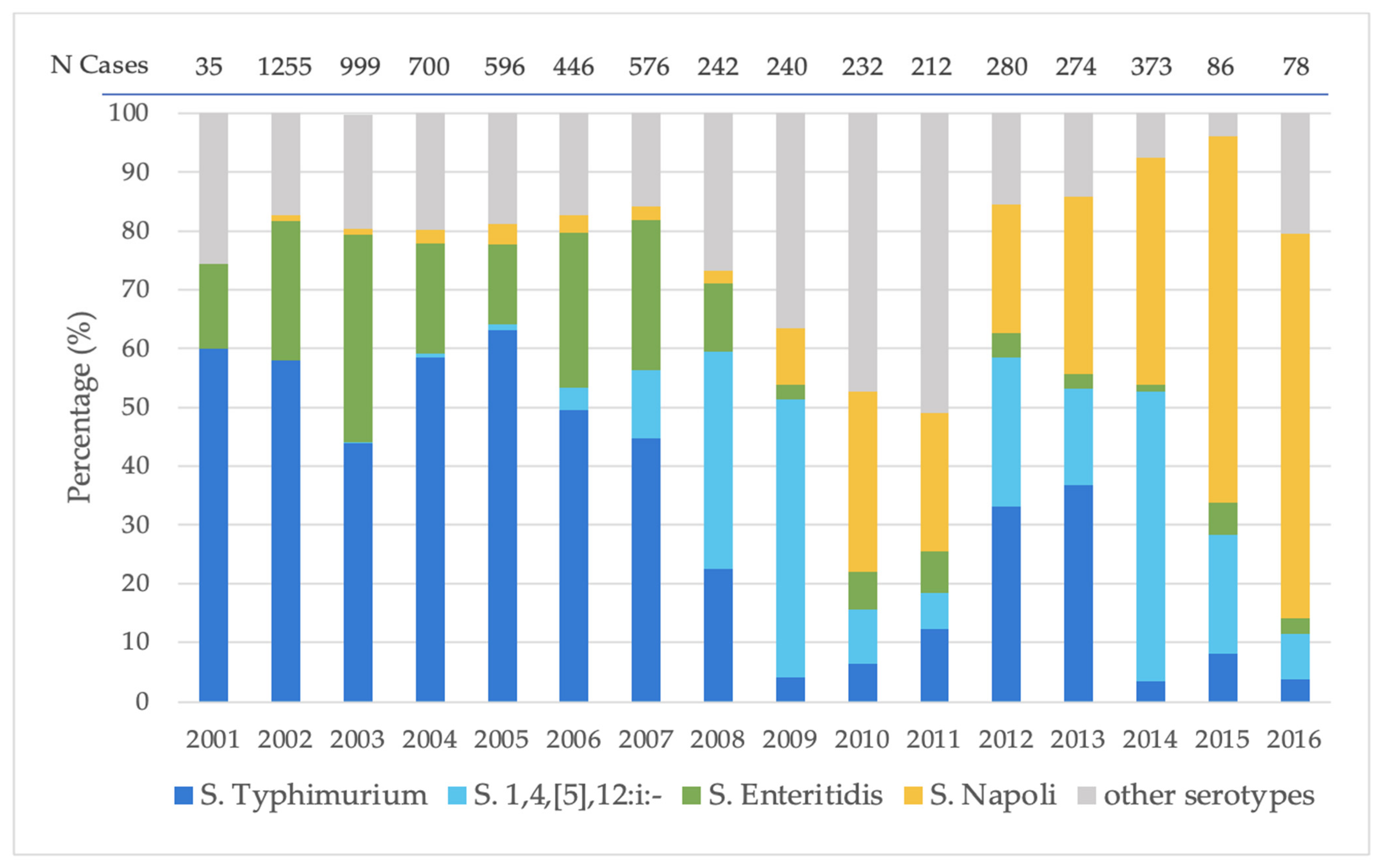
Serotype Distribution. Among the 6624 salmonellosis cases included in the study, 6566 (99%; 6566/6624) were caused by NTS belonging to 164 different serotypes, whereas 58 cases were caused by typhoidal serotypes (S. Typhi, S. Paratyphi A, B and C) (Supplementary Table S1). S. Typhimurium was the most frequently identified serotype (41.8%; 2772/6624), followed by S. Enteritidis (18.5%; 1223/6624), S. 1,4,[5],12:i:- (9.8%; 649/6624), and S. Napoli (9.6%; 636/6624). Overall, these four serotypes were identified in 79.7% (5280/6624) of all salmonellosis cases included in the study. Beyond these four predominant serotypes, S. Derby, S. Infantis, S. Choleraesuis, S. Muenchen, S. Goldcoast, and S. Virchow were collectively associated with a further 511 cases (7.7%). The relative distribution of the four most frequent serotypes changed over the study period (Figure 1). S. Typhimurium predominated during the early years, whereas the relative proportions of its monophasic variant (S. 1,4,[5],12:i:-) increased in the later years. Conversely, the relative contribution of S. Enteritidis declined over time, while S. Napoli showed an increasing relative representation after 2009.
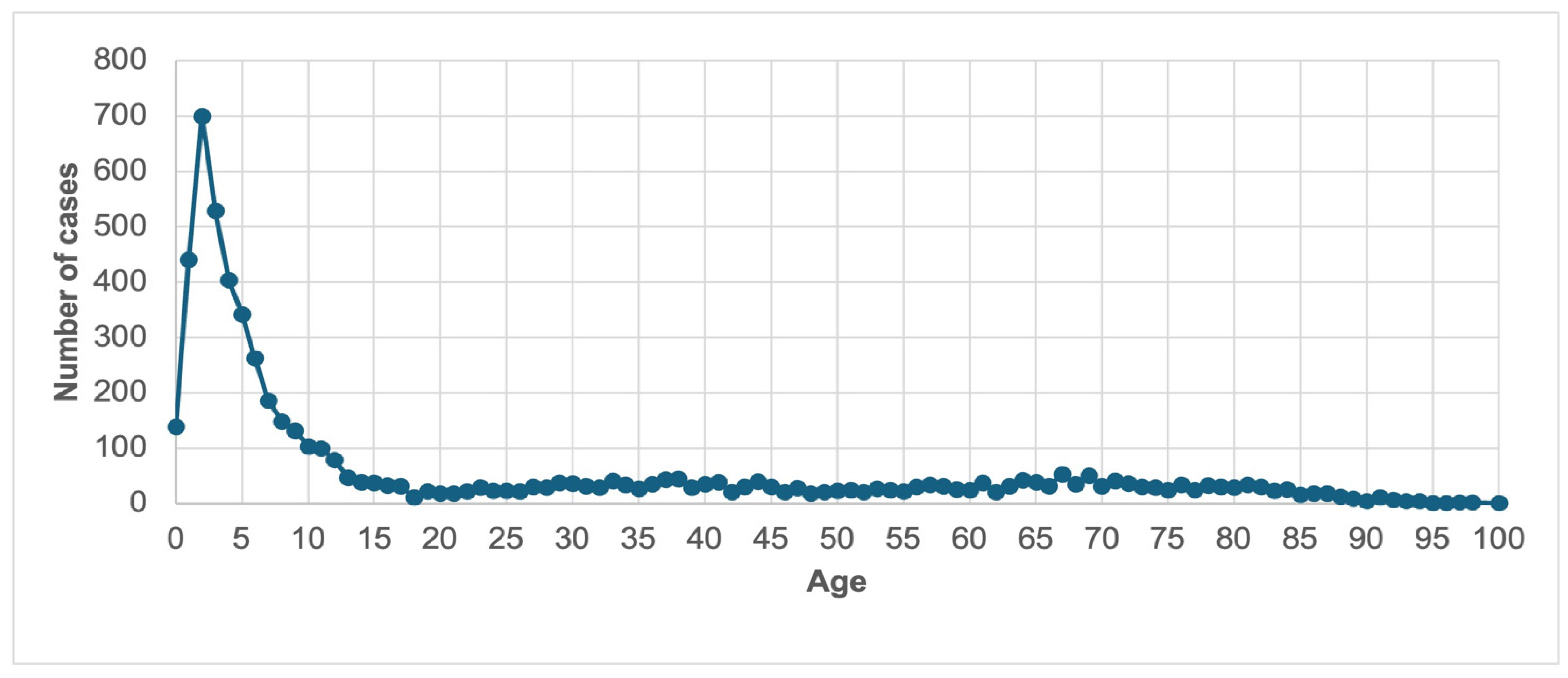
Demographic and clinical characteristics. Because data completeness varied across the archived datasheets, each analysis was carried out using the available data for the variable of interest. Among cases with available demographic information, 51.8% (3265/6304) occurred in male patients. The age distribution showed a marked peak at 2 years of age (Figure 2), and children 0–4 years accounted for the largest proportion of cases (37.7%; 2213/5870; p value < 0.0001; 0–4 age group vs. all other groups). Several serotypes showed distinct age-associated distributions (Table 1). S. Typhimurium was more frequently identified among children aged 0–4 years and 5–14 years (OR 1.78, 95% CI 1.59–1.99, p < 0.001; OR 1.21, 95% CI 1.07–1.36, p = 0.002, respectively). Conversely, S. Choleraesuis, S. Derby, S. Goldcoast and S. Muenchen were more frequently identified among patients older than 65 years (OR 16.09, 95% CI 9.80–26.40, p < 0.001; OR 2.85, 95% CI 1.95–4.15, p < 0.001; OR 3.53, 95% CI 1.95–6.38, p < 0.001; OR 2.63, 95% CI 1.58–4.39, p < 0.001, respectively) (Table 1 and Supplementary Table S2). Among patients aged 15–39 and 40–64 years, S. Enteritidis was more frequently identified than the other NTS serotypes (OR 2.80, 95% CI 2.37–3.30, p < 0.001; OR 1.80, 95% CI 1.51–2.15, p < 0.001, respectively).
Travel and food exposure. Travel history was available for 1361 of the 6624 salmonellosis cases (20.5%), of which 194 (14.3%) were associated with travel. The travel destination was reported for 142 cases, including domestic travel within Italy (62.7%; 89/142) and international travel (37.3%; 53/142). S. Typhimurium and S. Enteritidis were the serotypes most frequently identified among travel-associated cases. Information on food exposure was available for 466 cases (7.0%; 466/6624), while a specific suspected food item was reported for 300 cases (Table 2). Eggs and egg-containing foods were the most frequently reported exposure, followed by meat, fish and seafood products. Because no microbiological confirmation of Salmonella isolation from suspected food items was available, these data should be interpreted as patient-reported exposures only.
Clinical presentation. Information allowing classification of the clinical presentation was available for 5853 cases. Based on criteria described in the Section 2, 5529 cases (94.5%; 5529/5853) were classified as gastrointestinal infections, whereas 244 (4.2%; 244/5853) were classified as bloodstream infections. Among bloodstream isolates, S. Choleraesuis (24.6%; 60/244), S. Typhimurium (22.5%; 55/244) and S. Napoli (15.6%; 38/244) were the most frequently identified serotypes (Table 1).
Invasiveness. The highest Invasiveness Index (II) was observed for S. Choleraesuis (76.9%; 60/78), followed by S. Napoli (8.1%; 38/470) and S. Typhimurium (2.2%; 55/2518). S. Typhi accounted for 22 of the 244 bloodstream isolates (9.0%) and showed an II of 64.7% (22/34). A significant association was observed between serotype and the proportion of bloodstream isolates (p < 0.0001).
Hospitalization. Hospitalization data were available for 4345 cases, of which 1821 (41.9%) required hospital care. Hospitalization rates among the four most common serotypes ranged from 35.5% to 45.8%. (Table 1). S. Choleraesuis showed the highest hospitalization rate compared to all NTS (92.1%, 58/63 vs. 41.9%, 1821/4345; OR 16.07, 95% CI 6.89–45.4, p < 0.0001), comparable to that observed for S. Typhi (87.5%, 21/24 vs. 41.9%, 1821/4345, OR 9.70, 95% CI 3.17–40.9, p < 0.0001).
Association between age and source of isolation. A significant association was observed between age group and source of isolation (p < 0.001). Children aged 0–4 years were more likely to have stool isolates (42.0%, 656/1562, OR 5.64, 95% CI 3.48–9.14, p < 0.001), whereas patients older than 65 years were more likely to have bloodstream isolates (49.1%, 82/167, OR 4.60, 95% CI 3.28–6.46, p < 0.001).
Antimicrobial resistance. Antimicrobial susceptibility results were available for 5057 cases (76.3%). Because participating laboratories used different antimicrobial susceptibility testing panels during the study period, resistance was analyzed by antimicrobial class. Consequently, the number of isolates tested varied across antimicrobial classes (Table 3 and Supplementary Figure S1). High resistance rates were recorded for penicillins (46.9%; 2000/4264), tetracyclines (48%; 651/1355), and aminoglycosides (28.3%; 927/3272), whereas resistance to third-generation cephalosporins (1.5%; 41/2680) and carbapenems (0.7%; 13/1768) remained low. AMR analyses were further restricted to invasive non-typhoidal Salmonella (iNTS) infections. AMR profiles were available for 179 of the 213 (84.0%) iNTS cases (Supplementary Figure S2). Among these, 124 (69.3%, 124/179) were resistant to at least one antibiotic class, whereas 55 (30.7%, 55/179) were susceptible to all tested antimicrobial classes. Among the 124 antimicrobial-resistant iNTS isolates, S. Choleraesuis was the most frequently identified serotype (35.5%; 44/124), followed by S. Typhimurium (31.5%; 39/124) and S. Napoli (11.3%; 14/124). Of the resistant isolates, 39 (31.5%) were resistant to one antibiotic class, 42 (33.9%) to two classes, and 43 (34.7%) to three or more classes. The highest resistance rate was observed for penicillins (93.3%; 98/105), followed by aminoglycosides (73.1%; 49/67), sulfonamides (46%; 40/87) and tetracyclines (46.4%; 26/56), whereas no resistance was detected to carbapenems (0/31) or third-generation cephalosporins (0/81). Resistance to fluoroquinolones was observed in 13.9% (15/108) of iNTS isolates, markedly higher than observed among all isolates with available AMR profiles (2.9%, 136/4711; OR 5.43, 95% CI 2.97–9.42, p < 0.0001). Fluoroquinolone resistance was further characterized by serotype distribution. Among the 136 fluoroquinolone-resistant isolates, ciprofloxacin was the most frequently tested antimicrobial (112/136; 82.4%). Of these, 42 isolates (37.5%) belonged to S. Napoli, 12 (10.7%) to S. Typhimurium, and 11 (9.8%) to S. 1,4,[5],12:i:-.

4. Discussion

This study presents a retrospective analysis of a large historical collection of S. enterica in Italy, comprising strains collected from 6624 cases occurring in Lombardy between 2001 and 2016. By integrating microbiological data with epidemiological and clinical information recorded in the original datasheets, this study provides a retrospective overview of serovar distribution, bloodstream infections, and antimicrobial resistance over a 16-year period. The findings should be interpreted in the context of the study design, as the collection was assembled through passive laboratory-based surveillance and therefore was not intended to represent all salmonellosis cases occurring in Lombardy. Globally, S. Typhimurium and S. Enteritidis are the two serotypes most frequently associated with human salmonellosis worldwide [14,27]. In the present study, the distributions of three of the four predominant serotypes (S. Enteritidis, S. Typhimurium, and its monophasic variant S. 1,4,[5],12:i:-) were consistent with those reported by the Italian and European surveillance systems during the study period [28,29,30]. However, unlike in most European countries, where S. Enteritidis remained the predominant serotype, surveillance data from the Enter-Net Italy network showed that S. Typhimurium and its monophasic variant became the most frequently isolated serotypes in human infections after 2008 [31,32]. The serotype distribution observed in this collection was consistent with these national surveillance data [33].
Within the collection, S. Typhimurium and S. Enteritidis were the most frequently represented serotypes during the early 2000s. After 2007, however, the relative proportions of isolates belonging to these serotypes declined, whereas S. 1,4,[5],12:i:- and S. Napoli became progressively more represented. The apparent decline in S. Typhimurium may partly reflect the separate reporting of its monophasic variant, S. 1,4,[5],12:i:-, included in European surveillance systems from 2010 onwards [34]. Likewise, the reduced proportion of S. Enteritidis observed in our collection is consistent with the marked decline in human S. Enteritidis infections reported in Italy and across Europe following the implementation of coordinated European and national Salmonella control programs in poultry and laying hens [35,36].
The emergence of S. Napoli within the collection deserves particular attention. Across Europe, from 2000 to 2013, the incidence of S. Napoli increased by 256%, making it the 13th most frequently isolated serovar from human cases [37,38,39]. In the present collection, S. Napoli was the most frequently identified serotype among iNTS isolates collected during the period 2010–2014, accounting for 15.8% of invasive cases. Genomic and phylogenetic analyses indicated that S. Napoli belongs to the S. Typhi subclade of clade A, showing a close relationship with S. Paratyphi A and sharing genes associated with the typical typhoid fever virulence [18]. Together, these observations support the epidemiological and clinical relevance of this serotype. The most recent data indicate that S. Napoli remains an emerging serotype in Europe, with most reported infections concentrated in a limited number of countries, particularly Italy, France and Switzerland [40]. Within this European context, Italy represents one of the countries most affected by this serotype [41]. These findings, together with the association between S. Napoli and ciprofloxacin resistance observed in our collection, highlight the potential public health importance of this serotype and support the need for its continued surveillance in Italy.
Among non-typhoidal serovars, S. Choleraesuis showed the highest II (76.9%) and hospitalization rate (92.1%). Unlike the other predominant serotypes, which were mainly associated with gastrointestinal infections in young children, S. Choleraesuis was predominantly identified in bloodstream isolates from older adults [42,43]. This observation is consistent with previous findings that highlighted it not only as a swine-associated serotype but as a significant cause of invasive salmonellosis in humans [16,44]. Its close evolutionary relationship with S. Paratyphi C may help explain its enhanced invasiveness [45,46,47]. From a public health perspective, these findings highlight the importance of maintaining surveillance systems for S. enterica and ensuring systematic monitoring of invasive serotypes.
The antimicrobial resistance profile observed in the collection reflected the resistance patterns circulating during the study period. High levels of resistance to penicillins and tetracyclines (>45%) limited the therapeutic value of these antimicrobial classes and were consistent with the persistence of resistance to these antibiotics in Italy. According to the most recent ECDC and EFSA report, approximately 40% of Salmonella isolates in Italy remain resistant to these antimicrobial classes, whereas lower proportions are reported in many other European countries [48]. In contrast, resistance to fluoroquinolones and sulfonamides (2.9% and 10.2%, respectively) was lower than that currently reported in Italy (9.1% and 43.7%, respectively) and Europe (21.8% and 20.8%, respectively), suggesting that resistance to these antimicrobial classes has increased over time [33,48]. Notably, fluoroquinolone resistance reached 13.9% among iNTS isolates in our collection, emphasizing the clinical relevance of invasive Salmonella infections [49].
In particular, the increase in fluoroquinolone resistance is of particular concern, as fluoroquinolone-resistant non-typhoidal Salmonella has recently been included in the WHO list of high-priority bacterial pathogens [50]. In Italy, aminoglycosides were the only antimicrobial classes showing a lower resistance rate in the historical collection than that currently reported (28.3% vs. 5.2%), whereas resistance to third- and fourth-generation cephalosporins and carbapenems has remained low, supporting the continued effectiveness of these antibiotic classes. Importantly, one-third of iNTS isolates were multidrug-resistant, underscoring the importance of continuous AMR surveillance to support empirical therapy and antimicrobial stewardship.
This study has several limitations. The collection was generated through a passive laboratory-based reporting system and therefore does not represent a population-based sample of salmonellosis cases in Lombardy. In addition, the availability of demographic and clinical information varied over time. The incomplete availability of epidemiological and clinical information, particularly for travel history, food exposure, and hospitalization, may have influenced the strength of some subgroup analyses and should be considered when interpreting the findings. AST was performed by multiple participating laboratories, and although results were interpreted according to the standards in use at the time, differences in testing methodologies and changes in interpretative criteria over the study period may have affected the comparability of AMR data. Although grouping susceptibility results according to antimicrobial classes improved comparability across laboratories and years, some methodological heterogeneity cannot be excluded. Consequently, the findings should be interpreted as descriptive of the archived collection rather than representative of the epidemiology of salmonellosis in the region.
Despite these limitations, historical microbiological collections linked to epidemiological and microbiological metadata represent a valuable resource for retrospective investigations of serotype distribution and antimicrobial resistance. Recent whole-genome sequencing studies have considerably advanced the understanding of Salmonella evolution, virulence, and antimicrobial resistance [51,52]. Historical collections such as the one described here provide an important complementary resource by preserving well-documented archived isolates and associated metadata that can support future genomic investigations within a One Health framework.

5. Conclusions

This study highlights the value of a historical microbiological collection and its associated metadata for retrospectively investigating long-term changes in S. enterica serotype distribution, bloodstream infections, and antimicrobial resistance. The findings identify S. Napoli and S. Choleraesuis as serotypes of public health relevance and support the importance of continued surveillance of invasive Salmonella infections and AMR. More broadly, this study reinforces the scientific value of preserving historical microbiological collections as resources for future molecular epidemiology and One Health surveillance.

Supplementary Materials

The following supporting information can be downloaded at https://www.mdpi.com/article/10.3390/pathogens15070771/s1. Figure S1: Percentage of antibiotic resistance among S. enterica isolates collected from 2001 to 2016. Figure S2: Percentage of AMR profiles in iNTS. Table S1: Number of cases and percentage of 168 serotypes recorded in the period 2001–2016. Table S2: Distribution of minor serotypes (S. Derby, S. Goldcoast and S. Muenchen) per age.

Author Contributions

Conceptualization, P.P., S.B., A.A., M.P. and E.T.; methodology, P.P., M.G. (Marta Gusmeroli) and D.C.; formal analysis, P.P. and M.G. (Marta Gusmeroli); investigation, P.P. and M.P.; resources, S.B. and E.B.; data curation, P.P. and M.G. (Maria Gori); writing—original draft preparation, P.P. and S.B.; writing—review and editing, A.A., C.F., M.G. (Maria Gori) and E.T.; visualization, M.P. and E.B.; supervision, E.B. and S.B.; project administration, M.P., E.T. and S.B.; funding acquisition, E.B. and S.B. All authors have read and agreed to the published version of the manuscript.

Funding

This research received no external funding.

Institutional Review Board Statement

Ethical review and approval were waived for this study because it was conducted on archived bacterial isolates and related anonymized epidemiological and clinical data collected during routine microbiological diagnostic activities and collaborative strain collection programs involving hospitals and healthcare laboratories in Lombardy, Italy. No additional sampling or direct involvement of human subjects occurred for the purposes of this study.

Informed Consent Statement

Patient consent was waived because the study was performed retrospectively on archived bacterial isolates and anonymized data collected during routine diagnostic activities, without direct patient involvement or identifiable personal information.

Data Availability Statement

Data supporting the findings of this study are not publicly available due to institutional restrictions and the presence of sensitive epidemiological information but are available from the corresponding author upon reasonable request.

Acknowledgments

The Authors gratefully acknowledge all the microbiologists, laboratory technicians, clinicians, and collaborating diagnostic laboratories and hospitals that, over many years, contributed to the collection, preservation, serotyping, and documentation of the Salmonella enterica isolates archived at the University of Milan. Their continuous commitment to maintaining this historical microbiological collection and its associated epidemiological and clinical metadata created a unique scientific resource that made the present study possible.

Conflicts of Interest

The authors declare no conflicts of interest.

Abbreviations

The following abbreviations are used in this manuscript:
ASTAntimicrobial susceptibility testing
AMRAntimicrobial resistance
BPPLBacterial Priority Pathogens list
CIConfidence intervals
ECDCEuropean Centre for Disease Prevention and Control
EFSAEuropean Food Safety Authority
EUEuropean Union
IIInvasiveness Index
iNTSInvasive non-typhoidal Salmonella
MDRMultiple-drug resistance
NTSNon-typhoidal Salmonella
ORsOdds ratios
TSTyphoidal Salmonella
WGSWhole-genome sequencing

References

  1. De Paoli, P. Bio-banking in microbiology: From sample collection to epidemiology, diagnosis and research. FEMS Microbiol. Rev. 2005, 29, 897–910. [Google Scholar] [CrossRef] [Scilit] [PubMed]
  2. Müller, H.; Dagher, G.; Loibner, M.; Stumptner, C.; Kungl, P.; Zatloukal, K. Biobanks for life sciences and personalized medicine: Importance of standardization, biosafety, biosecurity, and data management. Curr. Opin. Biotechnol. 2020, 65, 45–51. [Google Scholar] [CrossRef] [Scilit] [PubMed]
  3. Medina, P.B.; Kealy, J.; Kozlakidis, Z. Integrating research infrastructures into infectious diseases surveillance operations: Focus on biobanks. Biosaf. Health 2022, 4, 410–413. [Google Scholar] [CrossRef] [Scilit] [PubMed]
  4. Muzambi, R.; Bhaskaran, K.; Rentsch, C.T.; Smeeth, L.; Brayne, C.; Garfield, V.; Williams, D.M.; Chaturvedi, N.; Warren-Gash, C. Are infections associated with cognitive decline and neuroimaging outcomes? A historical cohort study using data from the UK Biobank study linked to electronic health records. Transl. Psychiatry 2022, 12, 385. [Google Scholar] [CrossRef] [Scilit] [PubMed]
  5. Delpy, L.; Astbury, C.C.; Aenishaenslin, C.; Ruckert, A.; Penney, T.L.; Wiktorowicz, M.; Ciss, M.; Benko, R.; Bordier, M. Integrated surveillance systems for antibiotic resistance in a One Health context: A scoping review. BMC Public Health 2024, 24, 1717. [Google Scholar] [CrossRef] [Scilit] [PubMed]
  6. Gentile, B.; Grottola, A.; Orlando, G.; Fregni Serpini, G.; Venturelli, C.; Meschiari, M.; Anselmo, A.; Fillo, S.; Fortunato, A.; Lista, F.; et al. A retrospective whole-genome sequencing analysis of carbapenem and colistin-resistant Klebsiella pneumoniae nosocomial strains isolated during an MDR surveillance program. Antibiotics 2020, 9, 246. [Google Scholar] [CrossRef] [Scilit] [PubMed]
  7. Postiglione, U.; Batisti Biffignandi, G.; Corbella, M.; Merla, C.; Olivieri, E.; Petazzoni, G.; Feil, E.J.; Bandi, C.; Cambieri, P.; Gaiarsa, S.; et al. Combining genome surveillance and metadata to characterize the diversity of Staphylococcus aureus circulating in an Italian hospital over a 9-year period. Microbiol. Spectr. 2023, 11, e01010-23. [Google Scholar] [CrossRef] [Scilit] [PubMed]
  8. Rodrigues da Costa, M.; Pessoa, J.; Meemken, D.; Nesbakken, T. A systematic review on the effectiveness of pre-harvest meat safety interventions in pig herds to control Salmonella and other foodborne pathogens. Microorganisms 2021, 9, 1825. [Google Scholar] [CrossRef] [Scilit] [PubMed]
  9. European Food Safety Authority (EFSA); European Centre for Disease Prevention and Control (ECDC). The European Union One Health 2022 Zoonoses Report. EFSA J. 2023, 21, e8442. [Google Scholar] [CrossRef] [Scilit] [PubMed]
  10. Han, J.; Aljahdali, N.; Zhao, S.; Tang, H.; Harbottle, H.; Hoffmann, M.; Frye, J.G.; Foley, S.L. Infection biology of Salmonella enterica. EcoSal Plus 2024, 12, eESP0001-2023. [Google Scholar] [CrossRef] [Scilit] [PubMed]
  11. Brenner, F.W.; Villar, R.G.; Angulo, F.J.; Tauxe, R.; Swaminathan, B. Salmonella nomenclature. J. Clin. Microbiol. 2000, 38, 2465–2467. [Google Scholar] [CrossRef] [Scilit] [PubMed]
  12. Chattaway, M.A.; Langridge, G.C.; Wain, J. Salmonella nomenclature in the genomic era: A time for change. Sci. Rep. 2021, 11, 7494. [Google Scholar] [CrossRef] [Scilit] [PubMed]
  13. Galán-Relaño, Á.; Valero Díaz, A.; Huerta Lorenzo, B.; Gómez-Gascón, L.; Mena Rodríguez, M.Á.; Carrasco Jiménez, E.; Pérez Rodríguez, F.; Astorga Márquez, R.J. Salmonella and salmonellosis: An update on public health implications and control strategies. Animals 2023, 13, 3666. [Google Scholar] [CrossRef] [Scilit] [PubMed]
  14. World Health Organization (WHO). Salmonella. Available online: https://www.who.int/health-topics/salmonella (accessed on 1 May 2026).
  15. European Centre for Disease Prevention and Control (ECDC). Salmonellosis. Annual Epidemiological Report for 2022. Available online: https://www.ecdc.europa.eu/en/publications-data/salmonellosis-annual-epidemiological-report-2022 (accessed on 1 May 2026).
  16. Chiu, C.-H.; Su, L.-H.; Chu, C. Salmonella enterica serotype Choleraesuis: Epidemiology, pathogenesis, clinical disease, and treatment. Clin. Microbiol. Rev. 2004, 17, 311–322. [Google Scholar] [CrossRef] [Scilit] [PubMed]
  17. Mangat, C.S.; Bekal, S.; Avery, B.P.; Côté, G.; Daignault, D.; Doualla-Bell, F.; Finley, R.; Lefebvre, B.; Bharat, A.; Parmley, E.J.; et al. Genomic investigation of the emergence of invasive multidrug-resistant Salmonella enterica serovar Dublin in humans and animals in Canada. Antimicrob. Agents Chemother. 2019, 63, e00108-19. [Google Scholar] [CrossRef] [Scilit] [PubMed]
  18. Huedo, P.; Gori, M.; Zolin, A.; Amato, E.; Ciceri, G.; Bossi, A.; Pontello, M. Salmonella enterica serotype Napoli is the first cause of invasive nontyphoidal salmonellosis in Lombardy, Italy (2010–2014), and belongs to Typhi subclade. Foodborne Pathog. Dis. 2017, 14, 148–151. [Google Scholar] [CrossRef] [Scilit] [PubMed]
  19. Wang, Y.; Liu, Y.; Lyu, N.; Li, Z.; Ma, S.; Cao, D.; Pan, Y.; Hu, Y.; Huang, H.; Gao, G.F.; et al. The temporal dynamics of antimicrobial-resistant Salmonella enterica and predominant serovars in China. Natl. Sci. Rev. 2023, 10, nwac269. [Google Scholar] [CrossRef] [Scilit] [PubMed]
  20. Ferrari, R.G.; Rosario, D.K.A.; Cunha-Neto, A.; Mano, S.B.; Figueiredo, E.E.S.; Conte-Junior, C.A. Worldwide epidemiology of Salmonella serovars in animal-based foods: A meta-analysis. Appl. Environ. Microbiol. 2019, 85, e00591-19. [Google Scholar] [CrossRef] [Scilit] [PubMed]
  21. Westrell, T.; Monnet, D.L.; Gossner, C.; Heuer, O.; Takkinen, J. Drug-resistant Salmonella enterica serotype Kentucky in Europe. Lancet Infect. Dis. 2014, 14, 270–271. [Google Scholar] [CrossRef] [Scilit] [PubMed]
  22. Pitti, M.; Garcia-Vozmediano, A.; Tramuta, C.; CeRTiS Clinical Laboratories Group; Maurella, C.; Decastelli, L. Monitoring of antimicrobial resistance of Salmonella serotypes isolated from humans in Northwest Italy, 2012–2021. Pathogens 2023, 12, 89. [Google Scholar] [CrossRef] [Scilit] [PubMed]
  23. Petrin, S.; Orsini, M.; Massaro, A.; Olsen, J.E.; Barco, L.; Losasso, C. Phenotypic and genotypic antimicrobial resistance correlation and plasmid characterization in Salmonella spp. isolates from Italy reveal high heterogeneity among serovars. Front. Public Health 2023, 11, 1221351. [Google Scholar] [CrossRef] [Scilit] [PubMed]
  24. Gorietti, S.; Busani, L.; on behalf of the Gruppo ENTER-NET Italia. Salmonella Surveillance in 2000: The Italian ENTER-NET System. Bollettino Epidemiologico Nazionale. 2001. Available online: https://www.epicentro.iss.it/ben/2001/novembre/2_en (accessed on 1 May 2026).
  25. Issenhuth-Jeanjean, S.; Roggentin, P.; Mikoleit, M.; Guibourdenche, M.; de Pinna, E.; Nair, S.; Fields, P.I.; Weill, F.-X. Supplement 2008–2010 (No. 48) to the White–Kauffmann–Le Minor scheme. Res. Microbiol. 2014, 165, 526–530. [Google Scholar] [CrossRef] [Scilit] [PubMed]
  26. Parisi, A.; Crump, J.A.; Stafford, R.; Glass, K.; Howden, B.P.; Kirk, M.D. Increasing incidence of invasive nontyphoidal Salmonella infections in Queensland, Australia, 2007–2016. PLoS Negl. Trop. Dis. 2019, 13, e0007187. [Google Scholar] [CrossRef] [Scilit] [PubMed]
  27. Feasey, N.A.; Dougan, G.; Kingsley, R.A.; Heyderman, R.S.; Gordon, M.A. Invasive non-typhoidal Salmonella disease: An emerging and neglected tropical disease in Africa. Lancet 2012, 379, 2489–2499. [Google Scholar] [CrossRef] [Scilit] [PubMed]
  28. European Food Safety Authority (EFSA). The Community Summary Report on Trends and Sources of Zoonoses and Zoonotic Agents in the European Union in 2007. EFSA J. 2009, 7, 223R. [Google Scholar] [CrossRef] [Scilit]
  29. European Centre for Disease Prevention and Control (ECDC). Annual Epidemiological Report on Communicable Diseases in Europe. 2010. Available online: https://data.europa.eu/doi/10.2900/35039 (accessed on 1 May 2026).
  30. European Centre for Disease Prevention and Control (ECDC). Salmonellosis. Annual Epidemiological Report for 2015. Available online: https://www.ecdc.europa.eu/en/publications-data/salmonellosis-annual-epidemiological-report-2015 (accessed on 1 May 2026).
  31. Dionisi, A.M.; Filetici, E.; Owczarek, S.; Arena, S.; Benedetti, I.; Lucarelli, C.; Luzzi, I.; Scavia, G.; Minelli, F.; Ciaravino, G.; et al. ENTER-NET: Sorveglianza delle infezioni trasmesse da alimenti e acqua. Rapporto dell’attività 2007–2009. Not. Ist. Super. Sanità 2011, 24, 3–10. [Google Scholar]
  32. García Fernández, A.; Luzzi, I.; Dionisi, A.M.; Owczarek, S.; Arena, S.; Lucarelli, C. Salmonella. In Enter-Net Italia e Registro Italiano della Sindrome Emolitico Uremica: Sorveglianza delle Infezioni da Salmonella, Campylobacter, Escherichia coli Produttore di Shiga-Tossina e Listeria monocytogenes (2010–2015); Luzzi, I., García Fernández, A., Dionisi, A.M., Lucarelli, C., Gattuso, A., Gianfranceschi, M.V., Maugliani, A., Caprioli, A., Morabito, S., Scavia, G., Eds.; Istituto Superiore di Sanità: Rome, Italy, 2017; Rapporti ISTISAN 17/34; pp. 13–25. [Google Scholar]
  33. Lucarelli, C.; Garcia-Fernandez, A.; Dionisi, A.M.; Owczarek, S.; Arena, S.; Fortini, D.; Errico, G.; Maraglino, F.; Pilati, S.; Palamara, A.T.; et al. Sorveglianza Nazionale Delle Infezioni da Salmonella, Campylobacter, Shigella e Yersinia. Dati Enter-Net Italia 2016–2021. Rapporti ISS Sorveglianza 2024, RIS-1/2024. Available online: https://www.iss.it/documents/20126/0/RIS-1-2024.pdf/c2710a8c-fef8-1455-5d6a-90aa978bc4c3?t=1725524713756 (accessed on 20 June 2026).
  34. European Centre for Disease Prevention and Control (ECDC). Food- and Waterborne Diseases and Zoonoses. Annual Epidemiological Report 2012. Available online: https://www.ecdc.europa.eu/en/publications-data/food-and-waterborne-diseases-and-zoonoses-annual-epidemiological-report-2014-2012 (accessed on 1 May 2026).
  35. European Commission. Food-Borne Diseases (Zoonoses): Control of Salmonella. Available online: https://food.ec.europa.eu/food-safety/biological-safety/food-borne-diseases-zoonoses/control-salmonella_en (accessed on 1 May 2026).
  36. EFSA Panel on Biological Hazards (BIOHAZ); Koutsoumanis, K.; Allende, A.; Alvarez-Ordóñez, A.; Bolton, D.; Bover-Cid, S.; Chemaly, M.; De Cesare, A.; Herman, L.; Hilbert, F.; et al. Salmonella Control in Poultry Flocks and Its Public Health Impact. EFSA J. 2019, 17, e05596. [Google Scholar] [CrossRef] [Scilit] [PubMed]
  37. Gori, M.; Ebranati, E.; Scaltriti, E.; Huedo, P.; Ciceri, G.; Tanzi, E.; Pontello, M.; Zehender, G.; Pongolini, S.; Bolzoni, L. High-Resolution Diffusion Pattern of Human Infections by Salmonella enterica Serovar Napoli in Northern Italy Explained through Phylogeography. PLoS ONE 2018, 13, e0202573. [Google Scholar] [CrossRef] [Scilit] [PubMed]
  38. Graziani, C.; Luzzi, I.; Owczarek, S.; Dionisi, A.M.; Busani, L. Salmonella enterica Serovar Napoli Infection in Italy from 2000 to 2013: Spatial and Spatio-Temporal Analysis of Case Distribution and the Effect of Human and Animal Density on the Risk of Infection. PLoS ONE 2015, 10, e0142419. [Google Scholar] [CrossRef] [Scilit] [PubMed]
  39. Huedo, P.; Gori, M.; Amato, E.; Bianchi, R.; Valerio, E.; Magnoli, L.; Pontello, M. A Multischool Outbreak Due to Salmonella enterica Serovar Napoli Associated with Elevated Rates of Hospitalizations and Bacteremia, Milan, Italy, 2014. Foodborne Pathog. Dis. 2016, 13, 417–422. [Google Scholar] [CrossRef] [Scilit] [PubMed]
  40. Sabbatucci, M.; Dionisi, A.M.; Pezzotti, P.; Lucarelli, C.; Barco, L.; Mancin, M.; Luzzi, I. Molecular and Epidemiologic Analysis of Reemergent Salmonella enterica Serovar Napoli, Italy, 2011–2015. Emerg. Infect. Dis. 2018, 24, 562–565. [Google Scholar] [CrossRef] [Scilit] [PubMed]
  41. Leati, M.; Zaccherini, A.; Ruocco, L.; D’Amato, S.; Busani, L.; Villa, L.; Barco, L.; Ricci, A.; Cibin, V. The Challenging Task to Select Salmonella Target Serovars in Poultry: The Italian Point of View. Epidemiol. Infect. 2021, 149, e160. [Google Scholar] [CrossRef] [Scilit] [PubMed]
  42. Yahav, D.; Eliakim-Raz, N.; Leibovici, L.; Paul, M. Bloodstream Infections in Older Patients. Virulence 2016, 7, 341–352. [Google Scholar] [CrossRef] [Scilit] [PubMed]
  43. Chen, P.-L.; Lee, H.-C.; Lee, N.-Y.; Wu, C.-J.; Lin, S.-H.; Shih, H.-I.; Lee, C.-C.; Ko, W.-C.; Chang, C.-M. Non-Typhoidal Salmonella Bacteraemia in Elderly Patients: An Increased Risk for Endovascular Infections, Osteomyelitis and Mortality. Epidemiol. Infect. 2012, 140, 2037–2044. [Google Scholar] [CrossRef] [Scilit] [PubMed]
  44. Chiu, C.-H.; Tang, P.; Chu, C.; Hu, S.; Bao, Q.; Yu, J.; Chou, Y.-Y.; Wang, H.-S.; Lee, Y.-S. The Genome Sequence of Salmonella enterica Serovar Choleraesuis, a Highly Invasive and Resistant Zoonotic Pathogen. Nucleic Acids Res. 2005, 33, 1690–1698. [Google Scholar] [CrossRef] [Scilit] [PubMed]
  45. Liu, W.-Q.; Feng, Y.; Wang, Y.; Zou, Q.-H.; Chen, F.; Guo, J.-T.; Peng, Y.-H.; Jin, Y.; Li, Y.-G.; Hu, S.-N.; et al. Salmonella Paratyphi C: Genetic Divergence from Salmonella Choleraesuis and Pathogenic Convergence with Salmonella Typhi. PLoS ONE 2009, 4, e4510. [Google Scholar] [CrossRef] [Scilit] [PubMed]
  46. Fricke, W.F.; Mammel, M.K.; McDermott, P.F.; Tartera, C.; White, D.G.; Leclerc, J.E.; Ravel, J.; Cebula, T.A. Comparative Genomics of 28 Salmonella enterica Isolates: Evidence for CRISPR-Mediated Adaptive Sublineage Evolution. J. Bacteriol. 2011, 193, 3556–3568. [Google Scholar] [CrossRef] [Scilit] [PubMed]
  47. Zhou, Z.; Lundstrøm, I.; Tran-Dien, A.; Duchêne, S.; Alikhan, N.-F.; Sergeant, M.J.; Langridge, G.; Fotakis, A.K.; Nair, S.; Stenøien, H.K.; et al. Pan-Genome Analysis of Ancient and Modern Salmonella enterica Demonstrates Genomic Stability of the Invasive Para C Lineage for Millennia. Curr. Biol. 2018, 28, 2420–2428.e10. [Google Scholar] [CrossRef] [Scilit] [PubMed]
  48. European Food Safety Authority (EFSA); European Centre for Disease Prevention and Control (ECDC). Plain Language Summary of the European Union Summary Report on Antimicrobial Resistance in Zoonotic and Indicator Bacteria from Humans, Animals and Food in 2022–2023. EFSA J. 2025, 23, e230301. [Google Scholar] [CrossRef] [Scilit] [PubMed]
  49. Zhan, Z.; Xie, Z.; Lin, H.; Guo, J.; Cai, M.; Wu, Y.; Liang, S. Prevalence of Antibiotic Resistance in Invasive Non-Typhoidal Salmonella across 19 Developing Countries, 2000–2026: A Systematic Review and Meta-Analysis. J. Glob. Antimicrob. Resist. 2026, 50, 165–178. [Google Scholar] [CrossRef] [Scilit] [PubMed]
  50. World Health Organization (WHO). WHO Bacterial Priority Pathogens List, 2024: Bacterial Pathogens of Public Health Importance to Guide Research, Development and Strategies to Prevent and Control Antimicrobial Resistance. Available online: https://www.who.int/publications/i/item/9789240093461 (accessed on 1 May 2026).
  51. Wang, Y.; Xu, X.; Jia, S.; Qu, M.; Pei, Y.; Qiu, S.; Zhang, J.; Liu, Y.; Ma, S.; Lyu, N.; et al. A global atlas and drivers of antimicrobial resistance in Salmonella during 1900–2023. Nat. Commun. 2025, 16, 4611. [Google Scholar] [CrossRef] [Scilit] [PubMed]
  52. Wang, Y.; Xu, X.; Zhu, B.; Lyu, N.; Liu, Y.; Ma, S.; Jia, S.; Wan, B.; Du, Y.; Zhang, G.; et al. Genomic analysis of almost 8000 Salmonella genomes reveals drivers and landscape of antimicrobial resistance in China. Microbiol. Spectr. 2023, 11, e02080-23. [Google Scholar] [CrossRef] [Scilit] [PubMed]
Figure 1. Annual distribution of the main S. enterica serovars among serotyped isolates included in the historical collection (2001–2016). Bars show the percentage of each serovar relative to the total number of serotyped isolates available for each year. Numbers above the bars indicate the annual number of serotyped isolates (N). The distribution of serovars varied over time: S. Typhimurium predominated in the early years of the study period, while S. Enteritidis was consistently represented until 2007 and then declined, whereas the proportions of the monophasic variant S. 1,4,[5],12:i:- and S. Napoli increased in later years. Annual differences should be interpreted considering the variable number of isolates available for each year.
Pathogens 15 00771 g001
Figure 2. Age distribution of clinical cases associated with S. enterica isolates included in the historical collection (Lombardy, 2001–2016). A peak is present at 2 years of age with 700 cases.
Pathogens 15 00771 g002
Table 1. Demographic, clinical and microbiological characteristics of S. enterica cases, stratified by the six most relevant serotypes of the study. Data are presented as number (percentage). Percentages were calculated using the number of cases with available data for each variable.
All SerotypesS. TyphimuriumS. EnteritidisS. 1,4,[5],12:i:-S. NapoliS. CholeraesuisS. Typhi
N Cases (%)6624 (100)2772 (41.8)1223 (18.5)649 (9.80)636 (9.60)82 (1.24)37 (0.6)
Sex: N (%)
Available data6304269311995315708132
M 3265 (51.8)1432 (53.1)596 (49.7)280 (52.7)300 (52.6)51(63.0)25 (78.1)
Age: N (%)
Available data5870 256611464204997629
0–4 age 2213 (37.7)1163 (45.3)301 (26.3)183 (43.6)205 (41.1)0 (0)4 (13.8)
5–14 age 1438 (24.5)679 (26.5)283 (24.7)119 (28.3)159 (31.9)4 (5.3)7 (24.1)
15–39 age 736 (12.5)242 (9.4)269 (23.5)38 (9.0)24 (4.8)3 (3.9)13 (44,8)
40–64 age704 (12.0)233 (9.1)202 (17.6)31 (7.4)38 (7.6)16 (21.1)5 (17.2)
>65 age 779 (13.3)249 (9.7)91 (7.9)49 (11.7)73 (14.6)53 (69.7)0 (0.0)
Source of isolation: N (%)
Available data5853251811504334707834
Stool 5529 (94.5)2443 (97.0)1119 (97.3)426 (98.4)430 (91.4)8(10.3)12 (35.3)
Blood 244 (4.2)55 (2.2)25 (2.2)5 (1.2)38 (8.1)60 (76.9)22 (64.7)
Others *80(1.4)20 (0.8)6 (0.5)2 (0.5)2 (0.4)10 (12.8)0 (0.0)
Hospitalization: N (%)
Available data434519198892533536324
Hospitalized 1821 (41.9)796 (41.5)316 (35.5)116 (45.8)157 (44.5)58 (92.1)21 (87.5)
Note: * Specimens included urine, bile, abscess material, pus.
Table 2. Food and water sources involved in S. enterica infections.
Implicated Food and Water (%)
Available data300
Water 3 (1.0)
Meat 58 (19.3)
Crustaceans and mollusks 21 (7.0)
Fish 18 (6.0)
Milk, butter and derivatives 12 (4.0)
Eggs 67 (22.3)
Long-life canned foods 2 (0.7)
Fresh vegetables 2 (0.7)
Composite Food—eggs and milk 101 (33.7)
Composite Food—eggs 16 (5.3)
Table 3. Antibiotic resistance of S. enterica strains obtained from the datasheets. Resistance percentages were calculated using the number of isolates tested for each antimicrobial class as the denominator. The number of isolates tested varied according to the antimicrobial susceptibility panels routinely adopted during the study period (2001–2016).
Antibiotic ClassesAntimicrobials IncludedIsolates Tested (N)Resistant Isolates, N (%)
Beta-lactams
PenicillinsAmoxicillin, Ampicillin, Cloxacillin, Oxacillin, Mezlocillin, Piperacillin, Ticarcillin. 42642000 (46.9)
Penicillins + β-lactamase inhibitorAmpicillin/Sulbactam, Piperacillin/Tazobactam, Ticarcillin/Clavulanic acid 1729383 (22.2)
First-generation cephalosporinsCephalothin, Cefazolin3329565 (17.0)
Second-generation cephalosporinsCefuroxime, Cefuroxime Axetil, Cefoxitin 936251(26.8)
Third-generation cephalosporinsCefotaxime, Ceftazidime, Ceftriaxone, Cefixime, Cefpodoxime, Ceftiroxime 268041 (1.5)
Fourth-generation cephalosporins Only Cefepime tested92520 (2.2)
Monobactams Only Aztreonam tested78811(1.4)
CarbapenemsImipenem, Meropenem, Ertapenem 176813 (0.7)
Fluoroquinolones/QuinolonesCiprofloxacin, Levofloxacin, Norfloxacin, Ofloxacin4711136 (2.9)
AminoglycosidesAmikacin, Gentamicin, Netylminicin, Tobramycin, Streptomycin, Kanamycin 3272927 (28.3)
Tetracyclines/GlycylcyclinesTetracycline, Doxycycline, Tigecycline1355651(48.0)
SulfonamidesCotrimoxazole, Trimethoprim, Sulfamethoxazole, Trimethoprim/Sulfamethoxazole4722480 (10.2)
PhenicolsOnly Chloramphenicol668112 (16.8)
PolymyxinsOnly Colistin tested980 (0.0)
MacrolidesOnly Erythromycin tested42 (50.0)
OthersFosfomycin1504 (2.7)
Nitrofurantoin 274 (14.8)
Disclaimer/Publisher’s Note: The statements, opinions and data contained in all publications are solely those of the individual author(s) and contributor(s) and not of MDPI and/or the editor(s). MDPI and/or the editor(s) disclaim responsibility for any injury to people or property resulting from any ideas, methods, instructions or products referred to in the content.