Article

Evaluating the Impact of Little Cigar Use on the Oral Bacterial Microbiota of Cigarette Smokers


by
Suhana Chattopadhyay
1,
Leena Malayil
1,
Emmanuel F. Mongodin
2,† and
Amy R. Sapkota
1,*
1
Department of Global, Environmental, and Occupational Health, University of Maryland School of Public Health, College Park, MD 20742, USA
2
Institute for Genome Sciences, University of Maryland School of Medicine, Baltimore, MD 21201, USA
*
Author to whom correspondence should be addressed.
Current address: Division of Lung Diseases, National Heart, Lung and Blood Institute (NHLBI), National Institutes of Health (NIH), Bethesda, MD 20892, USA.
Pathogens 2026, 15(7), 732; https://doi.org/10.3390/pathogens15070732
Submission received: 5 June 2026 / Revised: 9 July 2026 / Accepted: 9 July 2026 / Published: 13 July 2026
(This article belongs to the Section Bacterial Pathogens)

Abstract

Tobacco products (e.g., cigarettes, little cigars) harbor diverse bacterial communities and long-term tobacco use alters the oral microbiome, potentially leading to oral disease. However, no studies have evaluated the immediate changes in the oral bacterial microbiota that could occur after using a new tobacco product. To address this knowledge gap, buccal swab and saliva samples were collected from forty cigarette smokers before and after a single use of a little cigar product on two separate visits. Total DNA was extracted from a total of 320 samples. The 16S rRNA gene was amplified from these samples and sequenced on the Illumina HiSeq to characterize the bacterial microbiota. Oral bacterial diversity was not significantly different between pre- and post-smoking samples. However, post-smoking buccal samples were enriched with Delftia, Leptotrichia, Pseudomonas and Stenotrophomonas (genera that can include opportunistic pathogens) and post-smoking saliva samples were enriched with Catonella when compared to the pre-smoking samples. In summary, single use of a little cigar product does not immediately impact overall oral bacterial diversity among cigarette smokers; however, post-smoking oral samples may have a higher relative abundance of some bacterial genera. Hence, smoking a new tobacco product could potentially alter the relative abundance of some bacterial types within the oral cavity of smokers, highlighting a potential microbiological pathway through which tobacco use could contribute to oral disease risk.

1. Introduction

Tobacco products have been shown to harbor a myriad of bacterial communities [1,2,3,4,5,6]. A recent study from our group also demonstrated that viable bacterial genera (e.g., Bacillus, Paenibacillus and Terribacillus) originating from cigarette tobacco can be aerosolized in mainstream cigarette smoke [7]. The oral cavity is the first to encounter this mainstream smoke and hence has the greatest potential to be affected by it. While the long term use of tobacco products has been well established to cause dysbiosis (community disturbance) in the oral microbiome of users [8,9,10,11,12], the potential transfer of tobacco-related bacteria to the oral cavity or the immediate impacts of using a specific tobacco product on a user’s oral bacterial microbiota have not been evaluated. This is of significant interest, since changes in bacterial diversity and community composition of the oral cavity can potentially initiate inflammation and subsequent disease development, including the growth of malignant lesions in the mouth [13,14,15].
These types of changes in the mouth can occur in users of a diverse range of tobacco products, from cigarettes to little cigars. Little cigars are comparable to traditional cigarettes in size and shape but contain more tobacco (100–200 mg) and other chemical compounds [16,17]. In comparison to cigarettes, little cigars also have a greater puff volume and puff duration, delivering more carbon monoxide, tobacco-specific nitrosamines (TSNAs), and benzo(a)pyrene to users than cigarettes [17,18,19], consequently causing greater cytotoxicity and inflammation [16]. Similarly to cigarettes, the tobacco in little cigars has been shown to harbor diverse bacterial communities [5,20]. For example, Smyth et al. (2019) demonstrated that the predominant bacterial genera within little cigar tobacco are Pantoea, Pseudomonas and Staphylococcus, including multiple bacterial pathogens such as Staphylococcus sciuri and Pseudomonas pseudoalcaligenes [5]. Nevertheless, to our knowledge there are no data concerning whether or not the bacterial communities within little cigars could be transferred to the oral cavity of smokers and effect changes in the oral microbiome.
Oral microbiome dysbiosis from extrinsic perturbations such as tobacco smoking is dependent on the amount and frequency of products smoked/used per day [21]; however, the overall composition of the oral microbiome has been shown to remain relatively stable over time [22,23,24]. Yet, to date, studies evaluating temporal changes in the oral microbiome [25,26,27,28] have not employed the use of tobacco products by study subjects, and hence, data on potential transient oral microbiome shifts among smokers are limited.
Other studies characterizing oral microbiome dysbiosis associated with tobacco smoking have focused on the use of traditional and/or electronic cigarettes [12,21,25,29,30,31] and indicated that specific oral bacterial genera are impacted by exposure to smoke over a one-week period [8]. For example, greater shifts in bacterial communities among smokers’ plaque samples were demonstrated compared to those of non-smokers over 7 days, with the enrichment of a pathogen-dominated bacterial community (e.g., Fusobacterium nucleatum, Acinetobacter johnsonii, A. baumannii, Streptococcus mutans) among smokers [8]. Moreover, while levels of predominant genera such as Streptococcus, Neisseria, and Veillonella were stable over seven days among non-smokers, less stability was observed among smokers during the same time period, while the colonization of pathogens associated with periodontitis occurred within 24 h of biofilm development [8]. However, to our knowledge, no study has evaluated the immediate potential changes in oral bacterial communities after a single use of a little cigar. Defining immediate changes in the oral microbiota is clinically important because even short-term shifts toward opportunistic pathogens may represent an early mechanistic step in tobacco-related oral disease development. Understanding these impacts can also inform risk assessment, product regulation, and targeted messaging around the harms of emerging tobacco products. Therefore, the objective of our study was to evaluate transient changes in the oral bacterial microbiota after cigarette users smoked a single little cigar product on two different occasions.

2. Methods

2.1. Product Selection

Swisher Sweets little cigars were chosen for inclusion in the study because they are characterized by the highest market sales among little cigar brands in the U.S. [32]. Two products (Swisher Sweets Original (SSORG) and Swisher Sweets Cherry (SSCHR)) were purchased in Columbus, OH, USA, and stored at 4 °C (to minimize variability and maintain consistent conditions prior to use) in their original packaging before being smoked by study participants.

2.2. Sample Size Calculation

Data from the Human Microbiome Project (HMP) Consortium, as detailed in a previously published article [33], provides a resource for power analysis by empirically quantifying the variability of the healthy oral microbiome. The HMP obtained taxonomic profiles from 233 oral 16S rRNA datasets (131 buccal, 102 saliva) and examined genus-level variability across the sampled population. They identified 30 genus-level groups (14 in buccal, 16 in saliva) that accounted for at least 1% of 16S rRNA sequences in the average oral microbiome and measured the variance in relative abundance for each genus. The power analysis conducted estimated the power for detecting various shifts in average relative abundance, with increased power found in higher-abundance genera. For this study, focusing on genera representing 1–3% of 16S rRNA sequences and requiring a significance level of α = 0.05, the analysis suggested that a sample size of at least 20 subjects per trial would provide robust statistical power to detect small percentage changes in low-abundance taxa. To account for potential loss to follow-up, we planned to over-recruit by 20%, which would result in an anticipated study population of 24 subjects per trial.

2.3. Study Population

Participants were recruited through word-of-mouth in Columbus, OH, USA. Once an individual expressed interest in participating in the study, they completed a phone screening process to ensure that they met our inclusion criteria. All participants had to be healthy individuals who self-recognized as current smokers (smoked at least six tobacco products on a typical day for the previous three years). Inclusion criteria included generally good oral health, with no antibiotic use, no heart or lung problems and no diagnosis of pneumonia in the past six months. Exclusion criteria included untreated lesions or oral abscesses in the mouth, clinically diagnosed candidiasis or halitosis, a pregnancy or plans to become pregnant in the next six months. The study was approved by the Battelle, OH, Institutional Review Board and all research was conducted in accordance with the Declaration of Helsinki. All participants completed the informed consent process and signed consent forms on their first visit.

2.4. Laboratory Visits, Smoking Process, and Sample Collection

Upon recruitment, study participants visited the laboratory twice, with 24 h to 35 days between visits. During their first visit, participants completed three questionnaires focused on demographics, tobacco product use, and oral health history. During each of the two visits, each participant provided pre-smoking (PRE) buccal swab and saliva samples then smoked one of two tobacco products (SSCHR on visit 1 or SSORG on visit 2), and then provided post-smoking (POST) buccal swab and saliva samples. For the purpose of this study, the term ‘new’ tobacco product refers to the little cigar product, which was ‘new’ to the participants, all of whom self-identified as traditional cigarette smokers.
For saliva samples, participants were asked to let saliva form in their mouth for at least one minute before collecting 2–5 mL in a 50 mL falcon tube. RNALater (3x volume) solution (Thermo Fisher, Waltham, MA, USA) was then added to the saliva sample, and the sample was vortexed and then incubated at 4 °C for 24 h. After incubation, all samples were frozen at −80 °C until DNA extractions could be completed. Buccal swab samples were collected with four e-swabs (Copan Diagnostics, Murrieta, CA, USA) from four oral sites: the tongue dorsum, the hard palate, and the left and right buccal mucosa. Surfaces were swabbed with e-swabs for 1 min, and all four e-swabs were added to a single 50 mL Falcon tube with 5 mL RNALater solution. Similarly to the saliva samples, all of the buccal swab samples were vortexed and incubated at 4 °C for 24 h. Afterwards, samples were then frozen at −80 °C until DNA extractions could be completed.

2.5. Total DNA Extraction, 16S rRNA Gene Amplification, and Sequencing

All buccal swab and saliva samples were thawed on ice. To 500 µL of each saliva sample, 500 µL of ice-cold 1× molecular-grade Phosphate-Buffered Solution (PBS) was added. Both sample types (buccal swabs and saliva) were then centrifuged at 10,000 rpm for 30 min. Next, the supernatant was discarded and the pellet was resuspended in 1 mL of ice-cold 1× PBS, and then transferred into Lysing Matrix B tubes (MP Biomedicals, Solon, OH, USA). DNA extraction was carried out following previously published protocols using enzymatic digestion and mechanical lysis of cells [34]. Briefly, samples were incubated twice in water baths with the addition of two enzymatic cocktails (Cocktail A: Mutanolysin, Lysostaphin, and Lysozyme; Cocktail B: Proteinase K and 10% sodium dodecyl sulfate). After mechanical lysis of cells using an MP Biomedical FastPrep 24 (Santa Ana, CA, USA), DNA lysate was purified using a QIAmp DSP DNA mini kit (Qiagen, Germantown, MD, USA) according to the manufacturer’s protocol. PCR amplification of the V3V4 hypervariable region of the 16S rRNA gene was then carried out using 319F (ACTCCTACGGGAGGCAGCAG) and 806R (GGACTACHVGGGTWTCTAAT) universal primers. Each primer was barcoded with a linker sequence and a 12 bp heterogeneity spacer index sequence. Amplicons were purified using the SequelPrep Normalization Kit (Invitrogen Inc. Carlsbad, CA, USA). Samples were pooled at a final concentration of 25 ng/amplicon and the pooled samples were sequenced on an Illumina HiSeq2500 (Illumina, San Diego, CA, USA), using previously published protocols [35,36].

2.6. Sequence Quality Filtering and Bioinformatic Analysis

16S rRNA sequencing reads were screened for low quality and short length, assembled using PANDAseq, Version 2.11, demultiplexed, and chimera-trimmed using UCHIME (v. 4). Quality reads were then incorporated into Quantitative Insights Into Microbial Ecology (QIIME v1.9.0) and clustered de novo using VSEARCH (v. 2.31.0). Taxonomies were then assigned using the Greengenes database (v. 132), using a 0.97 confidence threshold. The resulting operational taxonomic unit (OTU) table, reference sequences, and phylogenetic tree files were imported into R Statistical computing software (v. 0.99.473) using the phyloseq R package (1.22.3) for downstream analysis.
The Phyloseq package [37] in R was used to calculate alpha diversity and the results were tested for significance using ANOVA. Cumulative sum scaling (CSS) was used to normalize reads using the MetagenomeSeq (v. 1.16.0) package [38], and normalized reads were used to compute beta diversity using the vegan (v. 2.7-2) and Phyloseq (v. 1.22.3) packages in R and the results were tested for significance using ANOSIM. Core bacterial microbiota profiling was performed at the genera level when the genus was prevalent in at least 20% of the samples at a relative abundance of 0.2 on Microbiome Analyst [39,40]. Decision trees were generated using the Random Forest algorithm to predict bacterial biomarker taxa associated with each time point (PRE and POST) for each sample type (buccal swabs and saliva).

3. Results

3.1. Study Participants

We recruited a total of 40 participants, exceeding the target sample size of 24 by 67%, thereby increasing the statistical power of the study. Of these, 40% identified as female and 47.5% identified as Black (Table 1). The majority (55%) of the participants were single (or never married) and 58% were between 25 and 45 years old. All of the participants were current cigarette smokers: 35.13% smoked Newport brands and 21.6% smoked Marlboro products, with the majority (59.4%) smoking mentholated varieties of the cigarette brand that they used. Over 90% of the participants had previously smoked a little cigar product, with 23.5% currently smoking a little cigar every day and 53% smoking little cigars on some days (Table 2). Thirty-two percent of the subjects who had used little cigars had smoked Swisher products, followed by 20.5% who had used a Black & Mild product. Over 60% of the subjects smoked a flavored product.

3.2. Sequencing Data

A total of 320 samples yielded 2,472,070 sequences, comprising 713 OTUs, with an average number of sequences per sample of 11,392.03 (±8563.63 SD). After removing low-quality samples and samples with Good’s coverage values ≤ 0.95, there were 2,469,617 sequences from 208 samples, with an average number of sequences per sample of 11,873.16 (±8421.07 SD), which were used for downstream analysis.

3.3. Transient Changes in Oral Bacterial Diversity

Alpha diversity was measured using the observed number of species metric and the Shannon index (Figure 1). There were no significant differences in alpha diversity (p > 0.05) between PRE and POST smoking samples for each of the tobacco products stratified by sample type (buccal swab and saliva) across all study participants. When we looked at participant-level alpha diversity, there were also no significant differences between PRE and POST samples for each participant during both visits (Figure S1).
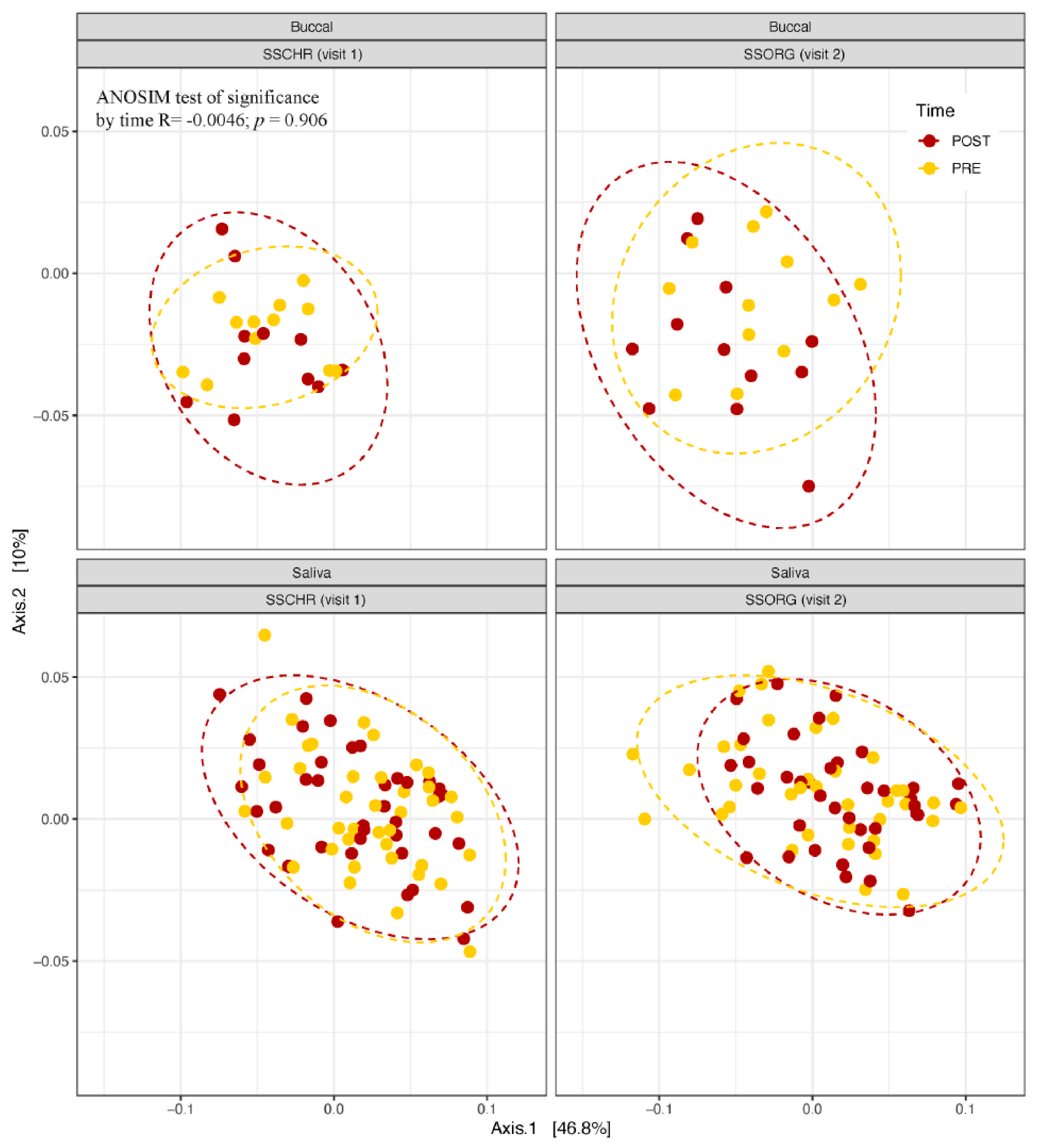
To evaluate beta diversity between time points (PRE and POST), we performed Principal Coordinates Analysis (PCoA) on weighted UniFrac distance matrices of normalized OTU relative abundances. (Figure 2). The first two principal axes explained 56.8% of the variation in bacterial community composition. Similarly to the alpha diversity results, there were no statistically significant changes (ANOSIM p > 0.05) in bacterial diversity between PRE and POST buccal swab samples or PRE and POST saliva samples for both visits. Performing further evaluation at the participant level, we also compared the UniFrac distances for each subject for each sample. PRE and POST smoking samples (of each sample type) from a single participant were characterized by the lowest UniFrac distances and clustered closer together when compared to samples from other participants.

3.4. Core Bacterial Microbiota and Correlation Between Bacterial Genera in Pre-Smoking Samples

The core oral bacterial microbiota in buccal swab and saliva samples from PRE smoking samples was significantly dominated by five taxa: Streptococcus, Veillonella, Rothia, Actinomyces, Granulicatella, Haemophilus and Prevotella (Figure S2). Among all bacterial genera, Streptococcus was at the highest relative abundance in both buccal swab (49%) and saliva (38.2%) samples. While Rothia (16%) and Veillonella (12%) were the other two bacterial genera at >10% relative abundance in buccal swabs, Veillonella was at a 14.2% relative abundance in saliva samples. Other genera identified in both buccal swab and saliva samples included Lactobacillus (1.5% buccal swabs and 2.2% in saliva), Pseudomonas (1.2% in buccal swabs and 2.4% in saliva), Stenotrophomonas (1% in buccal swabs) and Atopobium (1.3% in saliva).

3.5. Bacterial Taxa Associated with a Single Smoking Exposure

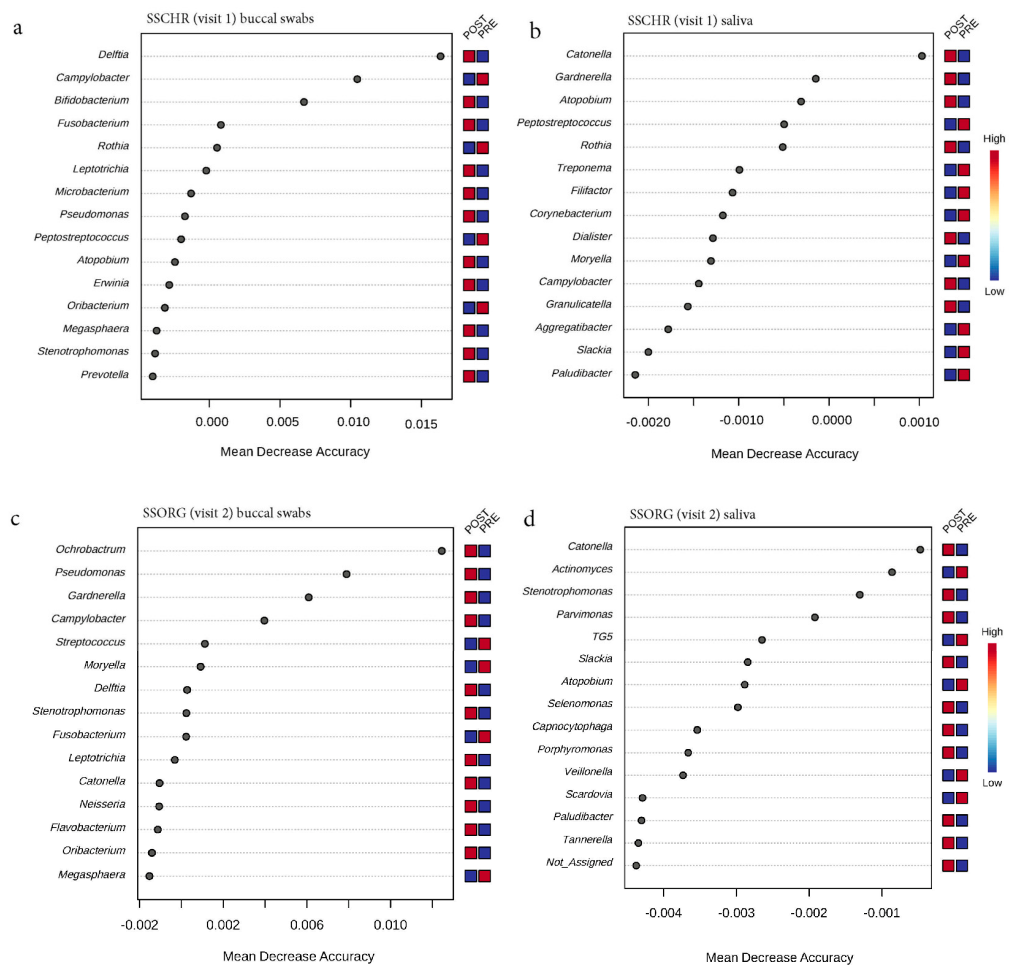
A Random Forest algorithm was applied to construct decision trees to predict the bacterial taxa best associated with PRE and POST samples for both buccal swab and saliva samples (Figure 3). The POST buccal swab samples from both visits were enriched with four bacterial genera, Delfia, Leptotrichia, Pseudomonas, and Stenotrophomonas, when compared to the PRE samples (Figure 3a,c). The POST saliva samples were enriched with Catonella when compared to the PRE samples from both visits (Figure 3b,d). After a single exposure to SSCHR, both buccal swab and saliva POST samples were associated with a depletion of Peptostreptococcus, with enrichment of Atopobium (Figure 3a,b). After a single use of an SSORG product, the higher relative abundance of Stenotrophomonas and Catonella were associated with POST samples, compared to PRE samples for both buccal swab and saliva samples (Figure 3c,d).

3.6. Changes in the Relative Abundance of Bacterial Taxa with a Single Exposure to Smoking

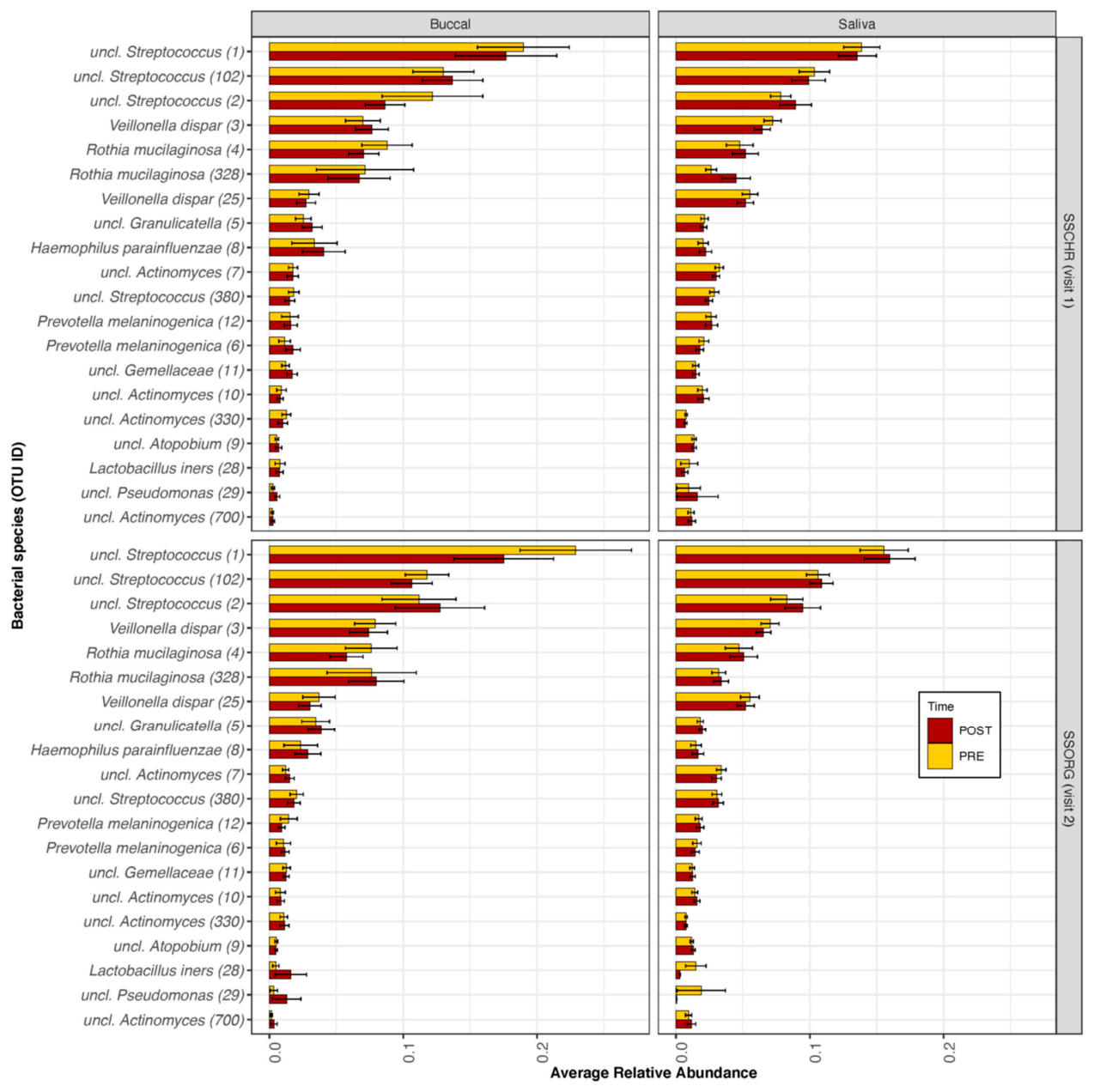
To evaluate the transient changes in the abundance of specific bacterial taxa, we compared the mean relative abundance of the top 20 bacterial OTUs between the PRE and POST smoking samples across buccal swab and saliva samples separately (Figure 4). Three OTUs among the Streptococcus genera (OTU # 1, 102, and 2), Veillonella dispar (OTU# 3 and 25), Rothia mucilaginosa (OTU # 4 and 328), and Prevotella melaninogenica (OTU# 12 and 6) were identified among all samples. There were no statistically significant differences in the relative abundance of the top 20 bacterial taxa between PRE and POST samples when stratified by product smoked and sample type.

3.7. Differences in the Oral Bacterial Microbiota Within and Between Participants

To evaluate changes in the oral bacterial microbiota within participants (intra-individual variation), we compared the PRE samples per participant across sample types and time points (visits). There were no significant differences in bacterial community composition between PRE and POST buccal swab and saliva samples across the two time points for a single individual participant (Table 3). However, the UniFrac distances between bacterial community members within an individual were lower in the buccal swab samples compared to the saliva samples.
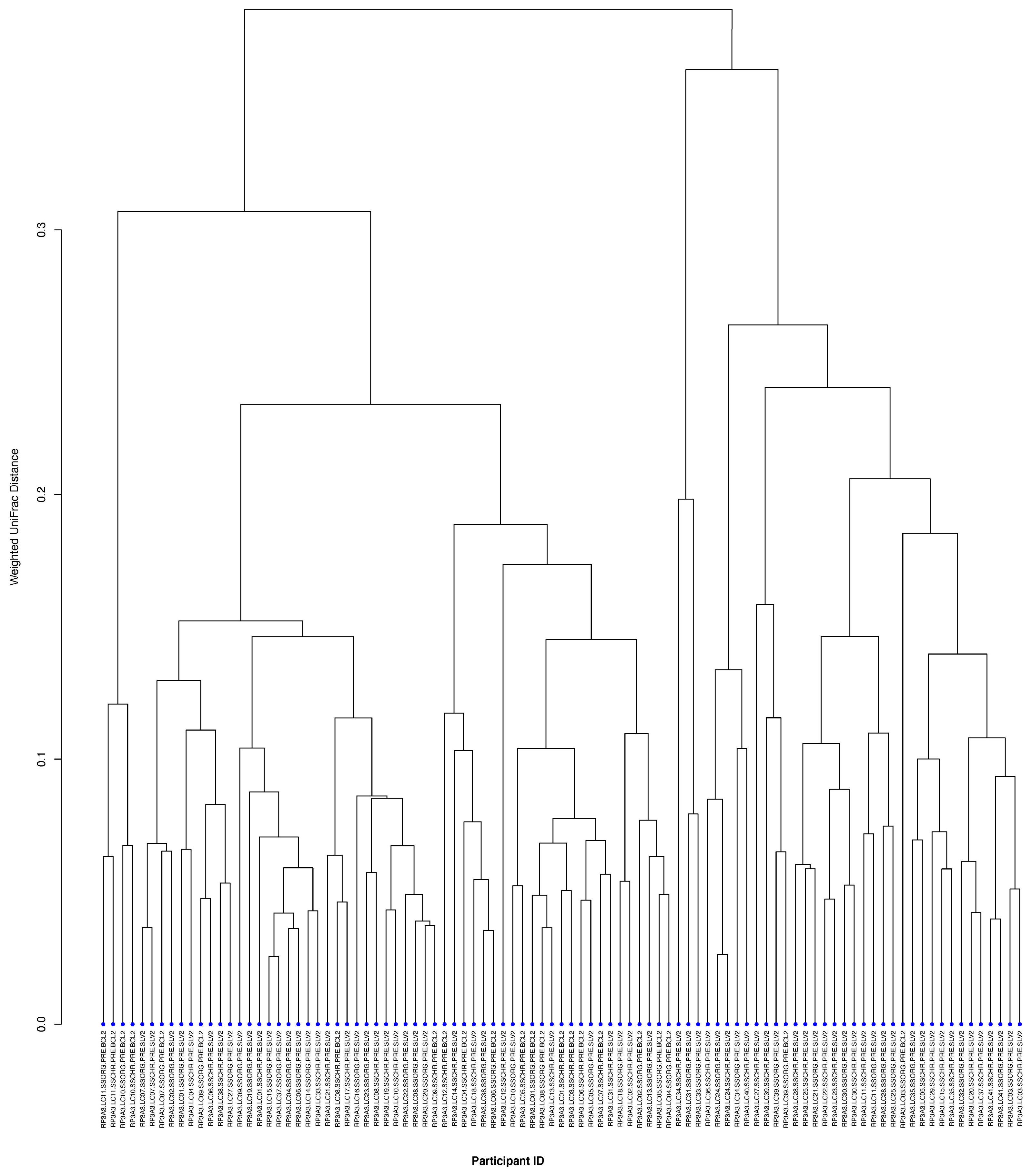
To evaluate bacterial diversity differences between individuals (inter-individual variation), we compared the PRE smoking samples from all 40 participants at each time point. Shannon diversity metrics among all participants were characterized by statistically significant inter-individual differences, among both buccal swab (p = 0.0195) and saliva samples (p = 0.0004) (Figure S1). Beta diversity was evaluated using weighted UniFrac distances between all pre-smoking samples (Figure 5). Across both sample types, while there were no significant differences on their first visit (buccal swabs ANOSIM R = −0.0799; p > 0.05; saliva ANOSIM R = 0.724; p > 0.05), there was significant variation in bacterial community composition across participants during their second visit (buccal swab ANOSIM R = 0.5004; p < 0.05; saliva ANOSIM R = 0.5003; p < 0.05). The inter-individual variation in bacterial genera was also different among the top ten most abundant bacterial genera present in the PRE smoking samples across all participants (Table S1).

4. Discussion

This study evaluated whether or not immediate changes in oral bacterial microbiota occurred after cigarette smokers used a new tobacco product. Overall, our data show that the oral bacterial communities of cigarette smokers remain stable after exposure to the mainstream smoke of a single little cigar product. Furthermore, our data provide a comprehensive characterization, as well as further evidence of the temporal stability, of the oral bacterial microbiota of cigarette smokers.
Previous studies from our group that have evaluated bacterial communities present in the tobacco of little cigars have identified multiple bacterial genera (e.g., Staphylococcus, Pseudomonas, Corynebacterium and Bacillus) in Swisher Sweets little cigars [5,20]. In the present study, the bacterial genera that were associated with our post-smoking samples were identified as Delftia, Leptotrichia, Pseudomonas, Stenotrophomonas and Catonella. While Pseudomonas has been identified in little cigar tobacco products, the other above-mentioned bacterial genera have not been identified in these products. Previous studies have demonstrated that the number of cigarettes smoked and duration of cigarette smoking influence the composition of periodontal pathogens found in smokers’ oral cavities [41,42]. Smokers have also been shown to exhibit relatively unstable biofilm occurrence (when compared to non-smokers) at least within 24 h, including early colonization of periodontal pathogens (e.g., Fusobacterium, Cardiobacterium and Selenomonas) [8]. Yet, in the present study, either the single exposure to little cigar smoke might not have been adequate to trigger these types of responses in smokers, or our methods were not sensitive enough to detect these changes.
Looking into specific bacterial taxa, we found that Streptococcus was the most abundant genera among all participants, followed by Rothia, Veillonella, and Actinomyces. While the relative abundance of Streptococcus, Veillonella and Actinomyces was lower in post-smoking samples (when compared to pre-smoking samples) at both visits, Rothia decreased in relative abundance only on visit two. While a number of studies have pointed out the role of commensals in oral disease development [43,44], the contribution of specific putative pathogens in oral disease initiation is still unclear [45]. In contrast, a higher relative abundance of all of these genera has been shown previously in healthy smokers [46], defining the “core microbiome” of the oral community [47].
While we observed the above-mentioned bacterial genera in all of our samples in comparatively higher relative abundance, we also identified a few genera (that could potentially include opportunistic pathogens) associated with the post-smoking samples (e.g., Delftia, Leptotrichia, Stenotrophomonas, Pseudomonas and Atopobium). Demonstrating resistance to multiple groups of antibiotics, ubiquitous Delftia is an emerging member of the opportunistic healthcare-associated pathogens [48,49,50], a few species of which are also used as plant growth-promoters [51]. Another common oral cavity member, Leptotrichia, has been frequently implicated in periodontal diseases and tooth decay [52]. With the production of potent endotoxin, Leptotrichia has been associated with a spectrum of human diseases [52,53]. Stenotrophomonas has been noted as a persistent colonizer of biofilms, but antibiotic resistance renders some species of this genus as major players in the development of infectious diseases, specifically among immunocompetent individuals [54,55]. Finally, Atopobium has been associated with periodontal diseases and gingival squamous cell carcinomas [56,57].
In addition to describing bacterial genera that were identified in the smokers’ mouths, we also compared variations within bacterial community composition (1) across all subjects at one visit (inter-individual variation) and (2) within each subject across two visits (intra-individual variation). Comparing across individuals, we found significantly more variation in the oral bacterial microbiota in comparison to intra-individual variation. This was consistent with previous studies that demonstrated that oral bacterial community composition is variable among participants, given the multiple intrinsic and extrinsic factors that can affect the oral microbiome of an individual [27,58,59,60]. We also found that the degree of temporal variability (measured with the Shannon diversity index per individual) in the composition of oral bacterial communities was different across all individuals. This was consistent with previous microbiome studies of samples from other areas of the body such as the palm, gut, tongue and vagina [58,61]. In our study, although there was considerable variation between subjects, bacterial profiles within subjects (intra-individual) were stable over the study period. Previous studies have shown similar results when evaluating a range of time periods (24 h to 10 months) [59,60].
There are multiple strengths to note in this study. To our knowledge, this is the first study to investigate potential transient changes in the oral bacterial microbiota resulting from a single exposure to a little cigar. While previous studies have evaluated oral microbiome impacts from tobacco smoking [11,62,63,64], none have sought to assess the immediate impacts on the oral bacteria of the smoker after a single use of a product. In addition, our data relied on high-throughput 16S rRNA sequencing from a robust sample size, and sampling from two different oral cavity locations.
Nevertheless, there were notable limitations to this study as well. First, we could not measure all of the possible factors that could contribute to individual-level variations in the oral bacterial microbiota. Possible factors driving such variations could be an individual’s genetic makeup, diet, or lifestyle/behavior [65,66]. Another limitation of our study was the inability to assess functional capabilities of the oral bacterial microbiota, since we were not able to subject our samples to a metagenomic sequencing approach due to costs. As a result, our analysis is limited to taxonomic compositions inferred from 16S rRNA gene sequencing, and we are unable to directly evaluate microbial gene content or metabolic potential that may underlie the observed community shifts. Additionally, since our work only included data from 16S rRNA gene sequencing, this limited our ability to complete species-level taxonomic assignments and prevented us from determining whether or not detected bacterial genera were viable/live or associated with relic DNA.

5. Conclusions

In conclusion, we found that a single use of a little cigar product did not immediately impact overall oral bacterial diversity among cigarette smokers. However, some bacterial genera were found to be associated with post-smoking samples (Delftia, Leptotrichia, Pseudomonas, Stenotrophomonas and Catonella). Future studies, utilizing a combination of alternative methods (e.g., culture methods, qPCR, metagenomic sequencing), would be needed to evaluate if those bacteria detected in post-smoking oral samples originated from the little cigar products/mainstream smoke and were transferred to the oral cavity, or if they were already present in the oral cavity and were potentially enriched after exposure to mainstream smoke.

Supplementary Materials

The following supporting information can be downloaded at https://www.mdpi.com/article/10.3390/pathogens15070732/s1. Figure S1: Alpha diversity of oral bacterial microbiota per individual subject from buccal and saliva samples collected pre- (red) and post-smoking (yellow) of two separate little cigar products: Swisher sweets original (SSORG) and Swisher sweets cherry (SSCHR); Figure S2: Core bacterial genera present in (a) buccal swab samples and (b) saliva samples. The color gradient shows the prevalence (%) of each genus from zero (blue) to all (red) samples; Table S1: Relative abundance of top 10 bacterial genera in pre-smoking samples across all participants during two visits.

Author Contributions

S.C. conducted laboratory processing and bioinformatic analyses following sequencing and wrote and edited the manuscript. L.M. performed laboratory analyses and edited the manuscript. A.R.S. and E.F.M. contributed to the study design, protocol development, data analysis and interpretation, and manuscript preparation. All authors have read and agreed to the published version of the manuscript.

Funding

This work was supported by the University of Maryland Tobacco Center of Regulatory Science (UMD TCORS) “Rapid Response Characterization of New and Manipulated Tobacco Products” awarded by the National Institute of Health (NIH) and the Food and Drug Administration (FDA)—Award # P50-CA-180523-01. S.C. and A.R.S. were supported by NRT-INFEWS: UMD Global STEWARDS (STEM Training at the Nexus of Energy, Water Reuse and FooD Systems) that was awarded to the University of Maryland School of Public Health by the National Science Foundation National Research Traineeship Program, Grant number 1828910. Authors (S.C., L.M. and A.R.S.) were supported by an Institutional Grant from the University of Maryland Grand Challenges Program that established the Global FEWture Alliance.

Institutional Review Board Statement

The study was conducted in accordance with the Declaration of Helsinki, and approved by the Battelle, OH, Institutional Review Board (IRB 0525-100020389 RPIA Rev 0.0) on 23 May 2016.

Informed Consent Statement

Informed consent was obtained from all subjects involved in the study.

Data Availability Statement

Data concerning the samples included in this study are deposited under the NCBI BioProject accession number PRJNA690810.

Acknowledgments

The authors are thankful to Lauren Hittle for her help with performing the sequencing of all samples.

Conflicts of Interest

The authors declare that they have no conflicts of interest.

Disclaimer

Mongodin contributed to this article as an employee of the University of Maryland School of Medicine. The views expressed are his own and do not necessarily represent the views of the National Institutes of Health or the United States Government.

References

  1. Sapkota, A.R.; Berger, S.; Vogel, T.M. Human pathogens abundant in the bacterial metagenome of cigarettes. Environ. Health Perspect. 2010, 118, 351–356. [Google Scholar] [CrossRef] [Scilit] [PubMed]
  2. Tyx, R.E.; Stanfill, S.B.; Keong, L.M.; Rivera, A.J.; Satten, G.A.; Watson, C.H. Characterization of Bacterial Communities in Selected Smokeless Tobacco Products Using 16S rDNA Analysis. PLoS ONE 2016, 11, e0146939. [Google Scholar] [CrossRef] [Scilit] [PubMed]
  3. Chopyk, J.; Chattopadhyay, S.; Kulkarni, P.; Smyth, E.M.; Hittle, L.E.; Paulson, J.N.; Pop, M.; Buehler, S.S.; Clark, P.I.; Mongodin, E.F.; et al. Temporal Variations in Cigarette Tobacco Bacterial Community Composition and Tobacco-Specific Nitrosamine Content Are Influenced by Brand and Storage Conditions. Front. Microbiol. 2017, 8, 358. [Google Scholar] [CrossRef] [Scilit] [PubMed]
  4. Hani, J.; Abdel Nour, G.; Matta, J.; Jazzar, B.; Pfaffl, M.W.; Hanna-Wakim, L.; Abdel Nour, A.M. Shisha microbiota: The good, the bad and the not so ugly. BMC Res. Notes 2018, 11, 446. [Google Scholar] [CrossRef] [Scilit] [PubMed]
  5. Smyth, E.M.; Chattopadhyay, S.; Babik, K.; Reid, M.; Chopyk, J.; Malayil, L.; Kulkarni, P.; Hittle, L.E.; Clark, P.I.; Sapkota, A.R.; et al. The Bacterial Communities of Little Cigars and Cigarillos Are Dynamic Over Time and Varying Storage Conditions. Front. Microbiol. 2019, 10, 2371. [Google Scholar] [CrossRef] [Scilit] [PubMed]
  6. Malayil, L.; Chattopadhyay, S.; Kulkarni, P.; Hittle, L.; Clark, P.I.; Mongodin, E.F.; Sapkota, A.R. Mentholation triggers brand-specific shifts in the bacterial microbiota of commercial cigarette products. Appl. Microbiol. Biotechnol. 2020, 104, 6287–6297. [Google Scholar] [CrossRef] [Scilit] [PubMed]
  7. Malayil, L.; Chattopadhyay, S.; Bui, A.; Panse, M.; Cagle, R.; Mongodin, E.F.; Sapkota, A.R. Viable bacteria abundant in cigarettes are aerosolized in mainstream smoke. Environ. Res. 2022, 212, 113462. [Google Scholar] [CrossRef] [Scilit] [PubMed]
  8. Kumar, P.S.; Matthews, C.R.; Joshi, V.; de Jager, M.; Aspiras, M. Tobacco smoking affects bacterial acquisition and colonization in oral biofilms. Infect. Immun. 2011, 79, 4730–4738. [Google Scholar] [CrossRef] [Scilit] [PubMed]
  9. Joshi, V.; Matthews, C.; Aspiras, M.; de Jager, M.; Ward, M.; Kumar, P. Smoking decreases structural and functional resilience in the subgingival ecosystem. J. Clin. Periodontol. 2014, 41, 1037–1047. [Google Scholar] [CrossRef] [Scilit] [PubMed]
  10. Mason, M.R.; Preshaw, P.M.; Nagaraja, H.N.; Dabdoub, S.M.; Rahman, A.; Kumar, P.S. The subgingival microbiome of clinically healthy current and never smokers. ISME J. 2015, 9, 268–272. [Google Scholar] [CrossRef] [Scilit] [PubMed]
  11. Wu, J.; Peters, B.A.; Dominianni, C.; Zhang, Y.; Pei, Z.; Yang, L.; Ma, Y.; Purdue, M.P.; Jacobs, E.J.; Gapstur, S.M.; et al. Cigarette smoking and the oral microbiome in a large study of American adults. ISME J. 2016, 10, 2435–2446. [Google Scholar] [CrossRef] [Scilit] [PubMed]
  12. Jia, Y.-J.; Liao, Y.; He, Y.-Q.; Zheng, M.-Q.; Tong, X.-T.; Xue, W.-Q.; Zhang, J.-B.; Yuan, L.-L.; Zhang, W.-L.; Jia, W.-H. Association Between Oral Microbiota and Cigarette Smoking in the Chinese Population. Front. Cell. Infect. Microbiol. 2021, 11, 658203. [Google Scholar] [CrossRef] [Scilit] [PubMed]
  13. Suárez, L.J.; Garzón, H.; Arboleda, S.; Rodríguez, A. Oral Dysbiosis and Autoimmunity: From Local Periodontal Responses to an Imbalanced Systemic Immunity. A Review. Front. Immunol. 2020, 11, 591255. Available online: https://www.frontiersin.org/article/10.3389/fimmu.2020.591255 (accessed on 19 April 2022). [CrossRef] [Scilit] [PubMed]
  14. Kadam, S.; Vandana, M.; Patwardhan, S.; Kaushik, K.S. Looking beyond the smokescreen: Can the oral microbiome be a tool or target in the management of tobacco-associated oral cancer? Ecancermedicalscience 2021, 15, 1179. [Google Scholar] [CrossRef] [Scilit] [PubMed]
  15. Kumar, P.S. Microbial dysbiosis: The root cause of periodontal disease. J. Periodontol. 2021, 92, 1079–1087. [Google Scholar] [CrossRef] [Scilit] [PubMed]
  16. Ghosh, A.; Abdelwahab, S.H.; Reeber, S.L.; Reidel, B.; Marklew, A.J.; Garrison, A.J.; Lee, S.; Dang, H.; Herring, A.H.; Glish, G.L.; et al. Little Cigars are More Toxic than Cigarettes and Uniquely Change the Airway Gene and Protein Expression. Sci. Rep. 2017, 7, 46239. [Google Scholar] [CrossRef] [Scilit] [PubMed]
  17. Hamad, S.H.; Johnson, N.M.; Tefft, M.E.; Brinkman, M.C.; Gordon, S.M.; Clark, P.I.; Buehler, S.S. Little Cigars vs 3R4F Cigarette: Physical Properties and HPHC Yields. Tob. Regul. Sci. 2017, 3, 459–478. [Google Scholar] [CrossRef] [Scilit] [PubMed]
  18. Pickworth, W.B.; Rosenberry, Z.R.; Koszowski, B. Toxicant exposure from smoking a little cigar: Further support for product regulation. Tob. Control 2017, 26, 269–276. [Google Scholar] [CrossRef] [Scilit] [PubMed]
  19. Edwards, S.H.; Hassink, M.D.; Taylor, K.M.; Watson, C.H.; Kuklenyik, P.; Kimbrell, B.; Wang, L.; Chen, P.; Valentín-Blasini, L. Tobacco-Specific Nitrosamines in the Tobacco and Mainstream Smoke of Commercial Little Cigars. Chem. Res. Toxicol. 2021, 34, 1034–1045. [Google Scholar] [CrossRef] [Scilit] [PubMed]
  20. Chattopadhyay, S.; Smyth, E.M.; Kulkarni, P.; Babik, K.R.; Reid, M.; Hittle, L.E.; Clark, P.I.; Mongodin, E.F.; Sapkota, A.R. Little cigars and cigarillos harbor diverse bacterial communities that differ between the tobacco and the wrapper. PLoS ONE 2019, 14, e0211705. [Google Scholar] [CrossRef] [Scilit] [PubMed]
  21. Al Bataineh, M.T.; Dash, N.R.; Elkhazendar, M.; Alnusairat, D.M.H.; Darwish, I.M.I.; Al-Hajjaj, M.S.; Hamid, Q. Revealing oral microbiota composition and functionality associated with heavy cigarette smoking. J. Transl. Med. 2020, 18, 421. [Google Scholar] [CrossRef] [Scilit] [PubMed]
  22. Costello, E.K.; Lauber, C.L.; Hamady, M.; Fierer, N.; Gordon, J.I.; Knight, R. Bacterial community variation in human body habitats across space and time. Science 2009, 326, 1694–1697. [Google Scholar] [CrossRef] [Scilit] [PubMed]
  23. Cameron, S.J.S.; Huws, S.A.; Hegarty, M.J.; Smith, D.P.M.; Mur, L.A.J. The human salivary microbiome exhibits temporal stability in bacterial diversity. FEMS Microbiol. Ecol. 2015, 91, fiv091. [Google Scholar] [CrossRef] [Scilit] [PubMed]
  24. Tamashiro, R.; Strange, L.; Schnackenberg, K.; Santos, J.; Gadalla, H.; Zhao, L.; Li, E.C.; Hill, E.; Hill, B.; Sidhu, G.; et al. Stability of healthy subgingival microbiome across space and time. Sci. Rep. 2021, 11, 23987. [Google Scholar] [CrossRef] [Scilit] [PubMed]
  25. The Human Microbiome Project Consortium. A framework for human microbiome research. Nature 2012, 486, 215–221. [Google Scholar] [CrossRef] [Scilit] [PubMed]
  26. Segata, N.; Haake, S.K.; Mannon, P.; Lemon, K.P.; Waldron, L.; Gevers, D.; Huttenhower, C.; Izard, J. Composition of the adult digestive tract bacterial microbiome based on seven mouth surfaces, tonsils, throat and stool samples. Genome Biol. 2012, 13, R42. [Google Scholar] [CrossRef] [Scilit] [PubMed]
  27. Hall, M.W.; Singh, N.; Ng, K.F.; Lam, D.K.; Goldberg, M.B.; Tenenbaum, H.C.; Neufeld, J.D.; Beiko, R.G.; Senadheera, D.B. Inter-personal diversity and temporal dynamics of dental, tongue, and salivary microbiota in the healthy oral cavity. npj Biofilms Microbiomes 2017, 3, 2. [Google Scholar] [CrossRef] [Scilit] [PubMed]
  28. Jiang, W.-X.; Hu, Y.-J.; Gao, L.; He, Z.-Y.; Zhu, C.-L.; Ma, R.; Huang, Z.-W. The impact of various time intervals on the supragingival plaque dynamic core microbiome. PLoS ONE 2015, 10, e0124631. [Google Scholar] [CrossRef] [Scilit] [PubMed]
  29. Pushalkar, S.; Paul, B.; Li, Q.; Yang, J.; Vasconcelos, R.; Makwana, S.; González, J.M.; Shah, S.; Xie, C.; Janal, M.N.; et al. Electronic Cigarette Aerosol Modulates the Oral Microbiome and Increases Risk of Infection. iScience 2020, 23, 100884. [Google Scholar] [CrossRef] [Scilit] [PubMed]
  30. Chopyk, J.; Bojanowski, C.M.; Shin, J.; Moshensky, A.; Fuentes, A.L.; Bonde, S.S.; Chuki, D.; Pride, D.T.; Crotty Alexander, L.E. Compositional Differences in the Oral Microbiome of E-cigarette Users. Front. Microbiol. 2021, 12, 599664. [Google Scholar] [CrossRef] [Scilit] [PubMed]
  31. Thomas, S.C.; Xu, F.; Pushalkar, S.; Lin, Z.; Thakor, N.; Vardhan, M.; Flaminio, Z.; Khodadadi-Jamayran, A.; Vasconcelos, R.; Akapo, A.; et al. Electronic Cigarette Use Promotes a Unique Periodontal Microbiome. mBio 2022, 13, e00075-22. [Google Scholar] [CrossRef] [Scilit] [PubMed]
  32. Wang, T.W.; Falvey, K.; Gammon, D.G.; Loomis, B.R.; Kuiper, N.M.; Rogers, T.; King, B.A. Sales Trends in Price-Discounted Cigarettes, Large Cigars, Little Cigars, and Cigarillos-United States, 2011–2016. Nicotine Tob. Res. 2018, 20, 1401–1406. [Google Scholar] [CrossRef] [Scilit] [PubMed]
  33. The Human Microbiome Project Consortium. Structure, Function and Diversity of the Healthy Human Microbiome. Nature 2012, 486, 207–214. [Google Scholar] [CrossRef] [Scilit] [PubMed]
  34. Chopyk, J.; Chattopadhyay, S.; Kulkarni, P.; Claye, E.; Babik, K.R.; Reid, M.C.; Smyth, E.M.; Hittle, L.E.; Paulson, J.N.; Cruz-Cano, R.; et al. Mentholation affects the cigarette microbiota by selecting for bacteria resistant to harsh environmental conditions and selecting against potential bacterial pathogens. Microbiome 2017, 5, 22. [Google Scholar] [CrossRef] [Scilit] [PubMed]
  35. Fadrosh, D.W.; Ma, B.; Gajer, P.; Sengamalay, N.; Ott, S.; Brotman, R.M.; Ravel, J. An improved dual-indexing approach for multiplexed 16S rRNA gene sequencing on the Illumina MiSeq platform. Microbiome 2014, 2, 6. [Google Scholar] [CrossRef] [Scilit] [PubMed]
  36. Holm, J.B.; Humphrys, M.S.; Robinson, C.K.; Settles, M.L.; Ott, S.; Fu, L.; Yang, H.; Gajer, P.; He, X.; McComb, E.; et al. Ultrahigh-Throughput Multiplexing and Sequencing of >500-Base-Pair Amplicon Regions on the Illumina HiSeq 2500 Platform. mSystems 2019, 4, e00029-19. [Google Scholar] [CrossRef] [Scilit] [PubMed]
  37. McMurdie, P.J.; Holmes, S. phyloseq: An R Package for Reproducible Interactive Analysis and Graphics of Microbiome Census Data. PLoS ONE 2013, 8, e61217. [Google Scholar] [CrossRef] [Scilit] [PubMed]
  38. Paulson, J.N.; Stine, O.C.; Bravo, H.C.; Pop, M. Differential abundance analysis for microbial marker-gene surveys. Nat. Methods 2013, 10, 1200–1202. [Google Scholar] [CrossRef] [Scilit] [PubMed]
  39. Dhariwal, A.; Chong, J.; Habib, S.; King, I.L.; Agellon, L.B.; Xia, J. MicrobiomeAnalyst: A web-based tool for comprehensive statistical, visual and meta-analysis of microbiome data. Nucleic Acids Res. 2017, 45, W180–W188. [Google Scholar] [CrossRef] [Scilit] [PubMed]
  40. Chong, J.; Liu, P.; Zhou, G.; Xia, J. Using MicrobiomeAnalyst for comprehensive statistical, functional, and meta-analysis of microbiome data. Nat. Protoc. 2020, 15, 799–821. [Google Scholar] [CrossRef] [Scilit] [PubMed]
  41. Shiloah, J.; Patters, M.R.; Waring, M.B. The prevalence of pathogenic periodontal microflora in healthy young adult smokers. J. Periodontol. 2000, 71, 562–567. [Google Scholar] [CrossRef] [Scilit] [PubMed]
  42. Gomes, S.C.; Piccinin, F.B.; Oppermann, R.V.; Susin, C.; Nonnenmacher, C.I.; Mutters, R.; Marcantonio, R.A. Periodontal Status in Smokers and Never-Smokers: Clinical Findings and Real-Time Polymerase Chain Reaction Quantification of Putative Periodontal Pathogens. J. Periodontol. 2006, 77, 1483–1490. [Google Scholar] [CrossRef] [Scilit] [PubMed]
  43. Rosier, B.T.; De Jager, M.; Zaura, E.; Krom, B.P. Historical and contemporary hypotheses on the development of oral diseases: Are we there yet? Front. Cell. Infect. Microbiol. 2014, 4, 92. [Google Scholar] [CrossRef] [Scilit] [PubMed]
  44. Kirst, M.E.; Li, E.C.; Alfant, B.; Chi, Y.-Y.; Walker, C.; Magnusson, I.; Wang, G.P. Dysbiosis and Alterations in Predicted Functions of the Subgingival Microbiome in Chronic Periodontitis. Appl. Environ. Microbiol. 2015, 81, 783–793. [Google Scholar] [CrossRef] [Scilit] [PubMed]
  45. Van Dyke, T.E.; Bartold, P.M.; Reynolds, E.C. The Nexus Between Periodontal Inflammation and Dysbiosis. Front. Immunol. 2020, 11, 511. [Google Scholar] [CrossRef] [Scilit] [PubMed]
  46. Al Kawas, S.; Al-Marzooq, F.; Rahman, B.; Shearston, J.A.; Saad, H.; Benzina, D.; Weitzman, M. The impact of smoking different tobacco types on the subgingival microbiome and periodontal health: A pilot study. Sci. Rep. 2021, 11, 1113. [Google Scholar] [CrossRef] [Scilit] [PubMed]
  47. Stahringer, S.S.; Clemente, J.C.; Corley, R.P.; Hewitt, J.; Knights, D.; Walters, W.A.; Knight, R.; Krauter, K.S. Nurture trumps nature in a longitudinal survey of salivary bacterial communities in twins from early adolescence to early adulthood. Genome Res. 2012, 22, 2146–2152. [Google Scholar] [CrossRef] [Scilit] [PubMed]
  48. Bilgin, H.; Sarmis, A.; Tigen, E.; Soyletir, G.; Mulazimoglu, L. Delftia acidovorans: A Rare Pathogen in Immunocompetent and Immunocompromised Patients. Can. J. Infect. Dis. Med. Microbiol. 2015, 26, 277–279. [Google Scholar] [CrossRef] [Scilit] [PubMed]
  49. Ranc, A.; Dubourg, G.; Fournier, P.E.; Raoult, D.; Fenollar, F. Delftia tsuruhatensis, an Emergent Opportunistic Healthcare-Associated Pathogen. Emerg. Infect. Dis. 2018, 24, 594–596. [Google Scholar] [CrossRef] [Scilit] [PubMed]
  50. Paun, V.I.; Lavin, P.; Chifiriuc, M.C.; Purcarea, C. First report on antibiotic resistance and antimicrobial activity of bacterial isolates from 13,000-year old cave ice core. Sci. Rep. 2021, 11, 514. [Google Scholar] [CrossRef] [Scilit] [PubMed]
  51. Ubalde, M.C.; Braña, V.; Sueiro, F.; Morel, M.A.; Martínez-Rosales, C.; Marquez, C.; Castro-Sowinski, S. The Versatility of Delftia sp. Isolates as Tools for Bioremediation and Biofertilization Technologies. Curr. Microbiol. 2012, 64, 597–603. [Google Scholar] [CrossRef] [Scilit] [PubMed]
  52. Eribe, E.R.K.; Olsen, I. Leptotrichia species in human infections. Anaerobe 2008, 14, 131–137. [Google Scholar] [CrossRef] [Scilit] [PubMed]
  53. Hou, H.; Chen, Z.; Tian, L.; Sun, Z. Leptotrichia trevisanii bacteremia in a woman with systemic lupus erythematosus receiving high-dose chemotherapy. BMC Infect. Dis. 2018, 18, 661. [Google Scholar] [CrossRef] [Scilit] [PubMed]
  54. Flores-Treviño, S.; Bocanegra-Ibarias, P.; Camacho-Ortiz, A.; Morfín-Otero, R.; Salazar-Sesatty, H.A.; Garza-González, E. Stenotrophomonas maltophilia biofilm: Its role in infectious diseases. Expert Rev. Anti Infect. Ther. 2019, 17, 877–893. [Google Scholar] [CrossRef] [Scilit] [PubMed]
  55. de Souza, M.L.M.; Castelo-Branco, I.V.M.; de Holanda, L.A.L.; Frade, A.L.; de Barros Santos, C.; Moura, V.C.; Taguchi, L.C.; de Lacerda Vida, A.K. Acute necrotizing ulcerative gingivitis associated with Stenotrophomonas maltophilia, a case report. J. Oral Diagn. 2020, 5, 1–6. [Google Scholar] [CrossRef] [Scilit]
  56. Kumar, P.S.; Griffen, A.L.; Moeschberger, M.L.; Leys, E.J. Identification of Candidate Periodontal Pathogens and Beneficial Species by Quantitative 16S Clonal Analysis. J. Clin. Microbiol. 2005, 43, 3944–3955. [Google Scholar] [CrossRef] [Scilit] [PubMed]
  57. Li, Y.; Tan, X.; Zhao, X.; Xu, Z.; Dai, W.; Duan, W.; Huang, S.; Zhang, E.; Liu, J.; Zhang, S.; et al. Composition and function of oral microbiota between gingival squamous cell carcinoma and periodontitis. Oral Oncol. 2020, 107, 104710. [Google Scholar] [CrossRef] [Scilit] [PubMed]
  58. Flores, G.E.; Caporaso, J.G.; Henley, J.B.; Rideout, J.R.; Domogala, D.; Chase, J.; Leff, J.W.; Vázquez-Baeza, Y.; Gonzalez, A.; Knight, R.; et al. Temporal variability is a personalized feature of the human microbiome. Genome Biol. 2014, 15, 531. [Google Scholar] [CrossRef] [Scilit] [PubMed]
  59. Belstrøm, D.; Holmstrup, P.; Bardow, A.; Kokaras, A.; Fiehn, N.-E.; Paster, B.J. Temporal Stability of the Salivary Microbiota in Oral Health. PLoS ONE 2016, 11, e0147472. [Google Scholar] [CrossRef] [Scilit] [PubMed]
  60. Vogtmann, E.; Hua, X.; Zhou, L.; Wan, Y.; Suman, S.; Zhu, B.; Dagnall, C.L.; Hutchinson, A.; Jones, K.; Hicks, B.D.; et al. Temporal Variability of Oral Microbiota over 10 Months and the Implications for Future Epidemiologic Studies. Cancer Epidemiol. Biomarkers Prev. 2018, 27, 594–600. [Google Scholar] [CrossRef] [Scilit] [PubMed]
  61. Gajer, P.; Brotman, R.M.; Bai, G.; Sakamoto, J.; Schütte, U.M.E.; Zhong, X.; Koenig, S.S.K.; Fu, L.; Ma, Z.; Zhou, X.; et al. Temporal Dynamics of the Human Vaginal Microbiota. Sci. Transl. Med. 2012, 4, 132ra52. [Google Scholar] [CrossRef] [Scilit] [PubMed]
  62. Jiang, Y.; Zhou, X.; Cheng, L.; Li, M. The Impact of Smoking on Subgingival Microflora: From Periodontal Health to Disease. Front. Microbiol. 2020, 11, 66. [Google Scholar] [CrossRef] [Scilit] [PubMed]
  63. Sato, N.; Kakuta, M.; Hasegawa, T.; Yamaguchi, R.; Uchino, E.; Kobayashi, W.; Sawada, K.; Tamura, Y.; Tokuda, I.; Murashita, K.; et al. Metagenomic analysis of bacterial species in tongue microbiome of current and never smokers. npj Biofilms Microbiomes 2020, 6, 11. [Google Scholar] [CrossRef] [Scilit] [PubMed]
  64. Suzuki, N.; Nakano, Y.; Yoneda, M.; Hirofuji, T.; Hanioka, T. The effects of cigarette smoking on the salivary and tongue microbiome. Clin. Exp. Dent. Res. 2021, 8, 449–456. [Google Scholar] [CrossRef] [Scilit] [PubMed]
  65. Murtaza, N.; Burke, L.M.; Vlahovich, N.; Charlesson, B.; O’Neill, H.M.; Ross, M.L.; Campbell, K.L.; Krause, L.; Morrison, M. Analysis of the Effects of Dietary Pattern on the Oral Microbiome of Elite Endurance Athletes. Nutrients 2019, 11, 614. [Google Scholar] [CrossRef] [Scilit] [PubMed]
  66. Cornejo Ulloa, P.; van der Veen, M.H.; Krom, B.P. Review: Modulation of the oral microbiome by the host to promote ecological balance. Odontology 2019, 107, 437–448. [Google Scholar] [CrossRef] [Scilit] [PubMed]
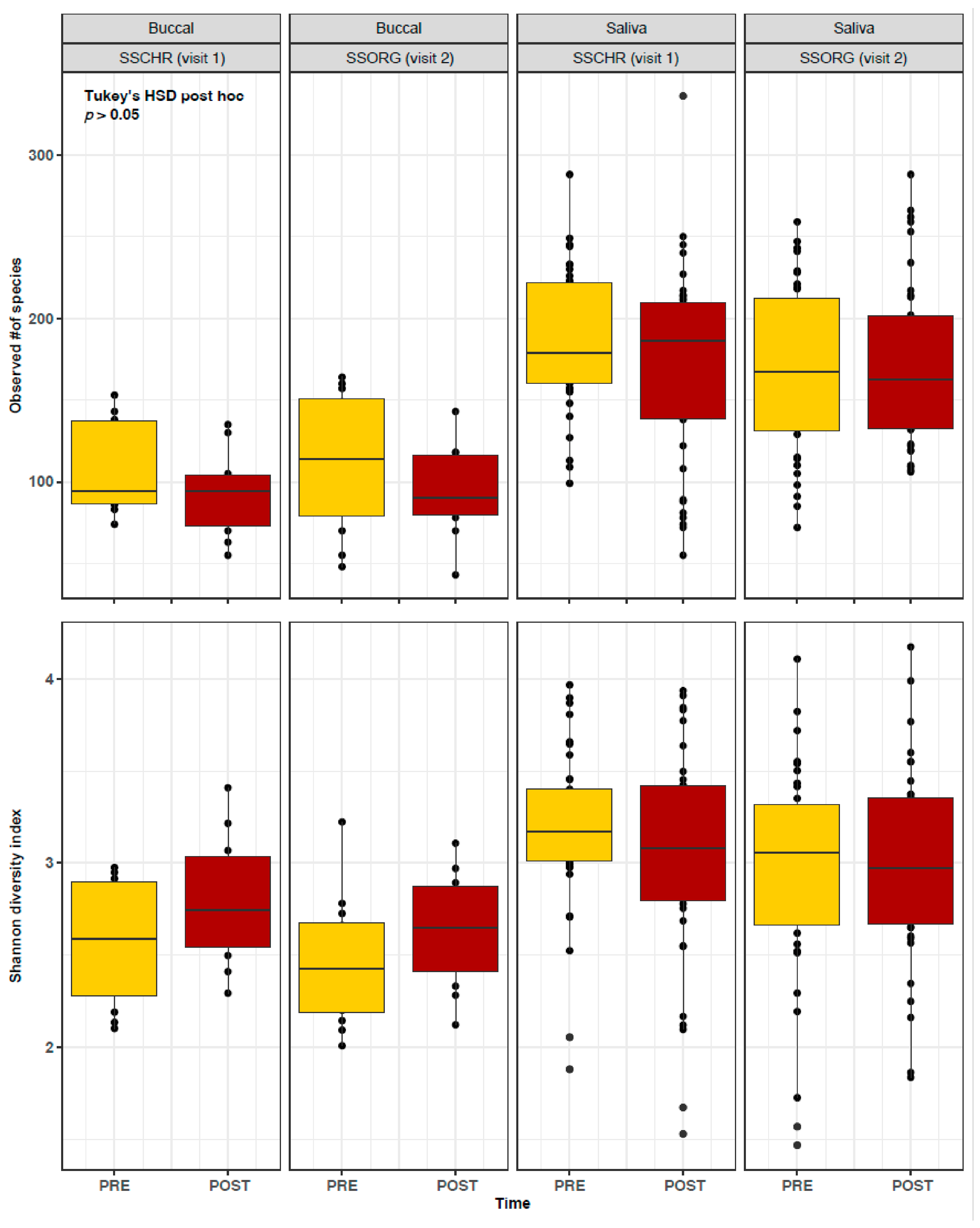
Figure 1. Alpha diversity analysis of buccal swab and saliva samples collected pre- and post-smoking of two little cigar products: Swisher sweets original (SSORG) and Swisher sweets cherry (SSCHR). The pre-smoking samples are denoted with yellow bars and post-smoking samples are denoted with red bars. Diversity was measured between pre-smoking (PRE) and post-smoking (POST) samples using ANOVA with Tukey’s HSD post hoc test.
Pathogens 15 00732 g001
Figure 2. PCoA plot measuring beta diversity between pre- and post-smoking samples from each visit. Ellipses are drawn at 95% confidence intervals. The statistical significance of beta diversity by time was measured by the ANOSIM test of significance and p-values < 0.05 were considered significant.
Pathogens 15 00732 g002
Figure 3. Predicted association of bacterial species between pre- (PRE) and post-smoking (POST) samples using the Random Forest algorithm in (a) buccal swabs from visit 1; (b) saliva from visit 1; (c) buccal swabs from visit 2; and (d) saliva from visit 2. The increasing abundance of each bacterial genera is represented by the color scale (blue to red).
Pathogens 15 00732 g003
Figure 4. Average relative abundance (±SE) of the top 20 bacterial species (OTU ID) present in buccal swab and saliva samples from pre- (denoted in yellow bars) and post-smoking (denoted in red bars) samples in each visit.
Pathogens 15 00732 g004
Figure 5. Histogram of weighted UniFrac distances of all pre-smoking samples across all participants.
Pathogens 15 00732 g005
Table 1. Demographics of the study participants.
n = 40%
Gender
Female1640
Male2152.5
Don’t know/Refused37.5
Age
25–351127.5
36–451127.5
46–55820
55+717.5
Don’t know/Refused37.5
Marital Status
Legally married25
Living with Partner410
Single/never married2255
Divorced717.5
Separated25
Don’t know/Refused37.5
Race
Black or African American1947.5
White1845
Don’t know/Refused37.5
Employment
Full-Time, 35+ h/week1332.5
Part-Time, irregular h/day work717.5
Part-Time, regular h512.5
Retired/Disabled12.5
Homemaker25
Unemployed922.5
Don’t know/Refused37.5
Table 2. Little cigar use of all study subjects. For frequency of smoking little cigars, ‘Everyday’ refers to smokers who used little cigars on all days of the past 30 days, ‘Some days’ refers to smokers who used little cigars only on few days in the past 30 days, and ‘Not at all’ refers to smokers who did not smoke a little cigar in the past 30 days.
Subject IDTried Little CigarLittle Cigar Use
Frequency of SmokingBrandFlavor
LC01YesSome daysSwisher SweetsFull Flavor
LC02YesSome daysDon’t knowMenthol
LC03YesNot at allWinchesterRegular
LC04YesEverydayGarcia gameGrape/honey
LC05YesNot at allBlack & MildRegular
LC06YesNot at allSwisherGrape
LC07YesEverydayBlack & MildRegular
LC08YesNot at allNANA
LC09YesSome daysSwisher SweetsRegular, grape, sour apple
LC10YesSome daysSwisher
LC11YesEverydaySenecaFull Flavor
LC12YesSome daysSwisher SweetsRegular
LC13YesSome daysSwisherRegular
LC14YesSome daysGarcia vega game and Swisher SweetsCherry
LC15YesEverydayBlack & MildCigarillo
LC16YesNot at allNANA
LC17YesEverydaySwisher and Black & MildTropical
LC18No Not at allNANA
LC19YesSome daysCigarillosCherry
LC20YesSome daysBlack & MildWine
LC21YesNot at allSwisherRegular
LC22YesEverydaySwisher SweetsGrape
LC23YesNot at allSwisherRegular
LC24YesSome daysBlack & MildRegular
LC25YesSome daysSwisher Sweet CigarillosPlain
LC27No Not at allNANA
LC28YesEverydayCheyenneFull flavor
LC29YesSome daysSwisher Sweet CigarillosPeach
LC30YesEverydaySwisher Sweet CigarillosSweet
LC31No Not at allNANA
LC32YesSome daysSenecaRegular
LC33YesNot at allNANA
LC34YesSome daysshowMango
LC35YesSome daysBlack & MildWine
LC36YesSome daysDjarumClove
LC37YesSome daysSwisherRegular
LC38YesSome daysBlack & MildCherry blend
LC39YesNot at allNANA
LC40YesSome daysAny/RandomCherry blend
LC41YesSome daysBlack & MildRegular
Table 3. Within and between individual variation in UniFrac distances.
VariationSample TypeProduct SmokedTimeUniFrac Distance
Visit # Average SD
Intra-individual (within a subject)SalivaVisits 1 + 2Pre0.110.07
Post0.110.08
BuccalVisits 1 + 2Pre0.090.04
Post0.110.06
Inter-individual (between subjects)SalivaVisits 1 + 2Pre0.160.07
Post0.180.08
SSCHR (visit 1)Pre0.160.06
Post0.170.07
SSORG (visit 2)Pre0.170.08
Post0.180.09
BuccalVisits 1 + 2Pre0.150.06
Post0.150.06
SSCHR (visit 1)Pre0.140.06
Post0.140.05
SSORG (visit 2)Pre0.170.06
Post0.160.07
Disclaimer/Publisher’s Note: The statements, opinions and data contained in all publications are solely those of the individual author(s) and contributor(s) and not of MDPI and/or the editor(s). MDPI and/or the editor(s) disclaim responsibility for any injury to people or property resulting from any ideas, methods, instructions or products referred to in the content.