Article

Diagnostic Pathways and Genotyping of Cases of Echinococcus granulosus from Polish Patients
by
Albert Gandurski
1,†,
Marta Tokaj
1,†,
Michał Jerzak
1,*,
Aleksandra Popławska-Ferenc
2,3,
Piotr Małkowski
2,3 and
Monika Dybicz
1,*
1
Department of General Biology and Parasitology, Medical University of Warsaw, 02-004 Warsaw, Poland
2
Department of General and Transplant Surgery, Medical University of Warsaw, 02-006 Warsaw, Poland
3
Department of Surgical and Transplantation Nursing and Extracorporeal Treatment, Faculty of Health Sciences, Medical University of Warsaw, 02-006 Warsaw, Poland
*
Authors to whom correspondence should be addressed.
These authors contributed equally to this work.
Pathogens 2026, 15(5), 459; https://doi.org/10.3390/pathogens15050459
Submission received: 6 March 2026 / Revised: 16 April 2026 / Accepted: 21 April 2026 / Published: 23 April 2026
(This article belongs to the Special Issue Updates on Zoonotic Parasites)

Abstract

Cystic echinococcosis (CE) is caused by a tapeworm of the Echinococcus granulosus s.l. species complex belonging to the Taeniidae family. CE affects more than 100 countries, including Poland, while remaining a significant public health threat to both humans and livestock. The aim of this study was to identify the genotypes responsible for cases of cystic echinococcosis in Poland by conducting molecular analysis of larvae isolated from Polish patients, and to investigate the diagnostic pathways leading to CE diagnosis. Between April 2023 and January 2025, tissue samples were collected from 10 patients following hepatectomy. Analysis of diagnostic pathways revealed that radiological findings followed by PCR or histopathological testing were sufficient to establish a reliable diagnosis of CE in 90% and 100% of cases, respectively. Serological tests showed lower sensitivity, reaching 86% for ELISA and 71% for Western blot. DNA extracted from all samples was used as the template in PCR to amplify and sequence the region of the mitochondrial NADH dehydrogenase 1 gene (nad1). PCR analysis confirmed presence of Echinococcus granulosus s.l. species in eight cases. All obtained nad1 sequences showed identity with the Echinococcus canadensis G7 (pig) strain. These results indicate that it remains the most frequent causative agent of human cystic echinococcosis in Poland.

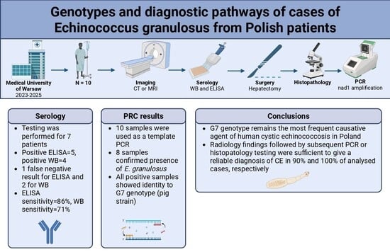
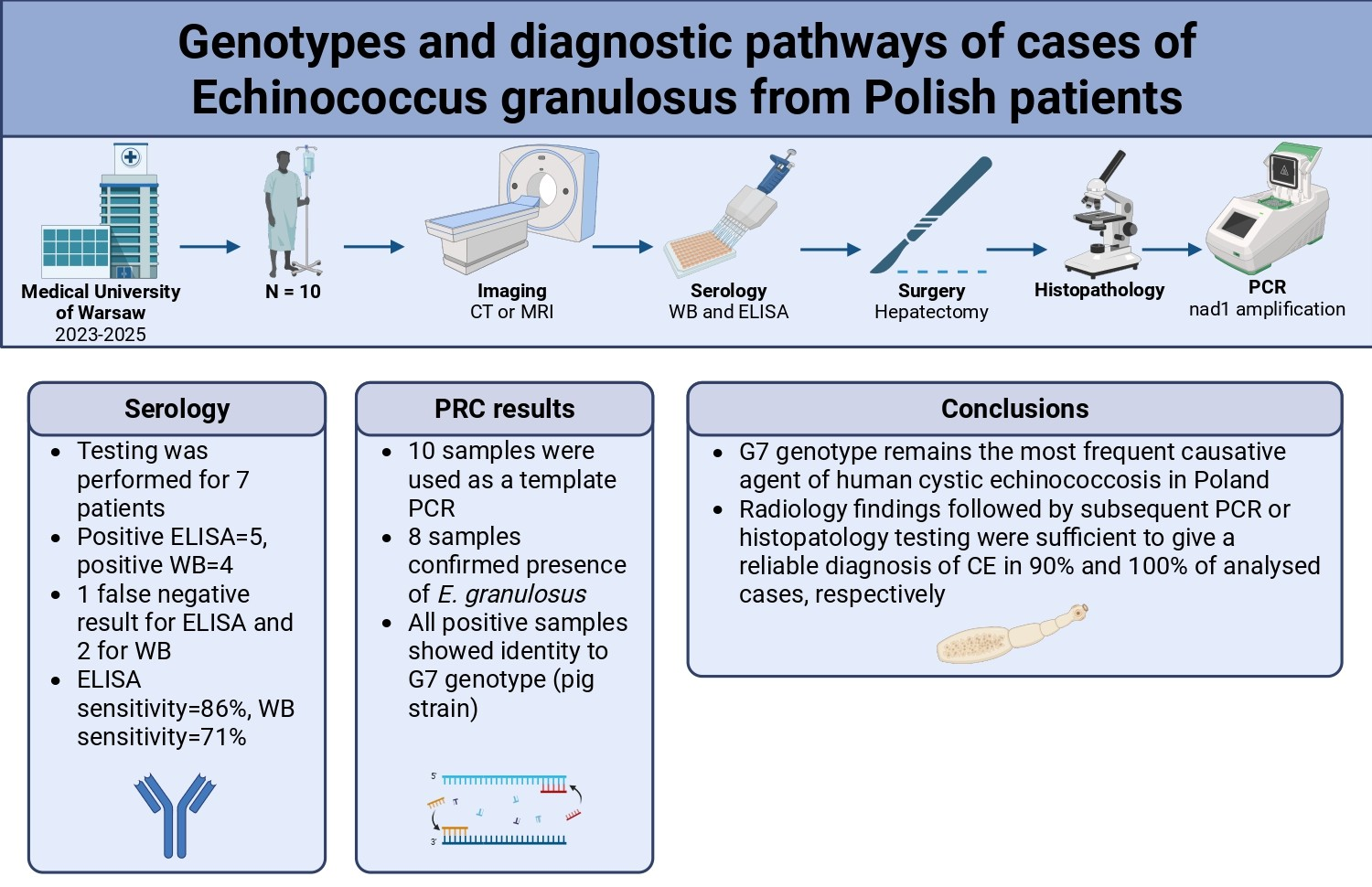
Graphical Abstract

1. Introduction

Cystic echinococcosis (CE) is a parasitic disease caused by the larval stage of the tapeworm belonging to Echinococcus granulosus sensu lato (s.l.) [1]. It is an important zoonotic disease with global distribution that affects more than 100 countries on every continent with the exception of Antarctica [2,3]. The highest prevalence has been reported in parts of Eurasia, northern and eastern Africa, Australia, and South America [4].
The global burden of CE remains substantial, causing significant economic losses and health problems in both rural and urban populations [2]. A recent study based on the Global Burden of Disease (GBD) 2021 database estimated CE mortality at 1364 (95% UI: 986–1776) deaths worldwide and attributed 105,071.6 (95% UI: 78,966.6–133,308.9) global Disability-Adjusted Life Years (DALYs) to the disease in 2021 [5].
In Poland, CE is not considered a frequent disease. According to the Polish National Institute of Public Health between 2014 and 2024 the number of reported human cases oscillated between 20 and 73 annually, as of 19 February 2025 [6,7,8,9,10,11,12,13,14,15,16]. Nevertheless, it remains expensive and complicated to treat and may require extensive surgery combined with prolonged drug therapy [17].
Usually, CE does not present clinical symptoms until cysts have reached a particular size. After a variable incubation period, growing cysts can exert pressure on adjacent tissues causing location-specific symptoms or rupture leading to systemic hypersensitivity reactions. In a considerable number of patients, CE is an incidental finding during routine imaging examinations. After initial suspicion further diagnosis is followed by a combination of imaging, serology, molecular, and histopathological testing [18]. Serological testing for Echinococcus antibodies is a common diagnostic method. However, a substantial number of patients do not elicit an immune response [19]. Ultrasonography (USG), magnetic resonance imaging (MRI) and to lesser extent computed tomography (CT) remain primary tools for detection and monitoring of hydatid cysts, particularly in seronegative cases. Nevertheless, atypical and varied imaging appearance depending on the parasite’s growth stage remains a major difficulty [17,20].
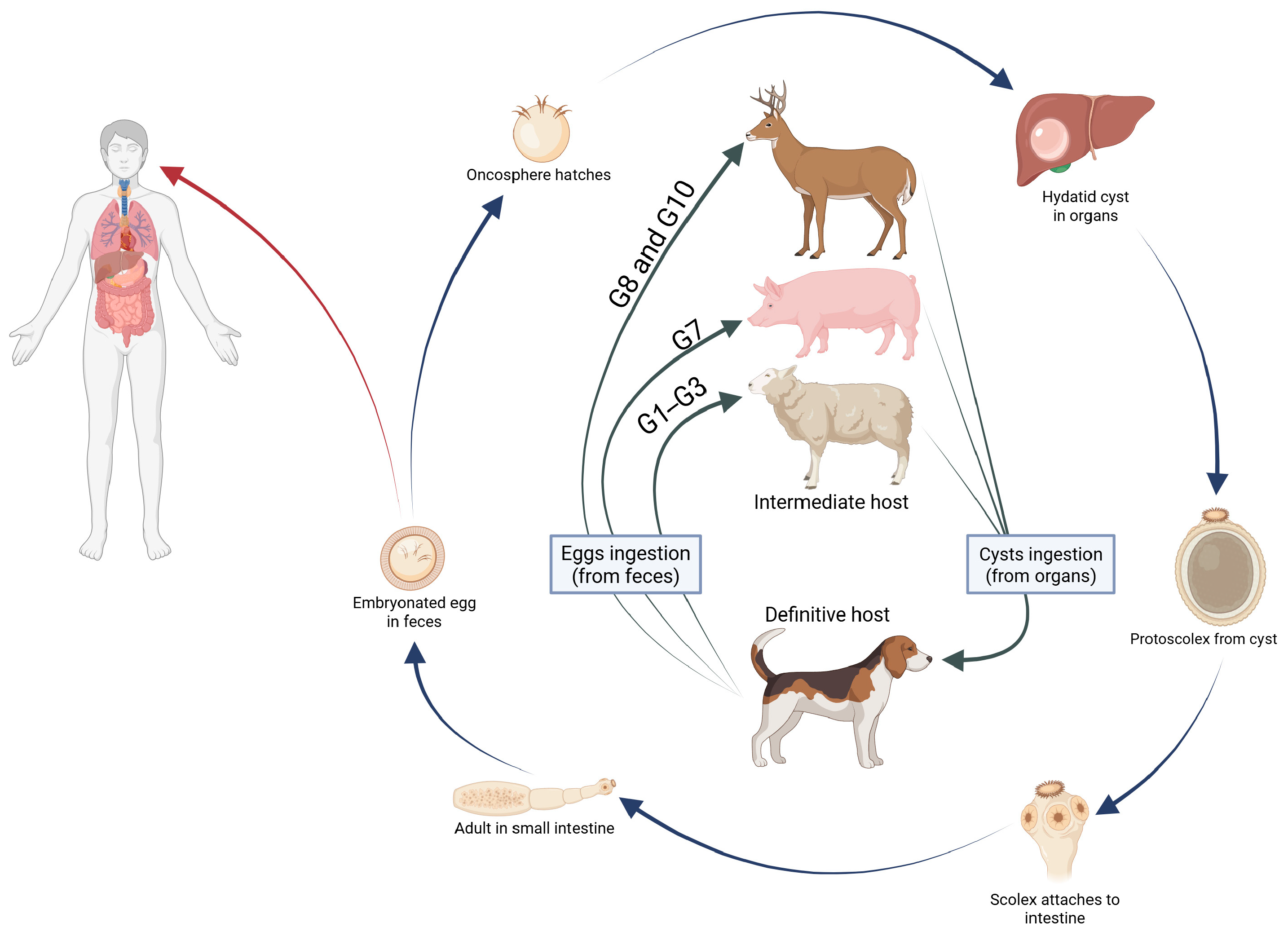
The life cycle of Echinococcus granulosus s.l. is mostly domestic, involving dogs as the definitive host, in which the adult worm lives in the small intestine and releases infective eggs. Wild canids such as wolves and red foxes can also serve as definitive hosts in the transmission cycle. Intermediate hosts, which include sheep, swine, and cattle, together with humans as accidental intermediate hosts, acquire the infection through accidental ingestion of the tapeworm eggs. After ingestion of echinococcal eggs, oncosphere larvae are released and penetrate the intestinal lamina propria. Following penetration, the oncosphere is passively transported via the bloodstream to the liver in approximately 70% of cases, lungs in 20% of cases, or other internal organs where it develops into a hydatid cyst [21,22] (Figure 1).
The current taxonomic consensus divides E. granulosus s.l. into five different species: E. granulosus sensu stricto (s.s.; G1, G3), E. equinus (G4), E. ortleppi (G5), E. canadensis (G6–G8, G10), and E. felidis, though only the first four have been directly linked to human CE cases. It is important to note that there is an ongoing debate concerning the heterogeneity within the E. canadensis cluster, with propositions to subdivide it into three separate species: E. intermedius (G6/7), E. borealis (G8), and E. canadensis (G10). Nevertheless, for the sake of clarity, the more established classification featuring an undivided E. canadensis was utilized in this manuscript [23,24,25,26].
In Poland, the most commonly identified causative agent of CE is E. canadensis G7 genotype. Moreover, G7 has also been detected in human patients in Slovakia, Ukraine, Austria and Turkey [4,27,28,29,30].
The aim of this study was to identify the genotypes responsible for cases of CE in Poland by conducting molecular analysis of larvae isolated from Polish patients and to investigate diagnostic pathways that have led to CE diagnosis.

2. Materials and Methods

2.1. Materials

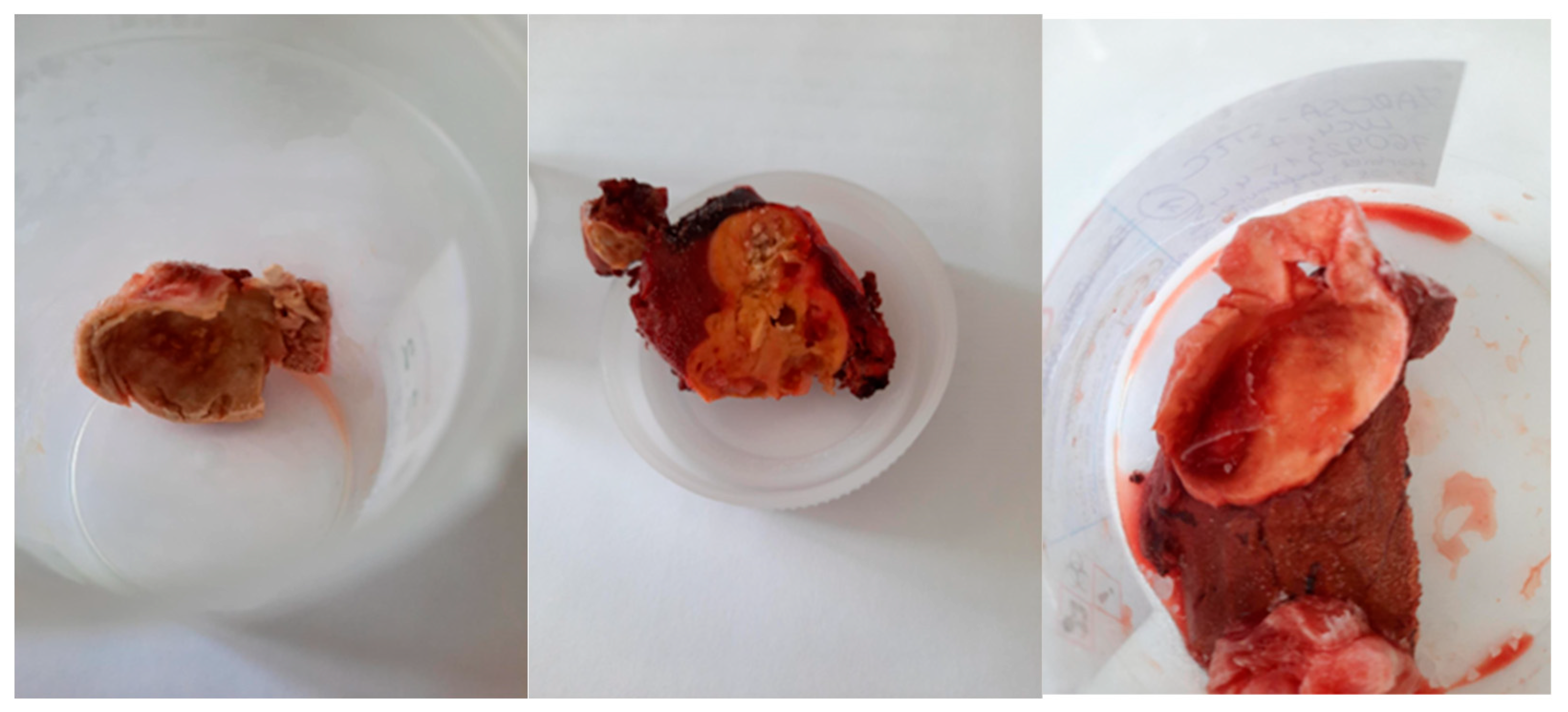
Between April 2023 and January 2025, tissue samples (Figure 2) were collected from 10 patients following hepatectomy at the Department of General, Transplant, and Liver Surgery, Medical University of Warsaw, Poland. Patient demographics, including age and gender, were recorded. Furthermore, imaging findings from USG, CT, and MRI were retrieved from the surgical department records.
Among the patients, 2 were male and 8 were female, with ages ranging from 33 to 70 years (Table 1). These patients were suspected of having CE. Nine out of ten patients underwent total pericystectomy, while the remaining one underwent partial pericystectomy (Table 1). The study was performed in accordance with the tenets of the Declaration of Helsinki.

2.2. Methods

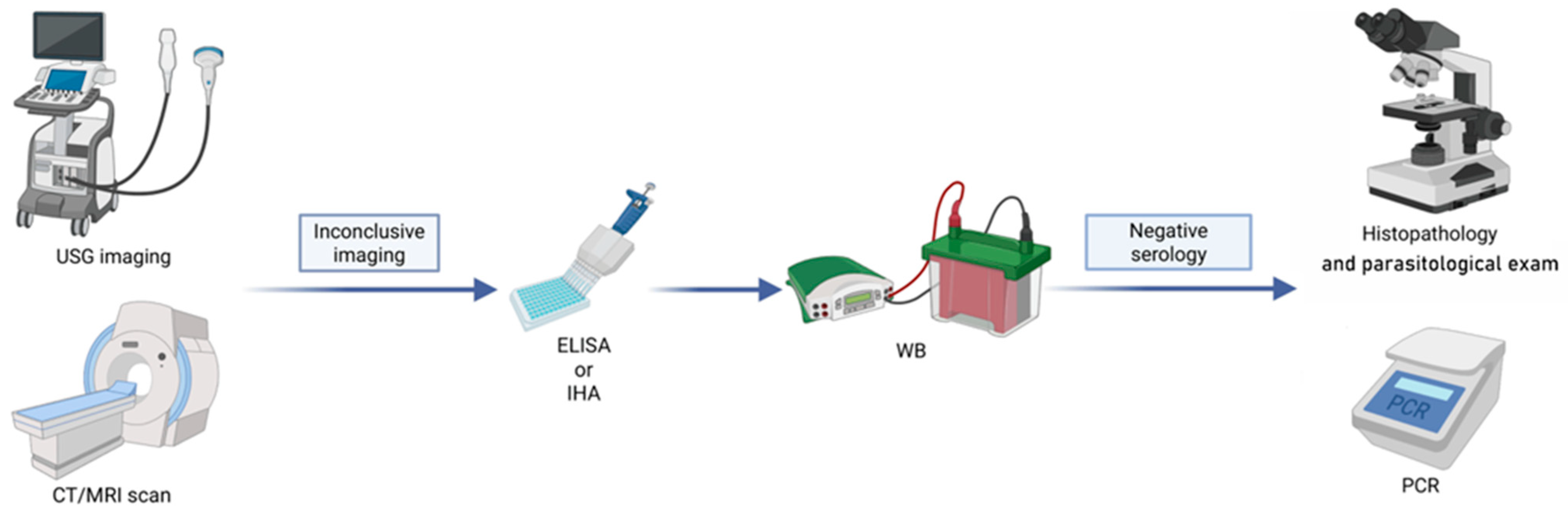
Sera from patients with inconclusive imaging results were investigated by ELISA for the detection of IgG antibodies against Echinococcus spp. (Bordier Affinity Products S.A., Crissier, Switzerland) according to the manufacturer’s instructions [31,32,33] (Table 2). This procedure is routinely used as a screening test in our laboratory. Positive ELISA results were confirmed by Western blot (WB) using the LDBIO Diagnostics kit (Lyon, France). The subsequent step involved parasitological examination via light microscopy and PCR analysis (Figure 3).
The examined samples represented part of a cyst containing protoscoleces and/or hooks. The morphology of the fresh samples was studied directly. The wet unstained sediment of the cyst fluid and the inner layers of cyst tissue were examined for the presence of larva fertile elements by light microscopy (Olympus BX40, Olympus, Tokyo, Japan). The tissue samples, which consisted of parts of cysts, were either stored frozen at −20 °C or fixed in 70% ethanol prior to molecular analysis.
Tissue samples from the cyst wall were fixed in 10% buffered formalin, embedded in paraffin, and sectioned at 4–5 μm. The slides were stained with Hematoxylin and Eosin (H&E) and Periodic Acid–Schiff (PAS). The diagnosis was confirmed by identifying the characteristic laminated layer and the germinal membrane.
The tissue samples were rinsed several times with phosphate-buffered saline (PBS). The resulting suspension was then centrifuged at 3000× g for 10 min to collect the sediment. Each pellet was resuspended in 100 μL PBS, and genomic DNA was extracted using the NucleoSpin kit (Macherey-Nagel, Düren, Germany), following the manufacturer’s instructions.
A mitochondrial region was amplified by PCR using the extracted DNA as a template. A fragment of the NADH dehydrogenase 1 (nad1) gene was amplified with primers JB11 (5′-AGATTCGTAAGGGGCCTAATA-3′) and JB12 (5′-ACCACTAACTAATTCACTTTC-3′) [34,35].
Molecular identification was performed using a previously described PCR protocol [4]. The PCR reaction (50 μL) consisted of 1 μL of DNA template, 0.5 μM of each primer, 0.2 mM of each dNTP, 1× PCR buffer containing 2.5 mM MgCl2, and 2 U of Taq DNA polymerase (Qiagen, Hilden, Germany). Amplification was performed in a PTC-200 thermal cycler (MJ Research, Waltham, MA, USA) with the following conditions: 3 min at 95 °C, followed by 35 cycles of 1 min at 95 °C, 1 min at 55 °C, and 1 min at 72 °C [4].
The PCR products were separated by electrophoresis on 2% agarose gel (MetaPhor, FMC BioProducts, Philadelphia, PA, USA), stained with ethidium bromide, and visualized under a UV transilluminator. The nad1 gene fragments were then purified and directly sequenced in both directions using the BigDye Ready Reaction Cycle Sequencing kit and an ABI 3730 Genetic Analyzer (Applied Biosystems, Foster City, CA, USA). Sequencing was performed independently by three experienced laboratory technicians.
The resulting chromatograms were manually reviewed and edited using Chromas 2.0 software. The obtained sequences were aligned with others from the NCBI GenBank database using ClustalW2 [4,36].

3. Results

A total of 10 samples were obtained from patients who underwent surgery to remove a cyst located in the liver (8 cases of women and 2 men) (Table 1).
All 10 portions of tissue underwent histopathological examinations, and we assessed other diagnostic procedures in relation to their findings (Table 2).
All patients were examined by radiological techniques, with eight undergoing computed tomography and two magnetic resonance imaging. All radiology findings revealed cyst-like lesions of suspected parasitic origin. The cysts ranged in size from 12 mm to 60 mm in diameter, and the largest one was 6 cm (Table 1). One of the radiological examinations was false negative (Table 2).
Serology testing, which consisted of ELISA and Western blot tests, was performed for seven patients. Western blot was positive for four patients, while ELISA was positive for five. Only one ELISA result was a false negative, compared to two false negatives in WB analysis (Table 2).
DNA extracted from all 10 samples and positive controls was used as the template in separate PCRs to amplify a region of the mitochondrial NADH dehydrogenase 1. Each PCR produced a single band on agarose gel electrophoresis.
Eight out of ten isolates and controls were diagnosed as positive by amplification of nad1 fragment (~500 bp). The nad1 fragments were sequenced and compared with sequences of Echinococcus genotypes available in NCBI GenBank. One of the samples turned out to be a false negative. The sequences of eight isolates showed 100% identity with that of the pig strain G7, designated E. canadensis (Table 2). All nad1 sequences were deposited in GenBank with accession numbers PX121313–PX121320.

4. Discussion

In human infections in Europe, the most prevalent genotypes of E. granulosus s.l. are G1–G3 (sheep strains) as well as genotype G7 (pig strain). In addition, genotypes G8 and G10 (cervid strains) have been reported in Scandinavian countries and northern Europe [3,37].
Sheep strains are not only globally distributed but are also considered responsible for up to 88.5% of all human E. granulosus s.s. infections [25]. In Europe, locally acquired human infections have been reported in the United Kingdom, France, Poland, Ukraine, Austria, the Balkans, and most frequently in southern Europe, including Italy, Portugal, and Spain [3,4,25].
Although G7 accounts for approximately 11% of human CE cases worldwide, Casulli et al. demonstrated that this genotype is responsible for up to 28.3% of human CE cases in Europe [25]. This genotype has been identified in Germany, Austria, Poland, Lithuania, Slovakia, Romania, and Serbia [3,4,28,30,36].
There are several possible reasons why the G7 genotype is particularly prevalent in eastern and southeastern Europe. In rural Polish households, dogs commonly live in close proximity to livestock. Because canines play a key role in the transmission of Echinococcus spp., the prevalence of infection in dogs has a direct impact on the transmission, since it increases risk of human echinococcosis [38,39]. Although data on E. granulosus s.l. infection in dogs in Europe are limited, prevalence rates of up to 31% have been reported in Spain and Italy, and 14.2% in Lithuania, which borders Poland [40,41,42]. Another important factor contributing to the presence of the G7 genotype in Poland is infection in livestock, which represents a major source of hydatid cysts [3].
In Poland, all cases of human CE are subject to mandatory reporting. According to the Polish National Institute of Public Health, the number of reported cases in 2023 and 2024 was 66 and 73, respectively (status as of 19 February 2026) [15,16]. Although the number of cases included in this analysis was limited, they nevertheless represent a substantial proportion of all reported CE cases in Poland during the study period.
Among the analyzed patients, imaging followed by PCR and histopathological testing was sufficient to establish a reliable diagnosis of CE in 90% and 100% of cases, respectively (Table 2). Notably, CT and MRI examinations achieved a specificity of 90%, indicating high diagnostic value in this patient group. Despite the small study group, these findings highlight the significant value of imaging techniques in the initial stages of the diagnostic process, which remains consistent with current state of knowledge [43,44].
Serological tests demonstrated lower sensitivity, reaching 86% for ELISA and 71% for Western blot (Table 2). Despite this sensitivity, all patients required additional diagnostic procedures, including PCR and histopathology, to confirm the final diagnosis. Although serological tests remain convenient and non-invasive diagnostic tools that can be used in an ambulatory setting, they should be regarded as supplementary rather than definitive methods [45]. Clinicians should always bear in mind the false negativity of these tests. Cyst stage, localization (hepatic or extra-hepatic), size, number and integrity are known factors that may influence the results of serological tests to be false negative [45,46]. On the other hand, false positivity is also a serious issue that should not be overlooked when it comes to discussing serological tests in CE. A serious drawback of this method is cross-reactivity, which occurs during tapeworm or helminth infections, neoplastic diseases or liver cirrhosis [45,47]. Furthermore, false seropositivity may occur in endemic regions and it can be related to exposure to the parasite [45,48]. For these reasons, serology alone would perform poorly both as a definitive diagnosis method and as a screening tool. Our results highlight the auxiliary role of serological tests in the diagnostic process of CE and underscore the need for PCR and histopathology to confirm the diagnosis.
According to World Health Organization (WHO) guidelines on treatment of patients with CE, in developed countries surgery remains the standard of care for uncomplicated, active, multivesicular liver cysts that are bigger than 5 cm and for complicated liver cysts [17]. The two main types of surgical approach are total pericystectomy and partial cystectomy. Total pericystectomy is a radical approach that aims to resect the whole cyst, while partial cystectomy is a conservative approach that involves draining the cyst’s content and unroofing it [49]. Radical surgery has the best potential to completely cure the disease, since the whole cyst is removed from the patient’s body. Furthermore, the damage to healthy parenchyma is minimal. However, it should be kept in mind that radical surgeries create a great risk of intraoperative hemorrhages and dangerous cyst ruptures. On the other hand, conservative techniques are better options in complicated situations, such as communication with biliary tract. Unfortunately, the recurrence is significantly higher compared to the radical approach, as the remaining part of the cyst is a serious source of complications [50,51]. Surgeon should choose the preferred method according to patient’s condition, size and location of the cyst, available equipment and their own operating skills. Nine out of 10 of our patients underwent pericystectomy and only one underwent partial cystectomy.
Our findings are consistent with previous reports from this region, confirming that the G7 genotype remains the most prevalent genotype of Echinococcus spp. in Poland [4,36,52].

Author Contributions

Conceptualization, A.G., M.T., M.J. and M.D.; methodology, A.G., M.T., M.J. and M.D.; validation, A.G., M.T., M.J. and M.D.; formal analysis, A.G., M.T., M.J. and M.D.; investigation, A.G., M.T., M.J. and M.D.; resources, A.P.-F., P.M. and M.D.; data curation, A.G., M.T., M.J. and M.D.; writing—original draft preparation, A.G., M.T., M.J. and M.D.; writing—review and editing, A.G., M.T., M.J. and M.D.; visualization, A.G., M.T., M.J. and M.D.; supervision, M.D.; project administration, A.G., M.T., M.J. and M.D.; funding acquisition, M.J. and M.D. All authors have read and agreed to the published version of the manuscript.

Funding

This research was funded by the Medical University of Warsaw, grant number 92/M/MG/N/23. The APC was funded by the Medical University of Warsaw.

Institutional Review Board Statement

In our manuscript we did not include information regarding Ethics Committee approval since this research had no components of a medical experiment. In our research project there was no interference with a patient’s diagnostic or therapeutic process for scientific purposes which requires the approval of an independent Bioethics Committee. Our study included retrospective analyses without direct contact with the patient and without interference with the treatment process. It also included diagnostic procedures that were required by the hospital and created in cooperation with co-authors from the surgery clinics. We used the surplus material for diagnostic tests that do not require committee approval.

Informed Consent Statement

Not applicable.

Data Availability Statement

Publicly available epidemiological datasets analyzed in this study can be found on the National Institute of Public Health—National Institute of Hygiene (NIZP-PZH) website (https://www.pzh.gov.pl) and the World Health Organization website (https://www.who.int). The primary data obtained from the analysis of post-mortem samples are available on request from the corresponding author. These data are not publicly available due to ethical and privacy restrictions.

Conflicts of Interest

The authors declare no conflicts of interest.

References

  1. Vuitton, D.A.; McManus, D.P.; Rogan, M.T.; Romig, T.; Gottstein, B.; Naidich, A.; Tuxun, T.; Wen, H.; Menezes da Silva, A. International consensus on terminology to be used in the field of echinococcoses. Parasite 2020, 27, 41. [Google Scholar] [CrossRef] [Scilit]
  2. Wen, H.; Vuitton, L.; Tuxun, T.; Li, J.; Vuitton, D.A.; Zhang, W.; McManus, D.P. Echinococcosis: Advances in the 21st Century. Clin. Microbiol. Rev. 2019, 32, e00075-18. [Google Scholar] [CrossRef] [Scilit]
  3. Deplazes, P.; Rinaldi, L.; Alvarez Rojas, C.A.; Torgerson, P.R.; Harandi, M.F.; Romig, T.; Antolova, D.; Schurer, J.M.; Lahmar, S.; Cringoli, G.; et al. Global Distribution of Alveolar and Cystic Echinococcosis. Adv. Parasitol. 2017, 95, 315–493. [Google Scholar] [CrossRef] [Scilit]
  4. Dybicz, M.; Gierczak, A.; Dąbrowska, J.; Rdzanek, Ł.; Michałowicz, B. Molecular diagnosis of cystic echinococcosis in humans from central Poland. Parasitol. Int. 2013, 62, 364–367. [Google Scholar] [CrossRef] [Scilit] [PubMed]
  5. Wang, Y.; Zhang, R.; Zhang, D.; Liang, Y.; Xu, C.; Zhang, C. Global burden of cystic echinococcosis: A 32-year analysis of 204 countries with 2036 projections. BMC Public Health 2025, 25, 3213. [Google Scholar] [CrossRef] [Scilit]
  6. National Institute of Public Health—National Institute of Hygiene, C.S.I. Infectious Diseases and Poisonings in Poland in 2014; NIZP-PZH: Warsaw, Poland, 2014.
  7. National Institute of Public Health—National Institute of Hygiene, C.S.I. Infectious Diseases and Poisonings in Poland in 2015; NIZP-PZH: Warsaw, Poland, 2015.
  8. National Institute of Public Health—National Institute of Hygiene, C.S.I. Infectious Diseases and Poisonings in Poland in 2016; NIZP-PZH: Warsaw, Poland, 2016.
  9. National Institute of Public Health—National Institute of Hygiene, C.S.I. Infectious Diseases and Poisonings in Poland in 2017; NIZP-PZH: Warsaw, Poland, 2017.
  10. National Institute of Public Health—National Institute of Hygiene, C.S.I. Infectious Diseases and Poisonings in Poland in 2018; NIZP-PZH: Warsaw, Poland, 2018.
  11. National Institute of Public Health—National Institute of Hygiene, C.S.I. Infectious Diseases and Poisonings in Poland in 2019; NIZP-PZH: Warsaw, Poland, 2019.
  12. National Institute of Public Health—National Institute of Hygiene, C.S.I. Infectious Diseases and Poisonings in Poland in 2020; NIZP-PZH: Warsaw, Poland, 2020.
  13. National Institute of Public Health—National Institute of Hygiene, C.S.I. Infectious Diseases and Poisonings in Poland in 2021; NIZP-PZH: Warsaw, Poland, 2021.
  14. National Institute of Public Health—National Institute of Hygiene, C.S.I. Infectious Diseases and Poisonings in Poland in 2022; NIZP-PZH: Warsaw, Poland, 2022.
  15. National Institute of Public Health—National Institute of Hygiene, C.S.I. Infectious Diseases and Poisonings in Poland in 2023; NIZP-PZH: Warsaw, Poland, 2023.
  16. National Institute of Public Health—National Institute of Hygiene, C.S.I. Infectious Diseases and Poisonings in Poland in 2024; NIZP-PZH: Warsaw, Poland, 2024.
  17. World Health Organization. WHO Guidelines for the Treatment of Patients with Cystic Echinococcosis; World Health Organization: Geneva, Switzerland, 2025. [Google Scholar]
  18. Kern, P.; Menezes da Silva, A.; Akhan, O.; Müllhaupt, B.; Vizcaychipi, K.A.; Budke, C.; Vuitton, D.A. The Echinococcoses: Diagnosis, Clinical Management and Burden of Disease. Adv. Parasitol. 2017, 96, 259–369. [Google Scholar] [CrossRef] [Scilit]
  19. Zhang, W.; Wen, H.; Li, J.; Lin, R.; McManus, D.P. Immunology and immunodiagnosis of cystic echinococcosis: An update. J. Immunol. Res. 2012, 2012, 101895. [Google Scholar] [CrossRef] [Scilit]
  20. Brunetti, E.; Kern, P.; Vuitton, D.A. Expert consensus for the diagnosis and treatment of cystic and alveolar echinococcosis in humans. Acta Trop. 2010, 114, 1–16. [Google Scholar] [CrossRef] [Scilit]
  21. World Health Organization. Echinococcosis in humans: Clinical aspects, diagnosis and treatment. In WHO/OIE Manual on Echinococcosis in Humans and Animals: A Public Health Problem of Global Concern; Eckert, J., Gemmell, M.A., Meslin, F., Pawłowski, Z.S., Eds.; World Organisation for Animal Health: Paris, France, 2001; pp. 20–66. [Google Scholar]
  22. Romig, T.; Deplazes, P.; Jenkins, D.; Giraudoux, P.; Massolo, A.; Craig, P.S.; Wassermann, M.; Takahashi, K.; de la Rue, M. Ecology and Life Cycle Patterns of Echinococcus Species. Adv. Parasitol. 2017, 95, 213–314. [Google Scholar] [CrossRef] [Scilit]
  23. Šnábel, V. Current aspects of the taxonomy and phylogeny of the genus Echinococcus Rudolphi, 1801. Ann. Parasitol. 2023, 69, s30–s32. [Google Scholar]
  24. Laurimäe, T.; Kinkar, L.; Moks, E.; Romig, T.; Omer, R.A.; Casulli, A.; Umhang, G.; Bagrade, G.; Irshadullah, M.; Sharbatkhori, M.; et al. Molecular phylogeny based on six nuclear genes suggests that Echinococcus granulosus sensu lato genotypes G6/G7 and G8/G10 can be regarded as two distinct species. Parasitology 2018, 145, 1929–1937. [Google Scholar] [CrossRef] [Scilit]
  25. Casulli, A.; Massolo, A.; Saarma, U.; Umhang, G.; Santolamazza, F.; Santoro, A. Species and genotypes belonging to Echinococcus granulosus sensu lato complex causing human cystic echinococcosis in Europe (2000–2021): A systematic review. Parasites Vectors 2022, 15, 109. [Google Scholar] [CrossRef] [Scilit]
  26. Thompson, R.C.A. The Molecular Epidemiology of Echinococcus Infections. Pathogens 2020, 9, 453. [Google Scholar] [CrossRef] [Scilit]
  27. Snábel, V.; Altintas, N.; D’Amelio, S.; Nakao, M.; Romig, T.; Yolasigmaz, A.; Gunes, K.; Turk, M.; Busi, M.; Hüttner, M.; et al. Cystic echinococcosis in Turkey: Genetic variability and first record of the pig strain (G7) in the country. Parasitol. Res. 2009, 105, 145–154. [Google Scholar] [CrossRef] [Scilit]
  28. Turceková, L.; Snábel, V.; D’Amelio, S.; Busi, M.; Dubinský, P. Morphological and genetic characterization of Echinococcus granulosus in the Slovak Republic. Acta Trop. 2003, 85, 223–229. [Google Scholar] [CrossRef] [Scilit]
  29. Kedra, A.H.; Swidersky, Z.; Tkach, V.V.; Dubinsky, P.; Pawlowsky, P.; Stefaniak, J.; Pawlowski, J. Genetic analysis of Echinococcus granulosus from humans and pigs in Poland, Slovakia and Ukraine. A multicenter study. Acta Parasitol. 1999, 44, 248–254. [Google Scholar]
  30. Schneider, R.; Gollackner, B.; Schindl, M.; Tucek, G.; Auer, H. Echinococcus canadensis G7 (pig strain): An underestimated cause of cystic echinococcosis in Austria. Am. J. Trop. Med. Hyg. 2010, 82, 871–874. [Google Scholar] [CrossRef] [Scilit]
  31. Gottstein, B. Molecular and immunological diagnosis of echinococcosis. Clin. Microbiol. Rev. 1992, 5, 248–261. [Google Scholar] [CrossRef] [Scilit]
  32. Gottstein, B.; Wang, J.; Blagosklonov, O.; Grenouillet, F.; Millon, L.; Vuitton, D.A.; Müller, N. Echinococcus metacestode: In search of viability markers. Parasite 2014, 21, 63. [Google Scholar] [CrossRef] [Scilit]
  33. Poretti, D.; Felleisen, E.; Grimm, F.; Pfister, M.; Teuscher, F.; Zuercher, C.; Reichen, J.; Gottstein, B. Differential immunodiagnosis between cystic hydatid disease and other cross-reactive pathologies. Am. J. Trop. Med. Hyg. 1999, 60, 193–198. [Google Scholar] [CrossRef] [Scilit]
  34. Bowles, J.; Blair, D.; McManus, D.P. Genetic variants within the genus Echinococcus identified by mitochondrial DNA sequencing. Mol. Biochem. Parasitol. 1992, 54, 165–173. [Google Scholar] [CrossRef] [Scilit]
  35. Bowles, J.; McManus, D.P. NADH dehydrogenase 1 gene sequences compared for species and strains of the genus Echinococcus. Int. J. Parasitol. 1993, 23, 969–972. [Google Scholar] [CrossRef] [Scilit]
  36. Dybicz, M.; Borkowski, P.K.; Dąbrowska, J.; Chomicz, L. Cases of Echinococcus granulosus Sensu Stricto Isolated from Polish Patients: Imported or Indigenous? Biomed. Res. Int. 2015, 2015, 728321. [Google Scholar] [CrossRef] [Scilit]
  37. Oksanen, A.; Lavikainen, A. Echinococcus canadensis transmission in the North. Vet. Parasitol. 2015, 213, 182–186. [Google Scholar] [CrossRef] [Scilit]
  38. Kachani, M.; Heath, D. Dog population management for the control of human echinococcosis. Acta Trop. 2014, 139, 99–108. [Google Scholar] [CrossRef] [Scilit]
  39. Deplazes, P.; van Knapen, F.; Schweiger, A.; Overgaauw, P.A. Role of pet dogs and cats in the transmission of helminthic zoonoses in Europe, with a focus on echinococcosis and toxocarosis. Vet. Parasitol. 2011, 182, 41–53. [Google Scholar] [CrossRef] [Scilit]
  40. Carmena, D.; Sánchez-Serrano, L.P.; Barbero-Martínez, I. Echinococcus granulosus infection in Spain. Zoonoses Public Health 2008, 55, 156–165. [Google Scholar] [CrossRef] [Scilit]
  41. Garippa, G.; Manfredi, M.T. Cystic echinococcosis in Europe and in Italy. Vet. Res. Commun. 2009, 33, 35–39. [Google Scholar] [CrossRef] [Scilit]
  42. Bruzinskaite, R.; Sarkūnas, M.; Torgerson, P.R.; Mathis, A.; Deplazes, P. Echinococcosis in pigs and intestinal infection with Echinococcus spp. in dogs in southwestern Lithuania. Vet. Parasitol. 2009, 160, 237–241. [Google Scholar] [CrossRef] [Scilit]
  43. Pakala, T.; Molina, M.; Wu, G.Y. Hepatic Echinococcal Cysts: A Review. J. Clin. Transl. Hepatol. 2016, 4, 39–46. [Google Scholar] [CrossRef] [Scilit]
  44. Leandru, A.; Neica, M. The role of different imaging modalities in detecting the complications of hydatid cysts and unusual locations. In Proceedings of the ECR 2017, Vienna, Austria, 1–5 March 2017. [Google Scholar]
  45. Siles-Lucas, M.; Uchiumi, L.; Tamarozzi, F. ‘No cyst, no echinococcosis’: A scoping review update on the diagnosis of cystic echinococcosis after the issue of the WHO-IWGE Expert Consensus and current perspectives. Curr. Opin. Infect. Dis. 2023, 36, 333–340. [Google Scholar] [CrossRef] [Scilit]
  46. Tamarozzi, F.; Silva, R.; Fittipaldo, V.A.; Buonfrate, D.; Gottstein, B.; Siles-Lucas, M. Serology for the diagnosis of human hepatic cystic echinococcosis and its relation with cyst staging: A systematic review of the literature with meta-analysis. PLoS Neglected Trop. Dis. 2021, 15, e0009370. [Google Scholar] [CrossRef] [Scilit]
  47. Mattwich, C.; Huber, K.; Bretzel, G.; Suerbaum, S.; Wieser, A.; Dichtl, K. Head-to-Head Comparison of Nine Assays for the Detection of Anti-Echinococcus Antibodies: A Retrospective Evaluation. Ann. Lab. Med. 2024, 44, 155–163. [Google Scholar] [CrossRef] [Scilit]
  48. Hernández, A.; Cardozo, G.; Dematteis, S.; Baz, A.; Trias, N.; Nuñez, H.; Barragué, A.; López, L.; Fuentes, J.; López, O.; et al. Cystic echinococcosis: Analysis of the serological profile related to the risk factors in individuals without ultrasound liver changes living in an endemic area of Tacuarembó, Uruguay. Parasitology 2005, 130, 455–460. [Google Scholar] [CrossRef] [Scilit]
  49. Berto, C.G.; Liou, P.; Coyle, C.M.; Emond, J.C. Surgical management of cystic echinococcosis of the liver. Curr. Opin. Infect. Dis. 2023, 36, 348–352. [Google Scholar] [CrossRef] [Scilit]
  50. A, J.-D.; Chai, J.-P.; Jia, S.-L.; A, X.-R. Historical changes in surgical strategy and complication management for hepatic cystic echinococcosis. World J. Gastrointest. Surg. 2023, 15, 1591–1599. [Google Scholar] [CrossRef] [Scilit]
  51. Georgiou, G.K.; Lianos, G.D.; Lazaros, A.; Harissis, H.V.; Mangano, A.; Dionigi, G.; Katsios, C. Surgical management of hydatid liver disease. Int. J. Surg. 2015, 20, 118–122. [Google Scholar] [CrossRef] [Scilit]
  52. Dybicz, M.; Borkowski, P.K.; Jonas, M.; Wasiak, D.; Małkowski, P. First Report of Echinococcus ortleppi in Human Cases of Cystic Echinococcosis in Poland. BioMed. Res. Int. 2019, 2019, 2474839. [Google Scholar] [CrossRef] [Scilit]
Figure 1. The life cycle of Echinococcus species.
Pathogens 15 00459 g001
Figure 2. Representative liver tissue samples that underwent histopathological and molecular testing.
Pathogens 15 00459 g002
Figure 3. Diagnostic pathway.
Pathogens 15 00459 g003
Table 1. General and characteristics of patients and type of surgery.
Case NumberSexAge in YearsThe Type of Surgery—Pericystectomy
1M33Total
2F40Total
3M41Total
4F57Partial
5F46Total
6F56Total
7F70Total
8F61Total
9F55Total
10F56Total
F Female, M Male.
Table 2. Results of diagnostic tests and procedures performed in patients during CE diagnosis and results of subsequent PCR analysis.
Case NumberRadiology Findings in the Liver
(Cyst Localization and Size)
Serology
ELISA WB
Parasitological
Examination
Histopathology
Diagnosis of Echinococcosis
PCR/Genotyping
1MRI: segment 7/8; 59 × 43 mmHooks+Echinococcus canadensis (G7)
2CT: segment 4b; 35 mm & segment 6/7++Lack of
parasite elements
+Echinococcus canadensis (G7)
3CT: segment 7/6; 60 mm+Lack of
parasite elements
+Echinococcus canadensis (G7)
4CT: segment 5/6/7/8++Lack of
parasite elements
+Echinococcus canadensis (G7)
5CT: segment 6; 41 × 31 × 31 mm++Lack of
parasite elements
+Echinococcus canadensis (G7)
6CT: segment 4/5 & segment 8/7; 30 mmNot
performed
Lack of
parasite elements
+Echinococcus canadensis (G7)
7CT: liver hilum+Lack of
parasite elements
8MRI: segment 8; 35 mm & segment 6/7; 12 mm+Not
performed
Lack of
parasite elements
+
9CT: segment 4; 30 mm & segment 7; 30 mm+Not
performed
Larva and protoscoleces+Echinococcus canadensis (G7)
10CT: segment 5/6/7/8++Lack of
parasite elements
+Echinococcus canadensis (G7)
WB, Western blot; ELISA, Enzyme-Linked Immunosorbent Assay; CT, computed tomography; MRI, magnetic resonance imaging; PCR, polymerase chain reaction.
Disclaimer/Publisher’s Note: The statements, opinions and data contained in all publications are solely those of the individual author(s) and contributor(s) and not of MDPI and/or the editor(s). MDPI and/or the editor(s) disclaim responsibility for any injury to people or property resulting from any ideas, methods, instructions or products referred to in the content.