Promising Drug Repurposing Candidates Targeting Free-Living Amoebae: A Systematic and Critical Review of Laboratory-Based Evidence

 ,
,  , ,
, ,  , ,
, ,  , and
, and
Abstract
1. Introduction
2. Materials and Methods
2.1. Review Question and Objectives
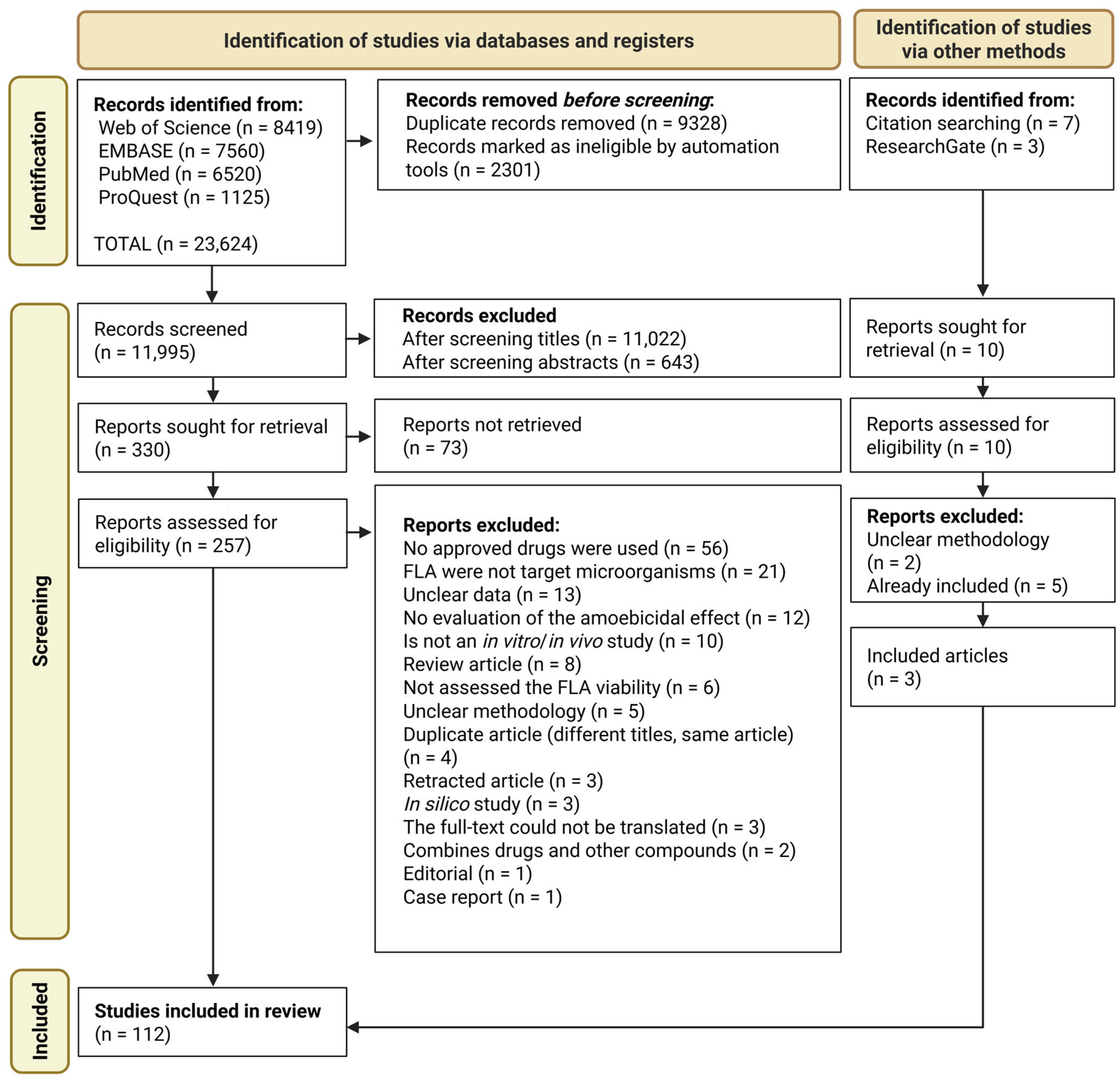
2.2. Article Collection and Screening Procedure
- •
- Inclusion and exclusion criteria: Articles were included if (1) the full text was available online; (2) they were written in English, Portuguese, Spanish, or another language that could be reliably translated using online tools; and (3) they reported findings from in vitro and/or in vivo evaluations of the amoebicidal activity of approved drugs against Acanthamoeba, Naegleria, Balamuthia, Vermamoeba, or Sappinia.
- •
- Exclusion criteria: Articles were excluded if they (1) were reviews; (2) were not peer-reviewed (e.g., preprints); or (3) reported on compounds not approved as drugs.
2.3. Data Extraction
2.4. Data Analysis
3. Results
3.1. In Vitro Anti-Free Living Amoeba Activity of Tested Drugs
3.1.1. Drugs with Amoebicidal Activity Against B. mandrillaris
3.1.2. Drugs with Acanthamoebicidal Activity
Cysticidal Drugs Against Acanthamoeba spp.
Trophocidal Drugs Against Acanthamoeba spp.
- Trophocidal drugs active at intermediate concentrations (3.0 to 5.0 µM)
- 2.
- Trophocidal drugs active at higher concentrations 5.6 to 14.6 µM
Drugs Active Against Naegleria spp.
- Cysticidal drugs against Naegleria spp.
- 2.
- Trophocidal drugs against Naegleria spp.
3.2. In Vivo Performance of Drugs Tested Against FLA Infections
3.2.1. Therapeutic Performance of Drugs Evaluated In Vivo in Models of Granulomatous Amoebic Encephalitis Caused by Acanthamoeba
3.2.2. In Vivo Efficacy of Drugs Tested in Animal Models of Acanthamoeba Keratitis
4. Discussion
4.1. In Vitro Studies
4.2. In Vivo Studies
4.2.1. In Vivo Models for Granulomatous Amoebic Encephalitis
4.2.2. In Vivo Models for Acanthamoeba Keratitis
4.2.3. In Vivo Models of Primary Amoebic Meningoencephalitis
5. Conclusions
Supplementary Materials
Author Contributions
Funding
Institutional Review Board Statement
Informed Consent Statement
Data Availability Statement
Acknowledgments
Conflicts of Interest
Abbreviations
| ADMET | Absorption, Distribution, Metabolism, Excretion, and Toxicity |
| AK | Acanthamoeba keratitis |
| CNS | Central Nervous System |
| DHFR | Dihydrofolate reductase |
| DHODH | Dihydroorotate dehydrogenase |
| DHPS | Dihydropteroate synthase |
| dpi | Days post-infection |
| FLA | Free-Living Amoebae |
| GAE | granulomatous amoebic encephalitis |
| HDAC | Histone deacetylases |
| IC50 | Inhibitory concentration of 50% of the parasites |
| PABA | Para-aminobenzoic acid |
| PAM | Primary Amoebic Meningoencephalitis |
| PDGFR | Platelet-Derived Growth Factor Receptor |
| PHMB | Polyhexamethylene Biguanide |
| PRISMA | Preferred Reporting Items for Systematic Reviews and Meta-Analyses |
| SD | Standard deviation. |
| VEGFR | Vascular Endothelial Growth Factor Receptor |
| ΔΨm | Mitochondrial membrane potential |
References
- Chaúque, B.J.M.; da Silva, T.C.B.; Dos Santos, D.L.; Benitez, G.B.; Chaúque, L.G.H.; Benetti, A.D.; Zanette, R.A.; Rott, M.B. Global prevalence of free-living amoebae in solid matrices—A systematic review with meta-analysis. Acta Trop. 2023, 247, 107006. [Google Scholar] [CrossRef] [Scilit]
- da Silva, T.C.B.; Chaúque, B.J.M.; Benitez, G.B.; Rott, M.B. Global prevalence of potentially pathogenic free-living amoebae in sewage and sewage-related environments—Systematic review with meta-analysis. Parasitol. Res. 2024, 123, 148. [Google Scholar] [CrossRef] [Scilit]
- Chaúque, B.J.M.; Dos Santos, D.L.; Anvari, D.; Rott, M.B. Prevalence of free-living amoebae in swimming pools and recreational waters, a systematic review and meta-analysis. Parasitol. Res. 2022, 121, 3033–3050. [Google Scholar] [CrossRef] [Scilit] [PubMed]
- Leal Dos Santos, D.; Chaúque, B.J.M.; Virginio, V.G.; Cossa, V.C.; Pettan-Brewer, C.; Schrekker, H.S.; Rott, M.B. Occurrence of Naegleria fowleri and their implication for health—A look under the One Health approaches. Int. J. Hyg. Environ. Health 2022, 246, 114053. [Google Scholar] [CrossRef] [Scilit] [PubMed]
- Choaji, M.; Samba-Louaka, A.; Fechtali-Moute, Z.; Aucher, W.; Pomel, S. Encystment and excystment processes in Acanthamoeba castellanii: An emphasis on cellulose involvement. Pathogens 2025, 14, 268. [Google Scholar] [CrossRef] [Scilit] [PubMed]
- Samba-Louaka, A.; Labruyère, E.; Matondo, M.; Locard-Paulet, M.; Olivo-Marin, J.C.; Guillen, N. Encystation and stress responses under the control of ubiquitin-like proteins in pathogenic amoebae. Microorganisms 2023, 11, 2670. [Google Scholar] [CrossRef] [Scilit]
- Dos Santos, D.L.; Chaúque, B.J.M.; Matiazo, F.F.; de Miranda Ribeiro, L.; Rott, M.B. Agar dehydration: A simple method for long-term storage of Acanthamoeba spp. collection at room temperature. Parasitol. Res. 2024, 123, 153. [Google Scholar] [CrossRef] [Scilit]
- Chaúque, B.J.M.; Rott, M.B. Photolysis of sodium chloride and sodium hypochlorite by ultraviolet light inactivates the trophozoites and cysts of Acanthamoeba castellanii in the water matrix. J. Water Health 2021, 19, 190–202. [Google Scholar] [CrossRef] [Scilit]
- Barrantes Murillo, D.F.; Berrocal, A.; Ali, I.K.M.; Uzal, F.A. Systemic Acanthamoeba T17 infection in a free-ranging two-toed sloth: Case report and literature review of infections by free-living amebas in mammals. J. Vet. Diagn. Investig. 2025, 37, 164–172. [Google Scholar] [CrossRef] [Scilit]
- Qvarnstrom, Y.; da Silva, A.J.; Schuster, F.L.; Gelman, B.B.; Visvesvara, G.S. Molecular confirmation of Sappinia pedata as a causative agent of amoebic encephalitis. J. Infect. Dis. 2009, 199, 1139–1142. [Google Scholar] [CrossRef] [Scilit]
- Jiang, Q.; Zhang, Z.; Cai, Y.; Chen, L.; Deng, L.; Xiong, Y. Disseminated Acanthamoeba castellanii infection in a patient with AIDS: A case report and literature review. Front. Med. 2024, 11, 1377302. [Google Scholar] [CrossRef] [Scilit]
- Haston, J.C.; O’Laughlin, K.; Matteson, K.; Roy, S.; Qvarnstrom, Y.; Ali, I.K.M.; Cope, J.R. The epidemiology and clinical features of non-keratitis Acanthamoeba infections in the United States, 1956–2020. Open Forum Infect. Dis. 2023, 10, ofac682. [Google Scholar] [CrossRef] [Scilit]
- Al Dhaheri, F.; Gunasekera, N.; Wardell, H.; Drobysheva, A.; Hussain, S. Disseminated Acanthamoeba infection in a bone marrow transplant recipient with CTLA-4 haploinsufficiency. J. Clin. Immunol. 2023, 43, 319–322. [Google Scholar] [CrossRef] [Scilit]
- Banerjee, C.T.; Conlan, S.; Mostaghim, A.; Michelin, A.; Arduino, M.; Mattioli, M.; Haston, J.C.; Das, S.; Seyedmousavi, A.; Chang, B.H.; et al. Acanthamoeba infection in a hematopoietic cell transplant recipient: Challenges in diagnosis, management, and source identification. Transpl. Infect. Dis. 2025, 27, e14425. [Google Scholar] [CrossRef] [Scilit]
- Haston, J.C.; Cope, J.R. Amebic encephalitis and meningoencephalitis: An update on epidemiology, diagnostic methods, and treatment. Curr. Opin. Infect. Dis. 2023, 36, 186–191. [Google Scholar] [CrossRef] [Scilit]
- Spottiswoode, N.; Haston, J.C.; Hanners, N.W.; Gruenberg, K.; Kim, A.; DeRisi, J.L.; Wilson, M.R. Challenges and advances in the medical treatment of granulomatous amebic encephalitis. Ther. Adv. Infect. Dis. 2024, 11, 20499361241228340. [Google Scholar] [CrossRef] [Scilit]
- Siddiqui, R.; Maciver, S.K.; Khan, N.A. Naegleria fowleri: Emerging therapies and translational challenges. Expert Rev. Anti-Infect. Ther. 2025, 23, 753–761. [Google Scholar] [CrossRef] [Scilit]
- Chaúque, B.J.M.; da Silva, T.C.B.; Chagas, L.B.; Kinape, L.F.G.; Bitencourt, P.E.R.; Gaspar, C.J.; Cheinquer, A.C.B.; Rott, M.B.; Zanette, R.A.; Goldim, J.R. Identifying promising novel compounds against free-living amoebae: A systematic review of in vitro and in vivo studies. Parasitologia 2025, 5, 56. [Google Scholar] [CrossRef] [Scilit]
- Chaúque, B.J.M.; da Silva, T.C.B.; Rott, E.B.; Rott, F.B.; Leite, A.P.M.C.; Benitez, G.B.; Neuana, N.F.; Goldim, J.R.; Rott, M.B.; Zanette, R.A. Effectiveness of phytoproducts against pathogenic free-living amoebae—A scoping and critical review paving the way toward plant-based pharmaceuticals. Fitoterapia 2025, 182, 106404. [Google Scholar] [CrossRef] [Scilit]
- Gutiérrez-Grijalva, E.P.; Contreras-Angulo, L.A.; Criollo-Mendoza, M.S.; Heredia, J.B.; López-Cervantes, J.; Sánchez-Machado, D.I.; Gonzalez-Galaviz, J.R.; Rodriguez-Anaya, L.Z. Phytochemicals as promising agents against pathogenic free-living amoebas. Phytochem. Rev. 2026, 25, 1109–1125. [Google Scholar] [CrossRef] [Scilit]
- Ahmed, U.; Manzoor, M.; Qureshi, S.; Mazhar, M.; Fatima, A.; Aurangzeb, S.; Hamid, M.; Khan, K.M.; Khan, N.A.; Rashid, Y.; et al. Anti-amoebic effects of synthetic acridine-9(10H)-one against brain-eating amoebae. Acta Trop. 2023, 239, 106824. [Google Scholar] [CrossRef] [Scilit]
- Akbar, N.; Siddiqui, R.; El-Gamal, M.I.; Zaraei, S.O.; Alawfi, B.S.; Khan, N.A. The anti-amoebic potential of carboxamide derivatives containing sulfonyl or sulfamoyl moieties against brain-eating Naegleria fowleri. Parasitol. Res. 2023, 122, 2539–2548. [Google Scholar] [CrossRef] [Scilit] [PubMed]
- Rodríguez-Expósito, R.L.; Reyes-Batlle, M.; Sifaoui, I.; Tejedor, D.; García-Tellado, F.; Piñero, J.E.; Lorenzo-Morales, J. Isobenzofuran-1(3H)-one derivatives: Amoebicidal activity and program cell death in Acanthamoeba castellanii Neff. Biomed. Pharmacother. 2022, 150, 113062. [Google Scholar] [CrossRef] [Scilit] [PubMed]
- Martín-Escolano, R.; Yiangou, L.; Kazana, E.; Robinson, G.K.; Michaelis, M.; Tsaousis, A.D. Repurposing in vitro approaches for screening anti-parasitic drugs against the brain-eating amoeba Naegleria fowleri. Int. J. Parasitol. Drugs Drug Resist. 2021, 17, 204–212. [Google Scholar] [CrossRef] [Scilit] [PubMed]
- Laurie, M.T.; White, C.V.; Retallack, H.; Wu, W.; Moser, M.S.; Sakanari, J.A.; Ang, K.; Wilson, C.; Arkin, M.R.; DeRisi, J.L. Functional assessment of 2177 U.S. and international drugs identifies the quinoline nitroxoline as a potent amoebicidal agent against the pathogen Balamuthia mandrillaris. mBio 2018, 9, e02051-18. [Google Scholar] [CrossRef] [Scilit]
- Taravaud, A.; Fechtali-Moute, Z.; Loiseau, P.M.; Pomel, S. Drugs used for the treatment of cerebral and disseminated infections caused by free-living amoebae. Clin. Transl. Sci. 2021, 14, 791–805. [Google Scholar] [CrossRef] [Scilit]
- Page, M.J.; McKenzie, J.E.; Bossuyt, P.M.; Boutron, I.; Hoffmann, T.C.; Mulrow, C.D.; Shamseer, L.; Tetzlaff, J.M.; Akl, E.A.; Brennan, S.E.; et al. The PRISMA 2020 statement: An updated guideline for reporting systematic reviews. BMJ 2021, 372, n71. [Google Scholar] [CrossRef] [Scilit]
- Abdelnasir, S.; Anwar, A.; Kawish, M.; Anwar, A.; Shah, M.R.; Siddiqui, R.; Khan, N.A. Metronidazole conjugated magnetic nanoparticles loaded with amphotericin B exhibited potent effects against pathogenic Acanthamoeba castellanii belonging to the T4 genotype. AMB Express 2020, 10, 127. [Google Scholar] [CrossRef] [Scilit]
- Abdullah, N.S.; Ghani, M.K.A.; Nordin, A.; Suboh, Y.; Rahim, N.A.; Ahmad, N. In vitro sensitivity testing of Acanthamoeba clinical isolates from patients with keratitis against polyhexamethylene biguanide (PHMB) and chlorhexidine. Sains Malays 2012, 41, 569–572. [Google Scholar]
- Ahmad, A.F.; Heaselgrave, W.; Andrew, P.W.; Kilvington, S. The in vitro efficacy of antimicrobial agents against the pathogenic free-living amoeba Balamuthia mandrillaris. J. Eukaryot. Microbiol. 2013, 60, 539–543. [Google Scholar] [CrossRef] [Scilit]
- Almusawi, S.A.; Samir, S.A.A.; Khalid, S.W. In vitro effect of insulin on Acanthamoeba spp. J. Med. Res. Health Sci. 2023, 6, 2880–2884. [Google Scholar] [CrossRef] [Scilit]
- Anwar, A.; Siddiqui, R.; Hussain, M.A.; Ahmed, D.; Shah, M.R.; Khan, N.A. Silver nanoparticle conjugation affects antiacanthamoebic activities of amphotericin B, nystatin, and fluconazole. Parasitol. Res. 2018, 117, 265–271. [Google Scholar] [CrossRef] [Scilit] [PubMed]
- Anwar, A.; Rajendran, K.; Siddiqui, R.; Raza Shah, M.; Khan, N.A. Clinically approved drugs against CNS diseases as potential therapeutic agents to target brain-eating amoebae. ACS Chem. Neurosci. 2019, 10, 658–666. [Google Scholar] [CrossRef] [Scilit]
- Anwar, A.; Mungroo, M.R.; Anwar, A.; Sullivan, W.J., Jr.; Khan, N.A.; Siddiqui, R. Repositioning of guanabenz in conjugation with gold and silver nanoparticles against pathogenic amoebae Acanthamoeba castellanii and Naegleria fowleri. ACS Infect. Dis. 2019, 5, 2039–2046. [Google Scholar] [CrossRef] [Scilit]
- Anwar, A.; Siddiqui, R.; Raza Shah, M.; Ahmed Khan, N. Gold nanoparticles conjugation enhances antiacanthamoebic properties of nystatin, fluconazole and amphotericin B. J. Microbiol. Biotechnol. 2019, 29, 171–177. [Google Scholar] [CrossRef] [Scilit]
- Aqeel, Y.; Siddiqui, R.; Anwar, A.; Shah, M.R.; Khan, N.A. Gold nanoparticle conjugation enhances the antiacanthamoebic effects of chlorhexidine. Antimicrob. Agents Chemother. 2016, 60, 1283–1288. [Google Scholar] [CrossRef] [Scilit]
- Asiri, S.; Ogbunude, P.O.; Warhurst, D.C. In vitro assessment of susceptibility of Acanthamoeba polyphaga to drugs using combined methods of dye-binding assay and uptake of radiolabelled adenosine. Int. J. Parasitol. 1994, 24, 975–980. [Google Scholar] [CrossRef] [Scilit] [PubMed]
- Baig, A.M.; Iqbal, J.; Khan, N.A. In vitro efficacies of clinically available drugs against growth and viability of an Acanthamoeba castellanii keratitis isolate belonging to the T4 genotype. Antimicrob. Agents Chemother. 2013, 57, 3561–3567. [Google Scholar] [CrossRef] [Scilit] [PubMed]
- Baig, A.M.; Katyara, P.; Khaleeq, A.; Nazim, F. Repurposing drugs: Ca2+ ion dependency that can be exploited to treat keratitis caused by Acanthamoeba castellanii. Eye 2019, 33, 1823–1825. [Google Scholar] [CrossRef] [Scilit]
- Behnia, M.; Latifi, A.; Rezaian, M.; Kharazi, S.; Mohebali, M.; Yasami, S.; Saghafi, S.; Chahardoli, R.; Anasori, N.; Torkian, H.; et al. In vitro activity of pentamidine isethionate against trophozoite and cyst of Acanthamoeba. Iran. J. Parasitol. 2021, 16, 560–566. [Google Scholar] [CrossRef] [Scilit]
- Berra, M.; Galperín, G.; Boscaro, G.; Zarate, J.; Tau, J.; Chiaradia, P.; Berra, A. Treatment of Acanthamoeba keratitis by corneal cross-linking. Cornea 2013, 32, 174–178. [Google Scholar] [CrossRef] [Scilit]
- Bouyer, S.; Imbert, C.; Daniault, G.; Cateau, E.; Rodier, M.H. Effect of caspofungin on trophozoites and cysts of three species of Acanthamoeba. J. Antimicrob. Chemother. 2007, 59, 122–124. [Google Scholar] [CrossRef] [Scilit]
- Burger, R.M.; Franco, R.J.; Drlica, K. Killing Acanthamoebae with polyaminopropyl biguanide: Quantitation and kinetics. Antimicrob. Agents Chemother. 1994, 38, 886–888. [Google Scholar] [CrossRef] [Scilit]
- Carter, R.F. Sensitivity to amphotericin B of a Naegleria sp. isolated from a case of primary amoebic meningoencephalitis. J. Clin. Pathol. 1969, 22, 470–474. [Google Scholar] [CrossRef] [Scilit]
- Chao, M.; Thongseesuksai, T.; Boonmars, T.; Laummaunwai, P. Investigation of the in vitro cysticidal activity of miltefosine against Acanthamoeba spp. J. Parasit. Dis. 2020, 44, 491–495. [Google Scholar] [CrossRef] [Scilit] [PubMed]
- Chao-Pellicer, J.; Arberas-Jiménez, I.; Fuchs, F.; Sifaoui, I.; Piñero, J.E.; Lorenzo-Morales, J.; Scheid, P. Repurposing of nitroxoline as an alternative primary amoebic meningoencephalitis treatment. Antibiotics 2023, 12, 1280. [Google Scholar] [CrossRef] [Scilit] [PubMed]
- Chao-Pellicer, J.; Arberas-Jiménez, I.; Sifaoui, I.; Piñero, J.E.; Lorenzo-Morales, J. Exploring therapeutic approaches against Naegleria fowleri infections through the COVID box. Int. J. Parasitol. Drugs Drug Resist. 2024, 25, 100545. [Google Scholar] [CrossRef] [Scilit]
- Chaúque, B.J.M.; Santos, P.A.; Lemos, T.M.; Varriento, G.O.; Pereira, P.; Fuentefria, A.M.; Goldim, J.R.; Rott, M.B.; Zanette, R.A. Pyrimethamine and amiodarone: Potent repositionable drugs for treating Acanthamoeba infections showing favorable toxicological profiles in Caenorhabditis elegans. Microbe 2025, 9, 100637. [Google Scholar] [CrossRef] [Scilit]
- Colon, B.L.; Rice, C.A.; Guy, R.K.; Kyle, D.E. Phenotypic screens reveal posaconazole as a rapidly acting amebicidal combination partner for treatment of primary amoebic meningoencephalitis. J. Infect. Dis. 2019, 219, 1095–1103. [Google Scholar] [CrossRef] [Scilit]
- Das, S.R.; Asiri, S.; El-Soofi, A.; Baer, H.P. Protective and curative effects of rifampicin in Acanthamoeba meningitis of the mouse. J. Infect. Dis. 1991, 163, 916–917. [Google Scholar] [CrossRef] [Scilit]
- Debnath, A.; Tunac, J.B.; Silva-Olivares, A.; Galindo-Gómez, S.; Shibayama, M.; McKerrow, J.H. In vitro efficacy of corifungin against Acanthamoeba castellanii trophozoites and cysts. Antimicrob. Agents Chemother. 2014, 58, 1523–1528. [Google Scholar] [CrossRef] [Scilit]
- Debnath, A.; Calvet, C.M.; Jennings, G.; Zhou, W.; Aksenov, A.; Luth, M.R.; Abagyan, R.; Nes, W.D.; McKerrow, J.H.; Podust, L.M. CYP51 is an essential drug target for the treatment of primary amoebic meningoencephalitis (PAM). PLoS Negl. Trop. Dis. 2017, 11, e0006104. [Google Scholar] [CrossRef] [Scilit]
- Duma, R.J.; Finley, R. In vitro susceptibility of pathogenic Naegleria and Acanthamoeba species to a variety of therapeutic agents. Antimicrob. Agents Chemother. 1976, 10, 370–376. [Google Scholar] [CrossRef] [Scilit]
- Dušeková, A.; Garajová, M.; Lukáč, M.; Mrva, M. Derivatisation of metronidazole enhances cytotoxic effect against Acanthamoeba genotype T4 isolates and leads to cytomorphological changes in trophozoites. Acta Trop. 2021, 216, 105830. [Google Scholar] [CrossRef] [Scilit]
- Ferrante, A.; Rowan-Kelly, B.; Thong, Y.H. In vitro sensitivity of virulent Acanthamoeba culbertsoni to a variety of drugs and antibiotics. Int. J. Parasitol. 1984, 14, 53–56. [Google Scholar] [CrossRef] [Scilit] [PubMed]
- Fürnkranz, U.; Nagl, M.; Gottardi, W.; Köhsler, M.; Aspöck, H.; Walochnik, J. Cytotoxic activity of N-chlorotaurine on Acanthamoeba spp. Antimicrob. Agents Chemother. 2008, 52, 470–476. [Google Scholar] [CrossRef] [Scilit]
- Gatti, S.; Cevini, C.; Bruno, A.; Penso, G.; Rama, P.; Scaglia, M. In vitro effectiveness of povidone-iodine on Acanthamoeba isolates from human cornea. Antimicrob. Agents Chemother. 1998, 42, 2232–2234. [Google Scholar] [CrossRef] [Scilit] [PubMed]
- Ghani, M.K.A.; Radzi, S.A.M.; Nordin, A.; Rahim, N.A.; Kannan, V. In vitro study of the cysticidal effect of antimicrobial agents on Acanthamoeba spp. from clinical isolates. Int. Med. J. 2019, 26, 396–398. [Google Scholar]
- Gueudry, J.; Le Goff, L.; Compagnon, P.; Lefevre, S.; Colasse, E.; Aknine, C.; Duval, F.; François, A.; Razakandrainibe, R.; Ballet, J.J.; et al. Evaluation of voriconazole anti-Acanthamoeba polyphaga in vitro activity, rat cornea penetration and efficacy against experimental rat Acanthamoeba keratitis. J. Antimicrob. Chemother. 2018, 73, 1895–1898. [Google Scholar] [CrossRef] [Scilit]
- Hahn, H.J.; Debnath, A. In vitro evaluation of farnesyltransferase inhibitor and its effect in combination with 3-hydroxy-3-methyl-glutaryl-CoA reductase inhibitor against Naegleria fowleri. Pathogens 2020, 9, 689. [Google Scholar] [CrossRef] [Scilit]
- Hahn, H.J.; Escrig, J.I.; Shing, B.; Debnath, A. In vitro effect of pitavastatin and its synergistic activity with isavuconazole against Acanthamoeba castellanii. Pathogens 2020, 9, 681. [Google Scholar] [CrossRef] [Scilit] [PubMed]
- Hargrove, T.Y.; Lamb, D.C.; Wawrzak, Z.; Hull, M.; Kelly, S.L.; Guengerich, F.P.; Lepesheva, G.I. Identification of potent and selective inhibitors of Acanthamoeba: Structural insights into sterol 14α-demethylase as a key drug target. J. Med. Chem. 2024, 67, 7443–7457. [Google Scholar] [CrossRef] [Scilit]
- Heredero-Bermejo, I.; Sánchez-Nieves, J.; Soliveri, J.; Gómez, R.; de la Mata, F.J.; Copa-Patiño, J.L.; Pérez-Serrano, J. In vitro anti-Acanthamoeba synergistic effect of chlorhexidine and cationic carbosilane dendrimers against both trophozoite and cyst forms. Int. J. Pharm. 2016, 509, 1–7. [Google Scholar] [CrossRef] [Scilit]
- Hernández-Martínez, D.; Reyes-Batlle, M.; Castelan-Ramírez, I.; Hernández-Olmos, P.; Vanzzini-Zago, V.; Ramírez-Flores, E.; Sifaoui, I.; Piñero, J.E.; Lorenzo-Morales, J.; Omaña-Molina, M. Evaluation of the sensitivity to chlorhexidine, voriconazole and itraconazole of T4 genotype Acanthamoeba isolated from Mexico. Exp. Parasitol. 2019, 197, 29–35. [Google Scholar] [CrossRef] [Scilit]
- Hoon, S.T.G.; Ghani, M.K.A.; Nordin, A.; Hashim, P.N.M.; Suboh, Y.; Rahim, N.A. Susceptibility of Acanthamoeba species isolated from environmental specimens to chlorhexidine, propamidine isethionate, gentamicin and chloramphenicol: An in vitro study in Malaysia. Int. Med. J. 2011, 18, 323–326. [Google Scholar]
- Iovieno, A.; Miller, D.; Ledee, D.R.; Alfonso, E.C. Cysticidal activity of antifungals against different genotypes of Acanthamoeba. Antimicrob. Agents Chemother. 2014, 58, 5626–5628. [Google Scholar] [CrossRef] [Scilit]
- Iqbal, K.; Abdalla, S.A.O.; Anwar, A.; Iqbal, K.M.; Shah, M.R.; Anwar, A.; Siddiqui, R.; Khan, N.A. Isoniazid conjugated magnetic nanoparticles loaded with amphotericin B as a potent antiamoebic agent against Acanthamoeba castellanii. Antibiotics 2020, 9, 276. [Google Scholar] [CrossRef] [Scilit]
- Jamieson, A. Effect of clotrimazole on Naegleria fowleri. J. Clin. Pathol. 1975, 28, 446–449. [Google Scholar] [CrossRef] [Scilit] [PubMed]
- Jha, B.K.; Jung, H.J.; Seo, I.; Kim, H.A.; Suh, S.I.; Suh, M.H.; Baek, W.K. Chloroquine has a cytotoxic effect on Acanthamoeba encystation through modulation of autophagy. Antimicrob. Agents Chemother. 2014, 58, 6235–6241. [Google Scholar] [CrossRef] [Scilit] [PubMed]
- Jha, B.K.; Seo, I.; Kong, H.H.; Suh, S.I.; Suh, M.H.; Baek, W.K. Tigecycline inhibits proliferation of Acanthamoeba castellanii. Parasitol. Res. 2015, 114, 1189–1195. [Google Scholar] [CrossRef] [Scilit]
- John, T.; Lin, J.; Sahm, D.; Rockey, J.H. Effects of corticosteroids in experimental Acanthamoeba keratitis. Rev. Infect. Dis. 1991, 13, S440–S442. [Google Scholar] [CrossRef] [Scilit] [PubMed]
- Kalsoom, H.; Baig, A.M.; Khan, N.A.; Siddiqui, R. Laboratory testing of clinically approved drugs against Balamuthia mandrillaris. World J. Microbiol. Biotechnol. 2014, 30, 2337–2342. [Google Scholar] [CrossRef] [Scilit]
- Khunkitti, W.; Lloyd, D.; Furr, J.R.; Russell, A.D. The lethal effects of biguanides on cysts and trophozoites of Acanthamoeba castellanii. J. Appl. Bacteriol. 1996, 81, 73–77. [Google Scholar] [CrossRef] [Scilit]
- Kim, J.H.; Jung, S.Y.; Lee, Y.J.; Song, K.J.; Kwon, D.; Kim, K.; Park, S.; Im, K.I.; Shin, H.J. Effect of therapeutic chemical agents in vitro and on experimental meningoencephalitis due to Naegleria fowleri. Antimicrob. Agents Chemother. 2008, 52, 4010–4016. [Google Scholar] [CrossRef] [Scilit]
- Kim, J.H.; Lee, Y.J.; Sohn, H.J.; Song, K.J.; Kwon, D.; Kwon, M.H.; Im, K.I.; Shin, H.J. Therapeutic effect of rokitamycin in vitro and on experimental meningoencephalitis due to Naegleria fowleri. Int. J. Antimicrob. Agents 2008, 32, 411–417. [Google Scholar] [CrossRef] [Scilit]
- Kong, H.H.; Chung, D.I. Ultrastructural changes of Acanthamoeba cysts of clinical isolates after treatment with minimal cysticidal concentration of polyhexamethylene biguanide. Korean J. Parasitol. 1998, 36, 7–13. [Google Scholar] [CrossRef] [Scilit] [PubMed]
- Latifi, A.; Esmaeili, F.; Mohebali, M.; Yasami-Khiabani, S.; Rezaeian, M.; Soleimani, M.; Kazemirad, E.; Amani, A. Chitosan nanoparticles improve the effectivity of miltefosine against Acanthamoeba. PLoS Negl. Trop. Dis. 2024, 18, e0011976. [Google Scholar] [CrossRef] [Scilit] [PubMed]
- Lazuana, T.; Astuty, H.; Sari, I.P. Effect of cellulase enzyme treatment on cyst wall degradation of Acanthamoeba sp. J. Parasitol. Res. 2019, 2019, 8915314. [Google Scholar] [CrossRef] [Scilit]
- Lee, H.A.; Moon, E.K.; Quan, F.S. Combination of tert-butyl hydroperoxide with vorinostat induces cell death of Acanthamoeba through cell cycle arrest. Exp. Parasitol. 2020, 210, 107833. [Google Scholar] [CrossRef] [Scilit]
- Lee, K.K.; Karr, S.L., Jr.; Wong, M.M.; Hoeprich, P.D. In vitro susceptibilities of Naegleria fowleri strain HB-1 to selected antimicrobial agents, singly and in combination. Antimicrob. Agents Chemother. 1979, 16, 217–220. [Google Scholar] [CrossRef] [Scilit]
- Lemke, A.; Kiderlen, A.F.; Petri, B.; Kayser, O. Delivery of amphotericin B nanosuspensions to the brain and determination of activity against Balamuthia mandrillaris amebas. Nanomedicine 2010, 6, 597–603. [Google Scholar] [CrossRef] [Scilit]
- Lim, C.C.; Peng, I.C.; Huang, Y.H. Safety of intrastromal injection of polyhexamethylene biguanide and propamidine isethionate in a rabbit model. J. Adv. Res. 2019, 22, 1–6. [Google Scholar] [CrossRef] [Scilit]
- Martín-Navarro, C.M.; López-Arencibia, A.; Arnalich-Montiel, F.; Valladares, B.; Piñero, J.E.; Lorenzo-Morales, J. Evaluation of the in vitro activity of commercially available moxifloxacin and voriconazole eye-drops against clinical strains of Acanthamoeba. Graefes Arch. Clin. Exp. Ophthalmol. 2013, 251, 2111–2117. [Google Scholar] [CrossRef] [Scilit]
- Martín-Navarro, C.M.; López-Arencibia, A.; Sifaoui, I.; Reyes-Batlle, M.; Valladares, B.; Martínez-Carretero, E.; Piñero, J.E.; Maciver, S.K.; Lorenzo-Morales, J. Statins and voriconazole induce programmed cell death in Acanthamoeba castellanii. Antimicrob. Agents Chemother. 2015, 59, 2817–2824. [Google Scholar] [CrossRef] [Scilit] [PubMed]
- Martín-Navarro, C.M.; Lorenzo-Morales, J.; Cabrera-Serra, M.G.; Rancel, F.; Coronado-Álvarez, N.M.; Piñero, J.E.; Valladares, B. The potential pathogenicity of chlorhexidine-sensitive Acanthamoeba strains isolated from contact lens cases from asymptomatic individuals in Tenerife, Canary Islands, Spain. J. Med. Microbiol. 2008, 57, 1399–1404. [Google Scholar] [CrossRef] [Scilit] [PubMed]
- Matoba, A.Y.; Pare, P.D.; Le, T.D.; Osato, M.S. The effects of freezing and antibiotics on the viability of Acanthamoeba cysts. Arch. Ophthalmol. 1989, 107, 439–440. [Google Scholar] [CrossRef] [Scilit]
- Mattana, A.; Biancu, G.; Alberti, L.; Accardo, A.; Delogu, G.; Fiori, P.L.; Cappuccinelli, P. In vitro evaluation of the effectiveness of the macrolide rokitamycin and chlorpromazine against Acanthamoeba castellanii. Antimicrob. Agents Chemother. 2004, 48, 4520–4527. [Google Scholar] [CrossRef] [Scilit] [PubMed]
- Megha, K.; Sharma, M.; Sharma, C.; Gupta, A.; Sehgal, R.; Khurana, S. Evaluation of in vitro activity of five antimicrobial agents on Acanthamoeba isolates and their toxicity on human corneal epithelium. Eye 2022, 36, 1911–1917. [Google Scholar] [CrossRef] [Scilit]
- Mehlotra, R.K.; Shukla, O.P. In vitro susceptibility of Acanthamoeba culbertsoni to inhibitors of folate biosynthesis. J. Eukaryot. Microbiol. 1993, 40, 14–17. [Google Scholar] [CrossRef] [Scilit]
- Morgan, S.R.; Pilia, N.; Hewitt, M.; Moses, R.L.; Moseley, R.; Lewis, P.N.; Morrison, P.W.J.; Kelly, S.L.; Parker, J.E.; Whitaker, D.; et al. Controlled in vitro delivery of voriconazole and diclofenac to the cornea using contact lenses for the treatment of Acanthamoeba keratitis. Int. J. Pharm. 2020, 579, 119102. [Google Scholar] [CrossRef] [Scilit]
- Moon, E.K.; Choi, H.S.; Kong, H.H.; Quan, F.S. Polyhexamethylene biguanide and chloroquine induce programmed cell death in Acanthamoeba castellanii. Exp. Parasitol. 2018, 191, 31–35. [Google Scholar] [CrossRef] [Scilit]
- Mrva, M.; Garajová, M.; Lukáč, M.; Ondriska, F. Weak cytotoxic activity of miltefosine against clinical isolates of Acanthamoeba spp. J. Parasitol. 2011, 97, 538–540. [Google Scholar] [CrossRef] [Scilit]
- Nacapunchai, D.; Phadungkul, K.; Kaewcharus, S. In vitro effect of artesunate against Acanthamoeba spp. Southeast Asian J. Trop. Med. Public Health 2002, 33, 49–52. [Google Scholar]
- Nagington, J.; Richards, J.E. Chemotherapeutic compounds and Acanthamoebae from eye infections. J. Clin. Pathol. 1976, 29, 648–651. [Google Scholar] [CrossRef] [Scilit] [PubMed]
- Nakaminami, H.; Tanuma, K.; Enomoto, K.; Yoshimura, Y.; Onuki, T.; Nihonyanagi, S.; Hamada, Y.; Noguchi, N. Evaluation of in vitro antiamoebic activity of antimicrobial agents against clinical Acanthamoeba isolates. J. Ocul. Pharmacol. Ther. 2017, 33, 629–634. [Google Scholar] [CrossRef] [Scilit]
- Noble, J.A.; Ahearn, D.G.; Avery, S.V.; Crow, S.A., Jr. Phagocytosis affects biguanide sensitivity of Acanthamoeba spp. Antimicrob. Agents Chemother. 2002, 46, 2069–2076. [Google Scholar] [CrossRef] [Scilit]
- Ondarza, R.N.; Iturbe, A.; Hernández, E. In vitro antiproliferative effects of neuroleptics, antimycotics and antibiotics on the human pathogens Acanthamoeba polyphaga and Naegleria fowleri. Arch. Med. Res. 2006, 37, 723–729. [Google Scholar] [CrossRef] [Scilit]
- Ortillés, Á.; Belloc, J.; Rubio, E.; Fernández, M.T.; Benito, M.; Cristóbal, J.Á.; Calvo, B.; Goñi, P. In vitro development of an effective treatment for Acanthamoeba keratitis. Int. J. Antimicrob. Agents 2017, 50, 325–333. [Google Scholar] [CrossRef] [Scilit]
- Panda, A.; Das, G.K.; Biswas, N.R.; Mittal, D.; Mohanty, S. Therapy of Acanthamoeba keratitis—An experimental study. Indian J. Physiol. Pharmacol. 2000, 44, 363–367. [Google Scholar] [PubMed]
- Papa, V.; van der Meulen, I.; Rottey, S.; Sallet, G.; Overweel, J.; Asero, N.; Minassian, D.C.; Dart, J.K.G. Safety and tolerability of topical polyhexamethylene biguanide: A randomised clinical trial in healthy adult volunteers. Br. J. Ophthalmol. 2022, 106, 190–196. [Google Scholar] [CrossRef] [Scilit]
- Pfaffman, M.A.; Klein, R.L. Effects of amebicides on growth of Acanthamoeba sp. Proc. Soc. Exp. Biol. Med. 1966, 121, 539–541. [Google Scholar] [CrossRef] [Scilit]
- Polat, Z.A.; Karakus, G. Cytotoxic effect of acriflavine against clinical isolates of Acanthamoeba spp. Parasitol. Res. 2013, 112, 529–533. [Google Scholar] [CrossRef] [Scilit]
- Polat, Z.A.; Obwaller, A.; Vural, A.; Walochnik, J. Efficacy of miltefosine for topical treatment of Acanthamoeba keratitis in Syrian hamsters. Parasitol. Res. 2012, 110, 515–520. [Google Scholar] [CrossRef] [Scilit]
- Polat, Z.A.; Vural, A. Effect of combined chlorhexidine gluconate and neosporin on experimental keratitis with two pathogenic strains of Acanthamoeba. Parasitol. Res. 2012, 110, 1945–1950. [Google Scholar] [CrossRef] [Scilit]
- Polat, Z.A.; Walochnik, J.; Obwaller, A.; Vural, A.; Dursun, A.; Arici, M.K. Miltefosine and polyhexamethylene biguanide: A new drug combination for the treatment of Acanthamoeba keratitis. Clin. Exp. Ophthalmol. 2014, 42, 151–158. [Google Scholar] [CrossRef] [Scilit]
- Rajendran, K.; Anwar, A.; Khan, N.A.; Siddiqui, R. Brain-eating amoebae: Silver nanoparticle conjugation enhanced efficacy of anti-amoebic drugs against Naegleria fowleri. ACS Chem. Neurosci. 2017, 8, 2626–2630. [Google Scholar] [CrossRef] [Scilit]
- Rased, N.M.; Johari, S.A.T.T.; Zakeri, H.A.; Ma, N.L.; Razali, S.A.; Hashim, F. Combinatorial treatment with β-glucanase enzyme and chlorhexidine induces cysticidal effects in Acanthamoeba cysts. Parasitol. Res. 2022, 121, 3105–3119. [Google Scholar] [CrossRef] [Scilit] [PubMed]
- Redd, T.K.; Talbott, M.; Cevallos, V.; Lalitha, P.; Seitzman, G.D.; Lietman, T.M.; Keenan, J.D. In vitro comparison of the Acanthamoeba cysticidal activity of povidone iodine, natamycin, and chlorhexidine. Ophthalmol. Sci. 2021, 1, 100025. [Google Scholar] [CrossRef] [Scilit]
- Rice, C.A.; Troth, E.V.; Russell, A.C.; Kyle, D.E. Discovery of anti-amoebic inhibitors from screening the MMV Pandemic Response Box on Balamuthia mandrillaris, Naegleria fowleri, and Acanthamoeba castellanii. Pathogens 2020, 9, 476. [Google Scholar] [CrossRef] [Scilit] [PubMed]
- Rowan-Kelly, B.; Ferrante, A.; Thong, Y.H. The chemotherapeutic value of sulphadiazine in treatment of Acanthamoeba meningoencephalitis in mice. Trans. R. Soc. Trop. Med. Hyg. 1982, 76, 636–638. [Google Scholar] [CrossRef] [Scilit] [PubMed]
- Russell, A.C.; Kyle, D.E. Differential growth rates and in vitro drug susceptibility to currently used drugs for multiple isolates of Naegleria fowleri. Microbiol. Spectr. 2022, 10, e0189921. [Google Scholar] [CrossRef] [Scilit]
- Saeed, B.Q.; Qalaji, M.R.; Akbar, N.; Siddiqui, R.; Roberta, C.; Manzoor, S.; Muhammad, J.S.; Adrees, A.O.; Al-Shahrabi, R.; Khan, N.A. Evaluation of nanoparticles with 5-fluorouracil and chloroquine on Acanthamoeba castellanii activity. Mol. Biochem. Parasitol. 2022, 250, 111492. [Google Scholar] [CrossRef] [Scilit]
- Sasaki, H.; Koda, T.; Morita, H. Anti-Acanthamoeba effect of potassium isostearate for use as a multipurpose solution. Biocontrol Sci. 2020, 25, 73–80. [Google Scholar] [CrossRef] [Scilit]
- Schuster, F.L.; Rechthand, E. In vitro effects of amphotericin B on growth and ultrastructure of the amoeboflagellates Naegleria gruberi and Naegleria fowleri. Antimicrob. Agents Chemother. 1975, 8, 591–605. [Google Scholar] [CrossRef] [Scilit]
- Schuster, F.L.; Visvesvara, G.S. Efficacy of novel antimicrobials against clinical isolates of opportunistic amebas. J. Eukaryot. Microbiol. 1998, 45, 612–618. [Google Scholar] [CrossRef] [Scilit]
- Sebastián-Pérez, V.; Sifaoui, I.; Reyes-Batlle, M.; Domínguez-De Barros, A.; López-Arencibia, A.; Campillo, N.E.; Piñero, J.E.; Lorenzo-Morales, J.; Gil, C. Discovery of amoebicidal compounds by combining computational and experimental approaches. Antimicrob. Agents Chemother. 2021, 65, e01749-20. [Google Scholar] [CrossRef] [Scilit] [PubMed]
- Shakour, T.Y.; Othman, A.A.; Fahmy, I.A.F.; Amin Ali, A.Y.; Mohamed Eid, A.M.; Elwakil, E.S.; Zoghroban, H.S. Therapeutic effect of nitazoxanide on Acanthamoeba keratitis in an experimental model. Exp. Eye Res. 2026, 265, 110858. [Google Scholar] [CrossRef] [Scilit] [PubMed]
- Sharma, V.; Madia, V.N.; Tudino, V.; Nguyen, J.V.; Debnath, A.; Messore, A.; Ialongo, D.; Patacchini, E.; Palenca, I.; Basili Franzin, S.; et al. Miconazole-like scaffold is a promising lead for Naegleria fowleri-specific CYP51 inhibitors. J. Med. Chem. 2023, 66, 17059–17073. [Google Scholar] [CrossRef] [Scilit] [PubMed]
- Shi, L.; Muthukumar, V.; Stachon, T.; Latta, L.; Elhawy, M.I.; Gunaratnam, G.; Orosz, E.; Seitz, B.; Kiderlen, A.F.; Bischoff, M.; et al. The effect of anti-amoebic agents and Ce6-PDT on Acanthamoeba castellanii trophozoites and cysts, in vitro. Transl. Vis. Sci. Technol. 2020, 9, 29. [Google Scholar] [CrossRef] [Scilit]
- Shing, B.; Balen, M.; Debnath, A. Evaluation of amebicidal and cysticidal activities of antifungal drug isavuconazonium sulfate against Acanthamoeba T4 strains. Pharmaceuticals 2021, 14, 1294. [Google Scholar] [CrossRef] [Scilit]
- Shing, B.; Singh, S.; Podust, L.M.; McKerrow, J.H.; Debnath, A. The antifungal drug isavuconazole is both amebicidal and cysticidal against Acanthamoeba castellanii. Antimicrob. Agents Chemother. 2020, 64, e02223-19. [Google Scholar] [CrossRef] [Scilit]
- Siddiqui, R.; Lakhundi, S.; Iqbal, J.; Khan, N.A. Effect of non-steroidal anti-inflammatory drugs on biological properties of Acanthamoeba castellanii belonging to the T4 genotype. Exp. Parasitol. 2016, 168, 45–50. [Google Scholar] [CrossRef] [Scilit]
- Sifaoui, I.; Capote Yanes, E.C.; Reyes-Batlle, M.; Rodríguez-Expósito, R.L.; Piñero, J.E.; Lorenzo-Morales, J. Combined amoebicidal effect of atorvastatin and commercial eye drops against Acanthamoeba castellanii Neff: In vitro assay based on mixture design. Pathogens 2020, 9, 219. [Google Scholar] [CrossRef] [Scilit]
- Soltow, S.M.; Brenner, G.M. Synergistic activities of azithromycin and amphotericin B against Naegleria fowleri in vitro and in a mouse model of primary amebic meningoencephalitis. Antimicrob. Agents Chemother. 2007, 51, 23–27. [Google Scholar] [CrossRef] [Scilit]
- Stevens, A.R.; O’Dell, W.D. In vitro and in vivo activity of 5-fluorocytosine on Acanthamoeba. Antimicrob. Agents Chemother. 1974, 6, 282–289. [Google Scholar] [CrossRef] [Scilit]
- Stevens, A.R.; Willaert, E. Drug sensitivity and resistance of four Acanthamoeba species. Trans. R. Soc. Trop. Med. Hyg. 1980, 74, 806–808. [Google Scholar] [CrossRef] [Scilit]
- Sunada, A.; Kimura, K.; Nishi, I.; Toyokawa, M.; Ueda, A.; Sakata, T.; Suzuki, T.; Inoue, Y.; Ohashi, Y.; Asari, S.; et al. In vitro evaluations of topical agents to treat Acanthamoeba keratitis. Ophthalmology 2014, 121, 2059–2065. [Google Scholar] [CrossRef] [Scilit]
- Talbott, M.; Cevallos, V.; Chen, M.C.; Chin, S.A.; Lalitha, P.; Seitzman, G.D.; Lietman, T.M.; Keenan, J.D. Synergy testing of antiamoebic agents for Acanthamoeba: Antagonistic effect of voriconazole. Cornea 2019, 38, 1309–1313. [Google Scholar] [CrossRef] [Scilit]
- Taravaud, A.; Loiseau, P.M.; Pomel, S. In vitro evaluation of antimicrobial agents on Acanthamoeba sp. and evidence of a natural resilience to amphotericin B. Int. J. Parasitol. Drugs Drug Resist. 2017, 7, 328–336. [Google Scholar] [CrossRef] [Scilit]
- Sifaoui, I.; Reyes-Batlle, M.; López-Arencibia, A.; Chiboub, O.; Bethencourt-Estrella, C.J.; San Nicolás-Hernández, D.; Rodríguez Expósito, R.L.; Rizo-Liendo, A.; Piñero, J.E.; Lorenzo-Morales, J. Screening of the pathogen box for the identification of anti-Acanthamoeba agents. Exp. Parasitol. 2019, 201, 90–92. [Google Scholar] [CrossRef] [Scilit]
- Thong, Y.H.; Rowan-Kelly, B.; Ferrante, A. Pyrimethamine in experimental amoebic meningoencephalitis. Aust. Paediatr. J. 1978, 14, 177–179. [Google Scholar] [CrossRef] [Scilit]
- Thong, Y.H.; Rowan-Kelly, B.; Ferrante, A. Treatment of experimental Naegleria meningoencephalitis with a combination of amphotericin B and rifamycin. Scand. J. Infect. Dis. 1979, 11, 151–153. [Google Scholar] [CrossRef] [Scilit]
- Thong, Y.H.; Rowan-Kelly, B.; Ferrante, A. Delayed treatment of experimental amoebic meningo-encephalitis with amphotericin B and tetracycline. Trans. R. Soc. Trop. Med. Hyg. 1979, 73, 336–337. [Google Scholar] [CrossRef] [Scilit]
- Thongseesuksai, T.; Wongwai, P.; Boonmars, T.; Sanpool, O.; Laummaunwai, P. Evaluating the in vitro efficacy of gatifloxacin, levofloxacin and gentamicin against Acanthamoeba cysts. Int. Ophthalmol. 2020, 40, 361–368. [Google Scholar] [CrossRef] [Scilit] [PubMed]
- Turner, N.A.; Russell, A.D.; Furr, J.R.; Lloyd, D. Resistance, biguanide sorption and biguanide-induced pentose leakage during encystment of Acanthamoeba castellanii. J. Appl. Microbiol. 2004, 96, 1287–1295. [Google Scholar] [CrossRef] [Scilit]
- Wysenbeek, Y.S.; Blank-Porat, D.; Harizman, N.; Wygnanski-Jaffe, T.; Keller, N.; Avni, I. The reculture technique: Individualizing the treatment of Acanthamoeba keratitis. Cornea 2000, 19, 464–467. [Google Scholar] [CrossRef] [Scilit] [PubMed]
- Ženíšková, K.; Mach, J.; Arbon, D.; Štursa, J.; Werner, L.; Zoltner, M.; Sutak, R. The 4-aminomethylphenoxy-benzoxaborole AN3057 as a potential treatment option for primary amoebic meningoencephalitis. Antimicrob. Agents Chemother. 2023, 67, e01506-22. [Google Scholar] [CrossRef] [Scilit]
- Zhang, L.; Marciano-Cabral, F.; Bradley, S.G. Effects of cyclophosphamide and a metabolite, acrolein, on Naegleria fowleri in vitro and in vivo. Antimicrob. Agents Chemother. 1988, 32, 962–965. [Google Scholar] [CrossRef] [Scilit]
- Schuster, F.L.; Guglielmo, B.J.; Visvesvara, G.S. In-vitro activity of miltefosine and voriconazole on clinical isolates of free-living amebas: Balamuthia mandrillaris, Acanthamoeba spp., and Naegleria fowleri. J. Eukaryot. Microbiol. 2006, 53, 121–126. [Google Scholar] [CrossRef] [Scilit]
- Hughes, J.P.; Rees, S.; Kalindjian, S.B.; Philpott, K.L. Principles of early drug discovery. Br. J. Pharmacol. 2011, 162, 1239–1249. [Google Scholar] [CrossRef] [Scilit]
- Liang, Y.; Wang, W. A Balamuthia amoebic encephalitis survivor in China, and literature review. Diagn. Microbiol. Infect. Dis. 2025, 111, 116698. [Google Scholar] [CrossRef] [Scilit]
- Xiong, Y.; Lou, X. Balamuthia mandrillaris encephalitis post-raw beef ingestion. Ann. Neurol. 2025, 97, 384–385. [Google Scholar] [CrossRef] [Scilit] [PubMed]
- Siddiqui, R.; Rizvi, Z.S.; Maciver, S.K.; Khan, N.A. Balamuthia mandrillaris—A brief overview of the parasite causing encephalitis. Microb. Pathog. 2026, 210, 108195. [Google Scholar] [CrossRef] [Scilit]
- Liu, J.; Zhang, W.; Wu, S.; Zeng, T.; Luo, F.; Jiang, Q.; Yang, R. A clinical case report of Balamuthia granulomatous amoebic encephalitis in a non-immunocompromised patient and literature review. BMC Infect. Dis. 2023, 23, 245. [Google Scholar] [CrossRef] [Scilit] [PubMed]
- Sakusic, A.; Chen, B.; McPhearson, K.; Badi, M.; Freeman, W.D.; Huang, J.F.; Siegel, J.L.; Jentoft, M.E.; Oring, J.M.; Verdecia, J.; et al. Balamuthia mandrillaris encephalitis presenting as a symptomatic focal hypodensity in an immunocompromised patient. Open Forum Infect. Dis. 2023, 10, ofad094. [Google Scholar] [CrossRef] [Scilit]
- Tsagkli, P.; Piperaki, E.T.; Chra, E.; Charakida, M.; Georgaki, I.; Syrimi, N.; Filias, P.; Markogiannakis, G.; Syggelou, A.; Zaoutis, T.; et al. Balamuthia mandrillaris encephalitis in a 12-year-old girl: Report of the first case diagnosed in Greece. Pediatr. Infect. Dis. J. 2026, 45, e25–e27. [Google Scholar] [CrossRef] [Scilit]
- Aiello, F.; Gallo Afflitto, G.; Ceccarelli, F.; Turco, M.V.; Han, Y.; Amescua, G.; Dart, J.K.; Nucci, C. Perspectives on the incidence of Acanthamoeba keratitis: A systematic review and meta-analysis. Ophthalmology 2025, 132, 206–218. [Google Scholar] [CrossRef] [Scilit]
- Chen, C.H.; Huang, J.M.; Wang, Y.J.; Tsai, C.M.; Lin, W.C. Recent in vitro advances in the ocular antimicrobial agents against Acanthamoeba. Int. J. Parasitol. Drugs Drug Resist. 2025, 27, 100586. [Google Scholar] [CrossRef] [Scilit]
- Atighehchian, M.; Latifi, A.; Kazemirad, E.; Kasiri, M.; Zarei-Ghanavati, M. Multidrug-resistant Acanthamoeba keratitis caused by the T4 genotype: A case report. Parasitol. Res. 2026, 125, 7. [Google Scholar] [CrossRef] [Scilit]
- Alkharashi, M.; Lindsley, K.; Law, H.A.; Sikder, S. Medical interventions for Acanthamoeba keratitis. Cochrane Database Syst. Rev. 2015, 2015, CD010792. [Google Scholar] [CrossRef] [Scilit] [PubMed]
- Singh, M.; Husain, N.; Dutta, S.; Sinha, B.P.; Mohan, L.; Dikshit, H.; Sharma, B. Efficacy and safety of polyhexamethylene biguanide for Acanthamoeba keratitis: Systematic review and proportional meta-analysis. Eur. J. Ophthalmol. 2026, 36, 421–433. [Google Scholar] [CrossRef] [Scilit]
- Büchele, M.L.C.; Nunes, B.F.; Filippin-Monteiro, F.B.; Caumo, K.S. Diagnosis and treatment of Acanthamoeba keratitis: A scoping review demonstrating unfavorable outcomes. Cont. Lens Anterior Eye 2023, 46, 101844. [Google Scholar] [CrossRef] [Scilit]
- Czerniecka, M.; Więcko, A.; Tylicki, A. Mechanism of the synergistic action of oxythiamine and ketoconazole against the yeast Malassezia pachydermatis. FEMS Yeast Res. 2026, 26, foaf059. [Google Scholar] [CrossRef] [Scilit]
- Smith, F.R.; Korn, E.D. 7-Dehydrostigmasterol and ergosterol: The major sterols of an amoeba. J. Lipid Res. 1968, 9, 405–408. [Google Scholar] [CrossRef] [Scilit]
- Zhou, W.; Warrilow, A.G.S.; Thomas, C.D.; Ramos, E.; Parker, J.E.; Price, C.L.; Vanderloop, B.H.; Fisher, P.M.; Loftis, M.D.; Kelly, D.E.; et al. Functional importance for developmental regulation of sterol biosynthesis in Acanthamoeba castellanii. Biochim. Biophys. Acta Mol. Cell Biol. Lipids 2018, 1863, 1164–1178. [Google Scholar] [CrossRef] [Scilit]
- Hall, A.D.; Kumar, J.E.; Golba, C.E.; Luckett, K.M.; Bryant, W.K. Primary amebic meningoencephalitis: A review of Naegleria fowleri and analysis of successfully treated cases. Parasitol. Res. 2024, 123, 84. [Google Scholar] [CrossRef] [Scilit]
- Friedrichs, S.P.; Doern, C.D.; Jamerson, M.J.; Korzun, W.J. Analytical performance of an assay for ergosterol, a potential biomarker for Naegleria fowleri in cerebrospinal fluid. Lab. Med. 2025, 56, 689–697. [Google Scholar] [CrossRef] [Scilit]
- Brisson-Noël, A.; Trieu-Cuot, P.; Courvalin, P. Mechanism of action of spiramycin and other macrolides. J. Antimicrob. Chemother. 1988, 22, 13–23. [Google Scholar] [CrossRef] [Scilit]
- Vázquez-Laslop, N.; Mankin, A.S. How macrolide antibiotics work. Trends Biochem. Sci. 2018, 43, 668–684. [Google Scholar] [CrossRef] [Scilit]
- Mathis, A.; Wild, P.; Boettger, E.C.; Kapel, C.M.; Deplazes, P. Mitochondrial ribosome as the target for the macrolide antibiotic clarithromycin in the helminth Echinococcus multilocularis. Antimicrob. Agents Chemother. 2005, 49, 3251–3255. [Google Scholar] [CrossRef] [Scilit]
- Goswick, S.M.; Brenner, G.M. Activities of azithromycin and amphotericin B against Naegleria fowleri in vitro and in a mouse model of primary amebic meningoencephalitis. Antimicrob. Agents Chemother. 2003, 47, 524–528. [Google Scholar] [CrossRef] [Scilit] [PubMed]
- Thorn, C.F.; Oshiro, C.; Marsh, S.; Hernandez-Boussard, T.; McLeod, H.; Klein, T.E.; Altman, R.B. Doxorubicin pathways: Pharmacodynamics and adverse effects. Pharmacogenet. Genom. 2011, 21, 440–446. [Google Scholar] [CrossRef] [Scilit]
- Linders, A.N.; Dias, I.B.; López Fernández, T.; Tocchetti, C.G.; Bomer, N.; Van der Meer, P. A review of the pathophysiological mechanisms of doxorubicin-induced cardiotoxicity and aging. NPJ Aging 2024, 10, 9. [Google Scholar] [CrossRef] [Scilit]
- Evison, B.J.; Sleebs, B.E.; Watson, K.G.; Phillips, D.R.; Cutts, S.M. Mitoxantrone, more than just another topoisomerase II poison. Med. Res. Rev. 2016, 36, 248–299. [Google Scholar] [CrossRef] [Scilit]
- Gandhi, V.; Plunkett, W. Cellular and clinical pharmacology of fludarabine. Clin. Pharmacokinet. 2002, 41, 93–103. [Google Scholar] [CrossRef] [Scilit]
- Mini, E.; Nobili, S.; Caciagli, B.; Landini, I.; Mazzei, T. Cellular pharmacology of gemcitabine. Ann. Oncol. 2006, 17, v7–v12. [Google Scholar] [CrossRef] [Scilit]
- Chen, C.Y.; Chang, Y.L.; Shih, J.Y.; Lin, J.W.; Chen, K.Y.; Yang, C.H.; Yu, C.J.; Yang, P.C. Thymidylate synthase and dihydrofolate reductase expression in non-small cell lung carcinoma: The association with treatment efficacy of pemetrexed. Lung Cancer 2011, 74, 132–138. [Google Scholar] [CrossRef] [Scilit]
- Popović-Bijelić, A.; Kowol, C.R.; Lind, M.E.; Luo, J.; Himo, F.; Enyedy, E.A.; Arion, V.B.; Gräslund, A. Ribonucleotide reductase inhibition by metal complexes of triapine (3-aminopyridine-2-carboxaldehyde thiosemicarbazone): A combined experimental and theoretical study. J. Inorg. Biochem. 2011, 105, 1422–1431. [Google Scholar] [CrossRef] [Scilit]
- Mitchell, B.S. The proteasome—An emerging therapeutic target in cancer. N. Engl. J. Med. 2003, 348, 2597–2598. [Google Scholar] [CrossRef] [Scilit]
- Atadja, P. Development of the pan-DAC inhibitor panobinostat (LBH589): Successes and challenges. Cancer Lett. 2009, 280, 233–241. [Google Scholar] [CrossRef] [Scilit]
- Sogbein, O.; Paul, P.; Umar, M.; Chaari, A.; Batuman, V.; Upadhyay, R. Bortezomib in cancer therapy: Mechanisms, side effects, and future proteasome inhibitors. Life Sci. 2024, 358, 123125. [Google Scholar] [CrossRef] [Scilit]
- Adnane, L.; Trail, P.A.; Taylor, I.; Wilhelm, S.M. Sorafenib (BAY 43-9006, Nexavar), a dual-action inhibitor that targets RAF/MEK/ERK pathway in tumor cells and tyrosine kinases VEGFR/PDGFR in tumor vasculature. Methods Enzymol. 2006, 407, 597–612. [Google Scholar] [CrossRef] [Scilit]
- Weisberg, E.; Manley, P.; Mestan, J.; Cowan-Jacob, S.; Ray, A.; Griffin, J.D. AMN107 (nilotinib): A novel and selective inhibitor of BCR-ABL. Br. J. Cancer 2006, 94, 1765–1769. [Google Scholar] [CrossRef] [Scilit]
- Zhu, Y.J.; Zheng, B.; Wang, H.Y.; Chen, L. New knowledge of the mechanisms of sorafenib resistance in liver cancer. Acta Pharmacol. Sin. 2017, 38, 614–622. [Google Scholar] [CrossRef] [Scilit] [PubMed]
- Liao, Z.; Li, F.; Zhang, C.; Zhu, L.; Shi, Y.; Zhao, G.; Bai, X.; Hassan, S.; Liu, X.; Li, T.; et al. Phase II trial of VEGFR2 inhibitor apatinib for metastatic sarcoma: Focus on efficacy and safety. Exp. Mol. Med. 2019, 51, 1–11. [Google Scholar] [CrossRef] [Scilit]
- Grüllich, C. Cabozantinib: Multi-kinase inhibitor of MET, AXL, RET, and VEGFR2. Recent Results Cancer Res. 2018, 211, 67–75. [Google Scholar] [CrossRef] [Scilit]
- Gundimeda, U.; Chen, Z.H.; Gopalakrishna, R. Tamoxifen modulates protein kinase C via oxidative stress in estrogen receptor-negative breast cancer cells. J. Biol. Chem. 1996, 271, 13504–13514. [Google Scholar] [CrossRef] [Scilit]
- Entner, N. Emetine binding to ribosomes of Entamoeba histolytica—Inhibition of protein synthesis and amebicidal action. J. Protozool. 1979, 26, 324–328. [Google Scholar] [CrossRef] [Scilit] [PubMed]
- Krstin, S.; Mohamed, T.; Wang, X.; Wink, M. How do the alkaloids emetine and homoharringtonine kill trypanosomes? An insight into their molecular modes of action. Phytomedicine 2016, 23, 1771–1777. [Google Scholar] [CrossRef] [Scilit]
- Srivastava, I.K.; Rottenberg, H.; Vaidya, A.B. Atovaquone, a broad spectrum antiparasitic drug, collapses mitochondrial membrane potential in a malarial parasite. J. Biol. Chem. 1997, 272, 3961–3966. [Google Scholar] [CrossRef] [Scilit] [PubMed]
- Kessl, J.J.; Lange, B.B.; Merbitz-Zahradnik, T.; Zwicker, K.; Hill, P.; Meunier, B.; Pálsdóttir, H.; Hunte, C.; Meshnick, S.; Trumpower, B.L. Molecular basis for atovaquone binding to the cytochrome bc1 complex. J. Biol. Chem. 2003, 278, 31312–31318. [Google Scholar] [CrossRef] [Scilit]
- Barton, V.; Fisher, N.; Biagini, G.A.; Ward, S.A.; O’Neill, P.M. Inhibiting Plasmodium cytochrome bc1: A complex issue. Curr. Opin. Chem. Biol. 2010, 14, 440–446. [Google Scholar] [CrossRef] [Scilit]
- Siregar, J.E.; Kurisu, G.; Kobayashi, T.; Matsuzaki, M.; Sakamoto, K.; Mi-ichi, F.; Watanabe, Y.; Hirai, M.; Matsuoka, H.; Syafruddin, D.; et al. Direct evidence for the atovaquone action on the Plasmodium cytochrome bc1 complex. Parasitol. Int. 2015, 64, 295–300. [Google Scholar] [CrossRef] [Scilit]
- Carvalho, L.; Luque-Ortega, J.R.; Manzano, J.I.; Castanys, S.; Rivas, L.; Gamarro, F. Tafenoquine, an antiplasmodial 8-aminoquinoline, targets Leishmania respiratory complex III and induces apoptosis. Antimicrob. Agents Chemother. 2010, 54, 5344–5351. [Google Scholar] [CrossRef] [Scilit]
- Carvalho, L.; Martínez-García, M.; Pérez-Victoria, I.; Manzano, J.I.; Yardley, V.; Gamarro, F.; Pérez-Victoria, J.M. The oral antimalarial drug tafenoquine shows activity against Trypanosoma brucei. Antimicrob. Agents Chemother. 2015, 59, 6151–6160. [Google Scholar] [CrossRef] [Scilit] [PubMed]
- Lu, K.Y.; Derbyshire, E.R. Tafenoquine: A step toward malaria elimination. Biochemistry 2020, 59, 911–920. [Google Scholar] [CrossRef] [Scilit]
- Wang, J.; Zhang, C.J.; Chia, W.N.; Loh, C.C.; Li, Z.; Lee, Y.M.; He, Y.; Yuan, L.X.; Lim, T.K.; Liu, M.; et al. Haem-activated promiscuous targeting of artemisinin in Plasmodium falciparum. Nat. Commun. 2015, 6, 10111. [Google Scholar] [CrossRef] [Scilit]
- Gutteridge, W.E.; Trigg, P.I. Action of pyrimethamine and related drugs against Plasmodium knowlesi in vitro. Parasitology 1971, 62, 431–444. [Google Scholar] [CrossRef] [Scilit] [PubMed]
- Foley, M.; Tilley, L. Quinoline antimalarials: Mechanisms of action and resistance. Int. J. Parasitol. 1997, 27, 231–240. [Google Scholar] [CrossRef] [Scilit] [PubMed]
- Sanchez, C.P.; Stein, W.D.; Lanzer, M. Dissecting the components of quinine accumulation in Plasmodium falciparum. Mol. Microbiol. 2008, 67, 1081–1093. [Google Scholar] [CrossRef] [Scilit]
- Gorka, A.P.; de Dios, A.; Roepe, P.D. Quinoline drug-heme interactions and implications for antimalarial cytostatic versus cytocidal activities. J. Med. Chem. 2013, 56, 5231–5246. [Google Scholar] [CrossRef] [Scilit]
- Rodrigues, T.; Santos, A.C.; Pigoso, A.A.; Mingatto, F.E.; Uyemura, S.A.; Curti, C. Thioridazine interacts with the membrane of mitochondria acquiring antioxidant activity toward apoptosis—Potentially implicated mechanisms. Br. J. Pharmacol. 2002, 136, 136–142. [Google Scholar] [CrossRef] [Scilit]
- Geary, T.G.; Divo, A.A.; Jensen, J.B. Effect of calmodulin inhibitors on viability and mitochondrial potential of Plasmodium falciparum in culture. Antimicrob. Agents Chemother. 1986, 30, 785–788. [Google Scholar] [CrossRef] [Scilit]
- Scheibel, L.W.; Colombani, P.M.; Hess, A.D.; Aikawa, M.; Atkinson, C.T.; Milhous, W.K. Calcium and calmodulin antagonists inhibit human malaria parasites (Plasmodium falciparum): Implications for drug design. Proc. Natl. Acad. Sci. USA 1987, 84, 7310–7314. [Google Scholar] [CrossRef] [Scilit]
- Thanacoody, H.K. Thioridazine: Resurrection as an antimicrobial agent? Br. J. Clin. Pharmacol. 2007, 64, 566–574. [Google Scholar] [CrossRef] [Scilit]
- Martins, M.; Dastidar, S.G.; Fanning, S.; Kristiansen, J.E.; Molnar, J.; Pagès, J.M.; Schelz, Z.; Spengler, G.; Viveiros, M.; Amaral, L. Potential role of non-antibiotics (helper compounds) in the treatment of multidrug-resistant Gram-negative infections: Mechanisms for their direct and indirect activities. Int. J. Antimicrob. Agents 2008, 31, 198–208. [Google Scholar] [CrossRef] [Scilit]
- Kasanami, Y.; Ishikawa, C.; Kino, T.; Chonan, M.; Toyooka, N.; Takashima, Y.; Iba, Y.; Sekiguchi, F.; Tsubota, M.; Ohkubo, T.; et al. Discovery of pimozide derivatives as novel T-type calcium channel inhibitors with little binding affinity to dopamine D2 receptors for treatment of somatic and visceral pain. Eur. J. Med. Chem. 2022, 243, 114716. [Google Scholar] [CrossRef] [Scilit] [PubMed]
- Varalda, M.; Antona, A.; Bettio, V.; Roy, K.; Vachamaram, A.; Yellenki, V.; Massarotti, A.; Baldanzi, G.; Capello, D. Psychotropic drugs show anticancer activity by disrupting mitochondrial and lysosomal function. Front. Oncol. 2020, 10, 562196. [Google Scholar] [CrossRef] [Scilit] [PubMed]
- Tai, B.Y.; Lu, M.K.; Yang, H.Y.; Tsai, C.S.; Lin, C.Y. Ziprasidone induces rabbit atrium arrhythmogenesis via modification of oxidative stress and sodium/calcium homeostasis. Biomedicines 2022, 10, 976. [Google Scholar] [CrossRef] [Scilit]
- Moraes, V.W.R.; Santos, V.M.; Suarez, E.R.; Ferraz, L.S.; Lopes, R.M.; Mognol, G.P.; Campeiro, J.D.; Machado-Neto, J.A.; Nascimento, F.D.; Hayashi, M.A.F.; et al. Targeting Ca2+ and mitochondrial homeostasis by antipsychotic thioridazine in leukemia cells. Life 2022, 12, 1477. [Google Scholar] [CrossRef] [Scilit]
- Zhang, Y.; Xu, X.; Wei, Z.; Cao, K.; Zhang, Z.; Liang, Q. The global epidemiology and clinical diagnosis of Acanthamoeba keratitis. J. Infect. Public Health 2023, 16, 841–852. [Google Scholar] [CrossRef] [Scilit]
- Jiang, S.; Wang, L.; Zhang, Z.; Wei, Z.; Cao, K.; Liang, Q. Corneal transplantation strategies for Acanthamoeba keratitis: A systematic review and meta-analysis. J. Infect. Public Health 2025, 19, 103114. [Google Scholar] [CrossRef] [Scilit] [PubMed]
- Seijo Martinez, M.; Gonzalez-Mediero, G.; Santiago, P.; Rodriguez De Lope, A.; Diz, J.; Conde, C.; Visvesvara, G.S. Granulomatous amebic encephalitis in a patient with AIDS: Isolation of Acanthamoeba sp. group II from brain tissue and successful treatment with sulfadiazine and fluconazole. J. Clin. Microbiol. 2000, 38, 3892–3895. [Google Scholar] [CrossRef] [Scilit]
- Hobbie, S.N.; Kaiser, M.; Schmidt, S.; Shcherbakov, D.; Janusic, T.; Brun, R.; Böttger, E.C. Genetic reconstruction of protozoan rRNA decoding sites provides a rationale for paromomycin activity against Leishmania and Trypanosoma. PLoS Negl. Trop. Dis. 2011, 5, e1161. [Google Scholar] [CrossRef] [Scilit] [PubMed]
- Kabir, M.H.B.; Recuenco, F.C.; Mohd Zin, N.K.; Watanabe, N.; Fukuda, Y.; Bando, H.; Watanabe, K.; Bochimoto, H.; Xuan, X.; Kato, K. Identification of potent anti-Cryptosporidium new drug leads by screening traditional Chinese medicines. PLoS Negl. Trop. Dis. 2022, 16, e0010947. [Google Scholar] [CrossRef] [Scilit] [PubMed]
- Zareen, S.; Adnan, M.; Khan, S.N.; Alotaibi, A. Anti-plasmodial potential of selected medicinal plants and a compound atropine isolated from Eucalyptus oblique. Open Chem. 2023, 21, 20220281. [Google Scholar] [CrossRef] [Scilit]
- Baig, A.M.; Ahmad, H.R. Evidence of a M1-muscarinic GPCR homolog in unicellular eukaryotes: Featuring Acanthamoeba spp. bioinformatics 3D-modelling and experimentations. J. Recept. Signal Transduct. Res. 2017, 37, 267–275. [Google Scholar] [CrossRef] [Scilit]
- Baig, A.M. Drug targeting in Acanthamoeba keratitis: Rational of using drugs that are already approved for ocular use in non-keratitis indications. Eye 2019, 33, 509–518. [Google Scholar] [CrossRef] [Scilit]
- Docampo, R.; Moreno, S.N.; Plattner, H. Intracellular calcium channels in protozoa. Eur. J. Pharmacol. 2014, 739, 4–18. [Google Scholar] [CrossRef] [Scilit]
- Sissons, J.; Kim, K.S.; Stins, M.; Jayasekera, S.; Alsam, S.; Khan, N.A. Acanthamoeba castellanii induces host cell death via a phosphatidylinositol 3-kinase-dependent mechanism. Infect. Immun. 2005, 73, 2704–2708. [Google Scholar] [CrossRef] [Scilit] [PubMed]
- Serrano-Luna, J.J.; Cervantes-Sandoval, I.; Calderón, J.; Navarro-García, F.; Tsutsumi, V.; Shibayama, M. Protease activities of Acanthamoeba polyphaga and Acanthamoeba castellanii. Can. J. Microbiol. 2006, 52, 16–23. [Google Scholar] [CrossRef] [Scilit]
- Bagga, B.; Sharma, S.; Gour, R.P.S.; Mohamed, A.; Joseph, J.; Rathi, V.M.; Garg, P. A randomized masked pilot clinical trial to compare the efficacy of topical 1% voriconazole ophthalmic solution as monotherapy with combination therapy of topical 0.02% polyhexamethylene biguanide and 0.02% chlorhexidine in the treatment of Acanthamoeba keratitis. Eye 2021, 35, 1326–1333. [Google Scholar] [CrossRef] [Scilit] [PubMed]
- Verma, N.K.; Singh, G.; Dey, C.S. Miltefosine induces apoptosis in arsenite-resistant Leishmania donovani promastigotes through mitochondrial dysfunction. Exp. Parasitol. 2007, 116, 1–13. [Google Scholar] [CrossRef] [Scilit] [PubMed]
- Dorlo, T.P.; Balasegaram, M.; Beijnen, J.H.; de Vries, P.J. Miltefosine: A review of its pharmacology and therapeutic efficacy in the treatment of leishmaniasis. J. Antimicrob. Chemother. 2012, 67, 2576–2597. [Google Scholar] [CrossRef] [Scilit] [PubMed]
- Freitas, P. Patient management strategies for oral miltefosine in Acanthamoeba keratitis. Trop. Med. Surg. 2024, 12, 362. [Google Scholar]
- Aqeel, Y.; Siddiqui, R.; Farooq, M.; Khan, N.A. Anaerobic respiration: In vitro efficacy of nitazoxanide against mitochondriate Acanthamoeba castellanii of the T4 genotype. Exp. Parasitol. 2015, 157, 170–176. [Google Scholar] [CrossRef] [Scilit]
- Hoffman, P.S.; Sisson, G.; Croxen, M.A.; Welch, K.; Harman, W.D.; Cremades, N.; Morash, M.G. Antiparasitic drug nitazoxanide inhibits the pyruvate oxidoreductases of Helicobacter pylori, selected anaerobic bacteria and parasites, and Campylobacter jejuni. Antimicrob. Agents Chemother. 2007, 51, 868–876. [Google Scholar] [CrossRef] [Scilit]
- Carvalho-Kelly, L.F.; da Costa, R.O.; Meyer-Fernandes, J.R. Identification of a pyruvate ferredoxin oxidoreductase in Acanthamoeba castellanii cysts: A key enzyme in cyst energy homeostasis. Cell Biol. Int. 2025, 49, 1319–1328. [Google Scholar] [CrossRef] [Scilit]
- Mesa-Arango, A.C.; Scorzoni, L.; Zaragoza, O. It only takes one to do many jobs: Amphotericin B as antifungal and immunomodulatory drug. Front. Microbiol. 2012, 3, 286. [Google Scholar] [CrossRef] [Scilit]
- Kumar, S.; Roy, V. Amphotericin B revisited. MAMC J. Med. Sci. 2021, 7, 104–108. [Google Scholar] [CrossRef] [Scilit]
- Cárdenas-Zúñiga, R.; Silva-Olivares, A.; Villalba-Magdaleno, J.A.; Sánchez-Monroy, V.; Serrano-Luna, J.; Shibayama, M. Amphotericin B induces apoptosis-like programmed cell death in Naegleria fowleri and Naegleria gruberi. Microbiology 2017, 163, 940–949. [Google Scholar] [CrossRef] [Scilit] [PubMed]
- Viaggi, B.; Cangialosi, A.; Langer, M.; Olivieri, C.; Gori, A.; Corona, A.; Finazzi, S.; Di Paolo, A. Tissue penetration of antimicrobials in intensive care unit patients: A systematic review—Part II. Antibiotics 2022, 11, 1193. [Google Scholar] [CrossRef] [Scilit] [PubMed]
- Sandman, Z.; Iqbal, O.A. Azithromycin. In StatPearls [Internet]; StatPearls Publishing: Treasure Island, FL, USA, 2025. [Google Scholar] [PubMed]
- Zhang, B.; Kopper, T.J.; Liu, X.; Cui, Z.; Van Lanen, S.G.; Gensel, J.C. Macrolide derivatives reduce proinflammatory macrophage activation and macrophage-mediated neurotoxicity. CNS Neurosci. Ther. 2019, 25, 591–600. [Google Scholar] [CrossRef] [Scilit]
- Lenz, K.D.; Klosterman, K.E.; Mukundan, H.; Kubicek-Sutherland, J.Z. Macrolides: From toxins to therapeutics. Toxins 2021, 13, 347. [Google Scholar] [CrossRef] [Scilit]
- Svetlov, M.S.; Koller, T.O.; Meydan, S.; Shankar, V.; Klepacki, D.; Polacek, N.; Guydosh, N.R.; Vázquez-Laslop, N.; Wilson, D.N.; Mankin, A.S. Context-specific action of macrolide antibiotics on the eukaryotic ribosome. Nat. Commun. 2021, 12, 2803. [Google Scholar] [CrossRef] [Scilit]
- Jahangeer, M.; Mahmood, Z.; Munir, N.; Waraich, U.E.; Tahir, I.M.; Akram, M.; Ali Shah, S.M.; Zulfqar, A.; Zainab, R. Naegleria fowleri: Sources of infection, pathophysiology, diagnosis, and management—A review. Clin. Exp. Pharmacol. Physiol. 2020, 47, 199–212. [Google Scholar] [CrossRef] [Scilit]
- Vale, N.; Pereira, M.; Mendes, R.A. Systemic inflammatory disorders, immunosuppressive treatment and increased risk of head and neck cancers—A narrative review of potential physiopathological and biological mechanisms. Cells 2023, 12, 2192. [Google Scholar] [CrossRef] [Scilit]
- Osen, A.; Stefoski, D.; Shoemaker, T.; Kaplan, T.; Morales, F.S. Cyclophosphamide for severe acute forms of central nervous system inflammatory disorders. J. Neurol. Sci. 2023, 451, 120693. [Google Scholar] [CrossRef] [Scilit]
- Chen, C.W.; Moseman, E.A. Pro-inflammatory cytokine responses to Naegleria fowleri infection. Front. Trop. Dis. 2022, 3, 1082334. [Google Scholar] [CrossRef] [Scilit]
- Tureyen, A.; Cesur, S.; Yalinbas-Kaya, B.; Zemheri-Navruz, F.; Demirel, H.H.; Ince, S. Syringaldehyde mitigates cyclophosphamide-induced liver and kidney toxicity in mice by inhibiting oxidative stress, inflammation, and apoptosis through modulation of the Nrf2/HO-1/NFκB pathway. J. Biochem. Mol. Toxicol. 2025, 39, e70172. [Google Scholar] [CrossRef] [Scilit]
- Shutter, M.C.; Akhondi, H. Tetracycline. In StatPearls [Internet]; StatPearls Publishing: Treasure Island, FL, USA, 2025. [Google Scholar] [PubMed]
- Chatzispyrou, I.A.; Held, N.M.; Mouchiroud, L.; Auwerx, J.; Houtkooper, R.H. Tetracycline antibiotics impair mitochondrial function and its experimental use confounds research. Cancer Res. 2015, 75, 4446–4449. [Google Scholar] [CrossRef] [Scilit] [PubMed]
- Garrido-Mesa, N.; Zarzuelo, A.; Gálvez, J. Minocycline: Far beyond an antibiotic. Br. J. Pharmacol. 2013, 169, 337–352. [Google Scholar] [CrossRef] [Scilit] [PubMed]
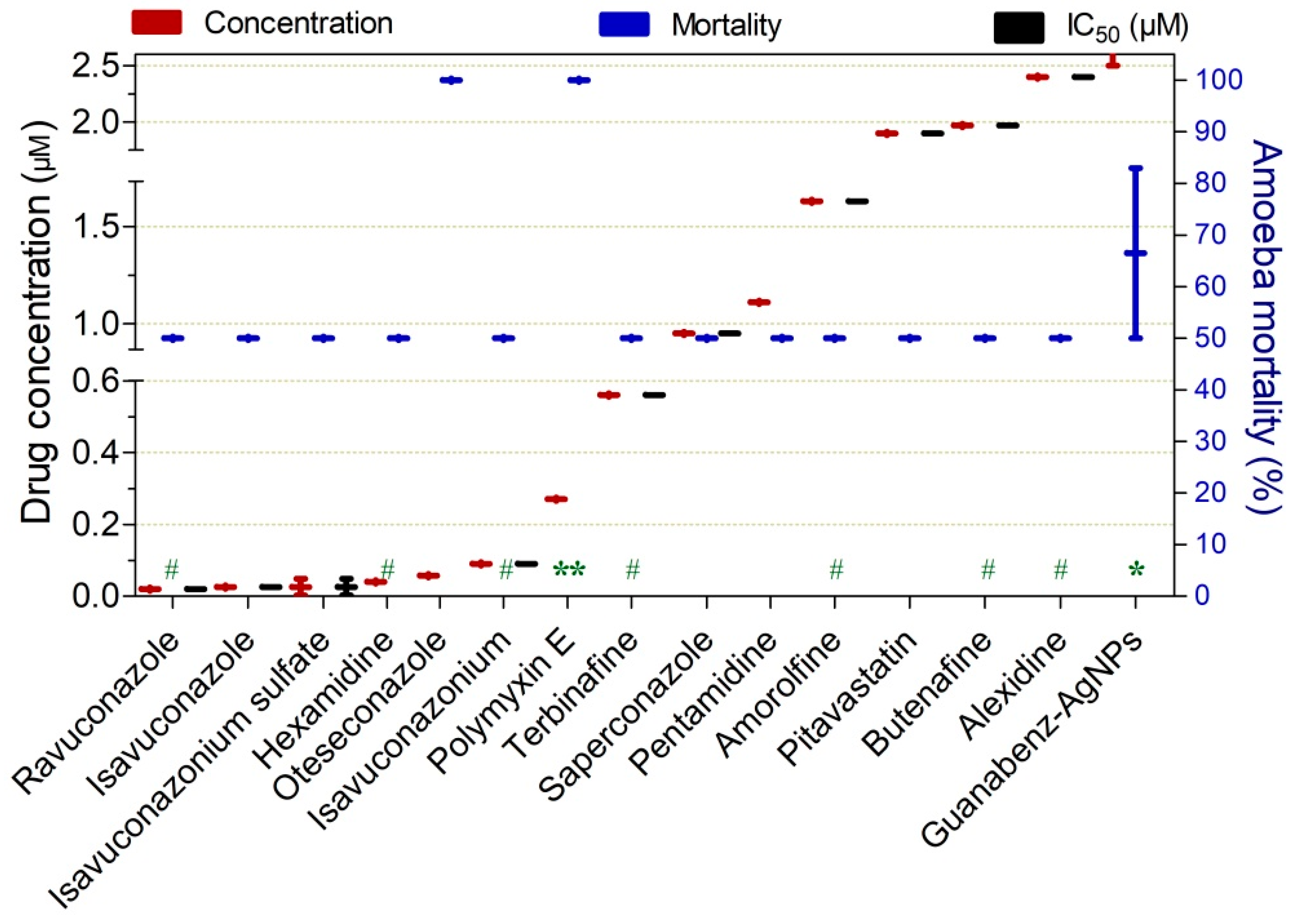
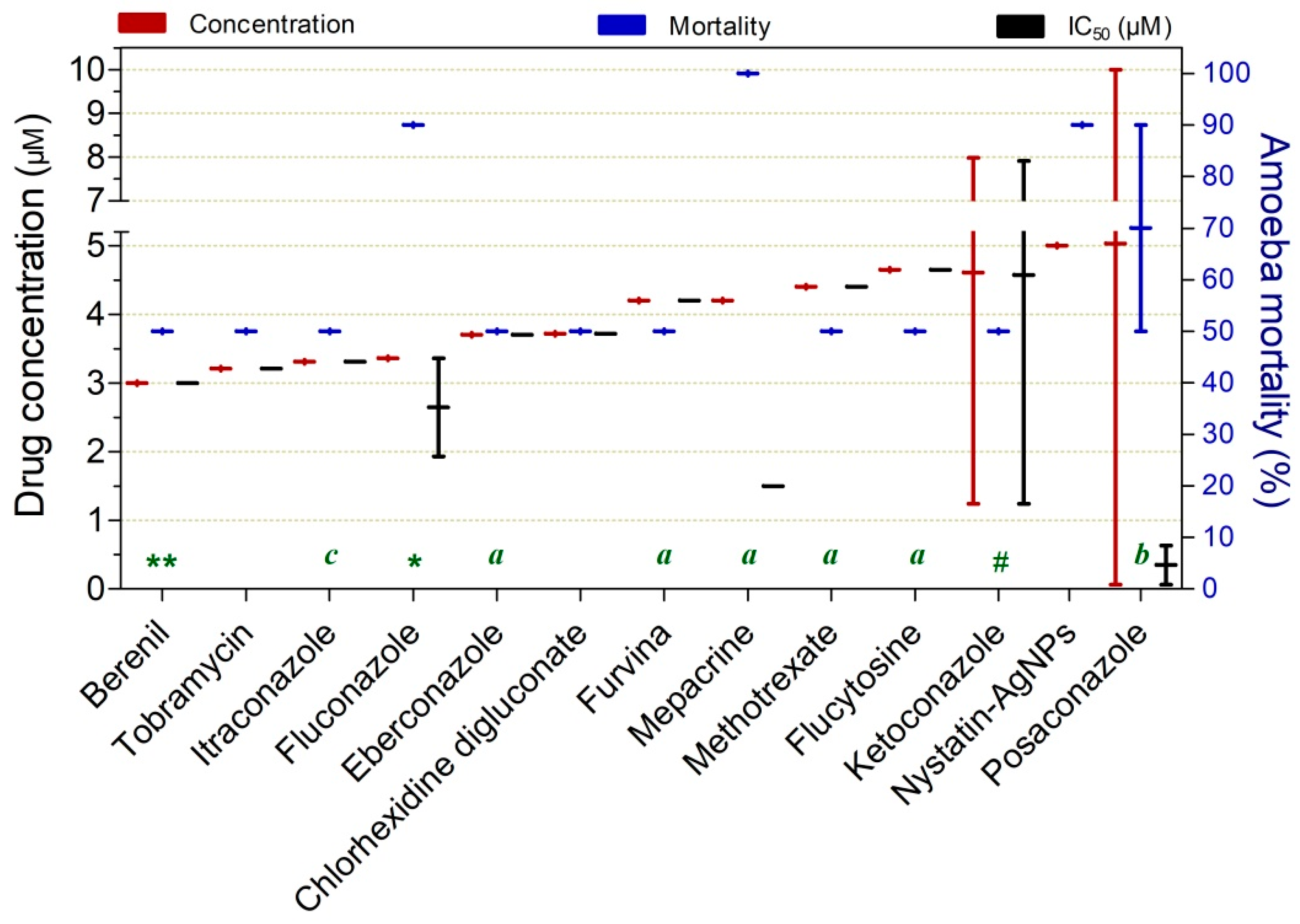


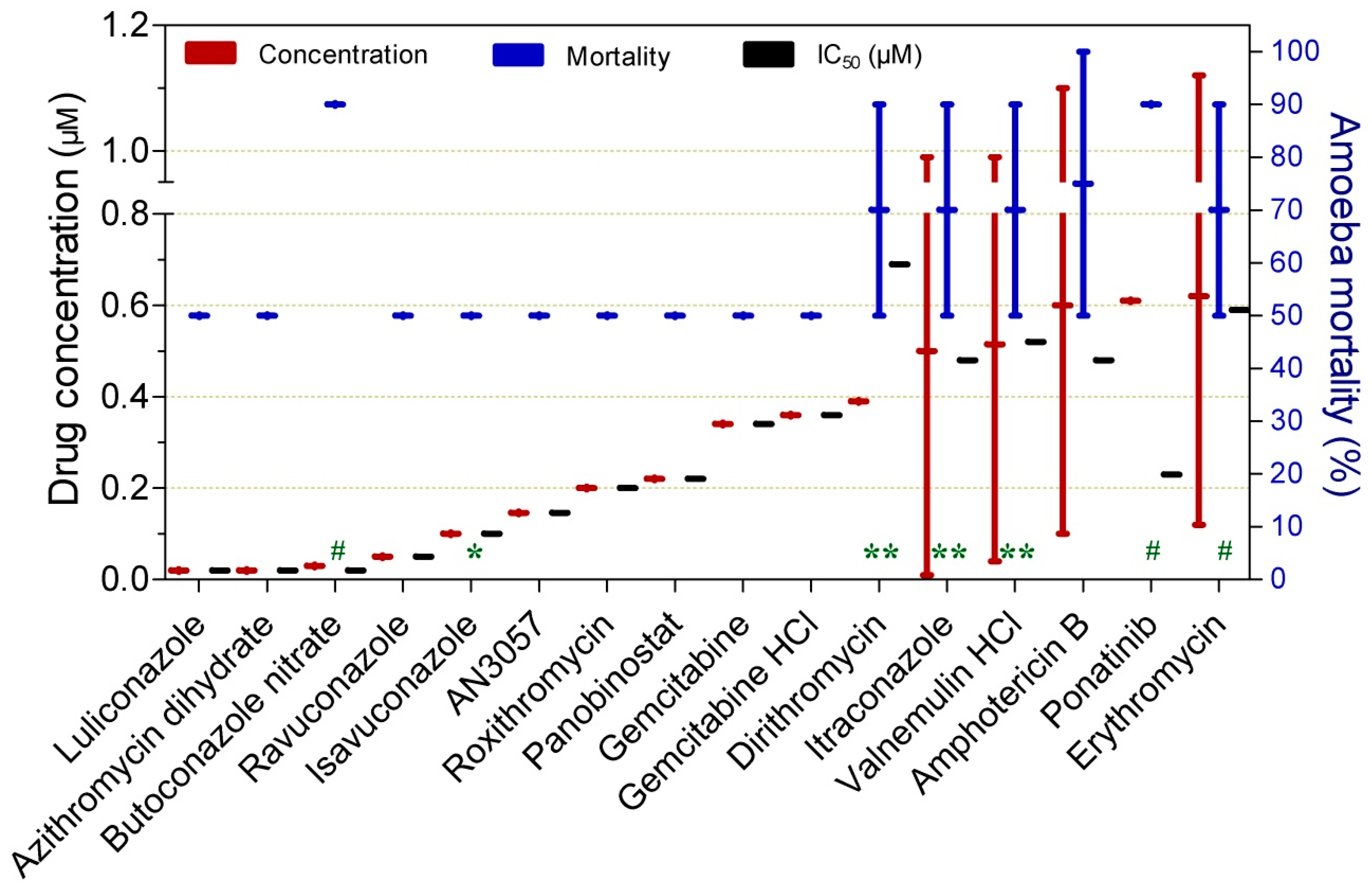

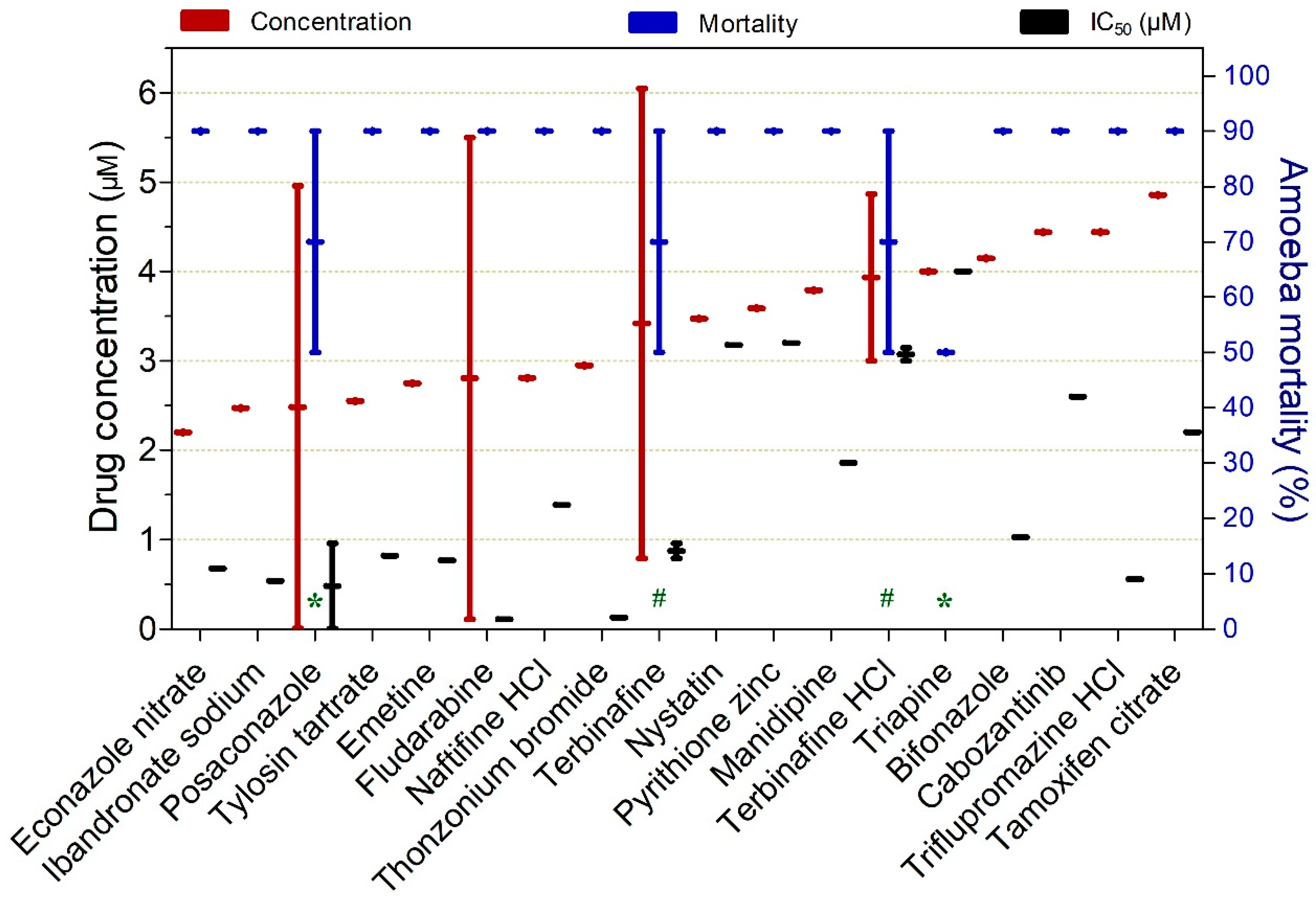



| Drug | Studies (n) | Dose (mg/kg) | Treatment Time (days) | Start of Treatment dpi | Cure Rate (%) |
|---|---|---|---|---|---|
| Sulphadiazine * | 1 | 200 | 10 | 1 | 100 |
| Rifampicin | 1 | 100 | 2 | 2 ** | 100 |
| Rifampicin | 1 | 100 | 5 | 2 | 88 |
| Drug (mg/mL) | Administration Route | Dose (mg/mL) | Treatment Time (days) | Start of Treatment dpi | Cure Rate (%) ±SD | Toxicity (%) |
|---|---|---|---|---|---|---|
| Aprotinin + neomycin (0.283) + Atropine ointment (10) | Eye drops ** | 13 | 17 (5×/day) | 2 | 100 | - |
| Fluconazole + neomycin (0.283) + Atropine ointment (10) | Eye drops ** | 2 | 22 (5×/day) | 2 | 100 | - |
| Neomycin (0.283) + atropine ointment (10) | Eye drops ** | 13 | 28 (5×/day) | 2 | 100 | - |
| PHMB + neomycin (0.283) + atropine ointment (10) | Eye drops ** | 0.2 | 13 (5×/day) | 2 | 100 | - |
| Povidone iodine + neomycin (0.283) + Atropine ointment (10) | Eye drops ** | 50 | 23 (5×/day) | 2 | 100 | - |
| Nitazoxanide (in liquid crystal) | Eye drops ** | 10 | 30 (5×/day) | 3 | 80 ± 0.0 | 20–70 |
| Voriconazole | Eye drops | 10 | 21 (13×/day/3 days; 7×/day/11 days and 4×/day/7 days) | 7 | 88.9 ± 0.0 | - |
| Miltefosine + polyhexanide | Eye drops | 0.06512 + 0.2 | 28 (8×/day/1st wk; 3×/day/2 wks) | 5 | 80 ± 0.0 | 69 |
| Miltefosine | Eye drops * | 0.06512 | 29 ± 1 (8×/day/1st wk; 3×/day/3 wks) | 5 | 73 ± 13 | 2 |
| Chlorhexidine + neomycin sulfate + polymyxin B sulfate + gramicidin | Eye drops | 0.02 + 4.49 + 0.5 + 0.025 | 7 (8×/day) + 21 (3×/day) | 5 | 71 ± 5 | |
| Miltefosine + propamidine isethionate | Eye drops | 0.06512 + 0.2 | 28 (8×/day/1st wk; 3×/day/2 wks) | 5 | 70 ± 0.0 | 79 |
| Miltefosine + chlorhexidine | Eye drops | 0.06512 + 0.2 | 28 (8×/day/1st wk; 3×/day/2 wks) | 5 | 70 ± 0.0 | 77 |
| Polyhexanide | Eye drops | 0.2 | 28 (8×/day/1st wk; 3×/day/2 wks) | 5 | 70 ± 0.0 | 65 |
| Drug | Studies (n) | Dose (mg/kg) | Treatment Time (Days) | Start of Treatment dpi | Cure Rate (%) | Toxicity |
|---|---|---|---|---|---|---|
| Amphotericin B + azithromycin | 1 | 2.5 + 25 | 5 | 3 | 100 | - |
| Cyclophosphamide | 1 | 30 | 10 | 0 | 92 | - |
| Amphotericin B + tetracycline | 1 | 2.5 + 150 | 7 | 3 | 87.5 | - |
| Rokitamycin | 1 | 20 | 3 (3×/day) * | 3 | 80 | No |
| Chlorpromazine | 1 | 20 | 3 * | 3 | 75 | - |
| Azithromycin + posaconazole | 1 | 25 + 20 | 3 | 3 | 70 | - |
| Amphotericin B | 1 | 7.5 | 14 | 1≥ | 60 | - |
| Azithromycin | 1 | 25 | 5 | 3 | 55 | |
| Azithromycin | 1 | 75 | 5 | 3 | 100 | |
| Miltefosine | 1 | 20 | 3 * | 3 | 55 | - |
| Drug | Drug Class | Approved for | Studies (n) | Concentration (µM), ± SD | Mortality (%) | IC50 (µM), ± SD | Exposure Time (h) |
|---|---|---|---|---|---|---|---|
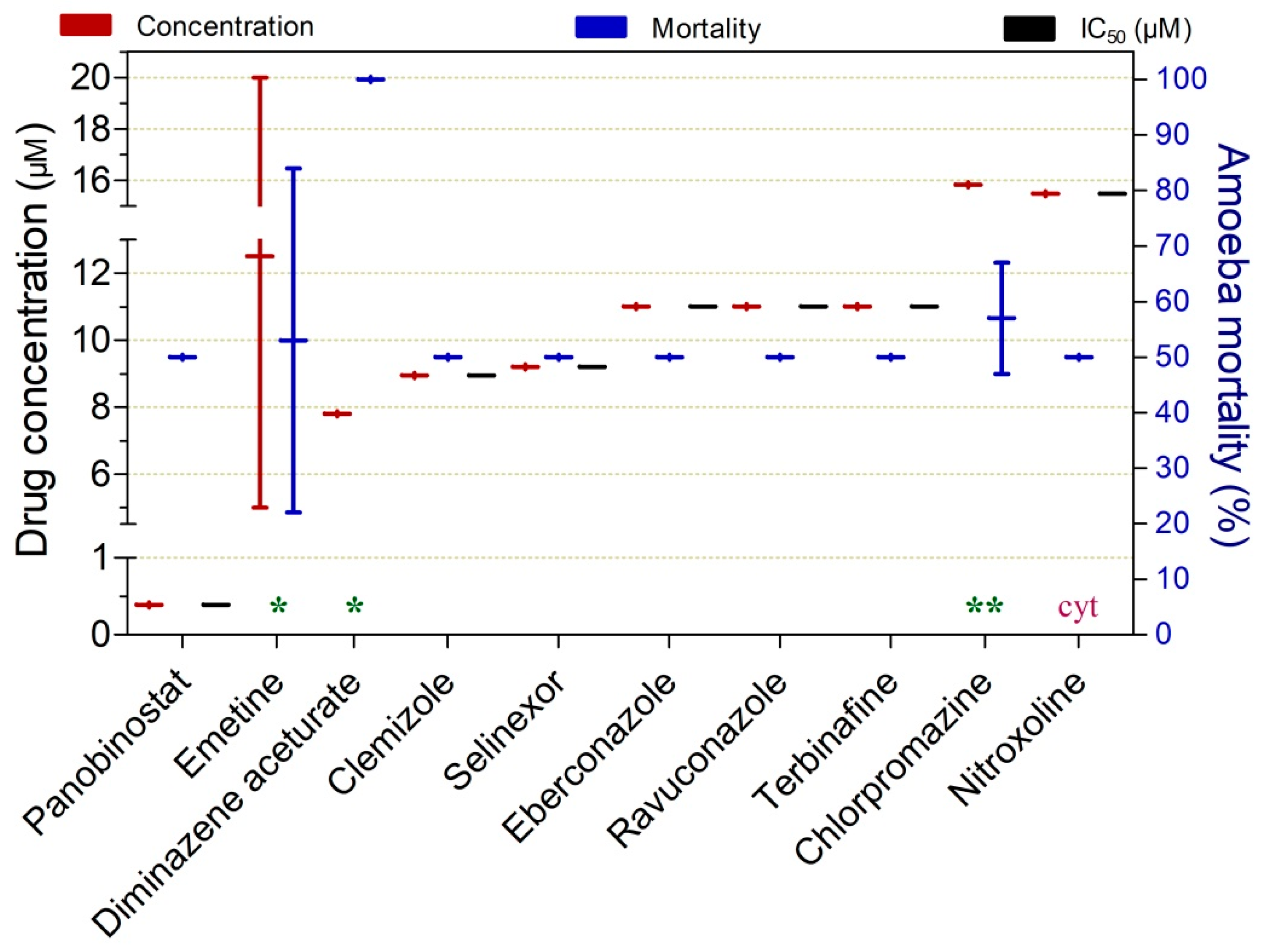
| Panobinostat | Histone deacetylase inhibitor | Cancer | 1 | 0.39 ± 0.0 | 50 | 0.39 ± 0.0 | 72 |
| Diminazene aceturate | Diamidines | Antiprotozoal | 1 | 7.8 ± 0.0 | 100 | 48 | |
| Clemizole | Benzimidazoles | Allergic rhinitis, urticaria | 1 | 8.95 ± 0.0 | 50 | 8.95 ± 0.0 | 72 |
| Selinexor | Triazoles | Cancer | 1 | 9.2 ± 0.0 | 50 | 9.2 ± 0.0 | 72 |
| Drug | Drug Class | Approved for | Studies (n) | Concentration ((SD), µM) | Mortality (%), ±SD | IC50 (µM), ±SD | Exposure Time (h) |
|---|---|---|---|---|---|---|---|
| Trophozoites | |||||||
| Ravuconazole | Azoles | Antifungal | 1 | 0.02 ± 0.0 | 50 ± 0.0 | 0.02 ± 0.0 | 72 |
| Isavuconazole | Azoles | Antifungal | 1 | 0.03 ± 0.0 | 50 ± 0.0 | 0.026 ± 0.0 | 48 |
| Isavuconazonium sulfate | Azoles | Antifungal | 3 | 0.03 ± 0.03 | 50 ± 0.0 | 0.03 ± 0.03 | 48 |
| Hexamidine | Hexamidine | Disinfectant | 1 | 0.04 ± 0.0 | 50 ± 0.0 | 72 | |
| Oteseconazole | Azoles | Antifungal | 1 | 0.06 ± 0.0 | 100 | 48 | |
| Isavuconazonium | Azoles | Antifungal | 1 | 0.09 ± 0.0 | 50 ± 0.0 | 0.09 ± 0.0 | 72 |
| Polymyxin E | Cationic polypeptide | Antibacterial | 1 | 0.27 ± 0.0 | 100 | 70 | |
| Terbinafine | Allylamine | Antifungal | 1 | 0.56 ± 0.0 | 50 ± 0.0 | 0.56 ± 0.0 | 72 |
| Saperconazole | Azoles | Antifungal | 1 | 0.95 ± 0.0 | 50 ± 0.0 | 0.95 ± 0.0 | 48 |
| Pentamidine | Diamidines | Disinfectant, antiseptic | 1 | 1.11 ± 0.0 | 50 ± 0.0 | 72 | |
| Amorolfine | Morpholine derivative | Antifungal | 1 | 1.63 ± 0.0 | 50 ± 0.0 | 1.63 ± 0.0 | 72 |
| Pitavastatin | Statins | Heart disorders | 1 | 1.90 ± 0.0 | 50 ± 0.0 | 1.9 ± 0.0 | 48 |
| Butenafine | Naftifine | Antifungal | 1 | 1.97 ± 0.0 | 50 ± 0.0 | 1.97 ± 0.0 | 72 |
| Alexidine | BisBiguanides | Antibacterial | 1 | 2.40 ± 0.0 | 50 ± 0.0 | 2.4 ± 0.0 | 72 |
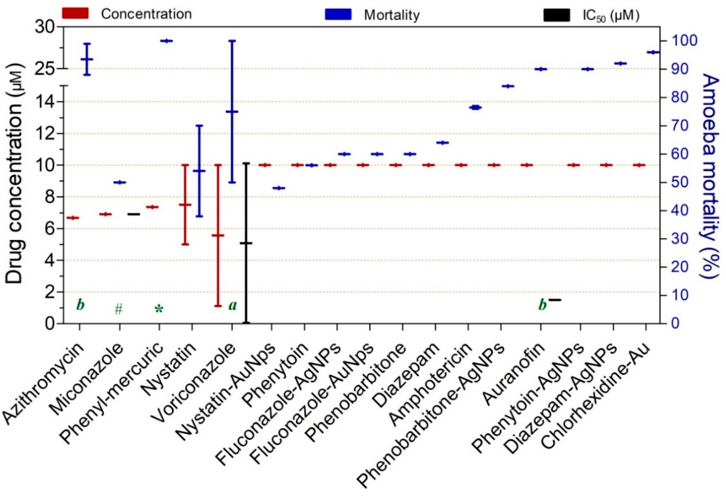
| Guanabenz-AgNPs | Alpha-2 adrenérgicos | Antihypertensive | 1 | 3.75 ± 1.77 | 66.5 ± 23.3 | 5 ± 0.0 | 24 |
| Berenil | Diamidines | Antiprotozoal | 1 | 3.00 ± 0.0 | 50 ± 0.0 | 3 ± 0.0 | 48 |
| Tobramycin | Aminoglycoside | Antibacterial | 1 | 3.21 ± 0.0 | 50 ± 0.0 | 3.21 ± 0.0 | 24 |
| Itraconazole | Azoles | Antifungal | 1 | 3.31 ± 0.0 | 50 ± 0.0 | 3.31 ± 0.0 | 96 |
| Fluconazole | Azoles | Antifungal | 5 | 3.36 ± 0.0 | 90 ± 0.0 | 2.7 (1.01) | 12 |
| Eberconazole | Azoles | Antifungal | 1 | 3.70 ± 0.0 | 50 ± 0.0 | 3.7 ± 0.0 | 72 |
| Chlorhexidine digluconate | Biguanides | Antiseptic | 1 | 3.72 ± 0.0 | 50 ± 0.0 | 3.72 ± 0.0 | 96 |
| Furvina | Nitrofurans | Antibacterial, Antifungal | 1 | 4.20 ± 0.0 | 50 ± 0.0 | 4.2 ± 0.0 | 72 |
| Mepacrine | Acridines | Antiprotozoal | 1 | 4.20 ± 0.0 | 100 ± 0.0 | 1.5 ± 0.0 | 72 |
| Methotrexate | Antimetabolites | Antineoplastic | 1 | 4.40 ± 0.0 | 50 ± 0.0 | 4.4 ± 0.0 | 72 |
| Flucytosine | Pyrimidine analogues | Antifungal | 1 | 4.65 ± 0.0 | 50 ± 0.0 | 4.65 ± 0.0 | 72 |
| Ketoconazole | Azoles | Antifungal | 5 | 4.61 ± 4.77 | 50 ± 0.0 | 4.6 ± 4.72 | 66 |
| Posaconazole | Azoles | Antifungal | 2 | 5.03 ± 7.03 | 70 ± 28.3 | 0.35 ± 0.4 | 84 |
| Azithromycin | Macrolide | Antibacterial | 13 | 6.68 ± 0.0 | 94 ± 7.8 | 120 | |
| Miconazole | Azoles | Antifungal | 1 | 6.90 ± 0.0 | 50 ± 0.0 | 6.9 ± 0.0 | 50 |
| Phenyl-mercuric nitrate | Organomercurials | Antiseptic | 1 | 7.36 ± 0.0 | 100 ± 0.0 | 4 | |
| Voriconazole | Azoles | Antifungal | 4 | 5.56 ± 6.3 | 75 ± 35.4 | 5.1 ± 7.1 | 96 |
| Chlorhexidine-Au | Biguanides | Antiseptic | 1 | 10 ± 0.0 | 96 ± 0.0 | 24 | |
| Diazepam-AgNP | Benzodiazepine | Antiseizure | 1 | 10 ± 0.0 | 92 ± 0.0 | 24 | |
| Auranofin | Gold derivative | Antirheumatic | 1 | 10 ± 0.0 | 90 ± 0.0 | 1.5 ± 0.0 | 120 |
| Phenytoin-AgNPs | Hydantoin derivative | Antiseizure | 1 | 10 ± 0.0 | 90 ± 0.0 | 24 | |
| Phenobarbitone-AgNPs | Barbiturate | Antiepileptic | 1 | 10 ± 0.0 | 84 ± 0.0 | 24 | |
| Amphotericin B-AuNps | Polyenes | Antifungal | 2 | 10 ± 0.0 | 76 ± 0.0 | 24 | |
| Diazepam | Benzodiazepine | Antiseizure | 1 | 10 ± 0.0 | 64 ± 0.0 | 24 | |
| Fluconazole-AuNps | Azoles | Antifungal | 1 | 10 ± 0.0 | 60 ± 0.0 | 24 | |
| Phenobarbitone | Barbiturate | Antiepileptic | 1 | 10 ± 0.0 | 60 ± 0.0 | 24 | |
| Phenytoin | Hydantoin derivative | Antiseizure | 1 | 10 ± 0.0 | 56 ± 0.0 | 24 | |
| Cysts | |||||||
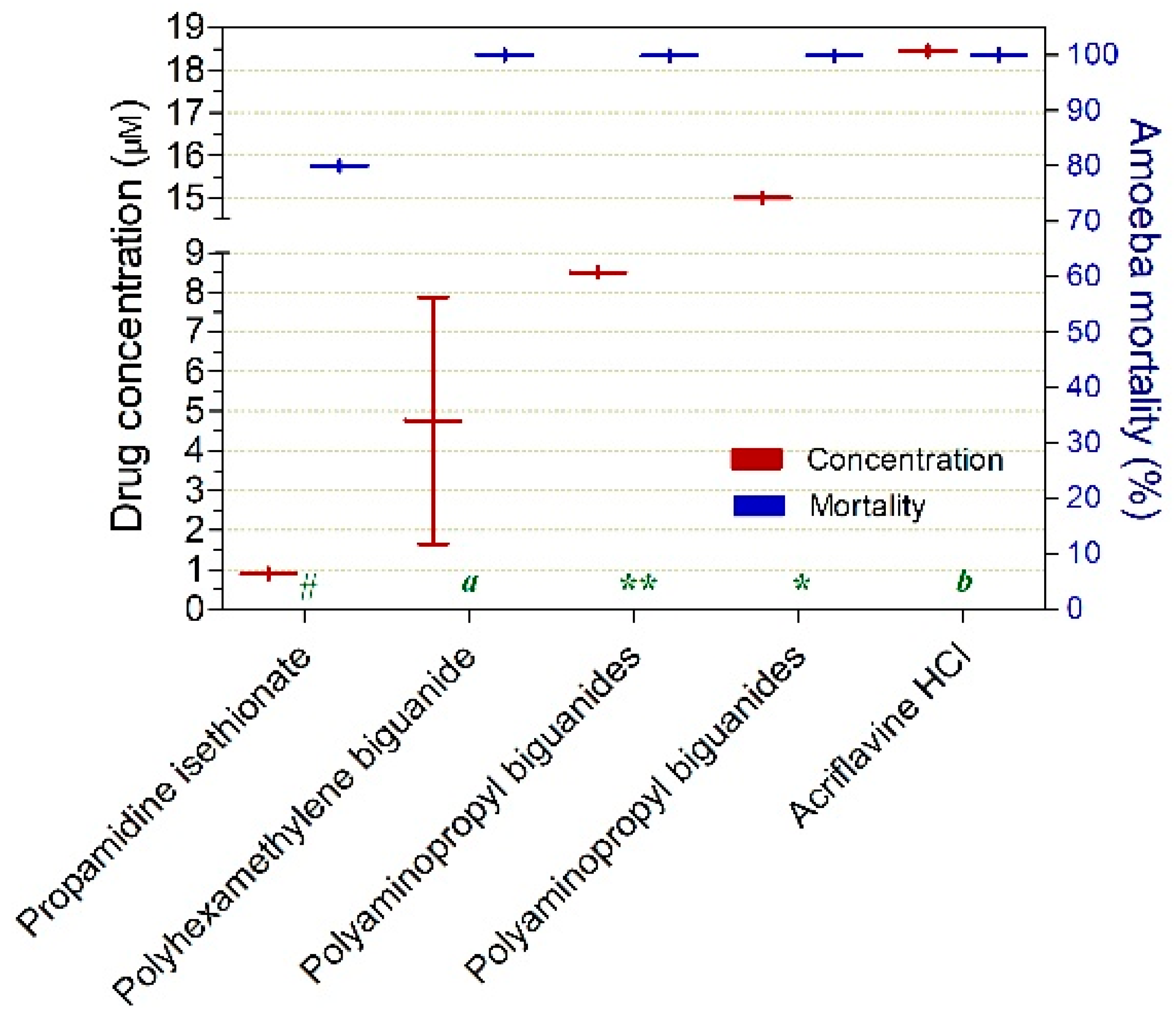
| Propamidine isethionate | Aromatic diamidines | Antimicrobial/Antiseptic | 1 | 0.89 ± 0.0 | 80 ± 0.0 | 4 | |
| Polyhexamethylene biguanide | Biguanides | Disinfectant, antiseptic | 2 | 1.64 ± 0.0 | 100 ± 0.0 | 18 ± 14 | |
| Polyaminopropyl biguanides | Biguanides | Antiseptic | 1 | 8.5 ± 0.0 | 99.9 ± 0.0 | 3 | |
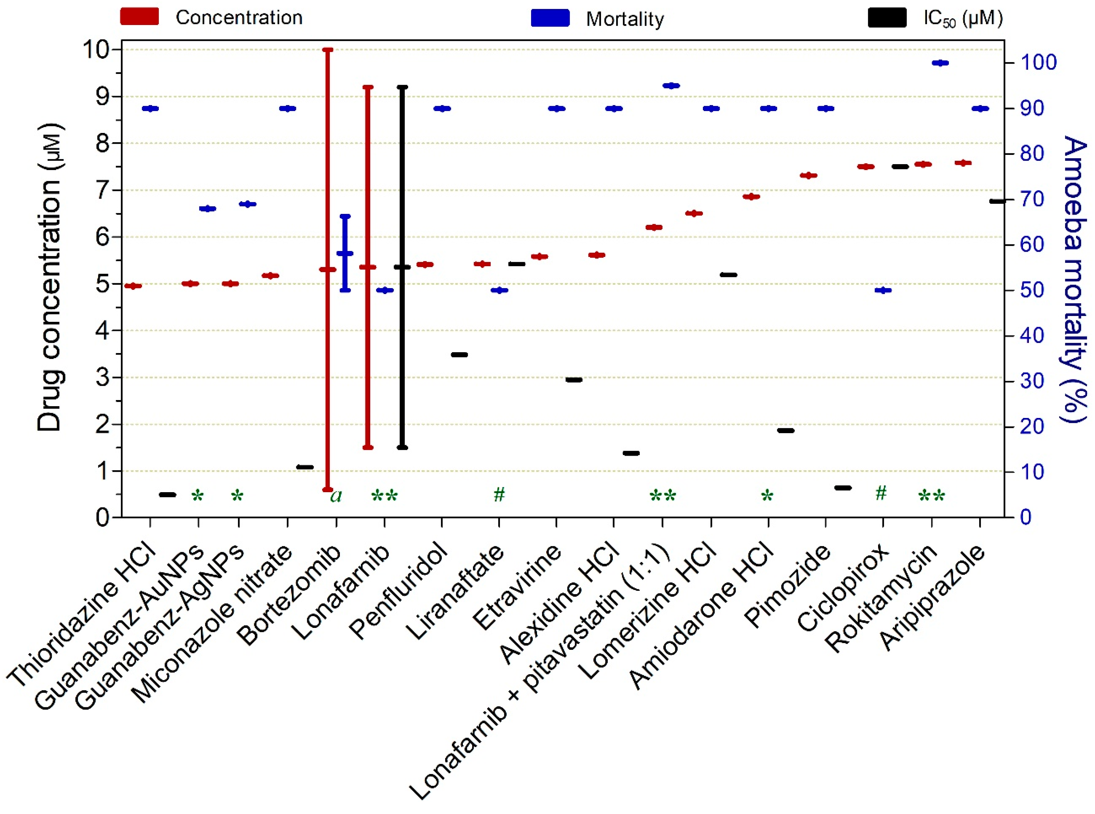
| Drug | Drug Class | Approved for | Studies (n) | Concentration (µM) ± SD, | Mortality (%) ± SD | IC50 (µM) ± SD | Exposure Time (h) |
|---|---|---|---|---|---|---|---|
| Trophozoites | |||||||
| Luliconazole | Azoles | Antifungal | 1 | 0.02 ± 0.0 | 50 ± 0.0 | 0.02 ± 0.0 | 72 |
| Azithromycin dihydrate | Macrolide | Antibacterial | 1 | 0.02 ± 0.0 | 50 ± 0.0 | 0.02 ± 0.0 | 72 |
| Butoconazole nitrate | Azoles | Antifungal | 1 | 0.03 ± 0.0 | 90 ± 0.0 | 0.02 ± 0.0 | 120 |
| Ravuconazole | Azoles | Antifungal | 1 | 0.05 ± 0.0 | 50 ± 0.0 | 0.05 ± 0.0 | 72 |
| Isavuconazole | Azoles | Antifungal | 1 | 0.1 ± 0.0 | 50 ± 0.0 | 0.1 ± 0.0 | 48 |
| AN3057 | Boronics | Antifungal | 1 | 0.146 ± 0.0 | 50 ± 0.0 | 0.146 ± 0.0 | 72 |
| Roxithromycin | Macrolide | Antibacterial | 1 | 0.2 ± 0.0 | 50 ± 0.0 | 0.2 ± 0.0 | 72 |
| Panobinostat | Histone deacetylase inhibitor | Antineoplastic | 1 | 0.22 ± 0.0 | 50 ± 0.0 | 0.22 ± 0.0 | 72 |
| Gemcitabine | Nucleosides | Antineoplastic | 1 | 0.34 ± 0.0 | 50 ± 0.0 | 0.34 ± 0.0 | 72 |
| Gemcitabine HCl | Nucleosides | Antineoplastic | 1 | 0.36 ± 0.0 | 50 ± 0.0 | 0.36 ± 0.0 | 72 |
| Dirithromycin | Macrolide | Antibacterial | 2 | 0.39 ± 0.0 | 70 ± 28.3 | 0.69 ± 0.0 | 84 |
| Itraconazole | Azoles | Antifungal | 2 | 0.5 ± 0.69 | 70 ± 28.3 | 0.48 ± 0.0 | 84 |
| Valnemulin HCl | Pleuromutilins | Antibacterial | 2 | 0.515 ± 0.67 | 70 ± 28.3 | 0.52 ± 0.0 | 84 |
| Ponatinib | Benzanilides | Antineoplastic | 1 | 0.61 ± 0.0 | 90 | 0.23 ± 0.0 | 120 |
| Clarithromycin | Macrolide | Antibacterial | 1 | 0.61 ± 0.82 | 70 ± 28.3 | 0.45 ± 0.6 | 72 |
| Erythromycin | Macrolide | Antibacterial | 1 | 0.62 ± 0.71 | 70 ± 28.3 | 0.59 ± 0.0 | 120 |
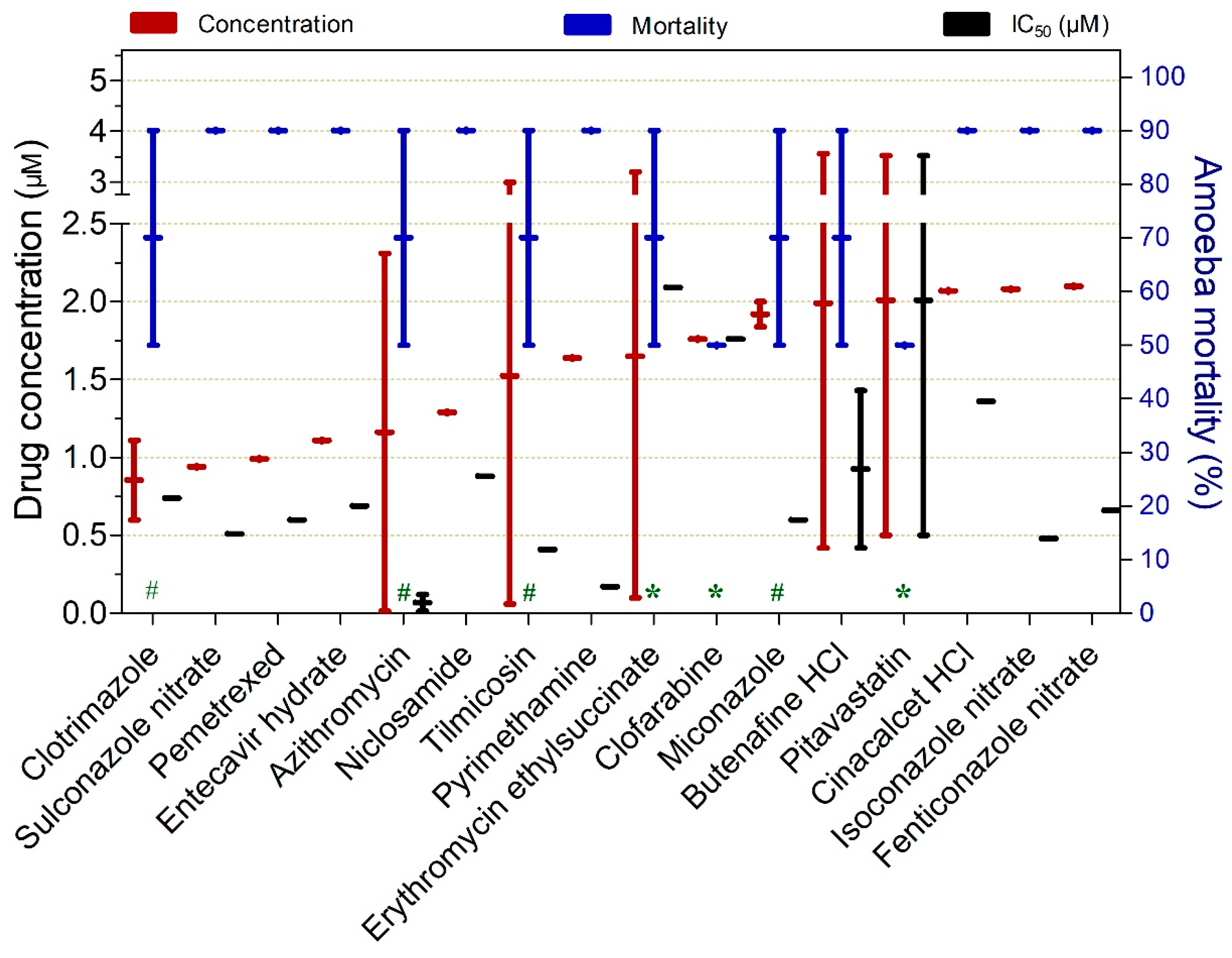
| Clotrimazole | Azoles | Antifungal | 2 | 0.855 ± 0.36 | 70 ± 28.3 | 0.74 ± 0.0 | 84 |
| Sulconazole nitrate | Azoles | Antifungal | 1 | 0.94 ± 0.0 | 90 ± 0.0 | 0.51 ± 0.0 | 120 |
| Pemetrexed | Antimetabolites | Antineoplastic | 1 | 0.99 ± 0.0 | 90 ± 0.0 | 0.6 ± 0.0 | 120 |
| Entecavir hydrate | Nucleoside analogues | Antiviral | 1 | 1.11 ± 0.0 | 90 ± 0.0 | 0.69 ± 0.0 | 120 |
| Azithromycin | Macrolide | Antibacterial | 12 | 1.16 ± 1.62 | 70 ± 28.3 | 0.07 ± 0.1 | 84 |
| Niclosamide | Salicylanilide | Anthelmintic | 1 | 1.29 ± 0.0 | 90 ± 0.0 | 0.88 ± 0.0 | 120 |
| Tilmicosin | Macrolide | Antibacterial | 2 | 1.525 ± 2.07 | 70 ± 28.3 | 0.41 ± 0.0 | 84 |
| Pyrimethamine | Pyrimedines | Antiprotozoal | 1 | 1.64 ± 0.0 | 90 ± 0.0 | 0.17 ± 0.0 | 120 |
| Erythromycin ethylsuccinate | Macrolide | Antibacterial | 1 | 1.65 (2.19) | 70 ± 28.3 | 2.09 ± 0.0 | 72 |
| Clofarabine | Nucleosides | Cancer | 1 | 1.76 ± 0.0 | 50 ± 0.0 | 1.76 ± 0.0 | 72 |
| Miconazole | Azoles | Antifungal | 1 | 1.92 ± 0.11 | 70 ± 28.3 | 0.6 ± 0.0 | 84 |
| Butenafine HCl | Naftifine | Antifungal | 2 | 1.99 ± 2.22 | 70 ± 28.3 | 0.93 ± 0.7 | 120 |
| Pitavastatin | Statins | Heart disorders | 1 | 2.01 ± 2.14 | 50 ± 0.0 | 2.01 ± 2.1 | 72 |
| Cinacalcet HCl | Naphthalenes | Hypercalcemia | 1 | 2.07 ± 0.0 | 90 ± 0.0 | 1.36 ± 0.0 | 120 |
| Isoconazole nitrate | Azoles | Antifungal | 1 | 2.08 ± 0.0 | 90 ± 0.0 | 0.48 ± 0.0 | 120 |
| Fenticonazole nitrate | Azoles | Antifungal | 1 | 2.1 ± 0.0 | 90 ± 0.0 | 0.66 ± 0.0 | 120 |
| Econazole nitrate | Azoles | Antifungal | 1 | 2.2 ± 0.0 | 90 ± 0.0 | 0.68 ± 0.0 | 120 |
| Ibandronate sodium | Bisphosphonate | Osteoporosis | 1 | 2.47 ± 0.0 | 90 | 0.54 ± 0.0 | 120 |
| Posaconazole | Azoles | Antifungal | 13 | 2.48 ± 3.5 | 70 ± 28.3 | 0.48 ± 0.7 | 72 |
| Tylosin tartrate | Macrolide | Antibacterial | 1 | 2.55 ± 0.0 | 90 ± 0.0 | 0.82 ± 0.0 | 120 |
| Emetine | Alkaloids | Antiprotozoal | 1 | 2.75 ± 0.0 | 90 ± 0.0 | 0.77 ± 0.0 | 120 |
| Fludarabine | Nucleosides | Antineoplastic | 2 | 2.81 ± 3.81 | 90 ± 0.0 | 0.11 ± 0.0 | 120 |
| Thonzonium bromide | Quaternary ammonium | Antiseptic | 1 | 2.95 ± 0.0 | 90 ± 0.0 | 0.13 ± 0.0 | 120 |
| Terbinafine | Allylamine | Antifungal | 1 | 3.42 ± 3.72 | 70 ± 28.3 | 0.88 ± 0.1 | 84 |
| Pyrithione zinc | Zinc complex | Antifungal | 1 | 3.59 ± 0.0 | 90 ± 0.0 | 3.2 ± 0.0 | 120 |
| Manidipine | Dihydropyridine | Antihypertensive | 1 | 3.79 ± 0.0 | 90 ± 0.0 | 1.86 ± 0.0 | 120 |
| Terbinafine HCl | Allylamine | Antifungal | 2 | 3.94 ± 1.32 | 70 ± 28.3 | 3.08 ± 0.1 | 84 |
| Triapine | Thiosemicarbazone derivative | Antineoplastic | 1 | 4 ± 0.0 | 50 ± 0.0 | 4 ± 0.0 | 72 |
| Bifonazole | Azoles | Antifungal | 1 | 4.15 ± 0.0 | 90 ± 0.0 | 1.03 ± 0.0 | 120 |
| Cabozantinib | Quinidines | Antineoplastic | 1 | 4.44 ± 0.0 | 90 ± 0.0 | 2.6 ± 0.0 | 120 |
| Triflupromazine HCl | Phenothiazines | Psychiatric disorders | 1 | 4.44 ± 0.0 | 90 ± 0.0 | 0.56 ± 0.0 | 120 |
| Tamoxifen citrate | Triphenylethylene | Antineoplastic | 1 | 4.86 ± 0.0 | 90 ± 0.0 | 2.2 ± 0.0 | 120 |
| Thioridazine HCl | Phenothiazines | Antipsychotic | 1 | 4.95 ± 0.0 | 90 ± 0.0 | 0.49 ± 0.0 | 120 |
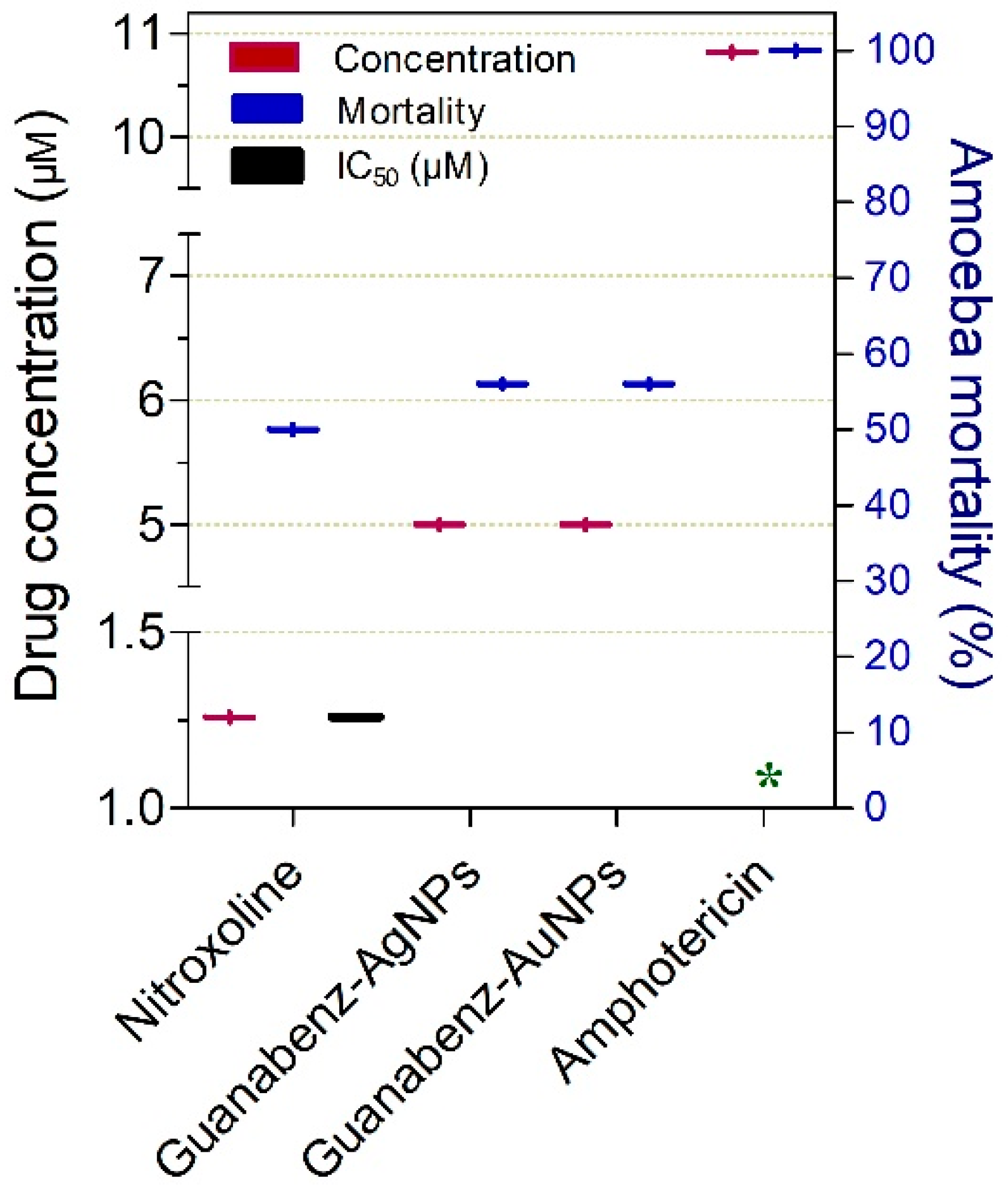
| Guanabenz-AuNPs | Alpha-2 adrenérgicos | Antihypertensive | 1 | 5 ± 0.0 | 68 ± 0.0 | 24 | |
| Guanabenz-AgNPs | Alpha-2 adrenérgicos | Antihypertensive | 1 | 5 ± 0.0 | 69 ± 0.0 | 24 | |
| Miconazole nitrate | Azoles | Antifungal | 1 | 5.17 ± 0.0 | 90 ± 0.0 | 1.08 ± 0.0 | 120 |
| Bortezomib | Proteasome inhibitor | Antineoplastic | 2 | 5.3 ± 6.7 | 58 ± 11.5 | 84 | |
| Lonafarnib | Benzocycloheptapyridine | Hutchinson-Gilford syndrome | 5 | 5.4 ± 5.4 | 50 ± 0.0 | 5.4 ± 5.4 | 48 |
| Penfluridol | Diphenylbutylpiperidines | Antiseptic | 1 | 5.41 ± 0.0 | 90 ± 0.0 | 3.48 ± 0.0 | 120 |
| Amphotericin B | Polyenes | Antifungal | 3 | 5.41 ± 5.0 | 85 ± 15 | 0.48 ± 0.0 | 48 |
| Liranaftate | Naftifine | Antifungal | 1 | 5.42 ± 0.0 | 50 ± 0.0 | 5.42 ± 0.0 | 72 |
| Etravirine | Diarylethers | Antivirals (antiretro) | 1 | 5.58 ± 0.0 | 90 ± 0.0 | 2.95 ± 0.0 | 120 |
| Alexidine HCl | BisBiguanides | Antibacterial | 1 | 5.61 ± 0.0 | 90 ± 0.0 | 1.38 ± 0.0 | 120 |
| Lonafarnib + pitavastatin (1:1) | 1 | 6.2 ± 0.0 | 95 ± 0.0 | 48 | |||
| Lomerizine HCl | Piperazines | Antimigraine, antivertigo | 1 | 6.5 ± 0.0 | 90 ± 0.0 | 5.19 ± 0.0 | 120 |
| Amiodarone HCl | Benzofurans | Antiarrhythmic | 1 | 6.86 ± 0.0 | 90 ± 0.0 | 1.86 ± 0.0 | 120 |
| Pimozide | Diphenylbutylpiperidine | Antipsychotic | 1 | 7.31 ± 0.0 | 90 ± 0.0 | 0.64 ± 0.0 | 120 |
| Ciclopirox | Pyridine derivatives | Antifungal | 1 | 7.5 ± 0.0 | 50 ± 0.0 | 7.5 ± 0.0 | 72 |
| Rokitamycin | Macrolide | Antibacterial | 1 | 7.55 ± 0.0 | 100 ± 0.0 | 48 | |
| Aripiprazole | Azoles | Antifungal | 1 | 7.58 ± 0.0 | 90 ± 0.0 | 6.76 ± 0.0 | 120 |
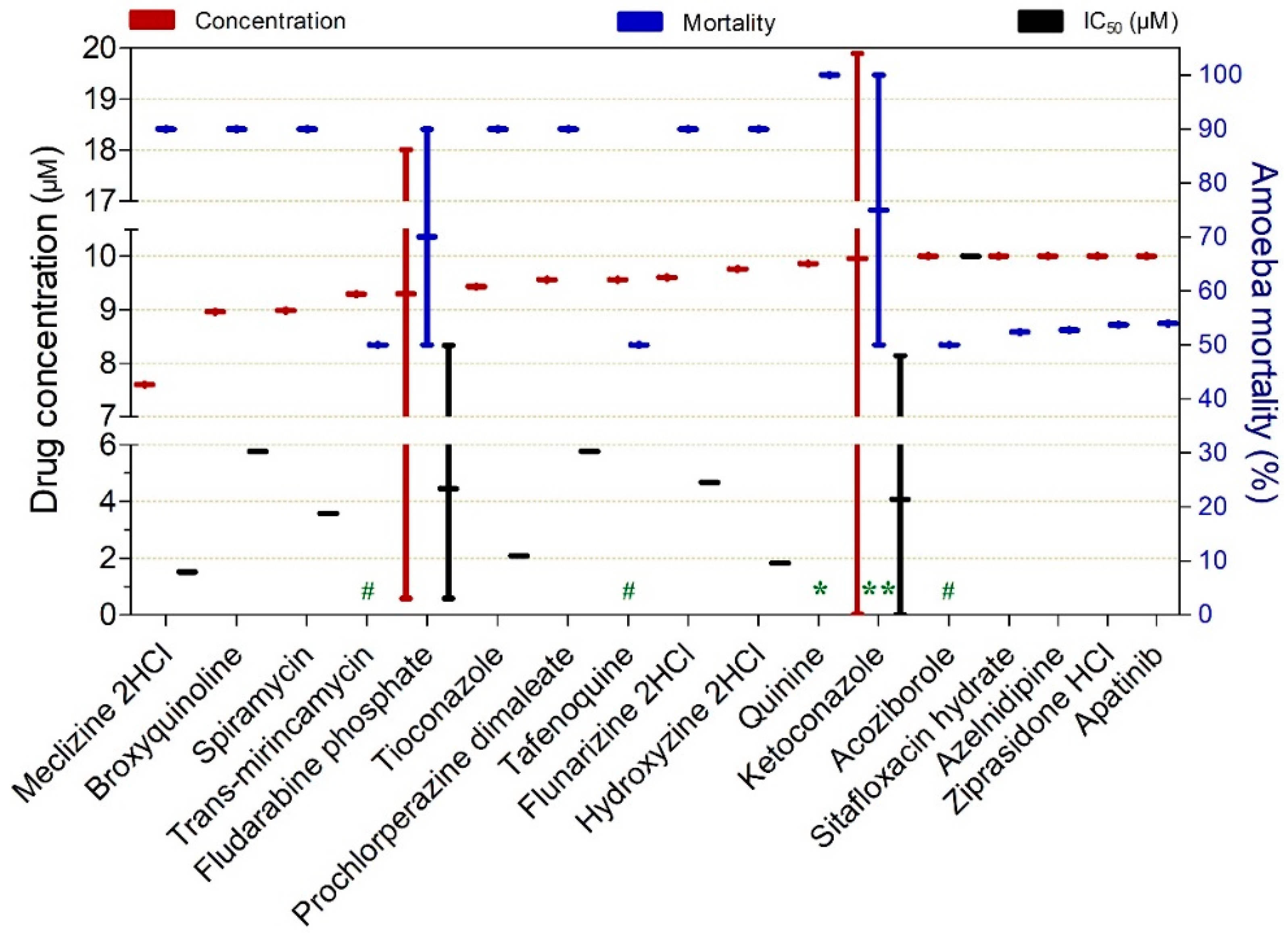
| Meclizine 2HCl | Piperazines | Antiallergic | 1 | 7.6 ± 0.0 | 90 ± 0.0 | 1.52 ± 0.0 | 120 |
| Broxyquinoline | Haloquinolines | Fungicides, antiprotozoal | 1 | 8.96 ± 0.0 | 90 ± 0.0 | 5.77 ± 0.0 | 120 |
| Spiramycin | Macrolide | Antibacterial | 1 | 8.98 ± 0.0 | 90 ± 0.0 | 3.58 ± 0.0 | 120 |
| Trans-mirincamycin | Lincosamide | Antibacterial | 1 | 9.29 ± 0.0 | 50 ± 0.0 | 9.29 ± 0.0 | 72 |
| Fludarabine phosphate | Nucleosides | Antineoplastic | 2 | 9.3 ± 12.3 | 70 ± 28.3 | 4.46 ± 5.5 | 84 |
| Tioconazole | Azoles | Antifungal | 1 | 9.43 ± 0.0 | 90 ± 0.0 | 2.08 ± 0.0 | 120 |
| Prochlorperazine dimaleate | Phenothiazines | Antipsychotic | 1 | 9.56 ± 0.0 | 90 ± 0.0 | 5.77 ± 0.0 | 120 |
| Tafenoquine | Aminoquinoline | Antiprotozoal | 1 | 9.56 ± 0.0 | 50 ± 0.0 | 9.56 ± 0.0 | 72 |
| Flunarizine 2HCl | Diphenylpiperazine | Antimigraine, antivertigo | 1 | 9.6 ± 0.0 | 90 ± 0.0 | 4.67 ± 0.0 | 120 |
| Hydroxyzine 2HCl | Piperazines | Antiallergic | 1 | 9.76 ± 0.0 | 90 ± 0.0 | 1.83 ± 0.0 | 120 |
| Quinine | Alkaloids | Antiprotozoal | 1 | 9.86 ± 0.0 | 100 ± 0.0 | 48 | |
| Ketoconazole | Azoles | Antifungal | 15 | 10 ± 14 | 75 ± 35.4 | 4.08 ± 5.7 | 84 |
| Climbazole | Azoles | Antifungal | 1 | 10 ± 0.0 | 100 ± 0.0 | 120 | |
| Chlorprothixene | Phenothiazines | Psychiatric disorders | 1 | 10 ± 0.0 | 78 ± 0.0 | 120 | |
| Clemastine | Benzyl ethers | Antihistamine | 1 | 10 ± 0.0 | 78 ± 0.0 | 120 | |
| Arbidol HCl | Phenylthiazoles | Antiviral | 1 | 10 ± 0.0 | 77 ± 0.0 | 120 | |
| Bekanamycin | Aminoglycosides | Antibacterial | 1 | 10 ± 0.0 | 76 ± 0.0 | 120 | |
| Clofazimine | Phenazine | Antibacterial | 1 | 10 ± 0.0 | 75 ± 0.0 | 120 | |
| Cidofovir | Pyrimidines | Antiviral | 1 | 10 ± 0.0 | 73 ± 0.0 | 120 | |
| Pitavastatin calcium | Statins | Heart disorders | 1 | 10 ± 0.0 | 69 ± 0.0 | 120 | |
| Atovaquone | Quinones | Antiprotozoal | 1 | 10 ± 0.0 | 68 ± 0.0 | 120 | |
| Mitoxantrone HCl | Anthracenedione | Antineoplastic | 1 | 10 ± 0.0 | 68 ± 0.0 | 120 | |
| Clodronate disodium | Bisphosphonate | Osteoporosis, hypercalcemia | 1 | 10 ± 0.0 | 66 ± 0.0 | 120 | |
| Bexarotene | Retinoid | Antineoplastic | 1 | 10 ± 0.0 | 65 ± 0.0 | 120 | |
| Cyclosporine | Cyclic lipopeptides | Immunosuppressant | 1 | 10 ± 0.0 | 65 ± 0.0 | 120 | |
| Cetylpyridinium chloride | Pyridinium | Antiseptic | 1 | 10 ± 0.0 | 64 ± 0.0 | 120 | |
| Sorafenib | Diarylethers | Antineoplastic | 1 | 10 ± 0.0 | 63 ± 0.0 | 120 | |
| Cyproheptadine HCl | Piperidines | Antiallergic | 1 | 10 ± 0.0 | 61 ± 0.0 | 120 | |
| Mevastatin | Delta valerolactones | Antihyperlipidemic | 1 | 10 ± 0.0 | 61 ± 0.0 | 120 | |
| Ethacridine lactate monohydrate | Acridines | Antiseptic, abortifacient | 1 | 10 ± 0.0 | 60 ± 0.0 | 120 | |
| Bismuth subcitrate potassium | Tricarboxylic acid | Antiulcer | 1 | 10 ± 0.0 | 59 ± 0.0 | 120 | |
| Flumequine | Fluoroquinolones | Antibacterial | 1 | 10 ± 0.0 | 59 ± 0.0 | 120 | |
| Fluvastatin sodium | Statins | Antihyperlipidemic | 1 | 10 ± 0.0 | 59 ± 0.0 | 120 | |
| Triclabendazole | Azoles | Antifungal | 1 | 10 ± 0.0 | 59 ± 0.0 | 120 | |
| Trimipramine maleate | Tricyclic | Antidepressant | 1 | 10 ± 0.0 | 59 ± 0.0 | 120 | |
| Dicyclomine HCl | Carboxylic acid ester | Antispasmodic | 1 | 10 ± 0.0 | 58 ± 0.0 | 120 | |
| Pridinol methanesulfonate | Piperidines | Muscle relaxant | 1 | 10 ± 0.0 | 57 ± 0.0 | 120 | |
| Crystal violet | Triphenylmethane dye | Antibacterial, antifungal | 1 | 10 ± 0.0 | 55 ± 0.0 | 120 | |
| Doxorubicin | Anthracycline | Antineoplastic | 1 | 10 ± 0.0 | 55 ± 0.0 | 120 | |
| Eprazinone 2HCl | Benzomorphan | Antitussive | 1 | 10 ± 0.0 | 55 ± 0.0 | 120 | |
| Apatinib | Quinazolines | Antineoplastic | 1 | 10 ± 0.0 | 54 ± 0.0 | 120 | |
| Artemether | Artemisinins | Antiprotozoal | 1 | 10 ± 0.0 | 54 ± 0.0 | 120 | |
| Nilotinib | Benzanilides | Antineoplastic | 1 | 10 ± 0.0 | 54 ± 0.0 | 120 | |
| Pentamidine | Diamidines | Disinfectant, antiseptic | 1 | 10 ± 0.0 | 54 ± 0.0 | 120 | |
| Ziprasidone HCl | Benzisothiazole | Antipsychotic | 1 | 10 ± 0.0 | 54 ± 0.0 | 120 | |
| Azelnidipine | Dihydropyridine derivative | Heart disorders | 1 | 10 ± 0.0 | 53 ± 0.0 | 120 | |
| Sitafloxacin hydrate | Fluoroquinolone | Antibacterial | 1 | 10 ± 0.0 | 52 ± 0.0 | 120 | |
| Acoziborole | Azoles | Antifungal | 1 | 10 ± 0.0 | 50 ± 0.0 | 10 ± 0.0 | 72 |
| Cysts | |||||||
| Nitroxoline | Quinoline derivative | Antibacterial | 1 | 1.26 ± 0.0 | 50 ± 0.0 | 1.26 ± 0.0 | 24 |
| Guanabenz-AgNPs | Alpha-2 adrenérgicos | Antihypertensive | 1 | 5 ± 0.0 | 56 ± 0.0 | 24 | |
| Guanabenz-AuNPs | Alpha-2 adrenérgicos | Antihypertensive | 1 | 5 ± 0.0 | 56 ± 0.0 | 24 | |
Disclaimer/Publisher’s Note: The statements, opinions and data contained in all publications are solely those of the individual author(s) and contributor(s) and not of MDPI and/or the editor(s). MDPI and/or the editor(s) disclaim responsibility for any injury to people or property resulting from any ideas, methods, instructions or products referred to in the content. |
© 2026 by the authors. Licensee MDPI, Basel, Switzerland. This article is an open access article distributed under the terms and conditions of the Creative Commons Attribution (CC BY) license.
Share and Cite
Chaúque, B.J.M.; Chagas, L.B.; da Silva, T.C.B.; dos Santos, D.L.; Rodrigues, L.P.; Baía, L.d.S.L.; Rodrigues, M.K.G.; Benitez, G.B.; Mendes, T.L.; Philippsen, H.K.; et al. Promising Drug Repurposing Candidates Targeting Free-Living Amoebae: A Systematic and Critical Review of Laboratory-Based Evidence. Pathogens 2026, 15, 294. https://doi.org/10.3390/pathogens15030294
Chaúque BJM, Chagas LB, da Silva TCB, dos Santos DL, Rodrigues LP, Baía LdSL, Rodrigues MKG, Benitez GB, Mendes TL, Philippsen HK, et al. Promising Drug Repurposing Candidates Targeting Free-Living Amoebae: A Systematic and Critical Review of Laboratory-Based Evidence. Pathogens. 2026; 15(3):294. https://doi.org/10.3390/pathogens15030294
Chicago/Turabian StyleChaúque, Beni Jequicene Mussengue, Luiza Bernardes Chagas, Thaisla Cristiane Borella da Silva, Denise Leal dos Santos, Luciano Palmeiro Rodrigues, Lucile da Silva Lins Baía, Manoella Kessler Gomes Rodrigues, Guilherme Brittes Benitez, Thais Lemos Mendes, Hellen Kempfer Philippsen, and et al. 2026. "Promising Drug Repurposing Candidates Targeting Free-Living Amoebae: A Systematic and Critical Review of Laboratory-Based Evidence" Pathogens 15, no. 3: 294. https://doi.org/10.3390/pathogens15030294
APA StyleChaúque, B. J. M., Chagas, L. B., da Silva, T. C. B., dos Santos, D. L., Rodrigues, L. P., Baía, L. d. S. L., Rodrigues, M. K. G., Benitez, G. B., Mendes, T. L., Philippsen, H. K., Rosa, L. D., Campos, F. S., Rott, M. B., Zanette, R. A., & Goldim, J. R. (2026). Promising Drug Repurposing Candidates Targeting Free-Living Amoebae: A Systematic and Critical Review of Laboratory-Based Evidence. Pathogens, 15(3), 294. https://doi.org/10.3390/pathogens15030294