


Managing Bone Infections Beyond Systemic Antibiotics: A Scoping Review

 and
and
Abstract
1. Introduction
2. Causes of Difficulty in Treating Osteomyelitis
2.1. Pathogen Factors
2.2. Blood Flow Impairment
2.3. Biofilm Formation
3. Non-Systemic Antibiotic Therapy
3.1. Local Antibiotic Delivery Systems
3.1.1. Polymethyl Methacrylate (PMMA)
3.1.2. Calcium Sulfate (CaS)
3.1.3. Hydroxyapatite (HA)
3.1.4. Hydrogels
3.1.5. Antibiotic-Impregnated Bone Grafts (AIBGs)
3.1.6. Nanoparticles
3.2. Immunomodulatory Approaches
3.2.1. Mesenchymal Stem Cells (MSCs)
3.2.2. Interleukins and Macrophages
3.2.3. Vaccination
3.2.4. Phages and Anti-Virulence Therapies
3.3. Non-Antibiotic Antimicrobials
3.3.1. Silver-Based Compounds
3.3.2. Povidone-Iodine (PVP-I)
3.3.3. Antimicrobial Peptides (AMPs)
3.3.4. Bacteriocins
3.3.5. Synthetic Antimicrobial Polymers
3.3.6. Honey and Essential Oils
4. Biomarkers and Personalized Systems-Based Approaches
5. Challenges, Controversies and Future Directions
6. Conclusions
Supplementary Materials
Author Contributions
Funding
Data Availability Statement
Conflicts of Interest
Abbreviations
| AIBGs | Antibiotic-Impregnated Bone Grafts |
| AgNP | Silver Nanoparticles |
| AI | Artificial Intelligence |
| ALBC | Antibiotic-Loaded Bone Cement |
| AMP | Antimicrobial Peptides |
| AAOS | American Academy of Orthopaedic Surgeons |
| BJIs | Bone and Joint Infections |
| CaS | Calcium Sulfate |
| CFU | Colony-Forming Unit |
| CNO | Chronic Nonbacterial Osteomyelitis |
| CRMO | Chronic Recurrent Multifocal Osteomyelitis |
| CRP | C-Reactive Protein |
| CT | Computed Tomography |
| DFI | Diabetic Foot Infection |
| ECM | Extracellular Matrix |
| E. coli | Escherichia coli |
| EGFR | Epidermal Growth Factor Receptor |
| EPS | Extracellular Polymeric Substances |
| ESR | Erythrocyte Sedimentation Rate |
| FRI | Fracture-Related Infection |
| HA | Hydroxyapatite |
| HBOT | Hyperbaric Oxygen Therapy |
| ICM | International Consensus Meeting |
| IL | Interleukin |
| MCP-1 | Monocyte Chemoattractant Protein-1 |
| MRI | Magnetic Resonance Imaging |
| MRSA | Methicillin-Resistant Staphylococcus aureus |
| MSCs | Mesenchymal Stem Cells |
| MT | Metatranscriptomics |
| NK | Natural Killer (cells) |
| OPG | Osteoprotegerin |
| PCT | Procalcitonin |
| PJI | Prosthetic Joint Infection |
| PLGA | Poly(lactic-co-glycolic acid) |
| PMMA | Polymethyl Methacrylate |
| PMNs | Polymorphonuclear Neutrophils |
| PRISMA | Preferred Reporting Items for Systematic Reviews and Meta-Analyses |
| PVA | Polyvinyl Alcohol |
| PVP-I | Povidone-Iodine |
| QS | Quorum Sensing |
| RANK-L | Receptor Activator of Nuclear Factor κB Ligand |
| ROS | Reactive Oxygen Species |
| S. aureus | Staphylococcus aureus |
| SCVs | Small-Colony Variants |
| SuPAR | Soluble Urokinase Plasminogen Activator Receptor |
| TNF-α | Tumor Necrosis Factor Alpha |
| TLR4 | Toll-Like Receptor 4 |
| VEGF | Vascular Endothelial Growth Factor |
| VRE | Vancomycin-Resistant Enterococci |
References
- Momodu, I.I.; Savaliya, V. Osteomyelitis; StatPearls Publishing LLC.: Treasure Island, FL, USA, 2025. [Google Scholar]
- Tande Aaron, J.; Patel, R. Prosthetic Joint Infection. Clin. Microbiol. Rev. 2014, 27, 302–345. [Google Scholar] [CrossRef] [Scilit]
- Lewis, S.S.; Dicks, K.V.; Chen, L.F.; Bolognesi, M.P.; Anderson, D.J.; Sexton, D.J.; Moehring, R.W. Delay in diagnosis of invasive surgical site infections following knee arthroplasty versus hip arthroplasty. Clin. Infect. Dis. 2015, 60, 990–996. [Google Scholar] [CrossRef] [Scilit] [PubMed]
- Tsukayama, D.T.; Estrada, R.; Gustilo, R.B. Infection after total hip arthroplasty. A study of the treatment of one hundred and six infections. J. Bone Jt. Surg. Am. 1996, 78, 512–523. [Google Scholar] [CrossRef] [Scilit]
- He, S.-y.; Yu, B.; Jiang, N. Current Concepts of Fracture-Related Infection. Int. J. Clin. Pract. 2023, 2023, 4839701. [Google Scholar] [CrossRef] [Scilit]
- Morgenstern, M.; Moriarty, T.; Kuehl, R.; Richards, R.; McNally, M.; Verhofstad, M.; Borens, O.; Zalavras, C.; Raschke, M.; Kates, S.; et al. International survey among orthopaedic trauma surgeons: Lack of a definition of fracture-related infection. Injury 2018, 49, 491–496. [Google Scholar] [CrossRef] [Scilit]
- Govaert, G.A.M.; Kuehl, R.; Atkins, B.L.; Trampuz, A.; Morgenstern, M.; Obremskey, W.T.; Verhofstad, M.H.J.; McNally, M.A.; Metsemakers, W.J.; Fracture-Related Infection (FRI) Consensus Group. Diagnosing Fracture-Related Infection: Current Concepts and Recommendations. J. Orthop. Trauma 2020, 34, 8–17. [Google Scholar] [CrossRef] [Scilit] [PubMed]
- Metsemakers, W.; Morgenstern, M.; McNally, M.; Moriarty, T.; McFadyen, I.; Scarborough, M.; Athanasou, N.; Ochsner, P.; Kuehl, R.; Raschke, M.; et al. Fracture-related infection: A consensus on definition from an international expert group. Injury 2018, 49, 505–510. [Google Scholar] [CrossRef] [Scilit] [PubMed]
- Walter, N.; Baertl, S.; Alt, V.; Rupp, M. What is the burden of osteomyelitis in Germany? An analysis of inpatient data from 2008 through 2018. BMC Infect. Dis. 2021, 21, 550. [Google Scholar] [CrossRef] [Scilit]
- Huang, C.-C.; Tsai, K.-T.; Weng, S.-F.; Lin, H.-J.; Huang, H.-S.; Wang, J.-J.; Guo, H.-R.; Hsu, C.-C. Chronic osteomyelitis increases long-term mortality risk in the elderly: A nationwide population-based cohort study. BMC Geriatr. 2016, 16, 72. [Google Scholar] [CrossRef] [Scilit]
- Grammatico-Guillon, L.; Baron, S.; Gettner, S.; Lecuyer, A.-I.; Gaborit, C.; Rosset, P.; Rusch, E.; Bernard, L. Bone and joint infections in hospitalized patients in France, 2008: Clinical and economic outcomes. J. Hosp. Infect. 2012, 82, 40–48. [Google Scholar] [CrossRef] [Scilit]
- Zhong, C.; Wu, Y.; Lin, H.; Liu, R. Advances in the antimicrobial treatment of osteomyelitis. Compos. Part B Eng. 2023, 249, 110428. [Google Scholar] [CrossRef] [Scilit]
- Darley, E.S.R.; MacGowan, A.P. Antibiotic treatment of Gram-positive bone and joint infections. J. Antimicrob. Chemother. 2004, 53, 928–935. [Google Scholar] [CrossRef] [Scilit]
- Ciampolini, J.; Harding, K.G. Pathophysiology of chronic bacterial osteomyelitis. Why do antibiotics fail so often? Postgrad. Med. J. 2000, 76, 479–483. [Google Scholar] [CrossRef] [Scilit]
- Hofstee, M.I.; Muthukrishnan, G.; Atkins, G.J.; Riool, M.; Thompson, K.; Morgenstern, M.; Stoddart, M.J.; Richards, R.G.; Zaat, S.A.J.; Moriarty, T.F. Current Concepts of Osteomyelitis: From Pathologic Mechanisms to Advanced Research Methods. Am. J. Pathol. 2020, 190, 1151–1163. [Google Scholar] [CrossRef] [Scilit]
- Al Ghaithi, A.; Husband, J.; Al Bimani, A.; Al Kindi, M.; Al Maskari, S. Biofilm-Induced Bone Degradation in Osteomyelitis: Insights from a comprehensive ex vivo pathogen interaction study. Sultan Qaboos Univ. Med. J. 2025, 25, 98–104. [Google Scholar] [CrossRef] [Scilit]
- de Mesy Bentley, K.L.; Trombetta, R.; Nishitani, K.; Bello-Irizarry, S.N.; Ninomiya, M.; Zhang, L.; Chung, H.L.; McGrath, J.L.; Daiss, J.L.; A Awad, H.; et al. Evidence of Staphylococcus aureus Deformation, Proliferation, and Migration in Canaliculi of Live Cortical Bone in Murine Models of Osteomyelitis. J. Bone Miner. Res. 2017, 32, 985–990. [Google Scholar] [CrossRef] [Scilit]
- Rong, Z.; Chen, X.; Qin, L.; Wang, X.; Luo, F.; Zou, Q.; Zeng, H. Immune escape of Staphylococcus aureus mediated by osteocyte lacuna-canalicular network leads to persistent and uncured bone infection. Front. Cell. Infect. Microbiol. 2025, 15, 1592086. [Google Scholar] [CrossRef] [Scilit]
- Butrico, C.E.; Cassat, J.E. Quorum Sensing and Toxin Production in Staphylococcus aureus Osteomyelitis: Pathogenesis and Paradox. Toxins 2020, 12, 516. [Google Scholar] [CrossRef] [Scilit]
- Wu, S.; Wu, B.; Liu, Y.; Deng, S.; Lei, L.; Zhang, H. Mini Review Therapeutic Strategies Targeting for Biofilm and Bone Infections. Front. Microbiol. 2022, 13, 936285. [Google Scholar] [CrossRef] [Scilit]
- Kahl, B.C.; Becker, K.; Löffler, B. Clinical Significance and Pathogenesis of Staphylococcal Small Colony Variants in Persistent Infections. Clin. Microbiol. Rev. 2016, 29, 401–427. [Google Scholar] [CrossRef] [Scilit]
- Croes, M.; van der Wal, B.C.H.; Vogely, H.C. Impact of Bacterial Infections on Osteogenesis: Evidence From In Vivo Studies. J. Orthop. Res. 2019, 37, 2067–2076. [Google Scholar] [CrossRef] [Scilit]
- Pineda, C.; Espinosa, R.; Pena, A. Radiographic imaging in osteomyelitis: The role of plain radiography, computed tomography, ultrasonography, magnetic resonance imaging, and scintigraphy. Semin. Plast. Surg. 2009, 23, 80–89. [Google Scholar] [CrossRef] [Scilit]
- Stadelmann, V.A.; Potapova, I.; Camenisch, K.; Nehrbass, D.; Richards, R.G.; Moriarty, T.F. In Vivo MicroCT Monitoring of Osteomyelitis in a Rat Model. BioMed Res. Int. 2015, 2015, 587857. [Google Scholar] [CrossRef] [Scilit]
- Croes, M.; Boot, W.; Kruyt, M.C.; Weinans, H.; Pouran, B.; van der Helm, Y.J.; Gawlitta, D.; Vogely, H.C.; Alblas, J.; Dhert, W.J.; et al. Inflammation-Induced Osteogenesis in a Rabbit Tibia Model. Tissue Eng. Part C Methods 2017, 23, 673–685. [Google Scholar] [CrossRef] [Scilit]
- Birt, M.C.; Anderson, D.W.; Bruce Toby, E.; Wang, J. Osteomyelitis: Recent advances in pathophysiology and therapeutic strategies. J. Orthop. 2017, 14, 45–52. [Google Scholar] [CrossRef] [Scilit]
- Oliveira, T.C.; Gomes, M.S.; Gomes, A.C. The Crossroads between Infection and Bone Loss. Microorganisms 2020, 8, 1765. [Google Scholar] [CrossRef] [Scilit]
- Claro, T.; Widaa, A.; O’Seaghdha, M.; Miajlovic, H.; Foster, T.J.; O’Brien, F.J.; Kerrigan, S.W. Staphylococcus aureus protein A binds to osteoblasts and triggers signals that weaken bone in osteomyelitis. PLoS ONE 2011, 6, e18748. [Google Scholar] [CrossRef] [Scilit]
- Widaa, A.; Claro, T.; Foster, T.J.; O’Brien, F.J.; Kerrigan, S.W. Staphylococcus aureus protein A plays a critical role in mediating bone destruction and bone loss in osteomyelitis. PLoS ONE 2012, 7, e40586. [Google Scholar] [CrossRef] [Scilit]
- Rao, R.R.; Stegemann, J.P. Cell-based approaches to the engineering of vascularized bone tissue. Cytotherapy 2013, 15, 1309–1322. [Google Scholar] [CrossRef] [Scilit]
- Mponponsuo, K.; Sibbald, R.G.; Somayaji, R. A Comprehensive Review of the Pathogenesis, Diagnosis, and Management of Diabetic Foot Infections. Adv. Skin. Wound Care 2021, 34, 574–581. [Google Scholar] [CrossRef] [Scilit]
- Fang, R.C.; Galiano, R.D. Adjunctive therapies in the treatment of osteomyelitis. Semin. Plast. Surg. 2009, 23, 141–147. [Google Scholar] [CrossRef] [Scilit]
- Noosak, C.; Jantorn, P.; Surassmo, S.; Chukaew, S.; Meesane, J.; Saeloh Sotthibandhu, D. Sericin/polyvinyl alcohol hydrogel optimization for enhanced angiogenesis: A promising strategy for treating chronic osteomyelitis. PLoS ONE 2025, 20, e0328846. [Google Scholar] [CrossRef] [Scilit]
- Sharma, S.; Mohler, J.; Mahajan, S.D.; Schwartz, S.A.; Bruggemann, L.; Aalinkeel, R. Microbial Biofilm: A Review on Formation, Infection, Antibiotic Resistance, Control Measures, and Innovative Treatment. Microorganisms 2023, 11, 1614. [Google Scholar] [CrossRef] [Scilit]
- Shree, P.; Singh, C.; Sodhi, K.; Surya, J.; Singh, D. Biofilms: Understanding the structure and contribution towards bacterial resistance in antibiotics. Med. Microecol. 2023, 16, 100084. [Google Scholar] [CrossRef] [Scilit]
- Zhou, L.; Zhang, Y.; Ge, Y.; Zhu, X.; Pan, J. Regulatory Mechanisms and Promising Applications of Quorum Sensing-Inhibiting Agents in Control of Bacterial Biofilm Formation. Front. Microbiol. 2020, 11, 589640. [Google Scholar] [CrossRef] [Scilit]
- Mah, T.F. Biofilm-specific antibiotic resistance. Future Microbiol. 2012, 7, 1061–1072. [Google Scholar] [CrossRef] [Scilit]
- Pinheiro, L.; Brito, C.I.; Pereira, V.C.; Oliveira, A.; Camargo, C.H.; Cunha Mde, L. Reduced susceptibility to vancomycin and biofilm formation in methicillin-resistant Staphylococcus epidermidis isolated from blood cultures. Mem. Inst. Oswaldo Cruz 2014, 109, 871–878. [Google Scholar] [CrossRef] [Scilit]
- Mills, H.; Donnelly, L.; Platt, S. Locally Delivered Antibiotics in Fracture-Related Infection. Cureus 2024, 16, e73210. [Google Scholar] [CrossRef] [Scilit]
- McConoughey, S.; Howlin, R.; Wiseman, J.; Stoodley, P.; Calhoun, J. Comparing PMMA and calcium sulfate as carriers for the local delivery of antibiotics to infected surgical sites: PMMA and Calcium Sulfate for Local Delivery of Antibiotics. J. Biomed. Mater. Res. Part B Appl. Biomater. 2014, 103, 870–877. [Google Scholar] [CrossRef] [Scilit]
- Zahar, A.; Hannah, P. Addition of antibiotics to bone cement for septic prosthesis exchange. Oper. Orthop. Traumatol. 2016, 28, 138–144. [Google Scholar] [CrossRef] [Scilit]
- Bistolfi, A.; Ferracini, R.; Albanese, C.; Vernè, E.; Miola, M. PMMA-Based Bone Cements and the Problem of Joint Arthroplasty Infections: Status and New Perspectives. Materials 2019, 12, 4002. [Google Scholar] [CrossRef] [Scilit]
- Vaishya, R.; Chauhan, M.; Vaish, A. Bone cement. J. Clin. Orthop. Trauma 2013, 4, 157–163. [Google Scholar] [CrossRef] [Scilit]
- Dietz, M.; McGowan, B.; Thomas, D.; Hunt, E.; Stewart, E.; Squire, M. Does Cement Viscosity Impact Antibiotic Elution and In Vitro Efficacy Against Common Prosthetic Joint Infection Pathogens? Clin. Orthop. Relat. Res. 2024, 483, 488–497. [Google Scholar] [CrossRef] [Scilit]
- van Vugt, T.A.G.; Arts, J.J.; Geurts, J.A.P. Antibiotic-Loaded Polymethylmethacrylate Beads and Spacers in Treatment of Orthopedic Infections and the Role of Biofilm Formation. Front. Microbiol. 2019, 10, 1626. [Google Scholar] [CrossRef] [Scilit]
- Campoccia, D.; Montanaro, L.; Speziale, P.; Arciola, C.R. Antibiotic-loaded biomaterials and the risks for the spread of antibiotic resistance following their prophylactic and therapeutic clinical use. Biomaterials 2010, 31, 6363–6377. [Google Scholar] [CrossRef] [Scilit]
- Gálvez-López, R.; Peña-Monje, A.; Antelo-Lorenzo, R.; Guardia-Olmedo, J.; Moliz, J.; Hernández-Quero, J.; Parra-Ruiz, J. Elution kinetics, antimicrobial activity, and mechanical properties of 11 different antibiotic loaded acrylic bone cement. Diagn. Microbiol. Infect. Dis. 2014, 78, 70–74. [Google Scholar] [CrossRef] [Scilit]
- Huang, M.-H.; Chen, Y.-H.; Pu, C.-C.; Ding, Y.-J.; Hsu, Y.-P.; Yang, N.-P.; Siu, L.K. Entrapping imipenem into poly(methyl methacrylate) assemblies by microfluidic process retains the activity against prosthetic joint infections following thermal treatment. Colloids Surf. B Biointerfaces 2025, 255, 114950. [Google Scholar] [CrossRef] [Scilit]
- Frank, F.A.; Krampitz, B.; Steiner, J.; Strathausen, R.; Morgenstern, M.; Clauss, M.; Kühn, K.-D. Evaluation and testing of polymethylmetacrylic (PMMA) bone cements with admixed Amphotericin B. J. Orthop. Surg. Res. 2025, 20, 151. [Google Scholar] [CrossRef] [Scilit]
- Humez, M.; Domann, E.; Thormann, K.M.; Fölsch, C.; Strathausen, R.; Vogt, S.; Alt, V.; Kühn, K.-D. Daptomycin-Impregnated PMMA Cement against Vancomycin-Resistant Germs: Dosage, Handling, Elution, Mechanical Stability, and Effectiveness. Antibiotics 2023, 12, 1567. [Google Scholar] [CrossRef] [Scilit]
- Patel, K.H.; Bhat, S.N.; H, M. Outcome analysis of antibiotic-loaded poly methyl methacrylate (PMMA) beads in musculoskeletal infections. J. Taibah Univ. Med. Sci. 2021, 16, 177–183. [Google Scholar]
- Wentao, Z.; Lei, G.; Liu, Y.; Wang, W.; Song, T.; Fan, J. Approach to osteomyelitis treatment with antibiotic loaded PMMA. Microb. Pathog. 2017, 102, 42–44. [Google Scholar] [CrossRef] [Scilit]
- Luo, S.; Jiang, T.; Yang, Y.; Yang, X.; Zhao, J. Combination therapy with vancomycin-loaded calcium sulfate and vancomycin-loaded PMMA in the treatment of chronic osteomyelitis. BMC Musculoskelet. Disord. 2016, 17, 502. [Google Scholar] [CrossRef] [Scilit] [PubMed]
- Wilairatana, V.; Sinlapavilawan, P.; Honsawek, S.; Limpaphayom, N. Alteration of inflammatory cytokine production in primary total knee arthroplasty using antibiotic-loaded bone cement. J. Orthop. Traumatol. 2017, 18, 51–57. [Google Scholar]
- von Hertzberg-Boelch, S.P.; Luedemann, M.; Rudert, M.; Steinert, A.F. PMMA Bone Cement: Antibiotic Elution and Mechanical Properties in the Context of Clinical Use. Biomedicines 2022, 10, 1830. [Google Scholar] [CrossRef] [Scilit]
- Fraval, A.; Zhou, Y.; Parvizi, J. Antibiotic-loaded cement in total joint arthroplasty: A comprehensive review. Arch. Orthop. Trauma Surg. 2024, 144, 5165–5175. [Google Scholar] [CrossRef] [Scilit]
- Samelis, P.V.; Papagrigorakis, E.; Sameli, E.; Mavrogenis, A.; Savvidou, O.; Koulouvaris, P. Current Concepts on the Application, Pharmacokinetics and Complications of Antibiotic-Loaded Cement Spacers in the Treatment of Prosthetic Joint Infections. Cureus 2022, 14, e20968. [Google Scholar] [CrossRef] [Scilit]
- Humez, M.; Citak, M.; Luck, S.; Linke, P.; Gehrke, T.; Paul, C.; Kühn, K. Enhancing PMMA Cements with Manually Added Antimicrobial Agents. APMIS 2025, 133, e70029. [Google Scholar] [CrossRef] [Scilit]
- Hoveidaei, A.H.; Sabaghian, A.; Basirat, E.; Ramezani, A.; Shu, H.T.; Conway, J.D. Local Antibiotic Delivery Systems and Their Applications in Orthopaedic Surgery. JBJS Open Access 2025, 10, e25.00157. [Google Scholar] [CrossRef] [Scilit] [PubMed]
- Schmolders, J.; Hischebeth, G.T.; Friedrich, M.J.; Randau, T.M.; Wimmer, M.D.; Kohlhof, H.; Molitor, E.; Gravius, S. Evidence of MRSE on a gentamicin and vancomycin impregnated polymethyl-methacrylate (PMMA) bone cement spacer after two-stage exchange arthroplasty due to periprosthetic joint infection of the knee. BMC Infect. Dis. 2014, 14, 144. [Google Scholar]
- Tan, H.L.; Lin, W.T.; Tang, T.T. The use of antimicrobial-impregnated PMMA to manage periprosthetic infections: Controversial issues and the latest developments. Int. J. Artif. Organs. 2012, 35, 832–839. [Google Scholar]
- Neut, D.; van de Belt, H.; Stokroos, I.; van Horn, J.R.; van der Mei, H.C.; Busscher, H.J. Biomaterial-associated infection of gentamicin-loaded PMMA beads in orthopaedic revision surgery. J. Antimicrob. Chemother. 2001, 47, 885–891. [Google Scholar] [CrossRef] [Scilit]
- Anagnostakos, K.; Hitzler, P.; Pape, D.; Kohn, D.; Kelm, J. Persistence of bacterial growth on antibiotic-loaded beads: Is it actually a problem? Acta Orthop. 2008, 79, 302–307. [Google Scholar] [CrossRef] [Scilit]
- Straub, J.; Sewing, A.; Walter, N.; Wong, R.M.Y.; Alt, V.; Heiss, C.; Rupp, M. Calcium Sulfate Bone Substitutes in Clinical Use: History, Material Properties, Application, and Outlook for the Future. J. Biomed. Mater. Res. B Appl. Biomater. 2025, 113, e35555. [Google Scholar] [CrossRef] [Scilit] [PubMed]
- Sheridan, G.A.; Falk, D.P.; Fragomen, A.T.; Rozbruch, S.R. Calcium sulfate in the management of osteomyelitis: A systematic review and meta-analysis of comparative studies. Medicine 2022, 101, e31364. [Google Scholar] [CrossRef] [Scilit] [PubMed]
- Maale, G.E.; Eager, J.J.; Mohammadi, D.K.; Calderon, F.A., 2nd. Elution Profiles of Synthetic CaSO(4) Hemihydrate Beads Loaded with Vancomycin and Tobramycin. Eur. J. Drug Metab. Pharmacokinet. 2020, 45, 547–555. [Google Scholar] [CrossRef] [Scilit]
- Levack, A.E.; Turajane, K.; Driscoll, D.A.; Yang, X.; Miller, A.O.; Bostrom, M.P.; Wellman, D.S.; Carli, A.V. Identifying alternative antibiotics that elute from calcium sulfate beads for treatment of orthopedic infections. J. Orthop. Res. 2022, 40, 1143–1153. [Google Scholar] [CrossRef] [Scilit] [PubMed]
- McPherson, E.J.; Chowdhry, M.; Dipane, M.V.; Marahrens, B.; Pena, D.D.; Stavrakis, A.I. Antibiotic-Loaded Calcium Sulphate Beads for Treatment of Acute Periprosthetic Joint Infection in Total Knee Arthroplasty: Results Based on Risk Stratification. J. Clin. Med. 2025, 14, 1531. [Google Scholar] [CrossRef] [Scilit]
- Shah, N.A.; Shah, R.V.; Patel, V.D.; Patel, D.V. Clinical Experience of Dissolvable Calcium Sulfate (Stimulan) Carrier for Antibiotic Delivery in Orthopedic Surgery: A Study of 143 Patients. J. Long-Term Eff. Med. Implant. 2025, 35, 31–44. [Google Scholar] [CrossRef] [Scilit]
- Cursaru, A.; Cursaru, R.; Iordache, S.; Costache, M.A.; Cretu, B.S.; Serban, B.; Popa, M.-I.; Cirstoiu, C. The Use of Dissolvable Synthetic Calcium Impregnated with Antibiotic in Osteoarticular Infection in Patients with Diabetes. Life 2024, 14, 1335. [Google Scholar] [CrossRef] [Scilit]
- Monami, M.; Bordoni, L.; Ragghianti, B.; Silverii, G.A.; Mannucci, E. Efficacy and safety of a bio-absorbable antibiotic delivery in calcium sulphate granules for the treatment of osteomyelitis in patients with diabetic foot: A randomized, double blinded, controlled clinical study The BIG D-FOOT study. Diabetes Obes. Metab. 2025, 27, 2552–2560. [Google Scholar] [CrossRef] [Scilit]
- Sun, X.; Tan, J.; Zhan, L.; Sheng, M.; Tang, Z.; Wu, L.; Xu, J.; Ma, H. Short-term follow-up of antibiotic-loaded calcium sulfate in treating chronic periprosthetic joint infection during two-stage revision. Front. Bioeng. Biotechnol. 2025, 13, 1352895. [Google Scholar]
- Luo, S.; Jiang, T.; Long, L.; Yang, Y.; Yang, X.; Luo, L.; Li, J.; Chen, Z.; Zou, C.; Luo, S. A dual PMMA/calcium sulfate carrier of vancomycin is more effective than PMMA-vancomycin at inhibiting Staphylococcus aureus growth in vitro. FEBS Open Bio 2020, 10, 552–560. [Google Scholar] [PubMed]
- Sigmund, I.K.; Palmer, A.J.R.; Hotchen, A.J.; A McNally, M.; Young, B.C.; Alvand, A.; Taylor, A.; Kendrick, B.J.L. The use of antibiotic-loaded calcium sulphate beads in debridement, antibiotics, and implant retention (DAIR) for periprosthetic infections: A retrospective comparative cohort on outcome. Acta Orthop. 2024, 95, 707–714. [Google Scholar] [CrossRef] [Scilit]
- Akiba, A.C.; Yousif, O.; Bairstow, D. Iatrogenic Hypercalcaemia Secondary to Antibiotic-Eluting Absorbable Calcium-Sulphate Beads in Orthopaedic Surgery. Cureus 2025, 17, e91009. [Google Scholar]
- Demidowich, A.P.; Motevalli, M.; Yi, K.; Kamali, A.; Batty, K.; Moseley, K.F.; Buber, R.R.; Hashemipour, M.; Zilbermint, M. Severe Hypercalcemia Following Hip Joint Implantation of Calcium Sulfate Antibiotic Beads: Case Series and Review of Literature. J. Community Hosp. Intern. Med. Perspect. 2025, 15, 107–110. [Google Scholar] [CrossRef] [Scilit]
- Thwaites, J.H.; Thwaites, J.F.; Ted, K.L.Y.; Chuang, T. Symptomatic hypercalcaemia following the use of calcium sulfate beads in periprosthetic joint infections. N. Z. Med. J. 2022, 135, 124–126. [Google Scholar]
- Lun, D.-X.; Li, S.-Y.; Li, N.-N.; Mou, L.-M.; Li, H.-Q.; Zhu, W.-P.; Li, H.-F.; Hu, Y.-C. Limitations and modifications in the clinical application of calcium sulfate. Front. Surg. 2024, 11, 1278421. [Google Scholar] [CrossRef] [Scilit] [PubMed]
- Tarar, M.Y.; Khalid, A.; Usman, M.; Javed, K.; Shah, N.; Abbas, M.W. Wound Leakage With the Use of Calcium Sulphate Beads in Prosthetic Joint Surgeries: A Systematic Review. Cureus 2021, 13, e19650. [Google Scholar] [CrossRef] [Scilit]
- Qin, C.-H.; Zhou, C.-H.; Song, H.-J.; Cheng, G.-Y.; Zhang, H.-A.; Fang, J.; Tao, R. Infected bone resection plus adjuvant antibiotic-impregnated calcium sulfate versus infected bone resection alone in the treatment of diabetic forefoot osteomyelitis. BMC Musculoskelet. Disord. 2019, 20, 246. [Google Scholar] [CrossRef] [Scilit]
- Du, B.; Su, Y.; Li, D.; Ji, S.; Lu, Y.; Xu, Y.; Yang, Y.; Zhang, K.; Li, Z.; Ma, T. Analysis of risk factors for serous exudation of biodegradable material calcium sulfate in the treatment of fracture-related infections. Front. Bioeng. Biotechnol. 2023, 11, 1189085. [Google Scholar] [CrossRef] [Scilit]
- Ozols, A.; ten Hoeve, I.; Saralegui, A.B.; Cachile, M.; Piol, M.N.; Boeykens, S.P. (Eds.) Functionalized Hydroxyapatite for Delivering Water Soluble Antibiotics in Bone Treatment Infections. In Advances in Bioengineering and Clinical Engineering; Springer Nature: Cham, Switzerland, 2024. [Google Scholar]
- Hoveidaei, A.H.; Shahul, S.; Esmaeili, S.; Pirahesh, K.; Ghaseminejad-Raeini, A.; Annasamudram, A.; Shrestha, R.K.; Conway, J.D. The Efficacy of Calcium Sulfate/Hydroxyapatite (CaS/HA) Gentamicin in Osteomyelitis Treatment: A Case Series. Antibiotics 2024, 13, 1068. [Google Scholar] [CrossRef] [Scilit]
- Hutting, K.H.; de Stegge, W.B.A.; van Netten, J.J.; Cate, W.A.T.; Smeets, L.; Welten, G.M.J.M.; Scharn, D.M.; de Vries, J.-P.P.M.; van Baal, J.G. Surgical Treatment of Diabetic Foot Ulcers Complicated by Osteomyelitis with Gentamicin-Loaded Calcium Sulphate-Hydroxyapatite Biocomposite. J. Clin. Med. 2021, 10, 371. [Google Scholar] [CrossRef] [Scilit]
- McNally, M.A.; Ferguson, J.Y.; Lau, A.C.; Diefenbeck, M.; Scarborough, M.; Ramsden, A.J.; Atkins, B.L. Single-stage treatment of chronic osteomyelitis with a new absorbable, gentamicin-loaded, calcium sulphate/hydroxyapatite biocomposite: A prospective series of 100 cases. Bone Jt. J. 2016, 98-b, 1289–1296. [Google Scholar] [CrossRef] [Scilit] [PubMed]
- Elkady, R.; McHugh, S.; Kheirelseid, E.; Naughton, P.; Moneley, D.; Aly, S.; Walsh, S.R. The role of Cerament® (Antibiotic impregnated bone void filler) in lower limb salvage for diabetic foot patients with osteomyelitis, a scoping review. Surgeon 2025, 16, S1479-666X(25)00136-2. [Google Scholar] [CrossRef] [Scilit]
- Bezstarosti, H.; Van Lieshout, E.M.M.; Van den Hurk, M.J.B.; Kortram, K.; Oprel, P.; Koch, B.C.P.; Croughs, P.D.; Verhofstad, M.H.J. In Vitro Elution of Gentamicin from CERAMENT® G Has an Antimicrobial Effect on Bacteria With Various Levels of Gentamicin Resistance Found in Fracture-related Infection. Clin. Orthop. Relat. Res. 2024, 482, 885–891. [Google Scholar] [CrossRef] [Scilit]
- Huang, J.; Sebastian, S.; Collin, M.; Tägil, M.; Lidgren, L.; Raina, D.B. A calcium sulphate/hydroxyapatite ceramic biomaterial carrier for local delivery of tobramycin in bone infections: Analysis of rheology, drug release and antimicrobial efficacy. Ceram. Int. 2023, 49, 33725–33734. [Google Scholar] [CrossRef] [Scilit]
- Yu, D.; Wong, J.; Matsuda, Y.; Fox, J.L.; Higuchi, W.I.; Otsuka, M. Self-Setting Hydroxyapatite Cement: A Novel Skeletal Drug-Delivery System for Antibiotics. J. Pharm. Sci. 1992, 81, 529–531. [Google Scholar] [CrossRef] [Scilit]
- Snoddy, B.; Jayasuriya, A.C. The use of nanomaterials to treat bone infections. Mater. Sci. Eng. C Mater. Biol. Appl. 2016, 67, 822–833. [Google Scholar] [CrossRef] [Scilit] [PubMed]
- Niemann, M.; Graef, F.; Ahmad, S.S.; Braun, K.F.; Stöckle, U.; Trampuz, A.; Meller, S. Outcome Analysis of the Use of Cerament(®) in Patients with Chronic Osteomyelitis and Corticomedullary Defects. Diagnostics 2022, 12, 1207. [Google Scholar] [CrossRef] [Scilit] [PubMed]
- Joshi Navare, K.; Eggermont, L.; Rogers, Z.; Mohammed, H.; Colombani, T.; Bencherif, S. Antimicrobial Hydrogels: Key Considerations and Engineering Strategies for Biomedical Applications; Springer: Cham, Switzerland, 2020; pp. 511–542. [Google Scholar]
- Doostmohammadi, M.; Ameri, A.; Mohammadinejad, R.; Dehghannoudeh, N.; Banat, I.M.; Ohadi, M.; Dehghannoudeh, G. Hydrogels For Peptide Hormones Delivery: Therapeutic And Tissue Engineering Applications. Drug Des. Dev. Ther. 2019, 13, 3405–3418. [Google Scholar] [CrossRef] [Scilit] [PubMed]
- Lupu, A.; Gradinaru, L.M.; Gradinaru, V.R.; Bercea, M. Diversity of Bioinspired Hydrogels: From Structure to Applications. Gels 2023, 9, 376. [Google Scholar] [CrossRef] [Scilit] [PubMed]
- Raina, N.; Pahwa, R.; Thakur, V.K.; Gupta, M. Polysaccharide-based hydrogels: New insights and futuristic prospects in wound healing. Int. J. Biol. Macromol. 2022, 223, 1586–1603. [Google Scholar] [CrossRef] [Scilit]
- Cota Quintero, J.L.; Ramos-Payán, R.; Romero-Quintana, J.G.; Ayala-Ham, A.; Bermúdez, M.; Aguilar-Medina, E.M. Hydrogel-Based Scaffolds: Advancing Bone Regeneration Through Tissue Engineering. Gels 2025, 11, 175. [Google Scholar] [CrossRef] [Scilit] [PubMed]
- Lei, L.; Bai, Y.; Qin, X.; Liu, J.; Huang, W.; Lv, Q. Current Understanding of Hydrogel for Drug Release and Tissue Engineering. Gels 2022, 8, 301. [Google Scholar] [CrossRef] [Scilit] [PubMed]
- Ortega, M.A.; De Leon-Oliva, D.; Boaru, D.L.; Fraile-Martinez, O.; García-Montero, C.; Diaz, R.; Coca, S.; Barrena-Blázquez, S.; Bujan, J.; García-Honduvilla, N.; et al. Unraveling the New Perspectives on Antimicrobial Hydrogels: State-of-the-Art and Translational Applications. Gels 2023, 9, 617. [Google Scholar] [CrossRef] [Scilit]
- Boot, W.; Foster, A.L.; Guillaume, O.; Eglin, D.; Schmid, T.; D’eSte, M.; Zeiter, S.; Richards, R.G.; Moriarty, T.F. An Antibiotic-Loaded Hydrogel Demonstrates Efficacy as Prophylaxis and Treatment in a Large Animal Model of Orthopaedic Device-Related Infection. Front. Cell. Infect. Microbiol. 2022, 12, 826392. [Google Scholar] [CrossRef] [Scilit]
- Yang, K.; Han, Q.; Chen, B.; Zheng, Y.; Zhang, K.; Li, Q.; Wang, J. Antimicrobial hydrogels: Promising materials for medical application. Int. J. Nanomed. 2018, 13, 2217–2263. [Google Scholar] [CrossRef] [Scilit] [PubMed]
- Zhu, L.; Chen, L. Facile design and development of nano-clustery graphene-based macromolecular protein hydrogel loaded with ciprofloxacin to antibacterial improvement for the treatment of burn wound injury. Polym. Bull. 2022, 79, 7953–7968. [Google Scholar] [CrossRef] [Scilit]
- Jiang, S.; Deng, J.; Jin, Y.; Qian, B.; Lv, W.; Zhou, Q.; Mei, E.; Neisiany, R.E.; Liu, Y.; You, Z.; et al. Breathable, antifreezing, mechanically skin-like hydrogel textile wound dressings with dual antibacterial mechanisms. Bioact. Mater. 2022, 21, 313–323. [Google Scholar] [CrossRef] [Scilit]
- Zhang, J.; Tan, W.; Li, Q.; Liu, X.; Guo, Z. Preparation of Cross-linked Chitosan Quaternary Ammonium Salt Hydrogel Films Loading Drug of Gentamicin Sulfate for Antibacterial Wound Dressing. Mar. Drugs 2021, 19, 479. [Google Scholar] [CrossRef] [Scilit]
- Thapa, R.K.; Kiick, K.L.; Sullivan, M.O. Encapsulation of collagen mimetic peptide-tethered vancomycin liposomes in collagen-based scaffolds for infection control in wounds. Acta Biomater. 2020, 103, 115–128. [Google Scholar] [CrossRef] [Scilit]
- Koumentakou, I.; Noordam, M.J.; Michopoulou, A.; Terzopoulou, Z.; Bikiaris, D.N. 3D-Printed Chitosan-Based Hydrogels Loaded with Levofloxacin for Tissue Engineering Applications. Biomacromolecules 2023, 24, 4019–4032. [Google Scholar] [CrossRef] [Scilit]
- Wei, S.; Liu, X.; Zhou, J.; Zhang, J.; Dong, A.; Huang, P.; Wang, W.; Deng, L. Dual-crosslinked nanocomposite hydrogels based on quaternized chitosan and clindamycin-loaded hyperbranched nanoparticles for potential antibacterial applications. Int. J. Biol. Macromol. 2020, 155, 153–162. [Google Scholar] [CrossRef] [Scilit]
- Pathania, D.; Verma, C.; Negi, P.; Tyagi, I.; Asif, M.; Kumar, N.S.; Al-Ghurabi, E.H.; Agarwal, S.; Gupta, V.K. Novel nanohydrogel based on itaconic acid grafted tragacanth gum for controlled release of ampicillin. Carbohydr. Polym. 2018, 196, 262–271. [Google Scholar] [CrossRef] [Scilit]
- Ge, B.; Xie, Q.; Wu, D.; Xu, J.; Jiao, H.; Zhao, D.; Li, J. Hydrogels as drug delivery platforms for orthopedic diseases treatment: A review. Int. J. Biol. Macromol. 2025, 304, 140902. [Google Scholar] [CrossRef] [Scilit] [PubMed]
- Zoccali, C.; Scoccianti, G.; Biagini, R.; Daolio, P.A.; Giardina, F.L.; Campanacci, D.A. Antibacterial hydrogel coating in joint mega-prosthesis: Results of a comparative series. Eur. J. Orthop. Surg. Traumatol. 2021, 31, 1647–1655. [Google Scholar] [CrossRef] [Scilit]
- De Meo, D.; Calogero, V.; Are, L.; Cavallo, A.U.; Persiani, P.; Villani, C. Antibiotic-Loaded Hydrogel Coating to Reduce Early Postsurgical Infections in Aseptic Hip Revision Surgery: A Retrospective, Matched Case-Control Study. Microorganisms 2020, 8, 571. [Google Scholar] [CrossRef] [Scilit] [PubMed]
- Romanò, C.L.; Malizos, K.; Capuano, N.; Mezzoprete, R.; D’ARienzo, M.; Van Der, C.; Scarponi, S.; Drago, L. Does an Antibiotic-Loaded Hydrogel Coating Reduce Early Post-Surgical Infection After Joint Arthroplasty? J. Bone Jt. Infect. 2016, 1, 34–41. [Google Scholar] [CrossRef] [Scilit] [PubMed]
- Malizos, K.; Blauth, M.; Danita, A.; Capuano, N.; Mezzoprete, R.; Logoluso, N.; Drago, L.; Romanò, C.L. Fast-resorbable antibiotic-loaded hydrogel coating to reduce post-surgical infection after internal osteosynthesis: A multicenter randomized controlled trial. J. Orthop. Traumatol. 2017, 18, 159–169. [Google Scholar] [CrossRef] [Scilit]
- Zagra, L.; Gallazzi, E.; Romanò, D.; Scarponi, S.; Romanò, C. Two-stage cementless hip revision for peri-prosthetic infection with an antibacterial hydrogel coating: Results of a comparative series. Int. Orthop. 2019, 43, 111–115. [Google Scholar] [CrossRef] [Scilit]
- Capuano, N.; Logoluso, N.; Gallazzi, E.; Drago, L.; Romanò, C.L. One-stage exchange with antibacterial hydrogel coated implants provides similar results to two-stage revision, without the coating, for the treatment of peri-prosthetic infection. Knee Surg. Sports Traumatol. Arthrosc. 2018, 26, 3362–3367. [Google Scholar] [CrossRef] [Scilit]
- Pellegrini, A.; Legnani, C. High rate of infection eradication following cementless one-stage revision hip arthroplasty with an antibacterial hydrogel coating. Int. J. Artif. Organs. 2022, 45, 113–117. [Google Scholar] [CrossRef] [Scilit]
- De Meo, D.; Ceccarelli, G.; Iaiani, G.; Torto, F.L.; Ribuffo, D.; Persiani, P.; Villani, C. Clinical Application of Antibacterial Hydrogel and Coating in Orthopaedic and Traumatology Surgery. Gels 2021, 7, 126. [Google Scholar] [CrossRef] [Scilit]
- Dantas, L.R.; Ortis, G.B.; Suss, P.H.; Tuon, F.F. Advances in Regenerative and Reconstructive Medicine in the Prevention and Treatment of Bone Infections. Biology 2024, 13, 605. [Google Scholar] [CrossRef] [Scilit]
- Anagnostakos, K.; Schröder, K. Antibiotic-impregnated bone grafts in orthopaedic and trauma surgery: A systematic review of the literature. Int. J. Biomater. 2012, 2012, 538061. [Google Scholar] [CrossRef] [Scilit]
- Witsø, E.; Persen, L.; Benum, P.; Bergh, K. Release of netilmicin and vancomycin from cancellous bone. Acta Orthop. Scand. 2002, 73, 199–205. [Google Scholar] [CrossRef] [Scilit] [PubMed]
- Winkler, H.; Kaudela, K.; Stoiber, A.; Menschik, F. Bone grafts impregnated with antibiotics as a tool for treating infected implants in orthopedic surgery—One stage revision results. Cell Tissue Bank. 2006, 7, 319–323. [Google Scholar] [CrossRef] [Scilit] [PubMed]
- Winkler, H.; Stoiber, A.; Kaudela, K.; Winter, F.; Menschik, F. One stage uncemented revision of infected total hip replacement using cancellous allograft bone impregnated with antibiotics. J. Bone Jt. Surg. Br. 2008, 90, 1580–1584. [Google Scholar] [CrossRef] [Scilit] [PubMed]
- Chan, Y.S.; Ueng, S.W.; Wang, C.J.; Lee, S.S.; Chen, C.Y.; Shin, C.H. Antibiotic-impregnated autogenic cancellous bone grafting is an effective and safe method for the management of small infected tibial defects: A comparison study. J. Trauma Acute Care Surg. 2000, 48, 246–255. [Google Scholar] [CrossRef] [Scilit]
- Elbers, J.B.; Leijtens, B.; van Werven, H.E.; Sturm, P.D.; Kullberg, B.J.; Schreurs, B.W. Antibiotic mixing through impacted bone grafts does not seem indicated in two-stage cemented hip revisions for septic loosening. Hip Int. 2014, 24, 596–603. [Google Scholar] [CrossRef] [Scilit]
- Michalak, K.A.; Khoo, P.P.; Yates, P.J.; Day, R.E.; Wood, D.J. Iontophoresed segmental allografts in revision arthroplasty for infection. J. Bone Jt. Surg. Br. 2006, 88, 1430–1437. [Google Scholar] [CrossRef] [Scilit]
- Reinert, N.; Bessems, L.; Onsea, J.; McNally, M.; Alt, V.; Zalavras, C.; Wouthuyzen-Bakker, M.; Obremskey, W.; Verhofstad, M.; Marais, L.; et al. Defining treatment outcome in fracture-related infections: A scoping review. Injury 2025, 56, 112563. [Google Scholar] [CrossRef] [Scilit]
- Hasan, S. A Review on Nanoparticles: Their Synthesis and Types. Res. J. Recent Sci. 2015, 4, 1–3. [Google Scholar]
- Mirza, A.Z.; Siddiqui, F.A. Nanomedicine and drug delivery: A mini review. Int. Nano Lett. 2014, 4, 94. [Google Scholar] [CrossRef] [Scilit]
- Rudramurthy, G.R.; Swamy, M.K.; Sinniah, U.R.; Ghasemzadeh, A. Nanoparticles: Alternatives Against Drug-Resistant Pathogenic Microbes. Molecules 2016, 21, 836. [Google Scholar] [CrossRef] [Scilit] [PubMed]
- Lam, P.L.; Wong, W.Y.; Bian, Z.; Chui, C.H.; Gambari, R. Recent advances in green nanoparticulate systems for drug delivery: Efficient delivery and safety concern. Nanomedicine 2017, 12, 357–385. [Google Scholar] [CrossRef] [Scilit]
- Jahangirian, H.; Lemraski, E.G.; Webster, T.J.; Rafiee-Moghaddam, R.; Abdollahi, Y. A review of drug delivery systems based on nanotechnology and green chemistry: Green nanomedicine. Int. J. Nanomed. 2017, 12, 2957–2978. [Google Scholar] [CrossRef] [Scilit]
- Zeng, M.; Xu, Z.; Song, Z.-Q.; Li, J.-X.; Tang, Z.-W.; Xiao, S.; Wen, J. Diagnosis and treatment of chronic osteomyelitis based on nanomaterials. World J. Orthop. 2023, 14, 42–54. [Google Scholar] [CrossRef] [Scilit] [PubMed]
- Víllora, G.; Montalbán, M.G.; Carissimi, G.; Fuster, M.G. Nanoparticles as Drug Delivery Systems. In 21st Century Nanostructured Materials—Physics, Chemistry, Classification, and Emerging Applications in Industry, Biomedicine, and Agriculture; Pham, P.V., Ed.; IntechOpen: London, UK, 2021. [Google Scholar]
- Chenxi, Z.; Hemmat, A.; Thi, N.H.; Afrand, M. Nanoparticle-enhanced drug delivery systems: An up-to-date review. J. Mol. Liq. 2025, 424, 126999. [Google Scholar] [CrossRef] [Scilit]
- Liu, S.; Yang, M.; Wang, X.; Yin, J.; Hong, W.; Chen, X.; Yin, X. Advances in metallic biomaterial-based osteomyelitis theranostics. Adv. Compos. Hybrid Mater. 2024, 8, 9. [Google Scholar] [CrossRef] [Scilit]
- Keck, C.M.; Specht, D.; Brüßler, J. Influence of lipid matrix composition on biopharmaceutical properties of lipid nanoparticles. J. Control. Release 2021, 338, 149–163. [Google Scholar] [CrossRef] [Scilit] [PubMed]
- Martins, S.; Sarmento, B.; Ferreira, D.C.; Souto, E.B. Lipid-based colloidal carriers for peptide and protein delivery--liposomes versus lipid nanoparticles. Int. J. Nanomed. 2007, 2, 595–607. [Google Scholar]
- Zegre, M.; Poljańska, E.; Caetano, L.A.; Gonçalves, L.; Bettencourt, A. Research progress on biodegradable polymeric platforms for targeting antibiotics to the bone. Int. J. Pharm. 2023, 648, 123584. [Google Scholar] [CrossRef] [Scilit]
- Singh, D.; Singh, S.; Sahu, J.; Srivastava, S.; Singh, M.R. Ceramic nanoparticles: Recompense, cellular uptake and toxicity concerns. Artif. Cells Nanomed. Biotechnol. 2016, 44, 401–409. [Google Scholar] [CrossRef] [Scilit] [PubMed]
- Kluin, O.S.; van der Mei, H.C.; Busscher, H.J.; Neut, D. Biodegradable vs non-biodegradable antibiotic delivery devices in the treatment of osteomyelitis. Expert Opin. Drug Deliv. 2013, 10, 341–351. [Google Scholar] [CrossRef] [Scilit]
- Shi, Z.; Neoh, K.G.; Kang, E.T.; Wang, W. Antibacterial and mechanical properties of bone cement impregnated with chitosan nanoparticles. Biomaterials 2006, 27, 2440–2449. [Google Scholar] [CrossRef] [Scilit] [PubMed]
- Bastari, K.; Arshath, M.; Ng, Z.H.M.; Chia, J.H.; Yow, Z.X.D.; Sana, B.; Tan, M.F.C.; Lim, S.; Loo, S.C.J. A controlled release of antibiotics from calcium phosphate-coated poly(lactic-co-glycolic acid) particles and their in vitro efficacy against Staphylococcus aureus biofilm. J. Mater. Sci. Mater. Med. 2014, 25, 747–757. [Google Scholar]
- Uskoković, V.; Desai, T.A. In vitro analysis of nanoparticulate hydroxyapatite/chitosan composites as potential drug delivery platforms for the sustained release of antibiotics in the treatment of osteomyelitis. J. Pharm. Sci. 2014, 103, 567–579. [Google Scholar] [CrossRef] [Scilit]
- Bernkop-Schnürch, A.; Dünnhaupt, S. Chitosan-based drug delivery systems. Eur. J. Pharm. Biopharm. 2012, 81, 463–469. [Google Scholar] [CrossRef] [Scilit]
- Ahmed, F.; Soliman, F.M.; Adly, M.A.; Soliman, H.A.M.; El-Matbouli, M.; Saleh, M. In vitro assessment of the antimicrobial efficacy of chitosan nanoparticles against major fish pathogens and their cytotoxicity to fish cell lines. J. Fish Dis. 2020, 43, 1049–1063. [Google Scholar] [CrossRef] [Scilit]
- Brennan, S.A.; Ní Fhoghlú, C.; Devitt, B.M.; O’Mahony, F.J.; Brabazon, D.; Walsh, A. Silver nanoparticles and their orthopaedic applications. Bone Jt. J. 2015, 97-b, 582–589. [Google Scholar] [CrossRef] [Scilit] [PubMed]
- Bruna, T.; Maldonado-Bravo, F.; Jara, P.; Caro, N. Silver Nanoparticles and Their Antibacterial Applications. Int. J. Mol. Sci. 2021, 22, 7202. [Google Scholar] [CrossRef] [Scilit]
- Guo, P.; Buttaro, B.A.; Xue, H.Y.; Tran, N.T.; Wong, H.L. Bone-targeting lipid-polymer hybrid nanoparticles for less invasive, injectable local antibiotic treatment of bone infections by methicillin-resistant Staphylococcus aureus (MRSA). Int. J. Pharm. 2026, 690, 126539. [Google Scholar] [CrossRef] [Scilit]
- McLaren, J.; White, L.; Cox, H.; Ashraf, W.; Rahman, C.; Blunn, G.; Goodship, A.; Quirk, R.; Shakesheff, K.; Bayston, R.; et al. A biodegradable antibiotic-impregnated scaffold to prevent osteomyelitis in a contaminated in vivo bone defect model. Eur. Cells Mater. 2014, 27, 332–349. [Google Scholar] [CrossRef] [Scilit]
- McKee, M.D.; Wild, L.M.; Schemitsch, E.H.; Waddell, J.P. The use of an antibiotic-impregnated, osteoconductive, bioabsorbable bone substitute in the treatment of infected long bone defects: Early results of a prospective trial. J. Orthop. Trauma 2002, 16, 622–627. [Google Scholar] [CrossRef] [Scilit]
- Wang, J.; Wang, L.; Fan, Y. Adverse Biological Effect of TiO2 and Hydroxyapatite Nanoparticles Used in Bone Repair and Replacement. Int. J. Mol. Sci. 2016, 17, 798. [Google Scholar] [CrossRef] [Scilit] [PubMed]
- Farjaminejad, S.; Farjaminejad, R.; Garcia-Godoy, F. Nanoparticles in Bone Regeneration: A Narrative Review of Current Advances and Future Directions in Tissue Engineering. J. Funct. Biomater. 2024, 15, 241. [Google Scholar] [CrossRef] [Scilit]
- Kumar, M.; Kulkarni, P.; Liu, S.; Chemuturi, N.; Shah, D.K. Nanoparticle biodistribution coefficients: A quantitative approach for understanding the tissue distribution of nanoparticles. Adv. Drug Deliv. Rev. 2023, 194, 114708. [Google Scholar] [CrossRef] [Scilit] [PubMed]
- Zhang, X.; Hou, Q.; Cao, S.; Lin, X.; Chen, X.; Wang, Z.; Wei, S.; Liu, S.; Dai, F.; Lu, X. Research status, opportunities, and challenges of cobalt phosphate based materials as OER electrocatalysts. Green Chem. 2023, 25, 7883–7903. [Google Scholar] [CrossRef] [Scilit]
- Chen, Y.; Liu, Z.; Lin, Z.; Lu, M.; Fu, Y.; Liu, G.; Yu, B. The effect of Staphylococcus aureus on innate and adaptive immunity and potential immunotherapy for S. aureus-induced osteomyelitis. Front. Immunol. 2023, 14, 1219895. [Google Scholar] [CrossRef] [Scilit]
- Muthukrishnan, G.; Masters, E.A.; Daiss, J.L.; Schwarz, E.M. Mechanisms of Immune Evasion and Bone Tissue Colonization That Make Staphylococcus aureus the Primary Pathogen in Osteomyelitis. Curr. Osteoporos. Rep. 2019, 17, 395–404. [Google Scholar] [CrossRef] [Scilit]
- Alves De Souza, C.; Queiroz Alves De Souza, A.; Queiroz Alves De Souza, M.D.S.; Dias Leite, J.A.; Silva De Morais, M.; Barem Rabenhorst, S.H. A link between osteomyelitis and IL1RN and IL1B polymorphisms—A study in patients from Northeast Brazil. Acta Orthop. 2017, 88, 556–561. [Google Scholar] [CrossRef] [Scilit]
- García-Alvarez, F.; Monzón, M.; Grasa, J.M.; Laclériga, A.; Amorena, B.; García-Alvarez, I.; Navarro-Zorraquino, M.; Alvarez, F.G.-A. Interleukin-1, interleukin-6, and interleukin-10 responses after antibiotic treatment in experimental chronic Staphylococcus aureus osteomyelitis. J. Orthop. Sci. 2006, 11, 370–374. [Google Scholar] [CrossRef] [Scilit]
- Huang, W.; Zhou, C.; Yu, Y.; Qin, S.; Chen, L.; Lin, H.; Zhang, S.; Xia, L.; Liang, W. Functionalized mesenchymal stem cells for enhanced bone regeneration: Advances and challenges. Stem Cell Res. Ther. 2025, 16, 600. [Google Scholar] [CrossRef] [Scilit]
- Peng, Y.; Jiang, H.; Zuo, H.D. Factors affecting osteogenesis and chondrogenic differentiation of mesenchymal stem cells in osteoarthritis. World J. Stem Cells 2023, 15, 548–560. [Google Scholar] [CrossRef] [Scilit] [PubMed]
- Wang, T.; Chen, Y.; Zhu, X.; Zheng, L.; Li, Y.; Ruan, X.; Yan, Z.; Guan, Z.; Sun, W.; Wang, H. IFT80 and TRPA1 cooperatively regulate bone formation by calcium signaling in response to mechanical stimuli. Metabolism 2025, 166, 156159. [Google Scholar] [CrossRef] [Scilit]
- Xue, Z.; Liao, Y.; Li, Y. Effects of microenvironment and biological behavior on the paracrine function of stem cells. Genes Dis. 2024, 11, 135–147. [Google Scholar] [CrossRef] [Scilit] [PubMed]
- Pittenger, M.F.; Discher, D.E.; Péault, B.M.; Phinney, D.G.; Hare, J.M.; Caplan, A.I. Mesenchymal stem cell perspective: Cell biology to clinical progress. NPJ Regen. Med. 2019, 4, 22. [Google Scholar] [CrossRef] [Scilit]
- Murphy, M.B.; Moncivais, K.; Caplan, A.I. Mesenchymal stem cells: Environmentally responsive therapeutics for regenerative medicine. Exp. Mol. Med. 2013, 45, e54. [Google Scholar] [CrossRef] [Scilit] [PubMed]
- Kangari, P.; Talaei-Khozani, T.; Razeghian-Jahromi, I.; Razmkhah, M. Mesenchymal stem cells: Amazing remedies for bone and cartilage defects. Stem Cell Res. Ther. 2020, 11, 492. [Google Scholar] [CrossRef] [Scilit]
- Rodham, P.; Khaliq, F.; Giannoudis, V.; Giannoudis, P.V. Cellular therapies for bone repair: Current insights. J. Orthop. Traumatol. 2024, 25, 28. [Google Scholar] [CrossRef] [Scilit] [PubMed]
- Cui, C.; Lin, F.; Xia, L.; Zhang, X. Mesenchymal stem cells therapy for the treatment of non-union fractures: A systematic review and meta-analysis. BMC Musculoskelet. Disord. 2025, 26, 245. [Google Scholar] [CrossRef] [Scilit]
- Yi, H.; Wang, Y.; Liang, Q.; Mao, X. Preclinical and Clinical Amelioration of Bone Fractures with Mesenchymal Stromal Cells: A Systematic Review and Meta-Analysis. Cell Transplant. 2022, 31, 9636897211051743. [Google Scholar] [CrossRef] [Scilit] [PubMed]
- Wang, H.; Li, X.; Lai, S.; Cao, Q.; Liu, Y.; Li, J.; Zhu, X.; Fu, W.; Zhang, X. Construction of Vascularized Tissue Engineered Bone with nHA-Coated BCP Bioceramics Loaded with Peripheral Blood-Derived MSC and EPC to Repair Large Segmental Femoral Bone Defect. ACS Appl. Mater. Interfaces 2023, 15, 249–264. [Google Scholar]
- Liebergall, M.; Schroeder, J.; Mosheiff, R.; Gazit, Z.; Yoram, Z.; Rasooly, L.; Daskal, A.; Khoury, A.; Weil, Y.; Beyth, S. Stem cell-based therapy for prevention of delayed fracture union: A randomized and prospective preliminary study. Mol. Ther. 2013, 21, 1631–1638. [Google Scholar] [CrossRef] [Scilit] [PubMed]
- Kim, S.J.; Shin, Y.W.; Yang, K.H.; Kim, S.B.; Yoo, M.J.; Han, S.K.; Im, S.A.; Won, Y.D.; Sung, Y.B.; Jeon, T.S.; et al. A multi-center, randomized, clinical study to compare the effect and safety of autologous cultured osteoblast(Ossron) injection to treat fractures. BMC Musculoskelet. Disord. 2009, 10, 20. [Google Scholar]
- Han, D.; Liu, W.; Gong, J.; Ma, Y.; Sun, Z. Challenges and future perspectives in using mesenchymal stem cells for efficient bone fracture healing. Front. Bioeng. Biotechnol. 2025, 13, 1568914. [Google Scholar] [CrossRef] [Scilit]
- Harris, A.R.; Walker, M.J.; Gilbert, F. Ethical and regulatory issues of stem cell-derived 3-dimensional organoid and tissue therapy for personalised regenerative medicine. BMC Med. 2022, 20, 499. [Google Scholar] [CrossRef] [Scilit]
- Dawalibi, A.; Alosaimi, A.A.; Mohammad, K.S. Balancing the Scales: The Dual Role of Interleukins in Bone Metastatic Microenvironments. Int. J. Mol. Sci. 2024, 25, 8163. [Google Scholar] [CrossRef] [Scilit]
- Kuroyanagi, G.; Kamiya, N.; Yamaguchi, R.; Kim, H.K. Interleukin-6 receptor blockade improves bone healing following ischemic osteonecrosis in adolescent mice. Osteoarthr. Cartil. Open 2023, 5, 100386. [Google Scholar] [CrossRef] [Scilit]
- Cui, K.; Chen, Y.; Zhong, H.; Wang, N.; Zhou, L.; Jiang, F. Transplantation of IL-10-Overexpressing Bone Marrow-Derived Mesenchymal Stem Cells Ameliorates Diabetic-Induced Impaired Fracture Healing in Mice. Cell. Mol. Bioeng. 2020, 13, 155–163. [Google Scholar]
- Martinez, C.E.; Allen, J.B.; Davidorf, F.H.; Cebulla, C.M. Endogenous endophthalmitis and osteomyelitis associated with interleukin 17 inhibitor treatment for psoriasis in a patient with diabetes. BMJ Case Rep. 2017, 2017, bcr2017219296. [Google Scholar] [CrossRef] [Scilit]
- Shi, M.; Zhang, P.; Zhao, Q.; Shen, K.; Qiu, Y.; Xiao, Y.; Yuan, Q.; Zhang, Y. Dual Functional Monocytes Modulate Bactericidal and Anti-Inflammation Process for Severe Osteomyelitis Treatment. Small 2020, 16, e1905185. [Google Scholar] [CrossRef] [Scilit] [PubMed]
- Chand, U.; Priyambada, P.; Kushawaha, P.K. Staphylococcus aureus vaccine strategy: Promise and challenges. Microbiol. Res. 2023, 271, 127362. [Google Scholar] [CrossRef] [Scilit]
- Masters, E.A.; Trombetta, R.P.; de Mesy Bentley, K.L.; Boyce, B.F.; Gill, A.L.; Gill, S.R.; Nishitani, K.; Ishikawa, M.; Morita, Y.; Ito, H.; et al. Evolving concepts in bone infection: Redefining “biofilm”, “acute vs. chronic osteomyelitis”, “the immune proteome” and “local antibiotic therapy”. Bone Res. 2019, 7, 20. [Google Scholar] [CrossRef] [Scilit]
- Mancini, F.; Monaci, E.; Lofano, G.; Torre, A.; Bacconi, M.; Tavarini, S.; Sammicheli, C.; Arcidiacono, L.; Galletti, B.; Laera, D.; et al. One Dose of Staphylococcus aureus 4C-Staph Vaccine Formulated with a Novel TLR7-Dependent Adjuvant Rapidly Protects Mice through Antibodies, Effector CD4+ T Cells, and IL-17A. PLoS ONE 2016, 11, e0147767. [Google Scholar]
- Schaffer, A.C.; Solinga, R.M.; Cocchiaro, J.; Portoles, M.; Kiser, K.B.; Risley, A.; Randall, S.M.; Valtulina, V.; Speziale, P.; Walsh, E.; et al. Immunization with Staphylococcus aureus clumping factor B, a major determinant in nasal carriage, reduces nasal colonization in a murine model. Infect. Immun. 2006, 74, 2145–2153. [Google Scholar] [CrossRef] [Scilit]
- Shinefield, H.; Black, S.; Fattom, A.; Horwith, G.; Rasgon, S.; Ordonez, J.; Yeoh, H.; Law, D.; Robbins, J.B.; Schneerson, R.; et al. Use of a Staphylococcus aureus conjugate vaccine in patients receiving hemodialysis. N. Engl. J. Med. 2002, 346, 491–496. [Google Scholar] [CrossRef] [Scilit] [PubMed]
- Hassanzadeh, H.; Baber, J.; Begier, E.; Noriega, D.C.; Konishi, H.; Yato, Y.; Wang, M.Y.; Le Huec, J.C.; Patel, V.; Varga, P.; et al. Efficacy of a 4-Antigen Staphylococcus aureus Vaccine in Spinal Surgery: The Staphylococcus aureus suRgical Inpatient Vaccine Efficacy (STRIVE) Randomized Clinical Trial. Clin. Infect. Dis. 2023, 77, 312–320. [Google Scholar] [CrossRef] [Scilit]
- Wong Fok Lung, T.; Chan, L.C.; Prince, A.; Yeaman, M.R.; Archer, N.K.; Aman, M.J.; Proctor, R.A. Staphylococcus aureus adaptive evolution: Recent insights on how immune evasion, immunometabolic subversion and host genetics impact vaccine development. Front. Cell. Infect. Microbiol. 2022, 12, 1060810. [Google Scholar] [CrossRef] [Scilit]
- Thabit, A.K.; Fatani, D.F.; Bamakhrama, M.S.; Barnawi, O.A.; Basudan, L.O.; Alhejaili, S.F. Antibiotic penetration into bone and joints: An updated review. Int. J. Infect. Dis. 2019, 81, 128–136. [Google Scholar] [CrossRef] [Scilit]
- Landersdorfer, C.B.; Bulitta, J.B.; Kinzig, M.; Holzgrabe, U.; Sörgel, F. Penetration of antibacterials into bone: Pharmacokinetic, pharmacodynamic and bioanalytical considerations. Clin. Pharmacokinet. 2009, 48, 89–124. [Google Scholar] [CrossRef] [Scilit] [PubMed]
- Liu, D.; Lu, Y.; Li, Z.; Pang, X.; Gao, X. Quorum Sensing: Not Just a Bridge Between Bacteria. Microbiologyopen 2025, 14, e70016. [Google Scholar] [CrossRef] [Scilit] [PubMed]
- Podkowik, M.; Perault, A.I.; Putzel, G.; Pountain, A.; Kim, J.; Dumont, A.; Zwack, E.; Ulrich, R.J.; Karagounis, T.K.; Zhou, C.; et al. Quorum-sensing agr system of Staphylococcus aureus primes gene expression for protection from lethal oxidative stress. elife 2024, 12, RP89098. [Google Scholar] [CrossRef]
- Hetta, H.F.; Ramadan, Y.N.; Rashed, Z.I.; Alharbi, A.A.; Alsharef, S.; Alkindy, T.T.; Alkhamali, A.; Albalawi, A.S.; Battah, B.; Donadu, M.G. Quorum Sensing Inhibitors: An Alternative Strategy to Win the Battle against Multidrug-Resistant (MDR) Bacteria. Molecules 2024, 29, 3466. [Google Scholar] [CrossRef] [Scilit] [PubMed]
- Sully, E.K.; Malachowa, N.; Elmore, B.O.; Alexander, S.M.; Femling, J.K.; Gray, B.M.; DeLeo, F.R.; Otto, M.; Cheung, A.L.; Edwards, B.S.; et al. Selective chemical inhibition of agr quorum sensing in Staphylococcus aureus promotes host defense with minimal impact on resistance. PLoS Pathog. 2014, 10, e1004174. [Google Scholar] [CrossRef] [Scilit]
- Mahdally, N.H.; George, R.F.; Kashef, M.T.; Al-Ghobashy, M.; Murad, F.E.; Attia, A.S. Staquorsin: A Novel Staphylococcus aureus Agr-Mediated Quorum Sensing Inhibitor Impairing Virulence in vivo Without Notable Resistance Development. Front. Microbiol. 2021, 12, 700494. [Google Scholar] [CrossRef] [Scilit]
- Todd, D.A.; Parlet, C.P.; Crosby, H.A.; Malone, C.L.; Heilmann, K.P.; Horswill, A.R.; Cech, N.B. Signal Biosynthesis Inhibition with Ambuic Acid as a Strategy To Target Antibiotic-Resistant Infections. Antimicrob. Agents Chemother. 2017, 61, e00263-17. [Google Scholar] [CrossRef] [Scilit]
- Plumet, L.; Ahmad-Mansour, N.; Dunyach-Remy, C.; Kissa, K.; Sotto, A.; Lavigne, J.-P.; Costechareyre, D.; Molle, V. Bacteriophage Therapy for Staphylococcus aureus Infections: A Review of Animal Models, Treatments, and Clinical Trials. Front. Cell. Infect. Microbiol. 2022, 12, 907314. [Google Scholar] [CrossRef] [Scilit]
- Kasman, L.M.; Porter, L.D. Bacteriophages; StatPearls Publishing LLC.: Treasure Island, FL, USA, 2025. [Google Scholar]
- Kishor, C.; Mishra, R.R.; Saraf, S.K.; Kumar, M.; Srivastav, A.K.; Nath, G. Phage therapy of staphylococcal chronic osteomyelitis in experimental animal model. Indian J. Med. Res. 2016, 143, 87–94. [Google Scholar]
- Sosa, B.R.; Niu, Y.; Turajane, K.; Staats, K.; Suhardi, V.; Carli, A.; Fischetti, V.; Bostrom, M.; Yang, X. 2020 John Charnley Award: The antimicrobial potential of bacteriophage-derived lysin in a murine debridement, antibiotics, and implant retention model of prosthetic joint infection. Bone Jt. J. 2020, 102-b, 3–10. [Google Scholar] [CrossRef] [Scilit]
- Yilmaz, C.; Colak, M.; Yilmaz, B.C.; Ersoz, G.; Kutateladze, M.; Gozlugol, M. Bacteriophage therapy in implant-related infections: An experimental study. J. Bone Jt. Surg. Am. 2013, 95, 117–125. [Google Scholar] [CrossRef] [Scilit] [PubMed]
- Mobarezi, Z.; Esfandiari, A.H.; Abolbashari, S.; Meshkat, Z. Efficacy of phage therapy in controlling staphylococcal biofilms: A systematic review. Eur. J. Med. Res. 2025, 30, 605. [Google Scholar] [CrossRef] [Scilit] [PubMed]
- Fedorov, E.; Samokhin, A.; Kozlova, Y.; Kretien, S.; Sheraliev, T.; Morozova, V.; Tikunova, N.; Kiselev, A.; Pavlov, V. Short-Term Outcomes of Phage-Antibiotic Combination Treatment in Adult Patients with Periprosthetic Hip Joint Infection. Viruses 2023, 15, 499. [Google Scholar] [CrossRef] [Scilit]
- Pirnay, J.P.; Djebara, S.; Steurs, G.; Griselain, J.; Cochez, C.; De Soir, S.; Glonti, T.; Spiessens, A.; Berghe, E.V.; Green, S.; et al. Personalized bacteriophage therapy outcomes for 100 consecutive cases: A multicentre, multinational, retrospective observational study. Nat. Microbiol. 2024, 9, 1434–1453. [Google Scholar] [CrossRef] [Scilit]
- Lin, J.; Du, F.; Long, M.; Li, P. Limitations of Phage Therapy and Corresponding Optimization Strategies: A Review. Molecules 2022, 27, 1857. [Google Scholar] [CrossRef] [Scilit]
- Kemah, B.; Uzer, G.; Turhan, Y.; Özturan, B.; Kılıç, B.; Gültepe, B.S.; Ceyran, A.B.; Ertürk, S.; Aksoylu, B.; Şenaydın, Ö.; et al. Effects of Local Application of Nano-silver on Osteomyelitis and Soft Tissue Infections: An Experimental Study in Rats. J. Bone Jt. Infect. 2018, 3, 43–49. [Google Scholar] [CrossRef] [Scilit]
- Köse, N.; Asfuroğlu, Z.M.; Köse, A.; Şahintürk, V.; Gürbüz, M.; Doğan, A. Silver ion-doped calcium phosphate-based bone-graft substitute eliminates chronic osteomyelitis: An experimental study in animals. J. Orthop. Res. 2021, 39, 1390–1401. [Google Scholar] [CrossRef] [Scilit] [PubMed]
- Choi, Y.S.; Kim, Y.H.; An, H.M.; Bae, S.K.; Lee, Y.K. Efficacy of Silver Nanoparticles-Loaded Bone Cement against an MRSA Induced-Osteomyelitis in a Rat Model. Medicina 2023, 59, 811. [Google Scholar] [CrossRef] [Scilit] [PubMed]
- Soma, T.; Iwasaki, R.; Sato, Y.; Kobayashi, T.; Ito, E.; Matsumoto, T.; Kimura, A.; Homma, F.; Saiki, K.; Takahashi, Y.; et al. An ionic silver coating prevents implant-associated infection by anaerobic bacteria in vitro and in vivo in mice. Sci. Rep. 2022, 12, 18387. [Google Scholar] [CrossRef] [Scilit]
- Webster, D.A.; Spadaro, J.A.; Becker, R.O.; Kramer, S. Silver anode treatment of chronic osteomyelitis. Clin. Orthop. Relat. Res. 1981, 161, 105–114. [Google Scholar] [CrossRef] [Scilit]
- Burchard, R.; Graw, J.A. Use of a silver-coated plate to treat a postoperative infection after high tibial osteotomy—A case report. J. Bone Jt. Infect. 2024, 9, 117–119. [Google Scholar] [CrossRef] [Scilit]
- Wafa, H.; Grimer, R.J.; Reddy, K.; Jeys, L.; Abudu, A.; Carter, S.R.; Tillman, R.M. Retrospective evaluation of the incidence of early periprosthetic infection with silver-treated endoprostheses in high-risk patients: Case-control study. Bone Jt. J. 2015, 97-B, 252–257. [Google Scholar] [CrossRef] [Scilit]
- Lepelletier, D.; Maillard, J.Y.; Pozzetto, B.; Simon, A. Povidone Iodine: Properties, Mechanisms of Action, and Role in Infection Control and Staphylococcus aureus Decolonization. Antimicrob. Agents Chemother. 2020, 64, e00682-20. [Google Scholar] [CrossRef] [Scilit]
- Inoue, D.; Kabata, T.; Kajino, Y.; Shirai, T.; Tsuchiya, H. Iodine-supported titanium implants have good antimicrobial attachment effects. J. Orthop. Sci. 2019, 24, 548–551. [Google Scholar] [CrossRef] [Scilit]
- Inoue, D.; Kabata, T.; Ohtani, K.; Kajino, Y.; Shirai, T.; Tsuchiya, H. Inhibition of biofilm formation on iodine-supported titanium implants. Int. Orthop. 2017, 41, 1093–1099. [Google Scholar] [CrossRef] [Scilit] [PubMed]
- Yang, X.; Chen, N.F.; Huang, X.L.; Lin, S.; Chen, Q.Q.; Wang, W.M.; Chen, J.S. Iodine-doped TiO2 nanotube coatings: A technique for enhancing the antimicrobial properties of titanium surfaces against Staphylococcus aureus. J. Orthop. Surg. Res. 2023, 18, 854. [Google Scholar] [CrossRef] [Scilit]
- Ueoka, K.; Kabata, T.; Tokoro, M.; Kajino, Y.; Inoue, D.; Takagi, T.; Ohmori, T.; Yoshitani, J.; Ueno, T.; Yamamuro, Y.; et al. Antibacterial Activity in Iodine-coated Implants Under Conditions of Iodine Loss: Study in a Rat Model Plus In Vitro Analysis. Clin. Orthop. Relat. Res. 2021, 479, 1613–1623. [Google Scholar] [CrossRef] [Scilit] [PubMed]
- Ikeda, N.; Fujibayashi, S.; Yamaguchi, S.; Goto, K.; Otsuki, B.; Kawai, T.; Shimizu, T.; Okuzu, Y.; Masamoto, K.; Shimizu, Y.; et al. Bioactivity and antibacterial activity of iodine-containing calcium titanate against implant-associated infection. Biomater. Adv. 2022, 138, 212952. [Google Scholar] [CrossRef] [Scilit] [PubMed]
- Tsuchiya, H.; Shirai, T.; Nishida, H.; Murakami, H.; Kabata, T.; Yamamoto, N.; Watanabe, K.; Nakase, J. Innovative antimicrobial coating of titanium implants with iodine. J. Orthop. Sci. 2012, 17, 595–604. [Google Scholar] [CrossRef] [Scilit]
- Shirai, T.; Tsuchiya, H.; Terauchi, R.; Tsuchida, S.; Mizoshiri, N.; Mori, Y.; Takeuchi, A.; Hayashi, K.; Yamamoto, N.; Ikoma, K.; et al. A retrospective study of antibacterial iodine-coated implants for postoperative infection. Medicine 2019, 98, e17932. [Google Scholar] [CrossRef] [Scilit]
- Shirai, T.; Tsuchiya, H.; Nishida, H.; Yamamoto, N.; Watanabe, K.; Nakase, J.; Terauchi, R.; Arai, Y.; Fujiwara, H.; Kubo, T. Antimicrobial megaprostheses supported with iodine. J. Biomater. Appl. 2014, 29, 617–623. [Google Scholar] [CrossRef] [Scilit]
- Ma, X.; Wang, Q.; Ren, K.; Xu, T.; Zhang, Z.; Xu, M.; Rao, Z.; Zhang, X. A Review of Antimicrobial Peptides: Structure, Mechanism of Action, and Molecular Optimization Strategies. Fermentation 2024, 10, 540. [Google Scholar] [CrossRef] [Scilit]
- Oliveira Júnior, N.G.; Souza, C.M.; Buccini, D.F.; Cardoso, M.H.; Franco, O.L. Antimicrobial peptides: Structure, functions and translational applications. Nat. Rev. Microbiol. 2025, 23, 687–700. [Google Scholar] [CrossRef] [Scilit]
- Lei, J.; Sun, L.; Huang, S.; Zhu, C.; Li, P.; He, J.; Mackey, V.; Coy, D.H.; He, Q. The antimicrobial peptides and their potential clinical applications. Am. J. Transl. Res. 2019, 11, 3919–3931. [Google Scholar]
- Pihl, M.; Galli, S.; Jimbo, R.; Andersson, M. Osseointegration and antibacterial effect of an antimicrobial peptide releasing mesoporous titania implant. J. Biomed. Mater. Res. Part B Appl. Biomater. 2021, 109, 1787–1795. [Google Scholar] [CrossRef] [Scilit] [PubMed]
- Keikhosravani, P.; Jahanmard, F.; Bollen, T.; Nazmi, K.; Veldhuizen, E.J.A.; Gonugunta, P.; Anusuyadevi, P.R.; van der Wal, B.C.; Vogely, C.; Bikker, F.J.; et al. Antibacterial CATH-2 Peptide Coating to Prevent Bone Implant-Related Infection. Adv. Mater. Technol. 2023, 8, 2300500. [Google Scholar] [CrossRef] [Scilit]
- Talapko, J.; Meštrović, T.; Juzbašić, M.; Tomas, M.; Erić, S.; Aleksijević, L.H.; Bekić, S.; Schwarz, D.; Matić, S.; Neuberg, M.; et al. Antimicrobial Peptides—Mechanisms of Action, Antimicrobial Effects and Clinical Applications. Antibiotics 2022, 11, 1417. [Google Scholar] [CrossRef] [Scilit] [PubMed]
- Tan, L.; Fu, J.; Feng, F.; Liu, X.; Cui, Z.; Li, B.; Han, Y.; Zheng, Y.; Yeung, K.W.K.; Li, Z.; et al. Engineered probiotics biofilm enhances osseointegration via immunoregulation and anti-infection. Sci. Adv. 2020, 6, eaba5723. [Google Scholar] [CrossRef] [Scilit]
- Jaekel, C.; Windolf, C.D.; Bieler, D.; Oezel, L.; Seiler, L.F.; Lakomek, F.N.; Beyersdorf, C.; Mertens, J.; Steuwe, A.; Windolf, J.; et al. Efficacy of lysostaphin-coated titanium plates on implant-associated MRSA osteitis in minipigs. Eur. J. Trauma Emerg. Surg. 2024, 50, 887–895. [Google Scholar] [CrossRef] [Scilit] [PubMed]
- Hart, E.; Azzopardi, K.; Taing, H.; Graichen, F.; Jeffery, J.; Mayadunne, R.; Wickramaratna, M.; O’Shea, M.; Nijagal, B.; Watkinson, R.; et al. Efficacy of antimicrobial polymer coatings in an animal model of bacterial infection associated with foreign body implants. J. Antimicrob. Chemother. 2010, 65, 974–980. [Google Scholar] [CrossRef] [Scilit]
- Akhtar, M.A.; Low, C.; Tiemessen, C.; Hoellwarth, J.S.; Al Muderis, M.; Tetsworth, K. Current Challenges and Future Prospects of Osseointegration Limb Reconstruction for Amputees. SN Compr. Clin. Med. 2023, 6, 4. [Google Scholar] [CrossRef] [Scilit]
- Lei, Z.; Liang, H.; Sun, W.; Chen, Y.; Huang, Z.; Yu, B. A biodegradable PVA coating constructed on the surface of the implant for preventing bacterial colonization and biofilm formation. J. Orthop. Surg. Res. 2024, 19, 175. [Google Scholar] [CrossRef] [Scilit]
- Liao, X.; Yu, X.; Yu, H.; Huang, J.; Zhang, B.; Xiao, J. Development of an anti-infective coating on the surface of intraosseous implants responsive to enzymes and bacteria. J. Nanobiotechnol. 2021, 19, 241. [Google Scholar] [CrossRef] [Scilit]
- Pal, T.; Ghosh, B.; Mukherjee, K.; Giri, T.K. Progress and prospects of synthetic antimicrobial polymers for the treatment of infectious diseases. J. Drug Deliv. Sci. Technol. 2025, 111, 107126. [Google Scholar] [CrossRef] [Scilit]
- Tashkandi, H. Honey in wound healing: An updated review. Open Life Sci. 2021, 16, 1091–1100. [Google Scholar] [CrossRef] [Scilit] [PubMed]
- Korani, S.; Khalesi, N.; Korani, M.; Jamialahmadi, T.; Sahebkar, A. Applications of honeybee-derived products in bone tissue engineering. Bone Rep. 2024, 20, 101740. [Google Scholar] [CrossRef] [Scilit] [PubMed]
- Mbanjwa, N.; Lenetha, G.; Molatlhegi, R.; Khasapane, N.G. Efficacy of Melaleuca alternifolia and Pelargonium graveolens Oils Against Staphylococcus aureus and Staphylococcus epidermidis: An In Vitro Study. Microorganisms 2025, 13, 2467. [Google Scholar] [CrossRef] [Scilit]
- Puvača, N.; Milenković, J.; Coghill, T.G.; Bursić, V.; Petrović, A.; Tanasković, S.; Pelić, M.; Pelić, D.L.; Miljković, T. Antimicrobial Activity of Selected Essential Oils against Selected Pathogenic Bacteria: In Vitro Study. Antibiotics 2021, 10, 546. [Google Scholar] [CrossRef] [Scilit] [PubMed]
- Babouee Flury, B.; Elzi, L.; Kolbe, M.; Frei, R.; Weisser, M.; Schären, S.; Widmer, A.F.; Battegay, M. Is switching to an oral antibiotic regimen safe after 2 weeks of intravenous treatment for primary bacterial vertebral osteomyelitis? BMC Infect. Dis. 2014, 14, 226. [Google Scholar] [CrossRef] [Scilit]
- Khan, M.H.; Smith, P.N.; Rao, N.; Donaldson, W.F. Serum C-reactive protein levels correlate with clinical response in patients treated with antibiotics for wound infections after spinal surgery. Spine J. 2006, 6, 311–315. [Google Scholar] [CrossRef] [Scilit]
- Michail, M.; Jude, E.; Liaskos, C.; Karamagiolis, S.; Makrilakis, K.; Dimitroulis, D.; Michail, O.; Tentolouris, N. The performance of serum inflammatory markers for the diagnosis and follow-up of patients with osteomyelitis. Int. J. Low. Extrem. Wounds 2013, 12, 94–99. [Google Scholar] [CrossRef] [Scilit] [PubMed]
- van Asten, S.A.; Jupiter, D.C.; Mithani, M.; La Fontaine, J.; Davis, K.E.; Lavery, L.A. Erythrocyte sedimentation rate and C-reactive protein to monitor treatment outcomes in diabetic foot osteomyelitis. Int. Wound J. 2017, 14, 142–148. [Google Scholar] [CrossRef] [Scilit]
- Lin, Z.; Vasudevan, A.; Tambyah, P.A. Use of erythrocyte sedimentation rate and C-reactive protein to predict osteomyelitis recurrence. J. Orthop. Surg. 2016, 24, 77–83. [Google Scholar] [CrossRef] [Scilit] [PubMed]
- Ansert, E.A.; Tarricone, A.N.; Coye, T.L.; Crisologo, P.A.; Truong, D.; Suludere, M.A.; Lavery, L.A. Update of biomarkers to diagnose diabetic foot osteomyelitis: A meta-analysis and systematic review. Wound Repair Regen. 2024, 32, 366–376. [Google Scholar] [CrossRef] [Scilit] [PubMed]
- Drago, L.; Vassena, C.; Dozio, E.; Corsi, M.; De Vecchi, E.; Mattina, R.; Romano, C. Procalcitonin, C-reactive protein, interleukin-6, and soluble intercellular adhesion molecule-1 as markers of postoperative orthopaedic joint prosthesis infections. Int. J. Immunopathol. Pharmacol. 2011, 24, 433–440. [Google Scholar] [CrossRef] [Scilit]
- Shen, C.J.; Wu, M.S.; Lin, K.H.; Lin, W.L.; Chen, H.C.; Wu, J.Y.; Lee, M.C.H.; Lee, C.C. The use of procalcitonin in the diagnosis of bone and joint infection: A systemic review and meta-analysis. Eur. J. Clin. Microbiol. Infect. Dis. 2013, 32, 807–814. [Google Scholar] [CrossRef] [Scilit]
- Qi, H.; Zhu, D.; Wang, X.; Wu, J. Meta-analysis of the accuracy of the serum procalcitonin diagnostic test for osteomyelitis in children. BMC Musculoskelet. Disord. 2024, 25, 578. [Google Scholar] [CrossRef] [Scilit]
- Ryan, E.; Ahn, J.; Wukich, D.K.; La Fontaine, J.; A Crisologo, P.; Malone, M.; Oz, O.; A Lavery, L. Effect of Sensory Neuropathy on the Predictive Value of Inflammatory Biomarkers for Osteomyelitis in Diabetic and Nondiabetic Patients with Foot Infections. J. Am. Podiatr. Med. Assoc. 2022, 112, 20–168. [Google Scholar]
- Scharrenberg, J.S.; Yagdiran, A.; Brinkmann, J.; Brune, M.; Siewe, J.; Jung, N.; Mahabir, E. The diagnostic value of soluble urokinase-type plasminogen activator receptor (suPAR) for the discrimination of vertebral osteomyelitis and degenerative diseases of the spine. J. Orthop. Surg. Res. 2019, 14, 367. [Google Scholar] [CrossRef] [Scilit]
- Marriott, I.; Gray, D.L.; Rati, D.M.; Fowler, V.G.; Stryjewski, M.E.; Levin, L.S.; Hudson, M.C.; Bost, K.L. Osteoblasts produce monocyte chemoattractant protein-1 in a murine model of Staphylococcus aureus osteomyelitis and infected human bone tissue. Bone 2005, 37, 504–512. [Google Scholar] [CrossRef] [Scilit] [PubMed]
- Crisologo, P.A.C.; Davis, K.E.; Ahn, J.; Farrar, D.; van Asten, S.; La Fontaine, J.; Lavery, L.A. The infected diabetic foot: Can serum biomarkers predict osteomyelitis after hospital discharge for diabetic foot infections? Wound Repair Regen. 2020, 28, 617–622. [Google Scholar] [CrossRef] [Scilit]
- Liu, Y.; Zheng, Y.; Ding, S. The correlation between serum calcium levels and prognosis in patients with severe acute osteomyelitis. Front. Immunol. 2024, 15, 1378730. [Google Scholar] [CrossRef] [Scilit]
- Mo, M.; Guilak, F.; Elward, A.; Quayle, K.; Thompson, D.M.; Brouillet, K.B.; Luhmann, S.J. The Use of Biomarkers in the Early Diagnosis of Septic Arthritis and Osteomyelitis—A Pilot Study. J. Pediatr. Orthop. 2022, 42, e526–e532. [Google Scholar] [CrossRef] [Scilit]
- Galliera, E.; Massaccesi, L.; Mangiavini, L.; De Vecchi, E.; Villa, F.; Romanelli, M.M.C.; Peretti, G.M. The Evaluation of New-Generation Biomarker sCD14ST Provides New Insight into COVID-19’s Effect on Bone Remodeling. J. Clin. Med. 2025, 14, 979. [Google Scholar] [CrossRef] [Scilit]
- Soleimani, Z.; Amighi, F.; Vakili, Z.; Momen-Heravi, M.; Moravveji, S.A. Diagnostic value of procalcitonin, erythrocyte sedimentation rate (ESR), quantitative C-reactive protein (CRP) and clinical findings associated with osteomyelitis in patients with diabetic foot. Hum. Antibodies 2021, 29, 115–121. [Google Scholar] [CrossRef] [Scilit] [PubMed]
- Tao, H.; Ge, G.; Liang, X.; Zhang, W.; Sun, H.; Li, M.; Geng, D. ROS signaling cascades: Dual regulations for osteoclast and osteoblast. Acta Biochim. Biophys. Sin. 2020, 52, 1055–1062. [Google Scholar] [CrossRef] [Scilit] [PubMed]
- Duygu, F.; Koruk, S.T.; Aksoy, N. Serum paraoxonase and arylesterase activities in various forms of hepatitis B virus infection. J. Clin. Lab. Anal. 2011, 25, 311–316. [Google Scholar] [CrossRef] [Scilit]
- Durrington, P.N.; Mackness, B.; Mackness, M.I. Paraoxonase and atherosclerosis. Arterioscler. Thromb. Vasc. Biol. 2001, 21, 473–480. [Google Scholar] [CrossRef] [Scilit]
- Massaccesi, L.; Galliera, E.; Pellegrini, A.; Banfi, G.; Corsi Romanelli, M.M. Osteomyelitis, Oxidative Stress and Related Biomarkers. Antioxidants 2022, 11, 1061. [Google Scholar] [CrossRef] [Scilit]
- Jyoti, A.; Singh, S.; Mukhopadhyay, B.; Gavel, R.; Mishra, S.P. Free radicals and antioxidant status in chronic osteomyelitis patients: A case control study. J. Clin. Diagn. Res. 2015, 9, Bc08-10. [Google Scholar] [CrossRef] [Scilit]
- Sun, H.; Heng, H.; Liu, X.; Geng, H.; Liang, J. Diagnostic Value of Neutrophil CD64 Index in Diabetic Foot Osteomyelitis. Diabetes Metab. Syndr. Obes. 2025, 18, 2985–2994. [Google Scholar] [CrossRef] [Scilit]
- Kim, J.; Ryu, H.; Kim, S.W.; Oh, J.K.; Kim, T.H. Prediction of Recurrence in Pyogenic Vertebral Osteomyelitis by Artificial Neural Network Using Time-series Data of C-Reactive Protein: A Retrospective Cohort Study of 704 Patients. Spine (Phila Pa 1976) 2021, 46, 1207–1217. [Google Scholar] [CrossRef] [Scilit]
- Nelson, S.B.; Pinkney, J.A.; Chen, A.F.; Tande, A.J. Periprosthetic Joint Infection: Current Clinical Challenges. Clin. Infect. Dis. 2023, 77, e34–e45. [Google Scholar] [CrossRef] [Scilit]
- Nicolas, A.; Deplanche, M.; Commere, P.-H.; Diot, A.; Genthon, C.; da Silva, W.M.; Azevedo, V.; Germon, P.; Jamme, H.; Guédon, E.; et al. Transcriptome Architecture of Osteoblastic Cells Infected with Staphylococcus aureus Reveals Strong Inflammatory Responses and Signatures of Metabolic and Epigenetic Dysregulation. Front. Cell. Infect. Microbiol. 2022, 12, 854242. [Google Scholar] [CrossRef] [Scilit]
- Goswami, K.; Shope, A.J.; Tokarev, V.; Wright, J.R.; Unverdorben, L.V.; Ly, T.; See, J.C.; McLimans, C.J.; Wong, H.T.; Lock, L.; et al. Comparative meta-omics for identifying pathogens associated with prosthetic joint infection. Sci. Rep. 2021, 11, 23749. [Google Scholar] [CrossRef] [Scilit]
- Zhao, M.; Tang, K.; Liu, F.; Zhou, W.; Fan, J.; Yan, G.; Qin, S.; Pang, Y. Metagenomic Next-Generation Sequencing Improves Diagnosis of Osteoarticular Infections From Abscess Specimens: A Multicenter Retrospective Study. Front. Microbiol. 2020, 11, 2034. [Google Scholar] [CrossRef] [Scilit]
- Zhang, B.; Chen, X.; Yao, X.; Li, M.; Li, Z.; Liu, B.; Liu, S.; Liu, Z.; Huo, J.; Han, Y. The diagnostic value of blood metagenomic next-generation sequencing in patients with acute hematogenous osteomyelitis. Front. Cell. Infect. Microbiol. 2023, 13, 1106097. [Google Scholar] [CrossRef] [Scilit] [PubMed]
- Cebecauerová, D.; Malcová, H.; Koukolská, V.; Kvíčalová, Z.; Souček, O.; Wagenknecht, L.; Bronský, J.; Šumník, Z.; Kynčl, M.; Cebecauer, M.; et al. Two phenotypes of chronic recurrent multifocal osteomyelitis with different patterns of bone involvement. Pediatr. Rheumatol. Online J. 2022, 20, 108. [Google Scholar] [CrossRef] [Scilit] [PubMed]
- Radzieta, M.; Malone, M.; Ahmad, M.; Dickson, H.G.; Schwarzer, S.; Jensen, S.O.; Lavery, L.A. Metatranscriptome sequencing identifies Escherichia are major contributors to pathogenic functions and biofilm formation in diabetes related foot osteomyelitis. Front. Microbiol. 2022, 13, 956332. [Google Scholar] [CrossRef] [Scilit] [PubMed]
- Cai, Y.; Fang, X.; Chen, Y.; Huang, Z.; Zhang, C.; Li, W.; Yang, B.; Zhang, W. Metagenomic next generation sequencing improves diagnosis of prosthetic joint infection by detecting the presence of bacteria in periprosthetic tissues. Int. J. Infect. Dis. 2020, 96, 573–578. [Google Scholar] [CrossRef] [Scilit] [PubMed]
- Chen, J.; Xiong, A.; Ma, Y.; Qin, C.; Ho, C.L. Impact of the Host-Microbiome on Osteomyelitis Pathogenesis. Front. Mol. Biosci. 2021, 8, 702484. [Google Scholar] [CrossRef] [Scilit]
- O’Leary, D.; Wilson, A.G.; MacDermott, E.J.; Lowry, C.; Killeen, O.G. Variability in phenotype and response to treatment in chronic nonbacterial osteomyelitis; the Irish experience of a national cohort. Pediatr. Rheumatol. Online J. 2021, 19, 45. [Google Scholar] [CrossRef] [Scilit]
- Fu, Z.; Wang, X.; Zou, L.; Zhang, Z.; Lu, M.; Zong, J.; Wang, S. Transcriptome analysis based on machine learning reveals a role for autoinflammatory genes of chronic nonbacterial osteomyelitis (CNO). Sci. Rep. 2023, 13, 6514. [Google Scholar] [CrossRef] [Scilit]
- Schröder, K. NADPH oxidases in bone homeostasis and osteoporosis. Free. Radic. Biol. Med. 2019, 132, 67–72. [Google Scholar] [CrossRef] [Scilit]
- Wu, Y.; Lu, X.; Hong, J.; Lin, W.; Chen, S.; Mou, S.; Feng, G.; Yan, R.; Cheng, Z. Detection of extremity chronic traumatic osteomyelitis by machine learning based on computed-tomography images: A retrospective study. Medicine 2020, 99, e19239. [Google Scholar] [CrossRef] [Scilit]
- Xia, Y.; Kang, Q.; Gao, Y.; Su, J. A transformer-based deep learning model for identifying the occurrence of acute hematogenous osteomyelitis and predicting blood culture results. Front. Microbiol. 2024, 15, 1495709. [Google Scholar] [CrossRef] [Scilit]
- Jia, Q.; Zheng, H.; Lin, J.; Guo, J.; Fan, S.; Alimujiang, A.; Wang, X.; Fu, L.; Xie, Z.; Ma, C.; et al. Optimizing diagnosis and surgical decisions for chronic osteomyelitis through radiomics in the precision medicine era. Front. Bioeng. Biotechnol. 2024, 12, 1315398. [Google Scholar] [CrossRef] [Scilit] [PubMed]
- Naik, N.; Hameed, B.M.Z.; Shetty, D.K.; Swain, D.; Shah, M.; Paul, R.; Aggarwal, K.; Ibrahim, S.; Patil, V.; Smriti, K.; et al. Legal and Ethical Consideration in Artificial Intelligence in Healthcare: Who Takes Responsibility? Front. Surg. 2022, 9, 862322. [Google Scholar] [CrossRef] [Scilit]
- Baldan, R.; Sendi, P. Precision Medicine in the Diagnosis and Management of Orthopedic Biofilm Infections. Front. Med. 2020, 7, 580671. [Google Scholar] [CrossRef] [Scilit] [PubMed]
- Vyas, H.K.N.; Xia, B.; Mai-Prochnow, A. Clinically relevant in vitro biofilm models: A need to mimic and recapitulate the host environment. Biofilm 2022, 4, 100069. [Google Scholar] [CrossRef] [Scilit]
- Meroni, G.; Tsikopoulos, A.; Tsikopoulos, K.; Allemanno, F.; Martino, P.A.; Soares Filipe, J.F. A Journey into Animal Models of Human Osteomyelitis: A Review. Microorganisms 2022, 10, 1135. [Google Scholar] [CrossRef] [Scilit]
- Cresti, L.; Cappello, G.; Pini, A. Antimicrobial Peptides towards Clinical Application-A Long History to Be Concluded. Int. J. Mol. Sci. 2024, 25, 4870. [Google Scholar] [CrossRef] [Scilit]
- Dijksteel, G.S.; Ulrich, M.M.W.; Middelkoop, E.; Boekema, B. Review: Lessons Learned From Clinical Trials Using Antimicrobial Peptides (AMPs). Front. Microbiol. 2021, 12, 616979. [Google Scholar] [CrossRef] [Scilit]
- Romanò, C.L.; Tsuchiya, H.; Morelli, I.; Battaglia, A.G.; Drago, L. Antibacterial coating of implants: Are we missing something? Bone Jt. Res. 2019, 8, 199–206. [Google Scholar] [CrossRef] [Scilit]
- Alt, V.; Chen, A.F. Antimicrobial coatings for orthopaedic implants—Ready for use? J. Bone Jt. Infect. 2020, 5, 125–127. [Google Scholar] [CrossRef] [Scilit]
- Cui, L.; Watanabe, S.; Miyanaga, K.; Kiga, K.; Sasahara, T.; Aiba, Y.; Tan, X.-E.; Veeranarayanan, S.; Thitiananpakorn, K.; Nguyen, H.M.; et al. A Comprehensive Review on Phage Therapy and Phage-Based Drug Development. Antibiotics 2024, 13, 870. [Google Scholar] [CrossRef] [Scilit] [PubMed]
- Tubb, C.C.; Polkowksi, G.G.; Krause, B. Diagnosis and Prevention of Periprosthetic Joint Infections. J. Am. Acad. Orthop. Surg. 2020, 28, e340–e348. [Google Scholar] [CrossRef] [Scilit]
- Berbari, E.F.; Kanj, S.S.; Kowalski, T.J.; Darouiche, R.O.; Widmer, A.F.; Schmitt, S.K.; Hendershot, E.F.; Holtom, P.D.; Huddleston, P.M., 3rd; Petermann, G.W.; et al. 2015 Infectious Diseases Society of America (IDSA) Clinical Practice Guidelines for the Diagnosis and Treatment of Native Vertebral Osteomyelitis in Adultsa. Clin. Infect. Dis. 2015, 61, e26–e46. [Google Scholar] [CrossRef] [Scilit] [PubMed]
- Metsemakers, W.J.; Fragomen, A.T.; Moriarty, T.F.; Morgenstern, M.; Egol, K.A.; Zalavras, C.; Obremskey, W.T.; Raschke, M.; McNally, M.A.; Fracture-Related Infection (FRI) Consensus Group. Evidence-Based Recommendations for Local Antimicrobial Strategies and Dead Space Management in Fracture-Related Infection. J. Orthop. Trauma. 2020, 34, 18–29. [Google Scholar] [CrossRef] [Scilit] [PubMed]
- Young, B.C.; Dudareva, M.; Vicentine, M.P.; Hotchen, A.J.; Ferguson, J.; McNally, M. Microbial Persistence, Replacement and Local Antimicrobial Therapy in Recurrent Bone and Joint Infection. Antibiotics 2023, 12, 708. [Google Scholar] [CrossRef] [Scilit] [PubMed]
- Lindqvist, E.K.; Sommar, P.; Stenius, M.; Lagergren, J.F. Complications after pressure ulcer surgery—A study of 118 operations in spinal cord injured patients. J. Plast. Surg. Hand Surg. 2020, 54, 145–150. [Google Scholar] [CrossRef] [Scilit] [PubMed]
- Biglari, B.; Büchler, A.; Reitzel, T.; Swing, T.; Gerner, H.J.; Ferbert, T.; Moghaddam, A. A retrospective study on flap complications after pressure ulcer surgery in spinal cord-injured patients. Spinal Cord 2014, 52, 80–83. [Google Scholar] [CrossRef] [Scilit] [PubMed]
- Arciola, C.R.; Campoccia, D.; Speziale, P.; Montanaro, L.; Costerton, J.W. Biofilm formation in Staphylococcus implant infections. A review of molecular mechanisms and implications for biofilm-resistant materials. Biomaterials 2012, 33, 5967–5982. [Google Scholar] [CrossRef] [Scilit]
- Peng, J.; Guo, C.; Yang, C.; Zhang, L.; Yang, F.; Huang, X.; Yu, Y.; Zhang, T.; Peng, J. Phage therapy for bone and joint infections: A comprehensive exploration of challenges, dynamics, and therapeutic prospects. J. Glob. Antimicrob. Resist. 2024, 39, 12–21. [Google Scholar] [CrossRef] [Scilit]
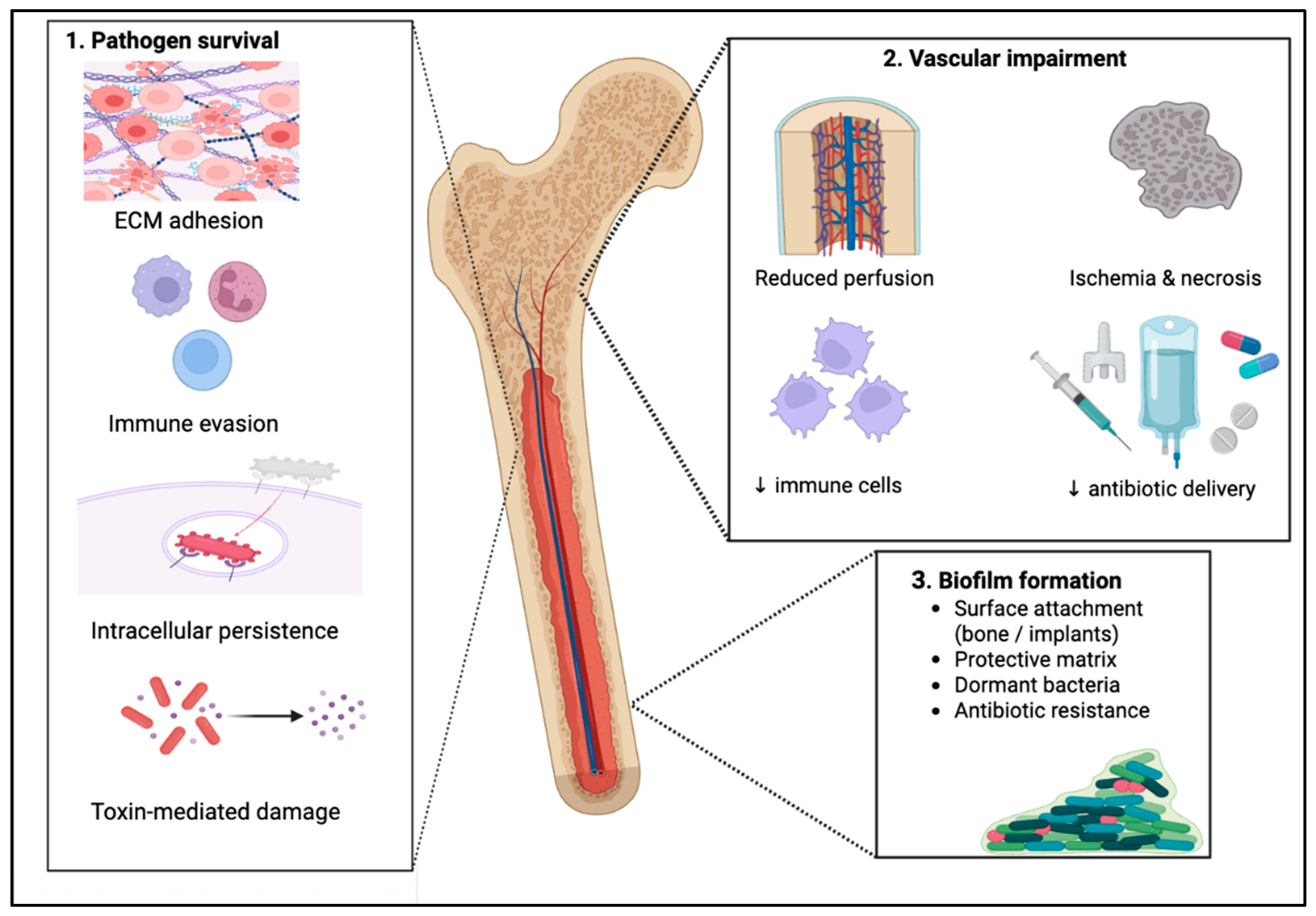

| Local Delivery System | Advantages | Disadvantages |
|---|---|---|
| PMMA (Polymethyl methacrylate) | - Good biocompatibility - Can sustain high local antibiotic concentrations - Proven effectiveness in chronic osteomyelitis - Allows for combination of antibiotics - Reduces infection recurrence | - Non-biodegradable; requires surgical removal - Limited antibiotic options (heat-stable, powder form) - Variable elution time - Reduced mechanical strength with high antibiotic load - Risk of bacterial resistance - Spacer-related mechanical complications |
| Calcium Sulfate (CaS) | - Biocompatible and biodegradable - Sustained antibiotic release - Achieves high local concentrations without systemic toxicity - Promotes bone regeneration | - Low mechanical strength - Risk of hypercalcemia - Postoperative wound drainage - Limited efficacy in immunocompromised patients - Variable degradation rates |
| Hydroxyapatite (HA) | - Excellent biocompatibility and bioactivity - Promotes bone regeneration - Effective carrier for water-soluble antibiotics | - Crystallizes and coagulates rapidly in vivo when used alone - Requires combination with other materials for stability - Potential wound complications and infection persistence - Occasional need for revision surgeries |
| Hydrogels | - High biocompatibility and biodegradability - Mimic extracellular matrix - Support bone tissue formation - Enable targeted and sustained drug release - Reduce systemic toxicity and bacterial resistance - Proven safety as implant coatings - Useful for both prevention and treatment of infection | - Limited robust clinical evidence -May require optimization for mechanical stability - Effectiveness varies depending on formulation and infection type |
| Antibiotic-Impregnated Bone Grafts (AIBGs) | - Combine structural support with infection control - Support bone regeneration and incorporation - Allow broad antibiotic selection - Long-term infection-free outcomes - Effective in trauma and revision surgeries | - Variable antibiotic elution when combining drugs - Preparation-dependent release kinetics - Potential risk of reinfection in impaction grafts - Limited availability of standardized protocols |
| Nanoparticles | - Nanoscale size allows precise, controlled drug release - Promote bone regeneration and tissue integration - Biodegradable—no removal surgery required - Reduce systemic toxicity - Effective against biofilm and resistant bacteria | - Possible complications (e.g., refracture, infection recurrence, wound necrosis) - Manufacturing complexity - Long-term safety data limited - Cost and scalability challenges |
| Name of the Study | Model/Subjects | Infection Type | Internation/Comparison | Main Outcomes |
|---|---|---|---|---|
| Kishor et al., 2016 [195] | In vivo—22 rabbits | MRSA osteomyelitis | Local administration of a 7-phage cocktail | Significant clinical, histopathological and radiological improvement and new bone formation. Safe and effective |
| Sosa et al., 2020 [196] | In vivo murine models and in vitro assays | Prosthetic joint osteomyelitis caused by S. aureus | PlySs2 lysin—alone or combined with vancomycin | Synergistic action with vancomycin—reduced bacteria load in periprosthetic tissue and on implant surfaces |
| Mobarezi et al., 2025 [198] | Systematic review | Various infections | Phages, Lysins and Depolymerases | Highlighted biofilm degradation ability of phages—mostly in vitro data—more animal/human research required |
| Yilmaz et al., 2013 [197] | In vivo and rat models | Implant-related infection (MRSA & P. aeruginosa) | 4 subgroups: control, phage only, antibiotic only, phage + antibiotic | In MRSA group: biofilm eradicated only in combined phage + antibiotic group. In P. aeruginosa: reduced CFU in all treatments, but biofilm thickness unchanged. |
| Fedorov et al., 2023 [199] | Clinical trial—Human patients | PJI | Bacteriophage therapy + antibiotics vs. antibiotics alone | Combined therapy improved outcomes (95.5% response); faster drop in inflammation markers; mild, transient adverse events (fever). |
| Pirnay et al., 2024 [200] | Multicenter, retrospective study—humans | Difficult-to-treat infections (including bone infections) | Bacteriophage therapy + antibiotics | Favorable clinical outcomes; supports combined approach in complex infections. |
| Strategy | Main Mechanism(s) of Action | Target Pathogens/Context | Key Advantages | Main Limitations | Technological/Clinical Maturity |
|---|---|---|---|---|---|
| Immunomodulatory approaches (MSCs, interleukins and macrophages) | Regulation of innate and adaptive immunity; suppression of excessive inflammation; promotion of osteogenesis and angiogenesis | Chronic osteomyelitis; impaired healing; inflammatory dysregulation | Supports bone regeneration; modulates host response | Variability in cell source and donor; limited homing efficiency; lack of standardized protocols; regulatory and ethical constraints | Preclinical to early clinical (fracture healing, nonunion); limited infection-specific trials |
| Vaccination strategies | Induction of pathogen-specific humoral and cellular immunity | S. aureus (including MRSA); infection prevention | Potential reduction in infection burden and antibiotic reliance | Failure to demonstrate clinical protection; inconsistent efficacy; possible new colonization | Preclinical to phase II/III trials; no approved vaccines |
| Quorum sensing/anti-virulence therapies | Inhibition of bacterial communication and virulence gene expression without bactericidal pressure | Biofilm-associated S. aureus infections | Reduces pathogenicity without strong selective pressure; preserves commensals | Mostly preclinical; strain-dependent effects; limited in vivo data | Preclinical |
| Bacteriophages and phage-derived lysins | Targeted bacterial lysis; biofilm degradation via lysins and depolymerases | Refractory, biofilm-dominated infections; MRSA; P. aeruginosa; PJI and osteomyelitis | High specificity; biofilm penetration; synergy with antibiotics | Narrow host range; resistance development; immune reactions; standardization and dosing challenges | Preclinical to early clinical |
| Silver-based compounds | Broad-spectrum antimicrobial activity; disruption of bacterial metabolism and biofilms | Chronic and refractory infections; implant-associated infections; MRSA | Activity against resistant bacteria; coating and local delivery options; tested in immunocompromised individuals | Lack of superiority when combined with standard care; limited clinical data | Preclinical to limited clinical use |
| Povidone-iodine (PVP-I) | Oxidative damage to proteins, lipids, and nucleic acids | Prevention and treatment of implant-related infections; S. aureus and P. aeruginosa | Broad-spectrum activity; preserved osseointegration; favorable clinical safety | Mainly implant/coating-based | Advanced preclinical and clinical use |
| AMPs | Membrane disruption; immunomodulation | Gram-positive and Gram-negative bacteria, viruses, parasitic infections | Broad activity | Poor in vivo stability; limited bioavailability; high production costs; lack of clinical data | Preclinical |
| Bacteriocins | Narrow-spectrum antibacterial activity against related strains | Implant-associated S. aureus infections | High specificity; reduced impact on microbiota | Lack of clinical trials; narrow activity spectrum | Preclinical |
| Synthetic antimicrobial polymers | Contact-killing or drug release depending on polymer properties | Implant-associated and local infections | Tunable properties; sustained release; reduced adhesion | Cytotoxicity risk; environmental concerns; structure-dependent performance | Preclinical |
| Honey and essential oils | Antimicrobial, anti-inflammatory, and antioxidant effects | Adjunctive therapy; experimental settings S. aureus and S. epidermidis | Multimodal biological activity | Variable efficacy; limited in vivo and clinical data | Preclinical |
Disclaimer/Publisher’s Note: The statements, opinions and data contained in all publications are solely those of the individual author(s) and contributor(s) and not of MDPI and/or the editor(s). MDPI and/or the editor(s) disclaim responsibility for any injury to people or property resulting from any ideas, methods, instructions or products referred to in the content. |
© 2026 by the authors. Licensee MDPI, Basel, Switzerland. This article is an open access article distributed under the terms and conditions of the Creative Commons Attribution (CC BY) license.
Share and Cite
Polyzou, E.; Gavatha, M.; Efthymiou, D.; Papageorgiou, D.; Ntalaki, E.; Stavropoulos, N.A.; Akinosoglou, K. Managing Bone Infections Beyond Systemic Antibiotics: A Scoping Review. Pathogens 2026, 15, 201. https://doi.org/10.3390/pathogens15020201
Polyzou E, Gavatha M, Efthymiou D, Papageorgiou D, Ntalaki E, Stavropoulos NA, Akinosoglou K. Managing Bone Infections Beyond Systemic Antibiotics: A Scoping Review. Pathogens. 2026; 15(2):201. https://doi.org/10.3390/pathogens15020201
Chicago/Turabian StylePolyzou, Eleni, Maria Gavatha, Dimitrios Efthymiou, Despoina Papageorgiou, Evangelia Ntalaki, Nikolaos A. Stavropoulos, and Karolina Akinosoglou. 2026. "Managing Bone Infections Beyond Systemic Antibiotics: A Scoping Review" Pathogens 15, no. 2: 201. https://doi.org/10.3390/pathogens15020201
APA StylePolyzou, E., Gavatha, M., Efthymiou, D., Papageorgiou, D., Ntalaki, E., Stavropoulos, N. A., & Akinosoglou, K. (2026). Managing Bone Infections Beyond Systemic Antibiotics: A Scoping Review. Pathogens, 15(2), 201. https://doi.org/10.3390/pathogens15020201

