Article

Phenotypic and Molecular Characterization of Clinical Isolates of Vancomycin-Resistant Enterococcus faecium in the Health District of Bolzano (Italy) During 2021–2023

by
Angela Maria Di Pierro
1,*,
Richard Aschbacher
1,
Maria Del Grosso
2,
Monica Monaco
2 and
Elisabetta Pagani
1
1
Laboratory of Microbiology and Virology, Provincial Hospital of Bolzano (SABES-ASDAA), Teaching Hospital of Paracelsus Medical University (PMU), 39100 Bolzano-Bozen, Italy
2
Department of Infectious Diseases, Istituto Superiore di Sanità, 00161 Rome, Italy
*
Author to whom correspondence should be addressed.
Pathogens 2026, 15(2), 143; https://doi.org/10.3390/pathogens15020143
Submission received: 5 December 2025 / Revised: 24 December 2025 / Accepted: 31 December 2025 / Published: 28 January 2026
(This article belongs to the Special Issue Advances in the Epidemiology of Human Infectious Diseases)

Abstract

Vancomycin-resistant Enterococcus faecium (VREfm) is an emerging pathogen responsible for healthcare-associated infections. For this reason, 44 VREfm isolates collected during 2021–2023 were characterized using phenotypic and genomic approaches. VREfm isolates were identified by MALDI-TOF and antimicrobial susceptibility tests were performed with Vitek 2, Sensititre, or E-test. Sequence type (ST), antibiotic resistance genes, virulence factors and genetic relatedness were determined using Next Generation Sequencing. Forty-three isolates had a VanA phenotype and vanHAX genotype and one had a VanB phenotype and vanHBX genotype. Isolates showed high antibiotic resistance to various antibiotics, but generally remained susceptible to quinupristin/dalfopristin, tigecycline and eravacycline. Two isolates were resistant to linezolid, showing the chromosomal mutation G2576T in domain V of the 23S rRNA gene in one isolate, and the transferable linezolid resistance genes cfr(D) and optrA in the other. Thirty-eight isolates belonged to ST80, one to ST17 (ST80 and ST17 are included in CC17) and one to ST697. Genomic analysis of the ST80 isolates showed that nearly all of them belonged to a single cluster. To prevent further spread of VREfm in the nosocomial environment, in addition to the application of up-to-date infection control strategies and antibiotic stewardship programs, the implementation of genomic surveillance is recommended.

1. Introduction

Vancomycin-resistant enterococci (VREs), mainly represented by Enterococcus faecium, are emerging pathogens responsible for a significant number of healthcare-associated infections. Even though most infections originate from the patient’s endogenous flora, VREs can spread from person to person by direct or indirect contact, such as through contaminated environmental surfaces or via caregivers’ hands. VREs can cause hospital outbreaks that can have significant impact on mortality, morbidity and length of patient stay, resulting in a significant financial burden on healthcare systems [1,2].
In particular, the rapid and continuous increase in bloodstream infections caused by vancomycin-resistant E. faecium (VREfm) in the European Union/European Economic Area, including Italy, is a cause for serious concern [3,4]. Consistent with national data, there has been a significant and gradual increase in the occurrence of VREfm infections in recent years up to 2022 in the Autonomous Province of Bolzano, located in Northern Italy, especially in the Bolzano Health District, with a reversing trend in 2023. Specifically, in the Autonomous Province of Bolzano, the percentage of VREfm isolated from blood and cerebrospinal fluid was 24.4% (10/41) in 2022, whereas in 2023 it decreased to 16.7% (6/36); all of these isolates had the VanA phenotype (defined as resistance to vancomycin and teicoplanin) [5].
This study shows phenotypic and genomic characteristics of 44 clinical VREfm isolates, 43 of them obtained from patients admitted to the Bolzano Regional Hospital during 2021–2023, together with one isolate from the Brunico Health District, also located in the Province of Bolzano. Microbial typing through Whole Genome Sequencing (WGS) was used to determine the antibiotic resistance and virulence gene content of the isolates and their possible clonal relationship.

2. Materials and Methods

2.1. Collection and Phenotypic Characterization of E. faecium VRE Isolates

Forty-four VREfm isolates (first isolate from each patient) were obtained during 2021–2023 from routine clinical specimens sent to the Clinical Microbiology Laboratory at the Bolzano Regional Hospital for diagnostic investigations of patients with suspected infections or from rectal surveillance screening. VREfm routine clinical isolates were grown on Columbia Agar and Columbia CNA Agar plates, both with sheep blood (Thermo Fisher Scientific, Waltham, MA, USA), whereas rectal surveillance isolates were grown on Brilliance VRE Agar plates (Thermo Fisher Scientific, Waltham, MA, USA), incubated for 24 h at 37 °C under aerobic conditions, and presumptive VREfm colonies have been identified by their characteristic morphology.
Identification of the isolates was performed using Matrix-assisted laser desorption/ionization-time of flight (MALDI-TOF) mass spectrometry (Bruker Daltonics MALDI Biotyper Sirius, Bremen, Germany) and antimicrobial susceptibility was determined by Vitek 2 AST-P658 cards. The isolates were subsequently stored at −80 °C using the CRYOBEADS system (bioMérieux, Marcy l’Etoile, France).
Three different systems were used for the determination of the antibiotic susceptibility of the stored isolates, in accordance with manufacturer’s specifications: Vitek 2 AST-P658 cards (bioMérieux, Marcy l’Etoile, France), Sensititre™ Streptococcus FDANDSF plates (Thermo Fisher Scientific, Waltham, MA, USA) or E-test strips (bioMérieux, Marcy l’Etoile, France); this allowed to test an expanded panel of antibiotics and the confirmation of susceptibility results between methods. The reference strains Staphylococcus aureus ATCC 29213, E. faecalis ATCC 29212 and VanB-E. faecalis ATCC 51299 were used for quality control.
Minimal inhibitory concentrations (MICs) were interpreted following the European Committee on Antimicrobial Susceptibility Testing breakpoints (EUCAST—version 13.0, 2023) [6]. Where clinical breakpoints were absent, EUCAST epidemiological cut-off (ECOFF) values were applied, and where neither clinical interpretation nor ECOFF values were available, MIC values alone are reported, because even without breakpoints MICs can provide comparable and meaningful data that can support clinical judgment, surveillance and future interpretation.

2.2. VREfm Genomic Characterization and Analysis

Single colonies of each E. faecium isolate, grown overnight on Columbia blood agar plates (Thermo Fisher Scientific, Waltham, MA, USA), were resuspended in 180 µL of enzyme solution (20 mg/mL Lysozyme, Tris HCl 20 nM, EDTA 2 mM, Triton 1.2%) and incubated for 30 min at 37 °C. Genomic DNA (gDNA) extraction was performed using the QIAmp®DNA Blood Mini Kit (QIAGEN Inc. GmbH, Hilden, Germany) according to the protocol provided by the manufacturer for Gram-positive bacteria; the quality of the extracted DNA was assessed with an automated electrophoresis system (Agilent Tape Station 4150, Santa Clara, CA, USA). Final samples were eluted in 50 µL of elution buffer and stored in the freezer at −20 °C.
Genomic libraries were prepared using the Nextera™ XT DNA Library Preparation Kit (Illumina, San Diego, CA, USA). The final concentration of the libraries was determined with the Qubit 4 fluorometer (ThermoFisher Scientific, Waltham, MA, USA), and their quality was assessed with the automated TapeStation 4150 platform (Agilent, Santa Clara, CA, USA). Sequencing of the bacterial genome was performed on the Miseq™ platform (Illumina). The quality of raw sequence data (Fastq) was assessed with the FastQC program (version 1.0.0, BaseSpace Labs); the trimming of the reads was performed with Trimmomatic (Galaxy Version 0.38.1) [7] and SPAdes (Galaxy Version 3.14.1+galaxy1) was employed for assembly, following the default settings of the server [8]. The quality of assembled genomes was assessed through the QUAST (Quality Assessment Tool for Genome Assemblies) program (Galaxy Version 5.0.2+galaxy1) [9]. Assembled genomes were analyzed by using tools available at the Center for Genomic Epidemiology (CGE-Technical University of Denmark) [10]: the identity of sequenced isolates was confirmed by KmerFinder (version 3.2); Multi Locus Sequencing Type (MLST) identification was performed by MLST (version 2.0.9), resistance determinants were searched by ResFinder (version 4.1), the LRE-Finder (version 1.0) was used for the identification of genes and mutations determining linezolid resistance and plasmid replicons were identified using PlasmidFinder (version 2.1). In addition, the AMRFinderPlus tool (Galaxy Version 1.0.19+galaxy1), developed by the National Center for Biotechnology Information (NCBI), was used for identifying point mutations. The presence of virulence factor (VF) determinants was analyzed by VFanalyzer, a platform for bacterial VF identification using the Virulence Factor Database-VFDB (https://www.mgc.ac.cn/VFs/ accessed on 16 January 2026) [11].
Genetic relatedness among the isolates was assessed using single nucleotide polymorphism (SNP)-based analysis with the CSI Phylogeny software (version 1.4), available on the CGE platform (https://cge.food.dtu.dk/services/CSIPhylogeny/ accessed on 16 January 2026 [12], with the E. faecium Aus0004 genome (Accession No. CP003351) as reference. The resulting SNP distance matrix was used to assess the genetic relationship among isolates, and an SNP difference cutoff < 20 was applied to define closely related isolates. The phylogenetic tree was visualized with Microreact (https://microreact.org/, accessed on 16 January 2026), developed by the Center for Genomic Pathogen Surveillance (CGPS) [13].

3. Results

3.1. Specimen Source for E. faecium VRE Isolates

A total of 44 E. faecium VRE isolates, stored by freeze preservation between 2021 and 2023, were selected based on total numbers of isolates per ward and year; forty-three of these isolates were from patients admitted to the Bolzano Regional Hospital, whereas one was from the Health District of Brunico.
Out of the examined isolates, 81.8% (36/44) were from clinical specimens of patients with suspected infections and 18.2% (8/44) were from rectal surveillance swabs (Table 1). Twenty-three patients (52.3%) were male and 21 (47.7%) were female. Patients’ mean age was 70.0 years (range 0–95 years), with 77.2% of isolates from patients over 60 years. Patients came from various hospital wards: Internal Medicine (No = 9), Intensive Care Unit (No = 7), General Surgery (No = 4), Gastroenterology (No = 4), Geriatrics Unit (No = 3), Neonatal Intensive Care Unit (No = 3), Vascular Surgery (No = 2), Hematology (No = 2), Urology (No = 2), Nephrology (No = 2), Casualty department (n = 1), Cardiology (No = 1), Dermatology (No = 1), Infectious Diseases Unit (No = 1), Gynecology (No = 1) and Otorhinolaryngology (No = 1).

3.2. Identification and Antimicrobial Susceptibility Testing Results

Isolates were identified by MALDI-TOF mass spectrometry, with scores > 2.0 (2.2 ± 0.1). Phenotypic susceptibility testing results are listed in Table 1. Resistance to vancomycin and teicoplanin (MICs > 16 mg/L, respectively) was found in all isolates except one, indicating a VanA phenotype; the exception was isolate No. 4424 with resistance to vancomycin and susceptibility to teicoplanin (MIC ≤ 0.5 mg/L), consistent with a VanB phenotype. For daptomycin, the MICs of all but one isolate (No. 5255) were ≤ the ECOFF value; for isolate No. 5255, the Vitek 2 test was also performed using the evaluation method for E. faecalis, which gave an MIC of 32 mg/L (Vitek 2 does not elaborate daptomycin MICs for E. faecium). Isolates No. 4497 and 5321 tested resistant to linezolid (>16 mg/L and 16 mg/L, respectively), whereas MICs for tedizolid were 0.5 mg/L and 2 mg/L, respectively (EUCAST clinical breakpoint not defined, ECOFF = 1 mg/L); for linezolid, Sensititre results were confirmed by Vitek 2. All isolates except one (No. 4750) were susceptible at a standard dosage regimen to quinupristin/dalfopristin, tigecycline and eravacycline. Oritavancin MICs for all isolates were ≤0.25 mg/L, but no EUCAST clinical breakpoints or ECOFF values have yet been defined. All MICs for dalbavancin except one (isolate No. 4424) were >2 mg/L, with Sensititre results confirmed by E-test. All isolates except two showed high-level resistance to gentamicin and streptomycin: isolate No. 5353 was high-level gentamicin susceptible, and isolate No. 4424 was high-level susceptible to gentamicin and streptomycin. All VREfm isolates except one (No. 4772) were resistant to the following antibiotics: ampicillin, ampicillin/sulbactam and amoxicillin/clavulanic acid (MICs >16 mg/L, Vitek 2), and had MICs >8 mg/L (Vitek 2) for imipenem and >8 mg/L for ceftaroline (Sensititre); isolate No. 4772, obtained in 2022 from the urine of a patient admitted to the Internal Medicine ward, was susceptible at a standard dosage regimen to ampicillin, ampicillin/sulbactam and amoxicillin/clavulanic acid (MICs ≤ 2 mg/L) and had an MIC ≤ 1 mg/L for imipenem and 0.06 mg/L for ceftaroline. Moreover, all isolates except one (No. 4615) were resistant to ciprofloxacin and levofloxacin (MICs > 4 mg/L, Vitek 2); the exception was isolate No. 4615, susceptible at a standard dosage regimen to ciprofloxacin (MIC ≤ 0.5 mg/L, Vitek 2) and levofloxacin (MIC = 2 mg/L, Vitek 2). All isolates except No. 4424 (MIC = 8 mg/L) had MICs > 32 mg/L for rifampicin (E-test). All isolates except No. 5353 and 4424 had MICs > 1 mg/L for clindamycin and >4 mg/L for erythromycin (Sensititre); MICs of isolates No. 5353 and 4424 for clindamycin and erythromycin were ≤0.125 mg/L.

3.3. Genomic Analysis

3.3.1. Antimicrobial Resistance Genes

Resistome analysis using ResFinder is shown in Table 2. The vanHAX gene cluster, determining the VanA resistance phenotype, was found in all E. faecium isolates except No. 4424; the latter isolate had the vanHBX cluster, determining the VanB phenotype (Table 2). Therefore, the phenotype and genotype agreed for glycopeptides.
Aminoglycoside modifying resistance genes were the following: intrinsic aac(6′)-Ii (all isolates), acquired aph(3′)-III (42/44 isolates), ant(6)-la (39/44 isolates) and aac(6)-aph(2″) (9/44 isolates). A single isolate (No. 4424) was phenotypically susceptible to high dosages of gentamicin and streptomycin, confirmed by the only presence of the intrinsic aac(6′)-Ii aminoglycoside resistance gene (if present alone it does not cause HLR to gentamicin and streptomycin), whereas all other isolates had a combination of this intrinsic aminoglycoside resistance gene with other aminoglycoside modifying genes, which, together with reduced intrinsic susceptibility and possibly other resistance mechanisms, could explain the HLR phenotype for gentamicin and streptomycin.
Other resistance genes were msr(C) (42/44 isolates) coding for a macrolide efflux pump, the 28S rRNA modification genes erm(B) (41/44 isolates) and erm(T) (12/44 isolates) and the lincosamide modification gene Inu(B) together with the macrolide-streptogramin efflux pump gene Isa(E) (1/44 isolate), conferring resistance to macrolides, lincosamides and group B streptogramins (MLSB phenotype) in 42/44 isolates. Low MICs for clindamycin and erythromycin of the isolates No. 5353 and 4424 could be explained by the absence of erm genes (presence only of the msr(C) gene).
Another frequent resistance gene was tet(L) (27/44 isolates) coding for an efflux pump conferring resistance to tetracyclines, but tetracycline resistance was not tested (intrinsic resistance of E. faecium). Isolate No. 4424 contained the trimethoprim resistance gene dfrG, but we did not test isolates for resistance to this antibiotic.
Chromosomal point mutations were observed in the quinolone resistance determining regions (QRDRs) of the DNA gyrase (gyrA, S83I) (43/44 isolates) and DNA topoisomerase IV (parC, S80R) (41/44 isolates) genes; all of these 43 isolates were resistant to ciprofloxacin and levofloxacin. The exception was isolate No. 4615, susceptible at a standard dosage regimen to ciprofloxacin and levofloxacin; this isolate had no gyrA or parC mutation in the QRDRs.
LRE-Finder software showed the presence of a mutation in domain V of the gene for 23S rRNA, specifically G2576T, responsible for a linezolid resistance in isolate No. 4497, and of the transferable linezolid resistance genes cfr(D) and optrA, coding for a 23S rRNA methyltransferase and a ribosomal protection protein, respectively, in isolate No. 5321 [14].
Isolate No. 5255 showed a daptomycin MIC > 256 mg/L. By AMRFinderPlus, no point mutations in the liaF, liaS and liaR genes, associated with daptomycin resistance [15], were found in this isolate. Other resistance mechanisms were not further investigated.

3.3.2. Multi Locus Sequence Typing (MLST)

Thirty-eight of the forty-four sequenced isolates belonged to ST80, 1 (No. 4424) to ST17 and 1 (No. 4615) to ST697 (Table 2). STs of 2 isolates (No. 4468 and No. 4861) are single locus variants (SLVs) of ST80, as they differ in only one of the seven canonical MLST typing loci; isolate No. 4791 differs in two alleles (double locus variant, DLV), whereas isolate No. 4809 differs by three alleles (tri locus variant, TLV).

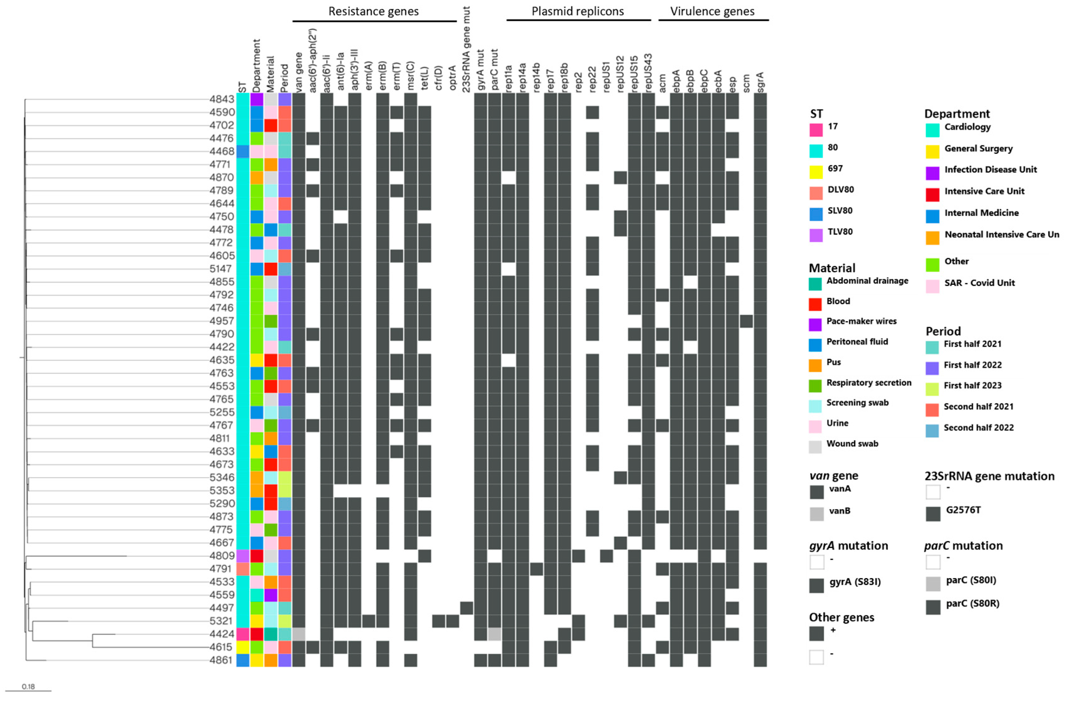
3.3.3. Virulence Factors

Analysis of the 44 VREfm genomes using VFDB revealed the presence of a total of 14 different virulence factor genes (Table S1, Figure 1). Most of the isolates harbored sgrA (43/44), ecbA (40/44) and esp (31/44), whereas acm was detected in 14 out of 44 isolates and scm in only one isolate (Figure 1). In addition, more than 90% of the isolates carried the pili gene cluster ebpA-ebpB-ebpC, which is involved in pilus formation. All of these genes encode surface proteins commonly associated with clinical isolates [16]. No significant differences in VF profiles were observed among the VREfm under study.

3.3.4. Plasmids

Eleven different plasmid replicons in thirteen different combinations were identified in the 44 VREfm: rep14a in all isolates; rep17 and rep18b each detected in 42 isolates; rep11a (n = 41); repUS43 and repUS15 each in 37 isolates; rep22 (n = 23), repUS12 (n = 6); rep2 (n = 3); rep14b and repUS1 each detected in only 1 isolate (Figure 1).

3.3.5. Phylogenetic Analysis

Phylogenetic analysis based on core genome SNP differences showed that the majority (35) of the VREfm isolates clustered within a single group (Cluster 1), whereas the remaining 9 isolates appeared genetically distinct, with 2 of these forming a separate group (Cluster 2). Cluster 1 comprised closely related isolates showing pairwise SNP differences below 20, except for a few isolates with SNP differences from twenty-one to sixty-one (Figure 1), and in total belonging to ST80 (34) and to a SLV of ST80 (1) (Figure 1). Isolates showing 1–2 pairwise SNP differences were obtained either from the same or distinct hospital wards on closely or distantly spaced days. Cluster 2 consisted of two closely related isolates (No. 4533 and No. 4559) with six SNP differences, both belonging to ST80 and isolated in the second half of 2021 in two different wards at a twenty-day interval. The seven remaining isolates were identified as singletons as SNP differences ranged from 252 to 1070, indicating considerable genetic distance among them. They included the following: two isolates, No. 4497 and No. 5321 both belonging to ST80, that exhibited linezolid resistance associated with mutation in a 23S rRNA gene (No. 4497) and the presence of cfr(D) and optrA genes (No. 5321); No. 4424 (ST17) carrying VanHBX; No. 4615 (ST697); No. 4791 (DLV of ST80); No. 4809 (TLV of ST80); and isolate No. 4861 (SLV of ST80).

4. Discussion

In the Autonomous Province of Bolzano, Northern Italy, a significant and gradual increase in clinical isolates of E. faecium VRE has been registered in recent years, consistent with national and European data [4,5]. During 2019–2023, a total of 217 non-duplicate clinical isolates of VREfm were collected in the Bolzano Health District from all clinical sample types and settings (hospital isolates, nursing homes, outpatients), excluding surveillance culture isolates. Between 2019 and 2022, we registered a significant upward trend, followed by a trend reversal in 2023. In 2023, the percentage of patients with positive fecal surveillance cultures for VREfm widely varied among departments in the Bolzano Regional Hospital, with an average of 8.1% (unpublished data). A point prevalence study in a Bolzano Long-Term Care Facility involving both residents and staff of the facility, and a comparison with the colonization of patients admitted to the Geriatrics ward of the referral Bolzano Regional Hospital, showed a significant increase in colonization by VREfm in 2022 compared to 2008, 2012 and 2016 [17]. The high percentages of VREfm found in the Province of Bolzano are not in line with the general epidemiology of multi-drug resistant (MDR) bacteria in the same geographical area, as MDR bacteria isolated from blood/liquor are generally significantly less prevalent than the Italian averages. In general, the Province of Bolzano records the lowest antibiotic resistance prevalences of all Italian provinces/regions and these are also generally below the respective European averages [5].
In the present study, 44 VREfm isolates collected during 2021–2023 were phenotypically and molecularly characterized for the first time in the Province of Bolzano. The tested isolates were highly multiresistant but generally maintained susceptibility and/or low MICs for daptomycin, linezolid, quinupristin/dalfopristin, oritavancin, tigecycline and eravacycline.
All isolates except one had the VanA phenotype, and genomic analysis showed the presence of the vanHAX gene cluster; the single isolate with a VanB phenotype was obtained in 2021 from a patient admitted to the Intensive Care Unit, and genomic analysis confirmed the presence of the vanHBX gene cluster. Isolates of VanB type VREfm have so far only sporadically been found in the Province of Bolzano. AR-ISS data suggest a high prevalence of the VanA phenotype in Italy in 2023, with resistance rates for blood/liquor isolates of 32.5% for vancomycin and 31.9% for teicoplanin [5].
Oritavancin is the only lipoglycopeptide active against all VREs, regardless of the Van resistance phenotype; in contrast, telavancin and dalbavancin appear to be active only against VanB-type VREs [18,19]. Although an ECOFF value and a EUCAST breakpoint for oritavancin have not yet been established, all MICs of our isolates were ≤0.25 mg/L, indicating this antibiotic as a potential therapeutic option for infections caused by VREfm [20].
Genomic analysis of the 44 isolates showed that 38 of them (86.3%) belonged to ST80, 4 were single (SLVs), double (DLV) or triple (TLV) locus variants of ST80 (9.1%), one was ST17 and one was ST697. The ST80 and ST17 strains of E. faecium belong to the clonal complex CC17, which is characterized by vancomycin-resistance, the presence of various virulence determinants and biofilm formation [21]. The hospital-adapted CC17 lineage of E. faecium, responsible for urinary tract infections, endocarditis and other infection types, is associated with severe morbidity and mortality and causes a considerable economic burden on public health [22].
As shown for our study isolates, the nosocomial transmission of E. faecium is reflected in the high overall clonality rate, with only one dominant sequence type found. Compared to community-adapted E. faecium isolates, those adapted to the hospital environment and belonging to CC17 contain more exogenously acquired genetic material, such as pathogenicity islands (PAIs) and plasmids or other mobile genetic elements frequently associated with antimicrobial resistance, virulence and/or colonization and carbohydrate metabolism [23]. These peculiarities give E. faecium CC17 selective advantages, making it difficult to treat in hospital settings and in certain patient niches [21].
A 2019 Danish study [24] and a retrospective study conducted in an Italian regional hospital [25] demonstrated that E. faecium ST80 was the most prevalent lineage in their hospital settings. Moreover, a single ST697 strain, not belonging to the CC17, was isolated from a patient admitted to the Geriatrics Unit but resident in a nursing home in Bolzano. In Public databases for molecular typing and microbial genome diversity (PubMLST), a single strain of E. faecium ST697 is reported, isolated in Spain in 2010 from a stool sample collected from a hospitalized patient [26].
Regarding virulence factors, all of our isolates except ST697 had a similar genetic profile. Phylogenetic analysis allowed to establish the clonality of most of our ST80 isolates, and, in addition, epidemiological and phylogenetic data were found to correspond. The isolates were found spatially dispersed throughout the hospital wards during the period from January 2021 to April 2023, suggesting a widespread transmission.
Two VREfm isolates included in the study were resistant to linezolid, but the percentage of linezolid-resistant E. faecium isolates in the Province of Bolzano in 2022 and 2023 was ≤0.5% (unpublished data). The two linezolid-resistant E. faecium isolates had MICs for tedizolid of 0.5 and 2 mg/L (ECOFF = 1 mg/L). In one linezolid-resistant isolate, sequencing analyses revealed the presence of a G2576T point mutation in the loop of the V-domain of the ribosomal 23S rRNA, which is the mutation most frequently associated with linezolid resistance, although it was not possible to specify in how many of the six chromosomal genes for 23S rRNA this mutation was present [27]. Furthermore, in one of the isolates, the transferable linezolid resistance genes cfr(D) and optrA were found. Other authors have described the presence of the cfr and optrA genes on a plasmid from an Italian E. faecium blood isolate obtained in 2015 [28].
In general, the resistance genes found in our isolates are in line with the respective phenotypes. However, the lack of identification of some genes linked to antibiotic resistance and virulence factors does not allow their absence from the genome to be ruled out with certainty, because, due to procedural limitations, genomes are not ‘closed’ but are instead subdivided into numerous contigs. The fragmentation of the genome means that there may be gaps (ranging from a few to several hundred base pairs) in which part or all of the gene sequence may fall, preventing the identification of the relevant gene.
A limitation of our study is the characterization of a small percentage of total isolates (16.5% of all first clinical routine VREfm isolates and a lower percentage of surveillance isolates), obtained during the period 2021–2023 in the Bolzano Health District. A further limitation is that it was performed in a single health district in the Province of Bolzano (except for a single isolate from another health district in the same province), not allowing for the extrapolation of data to other health districts in the same province or other provinces/regions in Italy. Moreover, to consolidate the significance of the data obtained, the evaluation of information regarding the clinical status of patients and its correlation with virulence factors and antimicrobial resistance profiles is desirable, but the limited number of sequenced isolates did not allow for a similar analysis.
VREfm strains are emerging pathogens causing a significant number of healthcare-associated infections (HAIs) [29]. Based on these considerations, it is essential to take effective preventive infection control and antimicrobial stewardship measures against VRE infections/colonizations, especially in hospitalized patients. The increasing trend of VREfm infections/colonization in the Bolzano Regional Hospital during the period 2019–2022, followed by a decreasing trend in 2023, correlates temporally with the COVID-19 pandemic and therefore a causal relationship is possible [30]. The combined implementation of hygienic–sanitary interventions is crucial for reducing or eliminating VRE infections/colonizations in hospital departments, as demonstrated by the decreasing trend for VREfm observed during 2023 in the Bolzano Regional Hospital, when enhanced surveillance and infection control procedures according to a locally adapted guideline have been applied, based on the infection prevention guideline for VRE infections of the Robert Koch Institute in Germany [31].

5. Conclusions

The present study shows that it is essential to provide the molecular typing of the VREfm isolates in association with phenotypic characterizations. This combined approach allows a better understanding of the spread and evolution of VREfm strains in hospital settings, providing a solid basis for the development of infection control and prevention strategies. The results can also serve as a basis for future studies to determine the origin and routes of infection, as well as to confirm or exclude outbreaks, track the cross-transmission of healthcare-associated pathogens, recognize particularly virulent strains and evaluate the effectiveness of control measures in the context of the infection prevention and control. In addition to the application of up-to-date infection control strategies and antibiotic stewardship programs, future activities should focus on the implementation of appropriate surveillance systems, including genomic analyses.

Supplementary Materials

The following supporting information can be downloaded at https://www.mdpi.com/article/10.3390/pathogens15020143/s1, Table S1: Analysis of virulence factor genes in 44 VREfm isolates.

Author Contributions

A.M.D.P. and R.A. organized and supervised the study. A.M.D.P. organized the ethics approval and the collection of samples from patients. A.M.D.P. and R.A. isolated and phenotypically tested the strains. A.M.D.P. conducted molecular analysis. A.M.D.P., R.A., M.D.G., M.M. and E.P. conducted data analysis, drafted, reviewed and edited the article. All authors have read and agreed to the published version of the manuscript.

Funding

This research received no external funding.

Institutional Review Board Statement

The study was conducted in accordance with the Declaration of Helsinki, and approved by the Ethics Committee of the South Tyrolean Health Authority at its meeting on 18 October 2023 (Opinion No. 107-2023).

Informed Consent Statement

Not applicable according to combined provisions of EU Regulation 2016/679 (directly applicable in all Member States) and Italian Legislative Decree No. 101/2018 (which supplements and amends the national Privacy Code for the parts not repealed by the Regulation).

Data Availability Statement

All genome sequences in this study were submitted to NCBI under the project accession number PRJNA1227293.

Acknowledgments

The authors thank the Department of Innovation, Research, University and Museums of the Autonomous Province of Bozen/Bolzano for covering the Open Access publication costs.

Conflicts of Interest

The authors declare no conflicts of interest. The authors have no other relevant affiliations or financial involvement with any organization or entity with a financial interest in or financial conflict with the subject matter or materials discussed in the manuscript.

Abbreviations

The following abbreviations are used in this manuscript:
DLVDouble Locus Variant
DNADeoxyribonucleic Acid
ECOFFEUCAST Epidemiological Cut-Off Value
EUCASTEuropean Committee on Antimicrobial Susceptibility Testing
HLRHigh-Level Resistance
MALDI-TOFMatrix-Assisted Laser Desorption/Ionization-Time of Flight
MDRMulti-Drug Resistant
MICMinimal Inhibitory Concentration
MLSTMulti Locus Sequencing Type
QRDRQuinolone Resistance Determining Region
RNARibonucleic Acid
SLVSingle Locus Variant
SNPsSingle Nucleotide Polymorphisms
STSequence Type
TLVTri Locus Variant
VFVirulence Factor
VREVancomycin-Resistant Enterococci
VREfmVancomycin-Resistant Enterococcus faecium
WGSWhole Genome Sequencing

References

  1. Puchter, L.; Chaberny, I.F.; Schwab, F.; Vonberg, R.P.; Bange, F.C.; Ebadi, E. Economic burden of nosocomial infections caused by vancomycin-resistant enterococci. Antimicrob. Resist. Infect. Control 2018, 7, 1. [Google Scholar] [CrossRef] [Scilit] [PubMed]
  2. Cattoir, V. The multifaceted lifestyle of enterococci: Genetic diversity, ecology and risks for public health. Curr. Opin. Microbiol. 2022, 65, 73–80. [Google Scholar] [CrossRef] [Scilit] [PubMed]
  3. Cassini, A.; Högberg, L.D.; Plachouras, D.; Quattrocchi, A.; Hoxha, A.; Simonsen, G.S.; Colomb-Cotinat, M.; Kretzschmar, M.E.; Devleesschauwer, B.; Cecchini, M.; et al. Burden of AMR Collaborative Group Attributable deaths and disability-adjusted life-years caused by infections with antibiotic-resistant bacteria in the EU and the European Economic Area in 2015: A population-level modelling analysis. Lancet Infect. Dis. 2019, 19, 56–66. [Google Scholar] [CrossRef] [Scilit]
  4. European Centre for Disease Prevention and Control. 2023. Available online: https://www.ecdc.europa.eu/en/publications-data/surveillance-antimicrobial-resistance-europe-2022 (accessed on 4 December 2025).
  5. AR-ISS: Sorveglianza Nazionale dell’Antibiotico-Resistenza. Dati 2022 e 2023. Available online: https://www.epicentro.iss.it/antibiotico-resistenza/ar-iss/RIS-4_2023.pdf (accessed on 4 December 2025).
  6. European Committee on Antimicrobial Susceptibility Testing (EUCAST). Breakpoint Tables for Interpretation of MICs and Zone Diameters. Version 13.0. 2023. Available online: https://www.eucast.org/fileadmin/eucast/pdf/Document_Archive/bacteria/breakpoint_tables/v_13.1_Breakpoint_Tables.pdf (accessed on 4 December 2025).
  7. Bolger, A.M.; Lohse, M.; Usadel, B. Trimmomatic: A flexible trimmer for Illumina sequence data. Bioinformatics 2014, 30, 2114–2120. [Google Scholar] [CrossRef] [Scilit] [PubMed]
  8. Bankevich, A.; Nurk, S.; Antipov, D.; Gurevich, A.A.; Dvorkin, M.; Kulikov, A.S.; Lesin, V.M.; Nikolenko, S.I.; Pham, S.; Prjibelski, A.D.; et al. SPAdes: A new genome assembly algorithm and its applications to single-cell sequencing. J. Comput. Biol. 2012, 19, 455–477. [Google Scholar] [CrossRef] [Scilit]
  9. Gurevich, A.; Saveliev, V.; Vyahhi, N.; Tesler, G. QUAST: Quality assessment tool for genome assemblies. Bioinformatics 2013, 29, 1072–1075. [Google Scholar] [CrossRef] [Scilit]
  10. Center for Genomic Epidemiology at the Technical University of Denmark. Available online: https://www.genomicepidemiology.org (accessed on 4 December 2025).
  11. Liu, B.; Zheng, D.D.; Zhou, S.Y.; Chen, L.; Yang, J. VFDB 2022: A general classification scheme for bacterial virulence factors. Nucleic Acids Res. 2022, 50, D912–D917. [Google Scholar] [CrossRef] [Scilit]
  12. Kaas, R.S.; Leekitcharoenphon, P.; Aarestrup, F.M.; Lund, O. Solving the Problem of Comparing Whole Bacterial Genomes across Different Sequencing Platforms. PLoS ONE 2014, 9, e104984. [Google Scholar] [CrossRef] [Scilit]
  13. Center for Genomic Pathogen Surveillance. Available online: https://microreact.org (accessed on 4 December 2025).
  14. Schwarz, S.; Zhang, W.; Du, X.D.; Krüger, H.; Feßler, A.T.; Ma, S.; Zhu, Y.; Wu, C.; Shen, J.; Wang, Y. Mobile Oxazolidinone Resistance Genes in Gram-Positive and Gram-Negative. Clin. Microbiol. Rev. 2021, 34, e0018820. [Google Scholar] [CrossRef] [Scilit]
  15. Arias, C.A.; Panesso, D.; McGrath, D.M.; Qin, X.; Mojica, M.F.; Miller, C.; Diaz, L.; Tran, T.T.; Rincon, S.; Barbu, E.M.; et al. Genetic Basis for In Vivo Daptomycin Resistance in Enterococci. N. Engl. J. Med. 2011, 365, 892–900. [Google Scholar] [CrossRef] [Scilit]
  16. Freitas, A.R.; Tedim, A.P.; Novais, C.; Coque, T.M.; Peixe, L. Distribution of putative virulence markers in Enterococcus faecium: Towards a safety profile review. J. Antimicrob. Chemother. 2018, 73, 306–319. [Google Scholar] [CrossRef] [Scilit]
  17. Nitti, M.T.; Sleghel, F.; Malgorzata, K.; Aschbacher, R.; Moroder, E.; Di Pierro, A.M.; Piscopiello, F.; Spalla, M.; Piazza, A.; Migliavacca, R.; et al. Colonization of Residents and Staff of an Italian Long-Term Care Facility and an Adjacent Acute Care Hospital Geriatrics Unit by Multidrug-Resistant Bacteria. Microb. Drug Resist. 2023, 29, 477–484. [Google Scholar] [CrossRef] [Scilit] [PubMed]
  18. Rosenthal, S.; Decano, A.G.; Bandali, A.; Lai, D.; Malat, G.E.; Bias, T.E. Oritavancin (Orbactiv): A New-Generation Lipoglycopeptide for the Treatment of Acute Bacterial Skin and Skin Structure Infections. Pharm. Ther. 2018, 43, 143–179. [Google Scholar]
  19. Pfaller, M.A.; Mendes, R.E.; Sader, H.S.; Castanheira, M.; Carvalhaes, C.G. Oritavancin in vitro activity against Gram-positive organisms from European medical centers: A 10-year longitudinal overview from the SENTRY Antimicrobial Surveillance Program (2010–2019). J. Chemother. 2023, 35, 689–699. [Google Scholar] [CrossRef] [Scilit] [PubMed]
  20. Cairns, K.A.; Udy, A.A.; Peel, T.N.; Abbott, I.J.; Dooley, M.J.; Peleg, A.Y. Therapeutics for Vancomycin-Resistant Enterococcal Bloodstream Infections. Clin. Microbiol. Rev. 2023, 36, e0005922. [Google Scholar] [CrossRef] [Scilit]
  21. Top, J.; Willems, R.; Bonten, M. Emergence of CC17 Enterococcus faecium: From commensal to hospital-adapted pathogen. FEMS Immunol. Med. Microbiol. 2008, 52, 297–308. [Google Scholar] [CrossRef] [Scilit]
  22. Lee, T.; Pang, S.; Abraham, S.; Coombs, G.W. Antimicrobial-resistant CC17 Enterococcus faecium: The past, the present and the future. J. Glob. Antimicrob. Resist. 2019, 16, 36–47. [Google Scholar] [CrossRef] [Scilit]
  23. Van Schaik, W.; Top, J.; Riley, D.R.; Boekhorst, J.; Vrijenhoek, J.E.; Schapendonk, C.M.; Hendrickx, A.P.; Nijman, I.J.; Bonten, M.J.; Tettelin, H.; et al. Pyrosequencing-based comparative genome analysis of the nosocomial pathogen Enterococcus faecium and identification of a large transferable pathogenicity island. BMC Genom. 2010, 11, 239. [Google Scholar] [CrossRef] [Scilit]
  24. Pinholt, M.; Bayliss, S.C.; Gumpert, H.; Worning, P.; Jensen, V.V.; Pedersen, M.; Feil, E.J.; Westh, H. WGS of 1058 Enterococcus faecium from Copenhagen, Denmark, reveals rapid clonal expansion of vancomycin-resistant clone ST80 combined with widespread dissemination of a vanA-containing plasmid and acquisition of a heterogeneous accessory genome. J. Antimicrob. Chemother. 2019, 74, 1776–1785. [Google Scholar] [CrossRef] [Scilit]
  25. Fioriti, S.; Simoni, S.; Caucci, S.; Morroni, G.; Ponzio, E.; Coccitto, S.N.; Brescini, L.; Cirioni, O.; Menzo, S.; Biavasco, F.; et al. Trend of clinical vancomycin-resistant enterococci isolated in a regional Italian hospital from 2001 to 2018. Braz. J. Microbiol. 2020, 51, 1607–1613. [Google Scholar] [CrossRef] [Scilit]
  26. PubMLST (Public Databases for Molecular Typing and Microbial Genome Diversity). Available online: https://pubmlst.org (accessed on 4 December 2025).
  27. Miller, W.R.; Munita, J.M.; Arias, C.A. Mechanisms of antibiotic resistance in enterococci. Expert Rev. Anti-Infect. Ther. 2014, 12, 10. [Google Scholar] [CrossRef] [Scilit]
  28. Morroni, G.; Brenciani, A.; Antonelli, A.; Maria D’Andrea, M.; Di Pilato, V.; Fioriti, S.; Mingoia, M.; Vignaroli, C.; Cirioni, O.; Biavasco, F.; et al. Characterization of a multiresistance plasmid carrying the optra and Cfr resistance genes from an Enterococcus faecium clinical isolate. Front. Microbiol. 2018, 9, 2189. [Google Scholar] [CrossRef] [Scilit]
  29. Morfin-Otero, R.; Perez-Gomez, H.R.; Gonzalez-Diaz, E.; Esparza-Ahumada, S.; Rodriguez-Noriega, E. Enterococci as Increasing Bacteria in Hospitals: Why Are Infection Control Measures Challenging for This Bacteria? Curr. Treat. Options Infect. Dis. 2018, 10, 129–142. [Google Scholar] [CrossRef] [Scilit]
  30. Toc, D.A.; Mihaila, R.M.; Botan, A.; Bobohalma, C.N.; Risteiu, G.A.; Simut-Cacuci, B.N.; Steorobelea, B.; Troanca, S.; Junie, L.M. Enterococcus and COVID-19: The Emergence of a Perfect Storm? Int. J. Transl. Med. 2022, 2, 220–229. [Google Scholar] [CrossRef] [Scilit]
  31. Hygienemaßnahmen zur Prävention der Infektion durch Enterokokken mit speziellen Antibiotikaresistenzen Empfehlung der Kommission für Krankenhaushygiene und Infektionsprävention (KRINKO) beim Robert Koch-Institut. Bundesgesundheitsblatt 2018, 61, 1310–1361. [CrossRef] [Scilit]
Figure 1. SNP-based core genome phylogeny of 44 VRE E. faecium isolates collected in the hospital of Bolzano (period 2021–2023). The phylogenetic tree was visualized by Microreact. Isolate names, clinical data and genome characteristics including resistance genes, plasmid replicons and virulence genes are at the right side of the tree. The legend includes the color-key used for each feature. SLV80, single locus variant of ST80; DLV80, dual locus variant of ST80; TLV80, tri locus variant of ST80; mut, mutation.
Pathogens 15 00143 g001
Table 1. Antimicrobial susceptibility testing results for 44 E. faecium VRE isolates and elaboration according to EUCAST breakpoints and/or ECOFF values.
IsolatesMaterialsVAN (∗)TPN (∗∗)DAL (∗)TLA (∗)ORI (∗)DAP (∗∗∗)LZD (∗)TZD (∗)QDA (∗∗)TGC (∗∗)ERV (∗∗∗)HLG (∗∗)HLS (∗∗)FOS (∗∗∗)NIT (∗∗∗)
4422Urine>16 (R)>16 (R)>210.0082 (S)1(S)0.250.5 (S)≤0.125(S)0.03 (S)SYN-RSYN-R128>512
4424Abdominal drainage>16 (R)<0.5 (S)0.030.0160.0011.5 (S)2 (S)0.25<0.25 (S)≤0.125 (S)0.016 (S)SYN-SSYN-S3232
4468Urine>16 (R)>16 (R)>210.0164 (S)2 (S)0.50.5 (S)≤0.125 (S)0.03 (S)SYN-RSYN-R128>512
4476Wound swab>16 (R)>16 (R)>220.0164 (S)2 (S)0.50.5 (S)≤0.125 (S)0.03 (S)SYN-RSYN-R>1024>512
4478Peritoneal fluid>16 (R)>16 (R)>210.0164 (S)2 (S)0.50.5 (S)≤0.125 (S)0.125 (S)SYN-RSYN-R128>512
4497Rectal surveillance swab>16 (R)>16 (R)>210.0084 (S)>16 (R)0.50.5 (S)≤0.125 (S)0.016 (S)SYN-RSYN-R128>512
4533Pus>16 (R)>16 (R)>210.0163 (S)2 (S)0.50.5 (S)≤0.125 (S)0.016 (S)SYN-RSYN-R128>512
4553Blood>16 (R)>16 (R)>210.0083 (S)1 (S)0.1250.5 (S)≤0.125 (S)0.06 (S)SYN-RSYN-R128>512
4559Pace-maker wires>16 (R)>16 (R)>220.0163 (S)2 (S)0.50.5 (S)≤0.125 (S)0.012(S)SYN-RSYN-R128>512
4590Urine>16 (R)>16 (R)>220.034 (S)2 (S)0.50.5 (S)≤0.125 (S)0.03 (S)SYN-RSYN-R128>512
4605Rectal surveillance swab>16 (R)32 (R)>210.0083 (S)1 (S)0.1250.5 (S)≤0.125 (S)0.06 (S)SYN-RSYN-R128>512
4615Urine>16 (R)>16 (R)>210.0083 (S)2 (S)0.50.5 (S)≤0.125 (S)0.03 (S)SYN-RSYN-R12864
4633Peritoneal fluid>16 (R)>16 (R)>220.0163 (S)2 (S)0.50.5 (S)≤0.125 (S)0.125 (S)SYN-RSYN-R192>512
4635Blood>16 (R)>16 (R)>220.0163 (S)2 (S)0.50.5 (S)≤0.125 (S)0.06 (S)SYN-RSYN-R128256
4644Urine>16 (R)16 (R)>220.0163 (S)2 (S)0.50.5 (S)≤0.125 (S)0.06 (S)SYN-RSYN-R>1024>512
4667Urine>16 (R)>16 (R)>220.033 (S)2 (S)0.51 (S)≤0.125 (S)0.125 (S)SYN-RSYN-S128256
4673Blood>16 (R)>16 (R)>220.033 (S)2 (S)0.250.5 (S)≤0.125 (S)0.125 (S)SYN-RSYN-R128>512
4702Blood>16 (R)>16 (R)>220.0163 (S)2 (S)0.50.5 (S)≤0.125 (S)0.06 (S)SYN-RSYN-R128256
4719Rectal surveillance swab>16 (R)>16 (R)>220.0163 (S)2 (S)0.50.5 (S)≤0.125 (S)0.06 (S)SYN-RSYN-R128256
4746Urine>16 (R)>16 (R)>220.0164 (S)2 (S)0.50.5 (S)≤0.125 (S)0.125 (S)SYN-RSYN-R256256
4750Urine>16 (R)>16 (R)>220.0162 (S)2 (S)0.52 (R)≤0.125 (S)0.06 (S)SYN-RSYN-R128256
4763Respiratory secretion>16 (R)>16 (R)>2>20.0163 (S)2 (S)0.50.5 (S)≤0.125 (S)0.06 (S)SYN-RSYN-R128>512
4765Ulcer swab>16 (R)>16 (R)>220.034 (S)2 (S)0.51 (S)≤0.125 (S)0.06 (S)SYN-RSYN-R128>512
4767Respiratory secretion>16 (R)>16 (R)>220.0163 (S)2 (S)0.50.5 (S)≤0.125 (S)0.06 (S)SYN-RSYN-R128>512
4771Pus>16 (R)>16 (R)>220.0163 (S)2 (S)0.50.5 (S)≤0.125 (S)0.032 (S)SYN-RSYN-R128256
4772Urine>16 (R)>16 (R)>220.0163 (S)2 (S)0.50.5 (S)≤0.125 (S)0.06 (S)SYN-RSYN-R64256
4775Respiratory secretion>16 (R)>16 (R)>220.0163 (S)2 (S)0.50.5 (S)≤0.125 (S)0.06 (S)SYN-RSYN-R128>512
4789Rectal surveillance swab>16 (R)>16 (R)>220.033 (S)2 (S)0.50.5 (S)≤0.125 (S)0.125 (S)SYN-RSYN-R128>512
4790Rectal surveillance swab>16 (R)>16 (R)>220.033 (S)2 (S)0.50.5 (S)≤0.125 (S)0.125 (S)SYN-RSYN-R128256
4791Rectal surveillance swab>16 (R)>16 (R)>220.034 (S)2 (S)0.50.5 (S)≤0.125 (S)0.06 (S)SYN-RSYN-R128>512
4792Rectal surveillance swab>16 (R)>16 (R)>220.032(S)2 (S)0.50.5 (S)≤0.125 (S)0.125 (S)SYN-RSYN-R128256
4809Wound swab>16 (R)>16 (R)>220.034 (S)2 (S)0.51 (S)≤0.125 (S)0.125 (S)SYN-RSYN-R128256
4811Pus>16 (R)>16 (R)>2>20.254 (S)4 (S)0.50.5 (S)≤0.125 (S)0.06 (S)SYN-RSYN-R128>512
4843Wound swab>16 (R)>16 (R)>220.0081(S)2 (S)0.50.5 (S)≤0.125 (S)0.06 (S)SYN-RSYN-R32256
4855Ulcer swab>16 (R)>16 (R)>220.032(S)2 (S)0.50.5 (S)≤0.125 (S)0.06 (S)SYN-RSYN-R128256
4861Pus>16 (R)>16 (R)>220.032(S)2 (S)0.50.5 (S)≤0.125 (S)0.03 (S)SYN-RSYN-R128>512
4870Wound swab>16 (R)>16 (R)>220.032(S)2 (S)0.50.5 (S)≤0.125 (S)0.06 (S)SYN-RSYN-R128256
4873Urine>16 (R)>16 (R)>220.064 (S)2 (S)0.50.5 (S)≤0.125 (S)0.125 (S)SYN-RSYN-R128256
4957Respiratory secretion>16 (R)>16 (R)>220.033 (S)2 (S)0.50.5 (S)≤0.125 (S)0.125 (S)SYN-RSYN-S128256
5018Respiratory secretion>16 (R)>16 (R)>2>20.034 (S)4 (S)0.50.5 (S)≤0.125 (S)0.06 (S)SYN-RSYN-R128>512
5033Urine>16 (R)>16 (R)>220.0163 (S)2 (S)0.250.5 (S)≤0.125 (S)0.06 (S)SYN-RSYN-R128256
5147Blood>16 (R)>16 (R)>220.0163 (S)2 (S)0.50.5 (S)≤0.125 (S)0.03 (S)SYN-RSYN-R128256
5255Rectal surveillance swab>16 (R)>16 (R)>220.016>256 (R)2 (S)0.50.5 (S)≤0.125 (S)0.03 (S)SYN-RSYN-R12832
5290Blood>16 (R)>16 (R)>220.0163 (S)2 (S)0.50.5 (S)≤0.125 (S)0.03 (S)SYN-RSYN-R128256
5321Rectal surveillance swab>16 (R)>16 (R)>2>20.0163 (S)16 (R)20.5 (S)≤0.125 (S)0.03 (S)SYN-RSYN-R6464
5346Rectal surveillance swab>16 (R)>16 (R)>220.032(S)2 (S)0.50.5 (S)≤0.125 (S)0.125 (S)SYN-RSYN-R128256
5353Blood>16 (R)>16 (R)>220.0164 (S)2 (S)0.50.5 (S)≤0.125 (S)0.03 (S)SYN-SSYN-R128>512
ECOFF (mg/L), EUCAST42/0.5/84120.250.125//128256
% >ECOFF100.0%97.9%/97.9%/2.1%4.3%2.1%0.0%0.0%0.0%//6.4%91.5%
Breakpoint S (mg/L), EUCAST≤4≤2////≤4/≤1≤0.250.125////
Breakpoint R (mg/L), EUCAST>4>2////>4/>1>0.250.125////
% R 100.0%97.9%///2.1%4.3%/0.0%0.0%0.0%95.7%95.7%//
Susceptibility testing method: ∗ Vitek 2, ∗∗ Sensititre, ∗∗∗ E-test. Antibiotics: VAN = vancomycin, TPN = teicoplanin, DAL = dalbavancin, TLA = telavancin, ORI = oritavancin, DAP = daptomycin, LZD = linezolid, TZD = tedizolid, QDA = quinupristin/dalfopristin, TGC = tigecycline, ERV = eravacycline, HLG = gentamicin high-level resistance, HLS = streptomycin high-level resistance, SYN-S = synergy between aminoglycoside and beta-lactam antibiotic or glycopeptide, SYN-R = no synergy between aminoglycoside and beta-lactam antibiotic or glycopeptide, FOS = fosfomycin, NIT = nitrofurantoin. MICs in mg/L. EUCAST interpretation in brackets (S: susceptible, standard dosing regimen; R: resistant).
Table 2. Sequence types (STs), antibiotic resistance genes and mutations, and plasmid replicons for 44 E. faecium VRE isolates.
Isolate NumberSTResistance Genes
4346, 4478, 4644, 4673, 4702, 4746, 4772, 4957, 4855, 4559, 4775, 4792, 4811, 4870, 4873, 5255, 529080aac(6′)-Ii, ant(6)-Ia, aph(3′)-III, erm(B), gyrA (S83I), msr(C), parC (S80R), tet(L), vanHAX
4422, 4533, 4559, 481180aac(6′)-Ii, ant(6)-Ia, aph(3′)-III, erm(B), gyrA (S83I), msr(C), parC (S80R), vanHAX
4476, 477180aac(6′)-aph(2″), aac(6′)-Ii, ant(6)-Ia, aph(3′)-III, erm(B), erm(T), gyrA (S83I), msr(C), parC (S80R), tet(L), vanHAX
4590, 463380aac(6′)-Ii, ant(6)-Ia, aph(3′)-III, erm(B), erm(T), gyrA (S83I), msr(C), parC (S80R), tet(L), vanHAX
4750, 484380aac(6′)-Ii, aph(3′)-III, erm(B), gyrA (S83I), msr(C), parC (S80R), vanHAX
4767, 478980aac(6′)-aph(2″), aac(6′)-Ii, ant(6)-Ia, aph(3′)-III, erm(B), erm(T), gyrA (S83I), msr(C), parC (S80R), tet(L), vanHAX
442417aac(6′)-Ii, dfrG, gyrA (S83I), msr(C), parC (S80I), vanHBX
4468SLV80aac(6′)-Ii, ant(6)-Ia, aph(3′)-III, erm(B), erm(T), gyrA (S83I), msr(C), parC (S80R), tet(L), vanHAX
449780aac(6′)-Ii, ant(6)-Ia, aph(3′)-III, erm(B), gyrA (S83I), msr(C), parC (S80R), G2576U 23S rRNA, vanHAX
455380aac(6′)-aph(2″), aac(6′)-Ii, ant(6)-Ia, aph(3′)-III, erm(B), gyrA (S83I), msr(C), parC (S80R), tet(L), vanHAX
460580aac(6′)-aph(2′’), aac(6′)-Ii, ant(6)-Ia, aph(3′)-III, erm(B), erm(T), gyrA (S83I), msr(C), parC (S80R), tet(L), vanHAX
4615697aac(6′)-aph(2″), aac(6′)-Ii, ant(6)-Ia, aph(3′)-III, erm(B), vanHAX
463580aac(6′)-Ii, ant(6)-Ia, aph(3′)-III, erm(B), erm(T), gyrA (S83I), msr(C), parC (S80R), tet(L), vanHAX
476380aac(6′)-aph(2″), aac(6′)-Ii, ant(6)-Ia, aph(3′)-III, erm(B), erm(T), gyrA (S83I), msr(C), parC (S80R), tet(L), vanHAX
476580aac(6′)-Ii, ant(6)-Ia, aph(3′)-III, erm(B), erm(T), gyrA (S83I), msr(C), parC (S80R), tet(L), vanHAX
479080aac(6′)-aph(2″), aac(6′)-Ii, ant(6)-Ia, aph(3′)-III, erm(B), erm(T), gyrA (S83I), msr(C), parC (S80R), tet(L), vanHAX
4791DLV80aac(6′)-Ii, ant(6)-Ia, aph(3′)-III, erm(B), gyrA (S83I), msr(C), parC (S80R), vanHAX
4809TLV80aac(6′)-Ii, ant(6)-Ia, aph(3′)-III, gyrA (S83I), lnu((B), Isa(E), tet(L), vanHAX
4861SLV80aac(6′)-Ii, aph(3′)-III, erm(B), gyrA (S83I), msr(C), parC (S80R), vanHAX
532180aac(6′)-Ii, ant(6)-Ia, aph(3′)-III, cfr(D), erm(A), erm(B), gyrA (S83I), msr(C), optr(A), parC (S80R), vanHAX
535380aac(6′)-Ii, gyrA (S83I), msr(C), parC (S80R), tet(L), vanHAX
Disclaimer/Publisher’s Note: The statements, opinions and data contained in all publications are solely those of the individual author(s) and contributor(s) and not of MDPI and/or the editor(s). MDPI and/or the editor(s) disclaim responsibility for any injury to people or property resulting from any ideas, methods, instructions or products referred to in the content.