Review

Trichinella-Derived Extracellular Vesicles: Implications and Future Prospects

Medical Parasitology Department, Faculty of Medicine, Tanta University, Tanta 31527, Egypt
*
Author to whom correspondence should be addressed.
Pathogens 2026, 15(2), 136; https://doi.org/10.3390/pathogens15020136
Submission received: 22 December 2025 / Revised: 20 January 2026 / Accepted: 21 January 2026 / Published: 26 January 2026
(This article belongs to the Special Issue Molecular Aspects of Host-Parasite Interactions)

Abstract

Parasite extracellular vesicles (EVs) are concise and versatile messages for parasite–parasite and parasite–host crosstalk. These vesicles are loaded with a cargo of diverse heterogeneous molecules, some of which are of potent immunomodulatory nature, and others have specific functions. Those EVs carrying the Trichinella signature are no exception. They play pivotal roles in the establishment of the parasite inside its niche within the host, ensuring better survival for both. They can also serve as biomarkers for diagnosis, follow-up, and prognosis of Trichinella infection. Owing to their immunogenicity and durability, they are excellent candidates for vaccine development. Moreover, enriched by the parasite’s elements, these intriguing EVs could protect the host from a wide array of inflammatory conditions associated with immune dysregulation such as inflammatory bowel disease and airway hyperreactivity, as evidenced by well-conducted experimental preclinical research. In sum, the potentials of Trichinella EVs seem enormous, awaiting only to be better characterized and conditioned for use in diagnostics and therapeutics. Detailed proteomic and transcriptomic analyses of the nature of these parasite-derived elements could provide invaluable insights into parasite biology and its interplay with the host at the same time.

1. Introduction

Extracellular vesicles (EVs) are nano-sized structures surrounded by a lipid bilayer, released by all cell types during physiological and pathological conditions. As crucial mediators of intracellular communication, EVs transfer a complex set of biomolecules including proteins, nucleic acids, and lipids between cells [1,2].
Parasites have evolved complex life cycles where intercellular communication is essential. Over the last few years, the study of EVs has revolutionized our understanding of how pathogens interact with the host in favor of their persistence. Moreover, advances in the comprehension of the biological roles of EVs have increased our knowledge about the development of several pathologies and opened up new avenues for the development of new diagnostic methods and new vaccines for parasitic infections [3].
Most parasites at some stages in their life cycle rely on the ability to communicate in parasite–parasite as well as in parasite–host interactions. Previous studies in this area have focused mainly on the soluble proteins secreted by parasites and their role in pathogenesis and modulation of the host immune response (reviewed in McKerrow et al. [4] and McSorley et al. [5]). Parasite-derived EVs are particularly interesting components of excretory–secretory (ES) products. They contain parasite-specific information for uptake and integration into other cells without direct contact between cells [6].

2. Biogenesis and Kinetics of EVs

EVs are membranous compartments formed through four stages: (1) initiation (selection of molecules such as nucleic acids, proteins, and others); (2) endocytosis (endocytosis of extracellular material by the plasma membrane invagination and intraluminal vesicle (ILV) formation); (3) multivesicular-like body (MVB) formation (formed through aggregation of various ILVs in a large body); and (4) secretion in the extracellular environment (upon exocytosis of MVBs through fusion with the plasma membrane). Vesicles generated in multivesicular endosomes are called exosomes. Other vesicles can be formed at the plasma membrane by direct budding into the extracellular space, giving rise to microvesicles and membrane particles [7,8]. However, in response to the uncertainty in many instances of their biogenesis, Minimal Information for Studies of Extracellular Vesicles (MISEV) guidelines have classified EVs into ‘large EVs’ (>200 nm) or ‘small EVs’ (˂200 nm) [9]. Furthermore, EVs have been classified based on their size into different types: exosomes, 30–120 nm; microvesicles, 150–1000 nm; apoptotic bodies/vesicles, 100–5000 nm; migrasomes, 500–3000 nm; large oncosomes, 1–10 μm; and exophers, 3.5–4 μm [10,11].
Much interest has been paid to EVs contents. EVs contain a varied series of bioactive cargos consisting of proteins, lipids, and diverse genetic materials, including DNAs, RNAs, mRNAs, miRNAs, and circRNAs [12]. Protein is the major component investigated in almost all studies of EVs. It includes two main categories: the specific components that exhibit unique protein signatures (cell-specific and varying depending on the cell of origin) [13] and the universal components, such as membrane transport and fusion-related proteins (like Rab, GTPases) [14], heat shock proteins (like HSP70, HSP90), annexins, and the lysobisphosphatidic acid (LBPA)-binding protein Alix [15]. Several adhesion molecules, such as CD146, CD9, CD18, and CD11 [16], and tetraspanin, including CD9, CD63, CD81, and CD82 [10], are also carried by EVs.
It is noteworthy that in addition to parasite proteins, EVs also contain host-derived proteins indicating the cross communication between the parasite and the host. For example, Marcilla et al. [17] identified 36 different host proteins in the EVs from Echinostoma caproni adults. In Fasciola hepatica EVs, they identified 19 host proteins. Similarly, parasite- and host-derived proteins were reported in Echinococcus multilocularis [18] and in intracellular parasites such as Plasmodium falciparum [19]. The most abundant host-derived proteins in parasite EVs were immunoglobulins, complement, and metabolic enzymes. It has been suggested that these host proteins may be packed into the parasite EVs during release into the extracellular space [18].
Moreover, EVs may carry bioactive lipids to recipient cells [20,21]. They contain 2–3 times the amount of cholesterol and glycosphingolipids relative to that in their progenitor cells, which vary among different cell origins [22]. However, only a few studies have yet explored the lipid content of EVs in parasites, and their specific role remains to be elucidated [23,24]. The current knowledge of helminth-derived EV molecular contents is found in a review by Sotillo et al. [25].
Once released from the secreting cells, EVs recognize and attach to their target cells through three possible ways: (1) endocytosis: the endocytosed EVs could fuse with the limiting membrane of the endosome; (2) receptor-ligand interaction: the binding of secreted vesicles to the surface of a recipient cell involves interactions between EV surface proteins (such as integrins, tetraspanins, proteoglycans, lectins, and immunoglobulins) and cellular receptors (such as intercellular adhesion molecule 1 (ICAM1), phosphatidylserine, and other unknown receptors); and (3) direct fusion of EVs with the plasma membrane. Finally, incorporation of proteins from the EV membrane into the plasma membrane and the release of EV contents into the cytoplasm of the recipient cell occur. The transfer of their content occurs as rapidly as 15 min after EV fusion with the target cells [26,27].
Here, we focus on the nematode Trichinella spiralis, as the research interest in T. spiralis EVs (Ts-EVs) has surged in the last few years. T. spiralis is a zoonotic parasite with a worldwide distribution acquired by the consumption of raw or undercooked meat infected with T. spiralis infective larvae (L1). It infects a wide variety of hosts, notably domestic pigs, rodents, and humans [28]. T. spiralis is distinctive among helminths, as it completes its entire life cycle within a single host. Following infection, the larvae are released in the stomach, invade the small intestinal mucosa, mature into adult worms (AWs), and reproduce. The resulting newborn larvae (NBL) disseminate through the lymphatic and circulatory systems to the striated muscles. Upon invasion of muscle cells, T. spiralis transforms them into nurse cells surrounded by a collagen capsule [29]. T. spiralis is a very successful parasite capable of producing long-term infections. The virulence of T. spiralis is attributed to its ability to manipulate the host immune responses aided by the release of a diverse array of functional proteins, such as heat shock proteins, proteinases, protein kinases, glycosidases, and EVs that promote invasion and long-term persistence [29,30,31].
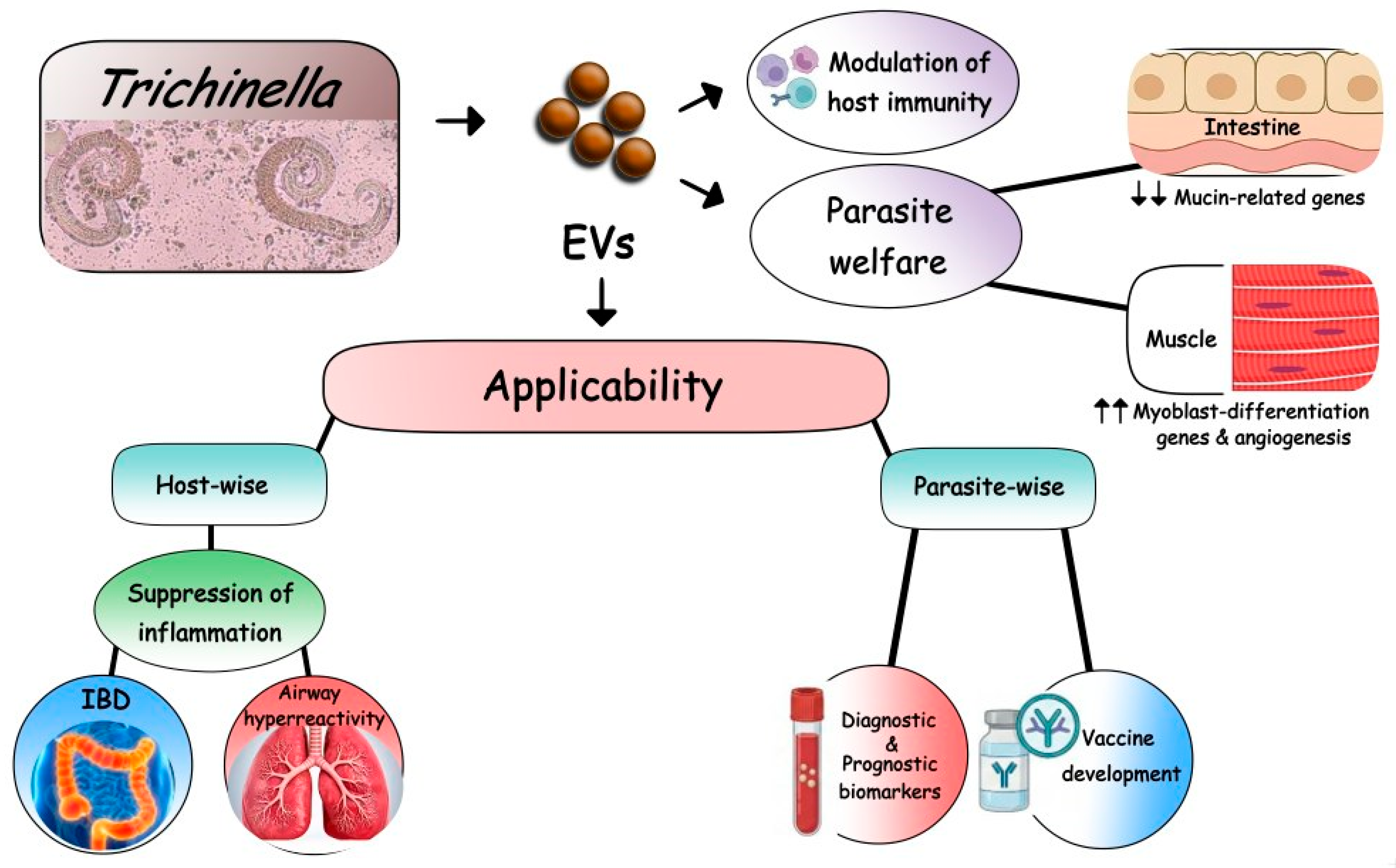
Despite much interest, information on the role of Ts-EVs in parasite–host communication is still limited. Therefore, in this review, we aim to demonstrate our current knowledge of Ts-EVs as well as their role in host–parasite interactions. Furthermore, implications of Ts-EVs as diagnostic biomarkers, novel potential therapeutic targets, and protective agents are discussed (outlined in Figure 1).

3. Isolation and Characterization of Trichinella spiralis EVs (Ts-EVs)

Kosanović et al. [30] submitted the first report on the production of EVs by T. spiralis muscle larvae (Ts-ML-EVs). Ts-EVs have been successfully isolated and characterized from the conditioned medium of T. spiralis muscle larvae (ML) [30,32,33,34,35] (summarized in Table 1). Generally, Ts-EVs are electron-lucent, intact, and rounded microvesicles with a size ˂ 200 nm. A few studies have reported larger sizes reaching 300 nm [36,37]. Thus, they are described as exosomes or EVs.
Moreover, Taylor et al. [38] quantified the number of EVs secreted by each life stage in vitro. Adult T. spiralis secreted about 3.15 × 10⁵ vesicles per parasite per 24 h, in comparison to 1.26 × 10⁵ vesicles per parasite per 24 h for ML. So, relative to their body mass, similar rates of EV production were detected. However, Glamoclija et al. [35] could isolate only 1100 vesicles per ML mostly due to differences in cultivation conditions of the larvae or EV isolation procedures.
Interestingly, Khueangchiangkhwang et al. [39] observed that both T. spiralis adult EVs (Ts-AW-EVs) and Ts-ML-EVs exhibited common functions such as inhibition of macrophage activation and suppression of the classical inflammatory pathways. However, different functions aligning with their specific locations in the host were reported. For example, Ts-AW-EVs—but not Ts-ML-EVs—significantly downregulated mRNA expression of mucin-related genes, e.g., MUC1, MUC5AC, trefoil factor 3 (Tff3), and chloride channel accessory 3 (Clca3). On the other hand, Ts-ML-EVs were uniquely effective at upregulating genes related to myoblast differentiation; including myogenin (MyoG) and myosin heavy chain embryonic (Myh3) mRNA, compared to Ts-AW-EVs-treated cells. These functional divergences might be dependent on their unique molecular cargos. Protein profiling revealed that while the two stages share 168 proteins, Ts-AW-EVs and Ts-ML-EVs possess 92 and 62 other stage-specific proteins, respectively.
Proteomic analysis of Ts-ML-EVs identified 753 proteins distributed throughout several compartments in T. spiralis ML, most notably in the plasma membrane, cytoplasm, nucleus, mitochondria, and extracellular regions. The most abundant Ts-ML-EV proteins included regulatory proteins of translation and signaling, as well as structural and metabolic enzymes [33]. Furthermore, mass spectrometry (MS)-based proteomics on T. spiralis newborn larva EVs (Ts-NBL-EVs) identified 231 proteins with similar subcellular distributions [40]. These findings align with the previous proteomic analysis of soluble antigens from Trichinella NBL by Hao et al. [41].
Interestingly, Western blotting and MS-based proteomics of NBL-EVs did not detect positive bands for the common proteins detected by Ts-ML-EVs (CD63 and CD81). Instead, they detected 14-3-3 protein and enolase (ENO1) [40]. The protein cargo into EVs is subjected to selective packaging before release; therefore, changes in EV protein composition between different developmental stages of Trichinella mostly reflect different conditions during release of EVs due to their different niches in the host [42]. Representatives of the most frequently identified protein cargo in Ts-EVs and their potential functions are listed in Table 2.

4. Trichinella EVs Are Key Strategic Tools for Infection

It is worth noting that parasite-derived EVs are involved in the pathogenetic mechanisms of parasitic diseases. Moreover, they have the ability to change the composition and function of the released vesicles during host–parasite interactions, thus mediating the disease [49].
During the early stage of infection, Wang et al. [34] investigated the effect of exosomes derived from T. spiralis ML (Ts-ML-Exos) on the intestinal epithelial cells in vitro. They observed that Ts-ML-Exos were significantly endocytosed in porcine small intestinal epithelial cells (IPEC-J2) within 6 h with a significant time-dependent increase in the number of endocytosed Ts-ML-Exos. They showed that Ts-ML-Exos decreased IPEC proliferation and could lead to increased levels of cytotoxicity, oxidative stress, and abnormal apoptosis. Moreover, Ts-ML-Exos significantly reduced the expression of different tight junction-related genes of ZO-1, CLDN-3, and OCLN at 24 h compared with the controls, leading to interruption of tight junctions and significantly increasing the permeability of the cells. Therefore, Ts-ML-Exos damaged gut epithelial integrity and played definite roles in the process of T. spiralis invasion inside the host. Previously, these findings were attributed to serine proteases in the T. spiralis ES proteins [50]; these proteases were recently detected in Ts-ML-EVs via liquid chromatography MS (LC-MS)/MS proteomic analysis [33].
Liu et al. [40] reported that the differentially expressed proteins and miRNAs in Ts-NBL-EVs were mainly involved in metabolic, autophagic, and other pathways, suggesting that these EVs may play an important role in evading host immune defense during early Trichinella infection. Among them, 14-3-3 protein was detected in Ts-NBL-EVs and let-7-5p had the highest expression. T. spiralis 14-3-3 protein is an immunodominant antigen during the early stage of Trichinella infection. It participates in regulating the growth and development of parasites and the process of invading the host [51], while let-7-5p activates the polarization of host macrophages to M2b type to evade the host’s immune response [37].
In the muscle phase of T. spiralis infection, Ts-ML-EVs play specific roles in nurse cell formation via the enhancement of myoblast differentiation. Ts-ML-EVs injected directly into striated muscle of mice caused a significantly higher expression of MyoG and Myogenic Regulatory Factor 4 (Mrf4) mRNAs at 2 days after the injection. Moreover, proteomic analysis revealed that Ts-ML-EVs contained proteins involved in the angiogenesis and vascular endothelial growth factor (VEGF) signaling pathways, involved in the encapsulation of larvae in the muscles. Thus, T. spiralis nurse cell formation and encapsulation in the muscle might be regulated at least in part by these EVs [39]. Therefore, an analysis of the proteins in Ts-ML-EVs against those in unencapsulated species (T. pseudospiralis) is essential to elucidate the specific cargo and molecular mechanism underlying the process of nurse cell formation.

5. MicroRNAs Abound in Ts-EVs

MicroRNAs (miRNAs), small non-coding RNAs, are key regulators of gene expression conserved across eukaryotes and involved in the regulation of numerous biological processes, such as cell development, differentiation, proliferation, and apoptosis, and in various diseases [52,53]. Many studies showed that miRNAs regulate the immune system functions and shape both innate and adaptive immunity, including Th1 and Th2 polarization [54,55].
Several species of helminths secrete abundant quantities of miRNAs that play pivotal roles in the host–parasite interaction [56]. Substantial amounts of secreted miRNAs are enclosed within EVs. These miRNAs could be taken up by host cells and potentially contribute to gene regulation with further manipulation of their host’s immune responses, through targeting host mRNAs for degradation or translational repression [56,57], but the exact mechanisms remain unclear.
Encapsulation of miRNAs in EVs protects them from degradation by RNases in the extracellular environment and allows their transport and delivery to host cells. However, some extracellular miRNAs are not enclosed in EVs mostly due to differences in the release mechanisms that can vary depending on the helminth species and life stage. However, the EV-enclosed miRNAs are considered the key for mediating interactions with the host [58,59]. Interestingly, Taylor et al. [38] observed that exonuclease treatment eliminated most of the small RNAs secreted by muscle larvae, yet it had little effect on the small RNAs released by adult worms under the same conditions. Thus, they proposed that adult T. spiralis secretes predominantly vesicular miRNAs, while most of the miRNAs secreted by T. spiralis ML are not contained within vesicles, suggesting they might be released directly into target cells. Moreover, several candidate proteins were identified in T. spiralis AW and ML secretomes that might bind and stabilize miRNAs. Two of them, namely KSRP and TsPUF, were characterized by Brown et al. [60].
Both Ts-AW-EVs and Ts-ML-EVs contain many miRNAs, such as tsp-miR-1, tsp-miR-100-5p, tsp-miR-125b-5p, tsp-miR-57-5p, and tsp-miR-72-5p, which are highly conserved across various helminth species. Sequencing Ts-EVs-miRNAs showed that 54 miRNAs were common between AW and ML. On the other hand, 78 and 43 were AW-specific- and ML-specific-miRNAs, respectively. These unique signatures suggest specialized biological roles. For instance, tsp-miR-36-5p is secreted solely by ML. This specific miRNA likely plays a targeted role in modulating host gene expression during the muscle phase of the infection, a process that remains to be fully characterized [39].
Regarding the potential role of miRNAs in Ts-EVs, an array of miRNAs was involved in immunoregulation, angiogenesis, apoptosis, cell communication, and other biological processes, including inflammation [36]. miRNA sequencing detected 64 miRNAs (10 of which are known miRNAs and 54 are unknown miRNAs). Among the known miRNAs of Ts-EVs, miRNA-1-3p and let-7-5p were the most highly expressed. Ts-miR-1-3p and Ts-let-7-5p induce the polarization of M2 macrophages and inhibit the activation of host fibroblasts in muscle cells; thereby, the parasite effectively evades the immune response and maintains a long-term existence necessary for its life cycle progression [37].
To investigate its role during the intestinal phase of T. spiralis infection, Wang et al. [61] constructed a miRNA library of Ts-Exos and then selected miR-153 for further studies. They demonstrated that miR-153 within Ts-Exos might be a key factor in the interaction between T. spiralis and host cells that participates in the regulation of various biological cellular processes. miR-153 acts as a proapoptotic factor in porcine intestinal epithelial cells (IPEC-J2) in vitro by inhibiting the target gene Bcl2 (anti-apoptotic pathway) and increasing the expression of proapoptotic proteins such as Bax, Bad, caspase 9, and caspase 3. Thus, miR-153 interacts with the intestinal epithelial cells and facilitates the invasion of T. spiralis larvae through induction of apoptosis and intestinal cellular damage as well as increased oxidative stress levels.
During the muscle phase, T. spiralis larvae induce a number of changes in the host muscle cells, involving direct manipulation of gene expression by factors secreted by T. spiralis larvae [62], which have been recently shown to include abundant small non-coding RNAs [38]. Among miRNAs enriched in the secreted material by T. spiralis ML, a homologue of the mammalian miR-31 was identified [38]. miR-31 has a well-documented role in muscle development by regulating translation of the myogenic determination gene Myf5. miR-31 is transcribed at high levels in quiescent satellite cells and is sequestered together with Myf5 RNA in messenger ribonucleoprotein (mRNP) granules. Following activation of satellite cells during development or after injury, mRNP granules are dissociated, miR-31 is reduced, and Myf5 mRNA is released from repression, allowing myogenesis to proceed [63]. Thus, miR-31 T. spiralis homologue is also predicted to target the Myf5 gene with further activation and proliferation of satellite cells, then remodel into a nurse cell.

6. Applications of Trichinella-Derived EVs

Research on EVs of parasites is promising, with many reports about parasite EV proteins at some stage in their life cycle. They are being investigated as new agents for diagnosis and vaccination candidates. Moreover, they may also be useful tools to ameliorate inflammation-associated diseases. Below, their potential applications are highlighted.

6.1. Modulating the Immune System to Treat Immunological Disorders

Trichinella spiralis ES products are characterized by strong immunoregulatory properties that enable modulation of the host’s immune system and thus ensure its long-term survival [31]. It is characterized by a Th2 phenotype with induction of regulatory T cells (Tregs), alternatively activated macrophages (AAMs), regulatory dendritic cells (DCregs), and increased expression of immunoregulatory factors (IL-10 and TGF-β) [31,64,65,66]. The immunomodulatory effects of T. spiralis and their implications for allergic and autoimmune diseases have been the subject of extensive and long-standing research [65,67,68,69,70,71,72,73].
Recently, these regulatory properties have been largely attributed to Ts-EVs in ES products. Studies on the properties of Ts-EVs highlighted their role in most of the previously observed immunomodulatory effects of T. spiralis infection or their ES products. Kosanović et al. [30] were the first to point to the potential immunomodulatory role of Ts-ML-EVs. They investigated their regulatory response using a peripheral blood mononuclear cell (PBMC) stimulation assay and observed an increase in IL-10 and IL-6 levels and a decrease in the production of IL-17a levels in culture supernatants of PBMC incubated with different concentrations of Ts-ML-EVs in a manner similar to that of the whole ES L1 products from which they were isolated.
Glamoclija et al. [35] showed that Ts-EVs possess the potential to promote the generation of tolerogenic DCs and subsequently polarize T cell responses towards Th2 and Treg responses. The most prominent effect of Ts-EV-treated DCs is the induced expansion of conventional CD4+ CD25+ Foxp3+ Treg cells and unconventional type 1 regulatory cells (Tr1) with a high production of regulatory cytokines IL-10 and TGF-β and low production of Th1 cytokines, namely IL-12, IFN-γ, IL-17, and IL-23. Moreover, Ts-EVs-treated DCs showed a high expression of genes associated with the tolerogenic status of DCs such as mTOR, AhR, and NFkB2; RelB, SOCS1, and SOCS3; and IDO.
Interestingly, these tolerogenic properties of Ts-EVs-treated DCs were retained even after challenge with a pro-inflammatory stimulus (lipopolysaccharides from Escherichia coli and IFN-γ). Therefore, Ts-EVs induce immune regulatory mechanisms that modulate the host’s immune response not only to itself, but also to other foreign or self-antigens which can result in the alleviation of inflammatory diseases [35]. Thus, EVs set the ground for developing safe helminth product-based therapeutics. Experimental studies have revealed some new potential applications of Ts-EVs for the treatment of some immunological disorders such as autoimmune diseases and allergies.

6.1.1. Inflammatory Bowel Diseases (IBD)

Ts-EV immunoregulatory properties were investigated in trinitrobenzene sulfonic acid (TNBS)-induced colitis in mice (a model for Crohn’s disease). Ts-EVs were injected intraperitoneally three times (50 μg/mice) every 3 days before induction of colitis. Ts-EVs markedly reduced Th1 and Th17 cells and pro-inflammatory cytokine secretion (IL-1β, IFN-γ, and TNF-α), upregulated the expression of Th2 cytokines (IL-4 and IL-13), and induced Treg differentiation with an increase in the regulatory cytokines (IL-10 and TGF-β) in colon tissues and in mesenteric lymph nodes. Subsequently, intestinal inflammation and colitis were ameliorated through regulation of Th1/Th2 balance. Moreover, Ts-EVs enhanced intestinal barrier integrity through induction of the expression levels of occludin and zo-1 tight junction protein in colonic tissues [36].
In further studies on the effect of Ts-EVs, Gao et al. [74] investigated the prophylactic outcome of the intraperitoneal injection of Ts-EVs on dextran sulfate sodium (DSS) colitis (a model for ulcerative colitis). They observed an improvement in disease activity index and macroscopic and histopathological scores of the Ts-EVs group compared to those of the DSS group. Ts-EVs upregulated the expression of IL-4, IL-10, TGF-β, and IL-13. In addition, Ts-EVs inhibited the classically activated macrophage polarization and increased the infiltration of AAMs into the colon. In addition, Ts-EVs promoted intestinal tissue repair and improved the intestinal barrier integrity by competition for L-arginine (Arg), upregulation of Arg-1, and proline production. Similarly, Wu et al. [37] showed that Ts-EVs can inhibit the levels of IL-1β, iNOS, TNF-α, IL-6, and IL-23 in macrophages, and at the same time, they promote the levels of anti-inflammatory factors Arg-1 and TGF-β in macrophages, as well as inhibiting fibroblast activation. Altogether, these findings suggest that Ts-EVs display a potent immunomodulatory capacity that may be a potential strategy to manage IBD.

6.1.2. Airway Hyperreactivity Diseases

Glamoclija et al. [75] explored the therapeutic potential of Ts-ML-EVs using an ovalbumin (OVA)-induced model of respiratory allergy. They demonstrated that intranasal delivery of Ts-EVs significantly alleviated airway inflammation and decreased lung infiltration with eosinophils. Moreover, Ts-EV suppressed the production of OVA-specific IgE- and Th2-mediated responses in sensitized mice. Notably, the EVs induced a systemic immunoregulatory response by expanding Treg cell populations and increasing IL-10 and TGF-β levels in both the lungs and spleen, while a localized increase in Tr1 cells within the lungs contributed to local immune modulation by inhibiting naïve and memory T cell responses. Therefore, these findings highlight Ts-EVs as a promising candidate for treating allergic inflammation.

6.2. Trichinella EVs as Potential Diagnostic and Prognostic Biomarkers

Parasite-derived EVs are structurally stable, rich sources of different molecules across diverse biofluids, including blood, saliva, urine, and cerebrospinal fluid (CSF) [76]. Due to their highly immunogenic cargo, they can serve as valuable biomarkers for the diagnosis of infectious diseases [77]. Furthermore, EVs provide a molecular snapshot of the parasite life cycle within the host. Notably, de Pontes et al. [78] established a positive correlation between EV concentration and infection intensity. Consequently, EV-based biomarkers represent a non-invasive approach for disease detection, prognosis, and therapeutic monitoring [12,79]. Given the increasing need for advanced diagnostics, EVs produced under Good Manufacturing Practice (GMP) protocols offer a promising frontier for clinical application [80].
The clinical diagnosis of human trichinellosis is challenging due to its non-specific clinical presentations. While muscle biopsy offers a definitive diagnosis, its utility is limited by its invasive nature and a high risk of false negatives during early-stage infections [81]. Although the International Commission on Trichinellosis (ICT) recommends serological testing to detect anti-Trichinella IgG against ML/ES antigens [28,82], these assays possess significant limitations including cross-reactivity with other nematodes, a delayed 3–4-week window before seroconversion, and low sensitivity in the early phases of the disease [83,84]. Furthermore, because antibody detection cannot reliably distinguish between active and past infections, the detection of circulating Trichinella antigens has emerged as a superior method for identifying current infections [85].
During the early stages of infection, Ts-ML-EVs and Ts-AW-EVs/miRNAs are released into the host’s bloodstream. Using quantitative PCR, Khueangchiangkhwang et al. [39] detected various miRNAs in T. spiralis-infected mice as early as one week post infection. Therefore, miRNA within Trichinella-secreted EVs can be a potential early-detection diagnostic tool for trichinellosis. Moreover, the host muscle cells themselves produce EVs that interact with satellite cells to stimulate their proliferation. The proteomic shifts in these muscle-derived EVs during infection may make them potential biomarkers for diagnosis. For instance, vimentin expression—an intermediate filament protein induced by T. spiralis NBL in muscle cells—was found to be significantly enriched in these EVs. Thus, it is suggested to be a novel candidate for the diagnosis of trichinellosis [86].
Interestingly, Ma et al. [87] characterized the profile of host circulating miRNAs in serum at 12, 18, and 30 days post infection (dpi) with T. spiralis. Significant miRNA alterations were observed during the early pre-encapsulation stages (12–18 dpi) when serological tests were still negative. They further identified distinct expression patterns of mmu-miR-467a-3p and mmu-miR-467d-3p that reached a peak at 30 dpi, whereas mmu-miR-376b-3p and mmu-miR-664-3p increased significantly at 18 dpi and then declined. In addition, mmu-miR-292a-5p expression gradually decreased from 12 to 30 dpi. Because these circulating miRNAs target specific signaling pathways involved in host–parasite interactions and vary in different phases of T. spiralis infection, they represent highly sensitive biomarkers for trichinellosis, especially in the early phase of infection.
Recently, Li et al. [43] used LC-MS/MS technology combined with bioinformatic analysis to characterize the proteomic profile of Ts-ML-EVs. They identified a candidate molecule, Ts-TTPA, a member of the S1 peptidase family containing a characteristic trypsin domain. Upon evaluation of the recombinant Ts-TTPA protein as a potential diagnostic antigen, it was found to detect anti-Trichinella IgG levels in swine serum as early as 7 dpi with no cross-reactivity with other common swine parasites. These findings suggest that EV-derived proteins, such as Ts-TTPA, are highly effective antigens for the early detection of trichinellosis.
Although not well documented, many of the previously mentioned proteins and miRNA cargo in Ts-EVs might be potential diagnostic markers. For instance, among the expressed miRNAs in Ts-NBL-EVs, let-7-5p exhibits the highest expression levels during early stages of Trichinella infection [40] and it is similarly expressed in Ts-ML-EVs [37]. Celik et al. [88] reported that let-7-5p serves as a biomarker for the early diagnosis of Echinococcus granulosus infection in dogs. Therefore, Ts-EVs offer significant potential as circulating biomarkers due to their abundance, structural stability, and ease of accessibility. Moreover, they represent an active biological process and maintain a proteomic profile that closely reflects their cell of origin.

6.3. Trichinella EVs as Potential Candidates for Vaccine Development

Single-antigen vaccines are often inadequate against the complex, multi-stage life cycle of T. spiralis. Therefore, multiepitope vaccines against T. spiralis were suggested as a better strategy [89]. EVs are potential vaccine candidates because of their protective lipid bilayer that allows slow release of antigens and protects them from degradation [90]. Moreover, structural proteins such as annexins and syntenin-1 identified in the EVs of many helminths could represent good candidates for vaccines because these homogenous proteins are not expressed in humans [91]. Importantly, the nano size of EVs ensures that they are efficiently internalized by antigen-presenting cells, thereby significantly boosting their antigenicity [92].
To date, no clinical trials have yet been reported; however, few experimental studies have investigated T. spiralis-derived EVs as novel vaccine candidates with a great potential for biotechnological development in the near future. Ashour et al. [32] evaluated Ts-ML-EVs as a potential vaccine candidate. They immunized experimental mice by subcutaneous injection of Ts-ML-EVs in two doses, one week apart, and then the mice were challenged with T. spiralis infection one week later. They demonstrated that Ts-ML-EVs induced a protective, mixed Th1/Th2 immune response with obvious Th1 polarization as well as increased serum T. spiralis-specific IgG resulting in a marked reduction in the numbers of adult worms (78.5%) and muscle larvae (~97%). Similarly, Gao et al. [33] used subcutaneous injection of Ts-ML-EVs in three doses, two weeks apart. Two weeks after the last immunization, experimental animals were challenged with T. spiralis infection. The intestinal adult worm burden of the Ts-ML-EV group was reduced by 23.4% and the ML burden was reduced by 43.7% in the immunized groups, suggesting that Ts-ML-EVs contain antigens that can effectively reduce the transmission and pathology of T. spiralis. They reported increased serum levels of IgM, IgG, and IgE in addition to an induced mixed response of Th1 and Th2.
Collectively, immunization with Ts-ML-EVs induces immune responses that confer resistance to T. spiralis through different proposed mechanisms: the rapid expulsion of adult worms from the intestine or delaying the larval invasion of the enteric mucosa, and the destruction of the AW and NBL larvae by antibody dependent cellular cytotoxicity (ADCC) or via the cytotoxic killing effect of eosinophils and granulocytes in immunized mice [32,33]. A notable finding in these vaccination models is the ability of EVs to regulate immune responses, thereby establishing a balanced Th1/Th2 profile and ameliorating the tissue pathology in the intestine and muscles. Whether Ts-AW-EVs could induce a protective immune response against T. spiralis awaits further investigation.

7. Limitations and Pitfalls

Despite the great potential of Ts-EVs, several significant challenges must be addressed before clinical implementation. Current methodologies lack the reproducibility and cost-effectiveness required for large-scale production [93,94,95,96]. Specifically, there are no standardized isolation and purification protocols or efficient drug-loading systems tailored for clinical-scale applications. These challenges represent critical barriers as Amruta et al. [97] suggested that any EV damage during isolation, drug loading, and/or storage may cause systemically administered EVs to exit the circulation within minutes and accumulate in the liver and spleen. Furthermore, the commonly used detection methods such as ELISA and Western blotting lack the necessary sensitivity for low-volume samples [98].
Moreover, much of the research has relied on in vitro parasite cultures rather than non-invasive biofluids such as plasma [99]. The yield of EVs in culture medium is usually low and insufficient for clinical translation [100]. As mentioned above, almost all studies focused on ML as a source of EVs. Very few used AW or NBL as a source of Ts-EVs. Different cargo detected in EVs isolated from different stages of T. spiralis can show different functions. Thus, further profiling and functional studies—particularly those aimed at identifying the core set of bioactive molecules—are essential to validate their therapeutic and diagnostic utility.
In almost all studies regarding Ts-EVs, information on the composition of Ts-EVs is limited. Even in the studies that performed proteomic analysis of Ts-EVs [33,39], they reported that the association of those proteins with Ts-EVs requires further confirmation. Furthermore, the existing studies on Ts-EVs cargo do not distinguish whether the contained proteins originate from T. spiralis or from the host.
Widespread clinical application of EVs remains constrained owing to multiple safety issues. EV preparations are considered to be a type of biopharmaceutical preparation from cells that may include co-isolated impurities and may be contaminated with culture-medium-derived components/reagents such as serum proteins, microorganisms, endotoxins, and virus particles or their fragments. Because EVs have high target specificity, it is unlikely to cause off-target toxicity. However, it may occur from these impurities. Thus, it is important to establish strict quality-controlled manufacturing processes to minimize the inclusion of components other than the intended EVs. Risks due to undesirable immune responses such as allergy or rejection are assumed with EVs derived from non-human sources, which is the case in parasite-derived EVs. Although there is no definitive evidence, the risk of carcinogenicity due to contamination with cell-derived DNA needs to be considered [101,102,103].
Although delivering miRNA via EVs is a promising solution that can overcome its instability and susceptibility to degradation in circulation, several challenges continue to delay their clinical development such as immunological off-target effects caused by partial complementarity with non-targeted transcripts’ “too many targets for miRNA effect” (TMTME), which may lead to unintended gene regulation of critical cellular pathways. Moreover, delivering miRNAs at excessively high concentrations that overwhelm the cellular machinery leading to miRNA-dependent cellular toxicity is another critical safety concern in miRNA -based therapeutics [104,105,106].

8. Conclusions and Future Perspectives

With the discovery of EVs and their importance in cellular communication, many studies have focused on parasite-derived EVs. They contain various parasitic factors and signaling molecules to modify the hostile microenvironment of their hosts for their benefit [107]. Over the past few years, Ts-EVs have attracted a significant interest that provides much information about their role in pathogenicity and immunomodulation of the host immune response with their potential applications. We collected almost all of the studies on Ts-EVs to determine their current status. Despite these insights, in-depth research into the whole picture and all the functions of EVs in T. spiralis infection is yet to be conducted, and much research is still needed in order to harness EVs for use in human clinical medicine.
Accurate and reliable methods for EV purification and characterization are crucial for using EVs as a tool for diagnosis and therapy. Exploring the composition of specific cargo molecules carried by Ts-EVs is critical not only to clarify their roles during T. spiralis infection and host–parasite communication but also to be used as a parasite-specific biomarker and as a rich molecular source for vaccine development [108]. Such knowledge would further enable the design of bioengineered synthetic vesicles or formulations that mimic the beneficial properties of Ts-EVs, and may lead to the development of scalable, high-quality, and safe biologics for clinical applications [75].
A great effort has been made by the International Society for Extracellular Vesicles (ISEV) that continuously updates the Minimal Information for Studies of Extracellular Vesicles (MISEV) guidelines for the standardization of procedures for EV isolation and characterization [109]. It is now the role of the researchers to stick to them to obtain reliable findings. Multiple new technologies have been developed in the past decade for EVs isolation and characterization. Micro/nanofluidic technology with affinity capture offers precise EV enrichment results in EVs that are highly purified compared to conventional methods [110]. Moreover, detection of a single EV is now possible by nanoplasmonic sensing technology for EV detection that allows sensitive multiplexed quantifiable detection of EVs through a simple assay, thus improving the potentiality and accuracy of EVs as diagnostic markers and for the prediction of treatment outcomes [12].
EVs have been tested as a drug delivery vehicle for the delivery of different molecules including drugs, which provide potential advantages over traditional synthetic delivery vehicles, such as liposomes and nanoparticles (reviewed in Meng et al. [111]). The nano size of EVs makes it one of the ideal carriers for the delivery of active molecules and drugs. They have successfully been applied in cancer therapy, inflammatory modulation, and immune response regulation [112,113,114]. Although their effectiveness has not yet been fully investigated, parasite-derived EVs may represent a promising carrier for drug delivery as they provide several advantages, such as easy bypass of cellular barriers [115] and delivered drug protection [116].
Regarding the treatment options of trichinellosis, benzimidazole derivatives such as albendazole, the most commonly used drug, have a poor bioavailability and hence it affects only the AW in the intestine with limited efficiency in eliminating ML [117]. Thus, based on EV tissue tropism to their cells of origin [118], we hypothesize that if albendazole, for example, is loaded on Ts-EVs, it may gain better access and reach ML more effectively. Yet, extensive studies on the pharmacokinetics of drug-loaded EVs, determining the optimal dosage for maximizing benefits while minimizing side effects and the availability of efficient drug loading systems for clinical-scale production, are required [95,119,120]. The stability and durability of EVs ensure the execution of such a mission with ease, and via genetic engineering techniques, we should be able to produce large quantities of EVs, all displaying the same molecular and physiological signature and all containing a single agent, or more likely, a cocktail of therapeutic molecules.
In summary, Ts-EVs hold promise as candidates for diverse future applications, including novel diagnostic biomarkers, effective vaccines, as well as therapeutic uses. Of equal importance is the assumption that these EVs carry molecules that facilitate their transfer into specific cells, for instance, intestinal and muscle cells in the case of Trichinella. Identification of these molecules may present a modality of targeted therapy where specific cells are selected and other normal cells are spared.

Author Contributions

Conceptualization and writing—original draft preparation, D.S.A. All authors contributed equally to writing—review and editing. All authors have read and agreed to the published version of the manuscript.

Funding

This work received no external funding.

Institutional Review Board Statement

Not applicable.

Informed Consent Statement

Not applicable.

Data Availability Statement

The original contributions presented in this review are included in the article.

Conflicts of Interest

The authors declare no conflicts of interest.

Abbreviations

AAMs, alternatively activated macrophage; ADCC, antibody-dependent cellular cytotoxicity; AW, adult worm; Bregs, regulatory B cells; ES, excretory–secretory; intercellular adhesion molecule 1 (ICAM1); ML, muscle larva; MS, mass spectrometry; miRNAs, microRNAs; NBL, newborn larva; PBMC, peripheral blood mononuclear cell; TGF-β, transforming growth factor–β; Tregs, regulatory T cells; Ts-AW-EVs, T. spiralis adult worm EVs; Ts-EVs, Trichinella spiralis extracellular vesicles, Ts-ML-EVs, T. spiralis muscle larva EVs; Ts-NBL-EVs, T. spiralis newborn larva EVs; VEGF, vascular endothelial cell growth factor.

References

  1. Mir, B.; Goettsch, C. Extracellular vesicles as delivery vehicles of specific cellular cargo. Cells 2020, 9, 1601. [Google Scholar] [CrossRef]
  2. Cuesta, C.M.; Guerri, C.; Ureña, J.; Pascual, M. Role of microbiota-derived extracellular vesicles in gut-brain communication. Int. J. Mol. Sci. 2021, 22, 4235. [Google Scholar] [CrossRef] [PubMed]
  3. Yáñez-Mó, M.; Siljander, P.R.; Andreu, Z.; Zavec, A.B.; Borràs, F.E.; Buzas, E.I.; Buzas, K.; Casal, E.; Cappello, F.; Carvalho, J.; et al. Biological properties of extracellular vesicles and their physiological functions. J. Extracell. Vesicles 2015, 4, 27066. [Google Scholar] [CrossRef]
  4. McKerrow, J.H.; Caffrey, C.; Kelly, B.; Loke, P.; Sajid, M. Proteases in parasitic diseases. Annu. Rev. Pathol. 2006, 1, 497–536. [Google Scholar] [CrossRef] [PubMed]
  5. McSorley, H.J.; Hewitson, J.P.; Maizels, R.M. Immunomodulation by helminth parasites: Defining mechanisms and mediators. Int. J. Parasitol. 2013, 43, 301–310. [Google Scholar] [CrossRef]
  6. Deatherage, B.L.; Cookson, B.T. Membrane vesicle release in bacteria, eukaryotes, and archaea: A conserved yet under-appreciated aspect of microbial life. Infect. Immun. 2012, 80, 1948–1957. [Google Scholar] [CrossRef] [PubMed]
  7. Théry, C.; Zitvogel, L.; Amigorena, S. Exosomes: Composition, biogenesis and function. Nat. Rev. Immunol. 2002, 2, 569–579. [Google Scholar] [CrossRef]
  8. Ovčar, A.; Kovačič, B. Biogenesis of extracellular vesicles (EVs) and the potential use of embryo-derived EVs in medically assisted reproduction. Int. J. Mol. Sci. 2024, 26, 42. [Google Scholar] [CrossRef]
  9. Théry, C.; Witwer, K.W.; Aikawa, E.; Alcaraz, M.J.; Anderson, J.D.; Andriantsitohaina, R.; Antoniou, A.; Arab, T.; Archer, F.; Atkin-Smith, G.K.; et al. Minimal information for studies of extracellular vesicles 2018 (MISEV2018): A position statement of the International Society for Extracellular Vesicles and update of the MISEV2014 guidelines. J. Extracell. Vesicles 2018, 7, 1535750. [Google Scholar] [CrossRef]
  10. Wu, Z.; Wang, L.; Li, J.; Wang, L.; Wu, Z.; Sun, X. Extracellular vesicle-mediated communication within host-parasite interactions. Front. Immunol. 2019, 9, 3066. [Google Scholar] [CrossRef]
  11. Jeppesen, D.K.; Zhang, Q.; Franklin, J.L.; Coffey, R.J. Extracellular vesicles and nanoparticles: Emerging complexities. Trends Cell Biol. 2023, 33, 667–681. [Google Scholar] [CrossRef]
  12. Neettiyath, A.; Chung, K.; Liu, W.; Lee, L.P. Nanoplasmonic sensors for extracellular vesicles and bacterial membrane vesicles. Nano Converg. 2024, 11, 23. [Google Scholar] [CrossRef] [PubMed]
  13. Zhang, Y.; Bi, J.; Huang, J.; Tang, Y.; Du, S.; Li, P. Exosome: A review of its classification, isolation techniques, storage, diagnostic and targeted therapy applications. Int. J. Nanomed. 2020, 15, 6917–6934. [Google Scholar] [CrossRef]
  14. Mears, R.; Craven, R.A.; Hanrahan, S.; Totty, N.; Upton, C.; Young, S.L.; Patel, P.; Selby, P.J.; Banks, R.E. Proteomic analysis of melanoma-derived exosomes by two-dimensional polyacrylamide gel electrophoresis and mass spectrometry. Proteomics 2004, 4, 4019–4031. [Google Scholar] [CrossRef]
  15. Gastpar, R.; Gehrmann, M.; Bausero, M.A.; Asea, A.; Gross, C.; Schroeder, J.A.; Multhoff, G. Heat shock protein 70 surface-positive tumor exosomes stimulate migratory and cytolytic activity of natural killer cells. Cancer Res. 2005, 65, 5238–5247. [Google Scholar] [CrossRef] [PubMed]
  16. Tang, N.; Sun, B.; Gupta, A.; Rempel, H.; Pulliam, L. Monocyte exosomes induce adhesion molecules and cytokines via activation of NF-κB in endothelial cells. FASEB J. 2016, 30, 3097–3106. [Google Scholar] [CrossRef] [PubMed]
  17. Marcilla, A.; Trelis, M.; Cortés, A.; Sotillo, J.; Cantalapiedra, F.; Minguez, M.T.; Valero, M.L.; Sánchez del Pino, M.M.; Muñoz-Antoli, C.; Toledo, R.; et al. Extracellular vesicles from parasitic helminths contain specific excretory/secretory proteins and are internalized in intestinal host cells. PLoS ONE 2012, 7, e45974. [Google Scholar] [CrossRef]
  18. Liu, C.; Cao, J.; Zhang, H.; Field, M.C.; Yin, J. Extracellular vesicles secreted by Echinococcus multilocularis: Important players in angiogenesis promotion. Microbes Infect. 2023, 25, 105147. [Google Scholar] [CrossRef]
  19. Platon, L.; Ménard, D. Plasmodium falciparum ring-stage plasticity and drug resistance. Trends Parasitol. 2024, 40, 118–130. [Google Scholar] [CrossRef]
  20. Xiang, X.; Poliakov, A.; Liu, C.; Liu, Y.; Deng, Z.B.; Wang, J.; Cheng, Z.; Shah, S.V.; Wang, G.J.; Zhang, L.; et al. Induction of myeloid-derived suppressor cells by tumor exosomes. Int. J. Cancer 2009, 124, 2621–2633. [Google Scholar] [CrossRef]
  21. Roma-Rodrigues, C.; Fernandes, A.R.; Baptista, P.V. Exosome in tumour microenvironment: Overview of the crosstalk between normal and cancer cells. BioMed Res. Int. 2014, 2014, 179486. [Google Scholar] [CrossRef]
  22. Skotland, T.; Sandvig, K.; Llorente, A. Lipids in exosomes: Current knowledge and the way forward. Prog. Lipid Res. 2017, 66, 30–41. [Google Scholar] [CrossRef]
  23. Yoon, Y.J.; Kim, O.Y.; Gho, Y.S. Extracellular vesicles as emerging intercellular communicasomes. BMB Rep. 2014, 47, 531–539. [Google Scholar] [CrossRef]
  24. Simbari, F.; McCaskill, J.; Coakley, G.; Millar, M.; Maizels, R.M.; Fabriás, G.; Casas, J.; Buck, A.H. Plasmalogen enrichment in exosomes secreted by a nematode parasite versus those derived from its mouse host: Implications for exosome stability and biology. J. Extracell. Vesicles. 2016, 5, 30741. [Google Scholar] [CrossRef]
  25. Sotillo, J.; Robinson, M.W.; Kimber, M.J.; Cucher, M.; Ancarola, M.E.; Nejsum, P.; Marcilla, A.; Eichenberger, R.M.; Tritten, L. The protein and microRNA cargo of extracellular vesicles from parasitic helminths—Current status and research priorities. Int. J. Parasitol. 2020, 50, 635–645. [Google Scholar] [CrossRef]
  26. Feng, D.; Zhao, W.L.; Ye, Y.Y.; Bai, X.C.; Liu, R.Q.; Chang, L.F.; Zhou, Q.; Sui, S.F. Cellular internalization of exosomes occurs through phagocytosis. Traffic 2010, 11, 675–687. [Google Scholar] [CrossRef] [PubMed]
  27. Mulcahy, L.A.; Pink, R.C.; Carter, D.R. Routes and mechanisms of extracellular vesicle uptake. J. Extracell. Vesicles 2014, 3. [Google Scholar] [CrossRef] [PubMed]
  28. Gottstein, B.; Pozio, E.; Nöckler, K. Epidemiology, diagnosis, treatment, and control of trichinellosis. Clin. Microbiol. Rev. 2009, 22, 127–145. [Google Scholar] [CrossRef] [PubMed]
  29. Mitic, I.; Glamoclija, S.; Radulovic, N.; Sabljic, L.; Tomic, S.; Gruden-Movsesijan, A.; Sofronic-Milosavljevic, L. Patients with Trichinella spiralis infection display unmodified antigen-specific immune response to SARS-CoV-2. Mem. Inst. Oswaldo Cruz 2025, 120, e250044. [Google Scholar] [CrossRef]
  30. Kosanović, M.; Cvetković, J.; Gruden-Movsesijan, A.; Vasilev, S.; Svetlana, M.; Ilić, N.; Sofronić-Milosavljević, L. Trichinella spiralis muscle larvae release extracellular vesicles with immunomodulatory properties. Parasite Immunol. 2019, 41, e12665. [Google Scholar] [CrossRef]
  31. Bruschi, F.; Ashour, D.S.; Othman, A.A. Trichinella-induced immunomodulation: Another tale of helminth success. Food Waterborne Parasitol. 2022, 27, e00164. [Google Scholar] [CrossRef]
  32. Ashour, D.S.; Ibrahim, F.M.K.; Elshamy, A.M.; Zoghroban, H.S. Trichinella spiralis-derived extracellular vesicles induce a protective immunity against larval challenge in mice. Pathog. Dis. 2022, 80, ftac040. [Google Scholar] [CrossRef]
  33. Gao, X.; Yang, Y.; Liu, X.; Xu, F.; Wang, Y.; Liu, L.; Yang, Y.; Liu, M.; Bai, X. Extracellular vesicles from Trichinella spiralis: Proteomic analysis and protective immunity. PLoS Negl. Trop. Dis. 2022, 16, e0010528–e0010544. [Google Scholar] [CrossRef]
  34. Wang, R.; Zhang, Y.; Zhen, J.; Zhang, J.; Pang, Z.; Song, X.; Lin, L.; Sun, F.; Lu, Y. Effects of exosomes derived from Trichinella spiralis infective larvae on intestinal epithelial barrier function. Vet. Res. 2022, 53, 87–97. [Google Scholar] [CrossRef]
  35. Glamočlija, S.; Sabljić, L.; Tomić, S.; Đokić, J.; Radulović, N.; Gruden-Movsesijan, A.; Kosanović, M. Trichinella spiralis extracellular vesicles induce anti-inflammatory and regulatory immune responses in vitro. Int. J. Parasitol. 2025, 55, 299–315. [Google Scholar] [CrossRef]
  36. Yang, Y.; Liu, L.; Liu, X.; Zhang, Y.; Shi, H.; Jia, W.; Zhu, H.; Jia, H.; Liu, M.; Bai, X. Extracellular vesicles derived from Trichinella spiralis muscle larvae ameliorate tnbs-induced colitis in mice. Front. Immunol. 2020, 11, 1174. [Google Scholar] [CrossRef]
  37. Wu, J.; Liao, Y.; Li, D.; Zhu, Z.; Zhang, L.; Wu, Z.; He, P.; Wang, L. Extracellular vesicles derived from Trichinella Spiralis larvae promote the polarization of macrophages to M2b type and inhibit the activation of fibroblasts. Front. Immunol. 2022, 13, 974332. [Google Scholar] [CrossRef]
  38. Taylor, P.J.; Hagen, J.; Faruqu, F.N.; Al-Jamal, K.T.; Quigley, B.; Beeby, M.; Selkirk, M.E.; Sarkies, P. Trichinella spiralis secretes abundant unencapsulated small RNAs with potential effects on host gene expression. Int. J. Parasitol. 2020, 50, 697–705. [Google Scholar] [CrossRef] [PubMed]
  39. Khueangchiangkhwang, S.; Wu, Z.; Hosoda-Yabe, R.; Yabe, T.; Okamoto, T.; Takada, C.; Okada, H.; Ogura, S.; Maekawa, Y. Trichinella spiralis secretes stage-specific extracellular vesicles for modulating host microenvironment for parasitism. bioRxiv 2023. [Google Scholar] [CrossRef]
  40. Liu, Y.; Cai, Y.; Chen, J.; Chen, S.; Yu, Y. Isolation, identification, and omics analysis of extracellular vesicles from Trichinella spiralis newborn larvae. Chin. J. Parasitol. Parasit. Dis. 2024, 42, 225–233. (In Chinese) [Google Scholar] [CrossRef]
  41. Hao, H.N.; Cheng, Y.K.; Zhang, R.; Han, L.; Song, Y.; Long, S.; Liu, R.; Zhang, X.; Wang, Z.; Cui, J. Immunoproteomic analysis on the soluble antigens of Trichinella spiralis newborn larvae. Chin. J. Parasitol. Parasit. Dis. 2023, 41, 176–182. [Google Scholar] [CrossRef]
  42. Nowacki, F.C.; Swain, M.T.; Klychnikov, O.I.; Niazi, U.; Ivens, A.; Quintana, J.F.; Hensbergen, P.J.; Hokke, C.H.; Buck, A.H.; Hoffmann, K.F. Protein and small non-coding RNA-enriched extracellular vesicles are released by the pathogenic blood fluke Schistosoma mansoni. J. Extracell. Vesicles 2015, 4, 28665. [Google Scholar] [CrossRef]
  43. Li, C.; Li, C.; Xu, F.; Wang, H.; Jin, X.; Zhang, Y.; Liu, X.; Wang, R.; You, X.; Liu, M.; et al. Identification of antigens in the Trichinella spiralis extracellular vesicles for serological detection of early stage infection in swine. Parasites Vectors 2023, 16, 387. [Google Scholar] [CrossRef]
  44. Wang, S.; Zhu, X.; Yang, Y.; Yang, J.; Gu, Y.; Wei, J.; Hao, R.; Boireau, P.; Cui, S. Molecular cloning and characterization of heat shock protein 70 from Trichinella spiralis. Acta Trop. 2009, 110, 46–51. [Google Scholar] [CrossRef] [PubMed]
  45. Nakada, T.; Nagano, I.; Wu, Z.; Takahashi, Y. Molecular cloning and functional expression of enolase from Trichinella spiralis. Parasitol. Res. 2005, 96, 354–360. [Google Scholar] [CrossRef] [PubMed]
  46. Yang, J.; Pan, W.; Sun, X.; Zhao, X.; Yuan, G.; Sun, Q.; Huang, J.; Zhu, X. Immunoproteomic profile of Trichinella spiralis adult worm proteins recognized by early infection sera. Parasites Vectors 2015, 8, 20. [Google Scholar] [CrossRef]
  47. Cvetkovic, J.; Sofronic-Milosavljevic, L.; Ilic, N.; Gnjatovic, M.; Nagano, I.; Gruden-Movsesijan, A. Immunomodulatory potential of particular Trichinella spiralis muscle larvae excretory-secretory components. Int. J. Parasitol. 2016, 46, 833–842. [Google Scholar] [CrossRef]
  48. Kang, Y.J.; Jo, J.O.; Cho, M.K.; Yu, H.S.; Cha, H.J.; Ock, M.S. Trichinella spiralis infection induces β-actin co-localized with thymosin β4. Vet. Parasitol. 2012, 187, 480–485. [Google Scholar] [CrossRef]
  49. Zhang, W.; Jiang, X.; Bao, J.; Wang, Y.; Liu, H.; Tang, L. Exosomes in pathogen infections: A bridge to deliver molecules and link functions. Front. Immunol. 2018, 9, 90. [Google Scholar] [CrossRef]
  50. Song, Y.Y.; Lu, Q.Q.; Han, L.L.; Yan, S.W.; Zhang, X.Z.; Liu, R.D.; Long, S.R.; Cui, J.; Wang, Z.Q. Proteases secreted by Trichinella spiralis intestinal infective larvae damage the junctions of the intestinal epithelial cell monolayer and mediate larval invasion. Vet. Res. 2022, 53, 19. [Google Scholar] [CrossRef]
  51. Aleem, M.T.; Yan, R.; Khan, A.; Asrar, R.; Shakoor, A.; Asif, A.; Wen, Z.; Yu, Z.; Malik, M.A.; Rehman, T.; et al. Perspective chapter: Advances in the development of anti-Trichinella spiralis vaccine, challenges, and future Prospective. In Parasitic Helminths and Zoonoses—From Basic to Applied Research; Morales-Montor, J., Hugo Del Río-Araiza, V., Hernandéz-Bello, R., Eds.; IntechOpen: London, UK, 2022. [Google Scholar] [CrossRef]
  52. Shi, J.; Chen, M.; Ouyang, L.; Wang, Q.; Guo, Y.; Huang, L.; Jiang, S. miR-142-5p and miR-130a-3p regulate pulmonary macrophage polarization and asthma airway remodeling. Immunol. Cell Biol. 2020, 98, 715–725. [Google Scholar] [CrossRef] [PubMed]
  53. Nazari, N.; Jafari, F.; Ghalamfarsa, G.; Hadinia, A.; Atapour, A.; Ahmadi, M.; Dolati, S.; Rostamzadeh, D. The emerging role of microRNA in regulating the mTOR signaling pathway in immune and inflammatory responses. Immunol. Cell Biol. 2021, 99, 814–832. [Google Scholar] [CrossRef] [PubMed]
  54. Naqvi, A.R.; Fordham, J.B.; Ganesh, B.; Nares, S. miR-24, miR-30b and miR-142-3p interfere with antigen processing and presentation by primary macrophages and dendritic cells. Sci. Rep. 2016, 6, 32925. [Google Scholar] [CrossRef]
  55. Pang, X.L.; Wang, Z.G.; Liu, L.; Feng, Y.H.; Wang, J.X.; Xie, H.C.; Yang, X.L.; Li, J.F.; Feng, G.W. Immature dendritic cells derived exosomes promotes immune tolerance by regulating T cell differentiation in renal transplantation. Aging 2019, 11, 8911–8924. [Google Scholar] [CrossRef]
  56. Wang, L.; Liao, Y.; Yang, R.; Yu, Z.; Zhang, L.; Zhu, Z.; Wu, X.; Shen, J.; Liu, J.; Xu, L.; et al. Sja-miR-71a in Schistosome egg-derived extracellular vesicles suppresses liver fibrosis caused by schistosomiasis via targeting semaphorin 4D. J. Extracell. Vesicles 2020, 9, 1785738. [Google Scholar] [CrossRef]
  57. Coakley, G.; McCaskill, J.L.; Borger, J.G.; Simbari, F.; Robertson, E.; Millar, M.; Harcus, Y.; McSorley, H.J.; Maizels, R.M.; Buck, A.H. Extracellular vesicles from a helminth parasite suppress macrophage activation and constitute an effective vaccine for protective immunity. Cell Rep. 2017, 19, 1545–1557. [Google Scholar] [CrossRef]
  58. Groot, M.; Lee, H. Sorting mechanisms for microRNAs into extracellular vesicles and their associated diseases. Cells 2020, 9, 1044. [Google Scholar] [CrossRef]
  59. Chowdhury, S.; Sais, D.; Donnelly, S.; Tran, N. The knowns and unknowns of helminth-host miRNA cross-kingdom communication. Trends Parasitol. 2024, 40, 176–191. [Google Scholar] [CrossRef] [PubMed]
  60. Brown, A.; Selkirk, M.E.; Sarkies, P. Identification of proteins that bind extracellular microRNAs secreted by the parasitic nematode Trichinella spiralis. Biol. Open 2023, 12, bio060096. [Google Scholar] [CrossRef]
  61. Wang, R.; Lin, L.; Han, Y.; Li, Z.; Zhen, J.; Zhang, Y.; Sun, F.; Lu, Y. Exosome-delivered miR-153 from Trichinella spiralis promotes apoptosis of intestinal epithelial cells by downregulating Bcl2. Vet. Res. 2023, 54, 52. [Google Scholar] [CrossRef]
  62. Wu, Z.; Nagano, I.; Takahashi, Y. Trichinella: What is going on during nurse cell formation? Vet. Parasitol. 2013, 194, 155–159. [Google Scholar] [CrossRef] [PubMed]
  63. Crist, C.G.; Montarras, D.; Buckingham, M. Muscle satellite cells are primed for myogenesis but maintain quiescence with sequestration of Myf5 mRNA targeted by microRNA-31 in mRNP granules. Cell Stem Cell 2012, 11, 118–126. [Google Scholar] [CrossRef]
  64. Ilic, N.; Gruden-Movsesijan, A.; Sofronic-Milosavljevic, L. Trichinella spiralis: Shaping the immune response. Immunol. Res. 2012, 52, 111–119. [Google Scholar] [CrossRef]
  65. Ashour, D.S. Trichinella spiralis immunomodulation: An interactive multifactorial process. Expert. Rev. Clin. Immunol. 2013, 9, 669–675. [Google Scholar] [CrossRef]
  66. Xu, N.; Liu, X.; Tang, B.; Wang, L.; Shi, H.N.; Boireau, P.; Liu, M.; Bai, X. Recombinant Trichinella pseudospiralis serine protease inhibitors alter macrophage polarization in vitro. Front. Microbiol. 2017, 8, 1834. [Google Scholar] [CrossRef]
  67. Gruden-Movsesijan, A.; Ilic, N.; Colic, M.; Majstorovic, I.; Vasilev, S.; Radovic, I.; Sofronic-Milosavljevic, L.J. The impact of Trichinella spiralis excretory-secretory products on dendritic cells. Comp. Immunol. Microbiol. Infect. Dis. 2011, 34, 429–439. [Google Scholar] [CrossRef] [PubMed]
  68. Park, H.K.; Cho, M.K.; Choi, S.H.; Kim, Y.S.; Yu, H.S. Trichinella spiralis: Infection reduces airway allergic inflammation in mice. Exp. Parasitol. 2011, 127, 539–544. [Google Scholar] [CrossRef]
  69. Ashour, D.S.; Othman, A.A.; Shareef, M.M.; Gaballah, H.H.; Mayah, W.W. Interactions between Trichinella spiralis infection and induced colitis in mice. J. Helminthol. 2014, 88, 210–218. [Google Scholar] [CrossRef]
  70. Wang, M.; Wu, L.; Weng, R.; Zheng, W.; Wu, Z.; Lv, Z. Therapeutic potential of helminths in autoimmune diseases: Helminth-derived immune-regulators and immune balance. Parasitol. Res. 2017, 116, 2065–2074. [Google Scholar] [CrossRef] [PubMed]
  71. Kang, S.A.; Park, M.K.; Park, S.K.; Choi, J.H.; Lee, D.I.; Song, S.M.; Yu, H.S. Adoptive transfer of Trichinella spiralis-activated macrophages can ameliorate both Th1- and Th2 activated inflammation in murine models. Sci. Rep. 2019, 9, 6547. [Google Scholar] [CrossRef]
  72. Yuan, Y.; Wang, L.Y.; Mei, J.; Cheng, Y.; Wang, W.; Chu, L.; Tang, J.; Li, N.; Li, H.H.; He, W.X.; et al. Protective effect of excretory-secretory protein from adult Trichinella spiralis on ovalbumin-induced allergic rhinitis in mice. Chin. J. Schistosomiasis Control 2019, 31, 504–509. [Google Scholar] [CrossRef]
  73. Shi, W.; Xu, Q.; Liu, Y.; Hao, Z.; Liang, Y.; Vallée, I.; You, X.; Liu, M.; Liu, X.; Xu, N. Immunosuppressive ability of Trichinella spiralis adults can ameliorate type 2 inflammation in a murine allergy model. J. Infect. Dis. 2024, 229, 1215–1228. [Google Scholar] [CrossRef]
  74. Gao, X.; Yang, Y.; Liu, X.; Wang, Y.; Yang, Y.; Boireau, P.; Liu, M.; Bai, X. Extracellular vesicles derived from Trichinella spiralis prevent colitis by inhibiting M1 macrophage polarization. Acta Tropica 2021, 213, 105761. [Google Scholar] [CrossRef] [PubMed]
  75. Glamočlija, S.; Sabljić, L.; Schmid, A.; Radulović, N.; Gruden-Movsesijan, A.; Vasilev, S.; Inić-Kanada, A.; Wiedermann, U.; Schabussova, I.; Kosanović, M. Trichinella spiralis-derived extracellular vesicles induce regulatory T cells and reduce airway allergy in mice. Front. Immunol. 2025, 16, 1637569. [Google Scholar] [CrossRef]
  76. Carriere, J.; Barnich, N.; Nguyen, H.T. Exosomes: From functions in hostpathogen interactions and immunity to diagnostic and therapeutic opportunities. Rev. Physiol. Biochem. Pharmacol. 2016, 172, 39–75. [Google Scholar] [CrossRef]
  77. Wang, Y.; Cheng, Z.; Lu, X.; Tang, C. Echinococcus multilocularis: Proteomic analysis of the protoscoleces by two-dimensional electrophoresis and mass spectrometry. Exp. Parasitol. 2009, 123, 162–167. [Google Scholar] [CrossRef]
  78. de Pontes, L.G.; Altei, W.F.; Galan, A.; Bilić, P.; Guillemin, N.; Kuleš, J.; Horvatić, A.; Ribeiro, L.N.M.; de Paula, E.; Pereira, V.B.R.; et al. Extracellular vesicles in infectious diseases caused by protozoan parasites in buffaloes. J. Venom. Anim. Toxins Incl. Trop. Dis. 2020, 26, e20190067. [Google Scholar] [CrossRef] [PubMed]
  79. Tkach, M.; Théry, C. Communication by extracellular vesicles: Where we are and where we need to go. Cell 2016, 164, 1226–1232. [Google Scholar] [CrossRef]
  80. Lau, H.C.; Han, D.W.; Park, J.; Lehner, E.; Kals, C.; Arzt, C.; Bayer, E.; Auer, D.; Schally, T.; Grasmann, E.; et al. GMP-compliant manufacturing of biologically active cell-derived vesicles produced by extrusion technology. J. Extracell. Biol. 2022, 1, e70. [Google Scholar] [CrossRef] [PubMed]
  81. Thawornkuno, C.; Nogrado, K.; Adisakwattana, P.; Thiangtrongjit, T.; Reamtong, O. Identification and profiling of Trichinella spiralis circulating antigens and proteins in sera of mice with trichinellosis. PLoS ONE 2022, 17, e0265013. [Google Scholar] [CrossRef]
  82. Gamble, H.R.; Pozio, E.; Bruschi, F.; Nöckler, K.; Kapel, C.M.; Gajadhar, A.A. International Commission on Trichinellosis: Recommendations on the use of serological tests for the detection of Trichinella infection in animals and man. Parasite 2004, 11, 3–13. [Google Scholar] [CrossRef] [PubMed]
  83. Liu, L.N.; Jing, F.J.; Cui, J.; Fu, G.Y.; Wang, Z.Q. Detection of circulating antigen in serum of mice infected with Trichinella spiralis by an IgY-IgM mAb sandwich ELISA. Exp. Parasitol. 2013, 133, 150–155. [Google Scholar] [CrossRef]
  84. Wang, N.; Bai, X.; Ding, J.; Lin, J.; Zhu, H.; Luo, X.; Fu, Z.; Zhu, C.; Jia, H.; Liu, M.; et al. Trichinella infectivity and antibody response in experimentally infected pigs. Vet. Parasitol. 2021, 297, 109111. [Google Scholar] [CrossRef]
  85. Gomaa, M.M. Early diagnosis of experimental Trichinella spiralis infection by nano-based enzyme-linked immunosorbent assay (nano-based ELISA). Exp. Parasitol. 2020, 212, 107867. [Google Scholar] [CrossRef] [PubMed]
  86. Maurer, M.; Chkhaydem, E.; Blaizot, A.; Estevez, L.; Destouches, D.; Vallée, I. Enrichment and characterization of muscle extracellular vesicles (EVs) during Trichinella spiralis infection in mice. In Proceedings of the 16th International Conference on Trichinellosis, Belgrade, Serbia, 30 August–1 September 2023. [Google Scholar]
  87. Ma, X.H.; Ren, H.J.; Peng, R.Y.; Li, Y.; Ming, L. Comparative expression profiles of host circulating miRNAs in response to Trichinella spiralis infection. Vet. Res. 2020, 51, 39. [Google Scholar] [CrossRef] [PubMed]
  88. Celik, F.; Tektemur, A.; Simsek, S. miRNA based biomarkers for the early diagnosis of Echinococcus granulosus in experimentally infected dogs. Vet. Parasitol. 2023, 324, 110075. [Google Scholar] [CrossRef]
  89. Andrade-Becerra, J.I.; Pompa-Mera, E.N.; Ribas-Aparicio, R.M.; Yépez-Mulia, L. Vaccination against Trichinella spiralis: Potential, limitations and future directions. In Natural Remedies in the Fight Against Parasites; Khater, H., Govindarajan, M., Benelli, G., Eds.; IntechOpen: London, UK, 2017; pp. 219–238. [Google Scholar] [CrossRef]
  90. Fais, S.; O’Driscoll, L.; Borras, F.E.; Buzas, E.; Camussi, G.; Cappello, F.; Carvalho, J.; Cordeiro da Silva, A.; Del Portillo, H.; El Andaloussi, S.; et al. Evidence-based clinical use of nanoscale extracellular vesicles in nanomedicine. ACS Nano 2016, 10, 3886–3899. [Google Scholar] [CrossRef]
  91. Isokpehi, R.D.; Mahmud, O.; Mbah, A.N.; Simmons, S.S.; Avelar, L.; Rajnarayanan, R.V.; Udensi, U.K.; Ayensu, W.K.; Cohly, H.H.; Brown, S.D.; et al. Developmental regulation of genes encoding universal stress proteins in Schistosoma mansoni. Gene Regul. Syst. Biol. 2011, 5, 61–74. [Google Scholar] [CrossRef]
  92. Shears, R.K.; Bancroft, A.J.; Hughes, G.W.; Grencis, R.K.; Thornton, D.J. Extracellular vesicles induce protective immunity against Trichuris muris. Parasite Immunol. 2018, 40, e12536. [Google Scholar] [CrossRef]
  93. Tian, Y.; Gong, M.; Hu, Y.; Liu, H.; Zhang, W.; Zhang, M.; Hu, X.; Aubert, D.; Zhu, S.; Wu, L.; et al. Quality and efficiency assessment of six extracellular vesicle isolation methods by nano-flow cytometry. J. Extracell. Vesicles 2019, 9, 1697028. [Google Scholar] [CrossRef]
  94. Liang, Y.; Duan, L.; Lu, J.; Xia, J. Engineering exosomes for targeted drug delivery. Theranostics 2021, 11, 3183–3195. [Google Scholar] [CrossRef]
  95. Ullah, M.; Kodam, S.P.; Mu, Q.; Akbar, A. Microbubbles versus extracellular vesicles as therapeutic cargo for targeting drug delivery. ACS Nano 2021, 15, 3612–3620. [Google Scholar] [CrossRef]
  96. Ullah, M.; Qian, N.P.M.; Yannarelli, G.; Akbar, A. Heat shock protein 20 promotes sirtuin 1-dependent cell proliferation in induced pluripotent stem cells. World J. Stem Cells 2021, 13, 659–669. [Google Scholar] [CrossRef] [PubMed]
  97. Amruta, A.; Iannotta, D.; Cheetham, S.W.; Lammers, T.; Wolfram, J. Vasculature organotropism in drug delivery. Adv. Drug Deliv. Rev. 2023, 201, 115054. [Google Scholar] [CrossRef]
  98. Logozzi, M.; De Milito, A.; Lugini, L.; Borghi, M.; Calabrò, L.; Spada, M.; Perdicchio, M.; Marino, M.L.; Federici, C.; Iessi, E.; et al. High levels of exosomes expressing CD63 and caveolin-1 in plasma of melanoma patients. PLoS ONE 2009, 4, e5219. [Google Scholar] [CrossRef]
  99. Barnadas-Carceller, B.; del Portillo, H.A.; Fernandez-Becerra, C. Extracellular vesicles as biomarkers in parasitic disease diagnosis. Curr. Top. Membr. 2024, 94, 187–223. [Google Scholar] [CrossRef] [PubMed]
  100. Syromiatnikova, V.; Prokopeva, A.; Gomzikova, M. Methods of the Large-Scale Production of Extracellular Vesicles. Int. J. Mol. Sci. 2022, 23, 10522. [Google Scholar] [CrossRef] [PubMed]
  101. Konoshenko, M.Y.; Lekchnov, E.A.; Vlassov, A.V.; Laktionov, P.P. Isolation of Extracellular Vesicles: General Methodologies and Latest Trends. Biomed. Res. Int. 2018, 2018, 8545347. [Google Scholar] [CrossRef]
  102. Tóth, E.Á.; Turiák, L.; Visnovitz, T.; Cserép, C.; Mázló, A.; Sódar, B.W.; Försönits, A.I.; Petővári, G.; Sebestyén, A.; Komlósi, Z.; et al. Formation of a protein corona on the surface of extracellular vesicles in blood plasma. J. Extracell. Vesicles 2021, 10, e12140. [Google Scholar] [CrossRef]
  103. Takakura, Y.; Hanayama, R.; Akiyoshi, K.; Futaki, S.; Hida, K.; Ichiki, T.; Ishii-Watabe, A.; Kuroda, M.; Maki, K.; Miura, Y.; et al. Quality and Safety Considerations for Therapeutic Products Based on Extracellular Vesicles. Pharm. Res. 2024, 41, 1573–1594. [Google Scholar] [CrossRef]
  104. Chen, Y.; Zhao, H.; Tan, Z.; Zhang, C.; Fu, X. Bottleneck limitations for microRNA-based therapeutics from bench to the bedside. Die Pharm. 2015, 70, 147–154. [Google Scholar] [CrossRef]
  105. Zhang, S.; Cheng, Z.; Wang, Y.; Han, T. The Risks of miRNA Therapeutics: In a Drug Target Perspective. Drug Des. Devel. Ther. 2021, 15, 721–733. [Google Scholar] [CrossRef]
  106. YiRan Li, V.; Rosas, N.; Fleischer, S.; Vunjak-Novakovic, G.; Cheng, K. MicroRNAs won the Nobel Prize. Now, can extracellular vesicles help them become drugs? Extracell. Vesicle 2025, 5, 100080. [Google Scholar] [CrossRef]
  107. Rodrigues, A.; Weber, J.I.; Durães-Oliveira, J.; Moreno, C.; Ferla, M.; Aires Pereira, M.; Valério-Bolas, A.; Freitas, B.E.; Nunes, T.; Antunes, W.T.; et al. Extracellular vesicles derived from Trypanosomatids: The key to decoding host-parasite communication. Int. J. Mol. Sci. 2025, 26, 4302. [Google Scholar] [CrossRef]
  108. Murphy, A.; Cwiklinski, K.; Lalor, R.; O’Connell, B.; Robinson, M.W.; Gerlach, J.; Joshi, L.; Kilcoyne, M.; Dalton, J.P.; O’Neill, S.M. Fasciola hepatica extracellular vesicles isolated from excretory-secretory products using a gravity flow method modulate dendritic cell phenotype and activity. PLoS Negl. Trop. Dis. 2020, 14, e0008626. [Google Scholar] [CrossRef]
  109. Welsh, J.A.; Goberdhan, D.C.I.; O’Driscoll, L.; Buzas, E.I.; Blenkiron, C.; Bussolati, B.; Cai, H.; Di Vizio, D.; Driedonks, T.A.P.; Erdbrügger, U.; et al. Minimal information for studies of extracellular vesicles (MISEV2023): From basic to advanced approaches. J. Extracell. Vesicles 2024, 13, e12404. [Google Scholar] [CrossRef] [PubMed]
  110. Kodam, S.P.; Ullah, M. Diagnostic and Therapeutic Potential of Extracellular Vesicles. Technol. Cancer Res. Treat. 2021, 20, 15330338211041203. [Google Scholar] [CrossRef]
  111. Meng, W.; He, C.; Hao, Y.; Wang, L.; Li, L.; Zhu, G. Prospects and challenges of extracellular vesicle-based drug delivery system: Considering cell source. Drug Deliv. 2020, 27, 585–598. [Google Scholar] [CrossRef] [PubMed]
  112. Raposo, G.; Stahl, P.D. Extracellular vesicles: A new communication paradigm? Nat. Rev. Mol. Cell Biol. 2019, 20, 509–510. [Google Scholar] [CrossRef]
  113. Ullah, M.; Akbar, A. Clinical relevance of RNA editing to early detection of cancer in human. Int. J. Stem Cell Res. Ther. 2020, 7, 066. [Google Scholar] [CrossRef] [PubMed]
  114. Ullah, M.; Ng, N.N.; Concepcion, W.; Thakor, A.S. Emerging role of stem cell-derived extracellular microRNAs in age-associated human diseases and in different therapies of longevity. Ageing Res. Rev. 2020, 57, 100979. [Google Scholar] [CrossRef]
  115. Elliott, R.O.; He, M. Unlocking the Power of Exosomes for Crossing Biological Barriers in Drug Delivery. Pharmaceutics 2021, 13, 122. [Google Scholar] [CrossRef]
  116. Goh, W.J.; Zou, S.; Ong, W.Y.; Torta, F.; Alexandra, A.F.; Schiffelers, R.M.; Storm, G.; Wang, J.W.; Czarny, B.; Pastorin, G. Bioinspired Cell-Derived Nanovesicles versus Exosomes as Drug Delivery Systems: A Cost-Effective Alternative. Sci. Rep. 2017, 7, 14322. [Google Scholar] [CrossRef]
  117. El Dib, N.; Adel, H.; Fahmy, A.; Amin, N. Allopurinol and Albendazole efficacy against trichinosis in experimentally-infected mice. Parasitol. United J. 2024, 17, 235–239. [Google Scholar] [CrossRef]
  118. Kooijmans, S.A.A.; Schiffelers, R.M.; Zarovni, N.; Vago, R. Modulation of tissue tropism and biological activity of exosomes and other extracellular vesicles: New nanotools for cancer treatment. Pharmacol. Res. 2016, 111, 487–500. [Google Scholar] [CrossRef] [PubMed]
  119. He, C.; Zheng, S.; Luo, Y.; Wang, B. Exosome theranostics: Biology and translational medicine. Theranostics 2018, 8, 237–255. [Google Scholar] [CrossRef]
  120. Arenaccio, C.; Chiozzini, C.; Ferrantelli, F.; Leone, P.; Olivetta, E.; Federico, M. Exosomes in therapy: Engineering, pharmacokinetics and future applications. Curr. Drug. Targets 2019, 20, 87–95. [Google Scholar] [CrossRef] [PubMed]
Figure 1. Schematic representation of the multifaceted role of Trichinella EVs in host–parasite interactions and their potential clinical applications. Trichinella spiralis releases EVs that actively modulate the host immunity, creating a tolerogenic environment that is beneficial for the host and the parasite. Ts-EVs act as key mediators for parasite survival. In the intestine, Ts-EVs suppress mucin-related genes to compromise the mucosal barrier. In the muscles, they upregulate myoblast-differentiation genes and promote angiogenesis, essential processes for the formation of the protective “nurse cells”. EVs, extracellular vesicles; IBD, inflammatory bowel diseases.
Pathogens 15 00136 g001
Table 1. An overview of stage-specific Trichinella EVs and their characteristics.
SourceIsolation MethodSize (Diameter)CharacterizationReferences
Muscle larvae
(Ts-ML-EVs)
Differential
centrifugation
30–80 nm (TEM)Two predominant protein bands at 49 and 53 kDa with less prominent bands at 32, 44, 65, and >200 kDa (SDS-PAGE); β-actin and 7C2C5 protein expression (Western blot).[30]
Ultracentrifugation30–150 nm (TEM),
50–250 nm, peak at 133 nm (NTA)
CD63 and enolase expression (Western blot).[36]
Ultracentrifugation24–86 nm (TEM)Prominent protein band at 49 kDa (SDS-PAGE); CD63 and CD73 expression (beads-based flow cytometry).[32]
Ultracentrifugation˂200 nm (TEM)EV proteins mainly within 45–70kDa kDa (SDS-PAGE); enolase expression (Western blot) and 753 total proteins defined (proteomic analysis).[33]
Ultracentrifugation60–120 nm (TEM & NTA)CD63 expression (Western blot).[34]
Ultracentrifugation30–300 nm (peak at 138 nm) (TEM & NTA)EVs contained 64 miRNAs (miRNA-1-3p and let-7-5p were
the most highly expressed) (miRNA sequencing).
[37]
Differential
centrifugation and ultrafiltration
30–80 nm (TEM),
average size 156 ± 74 nm (NTA)
Induced regulatory immune responses in vitro.[35]
Adult worms (Ts-AW-EVs) and muscle larvae (Ts-ML-EVs)UltracentrifugationUp to 150 nm (TEM & NTA)Unencapsulated small RNAs, peaking at 23 nt in length (high-throughput sequencing).[38]
Ultracentrifugation30–50 nm (electron microscopy
and flow cytometry)
AW-EVs and ML-EVs share 168 common proteins; ML-EVs had 62 stage-specific proteins and AW-EVs had 92 stage-specific proteins (LC-MS/MS).[39]
Newborn larvae
(Ts-NBL-EVs)
Differential ultracentrifugation~101 nm (TEM & NTA)Prominent protein band at ~65 kDa (Western blot), expression of 231 proteins such as 14-3-3 protein, and ENO1 protein. CD63 and CD81 not detected (MS-based proteomics). Identification of 183 miRNAs in EVs (miRNA sequencing).[40]
NTA, nanoparticle tracking analysis; SDS-PAGE, sodium dodecyl-sulfate polyacrylamide gel electrophoresis; TEM, transmission electron microscope.
Table 2. The most frequently identified protein cargo in Trichinella spiralis EVs and their potential functions.
Protein CargoPotential FunctionsReferences
CD63 (a tetraspanin protein)Surface markers of EVs but their specific function has not yet been defined.[32,34,43]
CD81 (a tetraspanin protein)[43]
Heat shock protein 70 (HSP70)Crucial for parasite survival under different stressors including oxidative stress of the host immune response [44].[30]
Enolase (metabolic enzyme)Multifunctional protein essential for parasite survival and invasion of the host tissues via degradation of the extracellular matrix [45].[33,36,40,43]
Serine proteases (e.g., Ts-TTPA protein)Involved in the process of T. spiralis invasion of intestinal epithelium of the host.[34]
14-3-3 proteinA conserved regulatory molecule that plays key roles in several eukaryotic biochemical processes, such as signal transduction, transport, regulation, cell differentiation, and cell survival [46].[40,43]
7C2C5 protein (glycoprotein triad containing 45, 49, and 53 kDa)Induction and polarization of immunomodulatory responses [47].[30]
Actin-5CCytoskeletal protein.[39]
β-actin Involved in nurse cell formation [48].[30]
Myogenin (MyoG)Nurse cell formation.[39]
Vascular endothelial cell growth factor (VEGF)Angiogenic molecule increased during nurse cell formation and encapsulation in the muscle.[39]
Disclaimer/Publisher’s Note: The statements, opinions and data contained in all publications are solely those of the individual author(s) and contributor(s) and not of MDPI and/or the editor(s). MDPI and/or the editor(s) disclaim responsibility for any injury to people or property resulting from any ideas, methods, instructions or products referred to in the content.