Article

Differential Exposure to Borrelia spp. and Spotted Fever Group Rickettsia in Serbia and North Macedonia: A Comparative Study

by
Dejan Jakimovski
1,2,3,*,
Sofija Mateska
2,3,
Marija Najdovska
2,3,
Angela Stamenkovska
3,4,
Verica Pavleva
3,5,
Mile Bosilkovski
1,2,
Dragana Mijatović
6,7,
Verica Simin
6,7,
Ivana Bogdan
6,7,
Jasmina Grujić
7,8,9,
Milica Simeunović
9,
Miodrag Vranješ
10,
Eleftherios Meletis
11,12,
Polychronis Kostoulas
11,12,
Olympia Lioupi
11 and
Pavle Banović
7,13,14,*
1
Faculty of Medicine, Ss. Cyril and Methodius University in Skopje, 1000 Skopje, North Macedonia
2
University Clinic for Infectious Diseases and Febrile Conditions, 1000 Skopje, North Macedonia
3
Clinical Medicine Task Force, Balkan Association for Vector-Borne Diseases, 21000 Novi Sad, Serbia
4
Public Health Institution General Hospital, 1480 Gevgelija, North Macedonia
5
Infectious Disease Department, Clinical Hospital Shtip, 2000 Shtip, North Macedonia
6
Department of Microbiology, Pasteur Institute Novi Sad, 21000 Novi Sad, Serbia
7
Diagnostics and Laboratory Research Task Force, Balkan Association for Vector-Borne Diseases, 21000 Novi Sad, Serbia
8
Department of Transfusiology, Faculty of Medicine in Novi Sad, University of Novi Sad, 21000 Novi Sad, Serbia
9
Blood Transfusion Institute of Vojvodina, 21000 Novi Sad, Serbia
10
Faculty of Medicine in Novi Sad, University of Novi Sad, 21000 Novi Sad, Serbia
11
Faculty of Public and One Health, University of Thessaly, 43100 Karditsa, Greece
12
Epidemiology and Biostatistics Research Task Force, Balkan Association for Vector-Borne Diseases, 21000 Novi Sad, Serbia
13
Clinic for Lyme Borreliosis and Other Tick-Borne Diseases, Department of Prevention of Rabies and Other Infectious Diseases, Pasteur Institute Novi Sad, 21000 Novi Sad, Serbia
14
Department of Microbiology with Parasitology and Immunology, Faculty of Medicine in Novi Sad, University of Novi Sad, 21000 Novi Sad, Serbia
*
Authors to whom correspondence should be addressed.
Pathogens 2025, 14(8), 814; https://doi.org/10.3390/pathogens14080814
Submission received: 16 July 2025 / Revised: 13 August 2025 / Accepted: 15 August 2025 / Published: 17 August 2025
(This article belongs to the Section Ticks)

Abstract

Several diseases caused by tick-borne pathogens, including Lyme borreliosis (LB) and spotted fever group rickettsioses, are endemic in the Balkan Peninsula, positioned between Central Europe and the Middle East. This cross-sectional study aimed to assess serological exposure to Borrelia spp. and spotted fever group Rickettsiae (SFGR) among individuals with recent tick bites and healthy controls in two Balkan countries—Serbia and North Macedonia. Serum samples from 223 participants were tested for anti-Borrelia and anti-SFGR IgG antibodies. SFGR exposure was significantly higher in tick-exposed individuals from Skopje (North Macedonia) compared to those from Novi Sad (Serbia) (30.9% vs. 8.0%; p = 0.003). In contrast, anti-Borrelia IgG was more frequently detected in Novi Sad, though differences did not reach statistical significance. The findings support a north-to-south gradient in Borrelia exposure and a reverse trend for SFGR, consistent with earlier studies and regional tick infection data. Given the high SFGR exposure and limited clinical reporting in North Macedonia, the results highlight the likelihood that tick-borne rickettsioses remain under-recognized. Additionally, Borrelia exposure in North Macedonia warrants further investigation. These findings emphasize the need for enhanced tick-borne disease surveillance, identification of endemic zones, and improved diagnostic and public health infrastructure in both countries.

1. Introduction

The Balkan Peninsula lies at the crossroads between Central Europe and the Middle East, characterized by diverse geographic landscapes and transitions between temperate and subtropical climate zones [1]. Numerous Balkan regions have been identified as endemic for specific tick-borne diseases (TBDs), such as Lyme borreliosis (LB), tick-borne encephalitis, Crimean–Congo hemorrhagic fever, and rickettsioses [2,3,4,5,6,7]. These endemic areas are scattered throughout the peninsula, including Serbia and North Macedonia—neighboring countries that span across the western and central Balkans.
Recent studies conducted in Serbia and North Macedonia revealed a remarkably high prevalence of spotted fever group Rickettsiae (SFGR) in ticks removed from humans [1,5]. Although cases of tick-borne rickettsioses have been reported in both countries [3,8], these diseases may be considerably under-recognized, as no dedicated surveillance systems are currently in place. Awareness of the frequency of SFGR exposure is particularly important during the early stages of disease presentation, as timely recognition can guide clinical management [9,10]. In severe forms of tick-borne rickettsiosis, early administration of appropriate antibiotic therapy is critical, as delayed treatment has been associated with serious complications and, in some cases, fatal outcomes [11,12]. Therefore, improving clinical awareness and strengthening diagnostic and surveillance capacities are essential to reducing morbidity and mortality associated with this neglected but emerging disease.
LB is traditionally considered the most prevalent TBD in Europe, with over 120,000 cases reported annually; thus, its incidence shows temporal and spatial variation across the continent [13,14]. Detecting these fluctuations through surveillance systems is crucial, as it enables more efficient allocation of resources for the prevention and control of LB and other TBDs. Like most European countries, North Macedonia has implemented LB surveillance and lists it as a reportable disease [13,15]. However, the effectiveness of this system is questionable, with only four LB cases reported at the national level between 2021 and 2024 [15]. In contrast, Serbia discontinued LB surveillance in 2017, despite being considered endemic for the disease, as the presence of both tick vectors and pathogenic Borrelia species has been confirmed in multiple studies and case reports [5,16].
In the absence of reliable surveillance systems, population-based seroprevalence studies offer valuable insights into the true extent of exposure to tick-borne pathogens [17]. Such studies can reveal patterns of silent or under-reported transmission and help estimate the public health burden more accurately [17]. Therefore, the aim of the present study is to assess the frequency of exposure to these pathogens in two distinct groups: individuals with recent tick infestations and healthy control participants representing the general population. This approach seeks to inform future risk assessment, clinical awareness, and public health planning in Serbia and North Macedonia.

2. Materials and Methods

2.1. Study Framework and Recruitment of Participants

This cross-regional retrospective study was carried out in Skopje (North Macedonia) and Novi Sad (Serbia), and included patients who had sought medical attention for tick infestation at two collaborating institutions: Clinic for Infectious Diseases in Skopje (CIDS) and Pasteur Institute in Novi Sad (PINS). Samples were collected during the period of January–December 2023. Individuals were invited to take part in the study four weeks after tick removal, allowing time for potential development of seroconversion and detectable immune response. Blood samples from patients who consented to participate were collected at their respective institutions between four weeks and six months following the tick bite. For each center, a control group was created, consisting of healthy people who lived in the same administrative area (i.e., City of Skopje and City of Novi Sad) and had no recent tick infestation nor history related to TBDs. Participants’ demographic information, such as age and sex, was documented. For clarity, in the following sections, participants are referred to as “Tick-exposed cohort Skopje” and “Healthy donors Skopje” for individuals enrolled through CIDS, and as “Tick-exposed cohort Novi Sad” and “Healthy donors Novi Sad” for those recruited via PINS.

2.2. Laboratory Analysis

After acquiring written informed consent, venous blood samples (3 mL) were collected from each participant using BD Vacutainer® SST™ tubes (BD, Franklin Lakes, NJ, USA). After collection, tubes were kept at room temperature to allow clot formation, followed by centrifugation at 2000× g for 10 min to separate the serum. The obtained serum samples were heat-inactivated at 56 °C for 30 min and subsequently stored at −80 °C until further analysis at the PINS.

2.3. Detection of Anti-Borrelia spp. IgG

Serum samples were analyzed for the presence of IgG antibodies against Borrelia burgdorferi sensu lato (s.l.) using a commercial enzyme-linked immunosorbent assay (ELISA) kit (recomWell Borrelia IgG, Mikrogen Diagnostik GmbH, Neuried, Germany; Cat. No. 4204). The assay utilizes recombinant OspC and VlsE antigens derived from B. burgdorferi sensu stricto (s.s.), Borrelia garinii, and Borrelia afzelii. All procedures were performed according to the manufacturer’s protocol, including the use of positive, negative, and cut-off controls provided with the kit. Results were interpreted qualitatively. Optical density (O.D.) was measured at 450 nm using an ELISA reader (ELX800, BioTek Instruments, Winooski, VT, USA). Samples with values ≥ 24 units/mL were considered positive, while values < 24 units/mL were classified as negative, in accordance with the manufacturer’s criteria.

2.4. Detection of Anti-SFGR IgG Antibodies

Serological detection of IgG antibodies reactive to Rickettsia spp. was performed using a commercial ELISA kit (Vircell S.L., Granada, Spain; Cat. No. G/M1005), with microplates coated with cells containing Rickettsia conorii as the representative SFGR antigen. The assay was carried out according to the manufacturer’s instructions. Results were interpreted qualitatively by calculating an antibody index (AI), derived by dividing the O.D. of the test sample by the mean O.D. of the cut-off control serum and then multiplying the result by 10. An AI value below 11 was considered negative, while values above 11 were interpreted as positive. O.D. was measured at 450 nm using the same ELISA reader (ELX800). Due to known antigenic cross-reactivity among SFGR members, the assay was used to assess genus-level exposure without species-specific discrimination.

2.5. Statistical Analysis

Descriptive statistics were used to summarize the demographic characteristics of the study population. Seroprevalence rates of IgG against Borrelia and SFGR antigens were calculated for each group and stratified by study site, exposure status, sex, and age category. The age groups were categorized as follows: young adults (18–30 years), middle-aged adults (31–50 years), and older adults (51–65 years). To assess the associations between seropositivity and selected risk factors, relative risk (RR) values were calculated. Comparisons between categorical variables were conducted using the Chi-square 2) test with Yates’s correction, or Fisher’s exact test when expected frequencies in any cell were below five. Statistical significance was defined as a p-value less than 0.05. All analyses were performed using GraphPad Prism version 9.0 (GraphPad Software, La Jolla, CA, USA).

3. Results

3.1. Participant Characteristics and Cohort Distribution

During the year 2023, a total of 492 and 530 patients presented for consultation to CIDS and PINS, from which 68 and 50 participants were enrolled, respectively (Table 1). None of the enrolled participants developed clinical signs of LB or SFGR during the follow-up period. Control groups for both regions consisted of healthy donors from Skopje (n = 55) and Novi Sad (n = 50) with no recent history of tick exposure or a history involving symptoms of the TBDs studied (Table 1).

3.2. Comparative Analysis of Anti-SFGR IgG Seropositivity

When seroprevalence values were compared by geographic region, tick-exposed individuals from the Skopje cohort showed almost four times higher exposure to SFGR compared to Novi Sad (30.9% [21/68] vs. 8.0% [4/50]; RR = 3.9, p = 0.003) (Figure 1A). Healthy participant comparison among cities did not reach statistical significance (p = 0.604); however, the Novi Sad healthy donor cohort showed a higher chance (RR = 2.2) of being exposed to SFGR compared to their Skopje counterparts (Figure 1A).
Among all individuals recruited in the Skopje region, tick-exposed individuals had a 17-fold higher relative risk of SFGR seropositivity compared to controls (30.9% [21/68] vs. 1.8% [1/55]; RR = 17.0; p < 0.00001). In contrast, the risk of SFGR exposure after a tick bite in the Novi Sad cohort was lower (RR = 2.0), without a statistically significant difference being observed between tick-exposed individuals and healthy donors (Figure 1A).
Although sex-stratified analysis did not reveal any statistically significant differences in seropositivity between the tick-exposed or non-exposed donors in either region, males from the Skopje tick-exposed group showed a slightly higher likelihood of seropositivity than females (34.2% [13/38] vs. 26.7% [8/30]; RR = 1.3, p = 0.504). Conversely, in the Novi Sad tick-exposed group, females were the ones who demonstrated a slightly higher risk of being seropositive (9.5% [2/21] vs. 6.9% [2/29]; RR = 1.4, p = 1.000) (Table 2).
No statistically significant differences in Borrelia and SFGR IgG seropositivity were observed when comparison was made across all age groups in both regions. Skopje tick-exposed group showed an age-associated rise in SFGR IgG seropositivity, with older adults exhibiting the highest rates and threefold risk to be seropositive when compared with young adults (32.5%, 13/40 vs. 10%, 1/10; RR = 3.3, p = 0.246). In contrast, the Novi Sad cohort seropositivity was confined predominantly to young adults (9.7%, 3/31), while no detectable levels of anti-SFGR IgG were found among older adults, suggesting the absence of a similar age-related trend.

3.3. Comparative Analysis of Anti-Borrelia IgG Seropositivity

Across the two study regions, tick-exposed individuals from Novi Sad showed four times higher risk for anti-Borrelia IgG seropositivity than those from Skopje (12.0% [6/50] vs. 2.9% [2/68]; RR = 4.1; p = 0.070), though this difference did not reach statistical significance (Figure 1B). Among healthy donors, the control group from Skopje had almost twice the risk of being seropositive to Borrelia spp. antigens than its Novi Sad counterpart, but again, without statistical significance (7.3% [4/55] vs. 4.0% [2/50]; RR = 1.8, p = 0.680) (Figure 1B).
Within the Novi Sad region, tick-exposed individuals had a threefold higher risk of exposure to Borrelia antigens (12.0% [6/50] vs. 4.0% [2/50]; RR = 3.0; p = 0.272). In Skopje, an opposite trend was observed, with seropositivity two times higher in controls than in tick-exposed individuals (7.3% [4/55] vs. 2.9% [2/68]; RR = 2.5; p = 0.406), though neither difference was statistically significant (Figure 1B).
Due to the low number of anti-Borrelia IgG-positive individuals, subgroup analyses were limited in interpretability. No significant sex-based differences were observed in either region, with comparable seropositivity rates between males and females (Table 2). Age-based comparisons were likewise inconclusive; in Skopje, only older adults tested positive, while in Novi Sad, seropositivity appeared in both older and young adults but not in middle-aged individuals, with no significant differences observed (14.3%, [2/14] vs. 12.9%, [4/31]; RR: 1.1, p = 1.000).

3.4. Comparative Analysis of Dual Seropositivity

Across the two study regions, co-exposure to both pathogens was observed in all cohorts except in healthy donors from Skopje (3/4; 75%) (Figure 2). Among tick-infested individuals, dual exposure was most commonly observed in Skopje (3.48%). Surprisingly, the rate of dual exposure was highest in healthy donors from Novi Sad (4.0%) (Figure 2).

4. Discussion

An examination of anti-SFGR and anti-Borrelia IgG seroprevalence revealed significant differences in pathogen exposure risk between individuals from two neighboring Balkan countries. Specifically, tick infestation in Skopje was significantly associated with a higher likelihood of SFGR exposure compared to tick-infested individuals from Novi Sad, as well as compared to individuals from Skopje without prior tick exposure. Conversely, the likelihood of Borrelia spp. exposure among individuals with confirmed tick infestation was approximately four times higher in the Novi Sad cohort compared to the matched group in Skopje; however, this difference was not statistically significant.
The results of this study suggest the existence of a latitudinal gradient in Borrelia spp. and SFGR exposure across the Balkans, with a higher likelihood of Borrelia spp. exposure in the north (Novi Sad), decreasing toward the south (Skopje), where the prevalence of SFGR exposure increases.
Previous serosurveys also demonstrated a higher seroprevalence of anti-Borrelia IgG in the northern and central regions of the Balkans (e.g., Serbia [18] and Bulgaria [19]) compared to the southern regions (e.g., North Macedonia [18] and Greece [20]).
The results of our study are consistent with previous findings, where Borrelia spp. was detected in 8.12% of ticks removed from patients in Serbia, while it was not detected in ticks removed from individuals in North Macedonia. Similarly, cases of LB are more frequently reported in the northern and central Balkans (e.g., Serbia, Romania, and Bulgaria) [14], whereas the status of the disease in North Macedonia and Greece remains unclear, with only sporadic cases reported [18,20]. In contrast, SFGR members were detected in 44.89% of ticks removed from the Serbian population, while nearly all ticks (94.11%) removed from the Macedonian population were infected with at least one SFGR member [1].
Due to the lack of previous data on SFGR circulation in North Macedonia, it remains unclear whether the high prevalence of SFGR in ticks reflects the presence of long-established natural foci or the emergence of newly formed ones, as has been described in northern regions of Romania [21]. Further studies are needed to determine whether the detected latitudinal gradient in SFGR prevalence could possibly be associated with ecological and environmental factors such as climate, landscape type, and vector distribution [22,23,24].
While clinical SFGR infections in Serbia are predominantly described as atypical and mild [3,5], neighboring countries such as Romania and Bulgaria are reporting cases of moderate and severe Mediterranean spotted fever (MSF) [21,25], and clinical data on SFGR infections in the Macedonian population remain limited [8]. Considering the findings of frequent SFGR exposure and previously reported high Rickettsia spp. infection rate in ticks removed from humans [1], there is a high possibility that tick-borne rickettsioses are endemic in North Macedonia. The high level of SFGR exposure in the Macedonian population, paired with the absence of clinical manifestations among the examined individuals, may suggest circulation of low virulence Rickettsia spp. in the local tick population [26,27,28].
Notably, the age distribution of the tick-exposed cohorts differed between Skopje and Novi Sad, most likely reflecting center-specific recruitment and catchment demographics rather than a biological difference; accordingly, city-level contrasts were interpreted with caution and are presented as stratified by age. The relatively small sample size represents a clear limitation of this study, nevertheless, the results are of foundational importance for establishing future SFGR endemicity zones in North Macedonia. This is particularly relevant because in settings lacking laboratory diagnostic capacities, epidemiological criteria, when combined with clinical criteria, can be sufficient to establish a diagnosis of MSF [29]. Spatiotemporal models implemented within a Bayesian framework could incorporate findings from current and future seroprevalence studies as prior information to construct multi-parameter models for identifying high-risk areas for SFGR exposure within each country [30]. Additionally, the routine implementation of rickettsial diagnostics in patients hospitalized within 30 days of a tick bite would allow for the implementation of early warning tools for early detection of tick-borne rickettsiosis outbreaks [31]. This approach would support timely public health responses and inform the deployment of targeted diagnostic and therapeutic protocols. Additionally, the detection of anti-Borrelia IgG in the Macedonian cohort warrants larger, multicenter seroprevalence studies to assess regional risk levels for LB. To enable the timely detection and treatment of potential cases of both TBDs, it is essential to identify endemic zones in both countries and to establish a surveillance system that supports case reporting, clinical characterization, and effective communication pipelines.

Author Contributions

Conceptualization, D.J. and P.B.; methodology, D.J. and P.B.; resources, S.M., M.N., A.S., V.P., J.G., M.S. and D.M.; data curation: D.J., I.B., V.S., M.V., J.G., M.S., E.M. and P.B.; writing—original draft preparation: D.J., P.B. and E.M.; writing—review and editing, P.B., P.K., V.S., I.B., D.M., J.G., M.V., O.L., M.B. and D.J.; supervision, P.B.; All authors have read and agreed to the published version of the manuscript.

Funding

This research received no external funding.

Institutional Review Board Statement

This study was reviewed and approved by the Ethics Committee of the Faculty of Medicine, Ss. Cyril and Methodius University in Skopje (Approval No. 03–1835/2) and the Ethics Committee of the Faculty of Medicine, University of Novi Sad (Approval No. 01–39/24/1). All procedures were conducted in accordance with the ethical standards of these institutions and in line with the Declaration of Helsinki. The study also complied with the applicable national legislation on patient rights in both North Macedonia and Serbia.

Informed Consent Statement

Informed consent was obtained from all subjects involved in the study.

Data Availability Statement

All data generated in this manuscript are available in the main text.

Acknowledgments

This research was supported by the COST (European Cooperation in Science and Technology Short Term Scientific Mission [COST Action project nr. CA21170, Prevention, anticipation, and mitigation of tick-borne disease risk applying the DAMA protocol (PRAGMATICK)] and Balkan Association for Vector-Borne Diseases (www.bavbd.org, accessed on 7 July 2025).

Conflicts of Interest

The authors declare no conflicts of interest.

Abbreviations

The following abbreviations are used in this manuscript:
AIAntibody index
CIDSClinic for Infectious Diseases in Skopje
ELISAEnzyme-linked immunosorbent assay
LBLyme borreliosis
MSFMediterranean spotted fever
O.D.Optical density
PINSPasteur Institute in Novi Sad
RRRelative risk
SFGRSpotted fever group Rickettsia
TBDsTick-borne diseases

References

  1. Banović, P.; Jakimovski, D.; Bogdan, I.; Simin, V.; Mijatović, D.; Bosilkovski, M.; Mateska, S.; Díaz-Sánchez, A.A.; Foucault-Simonin, A.; Zając, Z.; et al. Tick-borne diseases at the crossroads of the Middle East and Central Europe. Infect. Dis. Now. 2024, 54, 104959. [Google Scholar] [CrossRef]
  2. Taseva, E.; Christova, I.; Panayotova, E.; Ilieva, D.; Pavlova, V. Is there an outbreak of tick-borne encephalitis in Pernik District, Bulgaria? Four cases registered for a period of four years—Clinical manifestations and epidemiological relations. Probl. Infect. Parasit. Dis. 2021, 49, 19–25. [Google Scholar] [CrossRef]
  3. Banović, P.; Díaz-Sánchez, A.A.; Foucault-Simonin, A.; Mateos-Hernandez, L.; Wu-Chuang, A.; Galon, C.; Simin, V.; Mijatović, D.; Bogdan, I.; Corona-González, B.; et al. Emerging tick-borne spotted fever group rickettsioses in the Balkans. Infect. Genet. Evol. 2023, 107, 105400. [Google Scholar] [CrossRef] [PubMed]
  4. Sherifi, K.; Cadar, D.; Muji, S.; Robaj, A.; Ahmeti, S.; Jakupi, X.; Emmerich, P.; Krüger, A. Crimean-Congo hemorrhagic fever virus clades V and VI (Europe 1 and 2) in ticks in Kosovo, 2012. PLoS Negl. Trop. Dis. 2014, 8, e3168. [Google Scholar] [CrossRef]
  5. Banović, P.; Díaz-Sánchez, A.A.; Simin, V.; Foucault-Simonin, A.; Galon, C.; Wu-Chuang, A.; Mijatović, D.; Obregón, D.; Moutailler, S.; Cabezas-Cruz, A. Clinical aspects and detection of emerging rickettsial pathogens: A “One Health” approach study in Serbia, 2020. Front. Microbiol. 2022, 12, 797399. [Google Scholar] [CrossRef]
  6. Vilibic-Cavlek, T.; Barbic, L.; Mrzljak, A.; Brnic, D.; Klobucar, A.; Ilic, M.; Janev-Holcer, N.; Bogdanic, M.; Jemersic, L.; Stevanovic, V.; et al. Emerging and neglected viruses of zoonotic importance in Croatia. Pathogens 2021, 10, 73. [Google Scholar] [CrossRef]
  7. Jakimovski, D.; Banović, P.; Spasovska, K.; Rangelov, G.; Cvetanovska, M.; Cana, F.; Simin, V.; Bogdan, I.; Mijatović, D.; Cvetkovikj, A.; et al. One Health investigation following a cluster of Crimean–Congo haemorrhagic fever, North Macedonia, July to November 2023. Euro Surveill. 2025, 30, 2400286. [Google Scholar] [CrossRef]
  8. Jakimovski, D.; Mateska, S.; Simin, V.; Bogdan, I.; Mijatović, D.; Estrada-Peña, A.; Mateos-Hernández, L.; Foucault-Simonin, A.; Moutailler, S.; Cabezas-Cruz, A.; et al. Mediterranean spotted fever-like illness caused by Rickettsia sibirica mongolitimonae, North Macedonia, June 2022. Euro Surveill. 2022, 27, 2200735. [Google Scholar] [CrossRef] [PubMed]
  9. Baltadzhiev, I.G.; Popivanova, N.I.; Stoilova, Y.M.; Kevorkian, A.K. Mediterranean spotted fever—Classification by disease course and criteria for determining the disease severity. Folia Medica 2012, 54, 53–61. [Google Scholar] [CrossRef]
  10. Baltadzhiev, I.; Kevorkyan, A.; Popivanova, N. Mediterranean spotted fever in child and adult patients: Investigation from an endemic region in Bulgaria. Cent. Eur. J. Public Health 2020, 28, 187–192. [Google Scholar] [CrossRef]
  11. Abdeljelil, M.; Sakly, H.; Kooli, I.; Marrakchi, W.; Aouam, A.; Loussaief, C.; Toumi, A.; Ben Brahim, H.; Chakroun, M. Mediterranean spotted fever as a cause of septic shock. IDCases 2019, 15, e00528. [Google Scholar] [CrossRef]
  12. Esmaeili, S.; Latifian, M.; Khalili, M.; Farrokhnia, M.; Stenos, J.; Shafiei, M.; Mostafavi, E. Fatal case of Mediterranean spotted fever associated with septic shock, Iran. Emerg. Infect. Dis. 2022, 28, 485–488. [Google Scholar] [CrossRef] [PubMed]
  13. Nagarajan, A.; Skufca, J.; Vyse, A.; Pilz, A.; Begier, E.; Riera-Montes, M.; Gessner, B.D.; Stark, J.H. The landscape of Lyme borreliosis surveillance in Europe. Vector Borne Zoonotic Dis. 2023, 23, 142–155. [Google Scholar] [CrossRef]
  14. Burn, L.; Tran, T.M.P.; Pilz, A.; Vyse, A.; Fletcher, M.A.; Angulo, F.J.; Gessner, B.D.; Moïsi, J.C.; Jodar, L.; Stark, J.H. Incidence of Lyme borreliosis in Europe from national surveillance systems (2005–2020). Vector Borne Zoonotic Dis. 2023, 23, 156–171. [Google Scholar] [CrossRef] [PubMed]
  15. Veliкj Stefanovska, V.; Zafirova Ivanovska, B.; Kocinski, D.; Grivchevska, M.; Sazdovska, S.; Radevska, A. Zoonoses in Republic of North Macedonia: Tularemia. Lyme Disease (1980–2023); Zoonoses in the Republic of North Macedonia; University “Ss. Cyril and Methodius University”, Faculty of Medicine; Institute of Epidemiology and Biostatistics with Medical Informatics: Skopje, North Macedonia, 2024; p. 54. [Google Scholar]
  16. Banović, P.; Díaz-Sánchez, A.A.; Galon, C.; Simin, V.; Mijatović, D.; Obregón, D.; Moutailler, S.; Cabezas-Cruz, A. Humans infested with Ixodes ricinus are exposed to a diverse array of tick-borne pathogens in Serbia. Ticks Tick-Borne Dis. 2020, 12, 101609. [Google Scholar] [CrossRef] [PubMed]
  17. Angulo, F.J.; Colby, E.; Lebech, A.-M.; Lindgren, P.-E.; Moniuszko-Malinowska, A.; Strle, F.; Olsen, J.; Brestrich, G.; Vyse, A.; Shafquat, M.; et al. Incidence of symptomatic Lyme borreliosis in nine European countries. Int. J. Infect. Dis. 2024, 149, 107242. [Google Scholar] [CrossRef]
  18. Jakimovski, D.; Mateska, S.; Dimitrova, E.; Bosilkovski, M.; Mijatović, D.; Simin, V.; Bogdan, I.; Grujić, J.; Budakov-Obradović, Z.; Meletis, E.; et al. Tick-borne encephalitis virus and Borrelia burgdorferi seroprevalence in Balkan tick-infested individuals: A two-centre study. Pathogens 2023, 12, 922. [Google Scholar] [CrossRef]
  19. Ngoc, K.; Trifonova, I.; Gladnishka, T.; Taseva, E.; Panayotova, E.; Vladimirova, I.; Ivanova, V.; Kuteva, E.; Christova, I. Serological assessment of Lyme borreliosis in Bulgaria: A nationwide study. Pathogens 2024, 13, 754. [Google Scholar] [CrossRef]
  20. Karageorgou, I.; Koutantou, M.; Papadogiannaki, I.; Voulgari-Kokota, A.; Makka, S.; Angelakis, E. Serological Evidence of possible Borrelia afzelii Lyme disease in Greece. New Microbes New Infect. 2022, 46, 100978. [Google Scholar] [CrossRef]
  21. Cheran, C.A.; Panciu, A.M.; Riciu, C.D.; Nedelcu, I.M.; Iacob, D.G.; Hristea, A. Identifying new areas of endemicity and risk factors for Rickettsia conorii subsp. conorii infection: Serosurvey in rural areas of Romania. Pathogens 2024, 13, 783. [Google Scholar] [CrossRef]
  22. Arz, C.; Król, N.; Imholt, C.; Jeske, K.; Rentería-Solís, Z.; Ulrich, R.G.; Jacob, J.; Pfeffer, M.; Obiegala, A. Spotted fever group rickettsiae in ticks and small mammals from grassland and forest habitats in central Germany. Pathogens 2023, 12, 933. [Google Scholar] [CrossRef]
  23. Dyczko, D.; Błażej, P.; Kiewra, D. The influence of forest habitat type on Ixodes ricinus infections with Rickettsia spp. in south-western Poland. Curr. Res. Parasitol. Vector-Borne Dis. 2024, 6, 100200. [Google Scholar] [CrossRef]
  24. Estrada-Pena, A.; Ayllon, N.; De La Fuente, J. Impact of climate trends on tick-borne pathogen transmission. Front. Physiol. 2012, 3, 64. [Google Scholar] [CrossRef]
  25. Baltadzhiev, I.; Popivanova, N.; Zaprianov, Z. Malignant forms of Mediterranean spotted fever: Risk factors for fatal outcomes. Braz. J. Infect. Dis. 2016, 20, 511–512. [Google Scholar] [CrossRef]
  26. Tsokana, C.N.; Kapna, I.; Valiakos, G. Current data on Rickettsia felis occurrence in vectors, human and animal hosts in Europe: A Scoping Review. Microorganisms 2022, 10, 2491. [Google Scholar] [CrossRef]
  27. Brown, L.D.; Macaluso, K.R. Rickettsia felis, an emerging flea-borne rickettsiosis. Curr. Trop. Med. Rep. 2016, 3, 27–39. [Google Scholar] [CrossRef] [PubMed]
  28. Zhang, Y.-Y.; Sun, Y.-Q.; Chen, J.-J.; Teng, A.-Y.; Wang, T.; Li, H.; Hay, S.I.; Fang, L.-Q.; Yang, Y.; Liu, W. Mapping the global distribution of spotted fever group rickettsiae: A systematic review with modelling analysis. Lancet Digit. Health 2023, 5, e5–e15. [Google Scholar] [CrossRef] [PubMed]
  29. Brouqui, P.; Bacellar, F.; Baranton, G.; Birtles, R.J.; Bjoërsdorff, A.; Blanco, J.R.; Caruso, G.; Cinco, M.; Fournier, P.E.; Francavilla, E.; et al. Guidelines for the diagnosis of tick-borne bacterial diseases in Europe. Clin. Microbiol. Infect. 2004, 10, 1108–1132. [Google Scholar] [CrossRef]
  30. Malesios, C.; Kostoulas, P.; Dadousis, K.; Demiris, N. An early warning indicator for monitoring infectious animal diseases and its application in the case of a sheep pox epidemic. Stoch. Environ. Res. Risk Assess. 2017, 31, 329–337. [Google Scholar] [CrossRef]
  31. Meletis, E.; Poulakida, I.; Perlepe, G.; Katsea, A.; Pateras, K.; Boutlas, S.; Papadamou, G.; Gourgoulianis, K.; Kostoulas, P. Early Warning of Potential Epidemics: A pilot application of an early warning tool to data from the pulmonary clinic of the University Hospital of Thessaly, Greece. J. Infect. Public Health 2024, 17, 401–405. [Google Scholar] [CrossRef] [PubMed]
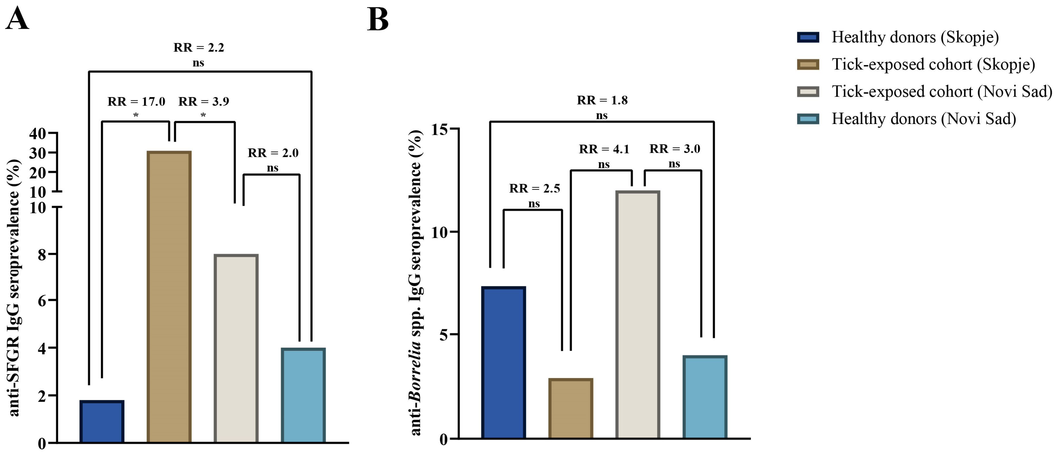
Figure 1. Seroprevalence of anti-SFGR IgG and anti-Borrelia IgG among tick-exposed and healthy donor groups in Skopje and Novi Sad: (A) anti-SFGR IgG seroprevalence by tick exposure and geographic location; (B) anti-Borrelia IgG seroprevalence by tick exposure and geographic location. RR: relative risk, indicating the probability of an individual in the studied groups being seropositive. The significance of the association was also tested by Fisher’s exact test; * statistically significant, p < 0.05; ns, non-significant, p > 0.05.
Pathogens 14 00814 g001
Figure 2. Single and dual exposure to examined tick-borne pathogens among tick-infested and healthy donor groups in Skopje (CIDS) and Novi Sad (PINS).
Pathogens 14 00814 g002
Table 1. Distribution of analyzed subjects according to risk groups in North Macedonia (Skopje) and Serbia (Novi Sad) (n = 223).
Risk GroupTick-Exposed CohortHealthy Donors
SkopjeNovi Sad SkopjeNovi Sad
n(%)n(%)pn(%)n(%)p
Gender
Male38(55.9)29(58)0.81825(45.5)27(54)0.382
Female30(44.1)21(42)30(54.5)23(46)
Total68(100)50(100)n/a55(100)50(100)n/a
Age
Young adults10(14.7)31(62)0.000 *
0.026 *
0.001 *
27(49.1)22(44)0.601
Middle-aged adults18(26.5)5(10)17(30.9)16(32)0.904
Older adults40(58.8)14(28)11(20)12(24)0.621
Total68(100)50(100)n/a55(100)50(100)n/a
The p-values represent the results of two-sided comparisons of Skopje vs. Novi Sad within each subgroup of each cohort, calculated with the Chi-square 2) test with Yates’s correction. Row-wise p-values result from comparing the listed age category vs. all other ages between Skopje and Novi Sad within each cohort; * statistically significant, p < 0.05; n/a: not applicable.
Table 2. Seroprevalence of IgG antibodies against Borrelia and SFGR antigens, by center, tick exposure, and sex (CIDS = Clinic for Infectious Diseases, Skopje, North Macedonia; PINS = Pasteur Institute Novi Sad, Serbia) (n = 223).
CentreGroupSexSamples Tested n (%)Anti-SFGR IgG
n (%)
pAnti-Borrelia spp. IgG
n (%)
p
CIDSTick-exposedMale38 (55.9)13 (34.2)0.5041 (2.6)1.000
Female30 (44.1)8 (26.7)1 (3.3)
Total68 (100)21 (30.9)n/a2 (2.9)n/a
Healthy donorsMale25 (45.5)0 (0.0)1.0002 (8.0)1.000
Female30 (54.5)1 (3.3)2 (6.6)
Total55 (100)1 (1.8)n/a4 (7.3)n/a
Total 123 (100)22 (17.9)n/a6 (4.9)n/a
PINSTick-exposedMale29 (58.0)2 (6.9)1.0003 (10.3)0.686
Female21 (42.0)2 (9.5)3 (14.3)
Total50 (100)4 (8.0)n/a6 (12.0)n/a
Healthy donorsMale27 (54.0)1 (3.7)1.0001 (3.7)1.000
Female23 (46.0)1 (4.3)1 (4.3)
Total50 (100)2 (4.0)n/a2 (4.0)n/a
Total 100 (100)6 (6.0)n/a8 (8.0)n/a
The p-values represent the results of the Chi-square 2) test with Yates’s correction (or Fisher’s exact test when any cell count < 5), comparing the proportion seropositive between males and females within the same center and exposure group. n/a: not applicable.
Disclaimer/Publisher’s Note: The statements, opinions and data contained in all publications are solely those of the individual author(s) and contributor(s) and not of MDPI and/or the editor(s). MDPI and/or the editor(s) disclaim responsibility for any injury to people or property resulting from any ideas, methods, instructions or products referred to in the content.