Article

Yeast Diversity on Sandy Lake Beaches Used for Recreation in Olsztyn, Poland

by
Tomasz Bałabański
1,2,
Anna Biedunkiewicz
1,* and
Jan P. Jastrzębski
3
1
Department of Microbiology and Mycology, Faculty of Biology and Biotechnology, University of Warmia and Mazury in Olsztyn, Oczapowski 1A St., 10-719 Olsztyn, Poland
2
The Center for the Popularization of Science and Innovation of the University of Warmia and Mazury in Olsztyn ″Kortosfera″, Dybowski 11A St., 10-723 Olsztyn, Poland
3
Department of Plant Physiology, Genetics and Biotechnology, Faculty of Biology and Biotechnology, University of Warmia and Mazury in Olsztyn, Oczapowski 1A St., 10-719 Olsztyn, Poland
*
Author to whom correspondence should be addressed.
Pathogens 2025, 14(8), 744; https://doi.org/10.3390/pathogens14080744
Submission received: 23 June 2025 / Revised: 15 July 2025 / Accepted: 25 July 2025 / Published: 29 July 2025

Abstract

Yeasts possess a range of environmental adaptations that allow them to colonize soil and sand. They can circulate seasonally between different components of lake ecosystems, including beach sand, water, and the coastal phyllosphere. The accumulation of people on beaches promotes the development and transmission of yeasts, posing an increasing sanitary and epidemiological risk. The aim of this study was to determine the species and quantitative composition of potentially pathogenic and pathogenic yeasts for humans present in the sand of supervised and unsupervised beaches along the shores of lakes in the city of Olsztyn (northeastern Poland). The study material consisted of sand samples collected during two summer seasons (2019; 2020) from 12 research sites on sandy beaches of four lakes located within the administrative boundaries of Olsztyn. Standard isolation and identification methods used in diagnostic mycological laboratories were applied and are described in detail in the following sections of this study. A total of 259 yeast isolates (264, counting species in two-species isolates separately) belonging to 62 species representing 47 genera were obtained during the study. Among all the isolates, five were identified as mixed (two species from a single colony). Eight isolated species were classified into biosafety level 2 (BSL-2) and risk group 2 (RG-2). The highest average number of viable yeast cells was found in sand samples collected in July 2019 (5.56 × 10² CFU/g), August, and September 2020 (1.03 × 10³ CFU/g and 1.94 × 10³ CFU/g, respectively). The lowest concentrations were in samples collected in April, September, and October 2019, and October 2020 (1.48 × 10² CFU/g, 1.47 × 10² CFU/g, 1.40 × 10² CFU/g, and 1.40 × 10² CFU/g, respectively). The results indicate sand contamination with yeasts that may pose etiological factors for human mycoses. In light of these findings, continuous sanitary-epidemiological monitoring of beach sand and further studies on its mycological cleanliness are warranted, along with actions leading to appropriate legal regulations.

Graphical Abstract

1. Introduction

Yeasts exhibit high physiological and ecological plasticity, and show a wide tolerance to various environmental factors such as temperature, UV radiation, light, humidity, salt content, and conditions of limited availability of nutrients and water [1,2,3,4,5,6,7,8]. These properties have allowed microfungi to achieve ecological success and colonize most ecosystems of the biosphere, including water and soil, which is also reflected in the 2021 WHO guidelines that recognize beach sand as a relevant ecological reservoir of fungi in recreational areas [1]. Studies have demonstrated the presence of yeasts in a variety of environments, including marine, deep-sea, and freshwater reservoirs, aquatic ecotone habitats, ontocenoses of aquatic organisms, the surfaces of coastal phyllosphere, and bottom sediment layers of water bodies, as well as sand from beaches and coastal areas [1,9,10,11,12,13,14,15,16,17,18]. The ability of yeasts to thrive in such a broad range of habitats, from extreme marine conditions to terrestrial and freshwater ecosystems, highlights their versatile metabolic pathways and resilience to fluctuating environmental factors [4,5,7,8].
Yeasts can not only inhabit but also circulate between different components of lake ecosystems, including beach sand, water, and coastal phyllosphere (Figure 1). They migrate from soil or sand into water through surface and subsurface runoff, temporarily settling on coastal phyllosphere, which acts as a natural filter for the water body (Figure 1). Humans, as beach and swimming area users, can serve as temporary vectors for the transmission of microfungi, transferring allochthonous species for which lake ecosystem components are not natural reservoirs [1,7,10,13,19,20,21,22]. This concern has been emphasized in the 2021 WHO guidelines, which recommend monitoring beach sand as a potential source of exposure to opportunistic fungi [1].
Soil and beach sand, in particular, represent environments of interest. Yeasts are a significant component of the taxonomic structure of microbiological soil biomass. Their adaptive capabilities enable them to thrive in soil ecosystems, which includes sand, where yeasts can adhere to granules in suspension or as biofilms [11,20,23,24].
The yeast nutrition method based on primary osmotrophy is a crucial factor in shaping the above-mentioned adaptive abilities. They secrete adaptive exoenzymes into the environment to break down nutritional substrates, with the resulting products then being absorbed into the cells via osmosis. When even small amounts of a new substrate appear or the proportions of existing ones change, yeasts stimulate their enzymatic apparatus to produce new exoenzymes. These properties make microfungi, including yeasts, key players in essential ecological processes such as the biogeochemical cycling of elements in nature [5,25,26].
A negative aspect of such advanced adaptive capabilities is the ability to cause diseases in humans. Research shows a continuous increase in the occurrence of yeasts in the human body, including species not previously associated with ontocenoses of the human body [8,27,28,29,30].
Mycological studies of lake ecosystems involve determining the species, taxonomic, and quantitative structure of microfungi and examining their biochemical and ecological activities. These parameters provide an overview of the sanitary and ecological condition of the water reservoir and the processes occurring within it, which play an important role in analyzing the dynamics of these ecosystems. Detailed analysis of these factors allows for the assessment of the sanitary-epidemiological status of the studied environments, related risks, and the development of rational water management plans, which aligns with the recommendations of the WHO, emphasizing the importance of including beach sand and fungal components in routine microbiological assessments of recreational water environments [1,10,31].
The aim of the study was to determine the species and quantitative composition of the overall yeast population present in the sand of supervised and unsupervised beaches along the shores of lakes in the city of Olsztyn (northeastern Poland), including species that are potentially pathogenic and pathogenic to humans. This research focus is relevant from both an ecological and epidemiological perspective, as beach sand may serve as a reservoir and transmission route for opportunistic yeasts, some of which can cause infections in humans, particularly in immunocompromised individuals or those with skin injuries. Moreover, their presence may reflect broader environmental imbalances and pose a public health concern in recreational freshwater areas. The research was based on the hypothesis that among the yeast species occurring in beach sand, there would be strains potentially harmful to human health, which may pose a risk to beach users.

2. Materials and Methods

2.1. Research Area and Material

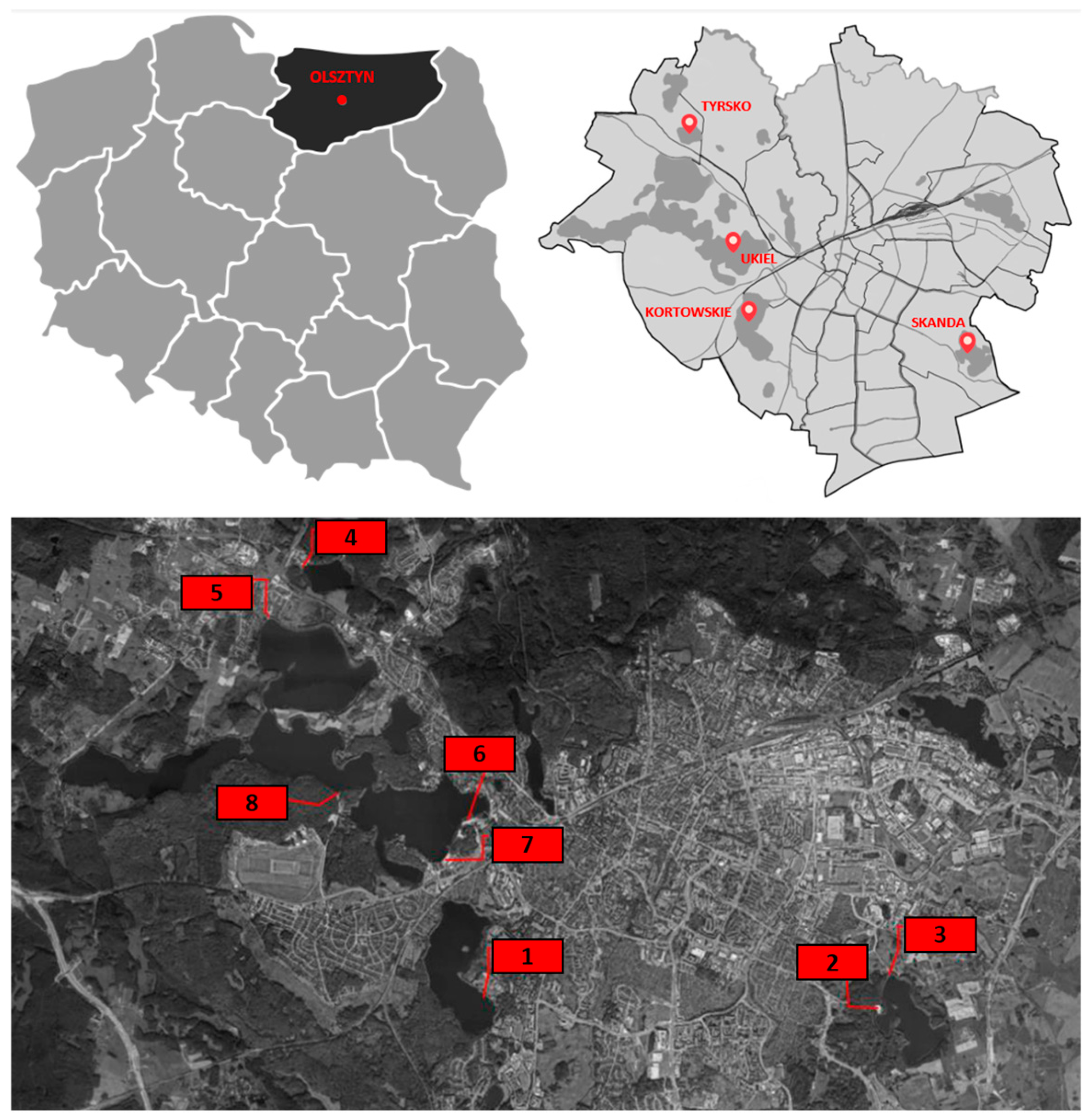
The research material consisted of yeasts isolated from sand during two summer seasons (April to October 2019 and 2020) from eight research sites located on supervised and unsupervised sandy beaches along the shores of lakes within the administrative borders of Olsztyn (northern Poland, Europe) (Figure 2). Four lakes were selected for the study: Kortowskie, Skanda, Tyrsko, and Ukiel, whose bathing waters undergo routine bacteriological testing by authorized state institutions (Provincial Sanitary and Epidemiological Station and County Sanitary and Epidemiological Station). Four of the beaches included in the study (sites 3, 6, 7, and 8) are supervised by authorized municipal institutions (Figure 2).

2.2. Sampling

Sampling sites were designated at the midpoint of the shoreline of each beach, 3 m from the water’s edge. Fifty-gram sand samples were collected from two depths (10 cm and 50 cm) using a soil auger (Fiskars) sterilized with a 70% alcohol solution. For sample unification, a 1 m × 1 m square was marked at the designated location, and 5 soil cores were taken (one from each corner of the square and one from the intersection of the diagonals). From each core, 10 g of soil were collected into a sterile Ziplock bag, sealed, and mixed to obtain a unified 50 g sample. Samples were collected during the first week of each month of the research season, twice a day (in the morning, 8:00–10:00 a.m., and in the evening, 8:00–10:00 p.m.). During sampling, the presence of beach users varied depending on the time of year. In the spring and summer months, a higher number of people were typically present on the beaches, particularly during afternoon and evening hours, which could potentially influence microbial loads. However, during periods of legal restrictions related to the COVID-19 pandemic—when access to public recreational infrastructure was limited or entirely prohibited—no beach users were present at the study sites due to the enforcement of national sanitary regulations. During two research seasons, a total of 448 sand samples of 50 g were collected from 8 study sites. During sample collection, air temperature and humidity were measured, and the UV index was recorded (Appendix A). Until yeast isolation, the collected sand samples were stored under refrigeration (4 °C) [32].

2.3. Yeast Isolation and Cultivation

The yeast isolation procedure was carried out within 48 h of sand sample collection. Yeasts were isolated from the sand samples using a modification of methods described in previous studies [32,33]. From the 50 g sand samples, 10 g sand samples were placed in 90 mL of sterile tap water (1:10 dilution) and shaken for 15 min. Yeasts were isolated by surface inoculation of 0.1 mL of the shaken suspension on Bengal Rose Agar with chloramphenicol (RBC) [32,33,34]. Each inoculation on the RBC medium was performed in triplicate. Given that 448 sand samples were tested, the total number of inoculations was 1344. In pilot studies, the tested media included RBC agar, Sabouraud agar with chloramphenicol, PDA (potato dextrose agar), and Czapek-Dox agar. The highest efficiency in yeast isolation from the studied sites was achieved using RBC medium. This medium was selected for further research due to the selective properties of Rose Bengal. Pilot studies demonstrated that RBC medium effectively inhibits the growth of filamentous fungi, particularly rapidly growing species such as Rhizopus and Mucor, thereby facilitating the growth and isolation of yeasts. Inoculated media were incubated at 25 °C for 48–72 h. In pilot studies conducted to validate the method, no growth was obtained at 37 °C, so a 25 °C incubation temperature was adopted for effective isolation in further studies. After incubation, macroscopic and microscopic evaluations of the colonies were conducted to differentiate yeast colonies from bacterial and filamentous fungal colonies. The yeast colonies were counted and passaged onto Sabouraud agar slants with chloramphenicol and incubated at 25 °C for 48 h. After incubation the cultures were stored under refrigeration (4 °C) for further studies.

2.4. Yeast Count Assessment

To assess yeast population density in the sand, the plate technique using solid media was employed [32]. According to the method, the number of viable cells in the sample is equal to the number of observed colonies. The results were calculated as colony-forming units (CFU) per 1 g of dry soil mass.

2.5. Yeast Identification

A macromorphological description of the verified yeast colonies (i.e., colonies that were microscopically confirmed as yeasts) was made, including features such as shape, color, consistency, surface prominence, edge structure, transparency, smell, and growth into the medium. The ability of the isolated yeasts to ferment a basic carbohydrate series (glucose, galactose, maltose, sucrose, lactose) was tested using a liquid medium for carbohydrate zymogram (tested sugar 20 g; peptone 0.5 g; distilled water 1000 mL; bromothymol blue with NaOH to achieve a blue color). Suspensions of the colonies of the tested strains were prepared in sterile tap water with turbidity of 3° on the McFarland scale. A 40 µL drop of the suspension was added to the zymogram medium tubes and incubated at 25 °C for 48 h. Fermentation ability was indicated by a change in the color of the solution from blue to yellow (lowering of the medium’s pH due to fermentation) and/or medium turbidity (increased yeast biomass).
To analyze micromorphological features, microcultures were prepared on Nickerson’s agar [8,31,35,36]. Microcultures were set up in sterile moist chambers by passaging colony inoculum onto the surface of Nickerson’s agar, followed by covering it with 20 µL of rabbit serum solution with bacteriological broth (1:1). The inoculum was then covered with a sterile coverslip. To maintain appropriate humidity, 1 mL of sterile distilled water was placed at the bottom of the chamber. The microcultures were incubated at 25 °C for 144 h to reveal diagnostically significant features, such as the size, shape, location, and formation of blastospores, the ability to produce chlamydospores, pseudohyphae, and true hyphae, as well as their size, shape, and arrangement [8,10,13,35,36]. Microculture on Nickerson’s agar remains a relatively underutilized yet highly valuable method for yeast morphology assessment and identification. This technique enables precise species determination by leveraging knowledge of the specific and spatial growth patterns of mycelium or pseudomycelium, allowing for natural, undisturbed development without external interference. Its ability to preserve the inherent structural characteristics of fungal growth makes it a crucial tool for accurate taxonomic differentiation [8,10,13,35]. Microscopic observations of the microcultures were made at 48, 72, and 144 h of incubation, with photographic documentation at each stage.
For strains requiring additional data for identification, Schaeffer–Fulton staining was performed to demonstrate the presence of asci with ascospores and to classify the strain as either ascomycetous or non-ascospore-forming yeasts [37].
The classification of strains into biosafety levels (BSL) and risk groups (RG) was based on multiple sources, as data on these categories vary across references and countries. However, diagnostic keys served as the primary basis for classification, with additional literature consulted to ensure accuracy and reliability [19,38,39,40].
To achieve more precise identification for a subset of the studied strains, molecular analyses were conducted in addition to phenotypic methods (including macro- and microculture observations). Genomic DNA was isolated using the Genomic Mini AX Yeast Spin kit (A&A Biotechnology, Gdańsk, Poland). Based on the convergence of phenotypic traits and selected biochemical analyses, 49 of the most representative samples (the most frequently occurring species identified phenotypically) were selected for molecular analysis to avoid redundant testing of multiple strains of the same species. PCR (polymerase chain reaction) was performed, followed by purification and sequencing of the ITS-1 (5′-TCCGTAGGTGAACCTGCGG-3′) and ITS-4 (5′-TCCTCCGCTTATTGATATGC-3′) regions of DNA for selected yeast isolates. The resulting nucleotide sequences were compared with the GenBank database using the Nucleotide BLAST tool (BLASTN, NCBI online version accessed in January 2025; https://blast.ncbi.nlm.nih.gov). The sequences used for identification have been deposited in the GenBank database under accession numbers PQ882533–PQ882625.
Final species classification of strains was conducted by integrating results from both phenotypic and molecular methods. In cases of uncertainty, final identification was achieved by comparing microculture observations on Nickerson’s agar with diagnostic keys [19,38,39].

2.6. Statistical Analysis

Statistical analysis was performed using STATISTICA 13.3 software (StatSoft). The following tests were used in the analysis: nonparametric Mann–Whitney U test, nonparametric Kruskal–Wallis H test, one-way ANOVA (analysis of variance), Tukey’s b test, paired Student’s t-test (for comparing the number of isolates per species between the 2019 and 2020 seasons), and Spearman’s rank correlation coefficient. For all tests, the significance level was set at p < 0.05.

3. Results

During the study, a total of 259 yeast isolates were obtained, representing 62 species from 47 genera (Table 1). Thirty-two of the identified genera belonged to ascomycetes (68.1%), and the remaining fifteen to basidiomycetes (31.9%) (Table 1). Among all the isolates, five were two-species isolates (19.3% of all isolates) (Table 1 and Table 2). Species from two-species isolates were counted separately in both quantitative and statistical analyses (Table 1 and Table 2). Of the sixty-two species, eight were classified as BSL-2 and RG-2 (12.9%) (Table 1). Preliminary macroscopic selection, including the macromorphological features of the colonies, indicated the need to distinguish different isolates, which were later classified as the same species during further diagnostic stages.
Analysis of individual research seasons showed differences in the number of isolates obtained from the beaches. Overall, more isolates were obtained in the second research season (2020; 149 isolates, including two two-species isolates), while 86 fewer isolates were obtained in the first research season (2019; 63 isolates, including three two-species isolates) (Table 1 and Table 2). The difference in the number of isolates was not statistically significant (p = 0.0927). Similarly, the paired t-test (p = 0.170) revealed no statistically significant difference in the average number of isolates per sampling event between the two seasons, although a tendency toward higher isolation frequency was observed in 2020 (mean = 4.45) compared to 2019 (mean = 3.43). This may reflect differences in environmental conditions or anthropogenic pressure.
In the total number of isolates, those obtained from sand in the morning hours predominated (147 isolates, 55.7% of all isolates). Fewer yeasts were isolated from sand collected in the evening (117 isolates, 44.3% of all isolates) (Table 1). No statistically significant differences were found in the number of isolates obtained in the morning and evening (p = 0.7955).
Larger differences were observed in the case of sand samples collected from different depths (Table 1). In the total pool of isolates, yeasts collected from a depth of 10 cm clearly dominated (175 isolates, 66.3% of all isolates). Fewer yeasts were isolated from soil at a depth of 50 cm, below the mixing layer of the sand (89 isolates, 33.7% of all isolates) (Table 1). These differences, however, were not statistically significant (p = 0.0927).
From the beaches supervised by authorized municipal institutions, 100 isolates were obtained (37.9% of all isolates), while 164 isolates were obtained from unsupervised beaches (62.1% of all isolates) (Table 1). These differences were not statistically significant (p = 0.2131).
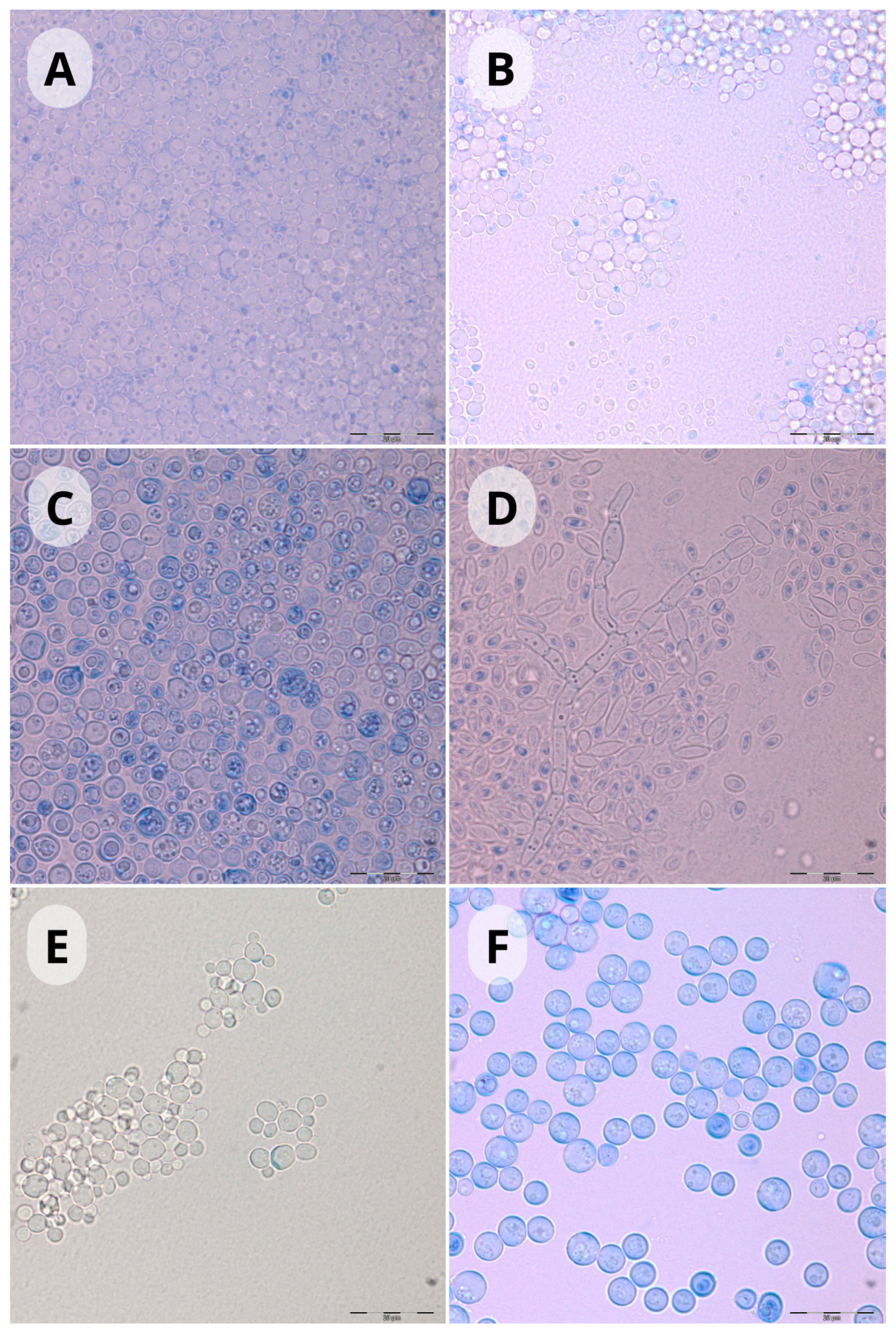
Among the yeasts isolated during the study, the most frequently represented species were: Barnettozyma californica, Nakasemomyces glabratatus, Cutaneotrichosporon moniliiforme, C. jirovecii, Debaryomyces hansenii, and Solicoccozyma aeria. Other species occurred in smaller numbers (eight isolates or less) (Table 1, Figure 3).
In the 2019 season, the most frequently isolated species were: B. californica, Cutaneotrochosporon jirovecii, C. moniliiforme, Schwanniomyces capriottii, and Tausonia pullulans (Table 1) Among the species isolated in the 2020 season, D. hansenii, Mycogloea nipponica, N. glabratatus, and S. aeria dominated (Table 1). Twenty-one species and five strains identified only to genus (one genus) were isolated in both research seasons, representing 33.3% of the species and genera pool obtained during the study. Twelve species and one strain identified only to genus one1 genus) were noted exclusively in the 2019 season (19.7% of the total species and genera pool). Twenty-nine species and four strains identified only to genus (two genera) were found only in the 2020 season (46.9% of the total species and genera pool) (Table 1).
From morning-collected sand samples, the most frequently isolated species were: B. californica, Cutaneotrichosporon jirovecii, C. moniliiforme, D. hansenii, and S. aeria (Table 1). In the evening-collected sand, the most common species were: C. glabrata, Cutaneotrichosporon jirovecii, C. moniliiforme, and D. hansenii (Table 1). Thirty-two species and nine strains identified to genus (two genera) were isolated both in the morning and evening, representing 51.5% of the total species and genera pool detected during the study (Table 1). Seventeen species and one strain identified to genus (one genus) were found only in the morning samples (27.3% of the total species and genera pool). Thirteen species and one genus were found only in evening samples (21.2% of the total species and genera pool) (Table 1).
From a depth of 10 cm, the most frequently isolated species were: B. californica, Citeromyces matritensis, Cutaneotrichosporon jirovecii, C. moniliiforme, D. hansenii, N. glabratus, and S. aeria (Table 1). Among species isolated from a depth of 50 cm, N. glabratus, C. moniliiforme, and D. hansenii dominated (Table 1). Twenty-four species and seven strains identified only to genus (two genera) were isolated from both depths (10 cm and 50 cm), representing 39.4% of the total species and genera pool (Table 1). Twenty-eight species and one genus were found only in the 10 cm deep samples (43.9% of the total species and genera pool). Ten species and one genus were isolated only from the 50 cm deep samples (16.6% of the total species and genera pool) (Table 1).
In the sand from supervised beaches, the most frequently isolated species were: B. californica, N. glabratus, C. moniliiforme, and D. hansenii (Table 1). From the sand of unsupervised beaches, the most common species were: Cutaneotrichosporon jirovecii, C. moniliiforme, D. hansenii, and S. aeria (Table 1). Twenty-six species and one genus were isolated from both types of beaches (supervised and unsupervised), representing 40.9% of the total species and genera pool detected during the study (Table 1). Twelve species and two genera were noted only in samples collected from supervised public beaches (21.2% of the total species and genera pool). Twenty-four species and one genus (strain identified only to genus) were found only in the sand from unsupervised beaches (37.8% of the total species and genera pool).
During the two-year study, the most isolates were obtained from the beaches of Lake Skanda (87 isolates; 32.95% of all isolates) and Lake Ukiel (87 isolates; 32.95% of all isolates). From Lake Kortowskie, 63 isolates were recorded (23.9% of all isolates). The fewest isolates were from Lake Tyrsko (27 isolates; 10.2% of all isolates) (Table 1).
Taking into account the strains identified to the species level, the greatest taxonomic diversity was observed in the case of Lakes Ukiel and Skanda (33 and 32 isolated species, respectively), and the least in the case of Lake Tyrsko (19 isolated species). From the sand of the beaches of Lake Kortowskie, 27 species were isolated (Table 1). The obtained results allowed for a detailed characterization of the species structure of the sand on the beaches of individual lakes (Table 1). Nine species were found exclusively in the sand of the beaches of Lake Kortowskie, the most frequently isolated was Papiliotrema pseudoalba. Seven species were isolated exclusively from the sand of the beaches of Lake Skanda, with Exophiala jeanselmei, classified as BSL-2 and RG-2, being the most frequent. Four species were found only in the sand of the beach of Lake Tyrsko, with Exophiala castellanii, also classified as BSL-2 and RG-2, being the most frequent. In the case of Lake Ukiel, nine species and five strains identified only to the genus (three genera) were noted exclusively in the sand of the beaches of this lake, with Nadsonia commutata being the dominant species.
Three species were recorded in the sand of the beaches of all the lakes (Cutaneotrichosporon jirovecii, C. moniliiforme, and D. hansenii). Two of them, C. jirovecii and C. moniliiforme, were classified as BSL-2 and RG-2 (Table 1). Fourteen species were found in the sand of the beaches of three out of the four studied lakes, including one (N. glabratus, formerly C. glabrata) classified as BSL-2 and RG-2 (Table 1). Fourteen species were isolated from the sand of the beaches of two lakes, while 35 species were found in the sand of beaches located near only one lake (Table 1). Five of the species present in the sand of the beaches of only one lake (Candida albicans, Clavispora lusitaniae, Exophiala bergeri, E. castellanii, and E. jeanselmei) were classified as BSL-2 and RG-2 (Table 1).
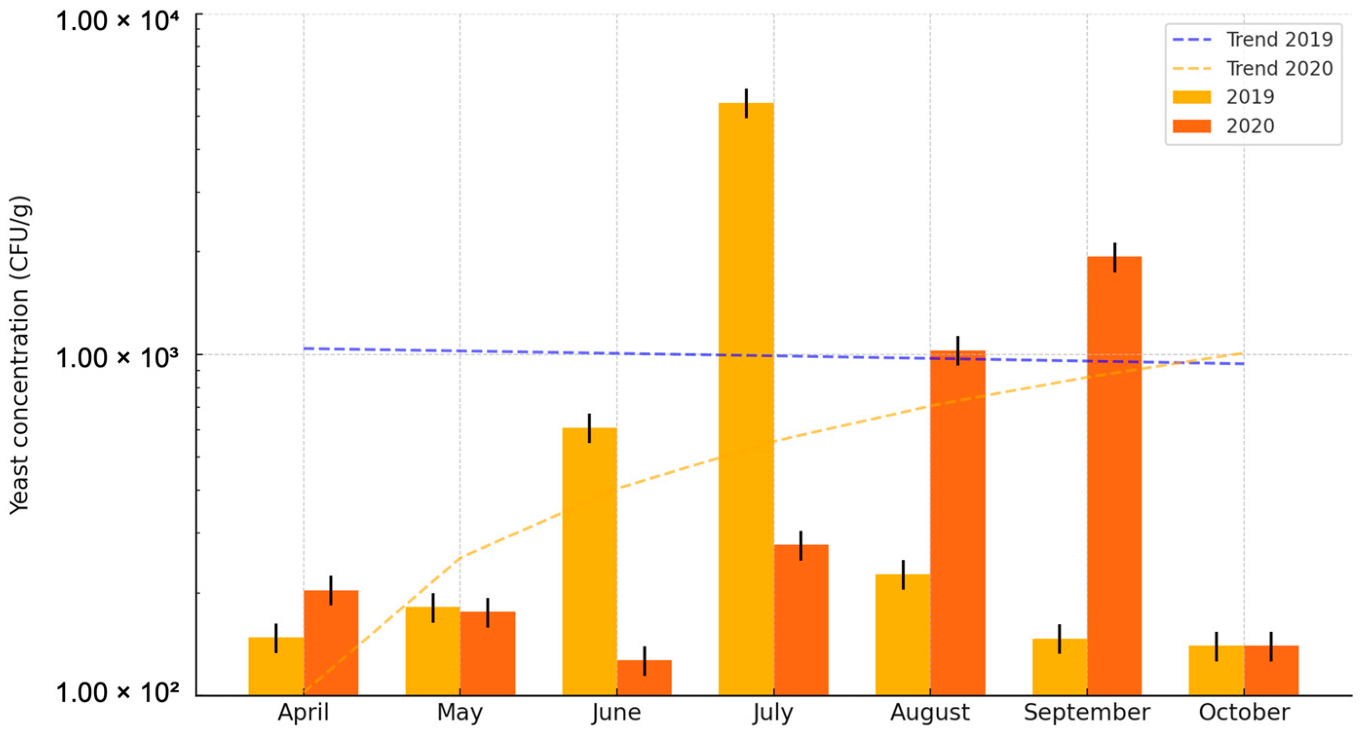
The number of viable cells of individual strains was counted, and the obtained data were compiled in tabular form (Table S1). On average, nearly twice as many viable yeast cells were isolated in the 2019 season (9.91 × 10² CFU/g) compared to the 2020 season (5.56 × 10² CFU/g) (Table S1; Figure 4). The fluctuations in the average number of yeasts in 1 g of sand in the 2019 season ranged from 1.40 × 10² CFU/g in October to 5.48 × 10³ CFU/g in July, while in the 2020 season, they ranged from 1.40 × 10² CFU/g in October to 1.94 × 10³ CFU/g in September. The highest average number of viable yeast cells was found in sand samples collected in July 2019 (5.56 × 10² CFU/g), and in August and September 2020 (1.03 × 10³ CFU/g and 1.94 × 10³ CFU/g, respectively). The lowest concentrations were in samples collected in April, September, and October 2019, and in October 2020 (1.48 × 10² CFU/g, 1.47 × 10² CFU/g, 1.40 × 10² CFU/g, and 1.40 × 10² CFU/g, respectively) (Table S1; Figure 4). In 2019, compared to 2020, more viable yeast cells were isolated in May, June, and July. In July 2019, the average number of viable yeast cells was an order of magnitude higher than in the 2020 season. The opposite trend was observed in April, August, and September. In August and September 2020, the average number of viable yeast cells was an order of magnitude higher than in the corresponding months of 2019. In October of both seasons, the same average number of viable yeast cells was recorded (Table S1; Figure 4). The differences in the average concentration of strains between individual months were statistically significant (p = 0.0300). Post hoc analysis showed that this difference occurred between April and July (the test considered numerical data from both research seasons and all studied beaches).
Statistical analysis was conducted to assess the impact of air temperature, air humidity, and UV index on the number of isolates and strain concentrations in the sand during individual months of the research seasons, revealing that only air temperature significantly affected the average strain concentration in the sand (p = 0.0210) (Table 3). The potential impact of air temperature on the abundance of the yeast population in the soil is justified in the later sections of this study.

4. Discussion

Research shows high ecological and taxonomic diversity of yeasts found in soil ecosystems [7,20,41]. The taxonomic structure of yeasts in sand is very uneven and diverse. It includes many species, classified both in the phylum Ascomycota and Basidiomycota. There is a relationship between the taxonomic and numerical structure of soil yeast populations and the cross-section of the soil profile [7,41]. The results of this study confirm this observation. According to the results, the largest number of yeasts occurs at a depth of about 10 cm, although their number is quite variable and may range from 10 to over 100 CFU (colony-forming units) per gram of dry soil mass. A clear correlation was found between the size of yeast populations and the content of carbon and nitrogen in the soil [7,20]. Thanks to their adaptive abilities, soil and sand provide good environments for yeast development due to the favorable microclimate, richness in available carbon and nitrogen sources, as well as other macro- and microelements [7,11,19,42].
Sanitary-epidemiological monitoring of recreational beaches currently includes only various parameters of bathing waters. This legal issue has been pointed out in other studies [42,43,44]. Other authors emphasize the unique and complex structure of sand, which requires going beyond standard beach monitoring parameters and developing independent quality control standards and microbiological norms to ensure the safety of beach users [45]. This view is supported by the 2021 WHO guidelines on recreational water quality, which recognize beach sand as a potential source of microbiological risk—including fungi—and recommend its inclusion in routine environmental monitoring of recreational sites [1,46].
Quantitative analysis and species pool of microfungi in beach sand depends on numerous atmospheric and environmental factors [11,42,43,45]. The present study found a statistically significant effect of air temperature on the average concentration of strains in the sand (higher temperature resulted in a higher average strain concentration). Other studies confirm the influence of air temperature on the temperature within the soil profile in its shallower layers. This influence decreases with depth [46]. This suggests that air temperature may have at least an indirect impact on the numerical and taxonomic structure of the yeast population in the upper layers of the soil profile. Other authors highlight the direct impact of temperature on the survival of microorganisms in water and sand [45,47]. Moreover, they suggest a possible connection between global warming and the growth kinetics of microorganisms, pointing out that, despite the general belief that increased temperatures favor microbial growth, in the case of mesophilic microorganisms, it will act as a limiting factor, while favoring the growth of pathogens that prefer conditions closer to the human body temperature [45]. An example could be yeasts of the genus Candida, which exhibit xerotolerant properties [48]. Furthermore, it is noted that increased temperatures affect other environmental parameters that shape the microbiological structure of beaches [45]. For instance, an indirect negative correlation between the abundance of fungi in sand and the length of exposure to sunlight has been demonstrated, with the latter causing an increase in sand temperature [11]. Similar to the present study, no statistically significant effect of air humidity on the number of yeasts per gram of soil was observed [11].
There were no clear statistical differences in the total number of isolates or overall strain concentration between the 2019 and 2020 seasons. Nevertheless, the data suggest a tendency toward a higher frequency of yeast isolation in 2020 compared to 2019. This trend may reflect seasonal variation in environmental factors such as temperature and humidity, as well as differences in anthropogenic pressure between the two research seasons. A broader analysis across individual months of the research season (April–October) also indicated that while monthly fluctuations in isolate counts were observed, they were not substantial. However, more pronounced variation was noted in strain concentrations between specific months, particularly in early and mid-summer. Similar findings have been reported by other authors, who emphasize that increased human activity during the peak summer months—including recreational use of beaches and the organization of public events—may lead to elevated microbial contamination of beach environments [45]. The positive correlation between the concentration of microfungi in beach sand and the number of users is confirmed by other studies [49,50]. This issue, however, requires further analysis, especially in the context of the number of users, due to incomplete data on population flow provided by supervisors of the studied municipal beaches (Appendix B). Additionally, an analysis of the average concentration of yeasts in the sand of beaches across studied lakes was conducted. In both research seasons, the lowest average concentration of yeasts in the sand was recorded for the lake with the smallest surface area. This observation is likely due to the varying number of beach users, which correlates with the size and availability of recreational areas at the studied water bodies. Lake Tyrsko has the shortest shoreline, significantly limiting the bathing area and the number of users. Other researchers have pointed out the possible influence of the water body’s structure on the taxonomic and numerical composition of yeast populations in the associated ecological niches [10,13,22].
There is no statistical relationship between the season and the number of isolates or the concentration of yeasts in the soil. Similar results were presented in the other study [11]. This suggests that the numerical structure of yeasts in beach sand is determined not by the season, but rather by long-term atmospheric conditions.
Moreover, no significant changes in the concentration and number of yeast isolates were observed between the times of day studied (morning; evening). The present study correlates with the results of other studies, which demonstrated the weakest seasonal correlation between air and soil temperature in the spring and summer. The authors noted that soil temperature is mainly influenced by factors unrelated to the diurnal cycle [46,51]. Furthermore, the authors found that the diurnal amplitude of soil temperatures and the dependence of soil temperature on air temperature decrease with soil profile depth [46,51]. This suggests that sand is less sensitive to temperature fluctuations with depth. In the present study, samples were collected from depths of 10 and 50 cm, which significantly reduced the influence of atmospheric conditions on the yeast population structure in the sand throughout the diurnal cycle.
Analyzing the yeast abundance at different depths, it was found that the obtained results are consistent with those reported by Grishkan and Kidron [52], who also observed higher average concentrations of yeast strains at a depth of 10 cm. This trend persisted across different research seasons. However, in the case of both parameters, the observed differences were not statistically significant. This observation may be linked to the higher content of organic carbon, water, and oxygen in the surface soil layers, where sand layers mix. Grishkan and Kidron’s [52] studies indicate decreased organic carbon content and oxygen availability with soil depth. The same study showed that this trend does not significantly change depending on the climate and that respiratory processes and biomass development of oxygen-affiliated organisms occur most intensively in the surface soil layers. Grishkan and Kidron’s [52] research suggests that the distribution of fungal communities in soil is related to the availability of organic matter, water, aeration, salinity, and UV radiation, emphasizing its complex and non-linear nature. Another factor influencing the distribution and abundance of microfungal consortia is soil and sand porosity [11,52]. Microbial biomass density reaches its maximum values in the surface soil layers (0.2–2 cm), with a second peak at a depth of 5–10 cm, followed by a sharp decline down to 50 cm [52]. This observation aligns with the present study.
During the research, differences in the taxonomic structure of soil yeast consortia depending on the soil profile depth were noted. The dominant part of the isolates obtained included species occurring at both depths (24 species; two genera). Species recorded in the number of three isolates or more were distinguished from this group. These were: N. glabratus, C. jirovecii, C. moniliiforme, D. hansenii, Oosporidium margaritiferum, and Schwanniomyces polymorphus. Based on these findings, it can be hypothesized that the mentioned species are insensitive to limited oxygen availability and are less susceptible to changes in oxygen content in the sand compared to other microorganisms. In the current study, 28 species and one genus were found exclusively at a depth of 10 cm, which may indicate their high oxygen affinity. At a depth of 50 cm, only 10 species and one genus were found, indicating their ability to grow in low-oxygen conditions. The taxonomic structure of the mycobiota at a depth of 50 cm was significantly poorer, showing a clear trend that yeasts can develop at various depths but are more commonly found in the surface soil layers. This is likely because most yeasts require oxygen for growth [53]. Nonetheless, other researchers suggest that yeasts may acquire the ability to develop in anaerobic conditions through horizontal gene transfer. It should be noted that other factors, such as soil structure (e.g., porosity), organic compound content (mainly carbon), water availability, UV radiation, salinity, and temperature, may also influence the species richness of yeasts in various soil layers [11,42,45,52].
Overall, more yeast strains were isolated from unsupervised beaches; however, a comparison of the number of isolates and yeast concentration in the sand did not reveal statistically significant differences. Similar results were obtained by other researchers [11,43]. The likely cause of this difference was the similar purpose of the beaches studied, as well as the cleaning and replenishment of sand on supervised beaches by municipal institutions, which may improve the sanitary condition of the sand. It should be noted, however, that the presence of municipal supervision at swimming beaches may encourage more people to use them, and the presence of users increases the quantitative and taxonomic structure of the mycobiota in the sand and water [10,43,45].
Among the most frequently reported yeasts on beaches are representatives of the genera: Aureobasidium, Candida, Geotrichum, Exophiala, Metschnikowia, Rhodothorula, and Yarrowia [14,42,43]. Yeasts from the genera Candida, Cryptococcus, and Rhodotorula are associated with sandy beaches [11]. In the present study, nearly 260 yeast strains were isolated from the sand of lake swimming beaches, among which 47 genera were recorded, including Aureobasidium, Candida, Cryptococcus, Exophiala, Metschnikowia, and Rhodotorula, confirming that beach sand is their natural ecological niche. Additionally, the literature indicates that the most commonly noted genera in lake waters are Candida, Cryptococcus, Geotrichum, Hansenula, Pichia, Rhodotorula, Saccharomyces, Sporobolomyces, and Trichosporon [10,11,31], and on the coastal phyllosphere, the genera Candida, Cryptococcus, Debaryomyces, Metschnikowia, Pichia, Saccharomyces, and Rhodotorula [9,10,11]. Five of the genera considered typical for bathing water and all those considered characteristic for coastal aquatic vegetation were isolated from the sand in this study. A summary of long-term studies conducted by Biedunkiewicz demonstrated the constant presence of over 40 yeast genera in lake waters in the Olsztyn region, and 16 associated with the aquatic vegetation found in their vicinity [10]. In the present study, 21 genera (48.8%) from those recorded by Biedunkiewicz and colleagues [10] in bathing water were noted, along with 13 genera (81.3%) that were isolated in their research from the phyllosphere. These observations indicate the continuous circulation of microfungi between the various components of the lake ecosystem, including during the washing of sand and vegetation by lake water, as well as through surface and subsurface runoff caused by precipitation. Beach users and animals living there, e.g., waterfowl, may also be an important vector of such transmission [10,11,42,45]. Numerous scientific papers indicate that species such as B. californica, N. glabratus, C. moniliiforme, C. jirovecii, D. hansenii, and S. aeria are soil microorganisms or are frequently isolated from soil, including beach sand [7,11,45,54,55]. The frequent notation of these species in the present study should therefore be considered a conventional observation.
A comparison of the taxonomic structure between the designated research seasons showed that 21 species and one genus (strain identified only to the genus level) were isolated in both seasons. Among this group, B. californica, Nakaseomyces glabratus, C. moniliiforme, D. hansenii, Schwanniomyces polymorphus, and S aeria were noted with three or more isolates in each season, suggesting that they are indigenous to the studied environments. This is supported by studies that classify these species as soil yeasts [7,11,19,56,57]. For the species C. moniliiforme and D. hansenii, the discrepancy in the number of isolates between seasons reached an order of magnitude. It can be hypothesized that these species may be more sensitive to seasonal changes in environmental parameters, such as weather conditions or beach user traffic and activity. This is corroborated by studies from other authors [7,10,11,13,21,45,55]. In the 2019 season, 29 species and two genera (strains identified only to the genus level) were recorded, while in 2020, 12 species and one genus were found. The probable cause of these observed differences is the limited traffic of beach users, a potential vector of microfungal transmission at bathing sites, due to the sanitary regulations related to the COVID-19 pandemic in 2020.
A total of 26 species and one genus (strains identified only to the genus level) were found on both supervised and unsupervised beaches. Among these, the species B. californica, N. glabratus, Cutaneotrichosporon jirovecii, C. moniliiforme, D. hansenii, and S. aeria were noted with three or more isolates on each type of beach. These species have previously been identified as characteristic or frequently isolated from soil and lake environments [7,10,11,45,54,55]. For the species B. californica, N. glabratatus, and D. hansenii, more isolates were found on supervised beaches. N. glabratatus often forms part of the physiological mycobiota of humans and, in immunocompromised individuals, can become pathogenic [10,11,13]. B. californica is classified as a soil yeast [7,19] and has also been isolated from water, plant debris, and animal droppings [58]. B. californica and D. hansenii are also found in food products [59,60]. The presence of these species suggests a link to human activity, as supervised beaches generally attract more visitors during the summer season. However, overall, half as many species were found exclusively on supervised beaches compared to those isolated solely from unsupervised beaches. This discrepancy is likely due to the better sanitary condition of supervised beaches, which are cleaned and have their sand replenished by supervisors.
The diurnal cycle was the parameter for which the greatest number of species were recorded in the sand, regardless of this parameter, compared to the other parameters adopted in the study (depth, season, type of supervision). Other researchers indicate that the taxonomic structure of yeasts in sand and soil generally changes due to seasonal dynamics rather than the diurnal cycle [11,46,51].
The taxonomic structure of yeast assemblages in sand was also compared between the beaches of different lakes. The most species were isolated from Lakes Ukiel and Skanda, and the fewest from Lake Tyrsko. The beaches of Ukiel and Skanda have the longest shorelines, the largest usable area, and are among the most frequently used for recreational purposes. The length of the shoreline increases the area where sand is washed by water, and beachgoers act as natural vectors of fungal transmission, promoting yeast circulation between the different components of the lake ecosystem [10,61]. The beach at Lake Tyrsko is managed by a private owner and is only accessible to guests of the nearby hotel, which significantly limits the number of users. Additionally, Lakes Ukiel, Kortowskie, and Skanda are eutrophic and exhibit high primary production, which is associated with high levels of nitrogen, oxygen, and phosphorus, and generally correlates with increased taxonomic diversity. In contrast, Lake Tyrsko, despite showing signs of aging, is characterized by low trophic status and poor water mineralization, which may limit the number of species transmitted to the sand [61].
Sixteen species and one genus were isolated from the beaches of more than two of the studied lakes. Among them, C. moniliiforme, C. jirovecii, and D. hansenii were isolated from the sand of all four studied lakes. Other studies confirm the presence of species belonging to these genera in lake water and beach sand [10,13,14,55]. These species can therefore be considered characteristic of the beach sand of the lakes studied and likely continuously associated with this type of ecological niche, which has sanitary and epidemiological significance.
All obtained yeast isolates were assigned BSL and RG index values in the context of their potential pathogenicity. Additionally, for the most frequently isolated species, a literature review was conducted regarding the infections they can cause.
Among the most frequently isolated species were Cutaneotrichosporon jirovecii and C. moniliiforme. These are potential human pathogens in immunocompromised individuals (Biedunkiewicz and Góralska [13]; Biedunkiewicz et al. [10]; Kulesza et al. [35]; Bałabański and Biedunkiewicz [8]). Studies indicate that species from the genus Cutaneotrichosporon can grow at human body temperature and produce hydrolytic enzymes that act as virulence factors promoting infections. Furthermore, the scientific literature indicates an increase in infections and resistance of Cutaneotrichosporon species to antifungal agents used by clinicians [62]. Researchers have noted that C. jirovecii can cause liver and skin damage, while C. moniliiforme tends to cause less invasive pathological changes [63]. The high risk these species pose to beachgoers is demonstrated by the fact that they were isolated from the sand of beaches at all the lakes studied.
The second most frequently noted species, D. hansenii, found on all the studied beaches, is associated with food products. It is generally considered saprotrophic, but there are reports in the literature of isolates from clinical material. Studies by Breuer and Harms [64] and Desnos-Ollivier et al. [59] showed that this species rarely causes infections such as bone infections, subcutaneous abscesses, keratitis, and allergic alveolitis in both immunocompetent and immunocompromised individuals.
During the study, numerous strains classified in the genus Candida were isolated. Species belonging to this genus are the most commonly isolated from the blood of hospitalized patients. Additionally, they are frequently found in the oral cavity, urinary tract, and female genital tract [65]. There is an increase in infections caused by Candida fungi in immunocompromised individuals, including cancer patients and postoperative patients [65]. Factors predisposing Candida species to infect humans include their ability to adhere to host epithelial cells and form biofilms [8,35,65,66,67]. In the present study, the dominant species was Nakaseomyces glabratus (formerly C. glabrata) with a single isolate of C. albicans also recorded. N. glabratus and C. albicans are among the four species responsible for approximately 95% of diagnosed candidiasis [65]. Both species are classified as BSL-2 and RG-2. They mainly cause infections of the skin and mucous membranes in immunocompromised individuals [10,13,43,56,65]. N. glabratus primarily causes infections of the oral cavity, urogenital system, and bloodstream, and it exhibits resistance to many antifungal agents [68]. C. albicans is considered an indicator of fecal contamination in the environment [11]. Among the frequently isolated species was also Citeromyces matritensis, the teleomorphic stage of Candida globosa. Our review of the literature did not reveal any reported clinical infections caused by this species.
Numerous strains of B. californica were isolated during the study This species is not pathogenic but rather saprotrophic [69]. Similarly, Solicoccozyma aeria was frequently noted. Research indicates that it can be isolated from animals, but there are no reports of human infections caused by this species [70]. S. aeria may be a part of the physiological mycobiota of the colon, as it has also been isolated from human feces [71]. Additionally, it has been linked to appetite suppression and the activation of interleukin 17 in sensitive individuals, which may be associated with disturbances in gut microbiota and mycobiota. Studies also show an increase in S. aeria expression in cancer patients, with the potential for considering this species as a marker of gastric cancer, while excluding it as an etiological factor in the disease [72]. In summary, the literature suggests that this species may have a positive impact on human health as a cancer marker. There are no reports of its potential pathogenicity to humans; however, its isolation from animals suggests it could be considered as a potential human pathogen.
Yeasts from the genus Cryptococcus were also noted in the beach sand. Many species from this genus exhibit pathogenic properties in both immunocompromised and immunocompetent individuals [73,74]. Fungi of the genus Cryptococcus cause cryptococcosis, which can affect the skin, lungs, and bloodstream, and, in advanced stages, lead to meningitis and brain edema [73,74]. The virulence factors of Cryptococcus include the production of polysaccharide capsules and high enzymatic activity (protease, urease, phospholipase) [73]. The only identified species, C. amylolentus, belongs to the non-pathogenic cryptococci, closely related to pathogenic species. This species has been shown to have infectious potential under induced conditions, but due to its sensitivity to thermal stress, it does not grow at human body temperature [73,74].
Six isolates from the genus Exophiala were recorded during the study. Of the identified species, E. jeanselmei was the most frequently isolated. This species can cause subcutaneous ulcers and nodules in transplant recipients [75]. In advanced cases, it leads to chronic, purulent infections of subcutaneous tissue [76]. The less frequently isolated species, E. bergeri, has been reported as an etiological agent of nail infections [77], while E. castellanii has been noted in subcutaneous infections [78]. All species from the genus Exophiala isolated in this study are potential pathogens for immunocompromised individuals, as they are classified as BSL-2 and RG-2 [8,10,13,35,78].
All remaining species were isolated in numbers of six or fewer strains. Due to their rare (episodic) occurrence in the studied environments and simultaneous classification as BSL-1 and RG-1, it was assumed that they do not pose a potential threat to beach users. This risk assessment is consistent with the approach outlined in the 2021 WHO guidelines on recreational water quality, which emphasize prioritizing microbiological monitoring efforts toward organisms with higher pathogenic potential and relevance to public health [1].

5. Conclusions

The examination of the sanitary condition of beach sand at supervised and unsupervised swimming areas near Lakes Kortowskie, Skanda, Tyrsko, and Ukiel revealed the presence of species classified as potential human pathogens. Particular attention should be given to N. glabratus, Cutaneotrichosporon jirovecii, and C. moniliiforme, which were among the five most frequently isolated species, exhibited a cosmopolitan character in relation to the ecological niches studied, and were classified as Biosafety Level 2 (BSL-2) and Risk Group 2 (RG-2). The research demonstrated that beach sand at swimming areas may pose a potential health risk for individuals with temporary or chronic immunocompromised conditions. In this context, it is essential to conduct continuous sanitary-epidemiological monitoring of beach sand and implement preventive measures to mitigate the risks associated with the presence of potentially pathogenic yeasts in the sand, such as regular sand cleaning and replenishment. The present study, combined with the literature data, revealed that knowledge regarding the ecological and taxonomic characteristics of yeast populations in beach sand at swimming areas is significantly limited. Furthermore, there is a lack of legal regulations defining reference standards for the mycological cleanliness of such sand. In light of this, the introduction of such standards and the supervision of beach sand at swimming areas by institutions authorized to conduct sanitary inspections is recommended, along with further research in this field.

Supplementary Materials

The following supporting information can be downloaded at: https://www.mdpi.com/article/10.3390/pathogens14080744/s1; Table S1. The number of isolated yeasts.

Author Contributions

Literature review, T.B.; Research design, A.B.; Conducting experiments, T.B. and A.B.; Writing the manuscript, T.B. and A.B.; Preparation and placement of sequences in GenBank, J.P.J. All authors have read and agreed to the published version of the manuscript.

Funding

This work was supported by the Department of Microbiology and Mycology, Faculty of Biology and Biotechnology, University of Warmia and Mazury in Olsztyn (12.610.009-300), and European Social Fund (POWR.03.05.00-00-Z310/17).

Institutional Review Board Statement

Not applicable.

Informed Consent Statement

Not applicable.

Data Availability Statement

Sequences of ITS regions have been deposited in the GenBank database. Accession number range: PQ882533–PQ882625.

Acknowledgments

We would like to extend our heartfelt gratitude to the staff of the Department of Microbiology and Mycology, Dorota Górniak, and Aleksander Świątecki, for their support in conducting molecular research, which enriched the substantive value of the study.

Conflicts of Interest

The authors declare no conflicts of interest.

Abbreviations

BSL—biosafety level; CFU—colony-forming unit; PCR—polymerase chain reaction; RBC—Rose Bengal Chloramphenicol Agar, RG—risk group; WHO—World Health Organization.

Appendix A. Values of Meteorological Parameters During Sand Sampling

SAMPLING PERIODTEMPERATUREAVERAGE TEMPERATUREAIR HUMIDITYUV INDEX
MORNINGEVENINGMORNINGEVENING
APRIL/20196 °C14 °CD 12 °C
N 5 °C
36% 32%3/10
MAY/20198 °C15 °CD 15 °C
N 8 °C
73% 29%4/10
JUNE/201917 °C25 °CD 25 °C
N 15 °C
64% 45%7/10
JULY/201920 °C21 °CD 24 °C
N 15 °C
75% 70%6/10
AUGUST/201916 °C19 °CD 22 °C
N 14 °C
85% 51%5/10
SEPTEMBER/201913 °C16 °CD 18 °C
N 11 °C
75% 51%4/10
OCTOBER/201912 °C9 °CD 13 °C
N 7 °C
91%91%2/10
APRIL/20204 °C12 °CD 13 °C
N 5 °C
69% 29%3/10
MAY/20206 °C6 °CD 17 °C
N 9 °C
93% 94%4/10
JUNE/202012 °C18 °CD 21 °C
N 12 °C
64% 42%6/10
JULY/202019 °C23 °CD 22 °C
N 14 °C
85% 59%6/10
AUGUST/202019 °C24 °CD 23 °C
N 15 °C
39%39%5/10
SEPTEMBER/202013 °C16 °CD 18 °C
N 11 °C
92%90%0/10
OCTOBER/202011 °C8 °CD 13 °C
N 6 °C
88%88%2/10
D—average temperature of the month during the day, N—average temperature of the month at night.

Appendix B. Estimated Number of Users of the “Ukiel” Recreational Complex and Supervised Beach on Lake Skanda During the Year—Data of the Sports and Recreation Center in Olsztyn

LAKE UKIEL
MONTH20192020
SUM OF ENTRIESSUM OF EXITSSUM OF ENTRIESSUM OF EXITS
JANUARY32,75129,60114,55713,889
FEBRUARY64,78684,38140153385
MARCH124,985133,03634583094
APRIL106,946126,38815621918
MAY142,397132,03584169846
JUNE110,436108,51543435640
JULY174,234160,329THE SUPERVISOR DID NOT KEEP A REGISTER DUE TO THE COVID-19 PANDEMIC
AUGUST72,88572,431
SEPTEMBER53,82557,711
OCTOBER24,17727,222
NOVEMBER12,46113,223
DECEMBER10,82811,499
SUM930,711956,37136,35137,772
LAKE SKANDA
MONTH20192020
PEOPLE PER DAYMONTHLYPEOPLE PER DAYMONTHLY
JANUARYTHE SUPERVISORY DOES NOT KEEP A REGISTER
(OFF-SEASON PERIOD)
FEBRUARY
MARCH
APRIL
MAY
JUNEApprox. 150 (weekends 500)Approx. 3600Approx. 150 (weekends 500)Approx. 3300
JULYApprox. 9300Approx. 10,200
AUGUSTApprox. 10,200Approx. 9000
SEPTEMBERApprox. 2100Approx. 1950
OCTOBERTHE SUPERVISORY DOES NOT KEEP A REGISTER
(OFF-SEASON PERIOD)
NOVEMBER
DECEMBER
SUM-Approx. 25,200-Approx. 24,450

References

  1. World Health Organization (WHO). Guidelines for Recreational Water Quality; World Health Organization: Geneva, Switzerland, 2021; Available online: https://www.who.int/publications/i/item/9789240031302 (accessed on 15 May 2025).
  2. Krajewska-Kułak, E.; Łukaszuk, C.; Gniadek, A.; Macura, A.B.; van Damme-Ostapowicz, K.; Lewko, J.; Rolka, H.; Rozwadowska, E.; Guzowski, A. Zanieczyszczenie Powietrza w Pomieszczeniach Mieszkalnych, Ze Szczególnym Uwzglednieniem Roli Grzybów [Air Pollution in Aviation Air, with Detailed Consideration of the Role of Fungi]. Mikol. Lek. 2010, 17, 177–181. [Google Scholar]
  3. Ejdys, E.; Dynowska, M.; Biedunkiewicz, A.; Sucharzewska, E. An Overview of the Species of Fungi Occurring in School Rooms—A Four-Year Study. Pol. J. Environ. Stud. 2013, 22, 1691–1700. [Google Scholar]
  4. Tedersoo, L.; Bahram, M.; Põlme, S.; Kõljalg, U.; Yorou, N.S.; Wijesundera, R.; Ruiz, L.V.; Vasco-Palacios, A.M.; Thu, P.Q.; Suija, A.; et al. Global Diversity and Geography of Soil Fungi. Science 2014, 346, 1078–1089. [Google Scholar] [CrossRef]
  5. Wrzosek, M.; Ruszkiewicz-Michalska, M.; Sikora, K.; Damszel, M.; Sierota, Z. The Plasticity of Fungal Interactions. Mycol. Prog. 2017, 16, 101–108. [Google Scholar] [CrossRef]
  6. Kowalski, M.; Pastuszka, J.S. Effect of Ambient Air Temperature and Solar Radiation on Changes in Bacterial and Fungal Aerosols Concentrationin the Urban Environment. Ann. Agric. Environ. Med. 2018, 25, 259–261. [Google Scholar] [CrossRef]
  7. Yurkov, A.M. Yeasts of the Soil—Obscure but Precious. Yeast 2018, 35, 369–378. [Google Scholar] [CrossRef] [PubMed]
  8. Bałabański, T.; Biedunkiewicz, A. Threats Resulting from the Presence of Potentially Pathogenic Fungi in the Sport Changing Rooms—A Preliminary Study. Pol. J. Environ. Stud. 2023, 32, 3033–3042. [Google Scholar] [CrossRef]
  9. Gouka, L.; Raaijmakers, J.M.; Cordovez, V. Ecology and Functional Potential of Phyllosphere Yeasts. Trends Plant Sci. 2022, 27, 1109–1123. [Google Scholar] [CrossRef]
  10. Biedunkiewicz, A.; Sucharzewska, E.; Kulesza, K.; Nowacka, K.; Kubiak, D. Phyllosphere of Submerged Plants in Bathing Lakes as a Reservoir of Fungi—Potential Human Pathogens. Microb. Ecol. 2020, 79, 552–561. [Google Scholar] [CrossRef]
  11. Brandão, J.; Gangneux, J.P.; Arikan-Akdagli, S.; Barac, A.; Bostanaru, A.C.; Brito, S.; Bull, M.; Çerikçioğlu, N.; Chapman, B.; Efstratiou, M.A.; et al. Mycosands: Fungal Diversity and Abundance in Beach Sand and Recreational Waters—Relevance to Human Health. Sci. Total Environ. 2021, 781, 146598. [Google Scholar] [CrossRef]
  12. Brandão, J.; Albergaria, I.; Albuquerque, J.; José, S.; Grossinho, J.; Ferreira, F.C.; Raposo, A.; Rodrigues, R.; Silva, C.; Jordao, L.; et al. Untreated Sewage Contamination of Beach Sand from a Leaking Underground Sewage System. Sci. Total Environ. 2020, 740, 140237. [Google Scholar] [CrossRef]
  13. Biedunkiewicz, A.; Góralska, K. Microfungi Potentially Pathogenic for Humans Reported in Surface Waters Utilized for Recreation. Clean-Soil Air Water 2016, 44, 599–609. [Google Scholar] [CrossRef]
  14. Libkind, D.; Buzzini, P.; Turchetti, B. Yeasts in Continental and Seawater. In Yeasts in Natural Ecosystems: Diversity; Buzzini, P., Lachance, M.-A., Yurkov, A.M., Eds.; Springer: Berlin/Heidelberg, Germany, 2017; pp. 1–61. [Google Scholar]
  15. Fedorova, M.D.; Kurakov, A.V. Microbiota of Bottom Sediments in the Coastal Zone of Lake Baikal. Contemp. Probl. Ecol. 2023, 16, 492–508. [Google Scholar] [CrossRef]
  16. Nagahama, T. Yeast Biodiversity in Freshwater, Marine and Deep-Sea Environments. In Biodiversity and Ecophysiology of Yeasts; Péter, G., Rosa, C., Eds.; Springer: Berlin/Heidelberg, Germany, 2006; pp. 241–262. [Google Scholar]
  17. Monapathi, M.E.; Bezuidenhout, C.C.; James Rhode, O.H. Aquatic Yeasts: Diversity, Characteristics and Potential Health Implications. J. Water Health 2020, 18, 91–105. [Google Scholar] [CrossRef] [PubMed]
  18. Sultana, S.; Biró, J.; Kucska, B.; Hancz, C. Factors Affecting Yeast Digestibility and Immunostimulation in Aquatic Animals. Animals 2024, 14, 2851. [Google Scholar] [CrossRef]
  19. Kurtzman, C.P.; Fell, J.W.; Boekhout, T. The Yeasts: A Taxonomic Study, 5th ed.; Elsevier: Amsterdam, The Netherlands, 2011. [Google Scholar]
  20. Botha, A. The Importance and Ecology of Yeasts in Soil. Soil Biol. Biochem. 2011, 43, 1–8. [Google Scholar] [CrossRef]
  21. Biedunkiewicz, A.; Dynowska, M.; Ejdys, E.; Sucharzewska, E. Species Diversity of Yeast-like Fungi in Some Eutrophic Lakes in Olsztyn. Acta Mycol. 2013, 48, 61–71. [Google Scholar] [CrossRef]
  22. Nowacka, K.; Kulesza, K.; Glinka, P.; Sucharzewska, E.; Dynowska, M. The Phyllosphere as a Little-Known Reservoir of the Fusarium Genus, a Fungi of Importance to Medical Mycology. Ann. Parasitol. 2018, 64, 225–228. [Google Scholar] [CrossRef] [PubMed]
  23. Schoeman, H.; Wolfaardt, G.M.; Botha, A.; Van Rensburg, P.; Pretorius, I.S. Establishing a Risk-Assessment Process for Release of Genetically Modified Wine Yeast into the Environment. Can. J. Microbiol. 2009, 55, 990–1002. [Google Scholar] [CrossRef] [PubMed]
  24. Maciel, N.O.P.; Johann, S.; Brandão, L.R.; Kucharíková, S.; Morais, C.G.; Oliveira, A.P.; Freitas, G.J.C.; Borelli, B.M.; Pellizzari, F.M.; Santos, D.A.; et al. Occurrence, Antifungal Susceptibility, and Virulence Factors of Opportunistic Yeasts Isolated from Brazilian Beaches. Mem. Inst. Oswaldo Cruz 2019, 114, e180566. [Google Scholar] [CrossRef]
  25. Dynowska, M. The Variability of Fungi and Its Significance. In Mykologia Medyczna; Kurnatowska, A., Kurnatowski, P., Eds.; Edra Urban & Partner: Wrocław, Poland, 2018; pp. 17–25. [Google Scholar]
  26. Walker, J.K.M.; Ward, V.; Jones, M.D. Ectomycorrhizal Fungal Exoenzyme Activity Differs on Spruce Seedlings Planted in Forests versus Clearcuts. Trees-Struct. Funct. 2016, 30, 497–508. [Google Scholar] [CrossRef]
  27. Menu, E.; Filori, Q.; Dufour, J.C.; Ranque, S.; L’Ollivier, C. A Repertoire of the Less Common Clinical Yeasts. J. Fungi 2023, 9, 1099. [Google Scholar] [CrossRef] [PubMed]
  28. Gregorczyk-Maga, I.; Kania, M.; Sulik-Tyszka, B.; Namysł, M.; Sepioło, A.; Romaniszyn, D.; Jachowicz-Matczak, E.; Wójkowska-Mach, J. Oral Myco- and Bacteriobiota and Yeast Infections in Mechanically Ventilated COVID-19 Patients. Microorganisms 2023, 11, 1442. [Google Scholar] [CrossRef] [PubMed]
  29. Kurnatowska, A.; Kurnatowski, P. Mykologia Medyczna [Medical Mycology]; Edra Urban & Partner: Wrocław, Poland, 2018. [Google Scholar]
  30. Dynowska, M.; Sucharzewska, E.; Biedunkiewicz, A. Enzymatic Activity of Fungi of the Genus Candida Isolated from the Skin and the Digestive Tract in People and from Municipal Sewage. Acta Mycol. 2001, 36, 293–302. [Google Scholar] [CrossRef]
  31. Dynowska, M.; Biedunkiewicz, A. Mikrogrzyby Wód o Potencjalnych Właściwościach Bioindykacyjnych [Water Microfungi with Potential Bioindicative Properties]. In Biologiczne Metody Oceny Stanu Środowiska. Tom 2. Ekosystemy Wodne [Biological Methods of Assessing the State of the Environment. Volume 2. Water Ecosystems]; Ciecierska, H., Dynowska, M., Eds.; Mantis: Olsztyn, Poland, 2013; pp. 284–310. [Google Scholar]
  32. Wójcik, A.; Kurnatowski, P.; Błaszkowska, J. Potentially Pathogenic Yeasts from Soil of Children’s Recreational Areas in the City of Łódź (Poland). Int. J. Occup. Med. Environ. Health 2013, 26, 477–487. [Google Scholar] [CrossRef]
  33. Wójcik, A.; Błaszkowska, J.; Kurnatowski, P.; Góralska, K. Sandpits as a Reservoir of Potentially Pathogenic Fungi for Children. Ann. Agric. Environ. Med. 2016, 23, 542–548. [Google Scholar] [CrossRef]
  34. Echevarría, L.; Iqbal, M.N. Identification of Fungi and Yeasts from the Sands of the Pyramids of Giza, in Cairo, Egypt. PSM Biol. Res. 2021, 6, 13–18. [Google Scholar]
  35. Kulesza, K.; Biedunkiewicz, A.; Nowacka, K.; Dynowska, M.; Urbaniak, M.; Stępień, Ł. Dishwashers as an Extreme Environment of Potentially Pathogenic Yeast Species. Pathogens 2021, 10, 446. [Google Scholar] [CrossRef]
  36. Dynowska, M.; Ejdys, E. Mikologia Laboratoryjna–Przygotowanie Materiału Badawczego i Diagnostyka [Laboratory Mycology–Preparation of Research Material and Diagnostics]; Wydawnictwo Uniwersytetu Warmińsko-Mazurskiego: Olsztyn, Poland, 2011. [Google Scholar]
  37. Ergül, C.; Çalişkan, E. Endospore Formed Bacteria and Staining Techniques. In Science, Ecology and Engineering Research in the Globalizing World; Christov, I., Strauss, E., Gad, A.-A., Curebal, I., Eds.; World. St. Kliment Ohridski University Press: Sofia, Bulgaria, 2018; pp. 362–374. [Google Scholar]
  38. De Hoog, G.; Guarro, J.; Gené, J.; Figueras, M.J. Atlas of Clinical Fungi, 2nd ed.; Centraalbureau voor Schimmelcultures/Universitat Rovira and Virgili: Reus, Spain, 2000. [Google Scholar]
  39. De Hoog, S.; Guarro, J.; Gené, J.; Ahmed, S.; Al-Hatmi, A.; Figueras, M.; Vitale, R. Atlas of Clinical Fungi: The Ultimate Benchtool for Diagnostics: Part α: Introductions, Lower Fungi, Basidiomycetes, Yeasts, Filamentous Ascomycetes A-B, 4th ed.; Westerdijk Institute/Universitat Rovira i Virgili: Reus, Spain, 2019. [Google Scholar]
  40. De Hoog, S.; Guarro, J.; Gené, J.; Ahmed, S.; Al-Hatmi, A.; Figueras, M.; Vitale, R. Atlas of Clinical Fungi: The Ultimate Benchtool for Diagnostics: Part β: Filamentous Ascomycetes C-Z, 4th ed.; Westerdijk Institute/Universitat Rovira i Virgili: Reus, Spain, 2019. [Google Scholar]
  41. Abdullabekova, D.A.; Magomedova, E.S.; Magomedov, G.G.; Kachalkin, A.V. Yeasts as an Element of Ampelocenosis Soil Biodiversity in an Arid Climate. Arid Ecosyst. 2021, 11, 299–303. [Google Scholar] [CrossRef]
  42. Novak Babič, M.; Gunde-Cimerman, N.; Breskvar, M.; Džeroski, S.; Brandão, J. Occurrence, Diversity and Anti-Fungal Resistance of Fungi in Sand of an Urban Beach in Slovenia—Environmental Monitoring with Possible Health Risk Implications. J. Fungi 2022, 8, 860. [Google Scholar] [CrossRef]
  43. Frenkel, M.; Serhan, H.; Blum, S.E.; Fleker, M.; Sionov, E.; Amit, S.; Gazit, Z.; Gefen-Halevi, S.; Segal, E. What Is Hiding in the Israeli Mediterranean Seawater and Beach Sand. J. Fungi 2022, 8, 950. [Google Scholar] [CrossRef]
  44. Jang, J.; Hur, H.G.; Sadowsky, M.J.; Byappanahalli, M.N.; Yan, T.; Ishii, S. Environmental Escherichia Coli: Ecology and Public Health Implications—A Review. J. Appl. Microbiol. 2017, 123, 570–581. [Google Scholar] [CrossRef]
  45. Brandão, J.; Weiskerger, C.; Valério, E.; Pitkänen, T.; Meriläinen, P.; Avolio, L.; Heaney, C.D.; Sadowsky, M.J. Climate Change Impacts on Microbiota in Beach Sand and Water: Looking Ahead. Int. J. Environ. Res. Public Health 2022, 19, 1444. [Google Scholar] [CrossRef]
  46. Kossowski, J. Relationship of Daily Amplitudes of Soil Temperature in the Near-Surface Layer with Air Temperature Amplitudes and Other Meteorological Elements [Związek Amplitud Dobowych Temperatury Gleby w Warstwie Przypowierzchniowej z Amplitudami Temperatury Powietrz. Acta Agrophysica 2005, 5, 657–667. [Google Scholar]
  47. Cuartero, J.; Querejeta, J.I.; Prieto, I.; Frey, B.; Alguacil, M.M. Warming and Rainfall Reduction Alter Soil Microbial Diversity and Co-Occurrence Networks and Enhance Pathogenic Fungi in Dryland Soils. Sci. Total Environ. 2024, 949, 175006. [Google Scholar] [CrossRef]
  48. Shah, A.H.; Abdelzaher, A.M.; Phillips, M.; Hernandez, R.; Solo-Gabriele, H.M.; Kish, J.; Scorzetti, G.; Fell, J.W.; Diaz, M.R.; Scott, T.M.; et al. Indicator Microbes Correlate with Pathogenic Bacteria, Yeasts and Helminthes in Sand at a Subtropical Recreational Beach Site. J. Appl. Microbiol. 2011, 110, 1571–1583. [Google Scholar] [CrossRef] [PubMed]
  49. Lindahl, J.F.; Grace, D.; Strand, T. The Consequences of Human Actions on Risks for Infectious Diseases: A Review. Infect. Ecol. Epidemiol. 2015, 5, 30048. [Google Scholar] [CrossRef]
  50. Kauppinen, A.; Al-Hello, H.; Zacheus, O.; Kilponen, J.; Maunula, L.; Huusko, S.; Lappalainen, M.; Miettinen, I.; Blomqvist, S.; Rimhanen-Finne, R. Increase in Outbreaks of Gastroenteritis Linked to Bathing Water in Finland in Summer 2014. Eurosurveillance 2017, 22, 1–8. [Google Scholar] [CrossRef] [PubMed]
  51. Skowera, B.; Wojkowski, J. Relation of Soil Temperature with Air Temperature at the Jurassic River Valley. Ecol. Eng. 2017, 18, 18–26. [Google Scholar] [CrossRef] [PubMed]
  52. Grishkan, I.; Kidron, G.J. Vertical Divergence of Cultured Microfungal Communities Through the Depth in Different Soil Formations at Nahal Nizzana, Western Negev Desert, Israel. Geomicrobiol. J. 2016, 33, 564–577. [Google Scholar] [CrossRef]
  53. Mooiman, C.; Bouwknegt, J.; Dekker, W.J.C.; Wiersma, S.J.; Ortiz-Merino, R.A.; De Hulster, E.; Pronk, J.T. Critical Parameters and Procedures for Anaerobic Cultivation of Yeasts in Bioreactors and Anaerobic Chambers. FEMS Yeast Res. 2021, 21, foab035. [Google Scholar] [CrossRef] [PubMed]
  54. Samarasinghe, H.; Lu, Y.; Aljohani, R.; Al-Amad, A.; Yoell, H.; Xu, J. Global Patterns in Culturable Soil Yeast Diversity. iScience 2021, 24, 103098. [Google Scholar] [CrossRef] [PubMed]
  55. Kaewkrajay, C.; Putchakarn, S.; Limtong, S. Cultivable Yeasts Associated with Marine Sponges in the Gulf of Thailand, South China Sea. Antonie Leeuwenhoek Int. J. Gen. Mol. Microbiol. 2021, 114, 253–274. [Google Scholar] [CrossRef]
  56. Góralska, K.; Biedunkiewicz, A. Human Mycobiota-Selected Ontocenoses of Students of Natural Science and Medicine. J. Bacteriol. Parasitol. 2016, 7, 3–8. [Google Scholar] [CrossRef]
  57. Salah, H.A.; Temerk, H.A.; Salah, N.A.; Alshehri, S.R.Z.; Al-Harbi, J.A.; Mawad, A.M.M.; Khaled, K.A.M.; Hesham, A.E.L.; Amein, K.A. Production and Optimization of Xylanase and α-Amylase from Non-Saccharomyces Yeasts (Pichia Membranifaciens). J. Pure Appl. Microbiol. 2021, 15, 452–461. [Google Scholar] [CrossRef]
  58. Mullen, A.A.; Lynch, C.D.; Hill, S.M.; Holohan, C.P.; Cróinín, T.Ó.; Wolfe, K.H.; Brien, C.E.O. Crossm Draft Genome Sequences of Two Natural Isolates of the Yeast. Genome Announc. 2018, 6, 1–2. [Google Scholar] [CrossRef]
  59. Desnos-Ollivier, M.; Ragon, M.; Robert, V.; Raoux, D.; Gantier, J.C.; Dromer, F. Debaryomyces Hansenii (Candida famata), a Rare Human Fungal Pathogen Often Misidentified as Pichia guilliermondii (Candida guilliermondii). J. Clin. Microbiol. 2008, 46, 3237–3242. [Google Scholar] [CrossRef]
  60. Drumonde-Neves, J.; Franco-Duarte, R.; Lima, T.; Schuller, D.; Pais, C. Yeast Biodiversity in Vineyard Environments Is Increased by Human Intervention. PLoS ONE 2016, 11, e0160579. [Google Scholar] [CrossRef]
  61. Lossow, K.; Gawrońska, H.; Mientki, C.; Łopata, M.; Wiśniewski, G. Olsztyn Lakes, Trophic Status, Threats [Jeziora Olsztyna, Stan Troficzny, Zagrożenia]; Studio Przygotowawcze Wydawnictw “Edycja” s.c.: Olsztyn, Poland, 2005. [Google Scholar]
  62. Do Espírito Santo, E.P.T.; Monteiro, R.C.; da Costa, A.R.F.; Marques-da-Silva, S.H. Molecular Identification, Genotyping, Phenotyping, and Antifungal Susceptibilities of Medically Important Trichosporon, Apiotrichum, and Cutaneotrichosporon Species. Mycopathologia 2020, 185, 307–317. [Google Scholar] [CrossRef]
  63. Ma, X.; Jiang, Y.; Wang, C.; Gu, Y.; Cao, S.; Huang, X.; Wen, Y.; Zhao, Q.; Wu, R.; Wen, X.; et al. Identification, Genotyping, and Pathogenicity of Trichosporon Spp. Isolated from Giant Pandas (Ailuropoda Melanoleuca). BMC Microbiol. 2019, 19, 113. [Google Scholar] [CrossRef] [PubMed]
  64. Breuer, U.; Harms, H. Debaryomyces hansenii—An Extremophilic Yeast with Biotechnological Potential. Yeast 2006, 23, 415–437. [Google Scholar] [CrossRef]
  65. Silva, S.; Negri, M.; Henriques, M.; Oliveira, R.; Williams, D.W.; Azeredo, J. Candida glabrata, Candida parapsilosis and Candida tropicalis: Biology, Epidemiology, Pathogenicity and Antifungal Resistance. FEMS Microbiol. Rev. 2012, 36, 288–305. [Google Scholar] [CrossRef]
  66. Martin, H.; Kavanagh, K.; Velasco-Torrijos, T. Targeting Adhesion in Fungal Pathogen Candida albicans. Future Med. Chem. 2021, 13, 313–334. [Google Scholar] [CrossRef] [PubMed]
  67. Zarnowski, R.; Sanchez, H.; Andreu, C.; Andes, D.; del Olmo, M. Formation and Characterization of Biofilms Formed by Salt-Tolerant Yeast Strains in Seawater-Based Growth Medium. Appl. Microbiol. Biotechnol. 2021, 105, 2411–2426. [Google Scholar] [CrossRef]
  68. Silva, L.; Ramirez, M.; Osorio, E. Yeast Diversity Associated to Sediments and Water from Two Colombian Artificial Lakes. Braz. J. Microbiol. 2014, 45, 135–142. [Google Scholar] [CrossRef] [PubMed]
  69. Ni, H.; Li, L.; Xiao, A.; Cao, Y.; Chen, Y.; Cai, H. Identification and Characterization of a New Naringinase-Producing Strain, Williopsis Californica Jmudeb007. World J. Microbiol. Biotechnol. 2011, 27, 2857–2862. [Google Scholar] [CrossRef]
  70. Bertout, S.; Gouveia, T.; Krasteva, D.; Pierru, J.; Pottier, C.; Bellet, V.; Arianiello, E.; Salipante, F.; Roger, F.; Drakulovski, P. Search for Cryptococcus neoformans/Gattii Complexes and Related Genera (Filobasidium, Holtermanniella, Naganishia, Papiliotrema, Solicoccozyma, Vishniacozyma) Spp. Biotope: Two Years Surveillance of Wild Avian Fauna in Southern France. J. Fungi 2022, 8, 227. [Google Scholar] [CrossRef]
  71. Guo, Y.; Guo, H.; Qiu, L.; Fu, Y.; Zhu, X.; Zhang, H.; Wang, J.; Chen, D. Appetite Suppression and Interleukin 17 Receptor Signaling Activation of Colonic Mycobiota Dysbiosis Induced by High Temperature and High Humidity Conditions. Front. Cell. Infect. Microbiol. 2021, 11, 657807. [Google Scholar] [CrossRef]
  72. Zhang, Z.; Feng, H.; Qiu, Y.; Xu, Z.; Xie, Q.; Ding, W.; Liu, H.; Li, G. Dysbiosis of Gastric Mucosal Fungal Microbiota in the Gastric Cancer Microenvironment. J. Immunol. Res. 2022, 2022, 6011632. [Google Scholar] [CrossRef]
  73. Gupta, S.; Paul, K.; Kaur, S. Diverse Species in the Genus Cryptococcus: Pathogens and Their Non-Pathogenic Ancestors. IUBMB Life 2020, 72, 2303–2312. [Google Scholar] [CrossRef]
  74. Passer, A.R.; Coelho, M.A.; Billmyre, R.B.; Nowrousian, M.; Mittelbach, M.; Yurkov, A.M.; Averette, A.F.; Cuomo, C.A.; Sun, S.; Heitman, J. Genetic and Genomic Analyses Reveal Boundaries between Species Closely Related to Cryptococcus Pathogens. mBio 2019, 10, e00764-19. [Google Scholar] [CrossRef]
  75. Lief, M.H.; Caplivski, D.; Bottone, E.J.; Lerner, S.; Vidal, C.; Huprikar, S. Exophiala Jeanselmeiinfection in Solid Organ Transplant Recipients: Report of Two Cases and Review of the Literature. Transpl. Infect. Dis. 2011, 13, 73–79. [Google Scholar] [CrossRef]
  76. Badali, H.; Najafzadeh, M.J.; Van Esbroeck, M.; van den Enden, E.; Tarazooie, B.; Meis, J.F.G.M.; de Hoog, G.S. The Clinical Spectrum of Exophiala jeanselmei, with a Case Report and in Vitro Antifungal Susceptibility of the Species. Med. Mycol. 2009, 48, 318–327. [Google Scholar] [CrossRef]
  77. Woo, P.C.Y.; Ngan, A.H.Y.; Tsang, C.C.C.; Ling, I.W.H.; Chan, J.F.W.; Leung, S.Y.; Yuen, K.Y.; Lau, S.K.P. Clinical Spectrum of Exophiala Infections and a Novel Exophiala Species, Exophiala hongkongensis. J. Clin. Microbiol. 2013, 51, 260–267. [Google Scholar] [CrossRef] [PubMed]
  78. Biedunkiewicz, A.; Schulz, Ł. Fungi of the Genus Exophiala in Tap Water—Potential Etiological Factors of Phaeohyphomycoses. Mikol. Lek. 2012, 19, 23–26. [Google Scholar]
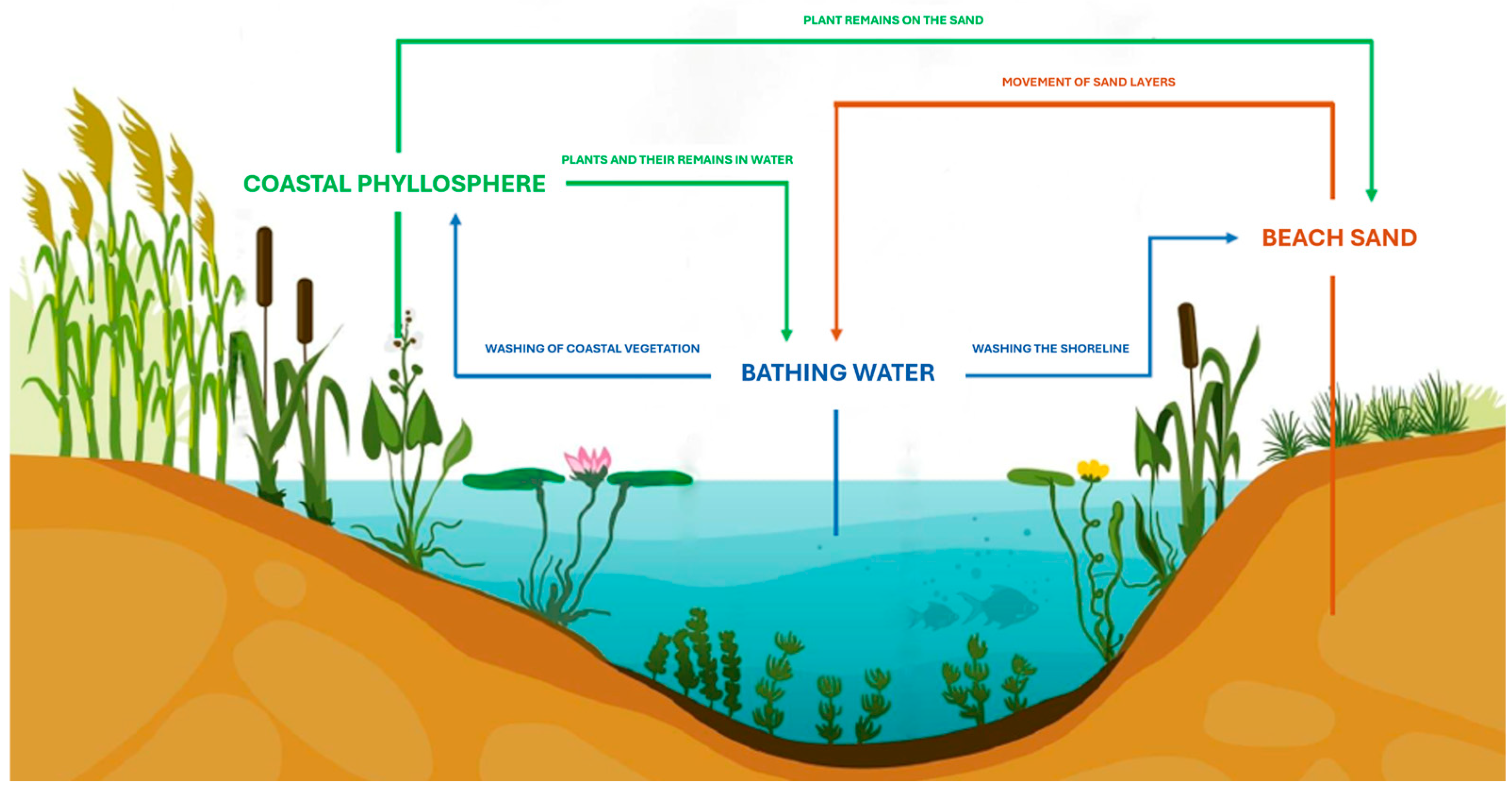
Figure 1. Seasonal circulation of yeasts in lake ecosystem [10].
Pathogens 14 00744 g001
Figure 2. Location of research sites against the background of the administrative map of Olsztyn (northeastern Poland) 1—Beach on Lake Kortowskie; 2—Beach on Lake Skanda; 3—Beach on Lake Skanda; 4—Beach on Lake Tyrsko; 5—Beach on Lake Ukiel; 6—Beach on Lake Ukiel (“Miejska”); 7—Beach on Lake Ukiel (“Omega”); 8—Beach on Lake Ukiel (“Słoneczna Polana”).
Pathogens 14 00744 g002
Figure 3. Most often isolated species (microcultures on Nickerson’s agar in moist chambers, incubated at 25 °C for 144 h) (A)—Barnettozyma californica 1000×; (B)—Nakasemomyces glabratatus 1000×; (C)—Cutaneotrichosporon jrovecii 1000×; (D)—Cutaneotrichosporon moniliiforme 1000×; (E)—Debaryomyces hansenii 1000×; (F)—Solicoccozyma aeria 1000×. Scale bar: 20 µm.
Pathogens 14 00744 g003
Figure 4. Average yeast concentration (CFU/g) in sand from monitored beaches across both research seasons (2019–2020), presented on a logarithmic scale. Error bars represent estimated variability (±10% of the measured values).
Pathogens 14 00744 g004
Table 1. List of species isolated in the study, including division, BSL and RG indicator values, and number of isolates obtained, categorized by research parameters.
No.SpeciesMIDVBSLRGSUMMEd10d50SUSs19s20LKLSLTLU
1Aureobasidium pullulans (de Bary & Löwenthal) G. Arnaud 1918+A115412314141031
2Barnettozyma californica (Lodder) Kurtzman, Robnett & Bas.-Powers 2008+A11127511184842019
3** Candida albicans (C.P. Robin) Berkhout 1923 A221101010010100
4Citeromyces matritensis (Santa María) Santa María 1957 A118538062262501
5Clavispora lusitaniae Rodr. Mir. 1979 (anamorfa: Candida lusitaniae) A221100110010001
6Cryptococcus amylolentus (Van der Walt, D.B. Scott & Klift) Golubev 1981 B114133104043010
7Cryptococcus uniguttulatus (Wolfram & Zach) Phaff & Fell 1970 B111101001010100
8Cutaneotrichosporon jirovecii (Frágner) Xin Zhan Liu, F.Y. Bai, M. Groenew. & Boekhout 2015+B2216881244121514615
9Cutaneotrichosporon moniliiforme (E. Guého & M.T. Sm.) Xin Zhan Liu, F.Y. Bai, M. Groenew. & Boekhout 2015+B2228181017116222538938
10Cyniclomyces guttulatus (C.P. Robin) Van der Walt & D.B. Scott 1971 A112200211020011
11** Debaryomyces hansenii (Zopf) Lodder & Kreger-van Rij 1984+A11247171014131151947211
12Dothiora sorbi (Wahlenb.) Fuckel 1870+A111010110010001
13Exophiala bergeri Haase & de Hoog 1999 A221010101010010
14Exophiala castellanii Iwatsu, Nishim. & Miyaji 1984 A222112002020020
15Exophiala jeanselmei (Langeron) McGinnis & A.A. Padhye 1977 A223300330030300
16** Geotrichum albidum (Lagerh.) H.Y. Zhu, X.Z. Liu & F.Y. Bai 2024 A113213021031200
17Geotrichum galactomycetum H.Y. Zhu, X.Z. Liu & F.Y. Bai 2024 A112111120020101
18Hanseniaspora osmophila (Niehaus) Phaff, M.W. Mill. & Shifrine 1956 A111101001100010
19Isabelozyma rhagii (Diddens & Lodder) Q.M. Wang, Yurkov, Boekhout & F.Y. Bai 2024 A111101001101000
20Komagataella pastoris (Guillierm.) Y. Yamada, M. Matsuda, K. Maeda & Mikata 1995 A113212121120102
21Kondoa malvinella (Fell & I.L. Hunter) Y. Yamada, Nakagawa & I. Banno 1989 B114222222220220
22Kregervanrija fluxuum (Phaff & E.P. Knapp) Kurtzman 2006 (anamorfa: Candida vini) A111101010010100
23Leucosporidium scottii Fell, Statzell, I.L. Hunter & Phaff 1970 B111011001010001
24Lipomyces lipofer (Den Dooren) Lodder & Kreger-van Rij 1952+A112202020020002
25Lodderomyces elongisporus (Recca & Mrak) Van der Walt 1971 A111011010010001
26Metschnikowia pulcherrima Pitt & M.W. Mill. 1968 A111101001100100
27Moniliella spathulata (de Hoog) C.A. Rosa & Lachance 2009 B112112002022000
28Mycogloea nipponica Bandoni 1998 B116244242061302
29** Nadsonia commutata Golubev 1973 A113301212210003
30Nadsonia fulvescens var. elongata (Konok.) Golubev & M.T. Sm. 1989 A115323205413200
31Naganishia albida (Saito) Xin Zhan Liu, F.Y. Bai, M. Groenew. & Boekhout 2015 B113123021120102
32** Nakaseomyces glabratus (H.W. Anderson) Sugita & M. Takash 2022 A2212668475390516
33Octosporomyces octosporus (Beij.) Kudryavtsev 1960 A111011001101000
34Ogataea angusta (Teun., H.H. Hall & Wick.) S.O. Suh & J.J. Zhou 2010 A112112002202000
35Ogataea minuta (Wick.) Y. Yamada, K. Maeda & Mikata 1994 A111011001010100
36Oosporidium margaritiferum Stautz 1931 A116513315062301
37Papiliotrema laurentii (Kuff.) Xin Zhan Liu, F.Y. Bai, M. Groenew. & Boekhout 2015 +B114313122040202
38Papiliotrema pseudoalba (Nakase & M. Suzuki) Xin Zhan Liu, F.Y. Bai, M. Groenew. & Boekhout 2015) B113300303033000
39** Pichia fermentans Lodder 1932 A111011010100001
40Pichia membranifaciens (E.C. Hansen) E.C. Hansen 1904 A116425124420501
41Pichia pseudolambica (M.T. Sm. & Poot) H.Y. Zhu, X.Z. Liu & F.Y. Bai 2024+A115413205140302
42Pichia terricola Van der Walt 1957 A111011010010001
43Rhodotorula diobovata (S.Y. Newell & I.L. Hunter) Q.M. Wang, F.Y. Bai, M. Groenew. & Boekhout 2015 B111101001011000
44Saccharomyces bayanus Sacc. 1895 A113120321031200
45Saccharomyces cerevisiae (Desm.) Meyen 1838 A115144114140311
46Saccharomyces mikatae G.I. Naumov, S.A. James, E.S. Naumova, E.J. Louis & I.N. Roberts 2000 A111100101010010
47** Saccharomycodes ludwigii (E.C. Hansen) E.C. Hansen 1904 A113213012121110
48Saitozyma podzolica (Babeva & Reshetova) Xin Zhan Liu, F.Y. Bai, M. Groenew. & Boekhout 2015+B113211221120102
49Schizosaccharomyces pombe Lindner 1893 A111101001100010
50Schwanniomyces capriottii M. Suzuki & Kurtzman 2010 +A115413223502012
51Schwanniomyces occidentalis Klöcker 1909 A112022002202000
52** Schwanniomyces polymorphus (Klöcker) M. Suzuki & Kurtzman 2010 A118445326350422
53Schwanniomyces vanrijiae (Van der Walt & Tscheuschner) M. Suzuki & Kurtzman 2010 A111011001101000
54Solicoccozyma aeria (Saito) Yurkov 2015+B11117410138387103
55Sporobolomyces xanthus (Nakase, G. Okada & Sugiy.) Boekhout 1991 B111101001010001
56Sydowia polyspora (Bref. & Tavel) E. Müll. 1953 A111011010010100
57Tausonia pullulans (Lindner) Xin Zhan Liu, F.Y. Bai, M. Groenew. & Boekhout 2015+B116245115602202
58Thelebolus globosus Brumm. & de Hoog 2005+A112200202022000
59Torulaspora globosa (Klöcker) Van der Walt & Johannsen 1975 A114224013220301
60Vanderwaltozyma polyspora (Van der Walt) Kurtzman 2003 A115323205321310
61Vanrija humicola (Dasz.) R.T. Moore 1980 B111010101101000
62Wickerhamomyces anomalus (E.C. Hansen) Kurtzman, Robnett & Bas.-Powers 2008 (anamorfa: Candida peliculosa) A111011010010001
63Bullera sp. B112111120020002
64* Cryptococcus sp.+B1/21/25323223322102
65Dipodascus sp. A111101001010001
66Kluyveromyces sp. A111010110100001
Sum * ---2641471171758910016411514963872787
No.SPECIESMIDVBSLRGSUMMEd10d50SUSs19s20LKLSLTLU
Number of isolates—how many times a given species was isolated during the study, and a pure culture was obtained on Sabouraud agar, and safely preserved for storage. e.g., Candida albicans—species classified to biosafety level 2 (BSL-2). ** e.g., Saccharomycodes ludwigii—species included in two-species isolates. e.g., * Cryptococcus sp.—BSL index value varies depending on species. SUM—total number of isolates of a given species. Sum *—total number of isolates (species of two-species isolates were counted separately and included in total sum of isolates). MI—“+” indicates that the species was confirmed using molecular identification methods (consistent with results obtained from phenotypic identification). DV—division (A—Ascomycota; B—Basidiomycota). BSL—Biosafety Level. RG—Risk Group. M—number of isolates obtained in the morning. E—number of isolates obtained in the evening. d10—number of isolates obtained from sand collected from a depth of 10 cm. d50—number of isolates obtained from sand collected from a depth of 50 cm. S—number of isolates obtained from sand of supervised beaches. US—number of isolates obtained from sand of unsupervised beaches. s19—number of isolates obtained in the 2019 research season. s20—number of isolates obtained in the 2020 research season. LK—number of isolates obtained from the beach of Lake Kortowskie. LS—number of isolates obtained from the beaches of Lake Skanda. LT—number of isolates obtained from the beach of Lake Tyrsko. LU—number of isolates obtained from the beach of Lake Ukiel.
Table 2. List of two-species isolates.
No.SpeciesResearch SeasonLake
1Nakaseomyces glabratus (H.W. Anderson) Sugita & M. Takash 2022 Pichia fermentans Lodder 19322019Ukiel
2Debaryomyces hansenii (Zopf) Lodder & Kreger-van Rij 1984
Nadsonia commutata Golubev 1973
2019Ukiel
3Debaryomyces hansenii (Zopf) Lodder & Kreger-van Rij 1984
Nadsonia commutata Golubev 1973
2019Ukiel
4Candida albicans (C.P. Robin) Berkhout 1923
Geotrichum albidum (Lagerh.) H.Y. Zhu, X.Z. Liu & F.Y. Bai 2024
2020Skanda
5Saccharomycodes ludwigii (E.C. Hansen) E.C. Hansen 1904
Schwanniomyces polymorphus (Klöcker) M. Suzuki & Kurtzman 2010
2020Tyrsko
Table 3. Statistical dependence of the number of isolates and strain concentration on meteorological parameters (Spearman rank correlation coefficient).
ParameterIsolates NumberAverage Strains Concentration
TemperatureIndependent of the parameter (R = 0.2991; p = 0.1355)* Depends on temperature
(R = 0.4339; p = 0.0210)
Air humidityIndependent of the parameter
(R = −0.0837; p = 0.6716)
Independent of the parameter
(R = −0.0766; p = 0.6984)
UV indexIndependent of the parameter (R = 0.1056; p = 0.5926)Independent of the parameter
(R = 0.3396; p = 0.0770)
* statistically significant results (p < 0.05).
Disclaimer/Publisher’s Note: The statements, opinions and data contained in all publications are solely those of the individual author(s) and contributor(s) and not of MDPI and/or the editor(s). MDPI and/or the editor(s) disclaim responsibility for any injury to people or property resulting from any ideas, methods, instructions or products referred to in the content.