Next Article in Journal
Outbreak Caused by VIM-1- and VIM-4-Positive Proteus mirabilis in a Hospital in Zagreb
Previous Article in Journal
Comprehensive Molecular and Epidemiological Characterization of Staphylococcus aureus Isolated from Bovine Mastitis in Water Buffalo of the Peshawar Division, Khyber Pakhtunkhwa, Pakistan
Previous Article in Special Issue
Antiprotozoal Effects of Pediococcus acidilactici-Derived Postbiotic on Blastocystis Subtypes ST1/ST3
 
 
pathogens-logo
Article Menu

Article Menu

Article

Trypanosoma cruzi Growth Is Impaired by Oleoresin and Leaf Hydroalcoholic Extract from Copaifera multijuga in Human Trophoblast and Placental Explants

by
Guilherme de Souza
1,†,
Clara Peleteiro Teixeira
1,†,
Joed Pires de Lima Júnior
1,
Marcos Paulo Oliveira Almeida
1,
Marina Paschoalino
1,
Luana Carvalho Luz
1,
Natália Carine Lima dos Santos
1,
Rafael Martins de Oliveira
1,
Izadora Santos Damasceno
1,
Matheus Carvalho Barbosa
1,
Guilherme Vieira Faria
1,
Maria Anita Lemos Vasconcelos Ambrosio
2,
Rodrigo Cassio Sola Veneziani
2,
Jairo Kenupp Bastos
3,
Angelica Oliveira Gomes
4,
Rosiane Nascimento Alves
5,
Carlos Henrique Gomes Martins
6,
Samuel Cota Teixeira
1,
Eloisa Amália Vieira Ferro
1 and
Bellisa Freitas Barbosa
1,*
1
Laboratory of Immunophysiology of Reproduction, Institute of Biomedical Science, Universidade Federal de Uberlândia, Uberlândia 38405-318, MG, Brazil
2
Nucleus of Research in Technological and Exact Sciences, Universidade de Franca, Franca 14404-600, SP, Brazil
3
School of Pharmaceutical Sciences of Ribeirão Preto, Universidade de São Paulo, Ribeirão Preto 14040-903, SP, Brazil
4
Institute of Natural and Biological Sciences, Universidade Federal do Triângulo Mineiro, Uberaba 38025-015, MG, Brazil
5
Department of Agricultural and Natural Science, Universidade do Estado de Minas Gerais, Ituiutaba 38302-192, MG, Brazil
6
Laboratory of Antimicrobial Testing, Institute of Biomedical Sciences, Universidade Federal de Uberlandia, Uberlandia 38405-318, MG, Brazil
*
Author to whom correspondence should be addressed.
These authors contributed equally to this work.
Pathogens 2025, 14(8), 736; https://doi.org/10.3390/pathogens14080736
Submission received: 16 June 2025 / Revised: 15 July 2025 / Accepted: 23 July 2025 / Published: 25 July 2025

Abstract

Congenital Chagas disease (CCD) is caused when Trypanosoma cruzi crosses the placental barrier during pregnancy and reaches the fetus, which can lead to serious consequences in the developing fetus. Current treatment is carried out with nifurtimox or benznidazole, but their effectiveness is limited, and they cause side effects, requiring the search for new therapeutic strategies. In this sense, many studies have demonstrated the potential of different compounds of the Copaifera genus in the control of parasitic diseases. Here, we aimed to evaluate the effect of oleoresin (OR) and leaf hydroalcoholic extract (LHE) of Copaifera multijuga on Trypanosoma cruzi infection in human villous trophoblast cells (BeWo line) and human placenta explants. Treatment with both compounds reduced invasion, proliferation, and release of trypomastigotes. Furthermore, OR and LHE affected the trypomastigotes and amastigote morphology, compromising their ability to invade and proliferate in BeWo cells, respectively. Also, treatment with OR decreased ROS production in infected BeWo cells, while LHE induced an increase. In addition, both compounds induced pro-inflammatory and anti-inflammatory cytokine production. In human placental explants, both compounds also decreased T. cruzi infection, in addition to inducing the production of pro-inflammatory cytokines. Thus, both OR and LHE of C. multijuga control T. cruzi infection at the human maternal–fetal interface, highlighting the possible therapeutic potential of these compounds for the treatment of CCD.

Graphical Abstract

1. Introduction

Chagas disease is caused by the protozoan parasite Trypanosoma cruzi, and it is estimated that around 6 to 7 million people worldwide are infected, especially in Latin America, where the disease is endemic [1]. Transmission can occur through blood-feeding insects from the subfamily Triatominae, but other routes are possible, such as organ transplant, blood transfusion, and ingestion of contaminated food or beverages, as well as vertical transmission from the mother to the fetus, characterizing the congenital Chagas disease (CCD) [2].
CCD has become prominent, especially in non-endemic countries, where this form is responsible for around 22.5% of cases [3,4]. The congenital infection rate is around 5%, with the risk of transmission higher in endemic areas (5%) compared to non-endemic countries (3%) [3,5]. Although most of the infected newborns are asymptomatic, some will present with low birth weight, prematurity, and reduced Apgar scores (analysis of respiration, heart rate, reflexes, muscle tone, and skin color). In more severe cases, miscarriage, hepatosplenomegaly, respiratory distress, anemia, myocarditis, and meningoencephalitis may occur. In addition, certain newborns may present with life-threatening symptoms early on and are at risk of developing serious chronic conditions later in life [6,7,8].
During pregnancy, the maternal immune system creates a tolerogenic profile that is important for fetal growth, characterized by an anti-inflammatory profile and the production of important cytokines, such as interleukin (IL)-4 and IL-10 [9], as well as the macrophage migration inhibitory factor (MIF), IL-6, and IL-8, which are pro-inflammatory cytokines that play an important role during pregnancy [10,11]. Failure to maintain this immune response can compromise the progression of pregnancy and cause deleterious effects to the fetus. It is known that T. cruzi infection in the placenta affects the immune response, with high production of IL-1β, IL-6, IL-8, IL-10, and tumor necrosis factor (TNF-α) [12]. It is worth mentioning that IL-1β, IL-6, and TNF-α are associated with cell proliferation and trophoblast differentiation [13,14], which are related to the trophoblast defense mechanisms, known as epithelial turnover, and this can explain the low rate of T. cruzi transmission through the placenta [15,16]. On the other hand, TNF-α secretion can lead to a disruption of the placental barrier, leading to fetal death [13]. In this sense, the maternal immune system has the challenge of maintaining an immunological balance for fetal development and, at the same time, dealing with possible factors that can cause an imbalance in the immune response, such as an infection by T. cruzi.
Over the last 60 years, two classical drugs have been preferentially used to treat CD: nifurtimox (Nfz, Lampit®,, Leverkusen, Germany) and benznidazole (BNZ, Rochagan®, and Roche, Basel, Switzerland). Although highly effective in the acute stage, both drugs have some shortcomings, such as adverse side effects and lengthy treatment, and their effectiveness is shortened during the chronic stage [17]. Although treating infected women of childbearing age with these drugs can prevent congenital transmission in future pregnancies [18], treatment during pregnancy remains contraindicated due to the lack of safety data related to the fetus. This underscores the importance of Chagas screening during pregnancy and early detection of infection [19,20]. These limitations highlight the need for new therapeutic drugs that are effective and safe [21].
In this context, many studies have demonstrated the potential of natural products as biologically competent compounds to treat diseases. As an example, we turned our attention to the genus Copaifera spp. (Fabaceae family), also known as “copaiba”, which has been widely used and studied since it has shown anti-inflammatory, anticancer, antioxidant, and antimicrobial potential, among other applications [22,23,24,25]. Previous studies showed that Copaifera spp. can control infection by different protozoans, such as Toxoplasma gondii [26,27], Leishmania spp. [28,29], Plasmodium spp. [30], and T. cruzi [31,32].
Taking into consideration its popular use, here, we aimed to show the potential of oleoresin (OR) and leaf hydroalcoholic extract (LHE) from Copaifera multijuga against T. cruzi infection at the human maternal–fetal interface, reinforcing the potential of this genus as a source of molecules that can be used to design new drugs.

2. Materials and Methods

2.1. Plant Material

Both oleoresin and leaf hydroalcoholic extract from C. multijuga were kindly provided by Professor Carlos Henrique Gomes Martins from the Department of Microbiology (DEMIC), Institute of Biomedical Sciences (ICBIM), Universidade Federal de Uberlândia (UFU), Uberlândia, MG, Brazil.
Authorization for scientific studies involving plant species from Brazilian biodiversity was obtained from the Council for Authorization and Information on Biodiversity (SIBIO/ICMBio/MMA/BRAZIL) and Genetic Heritage Management (CGEN/MMA/BRAZIL) under authorization numbers 35143-1 and 010225/2014-5, respectively.
Authentic oleoresin was collected in the northern region of Brazil, in Manacapuru, Amazonas state, by Jonas J. M. da Silva. The exudate was obtained by drilling tree trunks with a 2-inch drill bit and collected in glass bottles. All the plant material collected was identified by Silvane Tavares Rodrigues, a voucher specimen (NID 03/2013 and 62/2013) was deposited in the Herbarium of the Brazilian Agricultural Research Corporation (Embrapa Eastern Amazon), and identified by direct comparison with authentic herbarium vouchers, of which a taxonomic identity certificate is available upon request.
Air-dried leaves of C. multijuga (200 g), dried at 40 °C for 48 h, were ground and exhaustively extracted by maceration with 1.2 L of ethanol/water (7:3) at room temperature for 48 h. After lyophilization, this yielded 50 g of hydroalcoholic extract of leaves [33].

2.2. Cell Culture and Parasite Maintenance

Human villous trophoblast cells (BeWo cell line) were obtained from the American Type Culture Collection (ATCC, Manassas, VA, USA). Cells were cultured in RPMI 1640 medium (Cultilab, Campinas, SP, Brazil) supplemented with 100 U/mL penicillin, 100 µg/mL streptomycin (Sigma Chemical Co., St Louis, MO, USA), and 10% fetal bovine serum (FBS) (Cultilab) in a humidified incubator at 37 °C with 5% CO2. Although BeWo cells are not of immune origin, several studies have demonstrated their ability to produce and secrete cytokines, such as IL-4 [34], IL-6 [35], IL-8 [35], IL-10 [36], and MIF [25], under various physiological and pathological conditions.
Tissue culture-derived trypomastigotes of T. cruzi (TCT, Y strain) were propagated in VERO cells in RPMI 1640 medium supplemented with 100 U/mL penicillin, 100 µg/mL streptomycin, and 2% FBS at 37 °C with 5% CO2.

2.3. Human Placenta Explants

Full-term human placentas (36 to 40 weeks of gestation) (n = 4) were collected from pregnant patients following elective cesarean sections at the Clinics Hospital of the Universidade Federal de Uberlândia (HC-UFU), MG, Brazil. Exclusion criteria included preeclampsia, hypertension, infectious disease, such as Chagas disease and toxoplasmosis, chronic renal disease, cardiac disease, connective tissue disease, diabetes, and other diseases that might affect the study outcomes. After collection, placenta tissues were washed in sterile phosphate-buffered saline (PBS) within 1 h to remove excess blood. Floating terminal chorionic villi, each containing five to seven free tips, were isolated, washed with 1x PBS, and placed individually into 96-well plates with 200 µL of RPMI 1640 medium supplemented with 5% FBS, penicillin (100 U/mL), and streptomycin (100 µg/mL) overnight at 37 °C with 5% CO2 [35]. Written informed consent was obtained from all participants included in this study, which had been previously approved by the Ethics Committee of Universidade Federal de Uberlândia, MG, Brazil, with approval number 7.407.162.

2.4. Cell Viability Assay

BeWo cell viability after treatment with oleoresin (OR) and leaf hydroalcoholic extract (LHE) was carried out by MTT assay [(3-(4,5-dimethylthiazol-2-yl)-2,5-diphenyltertrazolin bromide)], as described by [37].
BeWo cells (1 × 10⁴/100 µL) were seeded in 96-well plates in 5% FBS medium overnight at 37 °C with 5% CO2. Then, the cells were treated for 72 h with increasing concentration of OR and LHE from C. multijuga (ranging from 4 to 256 µg/mL) [25,26], 50 µM benznidazole (BZN) (LAFEPE, Recife, PE, Brazil) [32], 0.38% DMSO (vehicle present in the highest concentration of both compounds), and 5% FBS medium (negative control).
The cells were then incubated with MTT (5 mg/mL) diluted in a 10% FBS medium for 4 h at 37 °C with 5% CO2. After incubation, the resulting formazan crystals were dissolved in 10% sodium dodecyl sulfate (SDS, Sigma) and 50% N, N-dimethylformamide (DMF) (Sigma). Absorbance was measured at 570 nm using a microplate spectrophotometer (VersaMax ELISA Microplate Reader, Molecular Devices, Sunnyvale, CA, USA). Cell viability was expressed as a percentage of viable cells (cell viability %) by comparing the treatments with the cells treated only with 5% FBS medium (medium, 100% viability). Three independent experiments with eight replicates were carried out.

2.5. Trypomastigote Viability

T. cruzi trypomastigote viability treated with OR and LHE was also carried out by MTT assay, as previously described with minor modifications.
T. cruzi trypomastigotes (1 × 10⁶/50 μL) were seeded in 96-well plates in 5% FBS medium and immediately treated for 72 h with OR and LHE from C. multijuga, 50 µM benznidazole, and 5% FBS medium (negative control).
Afterwards, the TCTs were incubated with MTT (5 mg/mL) for 4 h at 37 °C with 5% CO2 and processed, as described previously. Trypomastigote viability was expressed as a percentage of viable parasites (trypomastigote viability%) by comparing the treatments with the parasites treated only with 5% FBS medium (medium, 100% viability). Three independent experiments with eight replicates were carried out.

2.6. T. cruzi Intracellular Proliferation

Intracellular proliferation of T. cruzi in BeWo cells treated with OR and LHE was assessed by counting intracellular amastigotes under a light microscope.
BeWo cells (4 × 10⁴/500 µL) were seeded onto 13 mm glass coverslips placed in 24-well plates in 5% FBS medium overnight at 37 °C with 5% CO2. The cells were infected with TCTY at a multiplicity of infection (MOI) of 1.3:1 (parasite/cell) for 3 h. After infection, cells were washed three times with 1x PBS to remove non-internalized parasites and then treated for 72 h with non-cytotoxic concentrations of OR (16 and 32 µg/mL), LHE (32 and 64 µg/mL), BZN (50 µM), and 5% FBS medium (control).
After treatment, the cells were washed with 1x PBS, fixed with pure methanol (Dinamica Química, São Paulo, SP, Brazil), and stained with Giemsa (Neon, Suzano, SP, Brazil) diluted in distilled water at a ratio of 1:20 for 20 min. Cells were then washed several times with 1x PBS and examined under a light microscope (Opton Microscopios, São Paulo, SP, Brazil). The infection rate was determined by counting the number of infected cells out of 200 randomly selected cells, and intracellular proliferation was assessed by counting the number of amastigotes within infected cells [32]. Three independent experiments, with three replicates, were carried out. The MOI chosen for this set of experiments (1.3:1) was used to avoid overinfection of host cells, which could interfere with accurate quantification.

2.7. Trypomastigote Release Assay

The trypomastigotes released in the supernatant were analyzed, as described by Kian and colleagues [38] with minor modifications.
BeWo cells (4 × 10⁴/500 µL) were seeded in 24-well plates in 5% FBS medium overnight at 37 °C with 5% CO2. The cells were infected with TCTY (MOI 5:1; parasite/cell) for 3 h. Next, the cells were washed three times with 1x PBS to remove non-internalized parasites and then treated for 72 h with non-cytotoxic concentrations of OR (16 and 32 µg/mL), LHE (32 and 64 µg/mL), BZN (50 µM), and 5% FBS medium (control).
Afterwards, the supernatant was collected and analyzed for the number of trypomastigotes released, while 5% FBS medium was added to the same plate to analyze the release of trypomastigotes on the following three days (96, 120, and 144 h), with the media being replaced every 24 h. The supernatants were centrifuged at 4000 rpm for 10 min and resuspended in 20 µL of medium, and then, the trypomastigotes were counted under a light microscope using a Neubauer chamber (Kasvi, Pinhais, PR, Brazil). Three independent experiments, with three replicates, were carried out. The MOI selected for this set of experiments (5:1) was standardized by us, as it ensures sufficient trypomastigote release at different time points, allowing reliable quantification without overwhelming the counting method.

2.8. Invasion and Proliferation of T. cruzi Pretreated with OR and LHE

The effect of parasite pretreatment with OR and LHE on its invasion and proliferation was also carried out by counting the intracellular amastigotes under a light microscope.
BeWo cells (4 × 10⁴/500 µL) were seeded onto 13 mm glass coverslips placed in 24-well plates in 5% FBS medium overnight at 37 °C with 5% CO2. The cells were infected for 3 h with TCTY (MOI 10:1 and 1.3:1 parasites/cell for invasion and proliferation experiments, respectively) that were previously pretreated for 1 h with OR (16 and 32 µg/mL), LHE (32 and 64 µg/mL), BZN (50 µM), or 5% FBS medium (control). Two different experimental protocols were carried out: (I) Invasion: after 3 h, the cells were washed three times with 1x PBS to remove non-internalized parasites and then processed to Giemsa stain; (II) Proliferation: after 3 h, the cells were washed three times with 1x PBS to remove non-internalized parasites, and the cells were treated with 5% FBS medium for additional 72 h and then processed to Giemsa stain, as previously described.
For both situations (invasion and proliferation), the cells were examined under a light microscope (Opton Microscopios, SP, Brazil) to determine the infection rate (number of infected cells/200 cells counted randomly) and the T. cruzi intracellular proliferation (number of amastigotes observed in infected cells) [32]. Two independent experiments, with four replicates, were carried out. For the invasion assay, a MOI of 10:1 was used to ensure a sufficient number of parasites to infect the host cells within a short incubation period.

2.9. Reactive Oxygen Species (ROS) Production

ROS production was carried out using a 2′,7′-dichlorodihydrofluorescein diacetate probe (H2DCF-DA, Invitrogen, catalog number: D399) to form the highly fluorescent compound 2′,7′-dichlorofluorescein (DCF).
BeWo cells (1 × 10⁴/100 µL) were seeded in a 96-well black clear-bottom plate in 5% FBS medium overnight at 37 °C with 5% CO2. The cells were infected with TCTY (MOI 1.3:1; parasite/cell) for 3 h. Next, the cells were washed three times with 1x PBS to remove non-internalized parasites and then treated or not for 72 h with non-cytotoxic concentrations of OR (16 and 32 µg/mL), LHE (32 and 64 µg/mL), and BZN (50 µM). As controls, the cells were uninfected or infected and treated with 5% FBS medium.
Afterwards, the supernatants were collected and stored at −80 °C for cytokine analysis. The cells were then washed with 1x PBS and incubated with the H2DCF-DA probe (10 µM diluted in 1x PBS with 10% FBS) for 45 min at 37 °C with 5% CO2, protected from light. Following incubation, the probe was removed, and the cells were washed with 1x PBS and incubated with PBS with 10% FBS for fluorescence reading. The mean fluorescence intensity of DCF was measured using a GloMax Explorer microplate reader (Promega, Madison, WI, USA) at excitation and emission wavelengths of 488 nm and 522 nm, respectively. Three independent experiments with eight replicates were carried out.

2.10. Morphological Analysis of Trypomastigotes and Amastigotes

The trypomastigote morphology was analyzed by scanning electron microscopy (SEM) [32]. T. cruzi trypomastigotes (1 × 10⁷/1000 µL) were treated with OR (32 μg/mL) or LHE (64 μg/mL), BZN (50 μM), or 5% FBS for 1 h. Next, the parasites were centrifuged at 4000 rpm for 10 min, washed 1x with sodium cacodylate buffer, and fixed in Karnovsky’s solution containing 2% paraformaldehyde and 2.5% glutaraldehyde in 0.1 M sodium cacodylate buffer for 2 h. Then, the parasites were washed with sodium cacodylate buffer and incubated with 1% osmium tetroxide (OsO4) for 1 h. Afterwards, the parasites were washed twice with sodium cacodylate buffer, and 10 μL of parasite was added to circular coverslips and left overnight at room temperature to dry. Subsequently, the coverslips with parasites were dehydrated in increasing concentrations of ethanol (50, 70, 80, 90, 95, and 100%, 5 min each), and a thin layer of gold was applied to the samples and then analyzed under a scanning electron microscope (Tescan, VEJA 3 LMU, TESCAN Orsay Holding, Brno, Czech Republic). One experiment with three replicates was carried out, and around 10–15 pictures were taken from each condition.
Also, we analyze the amastigote morphology by transmission electron microscopy (TEM). BeWo cells (1 × 10⁶/500 µL) were seeded in 24-well plates in 5% FBS medium overnight at 37 °C with 5% CO2. The cells were infected with TCTY (MOI 1.3:1; parasite/cell) for 3 h. Next, the cells were washed three times with 1x PBS to remove non-internalized parasites and then treated for 72 h with OR (16 and 32 µg/mL), LHE (32 and 64 µg/mL), BZN (50 µM), and 5% FBS medium (control). After treatment, the cells were collected, centrifuged at 1500 rpm for 5 min, and fixed in Karnovsky’s solution containing 2% paraformaldehyde and 2.5% glutaraldehyde in 0.1 M sodium cacodylate buffer for 24 h. Next, the cells were incubated with OsO4 for 1 h and treated with potassium ferrocyanide for 30 min, dehydrated in increasing concentrations of ethanol (50, 70, 80, 90, 95, and 100%) and 100% propylene oxide, and then embedded in Epon resin. Ultrathin sections obtained were stained with 3% uranyl acetate and 1% lead citrate and then analyzed using a transmission electron microscope (Hitachi HT7700, Hitachi High-Tech Corporation, Tokyo, Japan). One experiment, with three replicates, was carried out.

2.11. Viability of Human Placenta Explants Treated with OR and LHE

The viability of human placenta explants treated with OR and LHE from C. multijuga was evaluated by MTT assay and histological analysis [25].
Human placenta explants were prepared as previously described and cultured in 96-well plates in 5% FBS medium overnight at 37 °C with 5% CO2. Next, the villous explants were treated for 72 h with increasing concentrations of OR and LHE from C. multijuga (ranging from 4 to 256 µg/mL), 50 µM benznidazole (BZN), and 5% FBS medium (negative control). After treatment, the supernatants were collected and stored at −80 °C for later cytokine analysis, and the villous explants were submitted to the MTT assay, as previously described. Villous viability was expressed as the percentage viability of villous explants in relation to explants treated only with medium (medium, 100% viability). Additionally, treated and untreated villi were collected for morphological analysis using 5% hematoxylin and 0.1% eosin staining [25,32]. Two independent experiments, with eight replicates, were carried out.

2.12. T. cruzi Proliferation in Human Placenta Explants Treated with OR and LHE

T. cruzi proliferation in human placenta explants treated with OR and LHE was evaluated by quantitative real-time PCR (qPCR) [32].
The villous explants were cultured in 96-well plates overnight in 10% FBS medium at 37 °C with 5% CO2. Next, the villi were infected with TCTY (1 × 10⁵/villi) for 24 h. Then, the villi were treated for 72 h with OR and LHE (64 and 128 µg/mL), BZN (50 µM), and 5% FBS medium (control). After treatment, the supernatants were collected for further cytokine measurements, while the villi were stored at −80 °C until quantification of parasite load by qPCR. It is important to highlight that parasite load in placental tissue was assessed after four days of infection, in contrast to the three-day timeline used for the cell model. This difference is because infection takes longer in placental tissue than in cells because the villi consist of multiple layers, including cytotrophoblasts, syncytiotrophoblasts, mesenchyme, and blood vessels. This placental barrier makes it harder and slower for the parasite to infect the placenta compared to the quicker process in the in vitro cell model.
The DNA from villi was extracted using a DNA purification kit (Promega, São Paulo, SP, Brazil) and proteinase K (20 mg/mL). DNA quantification was performed using a NanoDrop spectrophotometer (Thermo Fisher Scientific, Wilmington, DE, USA) by measuring absorbances at wavelengths of 260 and 280 nm. DNA purity was evaluated by the ratio of absorbance readings at 260/280 nm, with values between 1.8 and 2.0 considered acceptable. For the qPCR reaction, the amount of genetic material was normalized to 100 ng of tissue DNA per reaction, and the GoTaq® Master Mix kit (Promega) was used according to the manufacturer’s instructions. The primers used for DNA amplification were as follows: forward (5′-CGCAAACAGATATTGACAGAG-3′) and reverse (5′-TGTTCACACACTGGACACCAA-3′) [39]. Quantification of each sample was based on a standard curve prepared from serial two-fold dilutions starting at 100 ng of DNA, comprising six points. The results are expressed as TCTY equivalent to 100 ng of DNA tissue. Two independent experiments, with eight replicates, were carried out.

2.13. Cytokine Measurements

Human cytokines IL-4, IL-6, IL-8, IL-10, MIF, and TNF-α were quantified in the supernatants of BeWo cells and villous explants under different experimental conditions using a double-antibody sandwich enzyme-linked immunosorbent assay (ELISA), following the manufacturers’ protocol (BD Biosciences, San Jose, CA, USA or R&D Systems, Minneapolis, MN, USA). Cytokine levels in BeWo cells supernatants were expressed in pg/mL, whereas the villi values were normalized by the ratio between the concentration of cytokines (pg/mL) and the weight of each villous (mg), resulting in pg/mL/mg. The detection limits of each cytokine were as follows: IL-4, IL-10, TNF-α (7.8 pg/mL), IL-6 (4.7 pg/mL), IL-8 (3.12 pg/mL), and MIF (31.2 pg/mL).

2.14. Statistical Analysis

Statistical analysis was performed using GraphPad Prism Software version 8.0 (GraphPad Software Inc., San Diego, CA, USA, https://www.graphpad.com, Accessed on 15 June 2025). Data are presented as mean ± standard error of mean (SEM). Differences between groups were assessed by One-Way ANOVA test with Bonferroni’s multiple comparison post-test for the parametric data or Kruskal–Wallis’s test with Dunn’s multiple comparison post-test for the nonparametric data. A p-value of  <0.05 was considered statistically significant.

3. Results

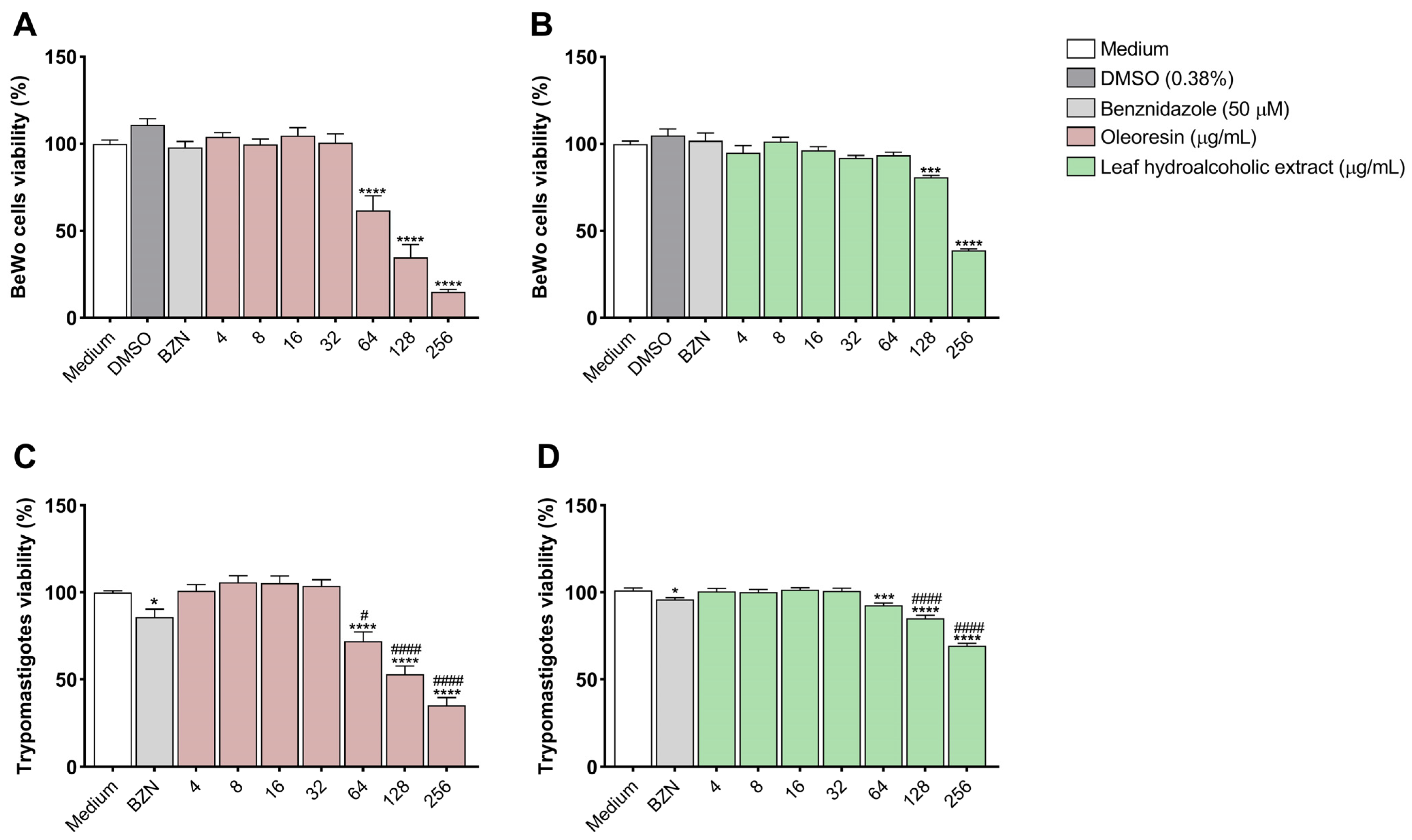
3.1. Oleoresin and Leaf Hydroalcoholic Extract from C. multijuga Are Toxic to BeWo Cells and T. cruzi Trypomastigotes Only at Highest Concentrations

An MTT assay was performed to evaluate the effect of oleoresin and the leaf hydroalcoholic extract from C. multijuga on both BeWo cells and T. cruzi trypomastigote viability.
In BeWo cells, we observed that treatment with OR was cytotoxic at concentrations of 64, 128, and 256 µg/mL when compared to cells treated with medium (****p < 0.0001) (Figure 1A) with CC50 = 91.73 µg/mL, while LHE was cytotoxic only at the concentrations of 128 (***p = 0.0002) and 256 µg/mL (****p < 0.0001) compared to cells treated with medium (Figure 1B), presenting CC50 = 221.93 µg/mL.
In trypomastigotes, we observed that treatment with BZN (*p = 0.0179), OR at concentrations of 64, 128, and 256 µg/mL (****p < 0.0001) (Figure 1C), as well as LHE at 64 (***p = 0.0009) and 128 and 256 µg/mL (****p < 0.0001), were also cytotoxic compared to trypomastigotes treated with medium (Figure 1D). Interestingly, both OR and LHE showed higher cytotoxicity in trypomastigotes compared to BZN (#p < 0.05). OR showed CC50 = 150.3 µg/mL, while LHE had a CC50 > 256 µg/mL.
Based on these results, we chose two non-cytotoxic concentrations of each compound for BeWo cells to perform the next experiments. For oleoresin, 16 and 32 µg/mL were chosen, while 32 and 64 µg/mL were chosen for the leaf hydroalcoholic extract.

3.2. Oleoresin and Leaf Hydroalcoholic Extract from C. multijuga Decrease T. cruzi Intracellular Proliferation in BeWo Cells

To evaluate the impact of oleoresin and leaf hydroalcoholic extract on T. cruzi proliferation, we counted the number of infected cells and intracellular amastigotes.
We observed that treatment with OR at concentrations of 16 (*p = 0.0262) and 32 μg/mL (*p = 0.0473), as well as LHE at concentrations of 32 (*p = 0.0343) and 64 μg/mL (**p = 0.0031) decreased the number of T. cruzi-infected cells compared to infected and untreated cells (infected medium) (Figure 2A). In addition, we observed that treatment with OR at concentrations of 16 (*p = 0.0185) and 32 μg/mL (**p = 0.0036), as well as LHE at concentrations of 32 (**p = 0.0038) and 64 μg/mL (***p = 0.0003), in addition to BZN (***p = 0.0008), decreased the number of amastigotes in infected cells compared to infected and untreated cells (infected medium) (Figure 2B).
Representative photomicrographs of BeWo cells (Figure 2C–H) are shown, where it is possible to observe several amastigotes (arrows) in the cytoplasm of infected and untreated BeWo cells (C) compared to cells infected and treated with BZN (D), OR 16 (E) and 32 μg/mL (F), and LHE 32 (G) and 64 μg/mL (H).

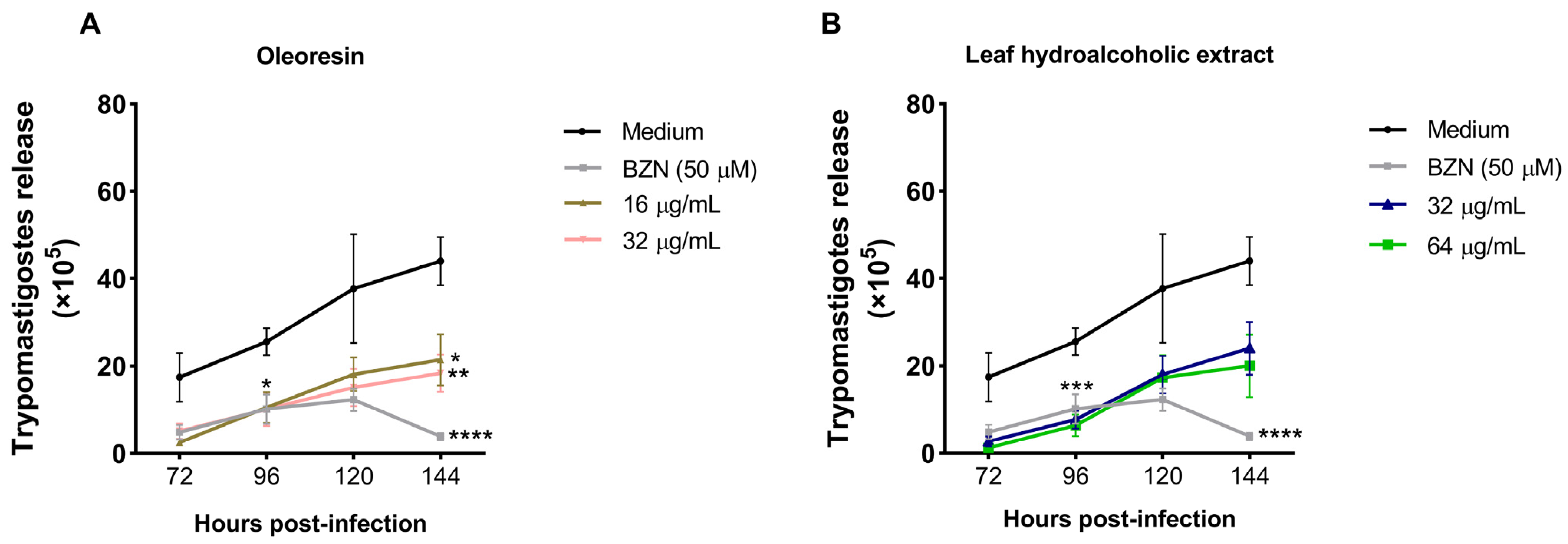
3.3. Oleoresin and Leaf Hydroalcoholic Extract from C. multijuga Decrease the Release of Trypomastigotes into the Supernatant of BeWo Cells

Since OR and LHE were affecting parasite proliferation, we also evaluated the effect of both compounds on trypomastigote release in the supernatant at different time points.
Regarding OR, we observed that both concentrations and BZN were able to decrease the release of trypomastigotes at all times compared to infected and untreated cells (medium), being statistically different at 96 (BZN: *p = 0.0139; 16 μg/mL: *p = 0.0166; 32 μg/mL: *p = 0.0157) and 144 h (BZN: ***p = 0.0004; 16 μg/mL: *p = 0.0388; 32 μg/mL: **p = 0.0072) (Figure 3A).
Concerning LHE, we observed that both concentrations also decreased the release of trypomastigotes at all times compared to infected and untreated cells (medium), being statistically different at 96 h (32 μg/mL: **p = 0.0036; 64 μg/mL: ***p = 0.0002), while BZN decreased at 96 (***p = 0.0005) and 144 h (***p = 0.0003) (Figure 3B).

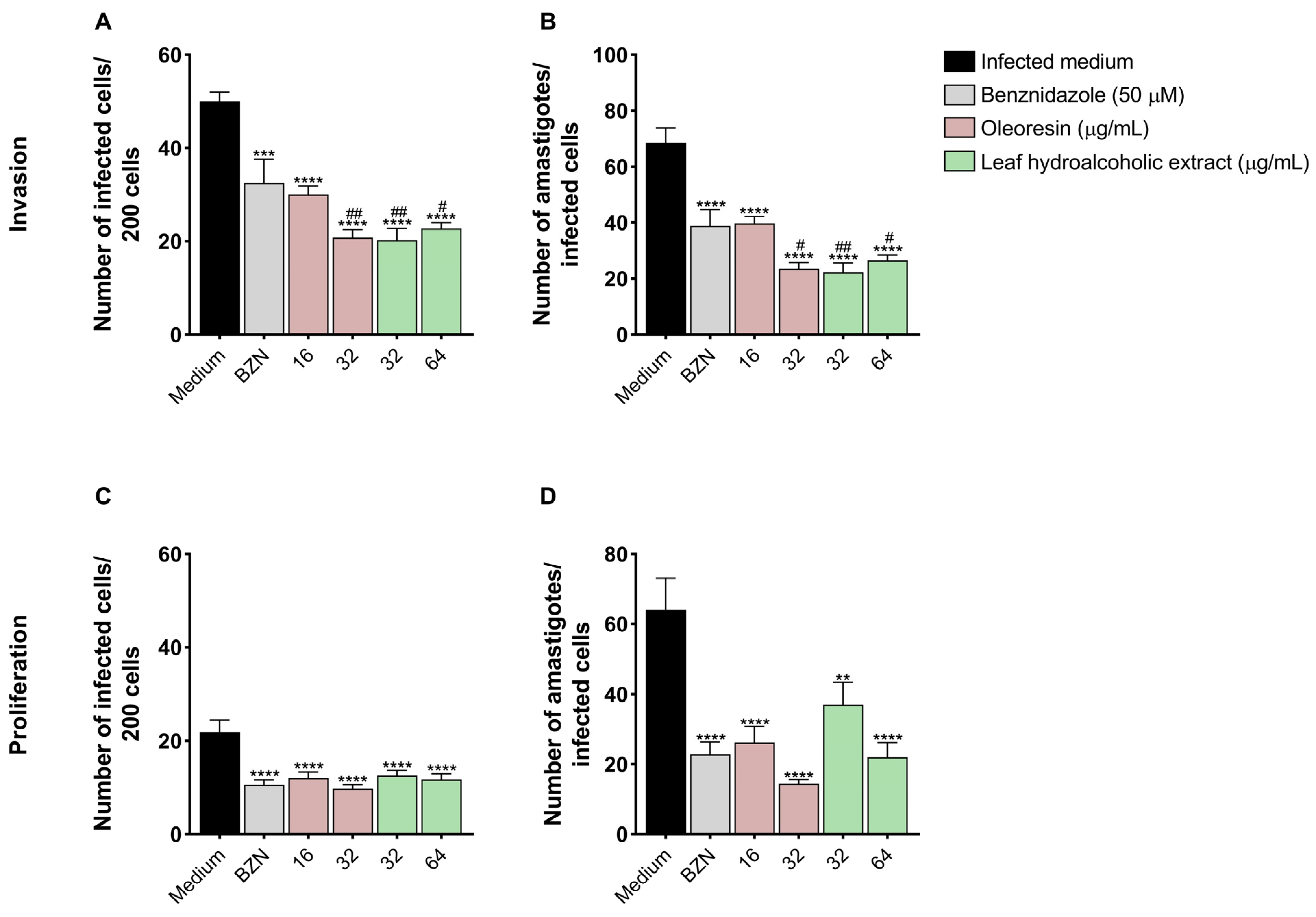
3.4. Pretreatment of T. cruzi Trypomastigotes with Oleoresin and Leaf Hydroalcoholic Extract Compromises Its Ability to Invade and Proliferate in BeWo Cells

To evaluate whether pretreatment with both compounds had a direct effect on T. cruzi trypomastigotes, we performed an invasion and proliferation assay.
In the invasion assay, we observed that pretreatment of the parasites with both concentrations of OR (****p < 0.0001) and LHE (****p < 0.0001), as well as BZN (***p = 0.0002), decreased the number of cells infected by T. cruzi compared to untreated parasites (medium), and both OR 32 μg/mL (##p < 0.0068) and LHE (32 μg/mL: ##p = 0.0002; 64 μg/mL: #p < 0.0001) reduced more compared to BZN (Figure 4A). Also, both concentrations of OR and LHE, in addition to BZN, decreased the number of amastigotes compared to cells infected with untreated parasites (medium) (****p < 0.0001) and OR 32 μg/mL (#p < 0.0119) and LHE (32 μg/mL: ##p = 0.0073; 64 μg/mL: #p < 0.0375) (Figure 4B).
Regarding the proliferation assay, we observed that pretreatment of parasites with both concentrations of OR (16 μg/mL: ***p = 0.0002; 32 μg/mL: ****p < 0.0001), LHE (****p < 0.0001), and BZN (****p < 0.0001) decreased the number of cells infected by T. cruzi compared to untreated parasites (medium) (Figure 4C). Furthermore, both concentrations of OR and LHE, as well as BZN, also decreased the number of amastigotes compared to cells infected with untreated parasites (medium) (****p < 0.0001) (Figure 4D).

3.5. Oleoresin and Leaf Hydroalcoholic Extract from C. multijuga Affect Both Trypomastigote and Amastigote Morphology

Since the invasion and proliferation of the parasite were being decreased by both compounds, we analyzed whether OR and LHE were affecting the trypomastigote and amastigote forms’ morphology by scanning electron microscopy (SEM) and transmission electron microscopy (TEM), respectively.
Regarding the trypomastigote forms (Figure 5A–D), untreated parasites (A) displayed normal morphology, while BZN (B), OR (C), and LHE (D) treatment induced morphological alterations, such as swelling of the parasite body, rounding of the cell surface, shortening of the flagellum, and cellular torsion.
As for the amastigote forms (Figure 5E–H), untreated T. cruzi amastigotes (E) exhibited typical morphology, with an intact plasma membrane, flagellum (f), nucleus (n), and kinetoplast (k). In contrast, treatment with BZN (F) caused cytoplasmic vacuolization (indicated by a star), while both OR (G) and LHE (H) induced ultrastructural changes, such as membrane swelling (thick arrow), cytoplasmic disorganization (arrowhead), and additional vacuolization (star).

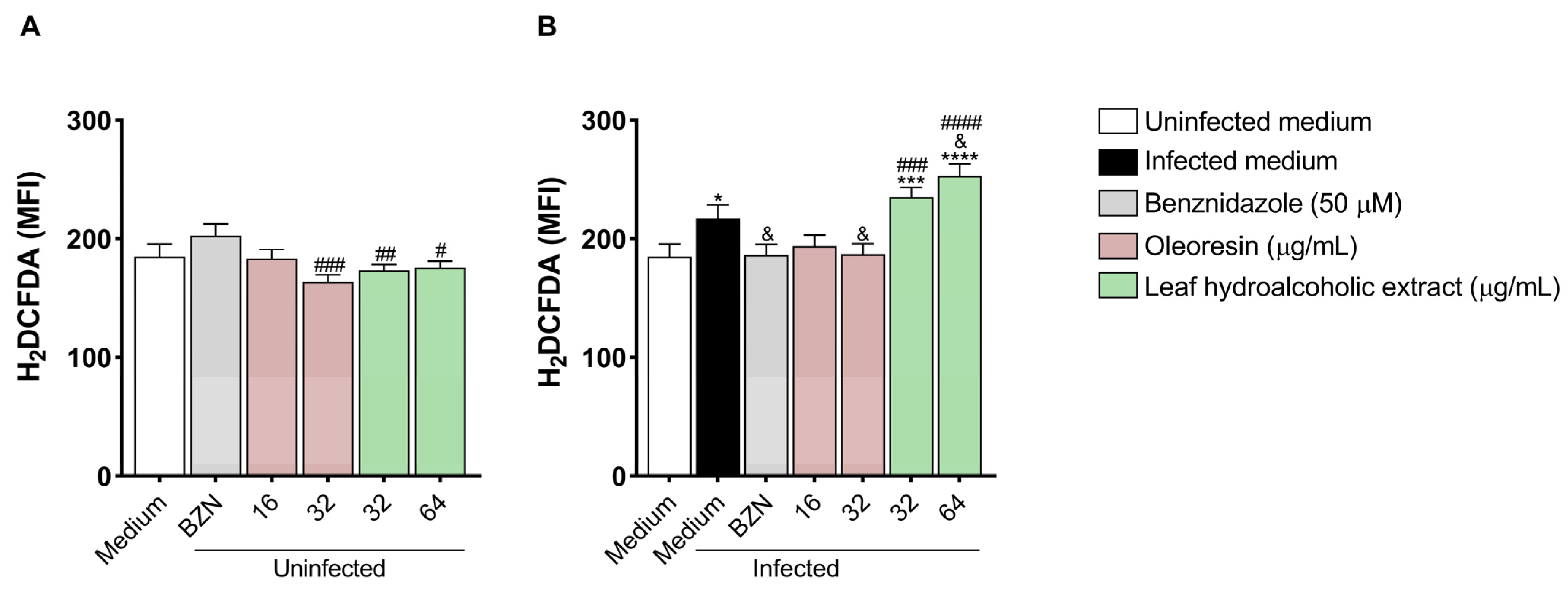
3.6. Oleoresin and Leaf Hydroalcoholic Extract from C. multijuga Modulate Reactive Oxygen Species in BeWo Cells

After observing that both compounds reduced T. cruzi proliferation and affected parasite morphology, we tested whether both OR and LHE modulate ROS production in infected and uninfected BeWo cells.
In uninfected cells, we observed that the concentration of 32 μg/mL of OR (###p = 0.0004) and both concentrations of LHE (32 μg/mL: ##p = 0.0078; 64 μg/mL: #p = 0.0133) induced lower ROS production compared to cells treated with BZN (Figure 6A).
In the presence of infection, BeWo cells infected and untreated (infected medium, *p = 0.0232) or infected and treated with both concentrations of LHE (32 μg/mL: ***p = 0.0006; 64 μg/mL: ****p < 0.0001) increased ROS production compared to uninfected and untreated cells (uninfected medium) (Figure 6B). In addition, cells infected and treated with BZN (&p = 0.0258) and OR (32 μg/mL) (&p = 0.0276) decreased ROS production compared to cells infected and untreated (infected medium), whereas LHE (64 μg/mL) increased (&p = 0.0125) (Figure 6B). Furthermore, both concentrations of LHE (32 μg/mL: ###p = 0.0006; 64 μg/mL: ####p < 0.0001) had a higher ROS production compared to cells infected and treated with BZN (Figure 6B).

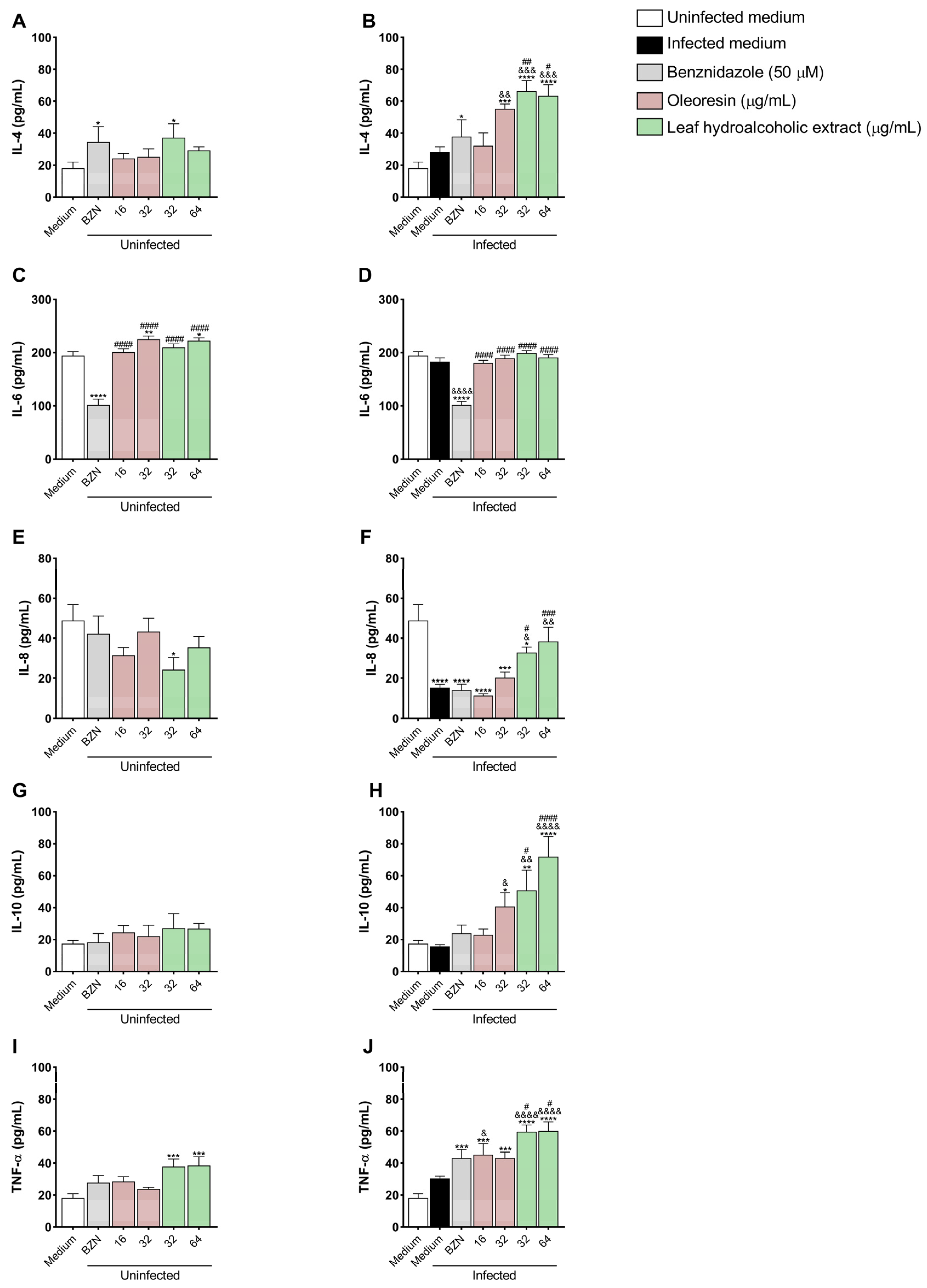
3.7. Oleoresin and Leaf Hydroalcoholic Extract from C. multijuga Increase IL-4, IL-8, IL-10, and TNF-α Production in BeWo Cells

We also tested whether OR and LHE would interfere with the host immune response through cytokine production.
IL-4: Uninfected cells treated with BZN (*p = 0.0432) and LHE at 32 μg/mL (*p = 0.0133) (Figure 7A), as well as infected cells treated with BZN (*p = 0.0448), OR at 32 μg/mL (***p = 0.0001), and both concentrations of LHE (****p < 0.0001) (Figure 7B) showed increased IL-4 production compared to uninfected untreated cells (uninfected medium). Infected cells treated with OR at 32 μg/mL (&&p = 0.0077), and both concentrations of LHE (32 μg/mL: &&&p = 0.0003; 64 μg/mL: &&&p = 0.0007) exhibited increased IL-4 levels compared to infected untreated cells (infected medium) (Figure 7B). In addition, both LHE concentrations (32 μg/mL: ##p = 0.0051; 64 μg/mL: #p = 0.0109) induced more IL-4 production compared to infected cells treated with BZN (Figure 7B).
IL-6: Uninfected cells treated with BZN (****p < 0.0001) showed decreased IL-6 levels, while OR at 32 μg/mL (**p = 0.0059) and LHE at 64 μg/mL (*p = 0.0109) showed increased IL-6 production compared to uninfected untreated cells (uninfected medium) (Figure 7C). In addition, both OR and LHE (####p < 0.0001) induced more IL-6 compared to those treated with BZN (Figure 7C). Infected cells treated with BZN showed decreased IL-6 production compared to uninfected untreated cells (uninfected medium) (****p < 0.0001) and infected untreated cells (infected medium) (&&&&p < 0.0001). In addition, infected cells treated either with OR or LHE induced more IL-6 (####p < 0.0001) compared to those infected cells treated with BZN (Figure 7D).
IL-8: In the absence of the parasite, only 32 μg/mL of LHE (*p = 0.0165) showed decreased IL-8 production compared to uninfected untreated cells (uninfected medium) (Figure 7E). Also, infected untreated cells (****p < 0.0001), or treated with BZN (****p < 0.0001), OR (16 μg/mL: ****p < 0.0001; 32 μg/mL: ***p = 0.0001) or LHE at 32 μg/mL (*p = 0.0389) exhibited decreased IL-8 levels compared to uninfected untreated cells (uninfected medium) (Figure 7F). In addition, both concentrations of LHE induced more IL-8 compared to only infected cells (32 μg/mL: &p = 0.0263; 64 μg/mL: &&p = 0.0011), as well as those infected and treated with BZN (32 μg/mL: #p = 0.0148; 64 μg/mL: ###p = 0.0004) (Figure 7F).
IL-10: Infected cells treated with OR at 32 μg/mL (*p = 0.0165) and both concentrations of LHE (32 μg/mL: **p = 0.003; 64 μg/mL: ****p < 0.0001) induced more IL-10 compared to uninfected untreated cells (uninfected medium), as well as infected untreated cells (infected medium) (OR 32 μg/mL: &p = 0.0107; LHE 32 μg/mL: &&p = 0.0026; LHE 64 μg/mL: &&&&p < 0.0001) (Figure 7H). In addition, LHE induced more IL-10 compared to those infected cells treated with BZN (32 μg/mL: #p = 0.0150; 64 μg/mL: ####p < 0.0004) (Figure 7H).
TNF-α: Uninfected cells treated with LHE (32 μg/mL: ***p = 0.001; 64 μg/mL: ***p = 0.0007) showed increased TNF-α levels compared to uninfected untreated cells (uninfected medium) (Figure 7I). In addition, infected cells treated with BZN (***p = 0.003), OR (16 μg/mL: ***p = 0.0002; 32 μg/mL: ****p = 0.0004), or LHE (****p < 0.0001) induced more TNF-α compared to uninfected untreated cells (uninfected medium) (Figure 7J). Also, treatment with OR at 16 μg/mL (&p = 0.0323) and LHE (&&&&p < 0.0001) exhibited increased TNF-α production compared to infected untreated cells (infected medium), and LHE induced more TNF-α compared to those infected cells treated with BZN (32 μg/mL: #p = 0.0112; 64 μg/mL: #p = 0.0121) (Figure 7J).
MIF: Levels were below the detection threshold in all experimental conditions.

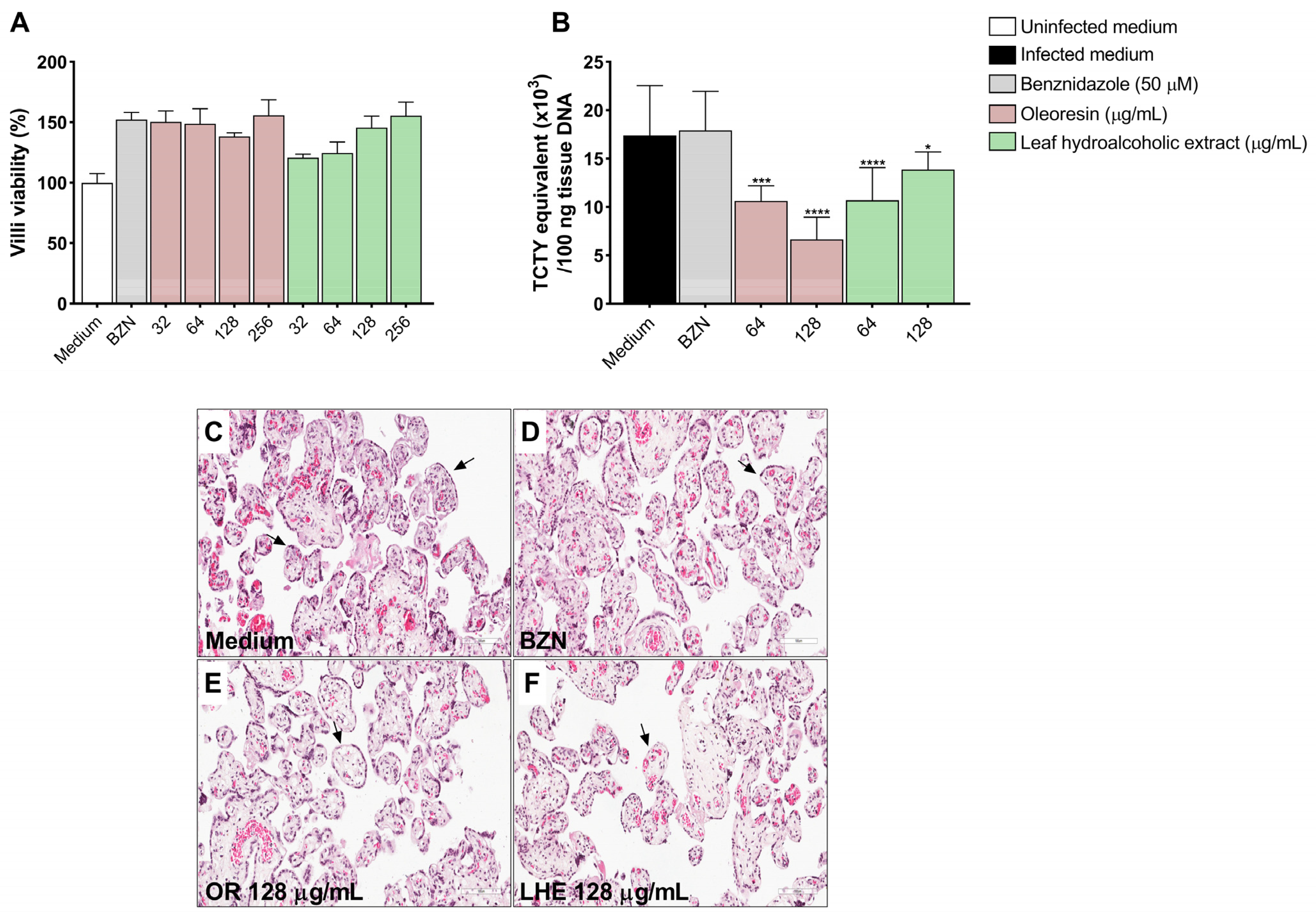
3.8. Oleoresin and Leaf Hydroalcoholic Extract from C. multijuga Reduce T. cruzi Infection in Human Placenta Explants

Following the assessment in the in vitro cell model, we evaluated whether OR and LHE could reduce T. cruzi infection using human placenta explants.
As measured by viability assays, no cytotoxic effects were observed at any concentrations of OR and LHE on villous explants (Figure 8A), and histological evaluation showed no evidence of tissue damage (Figure 8C–F). Treatment with OR (64 μg/mL: ***p = 0.0001; 128 μg/mL: ****p < 0.0001) and LHE (64 μg/mL: ****p < 0.0001; 128 μg/mL: *p = 0.0244) significantly reduced parasite DNA content compared to infected and untreated explants (Figure 8B).

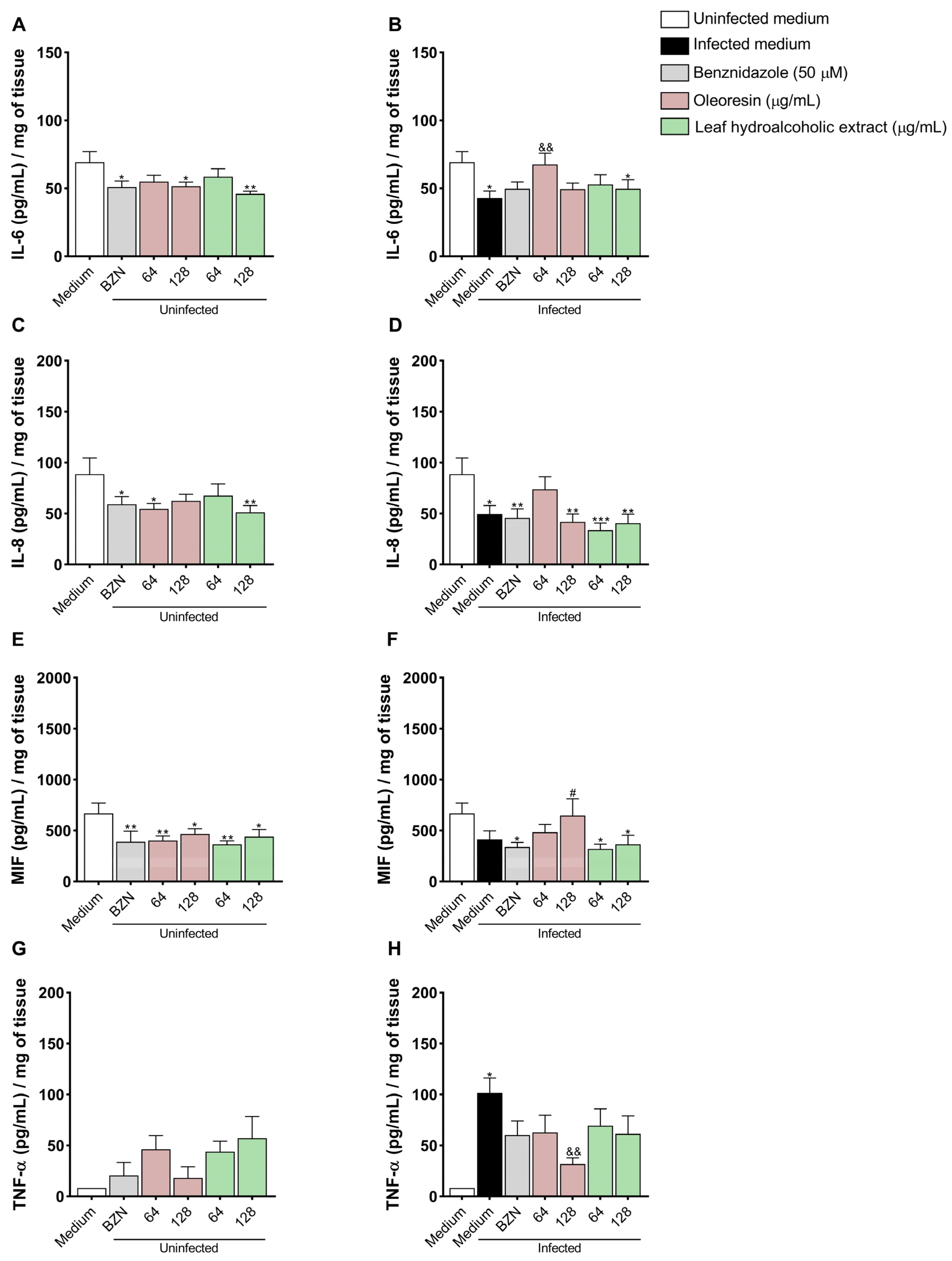
3.9. Oleoresin Increases IL-6 and Decreases TNF-α Production in Human Placenta Explants

Cytokine production was evaluated in villous explants following infection and/or treatment with OR and LHE.
IL-6: Uninfected villi treated with BZN (*p = 0.0188), OR at 128 μg/mL (*p = 0.0146), and LHE at 128 μg/mL (**p = 0.0026) exhibited significantly reduced IL-6 production compared to untreated and uninfected villi (uninfected medium) (Figure 9A). Additionally, infected and untreated villi (*p = 0.0108) and those treated with LHE at 128 μg/mL (*p = 0.0481) showed decreased IL-6 production compared to untreated uninfected villi (uninfected medium), whereas infected villi treated with OR at 64 µg/mL (&&p = 0.0083) exhibited increased IL-6 levels compared to infected untreated villi (infected medium) (Figure 9B).
IL-8: Uninfected villi treated with BZN (*p = 0.0435), OR at 64 μg/mL (*p = 0.0177), and LHE at 128 μg/mL (**p = 0.0097) showed reduced IL-8 production compared to untreated uninfected villi (uninfected medium) (Figure 9C). Similarly, infected and untreated villi (*p = 0.0109), or those treated with BZN (**p = 0.0065), OR at 128 μg/mL (**p = 0.0025), and LHE (64 μg/mL: ***p = 0.0003; 128 μg/mL: **p = 0.0013) also exhibited reduced IL-8 production compared to untreated uninfected villi (uninfected medium) (Figure 9D).
MIF: Uninfected villi treated with BZN (**p = 0.0089), OR (64 μg/mL: **p = 0.0092; 128 μg/mL: *p = 0.0424), and LHE (64 μg/mL: **p = 0.0032; 128 μg/mL: *p = 0.0273) exhibited decreased MIF levels compared to untreated uninfected villi (uninfected medium) (Figure 9E). Infected villi treated with BZN (*p = 0.03) and LHE (64 μg/mL: *p = 0.0159; 128 μg/mL: *p = 0.0349) also showed reduced MIF levels compared to untreated uninfected villi. However, infected villi treated with LHE at 128 μg/mL produced more MIF than those with BZN (#p = 0.0317) (Figure 9F).
TNF-α: Only infected untreated villi (infected medium, *p = 0.0384) showed increased TNF-α production compared to untreated uninfected villi. In contrast, infected villi treated with OR at 128 μg/mL (&&p = 0.0036) exhibited decreased TNF-α levels compared to infected untreated villi (Figure 9H).
IL-4 and IL-10: Levels were below the detection threshold in all experimental conditions.

4. Discussion

The limitation of benznidazole and nifurtimox for the treatment of congenital Chagas disease has arisen since they can cause side effects for the pregnant woman, with possible teratogenic effects, as well as the appearance of resistant parasites [40,41], making the search for novel drugs that are effective and safe an urgent need and of great importance. Plant-based compound studies have drawn attention as promising sources for novel drug discovery. Therefore, this study investigated the effects of oleoresin and leaf hydroalcoholic extract from C. multijuga on T. cruzi infection at the human maternal–fetal interface.
To be considered a potential new therapeutic agent against infectious diseases, it must not only control the infection but also exhibit low toxicity for the host. To address these challenges, we first evaluated the impact of OR and LHE on BeWo cell viability. Our results show that BeWo cells treated with OR and LHE exhibited reduced cell viability only at the highest concentrations. In agreement with our results, [22] sought to evaluate the genotoxicity of oleoresin from C. multijuga in Swiss mouse hepatocytes, and it was seen that it only caused some damage at the highest concentration. In a recent study from our group, both OR and LHE of C. multijuga were also toxic to BeWo and HTR-8/SVneo cells only at higher concentrations after 24 h of treatment [26], while in our current study, we analyzed after 72 h.
Furthermore, we evaluated the effect of OR and LHE on T. cruzi trypomastigote viability. Like the results observed in cells, both compounds exhibited cytotoxicity only at the highest concentrations (64, 128, and 256 µg/mL). Notably, the reduction in trypomastigote viability caused by OR and LHE was greater than that achieved with benznidazole. Although conventional treatment is more effective during the acute phase of the disease (around 60%), when trypomastigotes circulate in the bloodstream, studies have shown that the parasite has developed resistance mechanisms to BZN [42,43,44]. This underscores the need for new therapeutic strategies that are effective, especially in the acute phase of infection.
Our data shows that both compounds were able to decrease T. cruzi intracellular proliferation in BeWo cells. Corroborating our findings, Izumi and colleagues [45] demonstrated that terpenes found in oleoresins of Copaifera spp. promoted changes in oxidative metabolism, induced autophagy, and decreased the proliferation of the replicative forms of T. cruzi, especially amastigotes. In another study, Izumi and colleagues [31] evaluated the effect of oleoresin from eight species of Copaifera, including C. multijuga, and observed strong inhibitory effects on all life stages of the parasite. Additionally, oleoresin and kaurenoic acid from C. martii were shown to significantly reduce infection rates and the proliferation of T. cruzi amastigotes in murine peritoneal macrophages and HeLa cells [38]. Recently, our group reported that ORs from five different species of Copaifera controlled T. cruzi invasion, replication, and release in BeWo cells [32], further highlighting the trypanocidal potential of these compounds.
Once inside the host cell, T. cruzi amastigotes replicate in the cytoplasm and then differentiate back into trypomastigotes, which are released into the extracellular space to infect new cells and spread through the bloodstream. Since treatment with OR and LHE decreased the number of amastigotes in BeWo cells, we investigated whether this would also affect the differentiation and subsequent release of trypomastigotes. Although no statistical differences were observed at 72 and 120 h, our data showed a trend toward reduced trypomastigote release into the supernatant, suggesting a direct action of these compounds on the parasite that may compromise its ability to differentiate, lyse the host, infect new cells, and spread throughout the organism.
To test this hypothesis, we performed invasion and proliferation assays after pre-treating trypomastigotes for 1 h. Our data shows that pretreated parasites exhibited a reduced ability to invade and proliferate within BeWo cells. A recent study suggested that natural compounds may target different structures in T. cruzi, and it is hypothesized that mitochondria are strategic sites to induce parasite death [46]. To evaluate whether OR and LHE from C. multijuga affect parasite morphology, we analyzed trypomastigote forms pretreated for 1 h using a scanning electron microscope and observed that both compounds induced morphological alterations in the parasite body, such as shortening of the body and flagellum and torsion of the parasite, which may explain its difficulty in invading and proliferating inside the cell. Additionally, transmission electron microscopy of amastigotes treated for 72 h revealed further ultrastructural damage, such as cytoplasmic disorganization, membrane swelling, and vacuolization, supporting the observed reduction in parasite proliferation. Previous studies have reported similar effects of Copaifera spp. on other protozoa, such as membrane fusion in T. gondii, compromising its replicative capacity [25], rupture of the plasma membrane, retraction of the flagellum, and rounding of Leishmania spp. [29,47], as well as rupture of the plasma membrane and intracellular vacuole-like structures in T. cruzi [31,45]. Also, [32] showed that both T. cruzi amastigotes and trypomastigotes treated with Copaifera spp. undergo morphological modifications, such as disruption of the amastigote’s plasma membrane, kinetoplast swelling, and membrane shedding. These findings are important for the discovery of organelles and target molecules of the parasite to evaluate the possible therapeutic use of these compounds, and further studies are needed to discover which molecules present in these compounds have antiparasitic properties.
After demonstrating the direct effect of C. multijuga on T. cruzi trypomastigotes and amastigotes, we next evaluated whether OR and LHE could modulate the host immune response. We observed that T. cruzi infection increased ROS production compared to uninfected cells. Although the mechanisms are not fully understood, it is known that elevated ROS can promote T. cruzi infection and contribute to Chagas disease pathology [48,49]. Interestingly, treatment with BZN and the highest concentration of OR decreased ROS production compared to infected cells, while the highest concentration of LHE (64 μg/mL) increased it. Although both compounds are derived from the same plant species, these differences may be attributed to their distinct chemical composition, since ORs have more diterpenes and sesquiterpenes, while leaf extracts have more flavonoids and galloylquinic acids [50,51,52], so it is plausible that they can trigger different intracellular mechanisms.
Oleoresins from Copaifera spp. are known for their anti-inflammatory properties [53], which may help reduce ROS production. Supporting this, Teixeira and colleagues [25] observed that four Copaifera species reduced ROS levels in T. gondii-infected BeWo cells while also limiting parasite proliferation. Similarly, Kian and colleagues [38] demonstrated that C. martii oleoresin decreased ROS production in T. cruzi-infected macrophages and helped control parasite growth, highlighting that one of the antiparasitic mechanisms promoted by Copaifera spp. is through the induction of an anti-inflammatory response, which is important for a successful pregnancy, since an exacerbated inflammatory response may lead to a gestational imbalance, leading to miscarriage. In addition, in the context of T. cruzi infection, this decrease in ROS can contribute to controlling parasite proliferation. Although ROS has an important role in controlling pathogen infection, the parasite has developed mechanisms to overcome this response and survive within cells, and this oxidative stress induces cellular damage that contributes to parasite replication during the acute infection phase [54]. On the other hand, LHE increased ROS production compared to cells that were only infected. It is known that ROS are produced mainly in mitochondria and, at basal levels, play essential roles in cellular physiology, but in high concentrations, these molecules can induce oxidative stress, causing the oxidation of macromolecules, mainly lipids, proteins, and DNA, which can trigger cell death [55]. In this sense, Lazarin-Bidóia and colleagues [56] demonstrated that eupomatenoid-5 (isolated from the leaves of Piper regnellii var. pallescens) induced an oxidative imbalance in the three evolutionary forms of T. cruzi, especially trypomastigotes, shown by a downregulation in the activity of trypanothione reductase and an upregulation of ROS, which are key events in the induction of parasite death by several pathways, including apoptosis, necrosis, and autophagy. Thus, it is possible to suggest that the increase in ROS production induced by LHE may partially contribute to the control of T. cruzi proliferation in BeWo cells, but the trypanocidal effect promoted by LHE is more related to a direct action of this compound on the parasite than to the positive modulation of ROS.
Regarding cytokine production, OR treatment increased levels of IL-4, IL-10, and TNF-α, while LHE increased levels of IL-4, IL-8, IL-10, and TNF-α in BeWo cells. Santiago and colleagues [57] showed that oleoresins from different Copaifera species, including C. multijuga, can increase the production of IL-10 and TNF-α in human monocytes. Additionally, Kian and colleagues [38] demonstrated that C. martii oleoresins and the isolated compound kaurenoic acid reduced T. cruzi amastigote replication in macrophages without triggering an exacerbated pro-inflammatory response. This was evidenced by the downregulation of ROS and upregulation of IL-10, which is consistent with our findings. Given that BeWo cells are not classical immune cells, cytokine results derived from this model should be interpreted within the context of trophoblast biology.
T. cruzi is known to induce TNF-α production in human placenta, which can disrupt the placental barrier and contribute to low birth weight [58]. However, a study showed that elevated TNF-α levels are associated with the replication stage of the parasite within the chorionic villus (72 h after infection), which would hinder its transformation into a trypomastigote and consequently limit infection of the fetus [59]. This increase in TNF-α production induced by LHE can also induce the epithelial turnover [13], an important mechanism to limit the parasite’s congenital transmission [60]. Despite the high production of the pro-inflammatory cytokines TNF-α and IL-8 induced by OR and LHE, there was a simultaneous increase in IL-4 and IL-10, both anti-inflammatory cytokines, which would counterbalance the immune response towards a more anti-inflammatory environment, protecting the placenta and the fetus from an exacerbated pro-inflammatory immune response and maintaining a tolerogenic environment during pregnancy, in addition to controlling the T. cruzi proliferation.
Subsequently, we evaluated whether OR and LHE could control the infection in an ex vivo model using third-trimester human placenta explants. We observed that both compounds were non-cytotoxic, maintained villous morphology, and effectively controlled T. cruzi infection. Previous studies have demonstrated the potential of different Copaifera spp. to control T. cruzi and T. gondii infection in human placentas [25,26,27,32]. Interestingly, treatment with BZN did not reduce T. cruzi proliferation in our model. In agreement with our findings, Rojo and colleagues [61] showed that placenta explants infected with T. cruzi (DM28c strain) and treated with BZN (2, 20, and 200 µM) did not impair parasite infection, although BZN did prevent parasite-induced tissue damage. Conversely, Lima Júnior and colleagues [32] demonstrated that the same concentration used in this study was able to reduce parasitism. Nevertheless, despite using the same strain, the authors evaluated it after 48 h of treatment, while ours was at 72 h, so it is possible that BZN can control the proliferation of T. cruzi in earlier stages of infection, while in later stages, the parasite recovers its proliferative capacity. However, further studies are needed to verify, in terms of concentration, cytotoxicity, and control of parasitism against different strains, the efficacy and reliability of BZN in the treatment of congenital Chagas disease.
Regarding cytokine production in placenta explants, similar to the observations in BeWo cells, the parasite decreased IL-6 and IL-8 production, along with a trend toward reduced MIF production while increasing TNF-α levels. Other studies showed that T. cruzi downmodulates IL-6 and upregulates TNF-α, which can eliminate the parasite but at the same time cause tissue damage in high concentrations [12,62]. However, treatment with OR reversed this phenomenon, with upregulation of IL-6, IL-8, and MIF and downregulation of TNF-α. These findings suggest that OR helps restore the balance of key cytokines important for a healthy pregnancy while also limiting the overproduction of TNF-α, which, in excess, can increase the risk of pregnancy complications [63]. A similar TNF-α reduction induced by Copaifera ORs has been observed in human placentas infected by T. gondii [25,27] and T. cruzi [32].

5. Conclusions

Considering our results and the limited alternative treatments for CCD, we conclude that oleoresin and leaf hydroalcoholic extract of Copaifera multijuga effectively controlled the invasion, proliferation, and dissemination of T. cruzi in BeWo cells and human placenta explants. This effect appears to be associated with both direct action on the parasite and the modulation of ROS, pro-inflammatory, and anti-inflammatory cytokines. These findings highlight the potential of these natural compounds as promising therapeutic candidates for the treatment of congenital Chagas disease. However, further studies are needed to evaluate which bioactive molecules present in these compounds have antiparasitic activity.

Author Contributions

Conceptualization, B.F.B. and G.d.S.; methodology, G.d.S., C.P.T., J.P.d.L.J., M.P.O.A., M.P., L.C.L., N.C.L.d.S., R.M.d.O., I.S.D., M.C.B., G.V.F. and R.N.A.; validation, G.d.S., C.P.T. and B.F.B.; formal analysis, G.d.S., C.P.T. and B.F.B.; investigation, G.d.S., C.P.T. and B.F.B.; resources, B.F.B., E.A.V.F., M.A.L.V.A., R.C.S.V., J.K.B. and C.H.G.M.; data curation, G.d.S., C.P.T. and B.F.B.; writing—original draft preparation, G.d.S. and C.P.T.; writing—review and editing, G.d.S., C.P.T., A.O.G., C.H.G.M., M.A.L.V.A., R.C.S.V., J.K.B., S.C.T. and B.F.B.; visualization, G.d.S., C.P.T. and B.F.B.; supervision, E.A.V.F. and B.F.B.; project administration, G.d.S., C.P.T. and B.F.B.; funding acquisition, B.F.B., A.O.G., S.C.T. and E.A.V.F. All authors contributed to the article and approved the submitted version. All authors have read and agreed to the published version of the manuscript.

Funding

This work was supported by the Brazilian Researcher Agencies: Conselho Nacional de Desenvolvimento Científico e Tecnológico (CNPq), Fundação de Amparo à Pesquisa do Estado de Minas Gerais (FAPEMIG, APQ-01629-22), and the research productivity grant from Universidade do Estado de Minas Gerais (PQ/UEMG). This study was funded in part by the Coordenação de Aperfeiçoamento de Pessoal de Nıvel Superior’—Brazil (CAPES)—Finance Code 001.

Institutional Review Board Statement

This study was approved by the Ethics Committee of Universidade Federal de Uberlândia, MG, Brazil, with approval number 7.407.162 (approved on 24 February 2025).

Informed Consent Statement

A consent term was obtained from all participants included in this study.

Data Availability Statement

The original contributions presented in this study are included in the article. Further inquiries can be directed to the corresponding author.

Acknowledgments

We are thankful to the pregnant women who made these studies possible.

Conflicts of Interest

The authors declare no conflicts of interest.

References

  1. De Sousa, A.S.; Vermeij, D.; Ramos, A.N., Jr.; Luqueti, A.O. Chagas disease. Lancet 2024, 10422, 203–218. [Google Scholar] [CrossRef] [PubMed]
  2. Cucunubá, Z.M.; Gutiérrez-Romero, S.A.; Ramírez, J.D.; Velásquez-Ortiz, N.; Ceccarelli, S.; Parra-Henao, G.; Henao-Martínez, A.F.; Rabinovich, J.; Basáñez, M.G.; Nouvellet, P.; et al. The epidemiology of Chagas disease in the Americas. Lancet Reg. Health Am. 2024, 37, 100881. [Google Scholar] [CrossRef]
  3. Howard, E.J.; Xiong, X.; Carlier, Y.; Sosa-Estani, S.; Buekens, P. Frequency of the congenital transmission of Trypanosoma cruzi: A systematic review and meta-analysis. BJOG Int. J. Obstet. Gynaecol. 2014, 1, 22–33. [Google Scholar] [CrossRef] [PubMed]
  4. Palacios Gil-Antuñano, S.; Gold, S.; Abril, M.; Hernandéz, M.S.; Cancelo-Hidalgo, M.J.; Flores-Chávez, M.; Pelayo-Delgado, I. Mother-to-child Chagas disease transmission: The challenge of detection and prevention in areas without the risk of vectorial transmission. Int. J. Gynaecol. Obstet. 2024, 3, 835–842. [Google Scholar] [CrossRef]
  5. Messenger, L.A.; Bern, C. Congenital Chagas disease: Current diagnostics, limitations and future perspectives. Curr. Opin. Infect. Dis. 2018, 5, 415–421. [Google Scholar] [CrossRef]
  6. Antinori, S.; Galimberti, L.; Bianco, R.; Grande, R.; Galli, M.; Corbellino, M. Chagas disease in Europe: A review for the internist in the globalized world. Eur. J. Intern. Med. 2017, 43, 6–15. [Google Scholar] [CrossRef] [PubMed]
  7. Kemmerling, U.; Osuna, A.; Schijman, A.G.; Truyens, C. Congenital transmission of Trypanosoma cruzi: A review about the interactions between the parasite, the placenta, the maternal and the fetal/neonatal immune responses. Front. Microbiol. 2019, 10, 1854. [Google Scholar] [CrossRef]
  8. Rassi, A., Jr.; Rassi, A.; Marin-Neto, J.Á. Chagas disease. Lancet 2010, 9723, 1388–1402. [Google Scholar] [CrossRef]
  9. Chatterjee, P.; Chiasson, V.L.; Bounds, K.R.; Mitchell, B.M. Regulation of the anti-inflammatory cytokines interleukin-4 and interleukin-10 during pregnancy. Front. Immunol. 2014, 5, 253. [Google Scholar] [CrossRef]
  10. Todros, T.; Paulesu, L.; Cardaropoli, S.; Rolfo, A.; Masturzo, B.; Ermini, L.; Romagnoli, R.; Ietta, F. Role of the macrophage migration inhibitory factor in the pathophysiology of pre-eclampsia. Int. J. Mol. Sci. 2021, 4, 1823. [Google Scholar] [CrossRef] [PubMed]
  11. Vilotić, A.; Nacka-Aleksić, M.; Pirković, A.; Bojic-Trbojevic, Z.; Dekanski, D.; Krivokuca, M.J. IL-6 and IL-8: An overview of their roles in healthy and pathological pregnancies. Int. J. Mol. Sci. 2022, 23, 14574. [Google Scholar] [CrossRef] [PubMed]
  12. Castillo, C.; Muñoz, L.; Carrillo, I.; Liempi, A.; Gallardo, C.; Galanti, N.; Maya, J.D.; Kemmerling, U. Ex vivo infection of human placental chorionic villi explants with Trypanosoma cruzi and Toxoplasma gondii induces different Toll-like receptor expression and cytokine/chemokine profiles. Am. J. Reprod. Immunol. 2017, 78, e12660. [Google Scholar] [CrossRef]
  13. Haider, S.; Knöfler, M. Human tumour necrosis factor: Physiological and pathological roles in placenta and endometrium. Placenta 2009, 2, 111–123. [Google Scholar] [CrossRef]
  14. Hamilton, S.T.; Scott, G.; Naing, Z.; Iwasenko, J.; Hall, B.; Graf, N.; Arbuckle, S.; Craig, M.E.; Rawlinson, W.D. Human cytomegalovirus induces cytokine changes in the placenta with implications for adverse pregnancy outcomes. PLoS ONE 2012, 12, e52899. [Google Scholar] [CrossRef]
  15. Droguett, D.; Carrillo, I.; Castillo, C.; Gómez, F.; Negrete, M.; Liempi, A.; Muñoz, L.; Galanti, N.; Maya, J.D.; Kemmerling, U. Trypanosoma cruzi induces cellular proliferation in the trophoblastic cell line BeWo. Exp. Parasitol. 2017, 173, 9–17. [Google Scholar] [CrossRef]
  16. Liempi, A.; Castillo, C.; Duaso, J.; Droguett, D.; Sandoval, A.; Barahona, K.; Hernández, A.; Galanti, N.; Maya, J.D.; Kemmerling, U. Trypanosoma cruzi induces trophoblast differentiation: A potential local antiparasitic mechanism of the human placenta? Placenta 2014, 12, 1035–1042. [Google Scholar] [CrossRef]
  17. Pérez-Molina, J.A.; Molina, I. Chagas disease. Lancet 2018, 10115, 82–94. [Google Scholar] [CrossRef] [PubMed]
  18. Murcia, L.; Simón, M.; Carrilero, B.; Roig, M.; Segovia, M. Treatment of infected women of childbearing age prevents congenital Trypanosoma cruzi infection by eliminating the parasitemia detected by PCR. J. Infect. Dis. 2017, 9, 1452–1458. [Google Scholar] [CrossRef]
  19. Abras, A.; Ballart, C.; Fernández-Arévalo, A.; Pinazo, M.J.; Gascón, J.; Muñoz, C.; Gállego, M. Worldwide control and management of Chagas disease in a new era of globalization: A close look at congenital Trypanosoma cruzi infection. Clin. Microbiol. Rev. 2022, 2, e0015221. [Google Scholar] [CrossRef] [PubMed]
  20. Álvarez, M.G.; Vigliano, C.; Lococo, B.; Bertocchi, G.; Viotti, R. Prevention of congenital Chagas disease by Benznidazole treatment in reproductive-age women. An observational study. Acta Trop. 2017, 174, 149–152. [Google Scholar] [CrossRef]
  21. Pinazo, M.J.; Malchiodi, E.; Ioset, J.R.; Bivona, A.; Gollob, K.J.; Dutra, W.O. Challenges and advancements in the development of vaccines and therapies against Chagas disease. Lancet Microbe 2024, 10, 100972. [Google Scholar] [CrossRef] [PubMed]
  22. Alves, J.M.; Senedese, J.M.; Leandro, L.F.; Castro, P.T.; Pereira, D.E.; Carneiro, L.J.; Ambrósio, S.R.; Bastos, J.K.; Tavares, D.C. Copaifera multijuga oleoresin and its constituent diterpene (−)-copalic acid: Genotoxicity and chemoprevention study. Mutat. Res. Genet. Toxicol. Environ. Mutagen. 2017, 819, 26–30. [Google Scholar] [CrossRef]
  23. Gomes, N.M.; de Rezende, C.M.; Fontes, S.P.; Matheus, M.E.; Pinto, A.C.; Fernandes, P.D. Characterization of the antinociceptive and anti-inflammatory activities of fractions obtained from Copaifera multijuga Hayne. J. Ethnopharmacol. 2010, 1, 177–183. [Google Scholar] [CrossRef]
  24. Pinheiro, J.G.; Tavares, E.A.; Silva, S.S.; Silva, J.F.; Carvalho, Y.M.B.G.; Ferreira, M.R.A.; Araújo, A.A.S.; Barbosa, E.G.; Pedrosa, M.F.F.; Soares, L.A.L.; et al. Inclusion complexes of Copaiba (Copaifera multijuga Hayne) oleoresin and cyclodextrins: Physicochemical characterization and anti-inflammatory activity. Int. J. Mol. Sci. 2017, 11, 2388. [Google Scholar] [CrossRef]
  25. Teixeira, S.C.; De Souza, G.; Borges, B.C.; Araújo, T.E.; Rosini, A.M.; Aguila, F.A.; Ambrósio, S.R.; Veneziani, R.C.S.; Bastos, J.K.; Silva, M.J.B.; et al. Copaifera spp. oleoresins impair Toxoplasma gondii infection in both human trophoblastic cells and human placental explants. Sci. Rep. 2020, 1, 15158. [Google Scholar] [CrossRef]
  26. Martínez, A.F.F.; Teixeira, S.C.; de Souza, G.; Rosini, A.M.; Lima Júnior, J.P.; Melo, G.N.; Blandón, K.O.E.; Gomes, A.O.; Ambrósio, S.R.; Veneziani, R.C.S.; et al. Leaf hydroalcoholic extract and oleoresin from Copaifera multijuga control Toxoplasma gondii infection in human trophoblast cells and placental explants from third-trimester pregnancy. Front. Cell. Infect. Microbiol. 2023, 13, 1113896. [Google Scholar] [CrossRef]
  27. Teixeira, S.C.; Rosini, A.M.; de Souza, G.; Martínez, A.F.; Silva, R.J.; Ambrosio, S.R.; Veneziani, R.C.; Bastos, J.K.; Martins, C.H.; Barbosa, B.F.; et al. Polyalthic acid and oleoresin from Copaifera trapezifolia Hayne reduce Toxoplasma gondii growth in human villous explants, even triggering an anti-inflammatory profile. Exp. Parasitol. 2023, 250, 108534. [Google Scholar] [CrossRef]
  28. Rodrigues, I.A.; Ramos, A.d.S.; Falcão, D.Q.; Ferreira, J.L.P.; Basso, S.L.; Silva, J.R.A.; Amaral, A.C.F. Development of Nanoemulsions to Enhance the Antileishmanial Activity of Copaifera paupera Oleoresins. Biomed. Res. Int. 2018, 2018, 9781724. [Google Scholar] [CrossRef] [PubMed]
  29. Santos, A.O.D.; Izumi, E.; Ueda-Nakamura, T.; Dias-Filho, B.P.; Veiga-Júnior, V.F.; Nakamura, C.V. Antileishmanial activity of diterpene acids in copaiba oil. Mem. Inst. Oswaldo Cruz. 2013, 1, 59–64. [Google Scholar] [CrossRef] [PubMed]
  30. Souza, G.A.G.; Silva, N.C.; Souza, J.; Oliveira, K.R.M.; Fonseca, A.L.; Baratto, L.C.; Oliveira, E.C.P.; Varotti, F.P.; Moraes, W.P. In vitro and in vivo antimalarial potential of oleoresin obtained from Copaifera reticulata Ducke (Fabaceae) in the Brazilian Amazon rainforest. Phytomedicine 2017, 24, 111–118. [Google Scholar] [CrossRef]
  31. Izumi, E.; Ueda-Nakamura, T.; Veiga-Junior, V.F.; Nakamura, C.V. Toxicity of oleoresins from the genus Copaifera in Trypanosoma cruzi: A comparative study. Planta Med. 2013, 11, 952–958. [Google Scholar] [CrossRef]
  32. Lima Júnior, J.P.; Teixeira, S.C.; de Souza, G.; Faria, G.V.; Almeida, M.P.O.; Franco, P.S.; Luz, L.C.; Paschoalino, M.; Dos Santos, N.C.L.; Oliveira, R.M.; et al. Copaifera spp. oleoresins control Trypanosoma cruzi infection in human trophoblast cells (BeWo) and placental explants. Biomed. Pharmacother. 2024, 179, 117425. [Google Scholar] [CrossRef]
  33. Pimenta, I.P.; Abrão, F.; da Silva, J.J.M.; Oliveira, L.C.; Rogez, H.L.G.; Ambrósio, S.R.; Veneziani, R.C.S.; Bastos, J.K.; Martins, C.H.G. Antibacterial profile of Copaifera multijuga oleoresin and hydroalcoholic extract of leaves against oral pathogens. Curr. Dent. 2019, 1, 53–60. [Google Scholar] [CrossRef]
  34. Costa, I.N.; Ribeiro, M.; Franco, P.S.; Da Silva, R.J.; Araújo, T.E.; Milián, I.C.B.; Luz, L.C.; Guirelli, P.M.; Nakazato, G.; Mineo, J.R.; et al. Biogenic Silver Nanoparticles Can Control Toxoplasma gondii Infection in Both Human Trophoblast Cells and Villous Explants. Front. Microbiol. 2021, 11, 623947. [Google Scholar] [CrossRef]
  35. De Souza, G.; Teixeira, S.C.; Martínez, A.F.F.; Silva, R.J.; Luz, L.C.; Lima Júnior, J.P.; Rosini, A.M.; Dos Santos, N.C.L.; Oliveira, R.M.; Paschoalino, M.; et al. Trypanosoma cruzi P21 recombinant protein modulates Toxoplasma gondii infection in different experimental models of the human maternal-fetal interface. Front. Immunol. 2023, 14, 1243480. [Google Scholar] [CrossRef] [PubMed]
  36. Bennett, W.A.; Lagoo-Deenadayalan, S.; Whitworth, N.S.; Brackin, M.N.; Hale, E.; Cowan, B.D. Expression and production of interleukin-10 by human trophoblast: Relationship to pregnancy immunotolerance. Early Pregnancy 1997, 3, 190–198. [Google Scholar] [PubMed]
  37. Mosmann, T. Rapid colorimetric assay for cellular growth and survival: Application to proliferation and cytotoxicity assays. J. Immunol. Methods 1983, 1–2, 55–63. [Google Scholar] [CrossRef]
  38. Kian, D.; Lancheros, C.A.C.; Assolini, J.P.; Arakawa, N.S.; Veiga-Júnior, V.F.; Nakamura, C.V.; Pinge-Filho, P.; Conchon-Costa, I.; Pavanelli, W.R.; Yamada-Ogatta, S.F.; et al. Trypanocidal activity of copaiba oil and kaurenoic acid does not depend on macrophage killing machinery. Biomed. Pharmacother. 2018, 103, 1294–1301. [Google Scholar] [CrossRef]
  39. Diaz, C.; Nussenzweig, V.; Gonzalez, A. An improved polymerase chain reaction assay to detect Trypanosoma cruzi in blood. Am. J. Trop. Med. Hyg. 1992, 5, 616–623. [Google Scholar] [CrossRef]
  40. Morilla, M.J.; Romero, E.L. Nanomedicines against Chagas disease: An update on therapeutics, prophylaxis, and diagnosis. Nanomedicine 2015, 3, 465–481. [Google Scholar] [CrossRef]
  41. Tarleton, R.L. Effective drug discovery in Chagas disease. Trends Parasitol. 2023, 6, 423–431. [Google Scholar] [CrossRef]
  42. Campos, M.C.O.; Leon, L.L.; Taylor, M.C.; Kelly, J.M. Benznidazole-resistance in Trypanosoma cruzi: Evidence that distinct mechanisms can act in concert. Mol. Biochem. Parasitol. 2015, 201, 83. [Google Scholar] [CrossRef]
  43. Lima, D.A.; Gonçalves, L.O.; Reis-Cunha, J.L.; Guimarães, P.A.S.; Ruiz, J.C.; Liarte, D.B.; Murta, S.M.F. Transcriptomic analysis of benznidazole-resistant and susceptible Trypanosoma cruzi populations. Parasit. Vectors 2023, 1, 167. [Google Scholar] [CrossRef] [PubMed]
  44. Vela, A.; Coral-Almeida, M.; Sereno, D.; Costales, J.A.; Barnabé, C.; Brenière, S.F. In vitro susceptibility of Trypanosoma cruzi discrete typing units (DTUs) to benznidazole: A systematic review and meta-analysis. PLoS Negl. Trop. Dis. 2021, 3, e0009269. [Google Scholar] [CrossRef]
  45. Izumi, E.; Ueda-Nakamura, T.; Veiga, V.F., Jr.; Pinto, A.C.; Nakamura, C.V. Terpenes from Copaifera demonstrated in vitro antiparasitic and synergic activity. J. Med. Chem. 2012, 7, 2994–3001. [Google Scholar] [CrossRef]
  46. Lazarin-Bidóia, D.; Garcia, F.P.; Ueda-Nakamura, T.; Silva, S.O.; Nakamura, C.V. Natural compounds based chemotherapeutic against Chagas disease and leishmaniasis: Mitochondrion as a strategic target. Mem. Inst. Oswaldo Cruz. 2022, 117, e220396. [Google Scholar] [CrossRef]
  47. De Moraes, A.R.D.P.; Tavares, G.D.; Rocha, F.J.S.; De Paula, E.; Giorgio, S. Effects of nanoemulsions prepared with essential oils of copaiba- and andiroba against Leishmania infantum and Leishmania amazonensis infections. Exp. Parasitol. 2018, 187, 12–21. [Google Scholar] [CrossRef]
  48. Maldonado, E.; Rojas, D.A.; Morales, S.; Miralles, V.; Solari, A. Dual and Opposite Roles of Reactive Oxygen Species (ROS) in Chagas Disease: Beneficial on the Pathogen and Harmful on the Host. Oxid. Med. Cell. Longev. 2020, 2020, 8867701. [Google Scholar] [CrossRef]
  49. Paiva, C.N.; Medei, E.; Bozza, M.T. ROS and Trypanosoma cruzi: Fuel to infection, poison to the heart. PLoS Pathog. 2018, 4, e1006928. [Google Scholar] [CrossRef] [PubMed]
  50. Arruda, C.; Mejía, J.A.A.; Ribeiro, V.P.; Borges, C.H.G.; Martins, C.H.G.; Veneziani, R.C.S.; Ambrósio, S.R.; Bastos, J.K. Occurrence, chemical composition, biological activities and analytical methods on Copaifera genus—A review. Biomed. Pharmacother. 2019, 109, 1–20. [Google Scholar] [CrossRef] [PubMed]
  51. Leandro, L.M.; Vargas, F.S.; Barbosa, P.C.S.; Neves, J.K.O.; Da Silva, J.A.; Veiga-Júnior, V.F. Chemistry and biological activities of terpenoids from copaiba (Copaifera spp.) oleoresins. Molecules 2012, 4, 3866–3869. [Google Scholar] [CrossRef]
  52. Veiga-Junior, V.F.; Pinto, A.C. The Copaifera L. genus. Quím Nova 2002, 2, 163–169. [Google Scholar] [CrossRef]
  53. Trindade, R.D.; da Silva, J.K.; Setzer, W.N. Copaifera of the Neotropics: A Review of the Phytochemistry and Pharmacology. Int. J. Mol. Sci. 2018, 5, 1511. [Google Scholar] [CrossRef]
  54. Martínez, I.; Rivera-Santiago, L.; Rodríguez-Hernández, K.D.; Galván-Hernández, A.; Rodríguez-Fragoso, L.; Díaz-Peralta, L.; Torres-Martínez, L.; Agredano-Moreno, L.T.; Jiménez-García, L.F.; Ortega-Blake, I.; et al. A promising amphotericin B derivative induces morphological alterations, mitochondrial damage, and oxidative stress in vitro and prevents mice from death produced by a virulent strain of Trypanosoma cruzi. Microorganisms 2024, 6, 1064. [Google Scholar] [CrossRef]
  55. Kirkinezos, I.G.; Moraes, C.T. Reactive oxygen species and mitochondrial diseases. Semin. Cell Dev. Biol. 2001, 6, 449–457. [Google Scholar] [CrossRef] [PubMed]
  56. Lazarin-Bidóia, D.; Desoti, V.C.; Ueda-Nakamura, T.; Dias Filho, B.P.; Nakamura, C.V.; Silva, S.O. Further evidence of the trypanocidal action of eupomatenoid-5: Confirmation of involvement of reactive oxygen species and mitochondria owing to a reduction in trypanothione reductase activity. Free Radic. Biol. Med. 2013, 60, 17–28. [Google Scholar] [CrossRef] [PubMed]
  57. Santiago, K.B.; Conti, B.J.; Andrade, B.F.M.T.; Silva, J.J.M.; Rogez, H.L.G.; Crevelin, E.J.; Moraes, L.A.B.; Veneziani, R.; Ambrósio, S.R.; Bastos, J.K.; et al. Immunomodulatory action of Copaifera spp oleoresins on cytokine production by human monocytes. Biomed. Pharmacother. 2015, 70, 12–18. [Google Scholar] [CrossRef]
  58. Torrico, M.C.; Solano, M.; Guzmán, J.M.; Parrado, R.; Suarez, E.; Alonzo-Vega, C.; Truyens, C.; Carlier, Y.; Torrico, F. Estimation of the parasitemia in Trypanosoma cruzi human infection: High parasitemias are associated with severe and fatal congenital Chagas disease. Rev. Soc. Bras. Med. Trop. 2005, 38, 58–61. [Google Scholar]
  59. Benizio, E.; Moreira-Espinoza, M.J.; Triquell, M.F.; Mezzano, L.; Díaz-Luján, C.M.; Fretes, R.E. Pro-inflammatory cytokines are modified during the multiplication of Trypanosoma cruzi within the placental chorionic villi and are associated with the level of infection via the signaling pathway NF-κB. Am. J. Reprod. Immunol. 2023, 4, e13777. [Google Scholar] [CrossRef] [PubMed]
  60. Liempi, A.; Castillo, C.; Carrillo, I.; Muñoz, L.; Droguett, D.; Galanti, N.; Maya, J.D.; Kemmerling, U. A local innate immune response against Trypanosoma cruzi in the human placenta: The epithelial turnover of the trophoblast. Microb. Pathog. 2016, 99, 123–129. [Google Scholar] [CrossRef]
  61. Rojo, G.; Castillo, C.; Duaso, J.; Liempi, A.; Droguett, D.; Galanti, N.; Maya, J.D.; López-Muñoz, R.; Kemmerling, U. Toxic and therapeutic effects of Nifurtimox and Benznidazole on Trypanosoma cruzi ex vivo infection of human placental chorionic villi explants. Acta Trop. 2014, 132, 112–118. [Google Scholar] [CrossRef] [PubMed]
  62. Barbosa, C.G.; Gómez-Hernández, C.; da Silva, M.V.; Rezende-Oliveira, K.; Ferreira, P.T.M.; Oliveira, A.C.M.; Desidério, C.S.; Helmo, F.R.; Carvalho-Costa, T.M.; Dos Santos, I.K.P.; et al. Congenital transmission of Mexican strains of Trypanosoma cruzi TcIa: Interaction between parasite and human placental explants. Parasitology 2021, 3, 418–426. [Google Scholar] [CrossRef] [PubMed]
  63. Romanowska-Próchnicka, K.; Felis-Giemza, A.; Olesińska, M.; Wojdasiewicz, P.; Paradowska-Gorycka, A.; Szukiewicz, D. The role of TNF-α and anti-TNF-α agents during preconception, pregnancy, and breastfeeding. Int. J. Mol. Sci. 2021, 6, 2922. [Google Scholar] [CrossRef] [PubMed]
Figure 1. BeWo cell and trypomastigote viability treated with OR or LHE from C. multijuga. BeWo cells and T. cruzi trypomastigotes were treated with OR (A,C) or LHE (B,D) for 72 h and submitted to the MTT assay. Data is presented as percentage (%) of cell/trypomastigote viability compared to untreated cells/trypomastigotes (medium, 100% viability). Differences between groups were analyzed by One-Way ANOVA test with Bonferroni’s multiple comparisons post-test. Significant differences in relation to cells/trypomastigotes treated with medium (*p) or treated with benznidazole (#p). Three independent experiments, with eight replicates, were performed.
Pathogens 14 00736 g001
Figure 2. T. cruzi intracellular proliferation in BeWo cells treated with OR and LHE from C. multijuga. BeWo cells were infected with T. cruzi for 3 h and subsequently treated for 72 h with OR (16 and 32 µg/mL), LHE (32 and 64 µg/mL), BZN (50 µM), or FBS 5% medium. The number of infected cells (A) was analyzed under a light microscope to determine the infection index (the number of infected cells per 200 randomly observed cells) and the intracellular proliferation of T. cruzi (B) (the number of total amastigotes observed in the infected cells). Differences between groups were analyzed by the Kruskal–Wallis test with Dunn’s multiple comparisons post-test (A) or the One-Way ANOVA test with Bonferroni’s multiple comparisons post-test (B). Significant differences in relation to infected and untreated cells (medium) (*p). Three independent experiments with four replicates were performed. Representative images of BeWo cells infected with T. cruzi and untreated (C), treated with BZN (D), treated with OR 16 μg/mL (E) and 32 μg/mL (F), or LHE 32 μg/mL (G) and 64 μg/mL (H). Arrows indicate T. cruzi amastigotes in the cytoplasm of BeWo cells. Scale bar: 50 μm.
Pathogens 14 00736 g002
Figure 3. T. cruzi trypomastigote release in the supernatant of BeWo cells treated with (A) OR and (B) LHE from C. multijuga. The supernatant of BeWo cells infected and treated with OR (16 and 32 µg/mL), LHE (32 and 64 µg/mL), BZN (50 µM), or FBS 5% medium was collected after 72 h of treatment, as well as 96, 120, and 144 h after infection, and the trypomastigotes were counted in the Neubauer chamber. Data is presented as the number of trypomastigotes in the supernatant ×105/mL. Differences between groups were analyzed by the Two-Way ANOVA test with Bonferroni’s multiple comparisons post-test. Significant differences in relation to infected and untreated cells (medium) (*p). Three independent experiments, with four replicates, were performed.
Pathogens 14 00736 g003
Figure 4. T. cruzi invasion and proliferation after pretreatment with OR and LHE. BeWo cells were infected with T. cruzi previously pretreated for 1 h with OR (16 and 32 µg/mL), LHE (32 and 64 µg/mL), BZN (50 µM), or FBS 5% medium, and then, the invasion (A,B) and proliferation (C,D) were analyzed. The percentage of invasion/proliferation of the parasite was analyzed under a light microscope to determine the infection index (the number of infected cells per 200 randomly observed cells) (A,C) and the invasion/proliferation of T. cruzi (the number of total amastigotes observed in the infected cells) (B,D). Differences between groups were analyzed by the One-Way ANOVA test with Bonferroni’s multiple comparisons post-test. Significant differences in relation to cells infected with untreated parasites (medium) (*p) or cells infected with BZN-treated parasites (#p). Two independent experiments, with four replicates, were performed.
Pathogens 14 00736 g004
Figure 5. Effect of OR and LHE on T. cruzi trypomastigotes and amastigotes. T. cruzi trypomastigotes were pretreated for 1 h with OR (32 µg/mL), LHE (64 µg/mL), BZN (50 µM), or FBS 5% medium and then processed for scanning electron microscopy (AD). Representative images of trypomastigotes treated with medium (A) with normal morphology, while BZN (B), OR (C), and LHE (D) displayed some abnormalities, such as swelling of the parasite’s body, rounding of the cell surface, shortening of the flagellum, and torsion. Scale bars: 2 µm. Transmission electron microscopy images (EH) of T. cruzi amastigotes after 72 h of treatment with medium (E), BZN 50 µM (F), OR 32 µg/mL (G), and LHE 64 µg/mL (H). Thick arrow: membrane swelling; star: vacuolization; arrowhead: cytoplasm disorganization; N: nuclei; K: kinetoplast; F: flagellum. Scale bar: 0.5 µm.
Pathogens 14 00736 g005
Figure 6. ROS production in BeWo cells. BeWo cells were infected or not infected with T. cruzi for 3 h and then treated or not treated for 72 h with OR (16 and 32 µg/mL), LHE (32 and 64 µg/mL), BZN (50 µM), or FBS 5% medium. The cells were incubated with a probe 2′,7′-dichlorodihydrofluorescein diacetate (H2DCF-DA), and ROS production was measured by fluorescence in a plate reader and expressed as median fluorescence intensity (MFI). Differences between groups were analyzed by One-Way ANOVA with Bonferroni’s multiple comparisons post-test. Significant differences in relation to uninfected and untreated cells (uninfected medium) (*p), infected and untreated cells (&p), and cells infected or uninfected and treated with BZN (#p) (A,B). Three independent experiments, with eight replicates, were performed.
Pathogens 14 00736 g006
Figure 7. Cytokine production in BeWo cells. BeWo cells were infected or not infected with T. cruzi for 3 h and then treated or not treated for 72 h with OR (16 and 32 µg/mL), LHE (32 and 64 µg/mL), BZN (50 µM), or FBS 5% medium. The supernatant was submitted to an ELISA assay for the measurement of IL-4 (A,B), IL-6 (C,D), IL-8 (E,F), IL-10 (G,H), and TNF-α (I,J). Data are presented in pg/mL according to the standard curve for each cytokine. Differences between groups were analyzed by One-Way ANOVA with Bonferroni’s multiple comparisons. Significant differences in relation to uninfected and untreated cells (uninfected medium) (*p), infected and untreated cells (&p), and cells infected or not infected and treated with BZN (#p). Three independent experiments, with three replicates, were performed.
Pathogens 14 00736 g007
Figure 8. T. cruzi DNA content in human placenta explants. Human placental explants were treated for 72 h with OR, LHE, BZN (50 µM), or FBS 5% medium and submitted to an MTT assay (A). Data are presented as the percentage (%) of villi viability in relation to untreated villi (untreated medium). Two independent experiments, with eight replicates, were performed. In addition, the villi were infected with T. cruzi and treated for 72 h with OR (64 and 128 µg/mL), LHE (64 and 128 µg/mL), BZN (50 µM), and FBS 5% medium and then processed to PCR for quantification of T. cruzi DNA content (B). Data are presented as TCTY equivalent per 100 ng of DNA tissue. Representative images are shown of untreated villi (C), treated with BZN (50 µM) (D), OR (128 µg/mL) (E), and LHE (128 µg/mL) (F). Histological sections are stained by Harris hematoxylin and eosin, where it is possible to see no damage in the syncytiotrophoblast layer (arrow). Scale bar: 100 µm. Differences between groups were analyzed by One-Way ANOVA with Bonferroni’s multiple comparisons. Significant differences in relation to villi infected and untreated (medium) (*p). Two independent experiments, with eight replicates, were performed.
Pathogens 14 00736 g008
Figure 9. Cytokine production in human placenta explants. BeWo cells were infected or not infected with T. cruzi for 3 h and then treated or not treated for 72 h with OR (64 and 128 µg/mL), LHE (64 and 128 µg/mL), BZN (50 µM), or FBS 5% medium. The supernatant was submitted to an ELISA assay for the measurement of IL-6 (A,B), IL-8 (C,D), MIF (E,F), and TNF-α (G,H). Data are presented in pg/mL/mg of tissue. Differences between groups were analyzed by One-Way ANOVA with Bonferroni’s multiple comparisons post-test (AF,H) or the Kruskal–Wallis test with Dunn’s multiple comparisons post-test (G). Significant differences in relation to uninfected and untreated cells (uninfected medium) (*p), infected and untreated cells (&p), and cells infected or not infected and treated with BZN (#p). Two independent experiments, with eight replicates, were performed.
Pathogens 14 00736 g009
Disclaimer/Publisher’s Note: The statements, opinions and data contained in all publications are solely those of the individual author(s) and contributor(s) and not of MDPI and/or the editor(s). MDPI and/or the editor(s) disclaim responsibility for any injury to people or property resulting from any ideas, methods, instructions or products referred to in the content.

