Systematic Review

A Systematic Review on the Occurrence of Babesia spp. and Anaplasma spp. in Ticks and Wild Boar from Europe—A 15-Year Retrospective Study

1
Department of Parasitology and Parasitic Disease, Faculty of Veterinary Medicine, University of Life Sciences “King Mihai I” of Timisoara, 119, Calea Aradului, 300645 Timisoara, Romania
2
Veterinary and Food Safety Department 4, Surorile Martir Caceu, 300585 Timisoara, Romania
3
Department of Infectious Diseases and Preventive Medicine, Faculty of Veterinary Medicine, University of Life Sciences “King Mihai I” of Timisoara, 119, Calea Aradului, 300645 Timisoara, Romania
*
Authors to whom correspondence should be addressed.
Pathogens 2025, 14(7), 612; https://doi.org/10.3390/pathogens14070612
Submission received: 20 May 2025 / Revised: 18 June 2025 / Accepted: 19 June 2025 / Published: 20 June 2025
(This article belongs to the Special Issue Parasitic Diseases in Wild Animals)

Abstract

The wild boar (Sus scrofa) has experienced significant population growth as well as geographic expansion across Europe over the past 15 years, leading to increased concerns regarding its role in the transmission of zoonotic pathogens. Among these, Babesia spp. and Anaplasma spp. are of particular importance due to their impact on both wildlife and domestic animals. This study systematically reviews the prevalence and distribution of Babesia and Anaplasma spp. in wild boars and associated tick vectors across multiple European countries, synthesizing data from literature published between 2010 and 2024. A comprehensive search of Scopus, Google Scholar, and PubMed databases was conducted using predefined keywords related to babesiosis, anaplasmosis, wild boars, Europe, and tick-borne diseases. A total of 281 studies were initially retrieved, of which 19 met the inclusion criteria following relevance assessment. Data extraction focused on pathogen identification, diagnostic methods, sample type, host species, and prevalence rates. Molecular detection methods, primarily PCR and sequencing, were the most used diagnostic tools. Results indicate substantial regional variations in the prevalence of Babesia and Anaplasma spp. A. phagocytophilum was detected in wild boar populations across multiple countries, with the highest prevalence rates observed in Slovakia (28.2%) and Poland (20.34%). Conversely, lower prevalence rates were recorded in France (2%) and Portugal (3.1%). Babesia spp. showed higher prevalence rates in Italy (6.2%), while its detection in other regions such as Romania and Spain was minimal or absent. Notably, spleen and multi-organ samples (spleen/liver/kidney) exhibited higher positivity rates compared to blood samples, suggesting an organotropic localization of these pathogens. The findings underscore the role of wild boars as reservoirs for tick-borne pathogens and highlight their potential to contribute to the epidemiological cycle of these infections. The increasing distribution of wild boars, coupled with climate-driven shifts in tick populations, may further facilitate pathogen transmission. Future studies should focus on integrating molecular, serological, and ecological approaches to improve surveillance and risk assessment. Standardized methodologies across different regions will be essential in enhancing comparative epidemiological insights and informing targeted disease management strategies.

1. Introduction

Ticks serve as vectors for numerous parasitic and infectious diseases affecting both domestic and wild swine. In Europe, key tick-borne diseases impacting pigs include African swine fever, monocytic ehrlichiosis, babesiosis, anaplasmosis, and borreliosis [1].
The wild boar (Sus scrofa, L. 1758) is a highly adaptable and opportunistic species with a vast distribution range, spanning from Western Europe and the Mediterranean to Eastern Russia, Japan, and Southeast Asia. As one of the most widely dispersed terrestrial mammals, it thrives in diverse ecosystems, encompassing various vegetation types and climatic conditions (Figure 1) [2,3].
The European wild boar (Sus scrofa) has experienced a marked population and range expansion in recent decades due to multiple factors, including ecological adaptability, high reproductive capacity, rural depopulation, reintroduction efforts, absence of predators, changes in agriculture, reduced hunting pressure, feeding [5,6,7,8], economic impact and damage to agriculture and biodiversity [9,10], improved management strategies [5], landscape changes [11], and climate-related influences such as global warming and milder winters [12,13]. This expansion continues despite natural mortality caused by disease, extreme weather, and predation by wolves (Canis lupus) [6,14].
In order to provide a clearer overview of the wild boar population trends across Europe, below is a comparative figure that reflects the number of individuals hunted annually in several countries (Portugal [15], Spain [16], Sweden [17], and Poland [18]) between 2010 and 2024. This measure, often referred to as the “hunting bag”, represents the officially reported number of wild boars harvested during each hunting season and serves as a useful indicator of population dynamics and regional densities (Figure 2).
Babesiosis is a parasitic disease caused by protozoa of the Babesia genus, affecting both domestic and wild swine. The disease has a complex etiology involving multiple Babesia species known to infect pigs and wild boars. A review of the relevant scientific literature includes the following key species associated with porcine babesiosis:
B. suis—Recognized as the primary Babesia spp. responsible for babesiosis in domestic pigs, this parasite is transmitted via tick vectors such as Ixodes ricinus and Dermacentor reticulatus. Once inside the host, B. suis targets erythrocytes, leading to anemia and other clinical manifestations [19].
Babesia spp. MO1—A recently discovered Babesia spp., Babesia spp. MO1 has been detected in swine populations in China and Thailand. This species is suspected to be a significant pathogen in these regions, causing clinical signs such as anemia and jaundice, with transmission occurring through tick infestations [20].
B. motasi—Although identified in pigs across multiple Asian countries, B. motasi is considered relatively rare and less virulent compared to B. suis and Babesia spp. MO1 [19].
B. bovis—While capable of infecting pigs, B. bovis is regarded as a minor pathogen with lower virulence than other species within the genus Babesia [21].
Due to its often-nonspecific clinical presentation, porcine babesiosis can be overlooked, particularly when peripheral blood smears are not performed or when it is not included as a differential diagnosis in regions with a low prevalence of tick-borne infections. Symptoms such as jaundice, anemia, and hemoglobinuria are not exclusive to babesiosis, necessitating consideration of alternative diagnoses such as Eperythrozoon suis infection and acute leptospirosis [22].
Porcine anaplasmosis is an infectious disease caused by bacteria of the Anaplasma genus, with A. phagocytophilum being the predominant species involved. This pathogen is transmitted via hematophagous ticks that acquire the bacteria from infected animals, such as pigs, and subsequently transmit it to new hosts during blood meals. Clinical manifestations in pigs include fever, anorexia, lethargy, ocular inflammation, and weight loss, with severe infections potentially leading to mortality [23,24,25].
Numerous domestic (Bos taurus, Sus domesticus, Canis familiaris) and wild mammals (Apodemus sylvaticus, Microtus agrestis) have been identified as potential reservoirs of this bacteria in Europe [26].
A. phagocytophilum exhibits broad host adaptability and has been found in multiple genetic variants. Ongoing research focuses on the existence of subpopulations within the species and their implications for host specificity and pathogenicity [27].
This pathogen is a small, Gram-negative bacterium, approximately 0.4–1.3 µm in size. In mammalian hosts, A. phagocytophilum primarily targets neutrophils but is also capable of infecting both myeloid and non-myeloid cell types [28]. Within Ixodidae ticks, the bacterium persists in the salivary glands and midgut cells. Ultrastructural analyses have identified two morphological forms of A. phagocytophilum: the reticulate form and the dense-core (DC) form [29]. Proteomic studies indicate that these forms differ in protein expression by more than 20% when infecting human promyelocytic leukemia cells (HL-60). Experimental data suggest that the reticulate form represents the replicative but non-infectious stage of the bacterial life cycle, while the DC form, characterized by a condensed nucleoid structure, is more resistant to environmental changes and serves as the infectious stage for mammalian hosts [30].
While ticks can harbor numerous microorganisms, ranging from symbionts to pathogenic agents, detected through DNA analysis, their presence does not necessarily imply an infectious risk. This is due to several factors: the detection of pathogen DNA does not confirm the presence of viable, infectious organisms, and even when live pathogens are present, the tick may not be competent to transmit them [31].
Moreover, transmission of bacteria or parasites from infected ticks does not occur immediately; pathogens typically require time to mature and/or migrate to the salivary glands before transmission can take place, a process that may take up to 24 h [32].
Ticks often parasitize wild animals, which can act as reservoir hosts for various pathogens, thereby facilitating the maintenance and spread of infectious agents within natural ecosystems. Through blood feeding, ticks may acquire pathogens from these wildlife hosts, contributing to their circulation and potential spillover to domestic animals and humans [33,34].
This review aims to synthesize and critically evaluate the available scientific data from the past 15 years regarding the presence of Babesia spp. and Anaplasma spp. in wild boars and their associated tick vectors across Europe.

2. Materials and Methods

This systematic review was conducted in line with the recommendations of PRISMA 2020 (Preferred Reporting Items for Systematic Reviews and Meta-Analyses). The available literature on babesiosis and anaplasmosis was systematically reviewed using three independent databases—Scopus, Google Scholar, and PubMed—by applying a set of predefined search terms—Babesia, Anaplasma, wild boar, ticks, zoonoses, and Europe—using Boolean operators. No language restrictions applied. The research encompassed studies published between 2010 and 2024, focusing on the prevalence of zoonotic Babesia and Anaplasma spp. in animals and ticks.
Although literature prior to 2010 was considered during the initial screening, the review focused on studies published between 2010 and 2024 to better reflect current knowledge and research interest. A broader search using Google Scholar indicated that most relevant publications on Babesia spp. and Anaplasma spp. in wild boars and ticks have emerged within this time frame. Therefore, extending the period to include earlier decades would not have significantly impacted the outcomes of the review.
The summary table for animals categorized studies by publication year, country, disease detection method, host species, identified Babesia spp., total sample size, and prevalence. The articles were structured based on the country or location, demographics, medical history, and Babesia and Anaplasma spp. identified. Additionally, maps depicting reported cases were generated and georeferenced using MapChart version 6.8.1.
We included studies that
  • Were conducted in Europe;
  • Analyzed wild boars and/or their associated ticks;
  • Reported the prevalence of Babesia spp. and/or Anaplasma spp. infections.
Review articles, experimental studies, case reports, unpublished studies, and duplicates were excluded.
This review has not been previously registered in a public register such as PROSPERO, as no formal registration was foreseen at its inception.

3. Results and Discussion

The PRISMA 2020 flow diagram provides a summary of the research selection procedure [35]. From three databases, 282 records in all were obtained.
The search yielded 32 total results from Scopus (with 6 deemed relevant), 86 from PubMed (of which 10 were relevant), and 164 from Google Scholar (14 relevant) (Figure 3).
In total, 30 studies investigating babesiosis and anaplasmosis in animals or ticks were identified from Europe. However, ten studies were excluded to eliminate duplication. The remaining studies were systematically organized into a summary table.
Hence, we examined 20 papers.
Over the past 15 years, most studies conducted in Europe to detect Babesia spp. and Anaplasma spp. in wild boars and their attached ticks have originated from Italy.
The following map (Figure 4) illustrates the distribution of Anaplasma and Babesia spp. in different European countries, categorized as follows:
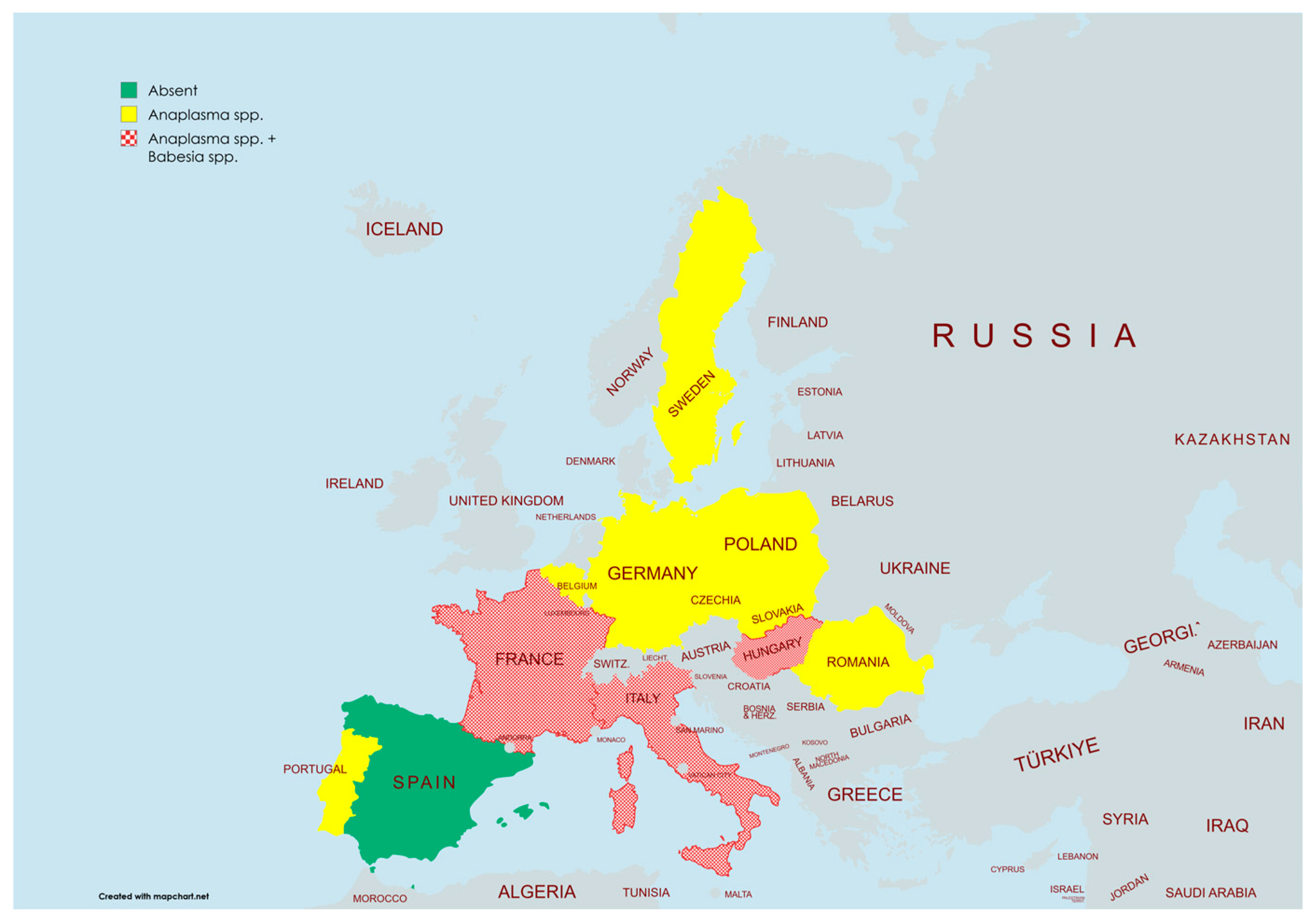
  • Green (Absent): Countries where no Anaplasma spp. or Babesia spp. have been detected (e.g., Spain).
  • Yellow (Anaplasma spp.): Countries where Anaplasma spp. has been detected but not Babesia spp. (e.g., Poland, Germany, Romania).
  • Patterned Red (Anaplasma spp. + Babesia spp.): Countries where both Anaplasma spp. and Babesia spp. have been detected (e.g., France, Italy, Hungary).
The map provides a visual representation of the geographical prevalence of these tick-borne pathogens, highlighting regions with a higher risk of infection.
Figure 4. Geographical distribution of Anaplasma and Babesia spp. in Europe [36].
Pathogens 14 00612 g004
The following tables (Table 1 and Table 2) provide a comprehensive overview of studies investigating Anaplasma and Babesia spp. in wild boars and ticks across various European countries. They include details such as the year of study, country, diagnostic methods, sample type, identified species, total number of analyzed samples, number of positive cases, and prevalence rates.
As PCR was consistently used as the diagnostic method across all studies, it was not specifically indicated in the table; the letter ‘S’ was added only in cases where PCR was followed by sequencing.
The reported prevalence of Anaplasma spp. in wild boar or in ticks collected from these animals has varied greatly across European countries over the past 15 years, reflecting differences in ecological conditions, host density, vector distribution, sampling strategies, and diagnostic methods employed. Some studies have reported relatively low prevalence rates, while others have documented significantly higher levels of infection, suggesting that regional factors, such as climate, habitat type, and wildlife management practices, may strongly influence the circulation and detection of these pathogens.
For formatting purposes, Anaplasma phagocytophilum was abbreviated as A. phag. in the table, as only a few species were identified for this pathogen.
Prevalence for Anaplasma spp. rates have ranged from as low as 0% in Spain [43] to 28.20% in Slovakia [57], and for Babesia spp., the prevalence ranges from 0% in Spain [43] to 1.50% in Hungary [38].
A. phagocytophilum was found in 5 of the 513 spleen samples that were analyzed in Belgium. These infections were determined by sequencing analysis to be members of the Dama 35 strain (GenBank accession number GQ450276.1) and the CE18 strain (GenBank accession number GQ450278.1). Compared to the previously reported prevalence of 85.6% in roe deer from the same location, the total prevalence in wild boars was 0.97% (95% CI: 0.12–1.82) [46].
Similar to these findings, A. phagocytophilum has been reported at low prevalence levels in other countries, such as the Czech Republic, where 5.10% (28 out of 550 samples) tested positive [47], and Romania (2.96%, 3.44%, 4.48%) [42,55,56].
Several studies from mainland France and Corsica have reported the presence of Anaplasma spp. and Babesia spp. in wild boars and ticks collected from them. In central France, Dugat et al. (2017) detected A. phagocytophilum in 44.8% of wild boar spleen samples collected between 2009 and 2015, the highest prevalence reported in the country [48]. Ticks collected from wild boars in the same region, including Rh. bursa and D. marginatus, were also found to carry Anaplasma spp., suggesting their role as potential vectors [37,38].
In Corsica, Grech-Angelini et al. (2019) analyzed ticks from wild and domestic animals, identifying A. phagocytophilum DNA in 2% of tick pools collected from wild boars [49]. The identified vector species included Rh. bursa, D. marginatus, I. ricinus, Hyalomma marginatum, and R. sanguineus.
More recently, Defaye et al. (2021) screened 158 wild boars from Corsica and reported three positive samples for Babesia spp., although sequencing results were inconclusive, preventing precise species identification [37]. Among the 113 ticks collected from these wild boars, 79.6% were positive for at least one vector-borne pathogen, and one D. marginatus tick tested positive for Anaplasma spp., though sequencing could not confirm the species [37].
In France (Corsica), the presence of Anaplasma spp. was minimal, with a prevalence rate of 0.88% (10/113 samples) [37]. Similarly, A. phagocytophilum in Corsican samples exhibited a low prevalence of 2% (4/177 samples) [49].
These findings suggest that both Anaplasma and Babesia spp. circulate in wild boars and their associated ticks in France, with marked regional differences in prevalence. The results also highlight the need for further studies to clarify the role of wild boars in the transmission of these pathogens and the involvement of various tick vectors across different French ecosystems.
Reported infection rates with A. phagocytophilum in wild boars from other regions have ranged between 0% and 28.2%. Higher prevalence levels have been observed in southern Germany (where 12.50% of sampled wild boars tested positive for A. phagocytophilum) [50], Poland (20.34%) [52], and Slovakia (28.2%) [57]. In contrast, studies conducted in Austria and northern Spain did not detect any wild boars infected with A. phagocytophilum [43,58,59].
A. phagocytophilum was found to be moderately prevalent in Hungary, with a frequency of 5.7% (19/333). However, just 5 out of 333 samples tested positive (1.5%) for B. canis and B. crassa, indicating a much lower frequency in Hungary [38].
Between 2010 and 2022, several studies reported the presence of Anaplasma and Babesia spp. in wild boars from different regions of Italy. In central Italy, Ebani et al. (2017) analyzed 100 spleen samples collected during hunting seasons between 2013 and 2015, detecting A. phagocytophilum in one sample (1%)—the first report of this pathogen in wild boars from Italy [51].
In northern Italy, Zanet et al. (2014) examined over 1000 wild ungulates, including 257 wild boars, and detected B. bigemina in 4.67% of wild boar samples using nested PCR and sequencing [39].
In southern Italy, Sgroi et al. (2023) analyzed 243 spleen samples collected between 2016 and 2022 and identified B. vulpes (5.3%) and Babesia capreoli (0.9%), with a total prevalence of 6.2% [41]. Although B. vulpes was not detected in blood samples, the presence of its suspected vectors (Ixodes hexagonus, Ixodes canisuga) [60,61], and that of I. ricinus for B. capreoli [62,63], suggests potential vector–host interaction in the area. These ticks are common on wildlife such as foxes, which share habitats with wild boars and may play a role in maintaining the transmission cycle [41].
In contrast, Zobba et al. (2014) found no evidence of Babesia spp. in 52 blood samples collected from wild boars in Sardinia [40]. The absence of positive cases highlights regional differences in prevalence across mainland Italy and the islands.
In Portugal, 141 free-ranging ungulates, comprising 73 red deer (Cervus elaphus), 65 wild boars (Sus scrofa), and 3 fallow deer (Dama dama), sampled from both sexes, were studied throughout the shooting seasons of December 2013 to March 2015. Five districts, Castello Branco (n = 31), Portalegre (n = 16), Lisboa (n = 19), Évora (n = 15), and Beja (n = 60), were used to collect the animals.
Two wild boars (3.1%; 95% CI: 0.4–10.7%) that were sampled had co-infections with Theileria capreoli and A. platys. Furthermore, red deer from Castelo Branco had a considerably higher incidence of Anaplasma infection than those from Beja (p = 0.019). Interestingly, the Beja district was the source of all wild boar blood samples that tested positive for T. capreoli or A. platys [54].
The locations that were sampled showed varying rates of pathogens discovered in ticks. With 93 out of 484 samples testing positive for A. phagocytophilum, Poland (East) had the greatest number of positive cases, with a 19.2% prevalence rate [52]. Germany (South), on the other hand, had the greatest relative prevalence (25%), even though there were only 16 samples, 4 of which tested positive [50].
Several studies have investigated the presence of A. phagocytophilum and Babesia spp. in wild boars from different regions of Romania. Kiss et al. (2014) conducted a large-scale survey between 2007 and 2012 on 870 organ samples (spleen, liver, kidney) collected from wild boars across 16 counties. Using nested PCR targeting the 16S rRNA gene, A. phagocytophilum was detected in 4.48% of samples, with most positive cases concentrated in central Transylvania [55].
Matei et al. (2023) analyzed 203 blood samples collected during two hunting seasons (2019–2020 and 2020–2021) in Sălaj County. They identified A. phagocytophilum in 3.0% of samples using conventional and nested PCR targeting both the 16S rRNA and groEL genes. No samples tested positive for Babesia spp. [42].
In another recent study, Dreghiciu et al. (2023) examined twenty-nine blood samples from wild boars in Hunedoara and Timiș counties, detecting A. phagocytophilum in one case (3.44%) using PCR targeting the epank1 gene [56].
Although prevalence rates remain relatively low, these findings confirm the circulation of A. phagocytophilum in wild boars from multiple regions of Romania. The absence of Babesia spp. in tested samples suggests limited distribution or lower host susceptibility, but further studies are needed to clarify this aspect.
In a Swedish study focusing on wild boars, spleen samples from 34 individuals were analyzed to detect the presence of tick-borne pathogens. The results revealed that A. phagocytophilum was present in 24 animals, indicating a notable infection rate of 70.6%. On the other hand, Babesia spp. was not detected in any of the samples tested [44]. These findings point toward a significant exposure of wild boars to A. phagocytophilum in this region, while suggesting a minimal or absent role in the transmission of Babesia under the conditions of this investigation.

General Discussion

This review provides an updated overview of the presence of Babesia spp. and Anaplasma spp. in wild boars and their associated ticks across Europe, based on studies published between 2010 and 2024. We acknowledge that differences in reported prevalence may be due to both real epidemiological variation and differences in study design or sampling. As these factors are often difficult to separate, this limitation has been noted. Even so, the collected data offers useful insights into the distribution of these pathogens and the potential role of wild boars and ticks in their circulation.
The hypothesis that wild boars can contract spontaneous infections but can also efficiently manage the pathogen is supported by the low incidence of A. phagocytophilum in these animals. Innate immune response activation and cytoskeletal reorganization are two potential mechanisms for this control, which might promote improved phagocytosis and autophagy and slow the spread of infection [64]. These findings demonstrate the geographical differences in pathogen dispersion, with France (Corsica) showing the lowest pathogen detection frequencies and Poland and Germany showing the greatest prevalence rates. Tick vector distribution, host reservoir dynamics, or environmental conditions may have an impact on the observed discrepancies, requiring more research (Figure 5).
Significant geographical diversity in infection rates was shown by the range of A. phagocytophilum prevalence in ticks, which varied from a low of 0.88% in Corsica (France) [37] to a maximum of 25% in southern Germany [50].
Spleen, liver, and kidney samples from wild boars are among the most informative for disease diagnosis, owing to both their higher sample counts and elevated positivity rates. In contrast, although numerous blood samples were collected, their relatively low prevalence rates suggest limited diagnostic value. As illustrated in Figure 6, spleen samples and combined multi-organ samples (spleen, liver, and kidney) appear to be the most reliable for detecting infections.
The examination of pathogen presence in several wild boar sample types showed notable differences in frequency, positive rate, and sample size. With the biggest sample size (n = 870) and the highest number of positive cases (n = 39), the wild boar spleen/liver/kidney group demonstrated a significant presence of pathogens in multi-organ samples.
The identification of pathogens in wild boars differed depending on the pathogen and tissue type. The efficacy of multi-tissue sampling was demonstrated by the highest prevalence of A. phagocytophilum in spleen/blood (28.20%) [57] and spleen/liver (20.34%) [52]. Other pathogens such as Babesia spp. showed very low to no detection across all sample types [37,38,39,40]. Overall, compared to blood alone, spleen and mixed tissue samples demonstrated greater reliability for pathogen surveillance.
This result implies that compared to spleen or multi-organ samples, blood samples can be less effective in identifying specific pathogen samples.
Until 2010, there was a clear lack of research focused on Babesia infections in wild boars or in ticks collected from them. The topic was rarely addressed, and detailed molecular studies were mostly missing. This made it difficult to understand the role that wild boars and their ectoparasites might have in the transmission cycle of Babesia spp. Overall, the limited data available from that period highlight how little was known about the involvement of wildlife in the ecology of these pathogens.
Despite its broad geographic range, considerable gaps remain in our understanding of A. phagocytophilum ecology, epidemiology, and vector–host dynamics. Limited data on the biological traits of its vectors, the diversity and pathogenic potential of circulating strains, and the infection sources continue to prevent the development of targeted strategies for disease control and prevention in both animals and humans [65]. The lack of evidence for transovarial transmission in the main tick vectors linked to human and animal infections highlights the critical role of vertebrate reservoirs in sustaining the pathogen in natural ecosystems [66,67]. Therefore, improving our understanding of strain variability, spatial distribution, and host–vector–pathogen interactions is essential for producing accurate risk assessments and informing veterinary and public health interventions.
Although traditionally considered a single bacterial species, A. phagocytophilum has shown considerable genetic diversity and a broad host range, as revealed by 16S rRNA gene analyses [68]. The detection of specific genetic variants in ticks appears to correlate with habitat characteristics and the presence of certain reservoir hosts, suggesting some degree of host-linked strain distribution. When more refined molecular approaches are applied, patterns of host specificity become increasingly evident, pointing to a complex network of host–pathogen interactions. These findings underscore the importance of considering the genetic variability of A. phagocytophilum when evaluating its transmission pathways, particularly the potential involvement of wild ruminants in maintaining and spreading strains that may also affect domestic animals and humans [69].
Studies conducted in Central Europe until 2010 have reported variable prevalence rates of A. phagocytophilum in wild boar tissues. In Poland, Michalik et al. (2012) identified the pathogen in 12% of 325 spleen samples using nested PCR [70]. In Slovakia, Smetanová et al. (2006) reported a prevalence of 5.5% based on 18 samples [71], while in the Czech Republic, Hulínská et al. (2004) found a detection rate of 4.4% in 69 wild boars [72]. A study from Slovenia conducted by Galindo et al., 2013, also found a relatively low prevalence of 2.7% in 113 wild boar samples [64]. These findings indicate a relatively low to moderate presence of A. phagocytophilum in wild boars across these regions.
In contrast, more recent investigations included in our review show notably higher rates, such as 20.34% in Poland [52], 28.2% in Slovakia [57], 44,8% in France [48], and 70.60% in Sweden [44]. Meanwhile, several countries such as Portugal (3.1%) [53] and Romania (2.96–4.48%) [42,55,56] continue to report consistently low levels of infection. These findings suggest both ecological and methodological sources of variation and emphasize the need for standardized diagnostic approaches to enable reliable comparisons between regions.
Prevalence studies of A. phagocytophilum in questing ticks across Europe show a wide range of values, as reported in multiple countries using PCR-based methods. While most prevalence estimates fall between 1% and 10%, a few countries report markedly higher or lower rates. For example, Denmark recorded one of the highest prevalences at 23.6% [73], and Bulgaria reported 33.9% in adult ticks, contrasting with just 2.2% in nymphs [74]. In Italy, values ranged from 1.5% [75] to 24.4% [76], reflecting regional and temporal variability. Conversely, countries such as Hungary (0.4%) [77], the Netherlands (0.6%) [78], Luxembourg (1.9%) [79], and Switzerland (1.2–2%) [80,81,82] consistently showed low infection rates. Germany presented predominantly low to moderate values, between 1.0% and 5.4%, though isolated studies reported higher figures up to 17.4%. Other countries, including Poland, France, and the UK, exhibited intermediate prevalence rates typically in the 2–10% range, although with noticeable intra-national variation [69].
These differences likely stem from variations in geographic region, tick developmental stage, detection method (PCR vs. qPCR), and host or reservoir presence. Collectively, the data highlights the uneven but widespread distribution of A. phagocytophilum in European tick populations.
When comparing the prevalence of Anaplasma phagocytophilum in questing ticks, as compiled by Stuen et al. (2013) [69], with the data from our review referring to ticks collected from wild boars, similar trends in variability are observed. In both reviews, prevalence values range from very low (e.g., 0.88–2% in France (Corsica) [37,49] and 5.70% in Hungary [38]) to relatively high prevalence, such as 19.2% in Poland and 25% [53] in Germany [50], which is consistent across both questing ticks and host-attached ticks. These parallels suggest comparable exposure prevalence in certain geographic areas. Differences between the two groups are also notable and may be influenced by the stage of the tick, the timing of sample collection, or differences in host availability and reservoir competence.
Overall, both datasets confirm the widespread yet not equal presence of A. phagocytophilum in European tick populations, whether questing or associated with wild boars.
It is important to recognize the limitations of this review. A formal risk of bias assessment was not conducted, and a meta-analysis could not be performed due to the heterogeneity of study designs and diagnostic techniques. Despite these limitations, the synthesis identifies important regional and epidemiological trends and offers a thorough summary of the incidence of Babesia spp. and Anaplasma spp. in wild boars and ticks throughout Europe.
We acknowledge that some of the variation in reported prevalence may be due to differences in study design, sampling methods, and molecular techniques. It is also possible that these differences reflect true variations in pathogen circulation by ecological, geographical, and host-specific factors.

4. Conclusions

The review demonstrates that wild boars across Europe serve as significant hosts for a diverse range of vector-borne pathogens, including Anaplasma and Babesia spp. Vector ticks, including D. marginatus and Rh. bursa, were frequently infected, suggesting that they may play a role in the transmission dynamics, which is not usually observed in I. ricinus.
The consistent detection of zoonotic agents, especially A. phagocytophilum and Babesia spp., across various studies highlights their potential role in maintaining and possibly amplifying these pathogens in natural environments. The reviewed literature also reveals substantial methodological heterogeneity, with differences in sample types (blood, spleen, ticks), molecular protocols, and reporting standards, which limits direct comparison between studies. To enhance epidemiological insight and inform public and veterinary health strategies, future research should prioritize harmonized sampling procedures and standardized molecular diagnostics.
These findings also underscore the need for standardized methodologies to improve data comparability across Europe.

Author Contributions

Conceptualization, I.C.D. and M.S.I.; methodology, I.C.D. and M.S.I.; validation, I.O., S.D., S.M., and M.S.I.; formal analysis, I.C.D.; data curation, I.C.D., V.I., and S.D.; writing—original draft preparation, I.C.D.; writing—review and editing, M.S.I., T.F., I.O., and S.M.; visualization, M.I., D.H., and A.P.; supervision, M.S.I. All authors have read and agreed to the published version of the manuscript.

Funding

This research received no external funding, and the APC was funded by the University of Life Science “King Mihai I” from Timișoara.

Institutional Review Board Statement

Not applicable.

Data Availability Statement

Data are contained within the article.

Conflicts of Interest

The authors declare no conflicts of interest.

References

  1. Stuen, S.; Scharf, W.; Schauer, S.; Freyburger, F.; Bergström, K.; von Loewenich, F.D. Experimental Infection in Lambs with a Red Deer (Cervus elaphus) Isolate of Anaplasma phagocytophilum. J. Wildl. Dis. 2010, 46, 803–809. [Google Scholar] [CrossRef]
  2. Ballari, S.A.; Barrios-García, M.N. A review of wild boar Sus scrofa diet and factors affecting food selection in native and introduced ranges. Mammal Rev. 2014, 44, 124–134. [Google Scholar] [CrossRef]
  3. Massei, G.; Genov, P.V. The environmental impact of wild boar. Galemys 2004, 16, 135–145. [Google Scholar] [CrossRef]
  4. Pittiglio, C.; Khomenko, S.; Beltran-Alcrudo, D. Wild boar mapping using population-density statistics: From polygons to high resolution raster maps. PLoS ONE 2018, 13, e0193295. [Google Scholar] [CrossRef]
  5. Apollonio, M.; Andersen, R.; Putman, R. European Ungulates and Their Management in the 21st Century; Cambridge University Press: Cambridge, UK, 2010. [Google Scholar]
  6. Massei, G.; Kindberg, J.; Licoppe, A.; Gačić, D.; Šprem, N.; Kamler, J.; Baubet, E.; Hohmann, U.; Monaco, A.; Ozoliņš, J.; et al. Wild boar populations up, numbers of hunters down? A review of trends and implications for Europe. Pest Manag. Sci. 2015, 71, 492–500. [Google Scholar] [CrossRef]
  7. Lewis, J.S.; Farnsworth, M.L.; Burdett, C.L.; Theobald, D.M.; Gray, M.; Miller, R.S. Biotic and abiotic factors predicting the global distribution and population density of an invasive large mammal. Sci. Rep. 2017, 7, 44152. [Google Scholar] [CrossRef] [PubMed]
  8. Sales, L.P.; Ribeiro, B.R.; Hayward, M.W.; Paglia, A.; Passamani, M.; Loyola, R. Niche conservatism and the invasive potential of the wild boar. J. Anim. Ecol. 2017, 86, 1214–1223. [Google Scholar] [CrossRef] [PubMed]
  9. McClure, M.L.; Burdett, C.L.; Farnsworth, M.L.; Lutman, M.W.; Theobald, D.M.; Riggs, P.D.; Grear, D.A.; Miller, R.S. Modeling and mapping the probability of occurrence of invasive wild pigs across the contiguous United States. PLoS ONE 2015, 10, e0133771. [Google Scholar] [CrossRef]
  10. Snow, N.P.; Jarzyna, M.A.; VerCauteren, K.C. Interpreting and predicting the spread of invasive wild pigs. J. Appl. Ecol. 2017, 54, 2022–2032. [Google Scholar] [CrossRef]
  11. Morelle, K.; Fattebert, J.; Mengal, C.; Lejeune, P. Invading or recolonizing? Patterns and drivers of wild boar population expansion into Belgian agroecosystems. Agric. Ecosyst. Environ. 2016, 222, 267–275. [Google Scholar] [CrossRef]
  12. Markov, N.; Pankova, N.; Morelle, K. Where winter rules: Modeling wild boar distribution in its north-eastern range. Sci. Total Environ. 2019, 687, 1055–1064. [Google Scholar] [CrossRef]
  13. Bieber, C.; Ruf, T. Population dynamics in wild boar Sus scrofa: Ecology, elasticity of growth rate and implications for the management of pulsed resource consumers. J. Appl. Ecol. 2005, 42, 1203–1213. [Google Scholar] [CrossRef]
  14. Jędrzejewski, W.; Jędrzejewska, B.; Okarma, H.; Ruprecht, A.L. Wolf predation and snow cover as mortality factors in the ungulate community of the Białowieża National Park, Poland. Oecologia 1992, 90, 27–36. [Google Scholar] [CrossRef] [PubMed]
  15. Carvalho, J.; Hipólito, D.; Teixeira, D.; Fonseca, C.; Tinoco, T.R. Hunting bag statistics of wild mammals in Portugal (1989–2022): On the need to improve data report and compilation. Eur. J. Wildl. Res. 2024, 70, 96. [Google Scholar] [CrossRef]
  16. Ruiz-Rodríguez, C.; Fernández-López, J.; Vicente, J.; Blanco-Aguiar, J.A.; Acevedo, P. Revisiting wild boar spatial models based on hunting yields to assess their predictive performance on interpolation and extrapolation areas. Ecol. Model. 2022, 471, 110041. [Google Scholar] [CrossRef]
  17. Bergqvist, G.; Kindberg, J.; Elmhagen, B. From virtually extinct to superabundant in 35 years: Establishment, population growth and shifts in management focus of the Swedish wild boar (Sus scrofa) population. BMC Zool. 2024, 9, 14. [Google Scholar] [CrossRef]
  18. Kopij, G. Population density of the wild boar (Sus scrofa) in south-western Poland in 1981–2020. Theriologia Ukrainica 2022, 24, 171–183. [Google Scholar] [CrossRef]
  19. Yin, H.; Luo, J.; Liu, Z.; Yang, J.; Mu, Y.; Zhang, P.; Guan, G. Babesiosis in domestic animals in China: Epidemiology and diagnosis. Transbound. Emerg. Dis. 2020, 67, 1294–1307. [Google Scholar]
  20. Hikosaka, K.; Nakamura, Y.; Tateno, M.; Matsuu, A.; Asada, M. A new species of Babesia infective to domestic pigs. J. Vet. Med. Sci. 2015, 77, 1655–1657. [Google Scholar]
  21. René-Martellet, M.; Lebert, I.; Chêne, J.; Massot, R.; Leon, M.; Leal, A.; Badavelli, S.; Chalvet-Monfray, K.; Ducrot, C.; Abrial, D.; et al. Diagnosis and incidence risk of clinical canine monocytic ehrlichiosis under field conditions in Southern Europe. Parasit. Vectors 2015, 8, 3. [Google Scholar] [CrossRef]
  22. de Waal, D.T. Porcine babesiosis. In Infectious Diseases of Livestock; Coetzer, J.A.W., Thomson, G.E., Maclachlan, N.J., Penrith, M.L., Eds.; Anipedia: Pretoria, South Africa, 2019. [Google Scholar]
  23. Dumitrache, M.O.; Matei, I.A.; Ionică, A.M.; Kalmár, Z.; Sándor, A.D.; Mărcuțan, I.D.; Gherman, C.M. Molecular detection and characterization of Anaplasma species in wild boars (Sus scrofa) from North-Eastern Romania. Parasit Vectors 2006, 11, 1–9. [Google Scholar]
  24. Galon, N.; Kohn, B.; Burridge, M.J. Anaplasma phagocytophilum and Anaplasma marginale: Comparison of nucleotide sequences for phylogenetic studies. BMC Microbiol. 2018, 18, 1–10. [Google Scholar]
  25. Mărcuţan, I.D.; Mihalca, A.D.; D’Amico, G.; Sándor, A.D.; Modrý, D.; Gherman, C.M. Molecular Detection of Anaplasma phagocytophilum in Wild Boars (Sus scrofa) from Romania. Vet. Parasitol. 2016, 228, 98–101. [Google Scholar]
  26. Pilloux, L.; Baumgartner, A.; Jaton, K.; Lienhard, R.; Ackermann-Gäumann, R.; Beuret, C.; Greub, G. Prevalence of Anaplasma phagocytophilum and Coxiella burnetii in Ixodes ricinus Ticks in Switzerland: An Underestimated Epidemiologic Risk. New Microbes New Infect. 2019, 27, 22–26. [Google Scholar] [CrossRef] [PubMed]
  27. Scharf, W.; Schauer, S.; Freyburger, F.; Petrovec, M.; Schaarschmidt-Kiener, D.; Liebisch, G.; Runge, M.; Ganter, M.; Kehl, A.; Dumler, J.S.; et al. Distinct Host Species Correlate with Anaplasma phagocytophilum ankA Gene Clusters. J. Clin. Microbiol. 2011, 49, 790–796. [Google Scholar] [CrossRef]
  28. Rikihisa, Y. Anaplasma phagocytophilum and Ehrlichia chaffeensis: Subversive Manipulators of Host Cells. Nat. Rev. Microbiol. 2010, 8, 328–339. [Google Scholar] [CrossRef]
  29. Popov, V.L.; Han, V.C.; Chen, S.M.; Dumler, J.S.; Feng, H.M.; Andreadis, T.G.; Tesh, R.B.; Walker, D.H. Ultrastructural Differentiation of the Genogroups in the Genus Ehrlichia. J. Med. Microbiol. 1998, 47, 235–251. [Google Scholar] [CrossRef]
  30. Troese, M.J.; Kahlon, A.; Ragland, S.A.; Ottens, A.K.; Ojogun, N.; Nelson, K.T.; Walker, N.J.; Borjesson, D.L.; Carlyon, J.A. Proteomic Analysis of Anaplasma phagocytophilum During Infection of Human Myeloid Cells Identifies a Protein that is Pronouncedly Upregulated on the Infectious Dense-Cored Cell. Infect. Immun. 2011, 79, 4696–4707. [Google Scholar] [CrossRef]
  31. Boulanger, N.; Boyer, P.; Talagrand-Reboul, E.; Hansmann, Y. Ticks and tick-borne diseases. Med. Mal Infect. 2019, 49, 87–97. [Google Scholar] [CrossRef]
  32. de la Fuente, J.; Contreras, M. Tick vaccines: Current status and future directions. Expert Rev. Vaccines 2015, 14, 1367–1376. [Google Scholar] [CrossRef]
  33. Estrada-Peña, A.; Gray, J.S.; Kahl, O.; Lane, R.S.; Nijhof, A.M. Research on the ecology of ticks and tick-borne pathogens—methodological principles and caveats. Front. Cell Infect Microbiol. 2013, 3, 29. [Google Scholar] [CrossRef]
  34. Medlock, J.M.; Hansford, K.M.; Bormane, A.; Derdakova, M.; Estrada-Peña, A.; George, J.C.; Golovljova, I.; Jaenson, T.G.; Jensen, J.K.; Jensen, P.M.; et al. Driving forces for changes in geographical distribution of Ixodes ricinus ticks in Europe. Parasit Vectors. 2013, 6, 1. [Google Scholar] [CrossRef]
  35. Page, M.J.; McKenzie, J.E.; Bossuyt, P.M.; Boutron, I.; Hoffmann, T.C.; Mulrow, C.D.; Shamseer, L.; Tetzlaff, J.M.; Akl, E.A.; Brennan, S.E.; et al. The PRISMA 2020 statement: An updated guideline for reporting systematic reviews. BMJ Clin. Res. Ed. 2021, 372, n71. [Google Scholar]
  36. MapChart. Available online: https://www.mapchart.net/europe.html (accessed on 16 March 2025).
  37. Defaye, B.; Moutailler, S.; Pietri, C.; Galon, C.; Grech-Angelini, S.; Pasqualini, V.; Quilichini, Y. Molecular Detection of Zoonotic and Non-Zoonotic Pathogens from Wild Boars and Their Ticks in the Corsican Wetlands. Pathogens 2021, 10, 1643. [Google Scholar] [CrossRef] [PubMed]
  38. Hornok, S.; Szekeres, S.; Horváth, G.; Takács, N.; Bekő, K.; Kontschán, J.; Gyuranecz, M.; Tóth, B.; Sándor, A.D.; Juhász, A.; et al. Diversity of Tick Species and Associated Pathogens on Peri-Urban Wild Boars—First Report of the Zoonotic Babesia cf. crassa from Hungary. Ticks Tick Borne Dis. 2022, 13, 101936. [Google Scholar] [CrossRef] [PubMed]
  39. Zanet, S.; Trisciuoglio, A.; Bottero, E.; de Mera, I.G.; Gortazar, C.; Carpignano, M.G.; Ferroglio, E. Piroplasmosis in Wildlife: Babesia and Theileria Affecting Free-Ranging Ungulates and Carnivores in the Italian Alps. Parasit. Vectors 2014, 7, 70. [Google Scholar] [CrossRef] [PubMed]
  40. Zobba, R.; Nuvoli, A.M.; Sotgiu, F.; Lecis, R.; Spezzigu, A.; Dore, G.M.; Masia, M.A.; Cacciotto, C.; Parpaglia, M.L.; Dessì, D.; et al. Molecular Epizootiology and Diagnosis of Porcine Babesiosis in Sardinia, Italy. Vector Borne Zoonotic Dis. 2014, 14, 716–723. [Google Scholar] [CrossRef]
  41. Sgroi, G.; D’Alessio, N.; Auriemma, C.; Salant, H.; Gallo, A.; Riccardi, M.G.; Alfano, F.; Rea, S.; Scarcelli, S.; Ottaviano, M.; et al. First Molecular Detection of Babesia vulpes and Babesia capreoli in Wild Boars from Southern Italy. Front. Vet. Sci. 2023, 10, 1201476. [Google Scholar] [CrossRef]
  42. Matei, I.A.; Kalmár, Z.; Balea, A.; Mihaiu, M.; Sándor, A.D.; Cocian, A.; Crăciun, S.; Bouari, C.; Briciu, V.T.; Fiț, N. The Role of Wild Boars in the Circulation of Tick-Borne Pathogens: The First Evidence of Rickettsia monacensis Presence. Animals 2023, 13, 1743. [Google Scholar] [CrossRef]
  43. Díaz-Cao, J.M.; Adaszek, Ł.; Dzięgiel, B.; Paniagua, J.; Caballero-Gómez, J.; Winiarczyk, S.; Winiarczyk, D.; Cano-Terriza, D.; García-Bocanegra, I. Prevalence of Selected Tick-Borne Pathogens in Wild Ungulates and Ticks in Southern Spain. Transbound. Emerg. Dis. 2022, 69, 1084–1094. [Google Scholar] [CrossRef]
  44. Fabri, N.D.; Sprong, H.; Hofmeester, T.R.; Heesterbeek, H.; Donnars, B.F.; Widemo, F.; Ecke, F.; Cromsigt, J.P.G.M. Wild ungulate species differ in their contribution to the transmission of Ixodes ricinus-borne pathogens. Parasit Vectors 2021, 14, 360. [Google Scholar] [CrossRef] [PubMed]
  45. Karlsson, M.E.; Andersson, M.O. Babesia species in questing Ixodes ricinus, Sweden. Ticks Tick Borne Dis. 2016, 7, 10–12. [Google Scholar] [CrossRef]
  46. Nahayo, A.; Bardiau, M.; Volpe, R.; Pirson, J.; Paternostre, J.; Fett, T.; Linden, A. Molecular Evidence of Anaplasma phagocytophilum in Wild Boar (Sus scrofa) in Belgium. BMC Vet. Res. 2014, 10, 80. [Google Scholar] [CrossRef]
  47. Hrazdilová, K.; Lesiczka, P.M.; Bardoň, J.; Vyroubalová, Š.; Šimek, B.; Zurek, L.; Modrý, D. Wild Boar as a Potential Reservoir of Zoonotic Tick-Borne Pathogens. Ticks Tick Borne Dis. 2021, 12, 101558. [Google Scholar] [CrossRef]
  48. Dugat, T.; Haciane, D.; Durand, B.; Lagrée, A.C.; Haddad, N.; Boulouis, H.J. Identification of a Potential Marker of Anaplasma phagocytophilum Associated with Cattle Abortion. Transbound. Emerg. Dis. 2017, 64, e1–e3. [Google Scholar] [CrossRef]
  49. Grech-Angelini, S.; Stachurski, F.; Vayssier-Taussat, M.; Devillers, E.; Casabianca, F.; Lancelot, R.; Uilenberg, G.; Moutailler, S. Tick-Borne Pathogens in Ticks (Acari: Ixodidae) Collected from Various Domestic and Wild Hosts in Corsica (France), a Mediterranean Island Environment. Transbound. Emerg. Dis. 2019, 67, 745–757. [Google Scholar] [CrossRef]
  50. Silaghi, C.; Pfister, K.; Overzier, E. Molecular Investigation for Bacterial and Protozoan Tick-Borne Pathogens in Wild Boars (Sus scrofa) from Southern Germany. Vector Borne Zoonotic Dis. 2014, 14, 371–373. [Google Scholar] [CrossRef] [PubMed]
  51. Ebani, V.V.; Bertelloni, F.; Cecconi, G.; Sgorbini, M.; Cerri, D. Zoonotic Tick-Borne Bacteria among Wild Boars (Sus scrofa) in Central Italy. Asian Pac. J. Trop. Med. Press 2017, 7, 141–143. [Google Scholar] [CrossRef]
  52. Myczka, A.W.; Steiner-Bogdaszewska, Ż.; Filip-Hutsch, K.; Oloś, G.; Czopowicz, M.; Laskowski, Z. Detection of Anaplasma phagocytophilum in Wild and Farmed Cervids in Poland. Pathogens 2021, 10, 1190. [Google Scholar] [CrossRef]
  53. Michalski, M.M.; Kubiak, K.; Szczotko, M.; Dmitryjuk, M. Tick-Borne Pathogens in Ticks Collected from Wild Ungulates in North-Eastern Poland. Pathogens 2021, 10, 587. [Google Scholar] [CrossRef]
  54. Pereira, A.; Parreira, R.; Nunes, M.; Casadinho, A.; Vieira, M.L.; Campino, L.; Maia, C. Molecular Detection of Tick-Borne Bacteria and Protozoa in Cervids and Wild Boars from Portugal. Parasit. Vectors 2016, 9, 251. [Google Scholar] [CrossRef] [PubMed]
  55. Kiss, T.; Cadar, D.; Krupaci, F.A.; Bordeanu, A.D.; Spînu, M. Prevalence of Anaplasma phagocytophilum Infection in European Wild Boar (Sus scrofa) Populations from Transylvania, Romania. Epidemiol. Infect. 2014, 142, 246–250. [Google Scholar] [CrossRef] [PubMed]
  56. Dreghiciu, I.C.; Imre, M.; Oprescu, I.; Florea, T.; Ghilean, B.M.; Sîrbu, B.A.M.; Iorgoni, V.; Badea, C.; Giubega, S.; Mederle, N.; et al. Molecular Detection of Anaplasma phagocytophilum in Wild Boar (Sus scrofa) from Hunedoara and Timiș Counties—Preliminary Study. Rev. Rom. Med. Vet. 2023, 33, 59–62. [Google Scholar]
  57. Kazimírová, M.; Hamšíková, Z.; Špitalská, E.; Minichová, L.; Mahríková, L.; Caban, R.; Sprong, H.; Fonville, M.; Schnittger, L.; Kocianová, E. Diverse Tick-Borne Microorganisms Identified in Free-Living Ungulates in Slovakia. Parasit. Vectors 2018, 11, 495. [Google Scholar] [CrossRef]
  58. Polin, H.; Hufnagl, P.; Hauschmid, R.; Gruber, F.; Guther, L. Molecular Evidence of Anaplasma phagocytophilum in Ixodes ricinus Ticks and Wild Animals in Austria. J. Clin. Microbiol. 2004, 42, 2285–2286. [Google Scholar] [CrossRef]
  59. Portillo, A.; Perez-Martinez, L.; Santibanez, S.; Santibanez, P.; Palomar, A.M.; Oteo, J.A. Anaplasma spp. in Wild Mammals and Ixodes ricinus from the North of Spain. Vector Borne Zoonotic Dis. 2011, 11, 3–8. [Google Scholar] [CrossRef] [PubMed]
  60. Camacho, A.T.; Pallas, E.; Gestal, J.J.; Guitián, F.J.; Olmeda, A.; Telford, S.R. Ixodes hexagonus is the Main Candidate as Vector of Theileria annae in Northwest Spain. Vet. Parasitol. 2003, 112, 157–163. [Google Scholar] [CrossRef]
  61. Obsomer, V.; Wirtgen, M.; Linden, A.; Claerebout, E.; Heyman, P.; Heylen, D.; Madder, M.; Maris, J.; Lebrun, M.; Tack, W.; et al. Spatial Disaggregation of Tick Occurrence and Ecology at a Local Scale as a Preliminary Step for Spatial Surveillance of Tick-Borne Diseases: General Framework and Health Implications in Belgium. Parasit. Vectors 2013, 6, 190. [Google Scholar] [CrossRef]
  62. Estrada-Peña, A.; Mihalca, A.D.; Petney, T.N. Ticks of Europe and North Africa: A Guide to Species Identification; Springer Nature: Cham, Switzerland, 2017; p. 368. [Google Scholar]
  63. Bajer, A.; Dwużnik-Szarek, D. The Specificity of Babesia-Tick Vector Interactions: Recent Advances and Pitfalls in Molecular and Field Studies. Parasit. Vectors 2021, 14, 507. [Google Scholar] [CrossRef]
  64. Galindo, R.C.; Ayllon, N.; Strasek, S.; Boadella, M.; Beltran-Beck, B.; Mazariegos, M. Gene Expression Profile Suggests that Pigs (Sus scrofa) are Susceptible to Anaplasma phagocytophilum but Control Infection. Parasites Vectors 2012, 5, 181. [Google Scholar] [CrossRef]
  65. Zhang, L.; Wang, G.; Liu, Q.; Chen, C.; Li, J.; Long, B.; Yu, H.; Zhang, Z.; He, J.; Qu, Z.; et al. Molecular analysis of Anaplasma phagocytophilum isolated from patients with febrile diseases of unknown etiology in China. PLoS ONE 2013, 8, e57155. [Google Scholar] [CrossRef] [PubMed]
  66. Ogden, N.H.; Bown, K.; Horrocks, B.K.; Woldehiwet, Z.; Bennett, M. Granulocytic Ehrlichia infection in ixodid ticks and mammals in woodlands and uplands of the U.K. Med. Vet. Entomol. 1998, 12, 423–429. [Google Scholar] [CrossRef] [PubMed]
  67. Liz, J.S.; Sumner, J.W.; Pfister, K.; Brossard, M. PCR detection and serological evidence of granulocytic ehrlichial infection in roe deer (Capreolus capreolus) and chamois (Rupicapra rupicapra). J. Clin. Microbiol. 2002, 40, 892–897. [Google Scholar] [CrossRef]
  68. Overzier, E.; Pfister, K.; Thiel, C.; Herb, I.; Mahling, M.; Silaghi, C. Anaplasma phagocytophilum in questing Ixodes ricinus Ticks: Comparison of prevalences and partial 16S rRNA gene variants in urban, pasture, and natural habitats. Appl. Environ. Microbiol. 2013, 79, 1730–1734. [Google Scholar] [CrossRef] [PubMed]
  69. Stuen, S.; Granquist, E.G.; Silaghi, C. Anaplasma phagocytophilum—A widespread multi-host pathogen with highly adaptive strategies. Front. Cell Infect. Microbiol. 2013, 3, 31. [Google Scholar] [CrossRef]
  70. Michalik, J.; Stanczak, J.; Cieniuch, S.; Racewicz, M.; Sikora, B.; Dabert, M. Wild boars as hosts of human-pathogenic Anaplasma phagocytophilum variants. Emerg. Infect. Dis. 2012, 18, 998–1001. [Google Scholar] [CrossRef]
  71. Smetanová, K.; Schwarzová, K.; Kocianová, E. Detection of Anaplasma phagocytophilum, Coxiella burnetii, Rickettsia spp., and Borrelia burgdorferi s. l. in Ticks, and wild-living animals in western and middle Slovakia. Ann. N. Y. Acad. Sci. 2006, 1078, 312–315. [Google Scholar] [CrossRef]
  72. Hulínská, D.; Langøová, K.; Pejèoch, M.; Pavlásek, I. Detection of Anaplasma phagocytophilum in animals by real-time polymerase chain reaction. Apmis 2004, 112, 239–247. [Google Scholar] [CrossRef]
  73. Skarphedinsson, S.; Lyholm, B.F.; Ljungberg, M.; Sogaard, P.; Kolmos, H.J.; Nielsen, L.P. Detection and identification of Anaplasma phagocytophilum, Borrelia burgdorferi, and Rickettsia helvetica in Danish Ixodes ricinus ticks. Apmis 2007, 115, 225–230. [Google Scholar] [CrossRef]
  74. Christova, I.; Schouls, L.; van De Pol, I.; Park, J.; Panayotov, S.; Lefterova, V.; Kantardjiev, T.; Dumler, J.S. High prevalence of granulocytic Ehrlichiae and Borrelia burgdorferi sensu lato in Ixodes ricinus ticks from Bulgaria. J. Clin. Microbiol. 2001, 39, 4172–4174. [Google Scholar] [CrossRef]
  75. Capelli, G.; Ravagnan, S.; Montarsi, F.; Ciocchetta, S.; Cazzin, S.; Porcellato, E.; Babiker, A.M.; Cassini, R.; Salviato, A.; Cattoli, G.; et al. Occurrence and identification of risk areas of Ixodes ricinus-borne pathogens: A cost-effectiveness analysis in north-eastern Italy. Parasit Vectors 2012, 5, 61. [Google Scholar] [CrossRef] [PubMed]
  76. Cinco, M.; Padovan, D.; Murgia, R.; Maroli, M.; Frusteri, L.; Heldtander, M.; Johansson, K.E.; Engvall, E.O. Coexistence of Ehrlichia phagocytophila and Borrelia burgdorferi sensu lato in Ixodes ricinus ticks from Italy as determined by 16S rRNA gene sequencing. J. Clin. Microbiol. 1997, 35, 3365–3366. [Google Scholar] [CrossRef]
  77. Egyed, L.; Elo, P.; Sreter-Lancz, Z.; Szell, Z.; Balogh, Z.; Sreter, T. Seasonal activity and tick-borne pathogen infection rates of Ixodes ricinus ticks in Hungary. Ticks Tick Borne Dis. 2012, 3, 90–94. [Google Scholar] [CrossRef] [PubMed]
  78. Wielinga, P.R.; Gaasenbeek, C.; Fonville, M.; de Boer, A.; de Vries, A.; Dimmers, W.; Akkerhuis Op Jagers, G.; Schouls, L.M.; Borgsteede, F.; van der Giessen, J.W. Longitudinal analysis of tick densities and Borrelia, Anaplasma, and Ehrlichia infections of Ixodes ricinus ticks in different habitat areas in The Netherlands. Appl. Environ. Microbiol. 2006, 72, 7594–7601. [Google Scholar] [CrossRef] [PubMed]
  79. Reye, A.L.; Hübschen, J.M.; Sausy, A.; Müller, C.P. Prevalence and seasonality of tick-borne pathogens in questing Ixodes ricinus ticks from Luxembourg. Appl. Environ. Microbiol. 2010, 76, 2923–2931. [Google Scholar] [CrossRef]
  80. Wicki, R.; Sauter, P.; Mettler, C.; Natsch, A.; Enzler, T.; Pusterla, N.; Kuhnert, P.; Egli, G.; Bernasconi, M.; Lienhard, R.; et al. Swiss Army Survey in Switzerland to determine the prevalence of Francisella tularensis, members of the Ehrlichia phagocytophila genogroup, Borrelia burgdorferi sensu lato, and tick-borne encephalitis virus in ticks. Eur. J. Clin. 2000, 19, 427–432. [Google Scholar] [CrossRef]
  81. Burri, C.; Dupasquier, C.; Bastic, V.; Gern, L. Pathogens of emerging tick-borne diseases, Anaplasma phagocytophilum, Rickettsia spp., and Babesia spp., in Ixodes ticks collected from rodents at four sites in Switzerland (Canton of Bern). Vector Borne Zoonotic Dis. 2011, 11, 939–944. [Google Scholar] [CrossRef]
  82. Leutenegger, C.M.; Pusterla, N.; Mislin, C.N.; Weber, R.; Lutz, H. Molecular evidence of coinfection of ticks with Borrelia burgdorferi sensu lato and the human granulocytic ehrlichiosis agent in Switzerland. J. Clin. Microbiol. 1999, 37, 3390–3391. [Google Scholar] [CrossRef]
Figure 1. Wild boar density in Europe and Asia by Pittiglio et al., 2018 [4].
Pathogens 14 00612 g001
Figure 2. Estimated wild boar hunting bag in selected European countries (2010–2024). Portugal [15], Spain [16], Sweden [17], Poland [18].
Pathogens 14 00612 g002
Figure 3. Identification and screening of studies on tick-borne pathogens in European wild boars.
Pathogens 14 00612 g003
Figure 5. Prevalence of pathogens in ticks from wild boar.
Pathogens 14 00612 g005
Figure 6. Prevalence of pathogens in wild boar samples.
Pathogens 14 00612 g006
Table 1. Evidence of Babesia in wild boars and ticks across European countries.
Years of StudyCountryDiag. MethodHost/SampleSpeciesNo. of SamplesPositivePrevalenceRef.
2018–2020France (Corsica) BloodBabesia spp.15832%[37]
2020Hungary Ticks (D. reticulatus, H. concinna)B. canis, B. crassa33351.50%[38]
2008–2012Italy (North) SpleenB. bigemina257124.67%[39]
2010–2013Italy (Sardinia) BloodBabesia spp.5200%[40]
2016–2022Italy (South)S.SpleenB. vulpes, B. capreoli243156.20%[41]
2019–2021RomaniaS.BloodBabesia spp.20300%[42]
2009–2015Spain SpleenBabesia spp.26900%[43]
2018–2019Sweden SpleenBabesia spp.3400%[44]
2013–2014Sweden Ticks (I. ricinus)B. microti, 519173.6%[45]
B. divergens, 10.2%
B. venatorum51%
Legend: PCR was consistently used as the diagnostic method across all studies; therefore, it is not specifically indicated in the table; S = Sequencing.
Table 2. Evidence of Anaplasma spp. in wild boars and ticks across European countries.
Years of StudyCountryDiag. MethodHost/SampleSpeciesNo. of SamplesPositivePrevalenceRef.
2011Belgium SpleenA. phag.51350.97%[46]
2018–2019Czech Rep.S.BloodA. phag.550285.10%[47]
2009–2015France (Central) SpleenA. phag.291344.80%[48]
2014–2015France (Corsica) Tick (Rhipicephalus bursa, D. marginatus)A. phag.17742%[49]
2018–2020France (Corsica) Tick (D. marginatus)Anaplasma spp.11310.88%[37]
2010–2013Germany (South)S.SpleenA. phag.24312.50%[50]
2010–2013Germany (South)S.TickA. phag.16425%[50]
2020Hungary Tick (I. ricinus, H. concinna, D. reticulatus)A. phag.333195.70%[38]
2013–2015Italy (Central) SpleenA. phag.10011%[51]
2017–2019PolandS.Spleen/liverA. phag.591220.34%[52]
2018–2020Poland (East) Tick (I. ricinus, D. reticulatus)A. phag.4849319.20%[53]
2013–2015PortugalS.BloodA. platys6523.10%[54]
2019–2020RomaniaS.BloodA. phag.20362.96%[42]
2007–2012Romania (Central) Spleen/liver/kidneyA. phag.870394.48%[55]
2023Romania (West) BloodA. phag.2913.44%[56]
2011–2014Slovakia Spleen/bloodA. phag.391128.20%[57]
2009–2015Spain SpleenAnaplasma spp.26900%[43]
2018–2019Sweden SpleenAnaplasma spp.342470.60%[44]
Legend: A. phag. = A. phagocytophilum; A. phag. was used as an abbreviation for Anaplasma phagocytophilum in the table; PCR was consistently used as the diagnostic method across all studies; therefore, it is not specifically indicated in the table; S = Sequencing.
Disclaimer/Publisher’s Note: The statements, opinions and data contained in all publications are solely those of the individual author(s) and contributor(s) and not of MDPI and/or the editor(s). MDPI and/or the editor(s) disclaim responsibility for any injury to people or property resulting from any ideas, methods, instructions or products referred to in the content.

