Article

Evaluation of the In Vitro Activity of Bedaquiline, Delamanid, and Clofazimine Against Mycobacterium abscessus Complex and Their Antibiofilm Potential

by
Katarzyna Kania
1,2,
Katarzyna Wójcik
3,
Alicja Skórkowska
4 and
Karolina Klesiewicz
1,*
1
Department of Pharmaceutical Microbiology, Faculty of Pharmacy, Jagiellonian University Medical College, St. Medyczna 9, 30-688 Krakow, Poland
2
Laboratory of Microbiology, The St. John Paul II Specialist Hospital, St. Pradnicka 80, 31-202 Krakow, Poland
3
Malopolska Central Laboratory of Tuberculosis Diagnostics, The St. John Paul II Specialist Hospital, St. Ulanow 29, 31-455 Krakow, Poland
4
BioImaging Laboratory, Center for the Development of Therapies for Civilization and Age-Related Diseases, Jagiellonian University Medical College, Medyczna 9, 30-688 Krakow, Poland
*
Author to whom correspondence should be addressed.
Pathogens 2025, 14(6), 582; https://doi.org/10.3390/pathogens14060582
Submission received: 6 May 2025 / Revised: 2 June 2025 / Accepted: 9 June 2025 / Published: 11 June 2025

Abstract

Mycobacterium abscessus complex (MABc) poses a major therapeutic challenge due to its intrinsic multidrug resistance and ability to form biofilms. This study evaluated the in vitro activity of three antimycobacterial agents—bedaquiline, delamanid, and clofazimine—on 20 clinical MABc isolates, including M. abscessus subsp. abscessus, massiliense, and bolletii, with a focus on biofilm-forming phenotypes. Biofilm analysis showed that the rough colony morphotypes were mostly weak biofilm formers, while the smooth and mixed morphotypes were predominantly moderate or strong biofilm formers. A statistically significant association was observed between the mixed colony morphology and strong biofilm formation (p = 0.032). Importantly, bedaquiline exhibited potent and consistent activity across all isolates, regardless of the biofilm-forming ability, with MIC values ranging from 0.125 to 1 µg/mL. In contrast, delamanid and clofazimine showed limited efficacy, with MIC values exceeding 16 µg/mL and 8 µg/mL, respectively. These findings strongly support the role of bedaquiline as a promising core agent for future combination therapies targeting drug-resistant MABc infections, including biofilm-associated infections. Our results, among the first from Poland, highlight the critical need for incorporating novel agents such as bedaquiline into therapeutic strategies against this difficult-to-treat pathogen.

Graphical Abstract

1. Introduction

Mycobacterium abscessus complex (MABc) is a rapidly growing non-tuberculous mycobacteria (NTM) that has emerged as a significant pathogen, causing severe pulmonary and extrapulmonary infections [1,2]. Treating patients with underlying conditions such as cystic fibrosis, chronic obstructive pulmonary disease, or those undergoing immunosuppressive therapy is particularly challenging [3,4,5,6]. In addition, the treatment of MABc is complicated by its intrinsic resistance to many antimicrobial agents and the ability of mycobacteria to form biofilms [7,8,9]. Resistance mechanisms include changes in genes encoding the drug’s molecular targets, inherent resistance or tolerance, and the limited permeability of the cell wall to drug penetration [10,11,12]. Biofilm formation increases antibiotic resistance, and protects bacteria from the host’s immune defences, making infections harder to treat [13,14,15].
The management of MABc infections remains a significant clinical challenge due to intrinsic resistance mechanisms and limited therapeutic options, highlighting the critical importance of performing susceptibility testing using a standardised antimicrobial panel to guide effective treatment. M. abscessus is particularly challenging to treat, with success rates ranging between 41% and 46% for all subspecies [16]. The recommended agents include amikacin, cefoxitin, ciprofloxacin, clarithromycin, doxycycline (or minocycline), imipenem, linezolid, moxifloxacin, trimethoprim-sulfamethoxazole, and tobramycin [17]. For M. abscessus subsp. abscessus isolates, additional testing for tigecycline and clofazimine may be warranted [17]. However, due to insufficient clinical correlation with in vitro data, the susceptibility breakpoints for these agents remain undefined, necessitating the reporting of minimum inhibitory concentrations (MICs) instead [17,18,19]. The effective use of antibiotics such as imipenem and clarithromycin is highly dependent on precise species-level identification, which should be performed using molecular methods [17].
Among the emerging therapeutic options, bedaquiline, a diarylquinoline, has shown promise for M. abscessus treatment [20]. However, there is limited information regarding its use for treating M. abscessus infections. By targeting ATP synthase, it disrupts bacterial energy metabolism and exhibits bactericidal activity. Although primarily approved for multidrug-resistant Mycobacterium tuberculosis (MDR-TB) [21,22], studies indicate that bedaquiline may have significant activity against M. abscessus, including drug-resistant and biofilm-forming strains [23,24,25].
Similarly, delamanid, a nitroimidazo-oxazole derivative, is another promising agent in the fight against M. abscessus [25,26]. It inhibits mycolic acid synthesis, a key component of the mycobacterial cell wall, effectively disrupting bacterial growth [27,28]. While delamanid has been approved for MDR-TB treatment, its potential against NTMs like MABc is an area of emerging research, with preliminary findings suggesting its efficacy in combination therapies [25]. Clofazimine, a lipophilic antibiotic, was initially developed for the treatment of leprosy [29,30,31]. In recent years, it has regained attention for its potential role in treating MDR-TB [32,33,34] and has also been discussed in the management of M. abscessus infections [35,36,37]. Its dual bactericidal and anti-inflammatory properties make it particularly effective against persistent bacterial populations, including those within biofilms. Clofazimine’s synergy with other drugs, such as bedaquiline, further enhances its therapeutic potential, especially in multidrug-resistant cases [37].
Biofilm formation remains a critical factor in MABc pathogenicity, contributing to its persistence and resistance [38]. Biofilms are complex bacterial communities encased in a self-produced extracellular matrix that provides protection from environmental stresses and antimicrobial agents. Understanding the interplay between biofilm formation and drug resistance is essential for developing effective therapeutic strategies [39].
This study explored the activity of bedaquiline, delamanid, and clofazimine against M. abscessus complex strains, with a focus on biofilm-forming isolates. Furthermore, we evaluated the MABc strains and correlated their phenotypes with drug susceptibility, providing insights into more effective diagnostic and therapeutic approaches.

2. Materials and Methods

2.1. Mycobacterial Strains

The study analysed 20 clinical strains of the M. abscessus complex isolated from pulmonary samples at the Central Laboratory of Tuberculosis Diagnostics, Saint John Paul II Specialist Hospital in Krakow, Poland, between January 2021 and September 2024. All isolates were identified as the causative agents of mycobacteriosis in accordance with the Infectious Diseases Society of America guidelines [17]. The strains were stored in cryobanks at −80 °C. After thawing, the strains were cultured on Löwenstein–Jensen (LJ) medium and, simultaneously, in liquid medium.
For Löwenstein–Jensen (LJ) culture, samples were incubated for 5–7 days to obtain visible colony growth. For the BACTEC MGIT 960 system, a full loop of bacterial growth from LJ medium was suspended in sterile saline solution. A 500 μL aliquot of the suspension was inoculated into BD BACTEC™ MGIT™ mycobacterial growth indicator tubes (BBL) (BD Diagnostics, Franklin Lakes, NJ, USA) containing liquid medium, supplemented with MGIT broth and PANTA™ (polymyxin B, amphotericin B, nalidixic acid, trimethoprim, azlocillin) and incubated at 37 °C in the BD BACTEC™ MGIT™ 960 instrument (BACTEC MGIT) (BD Diagnostics, Franklin Lakes, NJ, USA), until flagged positive by the system, typically after 5–7 days.

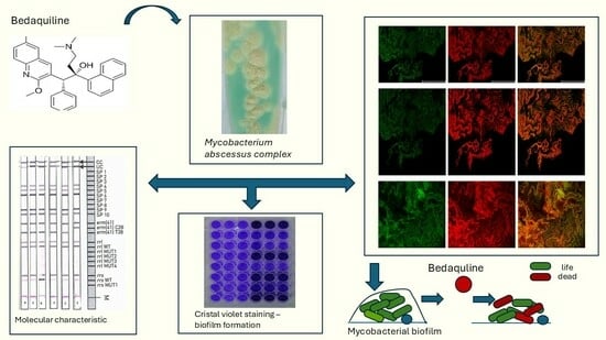
2.2. Molecular Identification of Subspecies Within the M. abscesuss Complex and Molecular Mechanisms of Resistance Assessed by the DNA Strip Method

The GenoType Mycobacterium assay was performed using total DNA extracted with the GenoLyse version 2.0 kit (Hain Lifescience, Bruker, Nehren, Germany) from monocultures of mycobacteria cultivated in liquid medium (BBL), within the BACTEC MGIT 960 system (BD Diagnostics, Franklin Lakes, NJ, USA), following the manufacturer’s protocol. The identification of the isolates as members of the MABc was confirmed using the CM Direct version 1.0 assay (Hain Lifescience, Bruker, Nehren, Germany) in accordance with the manufacturer’s protocol [40,41]. DNA identification was carried out via a hybridisation process coupled with an alkaline phosphatase reaction on a membrane strip. The amplification reagents, including polymerase and primers, were provided in mixes AM-A and AM-B. The amplification protocol comprised initial denaturation at 95 °C for 15 min, followed by 20 cycles of denaturation at 95 °C for 30 s and annealing at 65 °C for 120 s. This was followed by 30 cycles of denaturation at 95 °C for 25 s, annealing at 50 °C for 40 s, and extension at 70 °C for 30 s, concluding with a final elongation phase at 70 °C for 8 min. Molecular differentiation within the M. abscessus complex was performedusing the GenoType NTM-DR ver. 1.0 test (Hain Lifescience, Bruker, Nehren, Germany), following the manufacturer’s PCR protocol with specified primers and thermal cycling conditions. The 50 μL master mix included 10 μL of AM-A, 35 μL of AM-B reagents, and 5 μL of extracted DNA. The PCR cycle began with denaturation at 95 °C for 15 min, followed by 10 cycles (95 °C for 30 s, 65 °C for 120 s) and 20 cycles (95 °C for 25 s, 50 °C for 40 s, 70 °C for 40 s), ending with a final extension at 70 °C for 8 min. Hybridisation was performed using a manual protocol with a water bath or TwinCubator. After amplification, the samples were denatured with DEN solution, hybridised with prewarmed HYB buffer, and incubated at 45 °C for 30 min. The hybridised samples were then stringently washed with STR solution, and the strips were conjugated with diluted CON-C and incubated, followed by washing with RIN and distilled water. The substrate reaction with diluted SUB-C was conducted in the dark for 3–20 min until bands were visible. The strips were dried and interpreted according to the manufacturer’s guidelines [41].

2.3. Morphology of the Colony of Mycobacterial Strains

To distinguish the morphologies of M. abscessus, bacterial strains were inoculated onto Middlebrook 7H10 agar plates (BD Diagnostics, Franklin Lakes, NJ, USA) and incubated at 28 °C under aerobic conditions for up to 10 days. Colony growth was monitored daily. The morphology of colonies was evaluated directly on the agar plates using a light microscope (Zeiss Axiolab, equipped with ZEISS Axiocam 208 color, Carl Zeiss Microscopy GmbH, Jena, Germany) and visually. Smooth (S) morphotypes were identified by their moist, glossy, and translucent appearance with a uniform texture and rough (R) morphotypes were characterised by rugged, waxy colonies with pronounced cording patterns.

2.4. Phenotypic Drug Susceptibility Testing by Routinely Used Method

Drug susceptibility testing (DST) of NTM was carried out using the RAPMYCO Sensititre system (Thermo Fisher, Cleveland, OH, USA). Minimum inhibitory concentrations (MIC) were determined through microdilution in Sensititre Mueller–Hinton Broth (MHB) with TES (T3462), using 96-well polystyrene plates containing lyophilized antibiotics at a two-fold dilution. The plate contained antibiotics at doubling concentrations of the following drugs: amikacin (1–64 µg/mL), cefoxitin (4–128 µg/mL), ciprofloxacin (0.12–4 µg/mL), clarithromycin (0.06–16 µg/mL), doxycycline (0.12–16 µg/mL), imipenem (2–64 µg/mL), linezolid (1–32 µg/mL), minocycline (1–8 µg/mL), moxifloxacin (0.25–8 µg/mL), tigecycline (0.015–4 µg/mL), trimethoprim/sulfamethoxazole (0.25/4.75–8/152 µg/mL), cefepime (1–32 µg/mL), and amoxicillin/clavulanic acid in a 2:1 ratio (2/1–64/32 µg/mL), ceftriaxone (4–64 µg/mL), and tobramycin (1–16 µg/mL). MICs were recorded after 3–5 days of incubation if control growth was observed, except for clarithromycin, for which incubation was extended to 14 days to detect inducible macrolide resistance. The DST procedure was performed according to the manufacturer’s recommendations. In brief, 3–5 colonies were transferred into water to prepare a suspension with a 0.5 McFarland turbidity standard, as measured by a Nephelometer. A total of 50 μL of this suspension was combined with Sensititre Mueller–Hinton Broth (MHB), and 100 μL of the mixture was dispensed into each well of the Sensititre plate, using the Sensititre AIM. The plate was sealed and incubated at 35 °C in a non-CO2 incubator for 7–14 days. After incubation, the results were manually read using the Sensititre Vizion system. Interpretation was made according to the CLSI guidelines (M62, 3rd ed.), for Rapid Growing Mycobacteria (RGM) (Table A1) [19].

2.5. Susceptibility Testing to Bedaquiline, Delamanid and Clofazimine by the Broth Microdilution Method

MICs of bedaquiline (TMC207, ref. 20247, Cayman Chemical), delamanid (29697, Cayman Chemicals, Michigan, MI, USA) and clofazimine (1138904, Sigma Aldrich, Milwaukee, WI, USA) were determined using the broth microdilution method, following the Clinical and Laboratory Standards Institute (CLSI) guidelines (M24, 3rd Edition) [18,19]. Bacterial suspensions were inoculated into 96-well microplates containing cation-adjusted Mueller–Hinton broth (CAMHB) supplemented with 5% OADC (oleic acid, albumin, dextrose, and catalase; BD) [20,21]. Serial two-fold dilutions of antibiotics were prepared: 0.06–32 µg/mL for bedaquiline, 0.032–8 µg/mL for delamanid and 0.032–8 for clofazimine, respectively. The control wells included growth control (CAMHB with no antibiotic) and sterility control (no bacterial inoculum). Plates were incubated at 35 ± 2 °C in ambient air and examined after 72 h. MICs were recorded when growth in the control well reached at least a 2+ level (turbidity or visible pellet), as defined by CLSI guidelines [18]. If this threshold was not met, plates were re-incubated and re-read on days 4 and 5, if necessary. If growth remained inadequate on day 5, the test was repeated [18]. Tests were conducted in duplicate in two independent experiments.

2.6. Biofilm-Formation Assay by Crystal Violet Staining

Biofilm was analysed by the crystal violet staining method. The bacterial suspensions were inoculated into 96-well plates containing brain–heart infusion broth (BHI) supplemented with 5% OADC and incubated at 37 °C for 7 days. The wells were washed with phosphate-buffered saline (PBS), stained with 0.1% crystal violet for 15 min, and rinsed with distilled water. The bound dye was solubilized with 96% alcohol with acetic acid, and the optical density (OD) was measured at 595 nm (OD595). Strains were evaluated as non-biofilm formers (OD of the test isolate  ≤  ODcontrol), weak biofilm formers (OD of the test isolate between ODc and 2xODcontrol), moderate biofilm formers (OD of the test isolate between 2 and 4xODcontrol) and strong biofilm formers (OD of the test isolate  >  4xODcontrol) [42]. ODcontrol was calculated as the mean OD of the negative controls plus three standard deviations. All tests were run in duplicate in three independent experiments.

2.7. Evaluation of the Antibiofilm Activity of Bedaquiline

To evaluate the impact of bedaquline on biofilm, we evaluated the MBIC value (minimum biofilm inhibitory concentration), defined as the lowest concentration of an antimicrobial agent that prevents visible biofilm formation, assessed by biomass quantification methods such as crystal violet (CV) staining [42]. The MBIC for bedaquiline against 20 M. abscessus strains was determined by this method. The experiments were conducted in sterile flat-bottom microtiter plates [42]. Bacterial suspensions were prepared in Middlebrook 7H10 broth supplemented with 5% OADC and with adjusted density of 5 × 10⁵ CFU/mL [18]. Bedaquiline was added to the wells to achieve concentrations ranging from 0.125 to 2 µg/mL Plates were incubated at 37 °C for 7 days. After the incubation period, the wells were gently washed with PBS to remove planktonic cells. The plates were left at room temperature for 24 h until the biofilm was completely dry. The biofilm was stained with 0.1% crystal violet for 15 min and washed with PBS. Then, 96% ethanol with acetic acid was used to dissolve the crystal violet absorbed by the biofilm. The absorbance was measured at 595 nm using a spectrophotometer (Tecan Sunrise; Tecan Group Ltd., Männedorf, Switzerland). The tests were performed in triplicate, with the broth-only wells serving as a negative control.

2.8. Bacterial Cell-Viability Assessment and Microscopy

Bacterial cell viability after exposure to the MBIC concentration of bedaquiline was determined using the LIVE/DEAD® BacLight Bacterial Viability Kit (Invitrogen™, Thermo Fisher Scientific, Waltham, MA, USA). The staining procedure was performed according to the manufacturer’s instructions [43]. In brief, dried biofilm samples were stained with 10 µL of a mixture containing diluted fluorescent dyes (SYTO 9 and propidium iodide, PI, in a 1:1 ratio). The samples were incubated in the dark for 15 min and then observed using a Leica Stellaris 8 WLL DLS confocal microscope (Leica Microsystems GmbH, Wetzlar, Germany). Each scan was performed using the same microscope settings, such as resolution (2048 × 2048 pixels), power of the laser and detector, laser frequency (200 Hz) and pinhole (1.0 AU). The samples were scanned with 20×, NA 0.4 objective. The obtained images were analysed in LAS X software (version 4.5) (Leica, Wetzlar, Germany) to evaluate the mean fluorescence intensity. Results are expressed as the ratio of green-to-red fluorescence intensity (live-to-dead cells), according to the cell viability assay used in the study. The scale bar in the images was set to 200 µm.

2.9. Statistical Analysis

Associations between colony morphology and biofilm-formation ability were analysed using Fisher’s exact test. A p-value of <0.05 was considered statistically significant. The influence of biofilm-forming strength on the bedaquiline MIC values was evaluated using the Mann–Whitney U test. A p-value of <0.05 was considered statistically significant. Statistical analysis was performed using Statistica software version 13.0 (StatSoft Inc., Tulsa, OK, USA).

3. Results

3.1. Molecular Characterisation of Tested M. abscessus Isolates

Molecular analysis of 20 Mycobacterium abscessus isolates identified the subspecies distribution as follows: M. abscessus subsp. abscessus (n = 12), M. abscessus subsp. massiliense (n = 7), and M. abscessus subsp. bolletii (n = 1). Subspecies differentiation and resistance mechanism detection for aminoglycosides and macrolides were achieved using the GenoType NTM-DR ver. 1.0 system (Hain Lifescience, Nehren, Germany) (Table 1).
In the analysis of the 13 M. abscessus subsp. abscessus strains, the results revealed varied responses to macrolides (Table 1) and aminoglycosides. Twelve strains carried the erm(41) mutation involving a transition at position T28 (cytosine-to-thymine substitution), which led to macrolide resistance. One strain (no. 9) exhibited additional mutations, including erm(41) T28 and transversion in rrl gene (A2058C), both conferring resistance to macrolides. In addition, this strain displayed the transversion in rrs gene (T1408G), associated with aminoglycoside resistance. One M. abscessus subsp. abscessus strain (no.1) lacked detectable resistance mutations, suggesting susceptibility to macrolides and aminoglycosides.
Seven strains confirmed as M. abscessus subsp. massiliense revealed no mutations in the rrl and rrs genes. Due to a deletion in the erm(41) gene in this subspecies, the gene is non-functional, which confers macrolide sensitivity to these strains, regardless of the presence of the erm(41) T28 (Table 1).
One strain classified as M. abscessus subsp. bolletii carried the transition mutation at position T28 of the erm(41) gene, which leads to macrolide resistance (Table 1).
These findings underscore the complexity of antibiotic resistance in M. abscessus and highlight the necessity for comprehensive genetic analysis to inform effective treatment strategies. The observed variability in macrolide resistance, primarily associated with mutations in the erm(41) and rrl genes, emphasises the critical importance of individualised antimicrobial therapy based on molecular diagnostics.

3.2. Susceptibility of M. abscessus to Therapeutics Routinely Used in RGM Treatment

The MIC values for each antibiotic were evaluated by RAPMYCO2 Sensititre plates (Thermo Fisher, Cleveland, OH, USA). The susceptibility profiles of the tested drugs revealed varying levels of resistance among the strains of the M. abscessus complex.
In total, 100% of the strains were susceptible to amikacin, with MIC values ranging from 1 to 16 µg/mL, underlining its consistent efficacy and making it one of the most effective options in this study. For clarithromycin, 55% of the strains were susceptible, 40% exhibited intermediate susceptibility, and resistance was noted in 5% of the isolates. Linezolid showed promising activity, with 70% of the strains being susceptible, although intermediate susceptibility was present in 30% of cases. Ciprofloxacin showed high levels of resistance, with 95% of strains being resistant and 5% showing intermediate susceptibility (MIC values ranging from 4 µg/mL to 16 µg/mL). Doxycycline also showed limited efficacy, with 90% of the strains being resistant and only 10% susceptible. Among the tested drugs, tigecycline was evaluated, with MIC values ranging from 0.25 to 2 µg/mL However, the strains were not classified as susceptible or resistant, as the clinical breakpoints for tigecycline have not been defined (Figure 1, Table 2).

3.3. Potential of Bedaquiline, Delamanid, and Clofazimine Against M. abscessus

Simultaneously with standard drug-susceptibility testing, the microdilution method was conducted to evaluate the activity of bedaquiline, delamanid, and clofazimine against M. abscessus strains. According to the obtained results, low MIC values for bedaquiline were observed. Both MIC50 and MIC90 were determined to be 0.5 µg/mL, and the MIC range extended from 0.125 to 2 µg/mL.
In contrast, delamanid and clofazimine showed limited activity against the strains tested. Delamanid showed MIC values exceeding 16 µg/mL for all isolates, indicating a lack of efficacy in the concentration range tested. Similarly, clofazimine showed MIC values greater than 8 µg/mL for all isolates, suggesting minimal activity against M. abscessus (Table 3). Although breakpoints were not yet determined by EUCAST, the obtained results suggest limited clinical utility in treatment regimens.

3.4. Differentiation of Morphotypes and Biofilm Formation

Among the 20 MABc isolates, 16 isolates exhibited a clearly identifiable morphology; 2 isolates (10%) displayed a smooth (S) phenotype, 15 isolates (75%) exhibited a rough (R) phenotype, and 4 (20%) showed mixed morphology (S-R). The smooth morphotype produces glossy, uniform colonies that appear moist and translucent under standard laboratory culture conditions. In contrast, the rough morphotype forms colonies that are irregular, rough-edged, matte, and distinctly waxy in appearance (Figure 2).
M. abscessus biofilm-forming strains exhibit a smooth morphotype, forming colonies that are moist, glossy, and translucent. This appearance is attributed to the high expression of glycopeptidolipids (GPLs) on their cell surface, which confer hydrophilic properties. In contrast, rough morphotypes, which have lower levels of (GPLs), present as irregular, rugged colonies, as reported previously [1,42].
Based on the crystal violet (CV) staining method, seven strains were classified as strong biofilm producers (OD595/OD595 control > 4). These included all mixed morphotype strains and one smooth strain (M. abscessus subsp. bolletii), with the mixed morphotypes consistently showing the highest OD595 values, indicating a strong biofilm-forming ability. Moderate biofilm formation (OD595/OD595 control, ranging between 2 and 4) was observed in six strains, mainly exhibiting rough or smooth morphotypes, with the majority of these moderate forms belonging to M. abscessus subsp. abscessus. In contrast, seven strains were classified as weak biofilm producers (OD595/OD595 control < 2), with the rough morphotypes being the most common. These weak biofilm forms were primarily M. abscessus subsp. abscessus and subsp. massiliense, indicating minimal biofilm-formation ability. This distribution indicates a strong relationship between morphotype and biofilm-formation ability (Table 4).
The rough morphotype (Figure 3A) typically did not exhibit significant biofilm production. In contrast, the smooth morphotypes of the M. abscessus complex which form planktonic-like structures (Figure 3B) were strongly associated with biofilm formation. The mixed morphotype was also a strong biofilm producer. This distinction highlights the potential role of morphotype in determining the pathogenic potential and drug resistance of MABc.
There was a statistically significant association between colony phenotype and biofilm-formation ability among clinical MABc isolates. Strains with a mixed colony morphology were significantly more likely to form strong biofilms (p = 0.032, Fisher’s exact test). Although strains with a rough colony morphology tended to form weak biofilms more frequently, this association was not statistically significant (p = 0.141, Fisher’s exact test) (Table 5).

3.5. Bedaquilline as a Good Antibiofilm Agent

The efficacy of bedaquiline as anti-biofilm agent was demonstrated by MBIC values ranging from 0.25 µg/mL to 2 µg/mL, confirming its effectiveness against M. abscessus biofilms. A comparison of MIC and MBIC values revealed that the MBIC/MIC ratio varied between 1 and 4, indicating that higher concentrations of bedaquiline are typically required to inhibit biofilm formation compared to planktonic cells. Interestingly, five strains showed an MBIC/MIC ratio of 1, suggesting that biofilm inhibition in these cases required concentrations similar to those needed for planktonic cell inhibition. However, the most common MBIC/MIC ratio was 4, observed in half of the tested strains, indicating a four-fold increase in the concentration to effectively suppress biofilm growth (Table 6).
Additionally, the analysis of bacterial cell viability in biofilm structure was conducted using the LIVE/DEAD® bacterial viability assay with confocal microscopy visualization. The results revealed that bedaquiline significantly inhibited biofilm formation at the tested concentrations. At 0.5 µg/mL, corresponding to MIC values, the reduction in biofilm formation was 45.24% compared to the growth control, while at 0.25 µg/mL and 0.125 µg/mL, the reductions were 29.76% and 13.10%, respectively (Figure 4). These findings demonstrate that bedaquiline significantly inhibits biofilm formation at concentrations corresponding to its MIC, but does not fully eradicate biofilms. This supports its potential as a valuable agent for limiting biofilm formation by M. abscessus.
Additionally, we found that there was no statistically significant difference in bedaquiline MIC values between weak and moderate/strong biofilm-forming M. abscessus complex isolates (p = 0.828). The mean MIC was 0.438 µg/mL for the weak biofilm formers and 0.425 µg/mL for the moderate/strong biofilm formers.

4. Discussion

Treating M. abscessus complex infections is highly challenging, due to the bacterial resistance mechanisms and its ability to form biofilms. Furthermore, the accurate identification and differentiation of MABc subspecies, including M. abscessus subsp. abscessus, massiliense, and bolletii, remain critical because these subspecies exhibit significant differences in virulence, biofilm-formation capacity, and susceptibility to antimicrobials. Rapid genetic tests, such as molecular tests targeting resistance genes or subspecies-specific markers, provide valuable tools to guide treatment decisions and optimise antimicrobial therapy.
The analysis of 20 M. abscessus isolates revealed a diverse subspecies distribution, with M. abscessus subsp. abscessus being the most prevalent (n = 12), followed by M. abscessus subsp. massiliense (n = 7) and M. abscessus subsp. bolletii (n = 1). The identification of subspecies is clinically significant because of their differing resistance profiles and therapeutic implications [5,7]. A high prevalence of macrolide resistance was observed in 70% (14/20) of the isolates, consistent with the frequent occurrence of mutations in the erm(41) or rrl genes (in the one isolate). The erm(41) T28C mutation, which confers inducible macrolide resistance, was particularly prominent in subsp. abscessus. These findings underline the significant challenge macrolide resistance poses for effective treatment, especially given the reliance on macrolides as a cornerstone of MABc therapy [16,44,45,46]. M. abscessus subsp. abscessus is often associated with poor treatment outcomes due to inducible macrolide resistance, mediated by a functional erm(41) gene. In contrast. M. abscessus subsp. massiliense typically lacks this functional gene, resulting in higher macrolide susceptibility and better clinical responses [17,47]. Multiple studies have examined the roles of erm(41) and rrl mutations in M. abscessus resistance to clarithromycin [48,49], although their findings have been inconsistent. These mutations do not account for all clarithromycin-resistant M. abscessus strains [50], suggesting the presence of additional resistance mechanisms [44,50,51,52,53]. In contrast, aminoglycoside resistance was rare, detected in only 5% (1/20) of the isolates. This finding aligns with the established efficacy of aminoglycosides, such as amikacin, against MABc. The low rate of resistance supports the continued use of amikacin as a key therapeutic agent in combination regimens, while also underscoring the importance of routine surveillance to monitor potential resistance development.
The study also highlighted widespread resistance to ciprofloxacin, moxifloxacin, and doxycycline, limiting the effectiveness of these agents for MABc treatment. The susceptibility results for linezolid and trimethoprim–sulfamethoxazole were variable, with some isolates showing intermediate or resistant profiles. This variability underscores the importance of cautious clinical use and emphasises the need for tailored treatment plans based on individual susceptibility results.
Exploring new treatment options for M. abscessus infections among drugs already approved for M. tuberculosis therapy appears to be a promising alternative. Therefore, we evaluated the activity of bedaquiline, delamanid, and clofazimine against 20 clinical M. abscessus strains. Our results revealed low MIC values for bedaquiline, with MIC50 and MIC90 both determined to be 0.5 µg/mL and an MIC range of 0.125 to 2 µg/mL. The findings of this study underscore the significant potential of bedaquiline as an effective therapeutic option for M. abscessus infections. Bedaquiline has been approved by the US Food and Drug Administration (FDA) and the European Medicines Agency for the treatment of multidrug-resistant tuberculosis [53]. In vitro, it demonstrates bacteriostatic activity against M. abscessus isolates, with an MIC50 of 0.125 μg/mL and an MIC90 greater than 16 μg/mL [53].
Bedaquiline shows moderate activity based on epidemiological cutoff values, with its efficacy significantly enhanced when used in combination therapies [54]. Moigne et al. [54] demonstrated that the combination of bedaquiline and imipenem significantly enhanced infection clearance, emphasizing the importance of evaluating antibiotic combinations rather than relying solely on monotherapies when addressing highly drug-resistant mycobacteria. Similarly, Ruth et al. [55] evaluated the effectiveness of bedaquiline against M. abscessus infections. Their study revealed concentration-dependent bacteriostatic activity, although its efficacy was lower against M. abscessus than M. avium. A synergistic effect was observed when bedaquiline was combined with clofazimine, whereas no interaction was noted with clarithromycin. Resistance to bedaquiline developed during prolonged exposure, and, in vivo, bedaquiline monotherapy failed to prevent mortality in a murine M. abscessus model. These findings indicate that while bedaquiline exhibits bacteriostatic effects, its therapeutic potential against M. abscessus is limited, especially when used as monotherapy [25,54,55,56].
Delamanid and clofazimine exhibited limited activity against Mycobacterium abscessus in this study, with minimum inhibitory concentration (MIC) values exceeding 16 µg/mL and 8 µg/mL, respectively. These results align with previous reports indicating the poor efficacy of these agents against M. abscessus [25,57]. While clofazimine demonstrated effectiveness against other non-tuberculous mycobacteria (NTM), particularly the M. avium complex (MAC), its role in M. abscessus infections remains uncertain [17,44].
Combination therapies, particularly those involving bedaquiline, clofazimine, delamanid and other antimicrobials, are being increasingly explored. Studies suggest that combining delamanid with other agents may result in synergistic effects, offering a potential avenue for improving treatment efficacy. However, further investigation into optimal drug combinations, dosing regimens, and host-directed therapies is necessary to enhance treatment outcomes [21,25,26,56,58,59].
Beyond antimicrobial resistance, another key challenge in treating M. abscessus infections is biofilm formation, which enhances bacterial survival and limits antimicrobial efficacy in hostile environments [60,61]. This process is influenced by multiple factors, with evidence linking colony morphotype to biofilm-forming capacity [62,63].
We analysed the distribution of morphotypes among the tested MABc strains. The majority of the isolates (70%, 14/20) exhibited the rough morphotype, while only 10% (2/20) presented the smooth morphotype, typically linked to lower virulence and increased susceptibility to host immune defences [64]. Statistical analysis revealed a significant association between colony morphology and biofilm-formation capacity, with mixed morphotypes being significantly more likely to form strong biofilms (p = 0.032). However, due to the variability observed within each morphotype group, further studies with larger strain collections are necessary to confirm and better understand these associations.
The predominance of the rough morphotype in this study is consistent with previous reports, where this phenotype has been associated with more severe clinical outcomes, higher virulence, persistence in host tissues, and more pronounced inflammatory responses [60,65,66,67]. The underlying factors behind increased pathogenicity are not fully understood; however, rough M. abscessus strains exhibit this [46]. Rough variants also display increased cording, a virulence factor in mycobacterial infections that impairs macrophage phagocytosis [46,68]. The presence of both smooth and rough colonies within the same strain may represent a strategy to maximise survival in fluctuating environments, where one morphotype might be more suited for persistence in certain conditions, while the other could offer better survival under different circumstances. This finding aligns with the notion that M. abscessus populations are genetically diverse and capable of adapting rapidly to changes in their environment [69,70]. However, studies on the antibiotic susceptibility of M. abscessus isolates from both rough and smooth morphotypes have yielded inconsistent findings [60,66,71,72].
The formation of biofilms is a key factor in the pathogenicity of MABc [69]. The biofilm-forming capability of the MABc strains is a critical factor contributing to their persistence and resistance to antimicrobial agents. The success of new therapies will heavily depend on their ability to penetrate and dismantle biofilms, as biofilms are responsible for the majority of chronic and recurrent bacterial infections [73].
In this study, 25% (5/20) of the strains were strong biofilm formers, while 75% (15/20) demonstrated weak-to-moderate biofilm formation. Biofilm-positive strains pose a significant challenge in clinical settings, as biofilm formation increases the tolerance to antibiotics and host immune responses, complicating eradication efforts. The low prevalence of biofilm-positive strains may suggest that strain-specific differences in biofilm-forming capacity or environmental conditions influence this phenotype. Nevertheless, the identification of biofilm-positive strains highlights the need for therapies targeting biofilm-associated resistance mechanisms, such as combination treatments or biofilm-disrupting agents. Biofilm structures are complex, and present key challenges in measuring the number of active cells, biofilm morphology, and mass accumulation. The findings of this study emphasise the complexity of managing MABc infections due to diverse resistance profiles, morphotype variability, and biofilm-formation capabilities. The high prevalence of macrolide resistance necessitates alternative therapeutic strategies, such as the use of second-line agents or combination therapies. Additionally, the predominance of rough morphotypes underscores the aggressive nature of many MABc infections, which may require more intensive management. Future studies should focus on elucidating the molecular mechanisms underlying these phenotypes and their clinical impact. Integrating phenotypic and genotypic data will enhance our understanding of MABc pathogenicity and inform the development of a targeted therapeutic approach.
Bedaquiline’s antibiofilm activity, with MBIC values between 0.25 and 2 µg/mL, further strengthens its case for use as a key antimicrobial for tackling M. abscessus. The MBIC/MIC ratio, which was most commonly observed as 4, suggests that while higher concentrations are required to inhibit biofilm growth compared with planktonic cells, these values remain within achievable therapeutic ranges. Confocal microscopy and the LIVE/DEAD® assay revealed a significant reduction in biofilm viability, with bedaquiline reducing biofilm mass by 45% at MBIC concentrations. This ability to suppress biofilm formation, rather than simply eradicating existing biofilms, could play a critical role in preventing chronic biofilm-related complications. It is worth noting that the interpretation of viability staining in Mycobacterium spp. may be limited by the lipid-rich, hydrophobic cell wall, which can impair the penetration of fluorescent dyes such as propidium iodide. As previously reported, this may lead to inaccurate differentiation between viable and non-viable cells when using conventional viability kits based on membrane integrity [74].
Our study focused on evaluating the MBIC of bedaquiline, which measures its ability to prevent biofilm formation when administered at the onset of bacterial growth. While MBEC (Minimum Biofilm Eradication Concentration) is commonly used to assess the ability of an agent to eliminate pre-formed biofilms, MBIC is equally important in the context of M. abscessus infections. Inhibiting biofilm formation at an early stage can significantly impact bacterial persistence, as biofilms contribute to increased antimicrobial resistance, host immune evasion, and chronic infections. Moreover, MBIC testing provides insights into the effects of bedaquiline on the initial adhesion phase and early biofilm development, which are critical steps in biofilm formation.
The choice of culture medium is a critical factor influencing biofilm formation in M. abscessus [75,76]. To date, there is no universally standardized protocol for biofilm assays in RGM, and various media including Middlebrook 7H9, Sauton’s medium, or CAMHB have been employed across studies [76]. In the present study, BHI broth supplemented with 5% OADC was selected for the biofilm-formation assay. Although BHI is not the most commonly used medium for M. abscessus, it is widely applied in crystal violet-based biofilm quantification protocols for other bacteria [77,78], and this approach was extrapolated to M. abscessus in our experimental setup. The potential influence of medium composition on the characteristics and biomass of biofilms observed in this study should be considered when interpreting the results.
The findings of our study underscore the significant potential of bedaquiline as an effective therapeutic option for M. abscessus infections. This is particularly noteworthy, given the resistance mechanisms of M. abscessus and the role of biofilms in promoting persistence and the treatment failure.
In recent years, there has been a growing focus on M. abscessus biofilm research. However, the absence of standardised methods for culturing, characterising, and testing drug activity in biofilms makes it difficult to compare results across different studies. Variations in the biofilm models and experimental conditions used in M. abscessus biofilm research can influence the biofilm’s properties and behaviour, which helps explain the wide range of seemingly contradictory findings in this area of study. This study emphasizes the need for personalized treatment strategies and underscores the importance of frequent and thorough susceptibility testing in managing M. abscessus infections. This strategy will be crucial in addressing and overcoming the challenges posed by antimicrobial resistance in these highly resistant and difficult-to-manage bacterial infections.

5. Conclusions

To sum up, the results of our study highlight the significant therapeutic potential of bedaquiline against the M. abscessus complex, particularly in biofilm-forming strains, with consistent MIC50 and MIC90 values of 0.5 µg/mL. Importantly, despite the well-known challenge of biofilm-associated resistance, bedaquiline maintained its activity irrespective of the biofilm-forming capacity, with no statistically significant differences observed between weak and moderate/strong biofilm producers (p = 0.828). Furthermore, the MBIC values of bedaquiline were only up to four-fold higher than the MIC values, confirming effective biofilm penetration. Delamanid and clofazimine showed limited activity, with high MIC values exceeding 16 µg/mL and 8 µg/mL, respectively. The observation that 25% of the strains formed strong biofilms underscores the clinical importance of targeting biofilm structures, which contribute to persistence and therapeutic failure. These findings strongly support the inclusion of bedaquiline as a core component of personalised, combination regimens to overcome the dual challenges of biofilm formation and multidrug resistance in M. abscessus complex infections.

Author Contributions

Conceptualisation, K.K. (Katarzyna Kania); formal analysis, K.K. (Katarzyna Kania) and K.K. (Karolina Klesiewicz); funding acquisition, K.K. (Katarzyna Kania); investigation, K.K. (Katarzyna Kania), K.W. and A.S.; methodology, K.K. (Katarzyna Kania), K.W. and K.K. (Karolina Klesiewicz); project administration, K.K. (Katarzyna Kania); resources, K.W.; supervision, K.K. (Karolina Klesiewicz); visualization, A.S.; writing—original draft, K.K. (Katarzyna Kania) and K.K. (Karolina Klesiewicz); writing—review and editing, K.K. (Katarzyna Kania) and K.K. (Karolina Klesiewicz). All authors have read and agreed to the published version of the manuscript.

Funding

This research was partially supported by the statutory funds of the Jagiellonian University Medical College (N42/DBS/000293). Publication was funded by the Priority Research Area qLIFE under the Strategic Programme Excellence Initiative at Jagiellonian University. This research was carried out with the use of research infrastructure co-financed by the Smart Growth Operational Programme POIR 4.2 (project no. POIR.04.02.00-00-D023/20).

Institutional Review Board Statement

This study is approved by the Jagiellonian University Medical College Ethic Commission (118.0043.1.390.2024, approval date 12 December 2024). Our report is a retrospective analysis using anonymous survey data collected during routine practice at Malopolska Central Laboratory of Tuberculosis Diagnostics, the St. John Paul II Specialist Hospital.

Informed Consent Statement

Not applicable.

Data Availability Statement

The original contributions presented in this study are included in the article. Further inquiries can be directed to the corresponding author.

Conflicts of Interest

The authors declare no conflicts of interests.

Abbreviations

The following abbreviations are used in this manuscript:
BDQBedaquiline
CFUColony-Forming Units
CLOClofazimine
DELDelamanid
CVCrystal Violet
LJLöwenstein–Jensen medium
MABcMycobacterium abscessus complex
MBICMinimal Biofilm Inhibitory Concentration
MICMinimal Inhibitory Concentration
NTMNon-Tuberculous Mycobacteria
ODOptical Density
PIPropidium Iodide
DSTDrug Susceptibility Testing

Appendix A

Table A1. Antimycobacterial agents and breakpoints for testing rapidly growing Mycobacteria [19].
Antimicrobial Agent MIC (µg/mL)
S I R
Amikacin (IV)≤16 32 ≥64
Cefoxitin≤16 32–64 ≥128
Ciprofloxacin≤1 2 ≥4
Clarithromycin≤2 4 ≥8
Imipenem≤4 8–16 ≥32
Linezolid≤8 16 ≥32
Meropenem≤4 8–16 ≥32
Moxifloxacin≤1 2 ≥4
Trimetoprim-sulfametoxazol≤2/38 - ≥4/76
Tigecycline- - -
Tobramycin≤2 4 ≥8

References

  1. Victoria, L.; Gupta, A.; Gómez, J.L.; Robledo, J. Mycobacterium abscessus complex: A Review of Recent Developments in an Emerging Pathogen. Front. Cell. Infect. Microbiol. 2021, 11, 659997. [Google Scholar] [CrossRef] [PubMed]
  2. Abdela al, H.F.M.; Chan, E.D.; Young, L.; Baldwin, S.L.; Coler, R.N. Mycobacterium abscessus: It’s Complex. Microorganisms 2022, 10, 1454. [Google Scholar] [CrossRef] [PubMed]
  3. Prevots, D.R.; Shaw, P.A.; Strickland, D.; Jackson, L.A.; Raebel, M.A.; Blosky, M.A.; Montes De Oca, R.; Shea, Y.R.; Seitz, A.E.; Holland, S.M.; et al. Nontuberculous mycobacterial lung disease prevalence at four integrated health care delivery systems. Am. J. Respir. Crit. Care Med. 2010, 182, 970–976. [Google Scholar] [CrossRef] [PubMed]
  4. Snell, G.; Reed, A.; Stern, M.; Hadjiliadis, D. The evolution of lung transplantation for cystic fibrosis: A 2017 update. J. Cyst. Fibros. 2017, 16, 553–564. [Google Scholar] [CrossRef]
  5. Wetzstein, N.; Diricks, M.; Kohl, T.A.; Wichelhaus, T.A.; Andres, S.; Paulowski, L.; Schwarz, C.; Lewin, A.; Kehrmann, J.; Kahl, B.C.; et al. Molecular Epidemiology of Mycobacterium abscessus Isolates Recovered from German Cystic Fibrosis Patients. Microbiol. Spectr. 2022, 10, 0171422. [Google Scholar] [CrossRef]
  6. Faria, S.; Joao, I.; Jordao, L. General Overview on Nontuberculous Mycobacteria, Biofilms, and Human Infection. J. Pathog. 2015, 2015, 809014. [Google Scholar] [CrossRef]
  7. Nguyen, T.Q.; Heo, B.E.; Jeon, S.; Ash, A.; Lee, H.; Moon, C.; Jang, J. Exploring antibiotic resistance mechanisms in Mycobacterium abscessus for enhanced therapeutic approaches. Front. Microbiol. 2024, 15, 1331508. [Google Scholar] [CrossRef]
  8. Dokic, A.; Peterson, E.; Arrieta-Ortiz, M.L.; Pan, M.; Di Maio, A.; Baliga, N.; Bhatt, A. Mycobacterium abscessus biofilms produce an extracellular matrix and have a distinct mycolic acid profile. Cell Surf. 2021, 7, 100051. [Google Scholar] [CrossRef]
  9. Gloag, E.S.; Wozniak, D.J.; Stoodley, P.; Hall-Stoodley, L. Mycobacterium abscessus biofilms have viscoelastic properties which may contribute to their recalcitrance in chronic pulmonary infections. Sci. Rep. 2021, 11, 5020. [Google Scholar] [CrossRef]
  10. Luthra, S.; Rominski, A.; Sander, P. The Role of Antibiotic-Target-Modifying and Antibiotic-Modifying Enzymes in Mycobacterium abscessus Drug Resistance. Front. Microbiol. 2018, 9, 2179. [Google Scholar] [CrossRef]
  11. Nessar, R.; Cambau, E.; Reyrat, J.M.; Murray, A.; Gicquel, B. Mycobacterium abscessus: A new antibiotic nightmare. J. Antimicrob. Chemother. 2012, 67, 810–818. [Google Scholar] [CrossRef] [PubMed]
  12. Mulyukin, A.L.; Recchia, D.; Kostrikina, N.A.; Artyukhina, M.V.; Martini, B.A.; Stamilla, A.; Degiacomi, G.; Salina, E.G. Distinct Effects of Moxifloxacin and Bedaquiline on Growing and ‘Non-Culturable’ Mycobacterium abscessus. Microorganisms 2023, 11, 2690. [Google Scholar] [CrossRef] [PubMed]
  13. Sharma, S.; Mohler, J.; Mahajan, S.D.; Schwartz, S.A.; Bruggemann, L.; Aalinkeel, R. Microbial Biofilm: A Review on Formation, Infection, Antibiotic Resistance, Control Measures, and Innovative Treatment. Microorganisms 2023, 11, 1614, Erratum in Microorganisms 2024, 12, 1961. [Google Scholar] [CrossRef] [PubMed]
  14. Born, S.E.M.; Reichlen, M.J.; Bartek, I.L.; Benoit, J.B.; Frank, D.N.; Voskuil, M.I. Population heterogeneity in Mycobacterium smegmatis and Mycobacterium abscessus. Microbiology 2023, 169, 001402. [Google Scholar] [CrossRef]
  15. López-Roa, P.; Esteban, J.; Muñoz-Egea, M.C. Updated Review on the Mechanisms of Pathogenicity in Mycobacterium abscessus, a Rapidly Growing Emerging Pathogen. Microorganisms 2022, 11, 90. [Google Scholar] [CrossRef]
  16. Kwak, N.; Dalcolmo, M.P.; Daley, C.L.; Eather, G.; Gayoso, R.; Hasegawa, N.; Jhun, B.W.; Koh, W.-J.; Namkoong, H.; Park, J.; et al. Mycobacterium abscessus pulmonary disease: Individual patient data meta-analysis. Eur. Respir. J. 2019, 54, 1801991. [Google Scholar] [CrossRef]
  17. Daley, C.L.; Iaccarino, J.M.; Lange, C.; Cambau, E.; Wallace, R.J.; Andrejak, C.; Böttger, E.C.; Brozek, J.; Griffith, D.E.; Guglielmetti, L.; et al. Treatment of nontuberculous mycobacterial pulmonary disease: An official ATS/ERS/ESCMID/IDSA clinical practice guideline. Eur. Respir. J. 2020, 56, 2000535. [Google Scholar] [CrossRef]
  18. CLSI. Reference Method for Susceptibility Testing of Mycobacteria, Nocardia spp., and Other Aerobic Actinomycetes, Approved Standard, 3rd ed.; CLSI document, M24; Woods, G.L., Ed.; Clinical and Laboratory Standards Institute: Wayne PA, USA, 2018. [Google Scholar]
  19. CLSI. Performance Standards for Susceptibility Testing of Mycobacteria, Nocardia spp., and Other Aerobic Actinomycetes, 3rd ed.; Suplement Edition M62; Clinical and Laboratory Standards Institute: Wayne, PA, USA, 2018. [Google Scholar]
  20. Dupont, C.; Viljoen, A.; Thomas, S.; Roquet-Banères, F.; Herrmann, J.-L.; Pethe, K.; Kremer, L. Bedaquiline Inhibits the ATP Synthase in Mycobacterium abscessus and Is Effective in Infected Zebrafish. Antimicrob. Agents Chemother. 2017, 61, 01225–17. [Google Scholar] [CrossRef]
  21. Ahmad Khosravi, N.; Sirous, M.; Khosravi, A.D.; Saki, M. A Narrative Review of Bedaquiline and Delamanid: New Arsenals Against Multidrug-Resistant and Extensively Drug-Resistant Mycobacterium tuberculosis. J. Clin. Lab. Anal. 2024, 38, 25091. [Google Scholar] [CrossRef]
  22. Motta, I.; Boeree, M.; Chesov, D.; Dheda, K.; Günther, G.; Horsburgh, C.R.; Kherabi, Y.; Lange, C.; Lienhardt, C.; McIlleron, H.M.; et al. Recent advances in the treatment of tuberculosis. Clin. Microbiol. Infect. 2024, 30, 1107–1114. [Google Scholar] [CrossRef]
  23. Wang, M.; Men, P.; Zhang, W.; Wu, J.; Gu, Y.; Wang, F.; Huang, H.; Yu, X.; Duan, H. Bedaquiline susceptibility testing of Mycobacterium abscessus complex and Mycobacterium avium complex: A meta-analysis study. J. Glob. Antimicrob. Resist. 2024, 7, 135–140. [Google Scholar] [CrossRef] [PubMed]
  24. Li, B.; Ye, M.; Guo, Q.; Zhang, Z.; Yang, S.; Ma, W.; Yu, F.; Chu, H. Determination of MIC Distribution and Mechanisms of Decreased Susceptibility to Bedaquiline among Clinical Isolates of Mycobacterium abscessus. Antimicrob. Agents Chemother. 2018, 62, 00175–18. [Google Scholar] [CrossRef] [PubMed]
  25. Kim, D.H.; Jhun, B.W.; Moon, S.M.; Kim, S.-Y.; Jeon, K.; Kwon, O.J.; Huh, H.J.; Lee, N.Y.; Shin, S.J.; Daley, C.L.; et al. In Vitro Activity of Bedaquiline and Delamanid against Nontuberculous Mycobacteria, Including Macrolide-Resistant Clinical Isolates. Antimicrob. Agents Chemother. 2019, 63, 00665–19. [Google Scholar] [CrossRef]
  26. Meir, M.; Barkan, D. Alternative and Experimental Therapies of Mycobacterium abscessus Infections. Int. J. Mol. Sci. 2020, 21, 6793. [Google Scholar] [CrossRef]
  27. Andries, K.; Verhasselt, P.; Guillemont, J.; Göhlmann, H.W.; Neefs, J.M.; Winkler, H.; Van Gestel, J.; Timmerman, P.; Zhu, M.; Lee, E.; et al. A diarylquinoline drug active on the ATP synthase of Mycobacterium tuberculosis. Science 2005, 307, 223–227. [Google Scholar] [CrossRef] [PubMed]
  28. Xavier, A.S.; Lakshmanan, M. Delamanid: A new armor in combating drug-resistant tuberculosis. J. Pharmacol. Pharmacother. 2014, 5, 222–224. [Google Scholar] [CrossRef]
  29. Venkatesan, K.; Mathur, A.; Girdhar, B.K.; Bharadwaj, V.P. The effect of clofazimine on the pharmacokinetics of rifampicin and dapsone in leprosy. J. Antimicrob. Chemother. 1986, 18, 715–718. [Google Scholar] [CrossRef]
  30. Oommen, S.T.; Natu, M.V.; Mahajan, M.K.; Kadyan, R.S. Lymphangiographic evaluation of patients with clinical lepromatous leprosy on clofazimine. Int. J. Lepr. Other Mycobact. Dis. 1994, 62, 32–36. [Google Scholar]
  31. Mungroo, M.R.; Khan, N.A.; Siddiqui, R. Mycobacterium leprae: Pathogenesis, diagnosis, and treatment options. Microb. Pathog. 2020, 149, 104475. [Google Scholar] [CrossRef]
  32. Stadler, J.A.M.; Maartens, G.; Meintjes, G.; Wasserman, S. Clofazimine for the treatment of tuberculosis. Front. Pharmacol. 2023, 14, 1100488. [Google Scholar] [CrossRef]
  33. Riccardi, N.; Giacomelli, A.; Canetti, D.; Comelli, A.; Intini, E.; Gaiera, G.; Diaw, M.M.; Udwadia, Z.; Besozzi, G.; Codecasa, L.; et al. Clofazimine: An old drug for never-ending diseases. Clofazimine: An old drug for never-ending diseases. Future Microbiol. 2020, 15, 557–566. [Google Scholar] [CrossRef] [PubMed]
  34. Islam, M.; Alam, S.; Liu, Z.; Khatun, M.S.; Yusuf, B.; Hameed, H.M.A.; Tian, X.; Chhotaray, C.; Basnet, R.; Abraha, H.; et al. Molecular mechanisms of resistance and treatment efficacy of clofazimine and bedaquiline against Mycobacterium tuberculosis. Front. Med. 2024, 10, 1304857. [Google Scholar] [CrossRef] [PubMed]
  35. Hajikhani, B.; Nasiri, M.J.; Hosseini, S.S.; Khalili, F.; Karimi-Yazdi, M.; Hematian, A.; Nojookambari, N.Y.; Goudarzi, M.; Dadashi, M.; Mirsaeidi, M. Clofazimine susceptibility testing of Mycobacterium avium complex and Mycobacterium abscessus: A meta-analysis study. J. Glob. Antimicrob. Resist. 2021, 26, 188–193. [Google Scholar] [CrossRef]
  36. Schulthess, B.; Akdoğan Kittana, F.N.; Hömke, R.; Sander, P. In Vitro Bedaquiline and Clofazimine Susceptibility Testing in Mycobacterium abscessus. Antimicrob. Agents Chemother. 2022, 66, 0234621. [Google Scholar] [CrossRef]
  37. Omar, S.; Whitfield, M.G.; Nolan, M.B.; Ngom, J.T.; Ismail, N.; Warren, R.M.; Klopper, M. Bedaquiline for treatment of non-tuberculous mycobacteria (NTM): A systematic review and meta-analysis. J. Antimicrob. Chemother. 2024, 79, 211–240. [Google Scholar] [CrossRef]
  38. To, K.; Cao, R.; Yegiazaryan, A.; Owens, J.; Venketaraman, V. General Overview of Nontuberculous Mycobacteria Opportunistic Pathogens: Mycobacterium avium and Mycobacterium abscessus. J. Clin. Med. 2020, 9, 2541. [Google Scholar] [CrossRef]
  39. Zhao, A.; Sun, J.; Liu, Y. Understanding bacterial biofilms: From definition to treatment strategies. Front. Cell Infect. Microbiol. 2023, 13, 1137947. [Google Scholar] [CrossRef]
  40. GenoType NTM-DR VER 1.0 Molecular Genetic Assay for Detection of Resistance to Macrolides and Aminoglycosides in various Nontuberculous Mycobacterial Species (NTM) from Cultured Material. GenoType NTM-DR VER 1.0 Instructions for Use (IFU-297-03), 2019, 4–9. Available online: https://www.hain-lifescience.de/en/instructions-for-use.html (accessed on 15 July 2014).
  41. Stepanović, S.; Vuković, D.; Hola, V.; Bonaventura, G.D.; Djukić, S.; Ćirković, I.; Ruzicka, F. Quantification of biofilm in microtiter plates: Overview of testing conditions and practical recommendations for assessment of biofilm production by staphylococci. APMIS 2007, 115, 891–899. [Google Scholar] [CrossRef]
  42. Parrish, N.M.; Ko, C.G.; Dick, J.D.; Jones, P.B.; Ellingson, J.L. Growth, Congo Red agar colony morphotypes and antibiotic susceptibility testing of Mycobacterium avium subspecies paratuberculosis. Clin. Med. Res. 2024, 2, 107–114. [Google Scholar] [CrossRef]
  43. Invitrogen LIVE/DEAD® BacLight Bacterial Viability Kits. 2004. Available online: https://www.thermofisher.cn/order/catalog/product/L7012 (accessed on 15 July 2014).
  44. Griffith, D.E.; Daley, C.L. Treatment of Mycobacterium abscessus. Pulmonary Disease. Chest 2022, 161, 64–75. [Google Scholar] [CrossRef]
  45. Hedin, W.; Fröberg, G.; Fredman, K.; Chryssanthou, E.; Selmeryd, I.; Gillman, A.; Orsini, L.; Runold, M.; Jönsson, B.; Schön, T.; et al. A Rough Colony Morphology of Mycobacterium abscessus Is Associated With Cavitary Pulmonary Disease and Poor Clinical Outcome. J. Infect. Dis. 2023, 227, 820–827. [Google Scholar] [CrossRef] [PubMed]
  46. Nash, K.A.; Brown-Elliott, B.A.; Wallace, R.J., Jr. A novel gene, erm(41), confers inducible macrolide resistance to clinical isolates of Mycobacterium abscessus but is absent from Mycobacterium chelonae. Antimicrob. Agents Chemother. 2009, 53, 1367–1376. [Google Scholar] [CrossRef] [PubMed]
  47. Lee, S.H.; Yoo, H.K.; Kim, S.H.; Koh, W.J.; Kim, C.K.; Park, Y.K.; Kim, H.J. Detection and assessment of clarithromycin inducible resistant strains among Korean Mycobacterium abscessus clinical strains: PCR methods. J. Clin. Lab. Anal. 2014, 28, 409–414, Erratum in J. Clin. Lab. Anal. 2016, 30, 356. [Google Scholar] [CrossRef] [PubMed]
  48. Ramírez, A.; de Waard, J.H.; Araque, M. Molecular mechanisms of clarithromycin resistance in Mycobacterium abscessus complex clinical isolates from Venezuela. J. Glob. Antimicrob. Resist. 2015, 3, 205–209. [Google Scholar] [CrossRef]
  49. Carneiro, M.D.S.; Nunes, L.S.; David, S.M.M.; Barth, A.L. Lack of association between rrl and erm(41) mutations and clarithromycin resistance in Mycobacterium abscessus complex. Mem. Inst. Oswaldo Cruz 2017, 112, 775–778. [Google Scholar] [CrossRef]
  50. Ananta, P.; Kham-Ngam, I.; Chetchotisakd, P.; Chaimanee, P.; Reechaipichitkul, W.; Namwat, W.; Lulitanond, V.; Faksri, K. Analysis of drug-susceptibility patterns and gene sequences associated with clarithromycin and amikacin resistance in serial Mycobacterium abscessus isolates from clinical specimens from Northeast Thailand. PLoS ONE 2018, 13, 0208053. [Google Scholar] [CrossRef]
  51. Nie, W.; Duan, H.; Huang, H.; Lu, Y.; Chu, N. Species Identification and Clarithromycin Susceptibility Testing of 278 Clinical Nontuberculosis Mycobacteria Isolates. Biomed. Res. Int. 2015, 2015, 506598. [Google Scholar] [CrossRef]
  52. Pang, Y.; Zheng, H.; Tan, Y.; Song, Y.; Zhao, Y. In Vitro Activity of Bedaquiline against Nontuberculous Mycobacteria in China. Antimicrob. Agents Chemother. 2017, 1, 02627-16. [Google Scholar] [CrossRef]
  53. Le Moigne, V.; Raynaud, C.; Moreau, F.; Dupont, C.; Nigou, J.; Neyrolles, O.; Kremer, L.; Herrmann, J.-L. Efficacy of Bedaquiline, Alone or in Combination with Imipenem, against Mycobacterium abscessus in C3HeB/FeJ Mice. Antimicrob. Agents Chemother. 2020, 64, 00114–00120. [Google Scholar] [CrossRef]
  54. Ruth, M.M.; Sangen, J.J.N.; Remmers, K.; Pennings, L.J.; Svensson, E.; E Aarnoutse, R.; Zweijpfenning, S.M.H.; Hoefsloot, W.; Kuipers, S.; Magis-Escurra, C.; et al. A bedaquiline/clofazimine combination regimen might add activity to the treatment of clinically relevant non-tuberculous mycobacteria. J. Antimicrob. Chemother. 2019, 74, 935–943. [Google Scholar] [CrossRef]
  55. Brown-Elliott, B.A.; Nash, K.A.; Wallace, R.J., Jr. Antimicrobial susceptibility testing, drug resistance mechanisms, and therapy of infections with nontuberculous mycobacteria. Clin. Microbiol. Rev. 2012, 25, 545–582. [Google Scholar] [CrossRef] [PubMed]
  56. Pfaeffle, H.O.I.; Alameer, R.M.; Marshall, M.H.; Houpt, E.R.; Albon, D.P.; Heysell, S.K. Clofazimine for treatment of multidrug-resistant non-tuberculous mycobacteria. Pulm. Pharmacol. Ther. 2021, 70, 102058. [Google Scholar] [CrossRef] [PubMed]
  57. Watanabe, F.; Fujiwara, K.; Furuuchi, K.; Ito, M.; Hanada, K.; Kodama, T.; Aono, A.; Mitarai, S.; Yoshiyama, T.; Kurashima, A.; et al. Clofazimine serum concentration and safety/efficacy in nontuberculous mycobacterial pulmonary disease treatment. Respir. Med. 2024, 231, 107718. [Google Scholar] [CrossRef] [PubMed]
  58. Liu, Y.; Tan, Y.; Islam, M.M.; Cao, Y.; Lu, X.; Zeng, S.; Hameed, H.M.A.; Zhou, P.; Cai, X.; Wang, S.; et al. Assessment of Clofazimine and TB47 Combination Activity against Mycobacterium abscessus Using a Bioluminescent Approach. Antimicrob. Agents Chemother. 2020, 64, e02627-16. [Google Scholar] [CrossRef]
  59. Howard, S.T.; Rhoades, E.; Recht, J.; Pang, X.; Alsup, A.; Kolter, R.; Lyons, C.R.; Byrd, T.F. Spontaneous reversion of Mycobacterium abscessus from a smooth to a rough morphotype is associated with reduced expression of glycopeptidolipid and reacquisition of an invasive phenotype. Microbiology 2006, 152, 1581–1590. [Google Scholar] [CrossRef]
  60. Aguilera-Correa, J.J.; Boudehen, Y.M.; Kremer, L. Characterization of Mycobacterium abscessus colony-biofilms based on bi-dimensional images. Antimicrob. Agents Chemother. 2023, 67, 0040223. [Google Scholar] [CrossRef]
  61. Griffith, D.E.; Girard, W.M.; Wallace, R.J., Jr. Clinical features of pulmonary disease caused by rapidly growing mycobacteria. An analysis of 154 patients. Am. Rev. Respir. Dis. 1993, 147, 1271–1278. [Google Scholar] [CrossRef]
  62. Pawlik, A.; Garnier, G.; Orgeur, M.; Tong, P.; Lohan, A.; Le Chevalier, F.; Sapriel, G.; Roux, A.; Conlon, K.; Honoré, N.; et al. Identification and characterization of the genetic changes responsible for the characteristic smooth-to-rough morphotype alterations of clinically persistent Mycobacterium abscessus. Mol. Microbiol. 2013, 90, 612–629. [Google Scholar] [CrossRef]
  63. Nandanwar, N.; Gibson, J.E.; Neely, M.N. Growth medium and nitric oxide alter Mycobacterium abscessus morphotype and virulence. Microbiol. Res. 2021, 253, 126887. [Google Scholar] [CrossRef]
  64. Ferrell, K.C.; Johansen, M.D.; Triccas, J.A.; Counoupas, C. Virulence Mechanisms of Mycobacterium abscessus: Current Knowledge and Implications for Vaccine Design. Front. Microbiol. 2022, 13, 842017. [Google Scholar] [CrossRef]
  65. Rüger, K.; Hampel, A.; Billig, S.; Rücker, N.; Suerbaum, S.; Bange, F.C. Characterization of rough and smooth morphotypes of Mycobacterium abscessus isolates from clinical specimens. J. Clin. Microbiol. 2014, 52, 244–250. [Google Scholar] [CrossRef] [PubMed]
  66. Li, B.; Ye, M.; Zhao, L.; Guo, Q.; Chen, J.; Xu, B.; Zhan, M.; Zhang, Y.; Zhang, Z.; Chu, H. Glycopeptidolipid Genotype Correlates With the Severity of Mycobacterium abscessus Lung Disease. J. Infect. Dis. 2020, 221 (Suppl. S2), S257–S262. [Google Scholar] [CrossRef] [PubMed]
  67. Conyers, L.E.; Saunders, B.M. Treatment for non-tuberculous mycobacteria: Challenges and prospects. Front. Microbiol. 2024, 15, 1394220. [Google Scholar] [CrossRef] [PubMed]
  68. Esteban, J.; García-Coca, M. Mycobacterium Biofilms. Front. Microbiol. 2018, 8, 2651. [Google Scholar] [CrossRef]
  69. Kothavade, R.J.; Dhurat, R.S.; Mishra, S.N.; Kothavade, U.R. Clinical and laboratory aspects of the diagnosis and management of cutaneous and subcutaneous infections caused by rapidly growing mycobacteria. Eur. J. Clin. Microbiol. Infect. Dis. 2013, 32, 161–188. [Google Scholar] [CrossRef]
  70. Parmar, S.; Tocheva, E.I. The cell envelope of Mycobacterium abscessus and its role in pathogenesis. PLoS Pathog. 2023, 19, 1011318. [Google Scholar] [CrossRef]
  71. Lee, M.R.; Sheng, W.H.; Hung, C.C.; Yu, C.J.; Lee, L.N.; Hsueh, P.R. Mycobacterium abscessus Complex Infections in Humans. Emerg. Infect. Dis. 2015, 21, 1638–1646. [Google Scholar] [CrossRef]
  72. Mah, T.F. Biofilm-specific antibiotic resistance. Future Microbiol. 2012, 7, 1061–1072. [Google Scholar] [CrossRef]
  73. Shi, L.; Günther, S.; Hübschmann, T.; Wick, L.Y.; Harms, H.; Müller, S. Limits of propidium iodide as a cell viability indicator for environmental bacteria. Cytometry A 2007, 71, 592–598. [Google Scholar] [CrossRef]
  74. Clary, G.; Sasindran, S.J.; Nesbitt, N.; Mason, L.; Cole, S.; Azad, A.K.; McCoy, K.; Schlesinger, L.S.; Honda, J.R. Mycobacterium abscessus smooth and rough morphotypes form antimicrobial-tolerant biofilm phenotypes but are killed by acetic acid. Antimicrob. Agents Chemother. 2018, 62, e01782-17. [Google Scholar] [CrossRef]
  75. Johansen, M.D.; Herrmann, J.L.; Kremer, L. A laboratory perspective on Mycobacterium abscessus biofilm research. Front. Microbiol. 2024, 15, 1392606. [Google Scholar]
  76. O’Toole, G.A. Microtiter dish biofilm formation assay. J. Vis. Exp. 2011, 47, 2437. [Google Scholar] [CrossRef]
  77. Guerra, M.E.S.; Destro, G.; Vieira, B.; Lima, A.S.; Ferraz, L.F.C.; Hakansson, A.P.; Darrieux, M.; Converso, T.R. Klebsiella pneumoniae Biofilms and Their Role in Disease Pathogenesis. Front. Cell. Infect. Microbiol. 2022, 12, 877995, Erratum in Front. Cell. Infect. Microbiol. 2025, 15, 1564010. [Google Scholar] [CrossRef] [PubMed]
  78. Meliefste, H.M.; Mudde, S.E.; Ammerman, N.C.; de Steenwinkel, J.E.M.; Bax, H.I. A laboratory perspective on Mycobacterium abscessus biofilm culture, characterization, and drug activity testing. Front. Microbiol. 2024, 15, 1392606. [Google Scholar] [CrossRef]
Figure 1. Susceptibility of tested M. abscessus complex strains to amikacin (AN), clarithromycin (CLA), linezolid (LZD), ciprofloxacin (CIP), cefoxitin (FOX), doxycycline (DOX), moxifloxacin (MXF) and trimethoprim/sulfamethoxazole (STX). Breakpoint criteria are available in Appendix Table A1.
Pathogens 14 00582 g001
Figure 2. Analysis of M. abscessus complex colony morphology on Middlebrook 7H10 agar. (A) M. abscessus subsp. abscessus rough morphotype: irregular, matte colonies with cording. (B) M. abscessus subsp. bolletii smooth morphotype: glossy, translucent colonies with a uniform texture. (C) Close-up microscopic view (100× magnification) of a single rough morphotype colony, showing characteristic cording structures. (D) Close-up microscopic view (100× magnification) of a single smooth morphotype colony, displaying a uniform, non-cording appearance.
Pathogens 14 00582 g002
Figure 3. The differences in the cellular arrangement of M. abscessus complex strains observed under confocal microscopy using SYTO 9 staining. (A) Rough morphotype strain exhibiting dense, cord-like aggregates. (B) Smooth morphotype strain displaying a more dispersed, planktonic-like cellular distribution. Scale bar: 200 µm.
Pathogens 14 00582 g003
Figure 4. Confocal microscopy images of M. abscessus biofilm stained with LIVE/DEAD® assay and treated with different concentrations of bedaquiline. The panels show the growth control (untreated biofilm) and biofilms treated with 0.5 µg/mL and 0.25 µg/mL, illustrating live cells (green), dead cells (red), and merged fluorescence channels. Scale bar: 200 µm.
Pathogens 14 00582 g004
Table 1. Characteristics of M. abscessus complex strains—profiles of resistance to macrolides and aminoglycosides obtained by both phenotypic method and by molecular methods.
No.SpeciesMacrolide Resistance Aminoglycoside Resistance
Clarithromycin MIC
(µg/mL)
Detected
Mutation
Amikacin
MIC
(µg/mL)
Detected
Mutation
1 M. abscessus subsp. abscessus0.12 S-16S-
2 M. abscessus subsp. abscessus8 Rerm(41) T28 16S-
3 M. abscessus subsp. abscessus8 Rerm(41) T28 16S-
4 M. abscessus subsp. abscessus16 Rerm(41) T28 16S-
5 M. abscessus subsp. abscessus16 Rerm(41) T28 16S-
6 M. abscessus subsp. abscessus8 Rerm(41) T2816S-
7 M. abscessus subsp. abscessus16 Rerm(41) T28 16S-
8 M. abscessus subs massiliense0.125 S- 16S-
9 M. abscessus subsp. abscessus16 Rerm(41) T28, rrl16Srrs—T1408G
10 M. abscessus subs massiliense0.06 S- 4S-
11 M. abscessus subs massiliense0.06 S- 16S-
12 M. abscessus subsp. bolletii8 Rerm(41) T28 16S-
13 M. abscessus subsp. abscessus0.25 Serm(41) T28 16S-
14 M. abscessus subsp. abscessus16 Rerm(41) T28 16S-
15 M. abscessus subsp. abscessus0.5 Serm(41) T28 2S-
16 M. abscessus subs massiliense0.12 S- 4S-
17 M. abscessus subs massiliense≤0.06 S- ≤1S-
18 M. abscessus subs abscessus0.06 Serm(41) T28 16S-
19 M. abscessus subsp. massiliense0.06 S- 16S-
20 M. abscessus subsp. abscessus0.5 Serm(41) T28 2S-
R—resistant, S—susceptible.
Table 2. Distributions of MIC50 and MIC90 values for 20 isolates of M. abscessus complex in the RAPMYCO panel.
Antimicrobial AgentMIC (μg/mL)
MIC50MIC90Range
AN16161–16
FOX12812832–128
CIP442–4
DOX16161–16
LZD2161–16
MXF482–8
SXT4/768/1524/76–8/152
TGC0.510.25–2
CLA0.0640.5–16
AN—amikacin, FOX—cefoxitin, CIP—ciprofloxacin, DOX—doxycycline, LZD—linezolid, MXF—moxifloxacin, SXT—trimethoprim/sulfamethoxazole, TGC—tigecycline, CLA—clarithromycin. MIC50 refers to the minimum inhibitory concentration at which 50% of the tested bacterial isolates are inhibited. MIC90 is the concentration at which 90% of the isolates are inhibited.
Table 3. Distributions of MIC50 and MIC90 values for 20 isolates of M. abscessus.
MIC (μg/mL)
Antimicrobial Agent MIC50MIC90Range
BDQ0.50.50.125–1
DEL>16>16>16
CLO>8>8>8
BDQ—bedaquiline, DEL—delamanid, CLO—clofazimine. MIC50 refers to the minimum inhibitory concentration at which 50% of the tested bacterial isolates are inhibited. MIC90 is the concentration at which 90% of the isolates are inhibited.
Table 4. The analysis of M. abscessus morphotypes on Middlebrook 7H10 Agar and biofilm formation by crystal violet staining.
Strains No.SpeciesMorphotype
7H10 Agar
Crystal Violet Staining Method
OD595OD595/OD595control
Ratio *
Characterization
1M. abscessus subsp. abscessusRough2.451.99weak biofilm
2M. abscessus subsp. abscessusRough4.893.74moderate biofilm
3M. abscessus subsp. abscessusRough2.351.76weak biofilm
4M. abscessus subsp. abscessusSmooth4.693.59moderate biofilm
5M. abscessus subsp. abscessusRough3.783.15moderate biofilm
6M. abscessus subsp. abscessusMixed5.984.63strong biofilm
7M. abscessus subsp. abscessusMixed5.984.96strong biofilm
8M. abscessus subs massilienseRough2.081.64weak biofilm
9M. abscessus subsp. abscessusRough2.391.98weak biofilm
10M. abscessus subsp. massilienseRough1.271.02weak biofilm
11M. abscessus subsp. massilienseRough3.563.02moderate biofilm
12M. abscessus subsp. bolletiiSmooth5.434.55strong biofilm
13M. abscessus subsp. abscessusRough4.863.51moderate biofilm
14M. abscessus subsp. abscessusRough2.811.90weak biofilm
15M. abscessus subsp. abscessusRough2.251.73weak biofilm
16M. abscessus subsp. massilienseRough5.484.34strong biofilm
17M. abscessus subsp. massilienseMixed1.931.55weak biofilm
18M. abscessus subs abscessusRough2.231.44weak biofilm
19M. abscessus subsp. massilienseRough2.541.37weak biofilm
20M. abscessus subsp. abscessusMixed5.984.76strong biofilm
* OD595/OD595NC Ratio—strains were evaluated as non-biofilm producers (OD of the test isolate  ≤  ODcontrol), weak biofilm producers (OD of the test isolate between ODc and 2xODcontrol), moderate biofilm producers (OD of the test isolate between 2 ODc and 4xODcontrol) and strong biofilm producers (OD of the test isolate  >  4xODc). 7H10—Middlebrook agar.
Table 5. Association between colony morphotype and biofilm-formation ability in clinical MABc isolates.
Phenotype of
Colony
Ability to
Form Biofilm
No. of StrainsComparison and p-Value *
Roughweak biofilm9Rough vs. Weak:
p = 0.141 (ns a)
moderate biofilm 4
strong biofilm 1
Smoothmoderate biofilm1Smooth vs. Strong:
p = 0.447 (ns a)
strong biofilm1
Mixedweak biofilm1Mixed vs. Strong:
p = 0.032 (s b)
strong biofilm3
* Statistical significance assessed by Fisher’s Exact Test; p < 0.05 considered significant. a ns—non-significant, b s—significant.
Table 6. Comparison of bedaquiline activity against planktonic cells and biofilm defined by MIC and MBIC values, respectively.
Tested StrainsBedaquiline Activity
MIC (µg/mL)MBIC
(µg/mL)
MBIC/MIC Ratio
10.524
20.512
3111
40.512
50.50.51
60.512
70.524
80.512
90.2514
100.2514
110.524
120.2514
130.512
140.2514
150.512
160.2514
170.1250.252
180.524
190.524
200.2514
Disclaimer/Publisher’s Note: The statements, opinions and data contained in all publications are solely those of the individual author(s) and contributor(s) and not of MDPI and/or the editor(s). MDPI and/or the editor(s) disclaim responsibility for any injury to people or property resulting from any ideas, methods, instructions or products referred to in the content.

