Article

Pregnant Women Chronically Infected by Toxoplasma gondii with Depressive Disorder: Differential Modulation of Pro-Inflammatory and Anti-Inflammatory Cytokines

by
Carolina Salomão Lopes
1,†,
Ricardo José Victal Carvalho
1,2,†,
Tamires Lopes da Silva
1,
Heber Leão Silva Barros
1,
Lucas Vasconcelos Soares Costa
1,
Danielly Christine Adriani Maia Mota
1,
Bellisa Freitas Barbosa
3,
Luan Souza Vieira
2,
Talyene Marques de Araújo
2,
Alírio Resende Costa
2,
Ruth Opeyemi Awoyinka
1,
Tiago Wilson Patriarca Mineo
1,
Angélica Lemos Debs Diniz
4 and
José Roberto Mineo
1,*
1
Laboratory of Immunoparasitology, Department of Immunology, Institute of Biomedical Sciences, Federal University of Uberlândia, Uberlândia 380405-317, MG, Brazil
2
Clinical Department, Faculty of Medicine, Federal University of Uberlândia, Uberlândia 380405-317, MG, Brazil
3
Laboratory of Immunophysiology of Reproduction, Institute of Biomedical Sciences, Federal University of Uberlândia, Uberlândia 380405-317, MG, Brazil
4
Gynecology and Obstetrics Department, Faculty of Medicine, Federal University of Uberlândia, Uberlândia 380405-317, MG, Brazil
*
Author to whom correspondence should be addressed.
These authors contributed equally to this work.
Pathogens 2025, 14(4), 330; https://doi.org/10.3390/pathogens14040330
Submission received: 2 March 2025 / Revised: 22 March 2025 / Accepted: 26 March 2025 / Published: 30 March 2025

Abstract

:
Depressive disorder during pregnancy is a common condition, affecting approximately 10–15% of pregnant women, and is associated with adverse pregnancy outcomes such as inadequate prenatal care, substance abuse, and fetal growth restriction. Beyond neurotransmitter disturbances, increasing evidence suggests that infectious agents may play a role in the pathophysiology of depression through immune system modulation. Toxoplasma gondii infection has been linked to various mental disorders in the general population, including depression and anxiety. This study aimed to investigate whether depressive disorder during pregnancy is associated with chronic T. gondii infection by analyzing cytokine levels involved in inflammatory response modulation. Serum levels of TNF, IFN-γ, TGF-β1, IL-6, IL-8, IL-10, and MIF were measured in 79 pregnant women (18–40 years old) during the third trimester of an uncomplicated pregnancy. Participants were divided into four groups: Group I—depressive disorder and T. gondii seropositive (n = 19); Group II—no depressive disorder and T. gondii seropositive (n = 20); Group III—depressive disorder and T. gondii seronegative (n = 20); and Group IV—no depressive disorder and T. gondii seronegative (n = 20). Depressive symptoms were assessed using the Edinburgh Postnatal Depression Scale (EPDS) during routine prenatal visits, and blood samples were collected during standard prenatal examinations. Significant differences in cytokine levels were observed among the study groups. Notably, the group with both depressive disorder and chronic T. gondii infection exhibited a distinct cytokine profile characterized by significantly elevated TNF, IL-6, and IL-10 levels and significantly reduced IL-8 and MIF levels compared to the other groups. These findings suggest that pregnant women with depressive disorder and chronic T. gondii infection exhibit an altered balance of pro- and anti-inflammatory cytokines. This is the first study to investigate the association between serum cytokine levels, depressive disorder, and chronic T. gondii infection in pregnant women. Further research is needed to evaluate the potential of these immunobiomarkers as diagnostic tools or for monitoring therapeutic and prognostic strategies in this context.

1. Introduction

Depression, a mood disorder, has been considered by the World Health Organization to be one of the most common causes of disability and a public health problem [1]. During pregnancy and the postpartum period, cases of depression and anxiety are frequent, and negligence in the treatment of psychiatric illness has been associated with adverse effects in mothers and babies [2]. The estimated prevalence of depressive disorder (DD) in pregnancy ranges from 10 to 15% in high-income countries [3]; however, in developing countries such as Brazil, the average rates range from 10% to 25% [4,5,6], and it is associated with inadequate prenatal care, drug abuse, low birth weight, fetal growth restriction, preterm labor, risk of preeclampsia, and operative delivery [7].
Toxoplasma gondii is an obligate intracellular protozoan that is distributed worldwide. Most infections by T. gondii are considered asymptomatic; however, infection by this parasite has been linked to several mental illnesses, including schizophrenia [8,9], anxiety [10], depression [11,12,13], and cognitive impairment [14,15,16,17]. The association between T. gondii infection and mood disorders remains controversial. A study conducted in Durango City, Mexico, found no serological evidence of an association between T. gondii infection and depression in pregnant women [11]; however, a study carried out in the U.S. reported a link between prenatal depression and anxiety in T. gondii-positive women [10].
The pathophysiological mechanism by which T. gondii triggers mood disorders is not yet elucidated [13]. It is suggested that in psychosis, the potential mechanism involved in behavioral change may be the direct effect of the parasite on neuronal function and dopamine and serotonin synthesis [18]. Another mechanism is related to the side effects of the immune response against the parasite. Indeed, cytokine variations and mood disorders have been reported [19,20].
During acute T. gondii infection, the host immune response produces pro-inflammatory cytokines such as IL-6, TNF, IFN-γ, macrophage migration inhibitory factor (MIF), and IL-8, which help block parasite growth by inducing oxidative stress [21,22,23]. Interestingly, an imbalance in cytokines such as IL-6, TNF, IFN-γ, MIF, IL-17, TGF-β1, and IL-8 [19,24,25,26] are correlated to psychiatric disorders. In the context of mood disorders and infectious disease, the immune response to T. gondii has been suggested to contribute to depression through serotonin modulation [13]. It has been reported that during T. gondii infection, the production of pro-inflammatory cytokines, such as IL-6 and TNF, and the consequent activation of T CD4+ helper cells result in IFN-γ secretion, inducing the activation of IDO (indoleamine 2,3-dioxygenase), which depletes tryptophan, ultimately reducing serotonin levels in the brain.
Considering previous findings reported in the literature, the present work aimed to evaluate the cytokine profile associated with depressive disorder in T. gondii-seropositive pregnant women.

2. Methods and Materials

2.1. Participants

A cross-sectional study was conducted in 2016–2018 with pregnant women at Clinical Hospital of the Federal University of Uberlândia. This study was approved by the Committee of Ethics in Research from Federal University of Uberlândia (CEP-UFU), protocol # CAAE 55483716.2.0000.5152, on 10 May 2016, and all patients agreed to be participants and signed a consent form. Pregnant women during the third trimester were enrolled during routine consultation. Each participant completed a previously designed questionnaire consisting of short questions regarding demographic data such as age, average household income, occupational status, educational background, previous history of pregnancy, medication intake, relationship status (e.g., married or unmarried), and gestational age at the time of the interview (weeks). All questionnaires were evaluated carefully, and the following inclusion/exclusion criteria were used: patients should be between 18 and 41 years old; have no records of serious obstetric, infectious, and psychiatric diseases; and not be suspected to have congenital toxoplasmosis.
Depression was diagnosed using the EPDS scale (Edinburgh Postnatal Depressive Scale) (Cox et al., 1987 [27]; Santos et al., 2007 [28]). This test consists of 10 self-rating questions that were designed and validated for depression in postpartum women, and it was validated for depression during pregnancy (Castro-E-Couto et al., 2015 [29]). Patients were diagnosed with depression when EPDS scores were ≥11.

2.2. Study Design

The power and sample size were calculated. The sample size calculation was based on an expected prevalence of depression in pregnant woman of 15% (p), a 95% confidence interval (Z = 1.95), and a 5% desired absolute precision (d) [n = p(1 − p)Z2/d2], determining a sample of 194 individuals, according to Bakker et al., 2020 [30]. A total of 200 patients were included in this study, and their medical records were carefully reviewed to verify information regarding IgG anti-T. gondii and other comorbidities. However, only 79 patients met the criteria of not being under medication for depression or any other inflammatory comorbidity and/or agreed to participate in the study. The patients were divided into four groups according to the EPDS and serological screening for T. gondii IgG: Group I (IgG+/DD+)—pregnant women with depressive disorder (EPDS ≥ 11 points) and seropositive for T. gondii (n = 19); Group II (IgG+/DD−)—pregnant women without depressive disorder (EPDS < 11 points) and seropositive for T. gondii (n = 20); Group III (IgG−/DD+)—pregnant women with depressive disorder (EPDS ≥ 11 points) and seronegative to T. gondii (n = 20); and Group IV (IgG−/DD−)—pregnant women without depressive disorder EPDS < 11 points) and seronegative to T. gondii (n = 20). There were 19 patients in Group I; in this sense, we normalized all groups with 20 patients each. To validate our new sample size (n = 79), the power size of the sample was calculated according to Bakker et al., 2020 [30]. Assuming a 5% error, it was found to be 99.5%.

2.3. Assessment of T. gondii Seropositivity

Patients’ serology was confirmed by indirect ELISA, as described by Silva et al. (2002) [31], with modifications. Briefly, high-binding microtiter plates (Costar-Corning Incorporated) were coated with 10 μg/mL of STAg (soluble Toxoplasma antigen) diluted in 0.06 M carbonate buffer (pH 9.6) and incubated overnight at 4 °C. Then, plates were blocked with 5% skim fat milk in PBS-T for 1 h at room temperature. After that, serum samples were diluted in 1% skim fat milk diluted in PBS-T at 1:64 and placed into plate wells in duplicate and incubated for 1 h at 37 °C. In sequence, an incubation with peroxidase-labeled goat anti-human IgG (1:2000, Sigma-Aldrich Chemical Co., St. Louis, MO, USA) was performed for 1 h at 37 °C. Finally, the enzyme substrate (0.03% H2O2 and 0.01 M ABTS) was added for revealing enzymatic activity. Plates were washed with PBS-T between each step described before. The OD values were determined at 405 nm. Results were expressed as ELISA index (EI), according to the following formula: EI = OD sample/OD cutoff. Samples with EI values ≥ 1.2 were considered positive.

2.4. Cytokine Measurements

Blood samples were obtained from remnants of serum samples from prenatal routine tests. To avoid variations due to circadian rhythm, all appointments for blood collection were scheduled in the morning, between 7:00 and 8:00 a.m. After collection, the serum samples were stored at −70 °C until cytokine analysis. The IL-6, TNF, IFN-γ, IL-8, IL-10, TGF-β1, and MIF cytokines were measured by sandwich ELISA according to the manufacturer’s (BD Bioscience or R&D Systems, San Jose, CA, USA and Minneapolis, MN, USA, respectively) instructions in each cytokine kit. The data were expressed in pg/mL according to a standard curve of each cytokine.

2.5. Statistical Analysis

The data analysis was conducted using GraphPad Prism software, version 6.0 (GraphPad Software, Inc., San Diego, CA, USA). A bivariate analysis was first performed to select variables for the subsequent multivariate analysis. The association between depression, anti-T. gondii IgG, and the cytokine profile in pregnant women were evaluated using the Brunner–Munzel test, calculated in R, to account for differences between groups. The Brunner–Munzel test was used to compare cytokine levels between two groups, while the Kruskal–Wallis test was applied for multiple group comparisons, followed by Dunn’s post hoc test to identify specific group differences. Dunn’s test, followed by Holm’s method, was chosen for its suitability for non-parametric data and its ability to control Type I error, making it ideal for our dataset, which did not meet the assumptions of normality. Demographic data were evaluated using the chi-square test. Statistical significance was defined as p < 0.05.
To further explore the underlying patterns in the data while summarizing major sources of variation, Principal Component Analysis (PCA) was performed, and the first six principal components (PCs) were evaluated. PCA was applied to cytokine levels, ELISA indexes, and EPDS scores to reduce the dimensionality of the dataset and capture the most significant variance components.

3. Results

3.1. Pregnant Women Present Significant Differences Among EPDS Scores from Experimental Versus Control Groups Without Previous Occurrence of Depression

As shown in Table 1, pregnant women from groups I and III presented EPDS scores significantly higher than those from groups II and IV (p < 0.001), and the number of these patients without previous depression occurrence was higher than that with previous depression episode (p < 0.05). When the duration of pregnancy among participants was compared with the scores on the EPDS, no significant differences were observed (p > 0.05). The age means of the pregnant women presenting seropositivity for T. gondii antibodies were higher than those seronegative (p < 0.05). Related to marital status, the number of pregnant women living with their partners was higher than other statuses (p < 0.05). Regarding previous pregnancies, most participants were multiparous (p < 0.001) and predominantly had an education level of elementary or high school (p < 0.01).

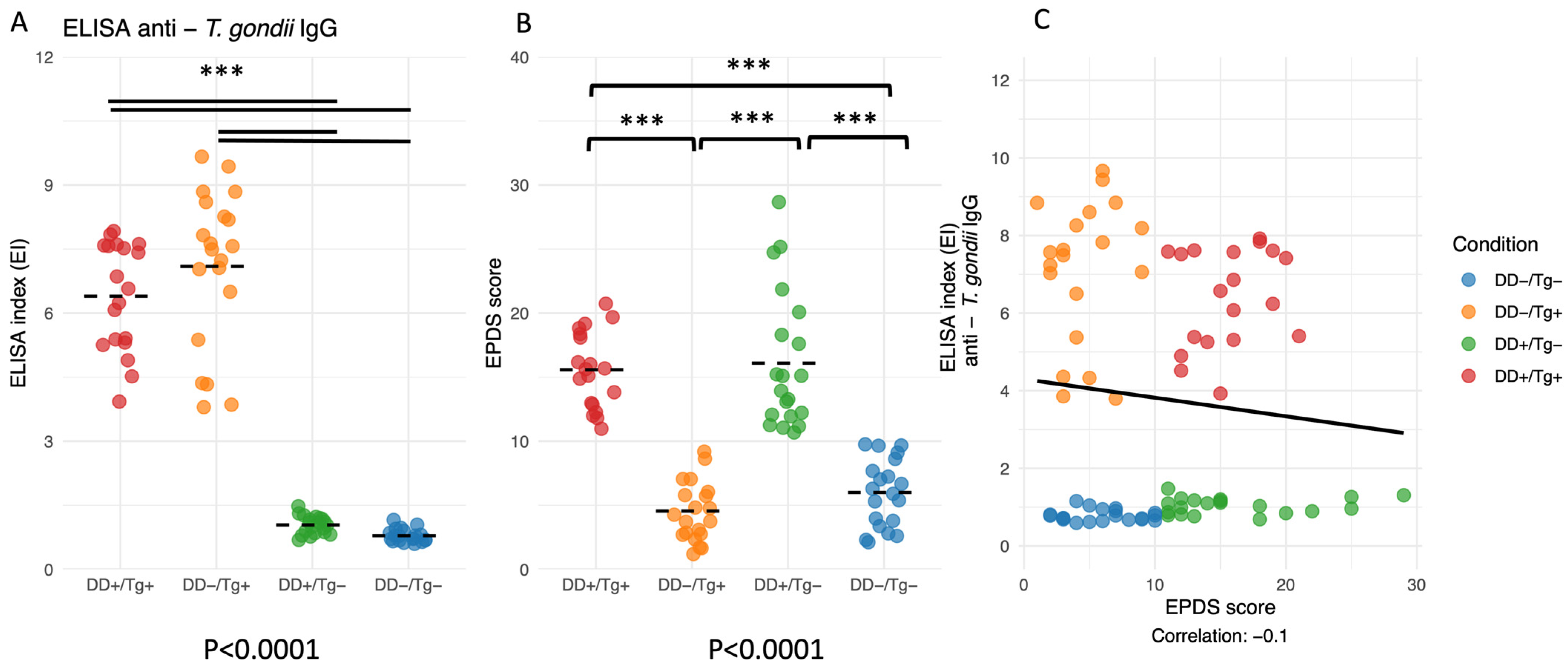
3.2. ELISA Indexes for IgG Antibodies to T. gondii and EPDS Scores Are Validated for Groups I, II, III, and IV of Patients, but No Correlation Was Found Between Both Parameters

When determining the levels of IgG antibodies to T. gondii, it was possible to observe that the ELISA indexes for Groups I and II were statistically higher than those observed for Groups III and IV, confirming that the former groups were from patients infected by this parasite, whereas the latter two groups were IgG-negative (p < 0.0001) (Figure 1A). Also, the EPDS scores for both groups of depressive patients (Group I and Group III) were statistically higher when compared to both groups of non-depressed patients (Group II and Group IV) (p < 0.0001) (Figure 1B). However, when a comparison was made between ELISA indexes and EPDS scores, no correlation was observed between the two parameters (p > 0.05) (Figure 1C).

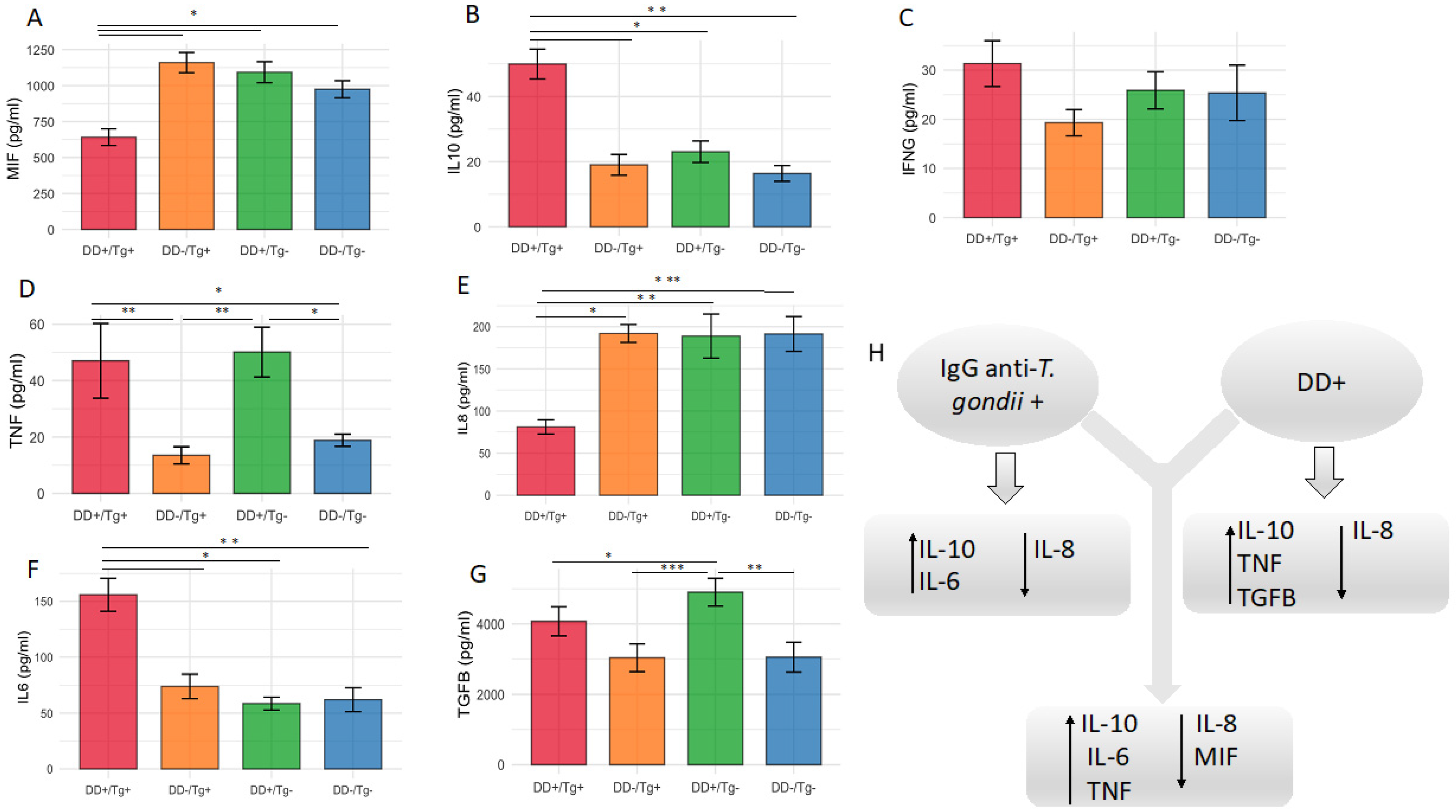
3.3. Higher Serum Levels of IL-10 and IL-6, but Lower Levels of IL-8, Cytokines Are Associated with Patients Chronically Infected by T. gondii

When analyzing the cytokine levels in the groups of patients with seropositivity for T. gondii (Groups I and II), compared to seronegative patients (Groups III and IV), no significant changes were observed in the levels of MIF, IFN-γ, TNF, and TGF-β1 associated with the positivity to T. gondii for the chronically infected patients (Figure 2A,C,D,G). However, higher levels of IL-10 and IL-6 (Figure 2B,F) and lower levels of IL-8 (Figure 2E) were found in the serum levels of patients infected by this parasite.

3.4. Higher Serum Levels of IL-10, TNF and TGF-β1, but Lower Levels of IL-8, Cytokines Are Associated with Patients Presenting Depressive Disorder

When analyzing the cytokine levels in the groups of patients with depressive disorder (Groups I and III), as determined by EPDS scores ≥ 11, in comparison with the groups without this disorder (Groups II and IV), there were no observed changes in the levels of MIF, IFN-γ, and IL-6 associated with this mental disorder (Figure 3A,C,F). In contrast, higher levels of IL-10, TNF, and TGF-β1 (Figure 3B,D,G) and lower levels of IL-8 (Figure 2E) were found in the serum levels of these groups of patients.

3.5. The Cytokine Level of MIF Is the Lowest for the Patients Presenting the Association of Infection by T. gondii and Depressive Disorder When Compared with All Other Groups of Patients

When evaluating cytokine levels considering the patients presenting the association of both clinical situations, i.e., infection by T. gondii and depressive disorder (Group I), compared with all remaining groups of patients (Groups II, III and IV), it was found that the mean level of MIF for these patients was the lowest one (Figure 4A) (p < 0.05). This group of patients also showed higher levels of IL-10, TNF, and IL-6 (Figure 4B,D,F) (p < 0.05; p < 0.01), associated with lower levels of IL-8 (Figure 4E) (p < 0.05; p < 0.01; p < 0.001).
To account for potential confounding factors and reduce noise, Principal Component Analysis (PCA) was performed to provide a lower-dimensional representation of the dataset. The analysis included cytokine levels, ELISA indexes, and EPDS scores to identify the primary sources of variance and elucidate the relationships between biological and psychological markers. As shown in Figure 5A–C, the first two principal components (PC1 and PC2) explained 21% and 16% of the total variance, respectively, with subsequent components contributing minimally. The clear separation of the DD+/Tg+ and DD−/Tg+ groups along PC1 and PC2 demonstrates the discriminatory power of PCA in distinguishing these groups and reinforces the findings presented in Figure 4.

4. Discussion

Toxoplasma gondii is a protozoan parasite that has the ability to invade the brain and can induce significant behavioral changes. The current literature presents substantial evidence of an association between T. gondii infection and various psychiatric disorders, including depressive disorder. However, additional studies are needed to confirm this association by examining both the seroepidemiology of T. gondii infection in patients with these disorders and the biological markers involved, particularly those linked to inflammatory damage in the brain.
The main aim of the present study was to assess the profiles of pro-inflammatory and anti-inflammatory cytokines associated with depressive disorder in T. gondii-seropositive pregnant women.
One of the most accepted current theories about mental disorders is their correlation with neurotransmitters, like dopamine, serotonin, and noradrenaline. However, many patients do not respond well to current antidepressant medications, likely due to other factors that may contribute to this lack of effectiveness. One of these suggests that T. gondii infection may be linked to certain mental disorders by directly impairing neuronal function in areas where parasite cysts are present. This could lead to apoptosis, changes in neuronal protein expression, or alterations in dopamine production. Alternatively, the presence of T. gondii in the brain might indirectly disrupt neuroplasticity, leading to neurodegeneration and neurotransmitter imbalances [32]. Thus, the neuropsychiatric disorders may be a result of T. gondii infection because of the tissue cyst location in the brain and the damage induced by host immune responses to the parasite [33,34].
Another potential mechanism is the effect of T. gondii infection on endocrine pathways, such as the upregulation of thyroid peroxidase autoantibodies (TPOs) in pregnant women [35]. It has been described that women who are TPO-positive during the postpartum period are more susceptible to depression [36]. Also, there are interesting findings related to two genes encoding tyrosine and phenylalanine hydroxylases due to T. gondii infection that led to the production of L-DOPA, a precursor of dopamine, and may directly affect the behavioral changes in infected hosts [33,37,38]. However, there are some disagreements related to the data described in the literature concerning this issue, as some investigations did not find an association of T. gondii IgG seropositivity with depressive disorder. In this context, Sugden et al. (2016) found that T. gondii seropositivity was not significantly associated with major depression in a population-based cohort in New Zealand [39]. Similarly, in a meta-analysis of 50 studies on T. gondii infection for major psychiatric disorders versus healthy controls, no association between T. gondii IgG seroprevalence and major depression was found [8]. In a case–control study, Alvarado-Esquivel did not find an association between T. gondii exposure and depression in pregnant women [40].
It is already known that cytokines are key mediators of the immune response, and they have been extensively studied for decades. In fact, considering that they play different functions, a fine adjustment of their expression and release is necessary; otherwise, instead of protection, they can cause immunopathological processes. Among their properties, some of them have synergistic action, in contrast with others exhibiting a clear antagonistic function. In this context, IFN-γ, TNF-α, and MIF are good examples of pro-inflammatory cytokines that play a key role in promoting inflammation and apoptosis, whereas others, such as IL-10, exhibit a significant anti-inflammatory effect, and insufficient production can lead to severe tissue destruction due to uncontrolled inflammation. In contrast, there are other cytokines, such as IL-6 and TGF-β1, with primary functions showing a dual-role cytokine with both pro- and anti-inflammatory effects depending on the cell type and context where they are released [21,22,23,24,25].
In the present study, our data revealed no association between the levels of seropositivity for IgG antibodies to T. gondii and levels of depressive disorder determined by the EPDSs, nor the correlation between levels of IgG and EPDS scores. However, when patients with depressive disorder were compared with those without depression, significant differences were observed in cytokine response, with higher levels of IL-10, TNF, and TGF-β1 and lower levels of IL-8 for depressive patients. Compared to pregnant women seronegative to T. gondii, pregnant women with positive serology showed higher levels of IL-10 and IL-6 and lower levels of IL-8. In addition, when cytokine levels from all groups were compared, the group of patients with both clinical situations, i.e., depressive disorder and T. gondii infection, was the only one that presented specifically lower levels of MIF, combined with higher levels of IL-10, IL-6, and TNF and lower levels of IL-8.
During T. gondii infection, the immune system balances pathogen control and tissue protection through a coordinated cytokine response. IL-6, TNF-α, and IL-8 drive early inflammation, with IL-8 recruiting neutrophils to enhance immune defense. IFN-γ, produced by T-helper 1 and natural killer cells, activates macrophages and antimicrobial mechanisms essential for limiting parasite replication. MIF, released by macrophages, amplifies inflammation by promoting TNF-α, IL-6, and IL-12 secretion. Interestingly, T. gondii expresses its own MIF homolog, modulating host immunity to aid parasite survival. However, while these pro-inflammatory responses are essential for controlling the parasite, excessive inflammation can cause tissue damage and pathology [19,24,25,26].
To counteract this, IL-10 and TGF-β1 act as key regulators. IL-10 suppresses pro-inflammatory cytokine production, while TGF-β1 modulates immune activation. This interplay between inflammatory and regulatory cytokines is crucial for maintaining immune homeostasis. A delicate balance between these responses ensures effective control of parasite replication while preventing immunopathology. Disruptions in this equilibrium can either lead to uncontrolled inflammation, exacerbating tissue damage, or insufficient immune activation, allowing parasite persistence [21,22].
In this context, the seropositivity for IgG antibodies to T. gondii in pregnant women during the third trimester may reflect an altered cytokine balance. This could indicate (i) a shift towards an anti-inflammatory/inflammatory cytokine profile, contrary to what is typically observed in healthy third-trimester pregnancies, and (ii) lower levels of MIF, in contrast to findings in pregnant women with depressive disorders during the third trimester. The Th1-associated immune profile is responsible for host resistance to T. gondii infection even during pregnancy, leading to the secretion of IFN-γ, in addition to other pro-inflammatory cytokines, such as IL-6 and TNF-α [33,41].
The immunologic adaptation of pregnant women changes according to the trimester of pregnancy, from pro-inflammatory in the first trimester to anti-inflammatory in the second and then returning to pro-inflammatory in the third. The release of placental cytokines (IL-1β, TNF-α, LIF/IL-6, and IFN-γ) from immune (Mus, NKs, and DCs) and nonimmune cells, the decidual cells and trophoblasts [42], during the third trimester, may contribute to triggering the changes observed in the hippocampus, prefrontal cortex, and anterior cingulate, as well as in the neuroendocrine activity of the hypothalamus and pituitary and adrenal glands. These features and the processes occurring in the placental tissues could elicit symptoms of depression during pregnancy. The release of pro-inflammatory cytokines enhances the altered neuroendocrine responses in women exhibiting depressive disorder during pregnancy [43].
The role of TNF and IFN-γ in inducing depressive symptoms is significant. These cytokines stimulate the production of indoleamine 2,3-dioxygenase (IDO) and then tryptophan (Trp) depletion along the kynurenine pathway, resulting in reduced 5-hydroxytryptamine (5-HT, serotonin) production in the brain [33,44].
Analyzing the immunologic changes that accompany depressive disorder, one meta-analysis of 24 studies on clinical patients reports significantly higher concentrations of the pro-inflammatory cytokines TNF-α and IL-6 in depressed patients compared with control ones [45]. In Brazil, Miranda et al. found elevated levels of IL-6 in patients with mood disorders [46]. Similarly, current depressive symptoms during pregnancy have been associated with elevated maternal serum IL-6 and TNF-α [47,48], showing an increasing trend across pregnancy and a significant increase at postpartum [47], and the relationship between symptom severity and both IL-6 and TNF-α was equivalent to a small effect size [48]. IL-8 is produced by human uterine cervical tissue and participates in its ripening and dilatation during labor [49]. In pregnant physiologic adaptation, the levels of IL-8 show a U-shaped curve, being high in the first trimester, decreasing in the second, and then returning to high titers at the end of pregnancy [47]. MIF is associated with the pathobiology of depression. Many investigations have identified MIF expression in the brain in significant areas associated with the behavioral symptoms of depression [50]. This cytokine may be involved in interaction with lifestyle, physical exercises, and pharmacological effects in antidepressant treatments, as it could be associated with neurogenesis. Thus, it has been investigated as a biomarker in patients presenting major depressive disorder and other mood disorders when its level is low. However, there are many aspects to be investigated concerning the association between MIF and depression, and future studies should work to clarify the relationship between central and peripheral MIF in depression. Further research is needed to clarify whether MIF acts as a pro-depressant or antidepressant and to define its role in the pathobiology of depression. Analyzing its connections with factors within and beyond the monoamine and neurogenic pathways, both known to influence depression, could provide valuable insights [50]. Also, Musil et al. (2011) reported that elevated MIF levels combined with reduced levels of TGF-β support the importance of the regulatory cytokines in major depressive disorder [51]. Given these findings and the unclear pro-inflammatory role of MIF, along with its emerging associations with perinatal outcomes and psychosocial influences during pregnancy, further studies are warranted, particularly to assess the dynamics of MIF changes over time during pregnancy [52]. Concerning the results of the present study, when evaluating the cytokine levels considering the patients presenting the association of both clinical situations, i.e., infection by T. gondii and depressive disorder (Group I), compared with all remaining groups of patients (Groups II, III and IV), it was found that the mean level of MIF for these patients was the lowest one, complemented by higher levels of IL-10, TNF, and IL-6 but lower levels of IL-8.
Even though many studies are highlighting the association between latent toxoplasmosis and mental disorders [53], it is difficult to establish a direct causal effect of toxoplasmosis in the pathogenesis of psychiatric disorders without cause–effect studies. Indeed, the simple presence of T. gondii during pregnancy is not definitely the causal factor per se, but it could be because of the profile of immune responses and the hormonal changes, which may result in the severity of the disease by infection of a specific strain type of parasite [33,34,36].

5. Limitation

This study has limitations that should be considered. Firstly, the pregnant women who need public health services in this region usually do not have access to preventive health assistance during the beginning of pregnancy. This is why we chose to enroll only the pregnant women in their third trimester in this study. This decision presented a limitation because it was not possible to observe the association between depressive disorder and the chronic phase of T. gondii during the whole period of pregnancy. Secondly, the body mass index and/or weight gain during the pregnancy would be relevant information, as these conditions may cause inflammation, which may interfere with cytokine production. Because only pregnant women were enrolled in this work, it was not possible to evaluate both parameters. Therefore, the limitations should be considered when interpreting the results of this study.

6. Conclusions

Taken together, it can be concluded that pregnant women in the third trimester of pregnancy with depressive disorder and the presence of chronic T gondii presented significant changes in the levels of immunomodulatory cytokines, as shown in Figure 6. However, these alterations were not sufficient to exacerbate this psychiatric disease during pregnancy, even considering that these molecules are key components of the immune response.

Author Contributions

Conceptualization, C.S.L., R.J.V.C., A.R.C., R.O.A., T.W.P.M., A.L.D.D. and J.R.M.; Methodology, C.S.L., T.L.d.S., H.L.S.B., L.V.S.C., D.C.A.M.M., B.F.B., L.S.V., T.M.d.A., A.R.C., R.O.A., T.W.P.M. and A.L.D.D.; Validation, D.C.A.M.M. and B.F.B.; Formal analysis, H.L.S.B., B.F.B., T.M.d.A., A.R.C., T.W.P.M., A.L.D.D. and J.R.M.; Investigation, R.J.V.C., T.L.d.S., L.V.S.C., L.S.V., T.M.d.A., A.R.C., A.L.D.D. and J.R.M.; Resources, T.W.P.M.; Data curation, J.R.M.; Writing—original draft, C.S.L., R.J.V.C., T.W.P.M. and J.R.M.; Writing—review & editing, L.S.V., R.O.A. and J.R.M.; Visualization, R.O.A.; Supervision, A.L.D.D.; Funding acquisition, T.W.P.M. and J.R.M. All authors have read and agreed to the published version of the manuscript.

Funding

This study was financially supported by Brazilian research agencies (Fundação de Amparo à Pesquisa do Estado de Minas Gerais-FAPEMIG, Grant # RED-00198-23 and #APQ-01313-14; Conselho Nacional de Desenvolvimento Científico e Tecnológico-CNPq, Grant # 312201/2021-4, INCT-Toxo 406572/2022-4, and # 445569/2020-4; Coordenação de Aperfeiçoamento de Pessoal de Nível Superior-CAPES, Grant # AUXPE-02450/09-7).

Institutional Review Board Statement

This study was approved by the Committee of Ethics in Research from Federal Univer-sity of Uberlândia (CEP-UFU), protocol # CAAE 55483716.2.0000.5152, from 10 May 2016 (can be found in Section 2.1).

Informed Consent Statement

All patients agreed to be participant and signed a consent form (can be found in Section 2.1).

Data Availability Statement

The original contributions presented in this study are included in the article. Further inquiries can be directed to the corresponding author.

Conflicts of Interest

The authors declare no conflicts of interest.

References

  1. Reddy, M.S. Depression: The disorder and the burden. Indian J. Psychol. Med. 2010, 32, 1–2. [Google Scholar] [CrossRef]
  2. Becker, M.; Weinberger, T.; Chandy, A.; Schmukler, S. Depression during pregnancy and postpartum. Curr. Psychiatry Rep. 2016, 18, 32. [Google Scholar] [CrossRef] [PubMed]
  3. Gelaye, B.; Rondon, M.B.; Araya, R.; Williams, M.A. Epidemiology of maternal depression, risk factors, and child outcomes in low-income and middle-income countries. Lancet Psychiatry 2016, 3, 973–982. [Google Scholar] [CrossRef] [PubMed]
  4. Fisher, J.; De Mello, M.C.; Patel, V.; Rahman, A.; Tran, T.; Holton, S.; Holmes, W. Prevalence and determinants of common perinatal mental disorders in women in low- and lower-middle-income countries: A systematic review. Bull. World Health Organ. 2012, 90, 139G–149G. [Google Scholar] [PubMed]
  5. Biratu, A.; Haile, D. Prevalence of antenatal depression and associated factors among pregnant women in Addis Ababa, Ethiopia: A cross-sectional study. Reprod. Health 2015, 12, 99. [Google Scholar] [CrossRef]
  6. Lima, M.D.O.P.; Tsunechiro, M.A.; Bonadio, I.C.; Murata, M. Depressive symptoms in pregnancy and associated factors: Longitudinal study. Acta Paul. Enferm. 2017, 30, 39–46. [Google Scholar] [CrossRef]
  7. Grote, N.K.; Bridge, J.A.; Gavin, A.R.; Melville, J.L.; Iyengar, S.; Katon, W.J. A meta-analysis of depression during pregnancy and the risk of preterm birth, low birth weight, and intrauterine growth restriction. Arch. Gen. Psychiatry 2010, 67, 1012–1024. [Google Scholar] [CrossRef]
  8. Sutterland, A.L.; Fond, G.; Kuin, A.; Koeter, M.W.J.; Lutter, R.; van Gool, T.; Yolken, R.; Szoke, A.; Leboyer, M.; de Haan, L. Beyond the association. Toxoplasma gondii in schizophrenia, bipolar disorder, and addiction: Systematic review and meta-analysis. Acta Psychiatr. Scand. 2015, 132, 161–179. [Google Scholar]
  9. Alvarado-Esquivel, C.; Urbina-Álvarez, J.D.; Estrada-Martínez, S.; Torres-Castorena, A.; Molotla-De-León, G.; Liesenfeld, O.; Dubey, J.P. Toxoplasma gondii infection and schizophrenia: A case control study in a low Toxoplasma seroprevalence Mexican population. Parasitol. Int. 2011, 60, 151–155. [Google Scholar]
  10. Groër, M.W.; Yolken, R.H.; Xiao, J.-C.; Beckstead, J.W.; Fuchs, D.; Mohapatra, S.S.; Seyfang, A.; Postolache, T.T. Prenatal depression and anxiety in Toxoplasma gondii positive women. Am. J. Obstet. Gynecol. 2011, 204, 433.e1–7. [Google Scholar] [CrossRef]
  11. Alvarado-Esquivel, C.; Sanchez-Anguiano, L.F.; Hernandez-Tinoco, J.; Berumen-Segovia, L.O.; Torres-Prieto, Y.E.; Estrada-Martinez, S.; Perez-Alamos, A.R.; Ortiz-Jurado, M.N.; Molotla-De-Leon, G.; Garcia, I.B.; et al. Toxoplasma gondii infection and mixed anxiety and depressive disorder: A case-control seroprevalence study in Durango, Mexico. J. Clin. Med. Res. 2016, 8, 519–523. [Google Scholar] [CrossRef]
  12. Kar, N.; Misra, B. Toxoplasma seropositivity and depression: A case report. BMC Psychiatry 2004, 4, 1. [Google Scholar]
  13. Hsu, P.C.; Groer, M.; Beckie, T. New findings: Depression, suicide, and Toxoplasma gondii infection. J. Am. Assoc. Nurse Pract. 2014, 26, 629–637. [Google Scholar] [CrossRef]
  14. Gajewski, P.D.; Falkenstein, M.; Hengstler, J.G.; Golka, K. Toxoplasma gondii impairs memory in infected seniors. Brain Behav. Immun. 2014, 36, 193–199. [Google Scholar]
  15. Alvarado-Esquivel, C.; Liesenfeld, O.; Márquez-Conde, J.A.; Estrada-Martínez, S.; Dubey, J.P. Seroepidemiology of infection with Toxoplasma gondii in workers occupationally exposed to water, sewage, and soil in Durango, Mexico. J. Parasitol. 2010, 96, 847–850. [Google Scholar] [CrossRef] [PubMed]
  16. Alvarado-Esquivel, C.; Campillo-Ruiz, F.; Liesenfeld, O. Seroepidemiology of infection with Toxoplasma gondii in migrant agricultural workers living in poverty in Durango, Mexico. Parasit. Vectors 2013, 6, 113. [Google Scholar] [CrossRef] [PubMed]
  17. Duffy, A.R.; Beckie, T.M.; Brenner, L.A.; Beckstead, J.W.; Seyfang, A.; Postolache, T.T.; Groer, M.W. Relationship between Toxoplasma gondii and mood disturbance in women veterans. Mil. Med. 2015, 180, 621–625. [Google Scholar] [CrossRef]
  18. Zhu, S. Psychosis may be associated with toxoplasmosis. Med. Hypotheses 2009, 73, 799–801. [Google Scholar] [CrossRef]
  19. Audet, M.C.; McQuaid, R.J.; Merali, Z.; Anisman, H. Cytokine variations and mood disorders: Influence of social stressors and social support. Front. Neurosci. 2014, 8, 416. [Google Scholar] [CrossRef]
  20. Del Grande, C.; Galli, L.; Schiavi, E.; Dell’Osso, L.; Bruschi, F. Is Toxoplasma gondii a trigger of bipolar disorder? Pathogens 2017, 6, E3. [Google Scholar] [CrossRef]
  21. Mammari, N.; Halabi, M.A.; Yaacoub, S.; Chlala, H.; Dardé, M.L.; Courtioux, B. Toxoplasma gondii modulates the host cell responses: An overview of apoptosis pathways. Biomed. Res. Int. 2019, 2019, 6152489. [Google Scholar] [CrossRef] [PubMed]
  22. Ruiz-Rosado, J.d.D.; Olguín, J.E.; Juárez-Avelar, I.; Saavedra, R.; Terrazas, L.I.; Robledo-Avila, F.H.; Vazquez-Mendoza, A.; Fernández, J.; Satoskar, A.R.; Partida-Sánchez, S.; et al. MIF Promotes classical activation and conversion of inflammatory Ly6Chigh monocytes into TipDCs during murine toxoplasmosis. Mediat. Inflamm. 2016, 2016, 9101762. [Google Scholar] [CrossRef] [PubMed]
  23. Park, E.A.; Han, I.H.; Kim, J.H.; Park, S.J.; Ryu, J.S.; Ahn, M.H. Production of inflammatory cytokines and nitric oxide by human mast cells incubated with Toxoplasma gondii lysate. Korean J. Parasitol. 2019, 57, 201–206. [Google Scholar] [CrossRef]
  24. Davami, M.H.; Baharlou, R.; Vasmehjani, A.A.; Ghanizadeh, A.; Keshtkar, M.; Dezhkam, I.; Atashzar, M.R. Elevated IL-17 and TGF-β serum levels: A positive correlation between T-helper 17 cell-related pro-inflammatory responses with major depressive disorder. Basic Clin. Neurosci. 2016, 7, 137–142. [Google Scholar] [CrossRef]
  25. Zou, W.; Feng, R.; Yang, Y. Changes in the serum levels of inflammatory cytokines in antidepressant drug-naïve patients with major depression. PLoS ONE 2018, 13, e0197267. [Google Scholar] [CrossRef]
  26. Conboy, L.; Varea, E.; E Castro, J.; Sakouhi-Ouertatani, H.; Calandra, T.; A Lashuel, H.; Sandi, C. Macrophage migration inhibitory factor is critically involved in basal and fluoxetine-stimulated adult hippocampal cell proliferation and in anxiety, depression, and memory-related behaviors. Mol. Psychiatry 2011, 16, 533–547. [Google Scholar] [PubMed]
  27. Cox, J.L.; JHolden, M.; Sagovsky, R. Detection of Postnatal Depression. Development of the 10-item Edinburgh Postnatal Depression Scale. Br. J. Psychiatry 1987, 150, 782–786. [Google Scholar]
  28. Santos, I.S.; Matijasevich, A.; Tavares, B.F.; Barros, A.J.D.; Botelho, I.P.; Lapolli, C.; da Silva Magalhães, P.V.; Barbosa, A.P.P.N.; Barros, F.C. Validation of the Edinburgh postnatal depression scale (EPDS) in a sample of mothers from the 2004 Pelotas Birth Cohort Study. Cad. Saude Publica 2007, 23, 2577–2588. [Google Scholar]
  29. Castro-E-Couto, T.; Brancaglion, M.Y.M.; Cardoso, M.N.; Protzner, A.B.; Garcia, F.D.; Nicolato, R.; Aguiar, R.A.L.P.; Leite, H.V.; Corrêa, H. What is the best tool for screening antenatal depression? J. Affect. Disord. 2015, 178, 12–17. [Google Scholar] [CrossRef]
  30. Bakker, M.; Veldkamp, C.L.S.; van den Akker, O.R.; van Assen, M.A.L.M.; Crompvoets, E.; Ong, H.H.; Wicherts, J.M. Recommendations in pre-registrations and internal review board proposals promote formal power analyses but do not increase sample size. PLoS ONE 2020, 15, e0236079. [Google Scholar] [CrossRef]
  31. Silva, D.A.O.; Silva, N.M.; Mineo, T.W.; Pajuaba Neto, A.A.; Ferro, E.A.; Mineo, J.R. Heterologous antibodies to evaluate the kinetics of the humoral immune response in dogs experimentally infected with Toxoplasma gondii RH strain. Vet. Parasitol. 2002, 107, 181–195. [Google Scholar] [CrossRef]
  32. Parlog, A.; Schluter, D.; Dunay, I.R. Toxoplasma gondii-induced neuronal alterations. Parasite Immunol. 2015, 37, 159–170. [Google Scholar] [CrossRef]
  33. McConkey, G.A.; Martin, H.L.; Bristow, G.C.; Webster, J.P. Toxoplasma gondii infection and behaviour—Location, location, location? J. Exp. Biol. 2013, 216, 113–119. [Google Scholar] [CrossRef] [PubMed]
  34. Webster, J.P.; McConkey, G.A. Toxoplasma gondii-altered host behaviour: Clues as to mechanism of action. Folia Parasitol. 2010, 57, 95–104. [Google Scholar] [CrossRef] [PubMed]
  35. Wasserman, E.E.; Nelson, K.; Rose, N.R.; Rhode, C.; Pillion, J.P.; Seaberg, E.; Talor, M.V.; Burek, L.; Eaton, W.; Duggan, A.; et al. Infection and thyroid autoimmunity: A seroepidemiologic study of TPOaAb. Autoimmunity 2009, 42, 439–446. [Google Scholar] [CrossRef] [PubMed]
  36. Lazarus, J.; Hall, R.; Othman, S.; Parkes, A.; Richards, C.; McCulloch, B.; Harris, B. The clinical spectrum of postpartum thyroid disease. QJM 1996, 89, 429–435. [Google Scholar] [CrossRef]
  37. Gaskell, E.A.; Smith, J.E.; Pinney, J.W.; Westhead, D.R.; McConkey, G.A. A unique dual activity amino acid hydroxylase in Toxoplasma gondii. PLoS ONE 2009, 4, e4801. [Google Scholar] [CrossRef]
  38. Skallova, A.; Kodym, P.; Frynta, D.; Flegr, J. The role of dopamine in Toxoplasma-induced behavioural alterations in mice: An ethological and ethopharmacological study. Parasitology 2006, 133, 525–535. [Google Scholar] [CrossRef]
  39. Sugden, K.; Moffitt, T.E.; Pinto, L.; Poulton, R.; Williams, B.S.; Caspi, A. Is Toxoplasma gondii infection related to brain and behavior impairments in humans? Evidence from a population-representative birth cohort. PLoS ONE 2016, 11, e0148435. [Google Scholar] [CrossRef]
  40. Alvarado-Esquivel, C.; Martínez-Martínez, A.L.; Sánchez-Anguiano, L.F.; Hernández-Tinoco, J.; Castillo-Orona, J.M.; Salas-Martínez, C.; Sifuentes-Álvarez, A.; Sandoval-Carrillo, A.A.; Salas-Pacheco, J.M.; Liesenfeld, O.; et al. Lack of association between Toxoplasma gondii exposure and depression in pregnant women: A case-control study. BMC Infect. Dis. 2017, 17, 190. [Google Scholar]
  41. Munoz, M.; Liesenfeld, O.; Heimesaat, M.O. Immunology of Toxoplasma gondii. Immunol. Rev. 2011, 240, 269–285. [Google Scholar] [PubMed]
  42. Leff-Gelman, P.; Mancilla-Herrera, I.; Flores-Ramos, M.; Cruz-Fuentes, C.; Reyes-Grajeda, J.P.; García-Cuétara, M.d.P.; Bugnot-Pérez, M.D.; Pulido-Ascencio, D.E. The immune system and the role of inflammation in perinatal depression. Neurosci. Bull. 2016, 32, 398–420. [Google Scholar] [CrossRef] [PubMed]
  43. Leff Gelman, P.; Flores-Ramos, M.; López-Martínez, M.; Fuentes, C.C.; Grajeda, J.P.R. Hypothalamic-pituitary-adrenal axis function during perinatal depression. Neurosci. Bull. 2015, 31, 338–350. [Google Scholar]
  44. Robinson, C.M.; Hale, P.T.; Carlin, J.M. The role of IFN-g and TNF-α-responsive regulatory elements in the synergistic induction of indoleamine dioxygenase. J. Interferon Cytokine Res. 2005, 25, 20–30. [Google Scholar]
  45. Dowlati, Y.; Herrmann, N.; Swardfager, W.; Liu, H.; Sham, L.; Reim, E.K.; Lanctôt, K.L. A meta-analysis of cytokines in major depression. Biol. Psychiatry 2010, 67, 446–457. [Google Scholar]
  46. Miranda, H.C.; Nunes, S.O.V.; Reiche, E.M.V.; Oda, J.M.M.; Watanabe, M.A.E. Higher than Normal Plasma Iinterleukin-6 Concentrations in Brazilian Patients with Mood Disorders. Braz. Arch. Biol. Technol. 2011, 54, 717–722. [Google Scholar]
  47. Christian, L.M.; Porter, K. Longitudinal Changes in Serum Proinflammatory Markers across Pregnancy and Postpartum: Effects of Maternal Body Mass Index. Cytokine 2014, 70, 134–140. [Google Scholar]
  48. Christian, L.M.; Franco, A.; Glaser, R.; Iams, J. Depressive symptoms are associated with elevated serum proinflammatory cytokines among pregnant women. Brain Behav. Immun. 2009, 23, 750–754. [Google Scholar] [CrossRef]
  49. Winkler, M. Role of cytokines and other inflammatory mediators. BJOG 2003, 110, 118–123. [Google Scholar]
  50. Bloom, J.; Al-Abed, Y. MIF: Mood Improving/Inhibiting Factor? J. Neuroinflamm. 2014, 11, 11. [Google Scholar] [CrossRef]
  51. Musil, R.; Schwarz, M.J.; Riedel, M.; Dehning, S.; Cerovecki, A.; Spellmann, I.; Arolt, V.; Müller, N. Elevated macrophage migration inhibitory factor and decreased transforming growth factor-beta levels in major depression—No influence of celecoxib treatment. J. Affect. Disord. 2011, 134, 217–225. [Google Scholar] [CrossRef] [PubMed]
  52. Christian, L.; Franco, A.; Iams, J.; Sheridam, J.; Glaser, R. Depressive symptoms predict exaggerated inflammatory responses to an in vivo immune challenge among pregnant Women. Brain Behav. Immun. 2010, 24, 49–53. [Google Scholar] [PubMed]
  53. Fallahi, S.; Rostami, A.; Shiadeh, M.N.; Behniafar, H.; Paktinat, S. An updated literature review on maternal-fetal and reproductive disorders of Toxoplasma gondii infection. J. Gynecol. Obstet. Hum. Reprod. 2018, 47, 133–140. [Google Scholar] [CrossRef] [PubMed]
Figure 1. (A) Detection of IgG antibodies against T. gondii in patient Groups I-IV using ELISA, with the results expressed as ELISA index values, which quantify relative antibody concentration by comparing the optical density (OD) of each sample to a cutoff value derived from control samples. (B) Determination of the Edinburgh Postnatal Depressive Scale (EPDS) scores for Groups I–IV of patients. (A,B) The mean for each group is indicated by a horizontal line in the plot, representing the average value of the data points within that group. (C) Correlation between ELISA index and EPDS scores for Groups I–IV of patients. *** p < 0.001.
Pathogens 14 00330 g001
Figure 2. (AH) Serum levels of pro-inflammatory and anti-inflammatory cytokines (MIF, IFNg, TNF, IL8, figures A, C, D and E, respectively; IL10, IL6, TGFB, figures B, F and G, respectively) were measured and compared between chronically T. gondii-infected patients (Groups I and II) and uninfected controls (Groups III and IV). Statistical significance was assessed using the Brunner–Munzel test, with significant differences (p < 0.05) displayed in the graph, indicating the decreasing corresponding p-values, as follows: * p = 0.013837; ** p = 0.007674; and *** p = 0.000789.
Pathogens 14 00330 g002
Figure 3. (AH) Serum levels of pro-inflammatory and anti-inflammatory cytokines (MIF, IFNg, TNF, IL8, figures A, C, D and E, respectively; IL10, IL6, TGFB, figures B, F and G, respectively) were measured and compared between groups with depressive disorder (Groups I and III) and groups without this mental problem (Groups II and IV). Statistical significance was assessed using the Brunner–Munzel test, with significant differences (p < 0.05) displayed in the graph, indicating the range of the corresponding p-values, as follows: *** p = 4.62 × 10⁻⁴; *** p = 6.21 × 10⁻⁵; *** p = 5.99 × 10⁻⁶; and *** p = 1.22 × 10⁻¹⁰.
Pathogens 14 00330 g003
Figure 4. (AH) Determination of the levels of pro-inflammatory and anti-inflammatory cytokines (MIF, IFNg, TNF, IL8, figures A, C, D and E, respectively; IL10, IL6, TGFB, figures B, F and G, respectively) in serum samples of patients presenting infection by T. gondii and depressive disorder (Group I) compared with all groups of patients (Groups II, III, and IV) The Kruskal–Wallis test was used for multiple groups comparisons, followed by Dunn’s post hoc test to identify specific group differences. Holm’s method was applied to reduce the risk of false positives * p < 0.05; ** p < 0.01; *** p < 0.001.
Pathogens 14 00330 g004
Figure 5. Principal Component Analysis of cytokine levels, ELISA indexes, and EPDS scores among the four diagnostic groups. The panels (AC) show different combinations of principal components (PC1 vs. PC2, PC3 vs. PC4, and PC5 vs. PC6) to demonstrate variance accounts for each component. Colored ellipses indicate the 95% confidence intervals for each diagnostic group: DD−/Tg− (blue stars), DD−/Tg+ (orange circles), DD+/Tg− (green squares), and DD+/Tg+ (red squares).
Pathogens 14 00330 g005
Figure 6. Summary of key findings. Illustration of the interaction between T. gondii infection and depressive disorder during pregnancy, highlighting their combined effects on cytokine balance. Chronic phase of T. gondii infection is associated with reduced IL-8 and increased IL-10 and IL-6, while depression leads to elevated TNF, IL-10, and TGF-β. When both conditions coexist, the imbalance intensifies, with higher TNF, IL-6, and IL-10 and lower IL-8 and MIF levels. This figure summarizes these cytokine shifts, emphasizing the immune dysregulation linked to both conditions.
Pathogens 14 00330 g006
Table 1. Characteristics of the patients from Groups I, II, III, and IV enrolled in the present study.
CharacteristicsGroup I DD+Tg+
(n = 19)
Group II
DD−Tg+
(n = 20)
Group III
DD+Tg−
(n = 20)
Group IV
DD−Tg−
(n = 20)
p-Value
Maternal age (year) ± SD *31 ± 6.63 a30 ± 5.88 ab27 ± 5.90 ab26 ± 5.31 b0.0174
Gestational age (weeks) ± SD29 ± 8.1031 ± 8.5032 ± 7.1532 ± 7.750.1571
EPDS score (score mean) ± SD *15.6 ± 2.9 a4.5 ± 2.3 b16.2 ± 5.5 a5.8 ± 2.8 b0.0001
Marital status
Married/living with a partner14 (74%)18 (90%)10 (50%)12 (60%)0.0359
Single/separated/divorced/
living without a partner
5 (26%)2 (10%)10 (50%)8 (40%)
Degree of education
Elementary/High school9 (47%)14 (70%)15 (75%)19 (95%)0.0099
Undergraduate10 (53%)6 (30%)5 (25%)1 (5%)
Occupancy
Housewife9 (47%)7 (35%)10 (50%)6 (30%)0.5387
Daily laborer/employed10 (53%)13 (65%)10 (50%)14 (70%)
Average household income
<minimum wage7 (37%)9 (45%)10 (50%)11 (55%)0.7326
>minimum wage12 (63%)11 (55%)10 (50%)9 (45%)
Parity
Primiparous7 (37%)7 (35%)3 (15%)15 (75%)0.0009
Multiparous12 (63%)13 (65%)17 (85%)5 (25%)
Type of previous delivery
Cesarean section5 (42%)5 (39%)6 (35%)3 (60%)0.8246
Spontaneous labor7 (58%)8 (61%)11 (65%)2 (40%)
Planed Pregnancy
Yes5 (26%)9 (45%)5 (25%)10 (50%)0.2659
No14 (74%)11 (55%)15 (75%)10 (50%)
Chronic disease
Yes3 (16%)4 (20%)5 (25%)6 (30%)0.8036
No16 (84%)16 (80%)15 (75%)14 (70%)
Previous depression
Yes2 (10%)1 (5%)5 (25%)00.0419
No17 (90%)19 (95%)15 (75%)20 (100%)
Alcohol consumption
Yes1 (5%)2 (10%)2 (10%)2 (10%)0.9999
No18 (95%)18 (90%)18 (90%)18 (90%)
Smoking
Yes02 (10%)3 (15%)00.1514
No19 (100%)18 (90%)17 (85%)20 (100%)
Negative life events
Yes6 (31%)3 (15%)10 (50%)5 (25%)0.1094
No13 (69%)17 (85%)10 (50%)15 (75%)
*,a or b: same letters, no significant difference; a and b: different letters, significant differences.
Disclaimer/Publisher’s Note: The statements, opinions and data contained in all publications are solely those of the individual author(s) and contributor(s) and not of MDPI and/or the editor(s). MDPI and/or the editor(s) disclaim responsibility for any injury to people or property resulting from any ideas, methods, instructions or products referred to in the content.

