Article

Prevalence and Molecular Characterization of Cryptosporidium Species in Diarrheic Children in Cameroon

1
Laboratory for Molecular Parasitology, Department of Microbiology and Parasitology, University of Buea, Buea P.O. Box 63, Cameroon
2
Centre for Research on Health and Priority Pathologies, Institute of Medical Research and Medicinal Plants Studies (IMPM), Yaoundé, P.O. Box 13033, Cameroon
3
Institute of Parasitology, Biomedical Research Center Seltersberg (B.F.S.), Justus Liebig University Giessen, 35392 Giessen, Germany
4
Department of Infectious Disease Epidemiology, Bernhard-Nocht Institute for Tropical Medicine, 20359 Hamburg, Germany
*
Authors to whom correspondence should be addressed.
Pathogens 2025, 14(3), 287; https://doi.org/10.3390/pathogens14030287
Submission received: 3 February 2025 / Revised: 9 March 2025 / Accepted: 11 March 2025 / Published: 14 March 2025
(This article belongs to the Section Parasitic Pathogens)

Abstract

:
Cryptosporidiosis remains a major cause of diarrhea-related childhood death, particularly in developing countries. Although effective anti-retroviral therapy has significantly reduced the cryptosporidiosis burden in western nations, the situation in developing countries remains alarming due to limited therapeutic options and a lack of preventive measures. To better control disease transmission and develop effective prevention strategies, a thorough understanding of the genetic diversity of circulating species is crucial. While cryptosporidiosis has previously been reported in Cameroon, information on the genetic diversity of parasite strains is lacking. In a large cross-sectional study conducted between March 2020 and March 2021 in four regions of Cameroon, Southwest, Littoral, Center and West, a total of 1119 fecal samples of children (n = 1119) were collected and genetically analyzed. This study aimed to assess the genetic diversity of Cryptosporidium strains circulating in this patient cohort in Cameroon. Using modified Ziehl–Neelsen fecal smear staining, an overall prevalence of 8.5% (96/1119) was recorded. PCR analysis revealed a prevalence of 15.4% in the Center, 7.2% in the Littoral, 10.5% in the West, and 13.1% in the Southwest regions. Molecular analysis identified Cryptosporidium. hominis and Cryptosporidium parvum as circulating species, with all subtype families suggesting anthroponotic transmission. No zoonotic subtypes of C. parvum were detected. These findings confirm that cryptosporidiosis transmission in Cameroon is primarily anthroponotic. Nonetheless, much larger epidemiological surveys, including other patient cohorts, are necessary for final confirmation of this statement.

1. Introduction

Cryptosporidium is an intestinal apicomplexan protozoan parasite of the Cryptosporiidae family within the phylum Alveolata and has been the cause of numerous water- and food-borne outbreaks of human and livestock enteric disease [1]. This parasite may produce a short-term and self-limiting illness in immunocompetent persons, clinically manifested by transient diarrhea, nausea, vomiting, fever, and abdominal pain [2,3]. Nonetheless, in immunocompromised patients, especially those with AIDS, or malnourished children, human cryptosporidiosis is characterized by a prolonged, life-threatening, and cholera-like diarrhea [4,5,6] Furthermore, infective Cryptosporidium oocysts are known to resist chlorination and thus are abundantly present in surface waters. Cryptosporidium hominis and Cryptosporidium parvum are the most important species infecting humans worldwide [3,7,8]. While anthroponotic C. hominis seems to mainly infect humans, C. parvum is zoonotic and infects a broad range of hosts, including humans and bovines. Especially in neonatal calves, it causes watery diarrhea resulting in significant economic losses for farmers and environmental oocyst contamination [9]. So far, preventive approaches, like vaccines, are completely lacking. Moreover, treatment of cryptosporidiosis in humans exclusively relies on nitazoxanide, which shows poor efficacy in immunocompromised patients and malnourished children, i.e., the main risk groups for human cryptosporidiosis [10].
The overall importance of human cryptosporidiosis in developing countries was highlighted by a global study covering all enteric diseases, ranking human cryptosporidiosis second to rotavirus infections concerning diarrhea in children [11]. While the use of highly active anti-retroviral therapies (HAARTs) against HIV infection has significantly reduced the burden of human cryptosporidiosis in industrialized countries, it is still a major cause of diarrhea in neonates and children, especially under two years of age, in developing countries [12,13,14]. Several subtypes of Cryptosporidium spp. have been associated with outbreaks of water-borne diseases in industrialized countries, like Korea [15], New Zealand [16], Hungary [17] and, more recently, the United Kingdom (Crypto Outbreak in Devon, UK, May 2024, BBC). In the USA, recreational water was associated with 156 of 444 (35.1%) reported cryptosporidiosis outbreaks from 2009 to 2017 [18].
Cryptosporidium species differ in host range and parasite–host coevolution, host adaptation, epizootiology, transmission, as well as geographic segregation, leading to the formation of subtype families with unique phenotypic traits within the major human–pathogenic species C. parvum and C. hominis [8,19,20]. In human populations, the most common species correspond to the anthroponotic species C. hominis. followed by the zoonotic species C. parvum [21]. However, only C. parvum can be cultured in vitro, driving it to become the focus of many experimental models used for basic research [22,23,24], vaccine and drug development [25]. Even though different C. parvum isolates from humans, farm animals, companion animals, and rodents are morphologically and developmentally similar, differences in host specificity, pre-patency, patency, and pathogenicity were documented [26,27,28,29]. Overall, more than 20 Cryptosporidium species and genotypes have been identified so far in humans [30] and a total of 44 Cryptosporidium species and over 120 wildlife genotypes described to date [31]. Identification of Cryptosporidium at the species or genotype level is essential for the assessment of infection sources and public health implications driven by parasite maintenance in domestic animals. Transmission intensity, genetic diversity, occurrence of genetic recombination and selective pressure have further shaped the parasite population’s genetic structures. Thus, the distribution of Cryptosporidium species in humans differs between developing and industrialized countries, with C. hominis dominating in developing countries, whilst both C. hominis and C. parvum are more or less equally present in industrialized nations [32,33,34]. These two species are responsible for almost all human cryptosporidiosis outbreaks reported so far [35]. However, there is a lack of information on the genetic diversity of Cryptosporidium amongst human populations, especially for developing countries.
Cryptosporidium spp. have been diagnosed in nearly all warm-blooded animals investigated so far [36,37,38,39] and consequently are present in both terrestrial and marine ecosystems [3,40,41,42,43,44]. The ubiquitous nature of this parasite makes it a serious threat to global health and emphasizes the urgent need to establish a comprehensive genotype mapping of Cryptosporidium, both geographically and across the different terrestrial and marine hosts. Importantly, given that efforts to develop a vaccine against cryptosporidiosis are ongoing, genetic diversity studies are also pivotal for optimized and efficient application of any protective vaccine.
Among the methods for genotypic analysis of Cryptosporidium, DNA sequencing of selected gene loci and restriction fragment length polymorphism analysis (RFLP) are the most popular. Reference genes, such as 18S rRNA, COWP, HSP70 and TRAP, have all successfully been applied to identify and classify Cryptosporidium spp. [45]. In addition, the highly polymorphic GP60 gene, which seems linked to pathogenicity [46,47], is preferentially used for sub-species genotyping.
In Cameroon, the burden of cryptosporidiosis in humans and livestock, as well as its transmission pattern, have been barely investigated. More importantly, Cameroonian Cryptosporidium spp. genetic diversity remains entirely unknown. Few studies have been conducted on the occurrence of cryptosporidiosis in HIV patients [48,49] and on environmental oocyst contamination of streams [50]. However, these studies all used microscopy for oocyst detection, which does not give any information on the genetic diversity of the species, thereby emphasizing the need for more in-depth studies in Cameroon. Referring to other regions of the African continent, a number of studies have been published so far, which include the molecular characterization of Cryptosporidium species, with examples from Ghana [51] Nigeria [52], Egypt [53], Gabon, Ghana, Madagascar and Tanzania [54], Zambia [55], Mozambique [56], Kenya [57], São Tomé and Principe [58], South Africa [59] and Uganda [60].
The aim of the current study was to determine the genetic diversity of Cryptosporidium spp. circulating among risk groups in Cameroon.

2. Materials and Methods

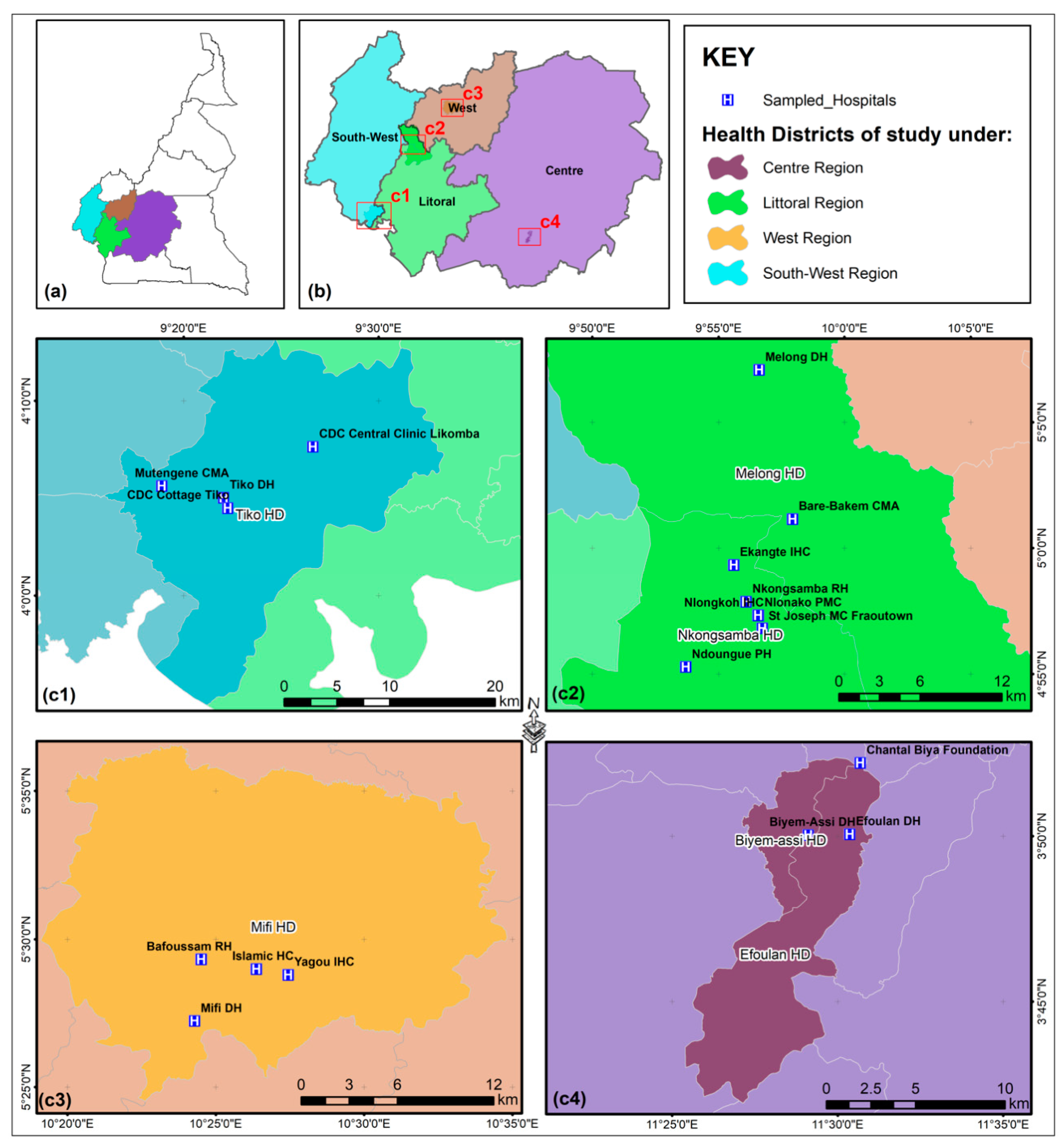
2.1. Sample Collection

In a cross-sectional study conducted between March 2020 and March 2021, fecal samples (n = 1119) were collected from children aged zero to ten years from four regions of Cameroon: Southwest, Littoral, Center and West (Figure 1). Participants included in the study were hospitalized patients (in- and outpatients) who presented with symptoms of loose, diarrheic stools (at least 3/day) and vomiting (at least once within the past 24 h). Patients who did not fulfil these criteria were excluded from the study. All health facilities included were in urban areas.

2.2. Modified Ziehl Neelsen (MZN) Staining

Freshly collected stool samples were homogenized, then applied as moderately thick fecal smears on standard microscope glass slides, using a sterile applicator stick. The smears were allowed to air dry for 20 min at room temperature (RT) before staining. The smears were stained using the Ziehl Neelsen Method (MZN) [61] with slight modifications. Briefly, the slides were fixed in absolute methanol for 3 min followed by staining with strong carbolfuchsin (Sigma-Aldrich, Darmstadt, Germany) for 20 min. The slides were thoroughly rinsed with tap water followed by a minimal decolorization by rapid agitation in 3% hydrochloric acid-alcohol for 10 s followed by a quick rinse with tap water. The smear was counterstained for 30 s in 1% methylene blue (Sigma-Aldrich, Darmstadt, Germany), rinsed with tap water, and allowed to air dry. The stained slides were examined under a phase contrast CHBS Olympus microscope (Olympus, Hamburg, Germany) using the 100× oil immersion objective.

2.3. DNA Extraction

DNA was extracted from approximately 200 mg of each fecal sample by the alkali freeze–boil method. For each sample, 800 μL of alkali washing solution (0.5 M NaOH and 0.05 M sodium citrate) was added and the samples were mixed for 10 min at RT by inversion using a rotary wheel (60 rpm). Thereafter, samples were centrifuged three times at 13,000× g for 5 min. The cell pellet was re-suspended in 0.5 mL Tris-HCl (pH 8.0) and re-centrifuged at 13,000× g for 5 min. This step was repeated once and the resulting pellet was re-suspended in Tris-EDTA (0.1 mL, 10 mM Tris HCl, pH 8.0, containing 1 mM EDTA). DNA was extracted by 4 cycles of freezing and thawing (−80 °C for 45 min and 100 °C for 12 min), respectively. The resulting extract was centrifuged at 13,000 g for 15 min and the supernatant containing total DNA was transferred to a sterile 1.5 mL Eppendorf tube [62]. The extracted DNA was quantified using a NanoDrop spectrophotometer (Thermo Fisher Scientific, Waltham, MA, USA) at 260 nm and stored at −20 °C until PCR amplification.

2.4. SSU-rRNA Nested PCR

For SSU-rRNA nested PCR, gene specific primers were used as follows: outer primers [(5′TTCTGAGCTAATACATGCG 3′) and (5′CCCATTTCCTTCGAAACAGGA 3′)] and inner primers [(5’GGAAGGGTTGTATTTATTAGATAAAG-3′) and (5′-AAGGAGTAAGGAACAACCTCCA-3′)] [45]. The amplification conditions were set as follows: initial denaturation of 95 °C for 3 min followed by 35 cycles of a three-step program (94 °C for 45 s, 55 °C for 45 s and 72 °C for 1 min), and a final extension step at 72 °C for 7 min. The same reaction conditions were used for primary and secondary PCR. The secondary amplicons were separated on 1.2% agarose gels and visualized using a UV-trans illuminator BIO-RAD Molecular Imager© Gel Doc™ XR+ imaging system (BIORAD, Feldkirchen, Germany). Positive (Cryptosporidium parvum oocysts) and negative (containing no template DNA) controls were included in each assay.

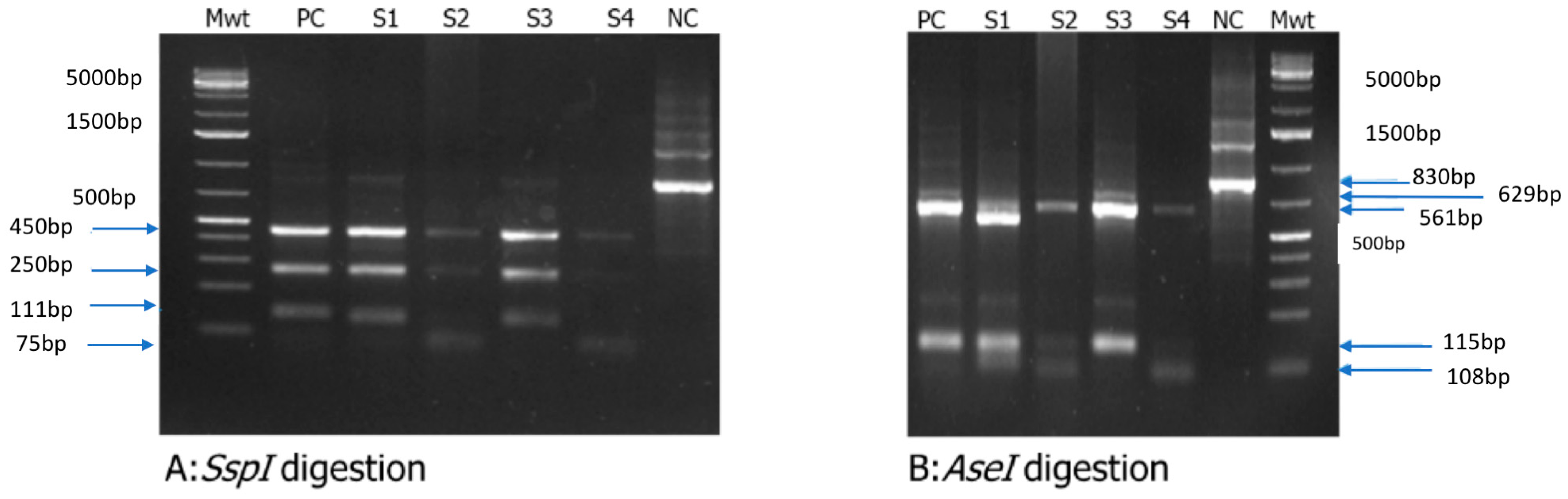
2.5. Restriction Fragment Length Polymorphism (RFLP) Analysis

For the RFLP analysis, 10 U of either SspI or AseI enzymes were used to digest 10 μL of DNA (amplicon from the 18S rRNA PCR) at 37 °C in a water bath for 2 h, as recommended by the supplier (New England Biolabs Inc., Ipswich, MA, USA). The samples were then separated by gel electrophoresis using 2% agarose and visualized after ethidium bromide staining (5 mg/mL, Sigma-Aldrich) using a BIO-RAD Molecular Imager© Gel Doc™ XR+ imaging system.

2.6. Cryptosporidium GP60 Gene Amplification

A gene fragment of the 60 kDa glycoprotein (GP60) was amplified by a two-step nested PCR, as previously described [27]. Briefly, 2 µL of extracted DNA was used at a final reaction volume of 20 µL. The primary PCR step was performed with the forward (Fp) and reverse (Rp) primers (5′-ATAGTCTCCGCTGTATTC-3′) and (5′-GGAAGGAACGATGTATCT-3′), respectively, using a GeneAmp® PCR System 9700 thermocycler (Applied Biosystems). The resulting amplicons were diluted 1:50 and used as template for secondary amplification with the primers (5′TCCGCTGTATTCTCA GCC-3′) and 5′GCAGAGGAACCAGCATC-3′). Cycling conditions were set as follows: initial denaturation at 95 °C for 3 min followed by 35 cycles of a three-step program (94 °C for 45 s, 50 °C for 45 s and 72 °C for 1 min), and a final extension step at 72 °C for 7 min. The secondary amplicons were separated on 1.2% agarose gels and visualized using a UV-trans illuminator BIO-RAD Molecular Imager© Gel Doc™ XR+ imaging system.

2.7. DNA Sequencing and Phylogenetic Analysis

Secondary GP60 PCR products were purified using the quick DNA purification kit from Qiagen (QIAquick® Gel Extraction kit, Qiagen, Hilden, Germany). Amplicons were subsequently sent for sequencing (Inqaba Biotec, Pretoria, South Africa). Direct DNA sequencing of GP60 amplicons purified from the gels was performed using the primers GP60-AL3532 (5′-TCCGCTGTATTCTCAGCC-3′) and GP60-AL3534 (5′-GCAGAGGAACCAGCATC-3′) on an ABI PRISM—3730 XL DNA Analyzer using the BigDye Terminator v3.1 Cycle Sequencing Kit (Applied Biosystems, Foster City, CA, USA). Sequence accuracy was confirmed by two-directional sequencing. The raw sequences obtained were edited with DNAstar Lasergene Editseq version 7.1.0 (http://www.dnastar.com/).
Raw sequence reads were initially assessed for quality using FastQC, then trimmed with fastp [63] to remove low-quality sequences. The quality threshold was set at Q20 to ensure 99% accuracy, and sequences shorter than 50 base pairs were discarded. High-quality sequences (n = 38) were aligned using MAFFT [64], and the resulting alignment was trimmed using trimAl [65] with the “automated1” option for low-quality alignment regions. A maximum likelihood phylogenetic tree was constructed using IQ-TREE [66], incorporating reference GP60 sequences (AY382670-Id, AY873783-Ia, GU214369-IIe, AY166807-Ib, FJ839876-IIc, and GU214365-IIc) of Cryptosporidium spp. from NCBI. The best-fit model of nucleotide substitution was selected using ModelFinder Plus within IQ-TREE, and branch support was assessed with 1000 ultrafast bootstrap approximations.

2.8. Statistical Analysis

Statistical analyses were performed using R statistical package version 4.4.1 (www.r-project.org). Descriptive statistics were performed to characterize the study population. To identify determinants of Cryptosporidium infection in study regions, multiple logistic regression was fitted with PCR results (1 for positive and 0 for negative) as outcome variable, and children’s age, gender, duration of diarrhea episode, and the mothers’ education level as predictor variables. The regression analysis results were presented as adjusted odds ratios, associated confidence intervals, and p-values. The confidence level in this study was set at 95%.

3. Results

3.1. Demographic Data

Overall, a total of 1119 stool samples were collected from children in the geographic regions of the Center (n = 272), Southwest (n = 320), Littoral (n = 223) and West (n = 304) in Cameroon (Figure 1). Samples were assessed for the presence of Cryptosporidium by both microscopic analysis of MZN-stained fecal smears and PCR. For socio-demographic characteristics of the sample population, see Table 1.

3.2. Results of Microscopic Analysis

Microscopic examination revealed a total of 96 samples (96/1119) as positive for Cryptosporidium oocysts, thereby representing a total prevalence of 8.5%.

3.3. Results of PCR Analysis

Positive samples showed a typical amplicon size of 830 bp in agarose gels after nested PCR (Figure 2). Of the 1119 samples collected, 132 were positive for Cryptosporidium, giving an overall prevalence of 11.8%. Referring to different regions of Cameroon, the prevalence varied between 7.2% and 15.4%: Center region 15.4% (42/272); Littoral region 7.2% (16/223); West region 10.5% (32/304) and Southwest region 13.1% (42/320) (Table 2).
Different parameters, like age, gender, duration of diarrhea and educational level of the mothers, were also analyzed but showed no statistically significant differences (Table 2).

3.4. Cryptosporidium spp. Genotyping

Enzymatic digestion of 18S rRNA amplicons with SspI yielded multiple DNA fragments with band sizes of 75 bp, 111 bp, 250 bp and 450 bp (Figure 3A), which are in line with reports for C. parvum or C. hominis [67]. Likewise, digestion with AseI generated two DNA fragments of 561 bp and 629 bp specific for C. hominis and C. parvum, respectively (Figure 3B).

3.5. Cryptosporidium spp. Sub-Genotyping and Evolutionary Relationships of Taxa

The next step in the characterization of field isolates consisted in analyzing the sequence of the GP60 gene. Therefore, 38 isolates were selected from all positive samples, sequenced after amplification of the GP60 gene locus and compared with reference GP60 DNA sequences obtained from the NCBI database. From the 38 isolates, 13 belonged to the C. hominis subtype family Id, 24 to the C. parvum subtype family IIc, and one isolate to the subtype family IIe (Figure 4).

4. Discussion

Cosmopolitan outbreaks of human cryptosporidiosis have regularly been linked to zoonotic transmission resulting from human contact with infected domestic and wildlife animals [68]. Despite this epizootiological fact, very few studies dealing with Cryptosporidium species/genotypes and their origin exist for Africa, where human cryptosporidiosis is a major cause of pediatric morbidity and mortality [67,68,69]. Thus, detailed knowledge of genotypes/sub-genotypes of Cryptosporidium species circulating in a defined human population will help to understand transmission routes in order to clarify if oocyst sources originate from zoonotic or anthroponotic origin. Additionally, knowledge of Cryptosporidium genotypes seems relevant for the design of optimal control programs by national public health authorities for human protection or even eradication of human cryptosporidiosis, as postulated elsewhere [67,69].
Multiple studies in sub-Saharan Africa have revealed a high prevalence of cryptosporidiosis in children and immunocompromised patients dominated by two species, C. hominis and C. parvum. C. parvum bears a high veterinary importance, particularly for neonatal livestock (e. g. calves, lambs, goat kids), and is considered as one of the most relevant etiological agent of neonatal enteritis [70,71,72]. Moreover, C. parvum occurs in a wide spectrum of terrestrial mammalian wildlife, including wild horses, deer, mountain gorillas, nutrias, raccoon dogs and camelids, among others [70,73]. Wildlife represents, therefore, an important natural reservoir and adds—together with humans and livestock—to the pool of C. parvum genotypes within the environment [69]. However, very few studies have characterized the genotypes or subtypes of human cryptosporidiosis in African countries [12,68,69].
With the current epidemiological study, we have shown a Cryptosporidium prevalence of 11.8% in children from 0 to 10 years of age for Cameroon by PCR analysis, representing a total of 132 positive children (n = 132/1119) from four different geographic regions of this West-African country. Although these results cannot be extrapolated to the nationwide situation, it is a representative distribution, since the four regions sampled represent a substantial fraction of the total population of the country. As expected, the current results further confirmed that PCR methodology is slightly more sensitive than MZN-fecal staining (11.8 % vs. 8.5 %), even though the difference is not statistically significant. Others have reported similar results, also with no statistically significant difference [21,74,75]. When considering the age-related sample distribution, the samples were divided in two groups, i.e., children below 5 years and children from 5 to 10 years. We observed a slightly higher prevalence of Cryptosporidium in children below 5 years (12%) compared to children aged 5 to 10 (7%), although this difference was not statistically significant. Furthermore, the consideration of symptoms, such as the duration of diarrhea, revealed that children with diarrhea for more than 2 days were slightly more likely to be Cryptosporidium-infected (12%) than those who had diarrhea for 2 days or less (8%). However, this difference was again not statistically significant. A meta-analysis of cryptosporidiosis in children under 5 years estimated this parasitic infection to be related, with 44.8 million diarrheal episodes and 48,300 deaths, globally [76]. In the present study, it seems likely that lack of a clear difference between the symptomatic (diarrhea for more than 2 days) and asymptomatic (diarrhea for 2 days or less) group is due to the inclusion of children up to 10 years, who might already have developed a parasite-specific T cell response. Hence, immunocompetent C. parvum re-infected children are more likely to develop a CD8+ T cell-based protective immunity to homologous infections, resulting in marginal intestinal damage and/or subclinical cryptosporidiosis [77,78,79].
The phylogenetic analysis of a representative number of isolates revealed that a substantial number belong to the subtype families Ia, Ib and Id, representing the anthroponotic C. hominis. Interestingly, these subtype families have been associated with human-to-human transmission in children and HIV-positive adults in several developing countries [35,80,81]. The largest group of isolates from this study belonged to the subtype family IIc, which represents the anthroponotic C. parvum. One sequence belonged to the subtype family IIe, which is also an anthroponotic C. parvum. Hence, these two anthroponotic subtype families have only been found in human specimens so far [51,82]. Strikingly, the zoonotically transmitted subtype families of C. parvum, i.e., IIa, IId and IIl, were not found in any of the current isolates. This study elucidates for the first time the genetic diversity of Cryptosporidium species circulating among Cameroonian children and supports the previously reported tendency that transmission of cryptosporidiosis in Africa is essentially based on human-to-human transfer, since all subtype families from both C. hominis and C. parvum identified in this study are anthroponotic subtypes.
Some limitations of this study were the fragmentary coverage of the entire country and the fact that not all isolates were sequenced, but were rather a representative fraction. Moreover, future identification analysis of the species may also include 18S rRNA gene sequencing to improve the robustness of the result. A larger study population covering a wider geographical area of Cameroon would certainly improve the robustness of these results.

5. Conclusions

From this study, it appears that cryptosporidiosis transmission in Cameroonian children is likely to proceed predominantly through the anthroponotic route, since no zoonotic subtypes were found. It is therefore plausible that implementation of a better control strategy to prevent human-to-human transmission would lead to a significant impact in the reduction of the overall disease burden.

Supplementary Materials

The following supporting information can be downloaded at: https://www.mdpi.com/article/10.3390/pathogens14030287/s1, Figure S1: Gene PCR gel map original image of Figure 2; Figure S2: Gene PCR gel map original image of Figure 3.

Author Contributions

F.K. and C.H. designed the Study; B.S., M.N.A., D.C. and C.A. conducted the experiments; F.K., C.H., L.A.A., B.S., M.N.A. and J.A.N. analyzed the data. F.K., L.A.A., C.H., A.T. and B.S. wrote the manuscript. All authors have read and agreed to the published version of the manuscript.

Funding

This research was funded by a German Federal Ministry of Education and Research [Bundesministerium für Bildung und Forschung (BMBF)]-financed project (DLR Projektträger; ‘CryptoNETs’; no. 01DG20023, awarded to FK and CH), the LOEWE Center DRUID (Novel Drug Targets against Poverty-Related and Neglected Tropical Infectious Diseases, awarded to CH and AT) and the Africa One Health University Network (AFROHUN) small grant Scheme.

Institutional Review Board Statement

Ethical approval was obtained from Institutional Review Board of the Faculty of Health Sciences (IRB-FHS) of the University of Buea (No. 805-05, approved 15 October 2019) and administrative clearance was obtained from the different hospitals where samples were collected. Informed consent was obtained from parents or guardians of participants after a clear explanation of the research objectives and participation was voluntary. The parents or guardians of study participants signed the consent forms on behalf of their children enrolled into the study.

Informed Consent Statement

Informed consent was obtained from all subjects involved in the study.

Data Availability Statement

All data generated or analyzed during the study are included in this published article. All sequences will be submitted to CrytoDB upon publication.

Acknowledgments

We thank all study participants for their availability, Julius Foyet for statistical analysis, Elma Arrey for help in sample collection and Martha Ngoe for writing assistance.

Conflicts of Interest

The authors declare no conflicts of interest. The funders had no role in the design of the study; in the collection, analyses, or interpretation of data; in the writing of the manuscript; or in the decision to publish the results.

References

  1. Dillingham, R.A.; Lima, A.A.; Guerrant, R.L. Cryptosporidiosis: Epidemiology and impact. Microbes Infect. 2002, 4, 1059–1066. [Google Scholar] [CrossRef]
  2. Olson, M.E.; Handley, R.M.O.; Ralston, B.J.; Mcallister, T.A.; Thompson, R.C.A. Update on Cryptosporidium and Giardia infections in cattle. Trends Parasitol. 2004, 20, 185–191. [Google Scholar] [CrossRef]
  3. Ryan, U.; Zahedi, A.; Paparini, A. Cryptosporidium in humans and animals-a one health approach to prophylaxis. Parasite Immunol. 2016, 38, 535–547. [Google Scholar] [CrossRef]
  4. Sterling, C.R.; Arrowood, M.J. Cryptosporidia; Elsevier: Amsterdam, The Netherlands, 2020; Volume 14, p. 293. [Google Scholar]
  5. Current, W.L.; Garcia, L.S. Cryptosporidiosis. Clin. Lab. Med. 1991, 11, 873–895. [Google Scholar] [CrossRef]
  6. Garcia, L.S.; Lilly, E. Cryptosporidiosis: Clinical features and diagnosis. Crit. Rev. Clin. Lab. Sci. II 1989, 27, 439–460. [Google Scholar] [CrossRef]
  7. Feltus, D.C.; Giddings, C.W.; Schneck, B.L.; Monson, T.; Warshauer, D.; McEvoy, J.M. Evidence Supporting Zoonotic Transmission of Cryptosporidium spp. in Wisconsin. J. Clin. Microbiol. 2006, 44, 4303–4308. [Google Scholar] [CrossRef]
  8. Feng, Y.; Torres, E.; Li, N.; Wang, L.; Bowman, D.; Xiao, L. Population genetic characterisation of dominant Cryptosporidium parvum subtype IIaA15G2R1. Int. J. Parasitol. 2013, 43, 1141–1147. [Google Scholar] [CrossRef]
  9. Thomson, S.; Hamilton, C.A.; Hope, J.C.; Katzer, F.; Mabbott, N.A.; Morrison, L.J.; Innes, E.A. Bovine cryptosporidiosis: Impact, host-parasite interaction and control strategies. Vet. Res. 2017, 48, 42. [Google Scholar] [CrossRef]
  10. Amadi, B.; Mwiya, M.; Sianongo, S.; Payne, L.; Watuka, A.; Katubulushi, M.; Kelly, P. High dose prolonged treatment with nitazoxanide is not effective for cryptosporidiosis in HIV positive Zambian children: A randomised controlled trial. BMC Infect. Dis. 2009, 9, 195. [Google Scholar] [CrossRef]
  11. Kotloff, M.M.L.K.L.; Nataro, J.P.; Blackwelder, W.C.; Nasrin, D.; Farag, T.H.; Panchalingam, S.; Wu, Y.; Sow, S.O.; Sur, D.; Breiman, R.F.; et al. Burden and aetiology of diarrhoeal disease in infants and young children in developing countries (the Global Enteric Multicenter Study, GEMS): A prospective, case-control study. Lancet 2013, 382, 209–222. [Google Scholar] [CrossRef]
  12. Sow, S.O.; Muhsen, K.; Nasrin, D.; Blackwelder, W.C.; Wu, Y.; Farag, T.H.; Panchalingam, S.; Sur, D.; Zaidi, A.K.M.; Faruque, A.S.G.; et al. The Burden of Cryptosporidium Diarrheal Disease among Children < 24 Months of Age in Moderate/High Mortality Regions of Sub-Saharan Africa and South Asia, Utilizing Data from the Global Enteric Multicenter Study (GEMS). PLoS Negl. Trop. Dis. 2016, 10, e0004729. [Google Scholar] [CrossRef]
  13. Checkley, W.; White, A.C.; Jaganath, D.; Arrowood, M.J.; Chalmers, R.M.; Chen, X.-M.; Fayer, R.; Griffiths, J.K.; Guerrant, R.L.; Hedstrom, L.; et al. A review of the global burden, novel diagnostics, therapeutics, and vaccine targets for Cryptosporidium. Lancet Infect. Dis. 2015, 15, 85–94. [Google Scholar] [CrossRef]
  14. Xiao, L.; Feng, Y. Zoonotic cryptosporidiosis. FEMS Immunol. Med. Microbiol. 2008, 52, 309–323. [Google Scholar] [CrossRef]
  15. Cho, M.-H.; Kim, A.-K.; Im, K.-I. Detection of Cryptosporidium oocysts from out-patients of the Severance Hospital Korea. Parasites Hosts Dis. 1993, 31, 193–199. [Google Scholar] [CrossRef]
  16. Garcia-R, J.C.; Hayman, D.T.S. A review and analysis of cryptosporidiosis outbreaks in New Zealand. Parasitology 2023, 150, 606–611. [Google Scholar] [CrossRef]
  17. Plutzer, J.; Kelen, K.; Varga, E.; Kucsera, I.; Reusz, G.; Szabó, A.J.; Fehér, Á.; Chalmers, R.M. First Cryptosporidium outbreak in Hungary, linked to a treated recreational water venue in 2015. Epidemiol. Infect. 2019, 147, e56. [Google Scholar] [CrossRef]
  18. Gharpure, R.; Perez, A.; Miller, A.D.; Wikswo, M.E.; Silver, R.; Hlavsa, M.C. Cryptosporidiosis outbreaks—United States, 2009–2017. Am. J. Transplant. 2019, 19, 2650–2654. [Google Scholar] [CrossRef]
  19. Mirdha, B.R. Evolving Patterns of Cryptosporidiosis: Issues and Implications in the Context of Public Health in India. Ann. Natl. Acad. Med. Sci. 2021, 57, 81–93. [Google Scholar] [CrossRef]
  20. O’Leary, J.K.; Sleator, R.D.; Lucey, B. Cryptosporidium spp. diagnosis and research in the 21st century. Food Waterborne Parasitol. 2021, 24, e00131. [Google Scholar] [CrossRef]
  21. Chalmers, R.M.; Smith, R.P.; Hadfield, S.J.; Elwin, K.; Giles, M. Zoonotic linkage and variation in Cryptosporidium parvum from patients in the United Kingdom. Parasitol. Res. 2011, 108, 1321–1325. [Google Scholar] [CrossRef]
  22. Vélez, J.; Velasquez, Z.; Silva, L.M.R.; Gärtner, U.; Failing, K.; Daugschies, A.; Mazurek, S.; Hermosilla, C.; Taubert, A. Metabolic signatures of Cryptosporidium parvum-infected hct-8 cells and impact of selected metabolic inhibitors on C. parvum infection under physioxia and hyperoxia. Biology 2021, 10, 60. [Google Scholar] [CrossRef]
  23. Vélez, J.; Silva, L.M.R.; Kamena, F.; Daugschies, A.; Mazurek, S.; Taubert, A.; Hermosilla, C. The Oesophageal Squamous Cell Carcinoma Cell Line COLO-680N Fails to Support Sustained Cryptosporidium parvum Proliferation. Pathogens 2022, 11, 49. [Google Scholar] [CrossRef]
  24. Nguyen-Ho-bao, T.; Ambe, L.A.; Berberich, M.; Hermosilla, C.; Taubert, A.; Daugschies, A.; Kamena, F. Octaarginine Improves the Efficacy of Nitazoxanide against Cryptosporidium parvum. Pathogens 2022, 11, 653. [Google Scholar] [CrossRef]
  25. Anschütz, N.H.; Gerbig, S.; Ghezellou, P.; Silva, L.M.R.; Vélez, J.D.; Hermosilla, C.R.; Taubert, A.; Spengler, B. Mass Spectrometry Imaging of In Vitro Cryptosporidium parvum-Infected Cells and Host Tissue. Biomolecules 2023, 13, 1200. [Google Scholar] [CrossRef]
  26. Cui, Z.; Song, D.; Qi, M.; Zhang, S.; Wang, R.; Jian, F.; Ning, C.; Zhang, L. Revisiting the infectivity and pathogenicity of Cryptosporidium avium provides new information on parasitic sites within the host. Parasites Vectors 2018, 11, 514. [Google Scholar] [CrossRef]
  27. Alves, M.; Xiao, L.; Sulaiman, I.; Lal, A.A.; Matos, O.; Antunes, F. Subgenotype analysis of Cryptosporidium isolates from humans, cattle, and zoo ruminants in Portugal. J. Clin. Microbiol. 2003, 41, 2744–2747. [Google Scholar] [CrossRef]
  28. Šlapeta, J. Cryptosporidiosis and Cryptosporidium species in animals and humans: A thirty colour rainbow? Int. J. Parasitol. 2013, 43, 957–970. [Google Scholar] [CrossRef]
  29. Sréter, T.; Egyed, Z.; Szell, Z.; Kovacs, G.; Nikolausz, M.; Marialigeti, K.; Varga, I. Morphologic, host specificity, and genetic characterization of a European Cryptosporidium andersoni isolate. J. Parasitol. 2000, 86, 1244–1249. [Google Scholar] [CrossRef]
  30. Feng, Y.; Ryan, U.M.; Xiao, L. Genetic Diversity and Population Structure of Cryptosporidium. Trends Parasitol. 2018, 34, 997–1011. [Google Scholar] [CrossRef]
  31. Ryan, U.M.; Feng, Y.; Fayer, R.; Xiao, L. Taxonomy and molecular epidemiology of Cryptosporidium and Giardia—A 50 year perspective (1971–2021). Int. J. Parasitol. 2021, 51, 1099–1119. [Google Scholar] [CrossRef]
  32. Gatei, W.; Greensill, J.; Ashford, R.W.; Cuevas, L.E.; Parry, C.M.; Cunliffe, N.A.; Beeching, N.J.; Hart, C.A. Molecular analysis of the 18S rRNA gene of Cryptosporidium parasites from patients with or without human immunodeficiency virus infections living in Kenya, Malawi, Brazil, the United Kingdom, and Vietnam. J. Clin. Microbiol. 2003, 41, 1458–1462. [Google Scholar] [CrossRef]
  33. Peralta, R.H.S.; Velásquez, J.N.; Cunha, F.d.S.; Pantano, M.L.; Sodré, F.C.; da Silva, S.; Astudillo, O.G.; Peralta, J.M.; Carnevale, S. Genetic diversity of Cryptosporidium identified in clinical samples from cities in Brazil and Argentina. Mem. Inst. Oswaldo Cruz 2016, 111, 30–36. [Google Scholar] [CrossRef] [PubMed]
  34. Takumi, K.; Cacciò, S.M.; Van Der Giessen, J.; Xiao, L.; Sprong, H. Hypothesis: Cryptosporidium genetic diversity mirrors national disease notification rate. Parasit. Vectors 2015, 8, 308. [Google Scholar] [CrossRef] [PubMed]
  35. Peng, M.M.; Wilson, M.L.; Holland, R.E.; Meshnick, S.R.; Lal, A.A.; Xiao, L. Genetic diversity of Cryptosporidium spp. in cattle in Michigan: Implications for understanding the transmission dynamics. Parasitol. Res. 2003, 90, 175–180. [Google Scholar] [CrossRef] [PubMed]
  36. Elmahallawy, E.K.; Sadek, H.A.; Aboelsoued, D.; Aloraini, M.A.; Alkhaldi, A.A.M.; Abdel-Rahman, S.M.; Bakir, H.Y.; Arafa, M.I.; Hassan, E.A.; Elbaz, E.; et al. Parasitological, Molecular, and Epidemiological Investigation of Cryptosporidium Infection Among Cattle and Buffalo Calves From Assiut Governorate, Upper Egypt: Current Status and Zoonotic Implications. Front. Vet. Sci. 2022, 9, 899854. [Google Scholar] [CrossRef]
  37. Bujila, I.; Troell, K.; Ögren, J.; Hansen, A.; Killander, G.; Agudelo, L.; Lebbad, M.; Beser, J. Cryptosporidium species and subtypes identified in human domestic cases through the national microbiological surveillance programme in Sweden from 2018 to 2022. BMC Infect. Dis. 2024, 24, 146. [Google Scholar] [CrossRef]
  38. Dąbrowska, J.; Sroka, J.; Cencek, T. Investigating Cryptosporidium spp. Using Genomic, Proteomic and Transcriptomic Techniques: Current Progress and Future Directions. Int. J. Mol. Sci. 2023, 24, 12867. [Google Scholar] [CrossRef]
  39. Berhanu, K.; Ayana, D.; Megersa, B.; Ashenafi, H.; Waktole, H. Cryptosporidium in human-animal-environment interphase at Adama and Asella areas of Oromia regional state, Ethiopia. BMC Vet. Res. 2022, 18, 402. [Google Scholar] [CrossRef]
  40. King, B.J.; Monis, P.T. Critical processes affecting Cryptosporidium oocyst survival in the environment. Parasitology 2007, 134, 309–323. [Google Scholar] [CrossRef]
  41. Zahedi, A.; Monis, P.; Aucote, S.; King, B.; Paparini, A.; Jian, F.; Yang, R.; Oskam, C.; Ball, A.; Robertson, I.; et al. Zoonotic Cryptosporidium species in animals inhabiting sydney water catchments. PLoS ONE 2016, 11, e0168169. [Google Scholar] [CrossRef]
  42. Kleinertz, S.; Hermosilla, C.; Ziltener, A.; Kreicker, S.; Hirzmann, J.; Abdel-Ghaffar, F.; Taubert, A. Gastrointestinal parasites of free-living Indo-Pacific bottlenose dolphins (Tursiops aduncus) in the Northern Red Sea, Egypt. Parasitol. Res. 2014, 113, 1405–1415. [Google Scholar] [CrossRef] [PubMed]
  43. Hermosilla, C.; Silva, L.M.R.; Navarro, M.; Taubert, A. Anthropozoonotic Endoparasites in Free-Ranging ‘Urban’ South American Sea Lions (Otaria flavescens). J. Vet. Med. 2016, 2016, 7507145. [Google Scholar] [CrossRef]
  44. Ebmer, D.; Navarrete, M.J.; Muñoz, P.; Flores, L.M.; Gärtner, U.; Brabec, J.; Poppert, S.; Taubert, A.; Hermosilla, C. Anthropozoonotic Parasites Circulating in Synanthropic and Pacific Colonies of South American Sea Lions (Otaria flavescens): Non-invasive Techniques Data and a Review of the Literature. Front. Mar. Sci. 2020, 7, 543829. [Google Scholar] [CrossRef]
  45. Xiao, L.; Morgan, U.M.; Limor, J.; Escalante, A.; Arrowood, M.; Shulaw, W.; Thompson, R.C.A.; Fayer, R.; Lal, A.A. Genetic Diversity within Cryptosporidium parvum and Related Cryptosporidium Species. Appl. Environ. Microbiol. 1999, 65, 3386–3391. [Google Scholar] [CrossRef]
  46. Leav, B.A.; Mackay, M.R.; Anyanwu, A.; Connor, R.M.O.; Cevallos, A.M.; Kindra, G.; Rollins, N.C.; Bennish, M.L.; Nelson, R.G.; Ward, H.D. Analysis of sequence diversity at the highly polymorphic Cpgp40/15 locus among Cryptosporidium isolates from human immunodeficiency virus-infected children in South Africa. Infect. Immun. 2002, 70, 3881–3890. [Google Scholar] [CrossRef]
  47. Waldron, L.S.; Ferrari, B.C.; Power, M.L. Glycoprotein 60 diversity in C. hominis and C. parvum causing human cryptosporidiosis in NSW, Australia. Exp. Parasitol. 2009, 122, 124–127. [Google Scholar] [CrossRef]
  48. Nkenfou, C.N.; Nana, C.T.; Payne, V.K. Intestinal Parasitic Infections in HIV Infected and Non-Infected Patients in a Low HIV Prevalence Region, West-Cameroon. PLoS ONE 2013, 8, e57914. [Google Scholar] [CrossRef] [PubMed]
  49. Tankoua-tchounda, R.; Nack, J.; Nchetnkou, C.M.; Tchankwé, D.L.K.; Lontsi-Demano, M.; Essangui, E.; Djimefo, A.K.T.; Lehman, L.G. Enteric Parasitosis of HIV-positive Patients in Cameroon: A Case of the Regional Hospital of Bafoussam (Western Region). Int. J. Trop. Dis. Heal. 2022, 43, 6–18. [Google Scholar] [CrossRef]
  50. Chia, P.N.; Ukaga, C.; Yongabi, K.A.; Nwoke, B.; Tih, P.M. Baseline Study on the Occurrence of Cryptosporidium Spp from Streams Water, After Torrential Rains in Bamenda, Cameroon. J. Biol. Agric. Health 2015, 4, 62–69. [Google Scholar]
  51. Eibach, D.; Krumkamp, R.; Al-Emran, H.M.; Sarpong, N.; Hagen, R.M.; Adu-Sarkodie, Y.; Tannich, E.; May, J. Molecular Characterization of Cryptosporidium spp. among Children in Rural Ghana. PLoS Negl. Trop. Dis. 2015, 9, e0003551. [Google Scholar] [CrossRef]
  52. Agba, A.A.; Aken’Ova, T.O.; Audu, P.A. Cryptosporidium infection among children attending some hospitals in Funtua Local Government Area, Katsina State, Nigeria. Zoologist 2018, 16, 1–5. [Google Scholar] [CrossRef]
  53. Gattan, H.S.; Alshammari, A.; Marzok, M.; Salem, M.; AL-Jabr, O.A.; Selim, A. Prevalence of Cryptosporidium infection and associated risk factors in calves in Egypt. Sci. Rep. 2023, 13, 17755. [Google Scholar] [CrossRef]
  54. Krumkamp, R.; Aldrich, C.; Maiga-Ascofare, O.; Mbwana, J.; Rakotozandrindrainy, N.; Borrmann, S.; Caccio, S.M.; Rakotozandrindrainy, R.; Adegnika, A.A.; A Lusingu, J.P.; et al. Transmission of Cryptosporidium Species Among Human and Animal Local Contact Networks in Sub-Saharan Africa: A Multicountry Study. Clin. Infect. Dis. 2021, 72, 1358–1366. [Google Scholar] [CrossRef] [PubMed]
  55. Mulunda, N.R.; Hayashida, K.; Yamagishi, J.; Sianongo, S. Molecular characterization of Cryptosporidium spp. from patients with diarrhoea in Lusaka, Zambia. Parasite 2020, 27, 53. [Google Scholar] [CrossRef]
  56. Messa, A., Jr.; Köster, P.C.; Garrine, M.; Nhampossa, T.; Massora, S.; Cossa, A.; Bassat, Q.; Kotloff, K.; Levine, M.M.; Alonso, P.L.; et al. Molecular Characterisation of Cryptosporidium spp. in Mozambican Children Younger than 5 Years Enrolled in a Matched Case-Control Study on the Aetiology of Diarrhoeal Disease. Pathogens 2021, 10, 452. [Google Scholar] [CrossRef]
  57. Mbae, C.; Mulinge, E.; Waruru, A.; Ngugi, B.; Wainaina, J.; Kariuki, S. Genetic Diversity of Cryptosporidium in Children in an Urban Informal Settlement of Nairobi, Kenya. PLoS ONE 2015, 151, e0142055. [Google Scholar] [CrossRef] [PubMed]
  58. Lobos, E.; Weiss, N.; Karam, M.; Taylort, H.R.; Otresen, E.A.; Nutman, T.B. Specific and early marker of infection. Science 1991, 16, 12–15. [Google Scholar]
  59. Samra, N.A.; Thompson, P.N.; Jori, F.; Frean, J.; Poonsamy, B.; du Plessis, D.; Mogoye, B.; Xiao, L. Genetic Characterization of Cryptosporidium spp. in Diarrhoeic Children from Four Provinces in South Africa. Zoonoses Public Heal. 2012, 60, 154–159. [Google Scholar] [CrossRef]
  60. Akiyoshi, D.E.; Tumwine, J.K.; Bakeera-Kitaka, S.; Tzipori, S. Subtype Analysis of Cryptosporidium Isolates From Children in Uganda Subtype Analysis of Cryptosporidium Isolates From Children in Uganda. J. Parasitol. 2006, 92, 1097–1100. [Google Scholar] [CrossRef]
  61. Khan, A.A.; Shams, S.; Khan, S.; Khan, M.I.; Khan, S.; Ali, A. Modified ZN Staining Protocol V.2 Version 1 is Forked from mZN Staining Protocol. 2018. Available online: https://www.protocols.io/view/modified-zn-staining-protocol-tb2eiqe.pdf (accessed on 11 November 2024).
  62. Millar, C.; Moore, J.; Lowery, C.; McCorry, K.; Dooley, J. Successful PCR amplification of genomic DNA from Cryptosporidium parvum oocysts extracted from a human faecal sample: A rapid and simple method suited for outbreak analysis. Int. J. Hyg. Environ. Health 2001, 204, 191–194. [Google Scholar] [CrossRef]
  63. Chen, S.; Zhou, Y.; Chen, Y.; Gu, J. Fastp: An ultra-fast all-in-one FASTQ preprocessor. Bioinformatics 2018, 34, i884–i890. [Google Scholar] [CrossRef] [PubMed]
  64. Nakamura, T.; Yamada, K.D.; Tomii, K.; Katoh, K. Parallelization of MAFFT for large-scale multiple sequence alignments. Bioinformatics 2018, 34, 2490–2492. [Google Scholar] [CrossRef]
  65. Capella-Gutiérrez, S.; Silla-Martínez, J.M.; Gabaldón, T. trimAl: A tool for automated alignment trimming in large-scale phylogenetic analyses. Bioinformatics 2009, 25, 1972–1973. [Google Scholar] [CrossRef]
  66. Minh, B.Q.; Schmidt, H.A.; Chernomor, O.; Schrempf, D.; Woodhams, M.D.; von Haeseler, A.; Lanfear, R. IQ-TREE 2: New Models and Efficient Methods for Phylogenetic Inference in the Genomic Era. Mol. Biol. Evol. 2020, 37, 1530–1534. [Google Scholar] [CrossRef]
  67. Okojokwu, O.J.; Ileigo, I.H.; Ezemuel, Y.S.; Oluyinka, O.O.; Akpakpan, E.E.; Kolawole, T.; Chima, N.J.; Anejo-Okopi, A.J. Molecular characterisation of Cryptosporidium species among Patients Presenting with Diarrhoea in Some Parts of Kaduna State, Nigeria. Am. J. Res. Commun. 2016, 4, 87–106. [Google Scholar]
  68. Robertson, L.J.; Johansen, Ø.H.; Kifleyohannes, T.; Efunshile, A.M.; Terefe, G. Cryptosporidium Infections in Africa—How Important Is Zoonotic Transmission? A Review of the Evidence. Front. Vet. Sci. 2020, 7, 575881. [Google Scholar] [CrossRef]
  69. Aldeyarbi, H.M.; El-Ezz, N.M.T.A.; Karanis, P. Cryptosporidium and cryptosporidiosis: The African perspective. Environ. Sci. Pollut. Res. 2016, 23, 13811–13821. [Google Scholar] [CrossRef] [PubMed]
  70. Squire, S.A.; Ryan, U. Cryptosporidium and Giardia in Africa: Current and future challenges. Parasit. Vectors 2017, 10, 195. [Google Scholar] [CrossRef]
  71. Appelbee, A.J.; Thompson, R.C.A.; Olson, M.E. Giardia and Cryptosporidium in mammalian wildlife—Current status and future needs. Trends Parasitol. 2005, 21, 370–376. [Google Scholar] [CrossRef]
  72. Lendner, M.; Daugschies, A. Cryptosporidium infections: Molecular advances. Parasitology 2014, 141, 1511–1532. [Google Scholar] [CrossRef]
  73. Grabbe, M.; Conejeros, I.; Velásquez, Z.D.; Hasheminasab, S.S.; Kamena, F.; Wehrend, A.; Gärtner, U.; Taubert, A.; Hermosilla, C.R. Cryptosporidium parvum-induced neutrophil extracellular traps in neonatal calves is a stage-independent process. Front. Vet. Sci. 2023, 10, 1256726. [Google Scholar] [CrossRef] [PubMed]
  74. Ryan, U.; Power, M. Cryptosporidium species in Australian wildlife and domestic animals. Parasitology 2012, 139, 1673–1688. [Google Scholar] [CrossRef] [PubMed]
  75. Morgan, U.M.; Sargent, K.D.; Deplazes, P.; Forbes, D.A.; Spano, F.; Hertzberg, H.; Elliot, A.; Thompson, R.C.A. Molecular characterization of Cryptosporidium from various hosts. Parasitology 1998, 117, 31–37. [Google Scholar] [CrossRef]
  76. Khalil, I.A.; Troeger, C.; Rao, P.C.; Blacker, B.F.; Brown, A.; Brewer, T.G.; Colombara, D.V.; De Hostos, E.L.; Engmann, C.; Guerrant, R.L.; et al. Articles Morbidity, mortality, and long-term consequences associated with diarrhoea from Cryptosporidium infection in children younger than 5 years: A meta-analyses study. Lancet Glob. Health 2016, 6, e758–e768. [Google Scholar] [CrossRef]
  77. Pantenburg, B.; Castellanos-Gonzalez, A.; Dann, S.M.; Connelly, R.L.; Lewis, D.E.; Ward, H.D.; White, A.C. Human CD8+ T cells clear Cryptosporidium parvum from infected intestinal epithelial cells. Am. J. Trop. Med. Hyg. 2010, 82, 600–607. [Google Scholar] [CrossRef]
  78. Haskins, B.E.; Gullicksrud, J.A.; Wallbank, B.A.; Dumaine, J.E.; Guérin, A.; Cohn, I.S.; O’Dea, K.M.; Pardy, R.D.; Merolle, M.I.; Shallberg, L.A.; et al. Dendritic cell-mediated responses to secreted Cryptosporidium effectors are required for parasite-specific CD8 (+) T cell responses. bioRxiv 2023, 2023, 553566. [Google Scholar] [CrossRef]
  79. Ross, R.; Hasheminasab, S.S.; Conejeros, I.; Gärtner, U.; Kamena, F.; Krueger, A.; Taubert, A.; Hermosilla, C.; Iv, G.U. Human dendritic cell interactions with the zoonotic parasite Cryptosporidium parvum result in activation and maturation. Front. Immunol. 2024, 15, 1388366. [Google Scholar] [CrossRef]
  80. Ajjampur, S.S.R.; Gladstone, B.P.; Selvapandian, D.; Muliyil, J.P.; Ward, H.; Kang, G. Molecular and spatial epidemiology of cryptosporidiosis in children in a semiurban community in South India. J. Clin. Microbiol. 2007, 45, 915–920. [Google Scholar] [CrossRef]
  81. Cama, V.A.; Ross, J.M.; Crawford, S.; Kawai, V.; Chavez-Valdez, R.; Vargas, D.; Vivar, A.; Ticona, E.; Ñavincopa, M.; Williamson, J.; et al. Differences in clinical manifestations among Cryptosporidium species and subtypes in HIV-infected persons. J. Infect. Dis. 2007, 196, 684–691. [Google Scholar] [CrossRef]
  82. Xiao, L. Molecular epidemiology of cryptosporidiosis: An update. Exp. Parasitol. 2010, 124, 80–89. [Google Scholar] [CrossRef]
Figure 1. Geographical map illustrating stool sample collection sites in Cameroon. (a) Represents the map of Cameroon; the four regions sampled (b) are projected in more detail to show the individual health centers from which samples were collected (c1c4).
Pathogens 14 00287 g001
Figure 2. Secondary amplicons from Cryptosporidium-nested PCR of the 18S rRNA gene indicates amplicon sizes at 830 bp and no amplification in the negative control. M = DNA ladder, 1–14 = samples, P = positive control. Gene PCR gel map original image can be found in Figure S1.
Pathogens 14 00287 g002
Figure 3. Restriction fragment length polymorphism (RFLP) analysis of Cryptosporidium 18S rRNA gene amplicons. (A) Ssp I digestion results in three bands of 450 bp, 250 bp and 111 bp size, which are typical for Cryptosporidium spp.; (B) Ase I digestion of the 18S rRNA gene amplicons resulted in typical bands sizes of 629 bp and 115 bp specific for C. parvum (S1 and S3) as well as of 561 bp and 108 bp specific for C.hominis (S2 and S4), Mwt = DNA ladder, PC = positive control, S1-S3 = samples 1 to 3, NC = negative control representing the undigested amplicon. Gene PCR gel map original image can be found in Figure S2.
Pathogens 14 00287 g003
Figure 4. Phylogenetic analysis of C. hominis and C. parvum subtypes and reference strains (in red) using maximum likelihood of the glycoprotein 60 (gp60) gene. Reference sequences for the subtypes are AY382670-Id, AY873783-Ia, GU214369-IIe, AY166807-Ib, FJ839876-IIc, GU2143365-IIc.
Pathogens 14 00287 g004
Table 1. Socio-demographic characteristics of the population.
Regions n (%)
VariablesCenterSouthwestLittoralWestOverall n (%)
Age
<5 yrs248 (91.18)150 (46.88)179 (80.27)189 (62.17)766 (68.45)
5–10 yrs24 (8.82)170 (53.12)44 (19.73)115 (37.83)353 (31.55)
Gender
Female119 (43.75)174 (54.37)95 (42.60)136 (44.74)524 (46.83)
Male153 (56.25)146 (45.62)128 (57.40)168 (55.26)595 (53.17)
Duration of Diarrhea (days)
<261 (22.43)98 (30.63)97 (43.50)107 (35.20)363 (32.44)
>2211 (77.57)222 (69.38)126 (56.50)197 (64.80)756 (67.56)
Education of mother
Primary30 (11.03)100 (31.25)46 (20.63)52 (17.11)228 (20.38)
Secondary242 (88.97)209 (65.31)139 (62.33)241 (79.28)831 (74.26)
Tertiary011 (3.44)38 (17.04)11 (3.62)60 (5.36)
Table 2. Prevalence of cryptosporidiosis based on PCR.
VariablesNumber PositiveTotal ExaminedPrevalence (%)
Age
<5 yrs9476612.27
5–10 yrs3835310.76
Gender
Female6652412.60
Male6659511.09
Duration of Diarrhea (days)
<23836310.47
>29475612.43
Education of Mother
Primary2422810.53
Secondary10283112.27
Tertiary66010.00
Overall Prevalence
Number of positiveTotal ExaminedPrevalenceLclUcl
132111911.810.0313.82
(Result = “Positive”)
PredictorsOdds RatiosCIp
(Intercept)0.230.12–0.43<0.001
Age [5–10 yrs]0.830.55–1.240.365
Gender [Male]0.830.57–1.200.317
Duration of Diarrhea [>2]1.230.83–1.860.321
Education [Secondary]1.280.81–2.110.308
Education [Tertiary]0.980.35–2.410.968
Observations1119
Disclaimer/Publisher’s Note: The statements, opinions and data contained in all publications are solely those of the individual author(s) and contributor(s) and not of MDPI and/or the editor(s). MDPI and/or the editor(s) disclaim responsibility for any injury to people or property resulting from any ideas, methods, instructions or products referred to in the content.

