Article

Integrated Analysis of Salmonella Infantis in Chicken Meat: Epidemiological Surveillance, Antibiotic Resistance, and Potential Bioactive Control Agents

1
General Directory of Meat and Milk Board, Adana Meat Directory, Adana 01330, Türkiye
2
Department of Food Hygiene and Technology, Faculty of Ceyhan Veterinary Medicine, Cukurova University, Adana 01330, Türkiye
3
Department of Microbiology, Faculty of Ceyhan Veterinary, Cukurova University, Adana 01330, Türkiye
*
Author to whom correspondence should be addressed.
Pathogens 2025, 14(11), 1178; https://doi.org/10.3390/pathogens14111178
Submission received: 24 September 2025 / Revised: 13 November 2025 / Accepted: 14 November 2025 / Published: 18 November 2025
(This article belongs to the Section Bacterial Pathogens)

Abstract

Salmonella species isolated from chicken meat pose an increasing threat to public health. According to ECDC data, salmonellosis cases have shown a significant upward trend in many European countries between 2019 and 2023, almost reaching pre-pandemic levels. EFSA reported 77,486 confirmed human cases in the EU in 2023. This corresponds to a notification rate of 18 cases per 100,000 people, compared to 15.4 cases per 100,000 in 2022. This study evaluated the prevalence of Salmonella spp., antimicrobial resistance (AMR) profiles, and the effectiveness of natural biological preservatives in raw chicken meat obtained from retail outlets in Southeast Turkey. Among 100 samples analyzed according to ISO 6579-1:2017, suspicious colonies were detected after selective enrichment in XLD and n = 3 isolates were confirmed to be Salmonella enterica subsp. enterica serovar Infantis by real-time PCR. Disk diffusion tests performed in accordance with EUCAST showed that all isolates were resistant to beta-lactam, tetracycline, trimethoprim, sulfonomid and aminoglycoside groups. All isolates were classified as multidrug-resistant. PCR detected blaTEM-1 (all isolates), aphA1-IAB (all isolates), aadA1 (two isolates), and sul1 (all isolates), while tetA/tetB genes were not detected. Among the natural compounds tested, carvacrol showed the strongest antimicrobial activity (MIC 1.56 µL/mL; MBC 3.125–6.25 µL/mL; inhibition zones 32–35 mm). Eugenol showed moderate effects with higher MIC/MBC values (3.125–6.25 µL/mL/12.25 µL/mL), while α-terpineol was effective only at higher concentrations. These findings are consistent with the global increase in Salmonella Infantis and AMR, supporting carvacrol followed by eugenol and α-terpineol as promising natural alternatives for controlling MDR Salmonella spp. in food safety applications.

1. Introduction

Salmonella spp. are among the most prevalent Gram-negative, foodborne zoonotic bacteria and represent a leading cause of gastrointestinal infections in humans worldwide [1]. The most common route of human infection is the consumption of contaminated food, particularly chicken and eggs, while pork and other meat products are less frequently implicated. Contact with infected animals or contaminated environments can also result in transmission [2,3].
It is reported that, between 200 million and 1 billion Salmonella spp. infection cases occur annually, resulting in 93 million gastroenteritis cases and approximately 155,000 deaths. Foodborne transmission accounts for about 85% of these cases [4,5]. Salmonella spp. cause salmonellosis, a disease characterized by fever, diarrhea, vomiting, and abdominal pain, typically appearing 6–48 h after the ingestion of contaminated food. Approximately 5% of patients experience additional symptoms, and the illness generally lasts between four and seven days [4,6]. More than 60,000 cases of nontyphoidal salmonellosis were reported in the European Union (EU) in 2021 [7]. Fewer than 1% of the 100 million people estimated to contract Salmonella Enterocolitis worldwide each year develop invasive nontyphoidal salmonellosis [8,9]. About 1.4 million instances of salmonellosis are recorded in the US annually, with foodborne illness accounting for the great majority of these cases.
The serotypes Salmonella Typhimurium (S. Typhimurium) and Salmonella Enteritidis (S. Enteritidis) notably account for over half of all illnesses [10]. Salmonella Dublin has also been related to outbreaks connected to ground beef in addition to these serotypes [11,12]. The S. Typhimurium and Salmonella Derby serotypes are the most frequently detected in raw pork slices [13]. Nevertheless, another investigation has shown that raw ground pork and raw pork slices could potentially include additional serotypes, such as S. Enteritidis, Salmonella Infantis (S. Infantis), and Salmonella Newport (S. Newport) [14]. Furthermore, S. Infantis has long been a prevalent isolate from poultry in Europe and other countries [9,15,16]. In recent years, it has become increasingly prevalent in broiler chicken populations in many countries, becoming the fourth most common serovar of human salmonellosis in the EU. According to 2022 data, 96% of food animal S. Infantis isolates originated from broiler chickens, and this serovar accounted for 50% of all Salmonella spp. isolates from broilers [17]. The most prevalent Salmonella serotype in Türkiye from 2012 to 2016 was Salmonella Enteritidis (57.3–74.1%), followed by Typhimurium (3.0–8.5%), Infantis (4.0–6.7%), Paratyphi B (3.8–3.2%), and Kentucky (3.8–2.7%) [18].
Additionally, salmonellosis also causes significant financial losses in terms of public health and the economy. According to research, the yearly cost is approximately USD 4.1 billion in the United States [19], EUR 3 billion in the EU [20], and AUD 811 million in Australia [21]. This emphasizes the critical importance of adhering to food safety protocols to ensure the proper handling, preparation, storage, and distribution of food appropriately.
One of the most critical public health concerns associated with non-typhoidal Salmonella (NTS) is the increasing prevalence of antibiotic-resistant strains. The widespread and often uncontrolled use of antibiotics in human and veterinary medicine, as well as in agriculture and aquaculture, is considered one of the main reasons for the emergence of antibiotic-resistant bacteria [22]. This uncontrolled use facilitates the evolution and dissemination of resistance genes, posing a serious global health threat. This pathogen has developed resistance over time to several critical antibiotic classes, including beta-lactams, aminoglycosides, tetracyclines, and sulfonamides, which are frequently prescribed to treat serious infections. The most frequently detected resistance genes to beta-lactam antibiotics are blaTEM-1 (ampicillin resistance) and blaTEM, blaSHV, and blaCTX-M (third-generation cephalosporin resistance). For aminoglycoside resistance, aadA1 and aphA1-IAB are the most common genes; for sulfonamide resistance, sul1 and sul2; for tetracycline resistance, tetA and tetB; and for chloramphenicol resistance, cat1, cat2, and floR. For azithromycin resistance, mph(A), ramAp, erm(B), and erm (42) have been reported [23,24,25,26].
Antibiotic resistance genes (ARGs) have been found in Salmonella spp. strains isolated primarily from meat and meat products, as well as in MDR Salmonella spp. Through the food chain, their presence has become a serious threat to human health, particularly with the emergence of multidrug-resistant (MDR) Salmonella spp. strains. Investigating the prevalence of Salmonella spp. in retail meats is essential for ensuring food safety and preventing economic losses, even though the spread of MDR remains challenging to control [27].
There is growing concern about the negative impacts of chemical preservatives used in the food chain to control these microorganisms harboring resistance genes on food safety and human health. Increasing public awareness of these risks has significantly increased interest in natural antimicrobial alternatives in the food industry [28]. In this context, plant extracts and essential oils with medicinal and antibacterial properties stand out not only for their preservative capacity but also for their potential to preserve the nutritional quality and sensory properties of foods, becoming the focus of current research on sustainable preservation strategies.
Bioactive compounds isolated from plants can be used as natural preservatives against foodborne zoonotic pathogens in various fields, including medicine, veterinary medicine, agriculture, aquaculture, and animal foods such as poultry and red meat. Carvacrol is a natural aromatic bioactive compound found in different plants, such as Oregano (Origanum vulgare L.) and Marjoram (Origanum majorana L.), which belong to the Lamiaceae family. Carvacrol is a phenolic monoterpene characterized by a strong odor [29]. It has been used in traditional medicine for centuries due to its antimicrobial, antifungal, antioxidant, anticancer, and anti-inflammatory properties. It also has potential applications in the food industry as a natural food preservative against pathogens, as it is classified as generally recognized as safe (GRAS) by the FDA and the European Commission [30,31]. Eugenol, a phenolic aromatic molecule mostly derived from clove oil (Syzygium aromaticum), exhibits various biological properties, including antifungal, antibacterial, and anticancer effects [32]. Furthermore, α-terpineol, a volatile monoterpene alcohol, has been investigated for its antibacterial, anticonvulsant, anticancer, insecticidal, anti-inflammatory, and antinociceptive properties, in addition to its potential to extend the shelf life of meat and meat products [33,34,35,36]. α-terpineol is the main component of the essential oils of several aromatic plant species, such as Origanum vulgare L. and Ocimum canum Sims, which are widely used for medicinal purposes. α-terpineol can also be isolated from various sources, such as cajeput oil (Melaleuca cajuputi/Melaleuca leucadendra), pine oil (Pinus spp.), and petitgrain oil (Citrus aurantium var. amara) [37]. Plant materials are typically processed using hydrodistillation, supercritical CO2 extraction, or ethanol-based solvent extraction methods. Quantification of the volatile fractions is commonly performed by GC-FID or GC-MS, and compound concentrations are determined in mg/g or % (w/w) using appropriate internal standards and calibration curves [38].
The present study aimed to comprehensively assess the prevalence, antibiotic resistance profiles, and genomic characteristics of Salmonella spp. strains isolated from chicken meat samples obtained from various retail sources. In addition, the antimicrobial effects of selected natural bioactive compounds (carvacrol, eugenol, and α-terpineol) were evaluated in this study for their potential applications in Salmonella spp. control within the meat industry.

2. Materials and Methods

2.1. Salmonella spp. Isolation and Identification

2.1.1. Sample Collection and Isolation

In this study, a total of 100 raw chicken meat samples were collected under aseptic conditions from 25 retail stores located in the central districts of Adana province (Çukurova, Seyhan, Yüreğir, and Sarıçam). Samples representing four product groups, skin-on wings, skin-on breasts, skin-on thighs, and skin-on drumsticks (25 packages each), were obtained with a minimum quantity of at least 500 g. All samples were transported to the laboratory under cold chain conditions on the day of purchase. Conventional Salmonella spp. isolation was performed using the standard microbiological procedures described by the International Organization for Standardization (ISO 6579-1:2017) [39], with minor modifications applied according to the method reported by Osivand et al. [23]. For the pre-enrichment process, 25 g of the sample was aseptically weighed directly and placed in sterile filtered stomacher bags (Fisher Scientific, Nepean, ON, Canada), and 225 mL of a Ringer solution was added. For each sample, both surface and internal portions of the meat were included to ensure representative microbiological assessment. Afterwards, the suspension, which was homogenized for 3 min with a stomacher (Masticator, IUL device, Barcelona, Spain), was incubated at 37 °C for 24 h. Pre-enriched inoculum (100 µL) was cultured in 10 mL of Rappaport Vassiliadis Broth (RVB; Merck, Germany) and Tetrathionat Broth (TB). RVB was incubated for 16–18 h at 41.5 °C, while TB was incubated for 24 h at 37 °C. Cultures were taken from MSRV and TB media with a loop and inoculated onto Xylose-Lysine-Deoxycolate (XLD) agar and incubated for 24 h at 37 °C. On XLD agar, fuchsia-colored formations and H2S-producing black colonies were evaluated as suspected Salmonella spp. isolates.

2.1.2. Salmonella spp. Identification

DNA Extraction and Real-Time PCR Assay
Genomic DNA was extracted from pure colonies of Salmonella spp. grown on Mueller–Hinton Agar using the DNeasy Blood and Tissue Kit with the QIAcube system (Qiagen, Hilden, Germany) and stored at −20 °C. For each sample, a 9 μL PCR master mix was prepared, consisting of 5 μL of 2× qPCR Mix, 3 μL of Salmonella spp. Oligo Mix, and 1 μL of inhibition/reaction control. Subsequently, 5 μL of DNA obtained after extraction was added to this mixture. Amplification was performed using a Roche LightCycler 480 real-time PCR system (Roche Diagnostics GmbH, Mannheim, Germany). The cycling procedure included an initial denaturation at 95 °C for 5 min, followed by 12 cycles of denaturation at 95 °C for 1 s and annealing at 67–56 °C for 1 s (touchdown stage). This was followed by 30 cycles consisting of denaturation at 95 °C for 10 s and annealing at 63 °C for 30 s (amplification stage).

2.2. Antimicrobial Susceptibility Test

Antimicrobial susceptibility testing was performed using the Kirby–Bauer disk diffusion method in accordance with the European Committee on Antimicrobial Susceptibility Testing (EUCAST). The following antibiotic disks (Bioanalyse Medical, Ankara, Türkiye) were used; Ampicillin (AM, 10 µg), amoxycillin–clavulanic acid (AMC, 10 µg), ceftriaxone (CRO, 30 µg), ceftazidime (CAZ, 30 µg), aztreonam (ATM, 30 µg), cefotaxime (CTX, 30 µg), gentamicin (CN, 10 µg), amikacin (AK, 30 µg), ciprofloxacin (CIP, 5 µg), levofloxacin (LEV, 5 µg), enrofloxacin (ENR, 5 µg), trimethoprim-sulfamethoxazole (TS, 1.25–23.75 µg), tetracycline (T, 30 µg), chloramphenicol (C, 30 µg), streptomycin (STR, 10 µg), and kanamycin (KAN, 30 µg) were used. Salmonella spp. isolates incubated at 37 °C for 24 h were standardized to a cell density of 0.5 McFarland. Mueller–Hinton Broth (MHB, Oxoid, CM0405, Basingstoke, UK) was used as the medium. A sterilized cotton swab was dipped into the suspension, and the bacteria were spread evenly over surface of a Muller–Hinton agar plate. Antibiotic disks were then placed on the Muller–Hinton agar (MHA) plates and incubated overnight at 37 °C. Zone diameters were evaluated according to EUCAST [40].

2.3. Detection of Resistance Genes by PCR

Resistance genes were detected by PCR using specific primers (Sentebiolab, Bilkent, Ankara, Turkey) and FirePol® Master Mix (Solis Biodyne, Tartu, Estonia), as described in previous studies (Table 1).

2.4. The Antimicrobial Effects of Selected Natural Bioactive Compounds (Carvacrol, Eugenol, and α-Terpineol) on Salmonella Isolates

2.4.1. Minimum Inhibitory (MIC) and Bactericidal Concentration (MBC) Assay

Clinical and Laboratory Standards Institute (2008) [51] methods were used to determine the MIC and MBC values of bioactive compounds, including carvacrol, eugenol, and α-terpineol, against three identified Salmonella spp. isolates. Test microorganisms incubated at 37 °C for 24 h were standardized to 0.5 MacFarland turbidity. Mueller–Hinton Broth (MHB, Oxoid, CM0405, Basingstoke, UK) was used as the medium. Briefly, 1 mL of the bioactive substance from a 50 mg/mL stock solution was added to the first tube of each series and serially diluted with sterile Muller–Hinton Broth (MHB, Merck, Darmstadt, Germany). The final concentrations of the bioactive compounds were 12.5, 6.25, 3.125, 1.56, 0.78, 0.39, and 0.19 mg/mL. The inoculum suspension (1 mL) of each bacterial isolate was then added to each tube containing bioactive compounds and MHB. Each tube was evaluated for bacterial growth and compared to the control. As a positive control, a tube containing MHB and bacterial suspension without bioactive compounds was used. The tube containing bioactive compounds and bacterial suspension was also used as a negative control. The tubes were then incubated at 37 °C for 24 h. After 24 h, the MIC value was determined by evaluating the turbidity of the growth in the tubes, and 100 µL from each tube was inoculated on MHA Petri plates and incubated overnight at 37 °C. The lowest concentration of the compound at which no growth was observed after one night was determined to be the MBC value.

2.4.2. Agar Well Diffusion Method

In vitro antimicrobial effects of three Salmonella spp. isolates were determined using the well diffusion method [52]. Wells with a diameter of 4 mm were utilized as Mueller–Hinton Agar (MHA, Merck, Germany) plates using a sterile cylinder in accordance with the well diffusion method standards. Salmonella spp. isolates incubated overnight on Mueller–Hinton agar were suspended in 2 mL of physiological serum, and McFarland turbidity values were standardized to 0.5. Sterile cotton swabs were dipped into the suspensions, impregnating the swabs with bacteria, and excess liquid was then drained from the edge of the tube. The suspension-impregnated swab was spread to cover the surface of the Mueller–Hinton medium. For each bacterial isolate, 100 µL of from stock solution of each food grade bioactive compounds at concentration of 1 g/mL [Carvacrol W224502 (Sigma-Aldrich, Steinheim, Germany), eugenol W246719 (Sigma-Aldrich, Steinheim, Germany), α-terpineol W304522, (Sigma-Aldrich, Steinheim, Germany)] was added to the wells, and the plates were incubated at 37 °C for 24 h. As a control group, only distilled water and MHA were added to the wells of Petri dishes inoculated with each bacterial strain on their plates. All tests were performed in triplicate for each sample. The results were expressed in millimeters (mm) as the arithmetic mean of the inhibition zone diameters around each well.

2.5. Statistical Analysis

The obtained data were expressed as mean ± standard deviation (SD). Statistical analyses were conducted using SPSS software version 18.0 (SPSS Inc., Chicago, IL, USA). One-way analysis of variance (ANOVA) and Duncan’s multiple range test were applied to determine significant differences among groups, and correlation variations were also evaluated where applicable. Statistical significance was considered at p < 0.05.

3. Results

3.1. Salmonella spp. Identification

Salmonella spp. are considered one of the most prevalent foodborne zoonotic pathogens worldwide and pose a serious public health problem. Despite the success of effective control and eradication programs in developed countries, consumption of contaminated poultry meat and other animal products remains an important source of Salmonella spp. infections in humans [53]. In this study, the prevalence of Salmonella spp. in raw chicken meat marketed in the Adana province of Türkiye was investigated. A total of 100 conventional raw chicken meat samples, including 25 wings, 25 breasts, 25 drumsticks, and 25 thighs, were collected from 25 different sales points in the Adana province and analyzed for Salmonella spp. contamination. The results demonstrated that bacterial growth was detected in all groups inoculated for selective enrichment. Following selective enrichment, suspected Salmonella spp. colonies were observed on xylose lysine deoxycholate (XLD) agar after 24 h of incubation at 37 °C. H2S-producing black colonies on XLD agar were evaluated as suspect Salmonella spp. isolates.
Presumptive Salmonella spp. isolates obtained from wing, thigh, breast, and drumstick samples were analyzed using the Bio-Speedy Salmonella spp. Real-Time PCR Detection Kit (Bioeksen, Istanbul, Türkiye), following the manufacturer’s instructions and employing the Touchdown-PCR protocol. Based on the PCR results, 2 isolates isolated from thighs and 1 isolate isolated from drumstick were confirmed as Salmonella spp. The real-time PCR amplification curves of the confirmed positive strains are presented in Figure 1. The colonies confirmed via real-time PCR were further analyzed at the Department of Microbiology, Faculty of Veterinary Medicine, Ankara University, for species-level identification. The results revealed that the isolates were identified as Salmonella Infantis (Table 2).

3.2. Determination of Antimicrobial Susceptibility

Three isolates of Salmonella Infantis were studied for their antibiotic resistance profiles using both phenotypic and molecular methods. All isolates exhibited resistance to ampicillin, amoxicillin–clavulanic acid, tetracycline, trimethoprim–sulfamethoxazole, streptomycin, and kanamycin, as indicated via zone diameter measurements. In contrast, all isolates exhibited susceptibility to aztreonam, cefotaxime, ceftazidime, ciprofloxacin, gentamicin, amikacin, levofloxacin, enrofloxacin, and chloramphenicol (Table 3). Two isolates were resistant to ceftriaxone, and isolate S1 had a zone diameter that was borderline suspicious for ESBL and was also resistant to amoxicillin-clavulanic acid. The detection of resistance to more than one antibiotic class in all isolates suggests that all three isolates can be classified as multidrug-resistant (MDR), posing a potential threat to both food safety and public health (Table 3).

Determination of Antibiotic Resistance Genes

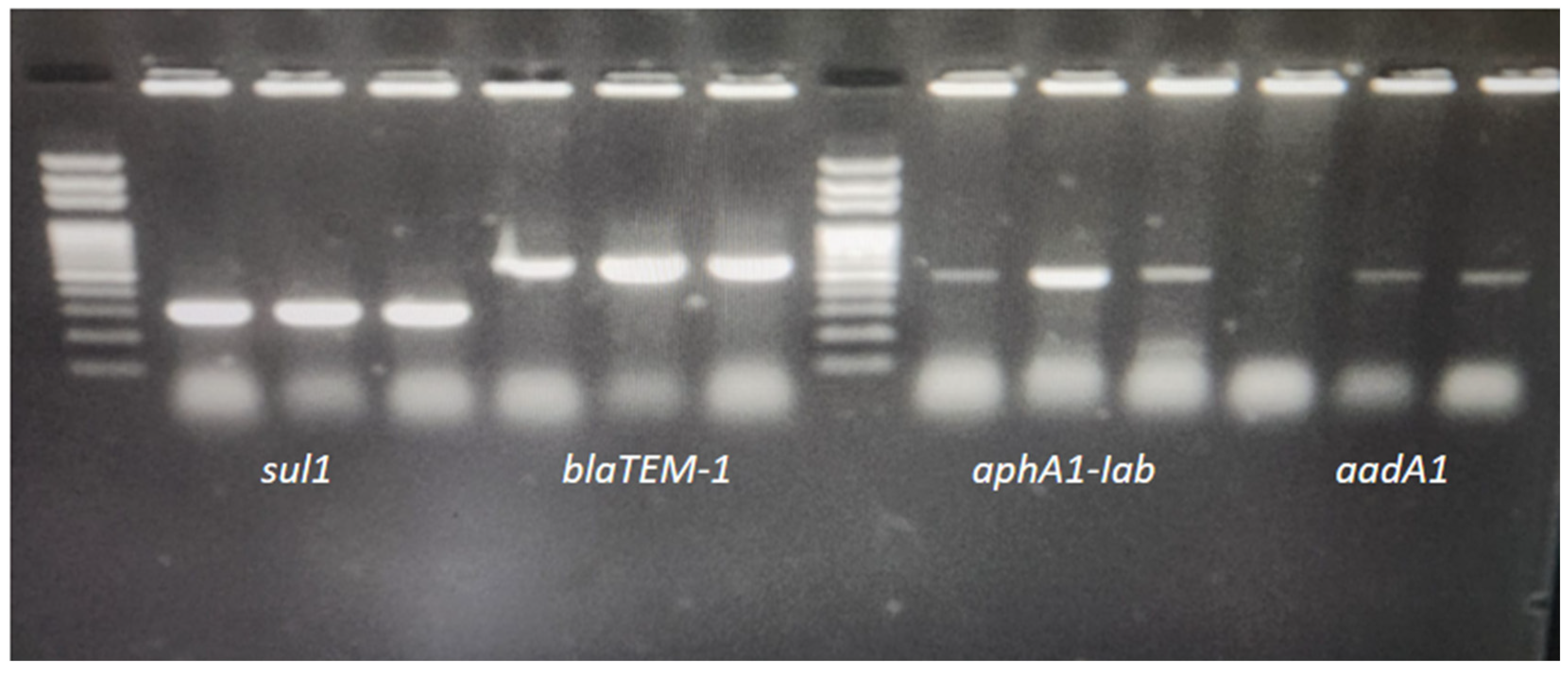
Antibiotic resistance genes were screened in all three isolates detected in this study, and the presence of the blaTEM-1 gene was detected in three isolates resistant to ampicillin and amoxicillin-clavulanic acid. All isolates in our study showed resistance to streptomycin and kanamycin via disk diffusion, and PCR detection targeting both the aadA1 and aphA1-IAB genes indicated that aminoglycoside-modifying enzymes contribute to this resistance. The aphA1-IAB gene was detected in all three isolates, and the aadA1 gene was detected in two (S2 and S3) (Figure 2).
The phenotype that is resistant to trimethoprim-sulfamethoxazole (TMZ) was confirmed at the molecular level via the detection of the sul1 gene in all isolates tested in the current study. But, for tetracycline, despite resistance being indicated in the antibiogram, the tetA and tetB genes were not found.

3.3. The Antimicrobial Effects of Carvacrol, Eugenol, and α-Terpineol on Salmonella Isolates

3.3.1. Minimum Inhibitory (MIC) and Bactericidal Concentration (MBC) Assay Values

The current study also evaluated the antimicrobial effects of natural bioactive compounds—including eugenol, carvacrol, and α-terpineol, which may serve as potential alternatives to synthetic preservatives in controlling foodborne pathogens—against three Salmonella Infantis isolates obtained from chicken meat. The antimicrobial activities of carvacrol, eugenol, and α-terpineol against S. Infantis isolates were evaluated based on their minimum inhibitory concentration (MIC) and minimum bactericidal concentration (MBC) values. Among the bioactive compounds tested on three different S. Infantis isolates (S1, S2, and S3), the lowest MIC and MBC values were observed in the carvacrol-treated samples (Table 4).
The MIC value of carvacrol was 1.56 µL/mL for all isolates, while MBC values varied depending on the isolate. These findings indicate that carvacrol exhibits higher antimicrobial activity against S. Infantis isolates. The minimum inhibitory concentration (MIC) of carvacrol was determined as 1.56 µL/mL for all isolates, whereas the minimum bactericidal concentration (MBC) values varied among the isolates. S2 and S3 isolates exhibited MBC values of 6.25 µL/mL and 3.125 µL/mL, respectively.
In this study, eugenol showed effective antimicrobial activity despite exhibiting higher MIC and MBC values compared to carvacrol. The MIC of eugenol was determined to be in the range of 3.125–6.25 µL/mL for all isolates, and the MBC value was 12.5 µL/mL (Table 4).
Comparing the α-terpineol MIC and MIC values to the other two compounds in the current work, it was found that this bioactive chemical exhibited the highest activity at high concentrations (Table 4).

3.3.2. Results of Agar Well Diffusion Method

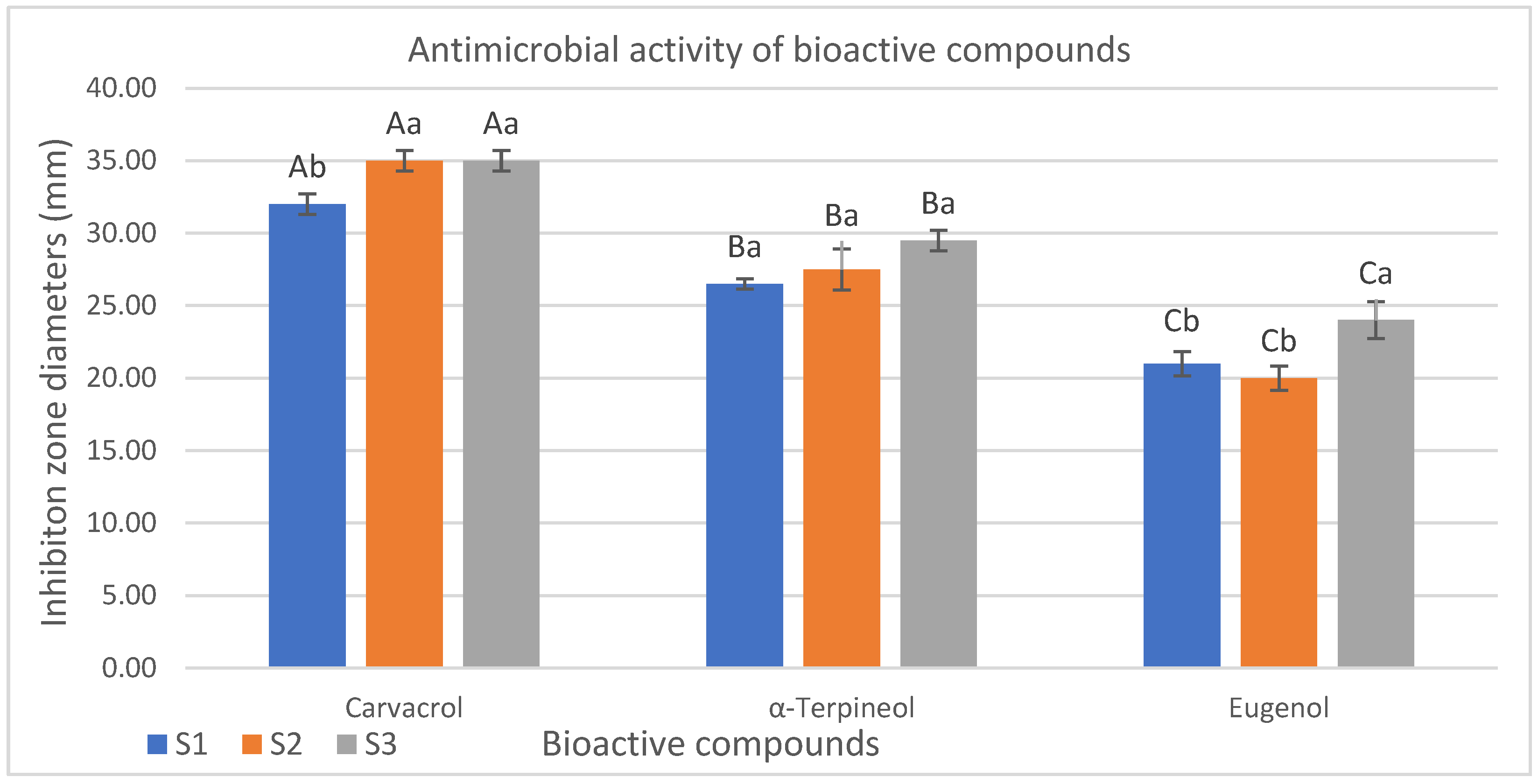
The antimicrobial effects of carvacrol, α-terpineol, and eugenol on Salmonella Infantis isolates were assessed based on inhibition zone diameters determined via the agar well diffusion method. Carvacrol demonstrated the strongest antimicrobial activity among the tested compounds, with inhibition zones ranging from 32.00 to 35.00 mm (p < 0.05). These findings confirm the potent activity of carvacrol against Salmonella spp. strains. The inhibition zone diameters obtained via the agar well diffusion method were evaluated as indicators of antimicrobial activity. Among the isolates tested, S3 was determined to be the most susceptible isolate to α-terpineol with an inhibition zone of 29.50 mm. In contrast, isolates S2 and S1 exhibited slightly lower inhibition zones of 27.50 mm and 26.50 mm, respectively. Although α-terpineol exhibited lower antimicrobial activity compared to carvacrol, statistically significant antibacterial effects were observed against the tested isolates. The lowest inhibition zone diameters were observed in isolates treated with eugenol ranging from 20.00 to 24.00 mm with S3 being the most sensitive isolate (24.00 mm) (Figure 3).

4. Discussion

4.1. Salmonella Infantis

Salmonella Infantis is a serotype that exhibits multidrug resistance (MDR) patterns and is gaining increasing importance among Salmonella spp. serotypes originating from poultry meat. When examined epidemiologically, there are significant differences in the prevalence of Salmonella spp. in poultry meat between countries. In this study, Salmonella Infantis was isolated from 4% of drumstick samples and 8% of thigh samples. In a similar study conducted in Japan, S. Infantis was found in 72.2% of ground chicken samples and 84% of boiler samples in Ecuador [54]. In addition, studies conducted in Turkey have shown that the prevalence of this serotype has increased significantly recently [55], suggesting that this serotype may be a source of concern for both poultry production and public health. The prevalence of S. Typhimurium and S. Enteritidis serovars was also evaluated in our study; however, neither serovar could be detected. Some studies in the literature have investigated the presence of these serovars in edible internal organ samples, but their isolation was not reported, which is consistent with our findings [56,57,58]. In a similar study on 150 chicken meat samples, Salmonella spp. was found in 42.66% of the samples, indicating a higher prevalence rate [59]. Babacan and Karadeniz detected Salmonella spp. contamination in 35% of packaged chicken meat samples using both the ISO 6579 standard [39] and the Mini Vidas Easy SLM (Biomerieux, Marcy-l’Étoile, France) systems [60]. In Tokyo, Salmonella species were reported to be present in 143 of 240 ground chicken samples [61]. Similarly, the prevalence of Salmonella spp. is also low. It was reported in 48.3% of 60 chicken meat samples from Surabaya, Indonesia [62]. These findings are consistent with the reports published by the European Food Safety Authority (EFSA) and the European Centre for Disease Prevention and Control (ECDC). According to the One Health Zoonoses Report (2018), the four most common Salmonella spp. serotypes isolated from human salmonellosis cases are S. Infantis, S. Typhimurium, S. Enteritidis, and the monophasic variant of S. Typhimurium [63]. More recent data presented in the 2022 report indicate that Salmonella spp. have been isolated throughout the European Union. Strains isolated throughout the European Union originate from chickens and related food products. The predominant serotypes are S. Infantis, S. Enteritidis, S. Typhimurium, monophasic S. Typhimurium, and S. Derby [7]. Our findings, which also identified S. Infantis as the predominant serotype in chicken meat samples, are consistent with this global upward trend and further emphasize its increasing epidemiological importance.

4.2. Determination of Antimicrobial Susceptibility and Antibiotic Resistance Genes

There is a global trend toward increasing ampicillin resistance, particularly among Enterobacteriaceae isolates. A study detected high ampicillin resistance in S. Infantis isolates [64]. The blaTEM-1 gene is one of the most prevalent β-lactamase genes and confers resistance to β-lactam antibiotics such as amoxicillin and ampicillin [65]. It forms the evolutionary basis of the extended-spectrum β-lactamase (ESBL) family, and ESBL enzymes originate from these β-lactamases [66]. The presence of blaTEM-1 in Salmonella spp. isolates explains the development of resistance, particularly with respect to first-line antibiotic agents. Furthermore, plasmid carriage of the blaTEM-1 gene facilitates horizontal gene transfer between species, increasing the spread of resistant Enterobacteriaceae within the community [67]. In many countries, the prevalence of blaTEM-1 in foodborne Salmonella spp. strains has been reported to range from 20% to 70% [68,69]. However, in a study examining 135 S. Infantis isolates from Japan, the blaTEM-1 gene was detected in 0.74% [16]. Salmonella Infantis strains isolated from poultry meat in Türkiye have been reported to contain additional TEM family enzymes, including blaTEM-70, blaTEM-148, and blaTEM-198. A single nucleotide mutation distinguishes these genes from blaTEM-1 [70].
In general, S. Infantis isolates in this study were found to be susceptible to third-generation cephalosporins, including ceftazidime and cefotaxime. However, S. Infantis 1 and 2 isolates showed resistance to cefotaxime (Table 3). Isolate S1 had an inhibition zone of 23 mm, and inhibition zones below 25 mm raised suspicion of ESBL resistance. However, resistance to amoxicillin/clavulanic acid eliminated this suspicion. However, three isolates were screened for the blaTEM, blaSHV, blaOXA, and blaCTX-M genes, and none were detectably positive.
Kanamycin and streptomycin resistance in Salmonella spp. isolates is clinically important, especially in invasive infections where treatment options are limited. Resistance to these aminoglycosides can often be transmitted via plasmid-borne genes such as aadA and aphA and can spread to other bacteria. Furthermore, the presence of resistant strains in the food chain poses a serious risk to public health, increasing the importance of antibiotic stewardship strategies. Our findings documented streptomycin and kanamycin resistance in S. Infantis isolates at the phenotypic and genetic levels. Multidrug-resistant (MDR) clones resistant to nalidixic acid, streptomycin, sulfamethoxazole, and tetracycline (NaSSuT) have been widely reported in the European poultry industry. Resistant Salmonella strains have been documented in many countries, including Hungary, Poland, Austria, Germany, and Switzerland [71,72]. Similarly, Rahmani et al. observed streptomycin resistance in all S. Infantis isolates obtained from poultry farms in Iran [73]. Numerous studies have reported a high prevalence of the aadA1 gene in S. Enterica serotypes isolated from poultry [73,74]. The aadA1 gene was found in 77.14% of Salmonella spp. isolates obtained from cloacal swabs and environmental samples at a chicken farm in China, and in Ghana, it was detected in 77% of Salmonella isolates from chicken eggs [75]. Another study reported that the aadA1 gene was detected in 97% of 135 S. Infantis isolates from Japan [16]. Acar et al. identified the aadA1 gene in S. Infantis strains isolated from chicken meat [70]. The aadA1 gene confers resistance by encoding the 3′-adenylate transferase enzymes, particularly streptomycin and spectinomycin. This gene is rapidly transferred between numerous bacterial species, mostly through class 1 integrons [74,76]. Although streptomycin has not been legally used in Türkiye in recent years, due to its long half-life and environmental persistence, this gene may be present in isolates [77]. The aadA1 gene was not detected in the S. Infantis 1 isolate, which may be due to the presence of other aadA or strA/B gene variants that were not investigated in our study.
The aphA1-IAB gene inactivates aminoglycosides such as kanamycin and neomycin through phosphorylation [78]. The aphA1-IAB gene can be transferred via transposons such as Tn903 [79]. The long-term and widespread use of aminoglycosides, especially in veterinary medicine, has caused this gene to be under selective pressure in zoonotic bacteria such as Salmonella [69,80]. The aphA1-IAB gene was reported to be detected in 4.4% of 135 S. Infantis isolates in Japan [71]. In Türkiye, the presence of the aphA1-IAB gene has been shown in S. Infantis isolates isolated from chicken meat in Şanlıurfa, Bolu, and Ankara [71,81].
Our findings indicated resistance to trimethoprim and sulfamethoxazole in all three S. Infantis isolates using disk diffusion tests, and the sul1 gene was identified. The variant of the dihydropteroate synthase enzyme that confers resistance to sulfonamides is encoded by the sul1 gene and is frequently associated with class 1 integrons [82]. The simultaneous identification of integron-associated genes such as aadA1 and sul1 introduces the possibility that these isolates may have developed resistance via mobile genetic elements. Furthermore, this process is known as the effect of co-selection because additional resistance genes on the same mobile genetic element can undergo co-selection under antibiotic pressure, such as trimethoprim and aminoglycosides [79]. This raises the possibility that zoonotic bacteria contain antibiotic resistance genes, particularly when combined with the widespread use of antibiotics for prophylaxis or treatment in animal production. The sul1 gene was detected in 70% of S. Indiana isolates isolated from frozen chicken meat in China [83]. It has been reported that the sul1 gene is present in 77.78% of S. Enteritidis and S. Typhimurium isolates in Türkiye [84]. The identification of this gene, particularly in zoonotic pathogens, highlights the need to address antimicrobial resistance throughout the food distribution chain and in clinics [17].
Although tetracycline resistance was demonstrated in the antibiogram, the tetA and tetB genes were not found. This could be explained by the presence of more than 40 different tet resistance genes. In our study, genes encoding ribosomal protection proteins or mediating enzymatic inactivation were not detected because their primers were not included in the analysis.

4.3. Evolution of the Antimicrobial Effects of Carvacrol, Eugenol, and α-Terpineol on Salmonella Isolates

4.3.1. Evolution of Minimum Inhibitory (MIC) and Bactericidal Concentration (MBC) Assay Values

One of the most effective preventive approaches against antibiotic-resistant food pathogens is to apply natural products with antibacterial activity to foods. Therefore, food-grade carvacrol, alpha-terpineol, and eugenol were used in our study due to their antibacterial activity. The MBC value of carvacrol for isolates S2 and S3 was 3.125 µL/mL. However, the MBC value of isolate S1 was 6.25. This difference suggests that carvacrol may be bactericidal at lower concentrations in isolates S2 and S3 and exhibits more potent antimicrobial activity against these strains. The results of this study are consistent with those reported by Dhifi et al., who reported that the most frequently used natural antibacterial agents, especially against Gram-negative bacteria, are phenolic compounds (thymol and carvacrol), aldehydes (cinnamaldehyde), and terpenes (eugenol and limonene) [85]. These active compounds disrupt the structural integrity of bacterial cell membranes, denature proteins, and negatively affect enzymatic activity, resulting in bacterial inhibition. Recent studies have shown that such bioactive compounds are effective in preserving the microbiological quality of foods [86]. The antibacterial effects of essential oil compounds, such as thymol, carvacrol, citral, and cinnamaldehyde, on E. coli O157:H7 and S. Typhimurium were investigated, and findings similar to ours were obtained [87]. These studies also reported that carvacrol-treated groups completely inhibited live cells within 60 and 30 min in the time-kill experiment, respectively. Khan et al. showed that carvacrol has a potent and rapid bactericidal effect against E. coli O157:H7 and S. Typhimurium pathogens in their studies [88]. Heckler et al. examined the antimicrobial effects of carvacrol and thymol against Staphylococcus aureus and Salmonella Enteritidis. They found the MIC value to be 200 µg/mL against both bacteria [89]. In another study conducted in Türkiye, the inhibitory effects of carvacrol, curcumin, and frankincense oil on S. Enteritidis and S. Enteritidis PT4 were investigated. The MIC values of carvacrol, curcumin, and frankincense oil were determined as 125.0 µg/mL, 132.5 µg/mL, and 31.3 mg/mL, respectively [90]. The MIC value of carvacrol against Salmonella Typhimurium was determined as 0.6 mg/mL, and the contact time required for bacterial inhibition was determined as 10 min [91]. In a similar study, the MICs and MBCs of carvacrol on S. Typhimurium were reported as 0.5 mg/mL and 1 mg/mL, respectively [92].
In our study, the MIC value of eugenol was determined to be within the range of 3.125–6.25 µL/mL, and the MBC value was 12.5 µL/mL for all isolates. It has been reported that the MIC values of eugenol against five clinical S. Typhimurium isolates varied between 0.00625% and 0.025% (v/v), and MBC values ranged between 0.0125% and 0.025% (v/v). In addition, the MIC values of 0.0125% and MBC values of 0.025% (v/v) were observed for the standard strain SL1344 [93,94]. The antimicrobial activity of eugenol was investigated with respect to three biofilm-forming isolates of ten S. Enteritidis isolates obtained from different chicken farms in Korea and the reference strain S. Enteritidis ATCC 13076. In this study, MIC values were reported to be 0.36% and 0.72% in the isolates and the reference strain, respectively [95]. As carried out by Balyan et al., Eugenol was encapsulated using an exopolysaccharide derived from Lactobacillus plantarum to increase its stability and antimicrobial activity. Their results showed that MBC values ranged from 2.6 to 4.4 mg/mL for L. monocytogenes and 1.6 to 3.2 mg/mL for Salmonella, with the lowest being 0.08 mg/mL for E. coli. They also found that MIC values varied depending on the strain [96].
When examining the MIC values of alpha-terpineol, the MIC value of isolates S1 and S2 was 6.25 µL/mL, while it was 12.5 µL/mL for isolate S3. The MBC value of all isolates was found to be 25 µL/mL. Similarly to our results, in a study evaluating the antimicrobial effects of α-terpineol, α-pinene, and 1,8-cineole on Gram-positive (S. aureus ATCC 25923, L. monocytogenes ATCC 1911, and Bacillus cereus ATCC 14579) and Gram-negative (S. Enterica ATCC 43972, and E. coli ATCC 25922) bacterial strains, it was reported that all three compounds exhibited significant antibacterial activity against five tested bacterial strains. In a study conducted by Gökmen, the antibacterial activities of carvacrol, α-terpineol, and eugenol were evaluated against extended-spectrum β-lactamase (ESBL)-producing E. coli strains isolated from chicken meat, and they were comparable to the scope of the present study. The broth dilution test revealed MIC and MBC values of 0.78 µL/mL for carvacrol, 3.125–6.25 µL/mL and 6.25–12.50 µL/mL for eugenol, and 3.125–6.25 µL/mL and 3.125–12.50 µL/mL for α-terpineol [97]. These results further support our findings by demonstrating that carvacrol exhibits a more potent antimicrobial effect at lower concentrations compared to the other tested compounds. The consistency between the two studies highlights the potent antimicrobial potential of carvacrol, particularly against multidrug-resistant Gram-negative pathogens, suggesting that it may play a promising role as a natural alternative in the control of foodborne bacteria.
In this study, carvacrol was identified as the most effective antimicrobial agent against S. Infantis isolates, while eugenol and α-terpineol exhibited relatively lower levels of activity. This discrepancy may be attributed to differences in the chemical structures, lipophilic properties, and modes of action of the tested compounds, in addition to isolate-specific resistance mechanisms. Carvacrol’s ability to induce membrane depolarization, disrupt bacterial cell integrity, and induce oxidative stress likely explains its effectiveness at lower concentrations. In contrast, the effects of eugenol and α-terpineol on membrane permeability appear to be more limited, which may explain their relatively weaker antimicrobial performance. This study also highlights the varying efficacy levels of natural antimicrobial agents against MDR S. Infantis strains and supports the potential of these three bioactive compounds (especially carvacrol) as alternative biological control agents. However, larger-scale studies are needed to comprehensively evaluate their efficacy against a wider range of bacterial strains.

4.3.2. Evolution of Agar Well Diffusion Method Results

In this study, the strongest activity in the agar well diffusion test was observed in carvacrol, showing inhibition zones of 32–35 mm. A comparable investigation examining the antibacterial effect of carvacrol against S. Typhimurium found that the application of 0.25 and 0.5 mg carvacrol concentrations, respectively, resulted in mean inhibition zones of 13.1 mm and 18.3 mm, which is consistent with our findings [96]. The antibacterial activities of thymol and carvacrol against Gram-negative bacteria, including Pseudomonas aeruginosa, E. coli, and S. Typhimurium, and antifungal activities against Candida albicans were investigated by Akermi et al. The inhibition zones in the carvacrol-treated groups ranged from 18 to 23 mm, while those in the thymol-treated groups ranged from 14 to 21 mm, with carvacrol reported to have significantly stronger effects than thymol (p < 0.05) [98]. Additionally, the antimicrobial activity of eugenol against E. coli strains was evaluated using the disk diffusion method, and mean inhibition zone diameters of 38 ± 5 mm, 30 ± 4 mm, and 18 ± 4 mm were obtained at concentrations of 1000, 500, and 250 µg/mL, respectively [99]. Hussein et al. reported in vitro inhibition zones of α-terpineol against S. Typhimurium as 2.33, 2.09, and 1.99 mm at 24, 48, and 72 h, respectively. The in vivo application of α-terpineol was also observed to completely inhibit Pseudomonas lundensis, Listeria monocytogenes, and S. Typhimurium in ground chicken stored at 4 °C for two weeks [36]. Discs impregnated with eugenol at 1%, 5%, and 10% (v/v) concentrations on S. Typhi exhibited inhibition zones of 7 mm, 11 mm, and 11 mm, respectively, while discs containing ciprofloxacin (500 ng/mL), used as a positive control, exhibited an inhibition zone of 13 mm. These results indicate that eugenol exhibits significant antibacterial activity against S. Typhi, especially at high concentrations, and that its activity is similar to that of the conventional antibiotic ciprofloxacin [100]. The antimicrobial effects of various plant essential oils were evaluated, including clove oil produced in Türkiye by Evrendilek [101], which contains 67.3% eugenol, on common foodborne Gram-negative and Gram-positive pathogenic bacteria. It showed significant antimicrobial activity against various foodborne pathogens [100]. The antibacterial activities of carvacrol, α-terpineol, and eugenol were evaluated against ESBL-producing E. coli isolates by Gökmen [97]. In the eugenol-treated groups, inhibition zones were determined as 35 mm for isolates CM1 and CM3, while isolates CM2 and CM4 exhibited slightly smaller zones of 30 mm [97]. Jeyakumar et al. evaluated the antibacterial effect of eugenol on E. coli isolates using the well diffusion method and reported the inhibition zone diameter as 24 mm [102].

5. Conclusions

In conclusion, the findings of this study demonstrate that S. Infantis isolates exhibit multidrug resistance (MDR), mediated not only by plasmid-borne genes (blaTEM-1, aadA1, aphA1-IAB, and sul1) but also, potentially, by chromosomal mutations (gyrA/parC QRDR). The presence of such MDR patterns in animal-derived isolates poses significant concerns, as it restricts therapeutic options in veterinary medicine and represents a public health threat due to the risk of zoonotic transmission. Therefore, the continuous monitoring of isolates at both the phenotypic and genetic levels is essential in order to improve our understanding of resistance mechanisms and inform the development of effective control strategies.
This study also compared the antibacterial properties of carvacrol, α-terpineol, and eugenol against Salmonella Infantis isolates from commercially marketed chicken meat produced in various integrated facilities. The findings revealed that all three compounds exhibited significant antimicrobial activity (p < 0.05). The compound with the strongest antibacterial activity was carvacrol, which had low MIC/MBC values and large inhibition zone diameters. In vitro, α-terpineol showed promising results. Eugenol exhibited antibacterial activity similar to antibiotics at high concentrations, although it was less effective than carvacrol and α-terpineol with respect to certain strains.

Author Contributions

H.Y.—conceptualization, data curation, formal analysis, investigation, methodology, project administration, supervision, writing—original draft, writing—review and editing. Y.T.—investigation, methodology, validation. T.G.G.—investigation, methodology, validation, review and editing. N.G.—data curation, review and editing. N.S.U.—methodology. All authors have read and agreed to the published version of the manuscript.

Funding

This research was financially supported by the Scientific Research Projects Unit of Adana Çukurova University, through the project “Detection of Salmonella species isolated from chicken meat produced in different integrated broiler farms and molecular characterization of antibiotic resistance genes” (TYL-2023-16378). The authors would like to thank the Scientific Research Projects Unit for their support.

Data Availability Statement

The data sets used are available from the corresponding author upon reasonable request.

Conflicts of Interest

Nuri Güngör is the deputy director of the General Directory of Meat and Milk Board, and Yasin Tekin is the operations manager of the General Directory of Meat and Milk Board. The company for which these authors work did not provide any financial support for this study.

References

  1. Talukder, H.; Roky, S.A.; Debnath, K.; Sharma, B.; Ahmed, J.; Roy, S. Prevalence and antimicrobial resistance profile of Salmonella isolated from human, animal and environment samples in South Asia: A 10-year Meta-analysis. J. Epidemiol. Glob. Health 2023, 13, 637–652. [Google Scholar] [CrossRef]
  2. Chanam’e Pinedo, L.; Mughini-Gras, L.; Franz, E.; Hald, T.; Pires, S.M. Sources and trends of human salmonellosis in Europe, 2015–2019: An analysis of outbreak data. Int. J. Food Microbiol. 2022, 379, 109850. [Google Scholar] [CrossRef] [PubMed]
  3. Papoula-Pereira, R.; Alvseike, O.; Cenci-Goga, B.T.; Grispoldi, L.; Nagel-Alne, G.E.; Ros-Lis, J.V.; Thomas, L.F. Economic evidence for the control of Salmonella in animal-derived food systems: A scoping review. Food Control 2025, 175, 111275. [Google Scholar] [CrossRef]
  4. Lamichhane, B.; Mawad, A.M.M.; Saleh, M.; Kelley, W.G.; Harrington, P.J.; Lovestad, C.W.; Amezcua, J.; Sarhan, M.M.; El Zowalaty, M.E.; Ramadan, H.; et al. Salmonellosis: An Overview of Epidemiology, Pathogenesis, and Innovative Approaches to Mitigate the Antimicrobial Resistant Infections. Antibiotics 2024, 13, 76. [Google Scholar] [CrossRef]
  5. Bermudez-Aguirre, D.; Carter, J.; Niemira, B.A. An investigation about the historic global foodborne outbreaks of Salmonella spp. in eggs: From hatcheries to tables. Compr. Rev. Food Sci. Food Saf. 2025, 24, e70202. [Google Scholar] [CrossRef]
  6. Adley, C.C.; Ryan, M.P. The nature and extent of foodborne disease. In Antimicrobial Food Packaging; Academic Press: New York, NY, USA, 2025; pp. 3–14. [Google Scholar]
  7. European Food Safety Agency/European Centre for Disease Prevention and Control (EFSA/ECDC). Rapid Outbreak Assessment: Multi- CountryOutbreak of Salmonella Enteritidis Sequence Type (ST)11 Infections Linked to Eggs and Egg Products; Third Update; EFSA/ECDC: Stockholm, Sweden, 2022.
  8. Stanaway, J.D.; Parisi, A.; Sarkar, K.; Blacker, B.F.; Reiner, R.C.; Hay, S.I.; Nixon, M.R.; Dolecek, C.; James, S.L.; Mokdad, A.H.; et al. The global burden of non-typhoidal salmonella invasive disease: A systematic analysis for the Global Burden of Disease Study 2017. Lancet Infect. Dis. 2019, 19, 1312–1324. [Google Scholar] [CrossRef]
  9. Cawthraw, S.; Wales, A.; Guzinski, J.; Trew, J.; Ring, I.; Huby, T.; Hussaini, A.; Petrovska, L.; Martelli, F. Salmonella Infantis outbreak on six broiler units in Great Britain: Investigation, epidemiology, and control. J. Appl. Microbiol. 2025, 136, lxaf040. [Google Scholar] [CrossRef]
  10. Nazari Moghadam, M.; Rahimi, E.; Shakerian, A.; Momtaz, H. Prevalence of Salmonella typhimurium and Salmonella Enteritidis isolated from poultry meat: Virulence and antimicrobial-resistant genes. BMC Microbiol. 2023, 23, 168. [Google Scholar] [CrossRef]
  11. Canning, M.; Birhane, M.G.; Dewey-Mattia, D.; Lawinger, H.; Cote, A.; Gieraltowski, L.; Schwensohn, C.; Tagg, K.A.; Watkins, L.K.F.; Robyn, M.P.; et al. Salmonella outbreaks linked to beef, United States, 2012–2019. J. Food Prot. 2023, 86, 100071. [Google Scholar] [CrossRef] [PubMed]
  12. Strickland, A.J.; Sampedro, F.; Hedberg, C.W. Quantitative risk assessment of salmonella in ground beef products and the resulting impact of risk mitigation strategies on public health. J. Food Prot. 2023, 86, 100093. [Google Scholar] [CrossRef] [PubMed]
  13. Campos, J.; Mourão, J.; Peixe, L.; Antunes, P. Non-typhoidal Salmonella in the pig production chain: A comprehensive analysis of its impact on human health. Pathogens 2019, 8, 19. [Google Scholar] [CrossRef] [PubMed]
  14. Broadway, P.R.; Brooks, J.C.; Mollenkopf, D.F.; Calle, M.A.; Loneragan, G.H.; Miller, M.F.; Carroll, J.A.; Sanchez, N.C.B.; Wittum, T.E. Prevalence and antimicrobial susceptibility of Salmonella Serovars isolated from U.S. retail ground pork. Foodborne Pathog. Dis. 2021, 18, 219–227. [Google Scholar] [CrossRef]
  15. EFSA. Report of the task force on zoonoses data collection on the analysis of the baseline survey on the prevalence of Salmonella in broiler flocks of Gallus gallus, in the EU, 2005–2006. EFSA J. 2007, 98, 1–85. [Google Scholar]
  16. Shahada, F.; Chuma, T.; Kosugi, G.; Okamoto, K. Genetic Characteristics of Antimicrobial Resistance of Salmonella Isolated from Chicken in Japan. J. Vet. Med. Sci. 2006, 68, 115–120. [Google Scholar]
  17. European Food Safety Authority (EFSA). The EU Summary Report on AMR in Zoonotic and Indicator Bacteria from Humans, Animals and Food in 2021. EFSA J. 2023, 21, e07784. [Google Scholar]
  18. Dishan, A.; Hizlisoy, H.; Onmaz, N.E.; Yildirim, Y.; Gonulalan, Z.; Al, S. Comprehensive analysis of Salmonella in poultry meat and products in Türkiye: Prevalence, antibiotic susceptibility and genomic characterisation. Int. J. Food Sci. Technol. 2024, 59, 3412–3422. [Google Scholar] [CrossRef]
  19. USDA. Cost Estimates of Foodborne Illnesses. U. S. Department of Agriculture (USDA) Economic Research Service; 2021. Available online: https://www.usda.gov (accessed on 18 August 2025).
  20. Li, L.; McWhorter, A.; Chousalkar, K. Ensuring Egg Safety: Salmonella Survival, Control, and Virulence in the Supply Chain. Compr. Rev. Food Sci. Food Saf. 2025, 24, e70075. [Google Scholar] [CrossRef]
  21. Simpson, K.M.J.; Hill-Cawthorne, G.A.; Ward, M.P.; Mor, S.M. Diversity of Salmonella Serotypes From Humans, Food, Domestic Animals and Wildlife in New South Wales, Australia. BMC Infect. Dis. 2018, 18, 623. [Google Scholar] [CrossRef]
  22. Kumar, G.; Kumar, S.; Jangid, H.; Dutta, J.; Shidiki, A. The rise of non-typhoidal Salmonella: An emerging global public health concern. Front. Microbiol. 2025, 16, 1524287. [Google Scholar] [CrossRef]
  23. Osivand, Z.; Rahimi, E.; Shakerian, A.; Khamesipour, F. Prevalence, Antibiotic Resistance, Virulence and Antimicrobial Resistance Gene Profiles of Salmonella Species Recovered from Retail Beef and Poultry Processing Environments. BMC Microbiol. 2025, 25, 174. [Google Scholar] [CrossRef]
  24. Rodrigues, G.L.; Panzenhagen, P.; Ferrari, R.G.; Paschoalin, V.M.F.; Conte-Junior, C.A. Antimicrobial resistance in nontyphoidal Salmonella isolates from human and swine sources in Brazil: A systematic review of the past three decades. Microb. Drug Resist. 2020, 26, 1260–1270. [Google Scholar] [CrossRef]
  25. Chiou, C.S.; Hong, Y.P.; Wang, Y.W.; Chen, B.H.; Teng, R.H.; Song, H.Y.; Liao, Y.S. Antimicrobial resistance and mechanisms of azithromycin resistance in nontyphoidal Salmonella isolates in Taiwan, 2017 to 2018. Microbiol. Spectr. 2023, 11, e03364-22. [Google Scholar] [CrossRef] [PubMed]
  26. Nuanmuang, N.; Leekitcharoenphon, P.; Njage, P.M.K.; Gmeiner, A.; Aarestrup, F.M. An overview of antimicrobial resistance profiles of publicly available Salmonella genomes with sufficient quality and metadata. Foodborne Pathog. Dis. 2023, 20, 405–413. [Google Scholar] [CrossRef]
  27. Wang, C.-Y.; Chen, Y.-W.; Hou, C.-Y. Antioxidant and Antibacterial Activity of Seven Predominant Terpenoids. Int. J. Food Prop. 2019, 22, 230–238. [Google Scholar] [CrossRef]
  28. Yazgan, H.; Kuley, E.; Özogul, Y. Investigation of Bioactive Compounds and Antimicrobial Properties of Aqueous Garlic Extracts. Biomass Convers. Biorefin. 2024, 14, 16673–16680. [Google Scholar] [CrossRef]
  29. Imran, M.; Aslam, M.; Alsagaby, S.A.; Saeed, F.; Ahmad, I.; Afzaal, M.; Arshad, M.U.; Abdelgawad, M.A.; El-Ghorab, A.H.; Khames, A.; et al. Therapeutic application of carvacrol: A comprehensive review. Food Sci. Nutr. 2022, 10, 3544–3561. [Google Scholar] [CrossRef]
  30. Mączka, W.; Twardawska, M.; Grabarczyk, M.; Wińska, K. Carvacrol—A natural phenolic compound with antimicrobial properties. Antibiotics 2023, 12, 824. [Google Scholar] [CrossRef]
  31. Ozogul, Y.; Karsli, G.T.; Yazgan, H.; Kuley, E.; Oztop, H.M.; Ozogul, F.; Esatbeyoglu, T. Enhanced pathogen control through thymol and carvacrol nanoemulsions: A microfluidization approach. Food Bioprocess Technol. 2025, 18, 5377–5387. [Google Scholar] [CrossRef]
  32. Ulanowska, M.; Olas, B. Biological Properties and Prospects for the Application of Eugenol—A Review. Int. J. Mol. Sci. 2021, 22, 3671. [Google Scholar] [CrossRef]
  33. de Oliveira, M.G.; Marques, R.B.; de Santana, M.F.; Santos, A.B.; Brito, F.A.; Barreto, E.O.; De Sousa, D.P.; Almeida, F.R.C.; Badauê-Passos, D., Jr.; Antoniolli, Â.; et al. α-Terpineol Reduces Mechanical Hypernociception and Inflammatory Response. Basic Clin. Pharmacol. Toxicol. 2012, 111, 120–125. [Google Scholar] [CrossRef]
  34. de Sousa, D.P.; Quintans, L., Jr.; de Almeida, R.N. Evaluation of the Anticonvulsant Activity of α-Terpineol. Pharm. Biol. 2007, 45, 69–70. [Google Scholar] [CrossRef]
  35. Hassan, S.B.; Gali-Muhtasib, H.; Göransson, H.; Larsson, R. Alpha-Terpineol: A Potential Anticancer Agent. Anticancer Res. 2010, 30, 1911–1919. [Google Scholar]
  36. Hussein, K.N.; Csehi, B.; József, S.; Ferenc, H.; Kiskó, G.; Dalmadi, I.; Friedrich, L. Effect of α-Terpineol on Chicken Meat Quality during Refrigerated Conditions. Foods 2021, 10, 1855. [Google Scholar] [CrossRef]
  37. Khaleel, C.; Tabanca, N.; Buchbauer, G. α-Terpineol, a natural monoterpene: A review of its biological properties. Open Chem. 2018, 16, 349–361. [Google Scholar] [CrossRef]
  38. Knez Hrnčič, M.; Cör, D.; Knez, Ž.; Hrnčič, M.K. Extraction techniques and analytical methods for characterization of active compounds in Origanum species. Molecules 2020, 25, 4735. [Google Scholar] [CrossRef]
  39. ISO 6579-1:2017; Microbiology of the Food Chain—Horizontal Method for the Detection, Enumeration and Serotyping of Salmonella—Part 1: Detection of Salmonella spp. International Organization for Standardization: Geneva, Switzerland, 2017. Available online: https://www.eucast.org (accessed on 23 September 2025).
  40. EUCAST (European Committee on Antimicrobial Susceptibility Testing). Breakpoint Tables for Interpretation of MICs and Zone Diameters, Version 15.0. 2025. Available online: https://www.eucast.org/clinical_breakpoints (accessed on 23 September 2025).
  41. Ng, L.K.; Martin, I.; Alfa, M.; Mulvey, M. Multiplex PCR for the Detection of Tetracycline Resistant Genes. Mol. Cell. Probes 2001, 15, 209–215. [Google Scholar] [CrossRef]
  42. Woodford, N.; Fagan, E.J.; Ellington, M.J. Multiplex PCR for Rapid Detection of Genes Encoding CTX-M ESBLs. J. Antimicrob. Chemother. 2006, 57, 154–155. [Google Scholar] [CrossRef]
  43. Dallenne, C.; Da Costa, A.; Decré, D.; Favier, C.; Arlet, G. Development of a Set of Multiplex PCR Assays for the Detection of Genes Encoding Important β-Lactamases in Enterobacteriaceae. J. Antimicrob. Chemother. 2010, 65, 490–495. [Google Scholar] [CrossRef]
  44. Reischl, U.; Linde, H.J.; Metz, M.; Leppmeier, B.; Lehn, N. Rapid Identification of MRSA and Species Confirmation Using Real-Time Fluorescence PCR. J. Clin. Microbiol. 2000, 38, 2429–2433. [Google Scholar] [CrossRef] [PubMed]
  45. Xu, Z.; Li, L.; Shirtliff, M.E.; Alam, M.J.; Yamasaki, S.; Shi, L. Expression of Antibiotic Resistance Genes in Salmonella Isolates from Poultry. Appl. Environ. Microbiol. 2007, 73, 7386–7389. [Google Scholar]
  46. Cattoir, V.; Poirel, L.; Rotimi, V.; Soussy, C.J.; Nordmann, P. Multiplex PCR for Detection of Plasmid-Mediated qnr Genes in ESBL-Producing Enterobacterial Isolates. J. Antimicrob. Chemother. 2007, 60, 394–397. [Google Scholar] [CrossRef] [PubMed]
  47. Cavaco, L.M.; Aarestrup, F.M.; Agersø, Y. Quinolone-Resistant Salmonella Hadar/Enteritidis from Danish Poultry. J. Antimicrob. Chemother. 2009, 64, 304–306. [Google Scholar]
  48. Wang, M.; Tran, J.H.; Jacoby, G.A.; Zhang, Y.; Wang, F.; Hooper, D.C. Plasmid-Mediated Quinolone Resistance in Clinical Escherichia coli from Shanghai, China. Antimicrob. Agents Chemother. 2003, 47, 2242–2248. [Google Scholar] [CrossRef]
  49. Antunes, P.; Machado, J.; Peixe, L. Characterization of Antimicrobial Resistance and Class 1 and 2 Integrons in Salmonella enterica Isolates from Different Sources in Portugal. J. Antimicrob. Chemother. 2005, 56, 1025–1029. [Google Scholar] [CrossRef]
  50. Sutcliffe, J.; Tait-Kamradt, A.; Wondrack, L. Detection of Erythromycin-Resistant Determinants by PCR. Antimicrob. Agents Chemother. 1996, 40, 2562–2566. [Google Scholar] [CrossRef]
  51. CLSI (Clinical and Laboratory Standards Institute). Performance Standards for Antimicrobial Susceptibility Testing, 34th ed.; CLSI Supplement M100; CLSI: Wayne, PA, USA, 2024. [Google Scholar]
  52. Aytar, F. Doğal Bileşiklerin Antibakteriyel Etkisinin Salmonella spp. Üzerine Karşılaştırmalı Olarak Araştırılması. Master’s Thesis, İstanbul Üniversitesi, İstanbul, Turkey, 2019. [Google Scholar]
  53. Antunes, P.; Mourão, J.; Campos, J.; Peixe, L. Salmonellosis: The Role of Poultry Meat. Clin. Microbiol. Infect. 2016, 22, 110–121. [Google Scholar] [CrossRef]
  54. Mattock, J.; Chattaway, M.A.; Hartman, H.; Dallman, T.J.; Smith, A.M.; Keddy, K.; Langridge, G.C. A One Health Perspective on Salmonella enterica Serovar Infantis, an Emerging Human Multidrug-Resistant Pathogen. Emerg. Infect. Dis. 2024, 30, 701–710. [Google Scholar] [CrossRef]
  55. Kürekçi, C.; Şahin, S. Salmonella infantis. J. Turk. Vet. Med. Soc. 2023, 94, 73–83. [Google Scholar]
  56. Raji, M.A.; Kazeem, H.M.; Magyigbe, K.A.; Ahmed, A.O.; Lawal, D.N.; Raufu, I.A. Salmonella Serovars, Antibiotic Resistance, and Virulence Factors Isolated from Chickens and Ready-to-Eat Gizzards in Nigeria. Int. J. Food Sci. 2021, 2021, 8872137. [Google Scholar] [CrossRef]
  57. Telli, A.E.; Biçer, Y.; Kahraman, H.A.; Telli, N.; Doğruer, Y. Presence and Antibiotic Resistance of Salmonella spp. Isolated from Chicken Meat and Giblets Consumed in Konya, Turkey. Eurasian J. Vet. Sci. 2018, 34, 164–170. [Google Scholar] [CrossRef]
  58. Hassan, A.H.; Salam, H.S.; Abdel-Latef, G.K. Identification of Virulence Genes, β-Lactams and Quinolone Resistance Genes, and Integrons in Salmonella from Retail Chicken in Egypt. J. Microbiol. Biotechnol. Food Sci. 2021, 10, 1320–1325. [Google Scholar]
  59. Sırıken, B.; Türk, H.; Yıldırım, T.; Durupınar, B.; Erol, İ. Prevalence and Characterization of Salmonella Isolated from Chicken Meat in Turkey. J. Food Sci. 2015, 80, M1044–M1050. [Google Scholar] [CrossRef]
  60. Babacan, O.; Karadeniz, H. Çiğ Tavuk Etlerinden İzole Edilen Salmonella spp. Suşlarının Antibiyotik Duyarlılıklarının Araştırılması. Vet. Hekimler Derneği Derg. 2019, 90, 105–114. [Google Scholar] [CrossRef]
  61. Ishihara, K.; Nakazawa, C.; Nomura, S.; Elahi, S.; Yamashita, M.; Fujikawa, H. Effects of Climatic Elements on Salmonella Contamination in Broiler Meat in Japan. J. Vet. Med. Sci. 2020, 82, 646–652. [Google Scholar] [CrossRef]
  62. Wardhana, D.K.; Haskito, A.E.P.; Purnama, M.T.E.; Safitri, D.A.; Annisa, S. Detection of Microbial Contamination in Chicken Meat from Local Markets in Surabaya, Indonesia. Vet. World 2021, 14, 3138–3144. [Google Scholar] [CrossRef]
  63. European Food Safety Authority (EFSA); European Centre for Disease Prevention and Control (ECDC). The European Union One Health 2018 Zoonoses Report. EFSA J. 2019, 17, e05926. [Google Scholar] [CrossRef]
  64. Sodagari, H.R.; Shrestha, R.D.; Agunos, A.; Gow, S.P.; Varga, C. Comparison of Antimicrobial Resistance among Salmonella enterica Serovars Isolated from Canadian Turkey Flocks, 2013–2021. Poult. Sci. 2023, 102, 102655. [Google Scholar] [CrossRef] [PubMed]
  65. Bush, K.; Jacoby, G.A. Updated Functional Classification of β-Lactamases. Antimicrob. Agents Chemother. 2010, 54, 969–976. [Google Scholar] [CrossRef] [PubMed]
  66. Bradford, P.A. Extended-Spectrum β-Lactamases in the 21st Century: Characterization, Epidemiology, and Detection. Clin. Microbiol. Rev. 2001, 14, 933–951. [Google Scholar] [CrossRef]
  67. Carattoli, A. Resistance Plasmid Families in Enterobacteriaceae. Antimicrob. Agents Chemother. 2009, 53, 2227–2238. [Google Scholar] [CrossRef]
  68. Guerra, B.; Junker, E.; Schroeter, A.; Malorny, B.; Lehmann, S.; Helmuth, R. Phenotypic and Genotypic Characterization of Antimicrobial Resistance in German Escherichia coli Isolates from Cattle, Swine and Poultry. J. Antimicrob. Chemother. 2003, 52, 489–492. [Google Scholar] [CrossRef]
  69. Wasyl, D.; Zając, M.; Lalak, A.; Skarżyńska, M.; Samcik, I.; Kwit, R.; Jabłoński, A.; Bocian, Ł.; Woźniakowski, G.; Hoszowski, A.; et al. Antimicrobial Resistance in Commensal Escherichia coli and Salmonella enterica from Food Animals in Poland. Microb. Drug Resist. 2018, 24, 807–815. [Google Scholar] [CrossRef]
  70. Acar, S.; Bulut, E.; Stasiewicz, M.J.; Soyer, Y. Genome Analysis of Antimicrobial Resistance, Virulence, and Plasmid Presence in Turkish Salmonella Serovar Infantis Isolates. Int. J. Food Microbiol. 2019, 307, 108275. [Google Scholar] [CrossRef]
  71. Nógrády, N.; Király, M.; Davies, R.; Nagy, B. Multidrug-Resistant Salmonella Infantis in Broiler Chickens and Humans in Hungary: The Need for Coordinated Monitoring. Food Res. Int. 2012, 45, 387–392. [Google Scholar]
  72. Hindermann, D.; Gopinath, G.; Chase, H.; Negrete, F.; Stephan, R. Salmonella enterica subsp. enterica Serovar Infantis from Meat Products and Human Infections in Switzerland: Phenotypic and Genotypic Antimicrobial Resistance and Phylogenetic Analysis. Food Control 2017, 73, 19–26. [Google Scholar]
  73. Rahmani, H.; Mardani, K.; Madani, M. Antimicrobial Resistance Patterns and Distribution of Resistance Genes in Salmonella Isolates from Iranian Poultry Farms. Vet. Res. Forum 2013, 4, 157–163. [Google Scholar]
  74. Bacci, C.; Torpdahl, M.; Nielsen, E.M. Resistance Genes in Salmonella Isolates from Food and Human Sources in Denmark. Microb. Drug Resist. 2014, 20, 146–153. [Google Scholar]
  75. Archer, J.R.; Otchere, J.; Osei, G.Y.; Pappoe, M.K.; Agyapong, A.; Newman, M.J.; Andoh, L.A.; Dickens, C.; Asante-Poku, A.; Harrison, E.M.; et al. Prevalence and Resistance Genes of Salmonella spp. Isolated from Poultry in Ghana. Front. Microbiol. 2023, 14, 1153205. [Google Scholar]
  76. Hall, R.M.; Collis, C.M. Mobile Gene Cassettes and Integrons: Capture and Spread of Genes by Site-Specific Recombination. Mol. Microbiol. 1995, 15, 593–600. [Google Scholar] [CrossRef]
  77. Seiffert, S.N.; Hilty, M.; Perreten, V.; Endimiani, A. Extended-Spectrum Cephalosporin-Resistant Gram-Negative Organisms in Livestock: An Emerging Problem for Human Health? Drug Resist. Updates 2013, 16, 22–45. [Google Scholar] [CrossRef]
  78. Ramirez, M.S.; Tolmasky, M.E. Aminoglycoside Modifying Enzymes. Drug Resist. Updates 2010, 13, 151–171. [Google Scholar] [CrossRef]
  79. Shaw, K.J.; Rather, P.N.; Hare, R.S.; Miller, G.H. Molecular Genetics of Aminoglycoside Resistance Genes and Familial Relationships of the Aminoglycoside-Modifying Enzymes. Microbiol. Rev. 1993, 57, 138–163. [Google Scholar] [CrossRef] [PubMed]
  80. van Hoek, A.H.; Mevius, D.; Guerra, B.; Mullany, P.; Roberts, A.P.; Aarts, H.J. Acquired Antibiotic Resistance Genes: An Overview. Front. Microbiol. 2011, 2, 203. [Google Scholar] [CrossRef]
  81. Abbasoğlu, D.; Akçelik, M. Phenotypic and Genetic Characterization of Multidrug-Resistant Salmonella Infantis Strains Isolated from Broiler Chicken Meats in Turkey. Biologia 2011, 66, 406–410. [Google Scholar] [CrossRef]
  82. Leverstein-van Hall, M.A.; Blok, H.E.; Donders, A.R.T.; Paauw, A.; Fluit, A.C.; Verhoef, J. Multidrug Resistance among Enterobacteriaceae Is Strongly Associated with the Presence of Integrons. J. Infect. Dis. 2003, 187, 251–259. [Google Scholar] [CrossRef]
  83. Peng, Y.; Wang, Z.; Li, X. Antimicrobial Resistance in Salmonella Isolated from Frozen Chicken Meat in China. Poult. Sci. 2024, 103, 102567. [Google Scholar]
  84. Telsaç, R.; Tuncay, M. Prevalence and Antibiotic Resistance Profiles of Salmonella spp. in Chicken Meat. Turk. J. Vet. Anim. Sci. 2022, 46, 708–717. [Google Scholar] [CrossRef]
  85. Dhifi, W.; Bellili, S.; Jazi, S.; Bahloul, N.; Mnif, W. Essential Oils’ Chemical Characterization and Investigation of Some Biological Activities: A Critical Review. Medicines 2016, 3, 25. [Google Scholar] [CrossRef] [PubMed]
  86. Angane, M.; Swift, S.; Huang, K.; Butts, C.A.; Quek, S.Y. Essential Oils and Their Major Components: Updated Review on Antimicrobial Activities, Mechanism of Action and Potential Food Industry Applications. Foods 2022, 11, 464. [Google Scholar] [CrossRef]
  87. Ma, M.; Zhao, J.; Yan, X.; Zeng, Z.; Wan, D.; Yu, P.; Xia, J.; Zhang, G.; Gong, D. Synergistic Effects of Monocaprin and Carvacrol against Escherichia coli O157:H7 and Salmonella Typhimurium in Chicken Meat Preservation. Food Control 2022, 132, 108480. [Google Scholar] [CrossRef]
  88. Khan, I.; Bahuguna, A.; Shukla, S.; Aziz, F.; Chauhan, A.K.; Ansari, M.B.; Bajpai, V.K.; Huh, Y.S.; Kang, S.C. Antimicrobial Potential of Carvacrol against Uropathogenic E. coli. Microb. Pathog. 2020, 142, 104046. [Google Scholar] [CrossRef]
  89. Heckler, C.; Brandelli, A.; Malheiros, P.S. Combined Effect of Carvacrol, Thymol and Nisin against Staphylococcus aureus and Salmonella Enteritidis. An. Acad. Bras. Cienc. 2021, 93, e20210550. [Google Scholar] [CrossRef]
  90. Keyvan, E.; Tutun, H.; Kahraman, H.A.; Şen, E.; Demirtaş, A.; Dönmez, S.; Akyüz, A.Ö. Determination of Time-Dependent Antibacterial Activities of Curcumin, Carvacrol and Styrax Liquidus on Salmonella Enteritidis. Ankara Univ. Vet. Fak. Derg. 2022, 69, 149–158. [Google Scholar]
  91. Boyer, E.; Galán-Relaño, Á.; Romero-Salmoral, A.; Barraza, P.; Gómez-Gascón, L.; Tarradas, C.; Luque, I.; de Aguiar, F.C.; Lorenzo, B.H. Post-Antibiotic and Post-Sub-MIC Effects of Carvacrol against Salmonella Typhimurium. Animals 2024, 14, 2631. [Google Scholar] [CrossRef]
  92. Cui, H.; Chen, X.; Aziz, T.; Mohamed, R.A.E.H.; Al-Asmari, F.; Alshammari, J.M.; Al-Joufi, F.A.; Shi, C.; Lin, L. Inactivation Mechanisms of Carvacrol on Salmonella Typhimurium and Its Combined Inhibitory Effects with 405 nm Blue Light. Int. J. Food Microbiol. 2025, 414, 111276. [Google Scholar]
  93. Zhao, X.; Zheng, S.; Wei, S.; Tian, Q.; Tao, Y.; Bo, R.; Liu, M.; Li, J. The Protective Effect and Potential Mechanisms of Eugenol against Salmonella Infections. Poult. Sci. 2022, 101, 101801. [Google Scholar] [CrossRef] [PubMed]
  94. Zhao, X.; Wei, S.; Tian, Q.; Peng, W.; Tao, Y.; Bo, R.; Liu, M.; Li, J. Eugenol Exposure in vitro Inhibits T3SS and TIF Virulence Genes in Salmonella Typhimurium. Microb. Pathog. 2022, 162, 105314. [Google Scholar] [CrossRef] [PubMed]
  95. Kim, Y.; Nahar, S.; Cho, A.J.; Mahamud, A.S.U.; Park, S.H.; Ha, S.D. Synergistic Antibacterial Effect of DNase I and Eugenol against Salmonella Enteritidis Biofilm. Food Control 2023, 150, 109764. [Google Scholar] [CrossRef]
  96. Balyan, S.; Dadwal, V.; Jha, D.K.; Patil, B.S. Innovative Food Safety Strategy: Eugenol Nanoemulsion with Post-Biotic Biopolymer for Biofilm Inhibition. Food Control 2025, 176, 111348. [Google Scholar] [CrossRef]
  97. Gökmen, T.G. Investigation of Antibacterial Activity of Carvacrol, Alpha-Terpineol and Eugenol on ESBL-Producing Escherichia coli Strains Isolated from Chicken Meat. Turk. J. Agric. Food Sci. Technol. 2025, 13, 1625–1630. [Google Scholar]
  98. Akermi, S.; Smaoui, S.; Chaari, M.; Elhadef, K.; Gentile, R.; Hait, M.; Roymahapatra, G.; Mellouli, L. Combined In Vitro/In Silico Approaches and Safety Assessment of Thymol and Carvacrol. Chem. Biodivers. 2024, 21, e202301575. [Google Scholar] [CrossRef]
  99. Asadi, S.; Nayeri-Fasaei, B.; Zahraei-Salehi, T.; Yahya-Rayat, R.; Shams, N.; Sharifi, A. Antibacterial and Anti-Biofilm Properties of Carvacrol Alone and in Combination with Cefixime against E. coli. BMC Microbiol. 2023, 23, 55. [Google Scholar] [CrossRef]
  100. Devi, K.P.; Nisha, S.A.; Sakthivel, R.; Pandian, S.K. Eugenol (an Essential Oil of Clove) Acts as an Antibacterial Agent against Salmonella Typhi. J. Ethnopharmacol. 2010, 130, 107–115. [Google Scholar] [CrossRef] [PubMed]
  101. Evrendilek, G. Empirical Prediction and Validation of Antibacterial Inhibitory Effects of Various Plant Essential Oils on Common Pathogenic Bacteria. Int. J. Food Microbiol. 2015, 202, 35–41. [Google Scholar] [CrossRef] [PubMed]
  102. Jeyakumar, G.E.; Lawrence, R. Mechanisms of Bactericidal Action of Eugenol against Escherichia coli. J. Herb. Med. 2021, 26, 100406. [Google Scholar] [CrossRef]
Figure 1. Salmonella spp. identification by Real-time PCR method.
Pathogens 14 01178 g001
Figure 2. Agarose gel image of resistance genes of Salmonella Infantis isolates.
Pathogens 14 01178 g002
Figure 3. Inhibition zone diameters of carvacrol, alpha-terpineol and eugenol determined by the agar well diffusion method. Values represent mean ± SD. There is a significant difference (p < 0.05) between the groups (a,b) and bacteria (A–C) for the values indicated by different letters in the same row and column.
Pathogens 14 01178 g003
Table 1. Primers For Antibiotic Resistance Gene.
GeneSequences°C *bpReferences
TEMF: CATTTCCGTGTCGCCCTTATTC
R: CGTTCATCCATAGTTGCCTGAC
52 °C800 bp[41,42,43]
SHVF: AGCCGCTTGAGCAAATTAAAC
R: ATCCCGCAGATAAATCACCAC
52 °C713 bp
OXAF: GGCACCAGATTCAACTTTCAAG
R: GACCCCAAGTTTCCTGTAAGTG
52 °C564 bp
CTXM1F: TTAGGAARTGTGCCGCTGYA
R: CGATATCGTTGGTGGTRCCAT
52 °C688 bp
CTXM2F: CGTTAACGGCACGATGAC
R: CGATATCGTTGGTGGTRCCAT
52 °C404 bp
CTXM9F: TCAAGCCTGCCGATCTGGT
R: TGATTCTCGCCGCTGAAG
52 °C561 bp
CTXM
8/25
F: AACRCRCAGACGCTCTAC
R: TCGAGCCGGAASGTGTYAT
52 °C326 bp
TEM-1F: CAGCGGTAAGATCCTTGAGA
R: ACTCCCCGTCGTGTAGATAA
46 °C643 bp[44]
tetAF: GGTTCACTCGAACGACGTCA
R: CTGTCCGACAAGTTGCATGA
55 °C210 bp[41]
tetBF: CCTCAGCTTCTCAACGCGTG
R: GCACCTTGCTGATGACTCTT
55 °C659 bp
cat1F: ATGAGAAAAAATCACTGGATATACC
R: TTACGCCCCGCCCTGCC
56 °C547 bp[44,45]
cat2F: TCCGGGCCTGCTGACAGGCATC
R: GAGTTGAGCGTCAGCGGGTG
56 °C352 bp
qnrAF:GGATGCCAGTTTCGAGGA
R:TGCCAGGCACAGATCTTG
50 °C492 bp[46,47,48]
qnrBF:GGMATHGAAATTCGCCACTG
R:TTTGCYGYYCGCCAGTCGAA
50 °C264 bp
qnrSF:TCGACGTGCTAACTTGCG
R:GATCTAAACCGTCGAGTTCGG
50 °C466 bp
qnrCF:GGGTTGTACATTTATTGAATCG
R:CACCTACCCATTTATTTTCA
50 °C307 bp
qnrDF:CGAGATCAATTTACGGGGAATA
R:AACAAGCTGAAGCGCCTG
50 °C582 bp
aadA1F:TATCAGAGGTAGTTGGCGTCAT
R:GTTCCATAGCGTTAAGGTTTCATT
45 °C484 bp[41]
aphA1-IABF: AAACGTCTTGCTCGAGGC
R: CAAACCGTTATTCATTCGTGA
46 °C500 bp
Sul1F: TCACCGAGGACTCCTTCTTC
R: CAGTCCGCCTCAGCAATATC
45 °C331 bp[49]
ermBF:GAAAAGGTACTCAACCAAATA
R:AGTAACGGTACTTAAATTGTTTAC
52 °C639 bp[50]
* °C: Annealing temperature.
Table 2. Distribution of Salmonella spp. according to chicken meat sample.
SampleSalmonella spp (n, %)S. Typhimurium (n, %)S. Infantis
(n, %)
S. Enteritidis
(n, %)
Wing (n: 25)NDNDNDND
Breasts (n: 25)NDNDNDND
Drumstick (n: 25)1, 4%ND%4ND
Thigh (n: 25)2, 8%ND%8ND
Table 3. Antibiotic susceptibility of Salmonella Infantis isolates determined by the disk diffusion method.
AntibioticsSalmonella
Infantis 1
(S1)
Salmonella
Infantis 2
(S2)
Salmonella
Infantis 3
(S3)
Ampicillin-AMRRR
Amoxicillin-clavulanic acid-AMCRRR
Ceftriaxone-CROR *RS
Ceftazidime-CAZSSS
Aztreonam-ATMSSS
Cefotaxime-CTXSSS
Gentamicin-CNSSS
Amikacin-AKSSS
Ciprofloxacin-CIPSSS
Levofloxacin-LEVSSS
Enrofloxacin-ENRSSS
Trimethoprim-sulfamethoxazole-TMZ RRR
Tetracycline-TRRR
Chloramphenicol-CSSS
Streptomycin-SRRR
Kanamycin-KRRR
*: Suspicious borderline zone, R: Resistant, S: Sensitive.
Table 4. The Minimum Inhibitory Concentration (MIC) and Minimum Bactericidal Concentration (MBC) values of bioactive compounds.
Salmonella
Infantis Isolate
Minimum Inhibitory Concentration (MIC)Minimum Bactericidal Concentration (MBC)
CarvacrolEugenolα-TerpineolCarvacrolEugenolα-Terpineol
S11.56 µL/mL3.125 µL/mL6.25 µL/mL6.25 µL/mL12.5 µL/mL25 µL/mL
S21.56 µL/mL6.25 µL/mL6.25 µL/mL3.125 µL/mL12.5 µL/mL25 µL/mL
S31.56 µL/mL6.25 µL/mL12.50 µL/mL3.125 µL/mL12.5 µL/mL25 µL/mL
Disclaimer/Publisher’s Note: The statements, opinions and data contained in all publications are solely those of the individual author(s) and contributor(s) and not of MDPI and/or the editor(s). MDPI and/or the editor(s) disclaim responsibility for any injury to people or property resulting from any ideas, methods, instructions or products referred to in the content.