Article

Association of SLC11A1 3′UTR (GT)n Microsatellite Polymorphisms with Resistance to Paratuberculosis in Sheep

by
Antonia Mataragka
1,*,
Anastasios Klavdianos Papastathis
2 and
John Ikonomopoulos
1
1
Laboratory of Anatomy and Physiology of Farm Animals, Department of Animal Science, School of Animal Biosciences, Agricultural University of Athens, 78 Iera Odos Str., GR-11855 Athens, Greece
2
Institute for Fundamental Biomedical Research, Biomedical Sciences Research Center “Alexander Fleming”, Fleming 34, 16672 Vari, Greece
*
Author to whom correspondence should be addressed.
Pathogens 2025, 14(11), 1150; https://doi.org/10.3390/pathogens14111150 (registering DOI)
Submission received: 12 October 2025 / Revised: 9 November 2025 / Accepted: 11 November 2025 / Published: 12 November 2025

Abstract

Paratuberculosis (Johne’s disease) is a chronic enteric infection of ruminants caused by Mycobacterium avium subsp. paratuberculosis (MAP), leading to significant economic losses in livestock production. While the solute carrier family 11 member 1 (SLC11A1) gene has been implicated in resistance to intracellular pathogens in several species, its role in ovine paratuberculosis remains largely uncharacterized. The present study investigated whether polymorphic variation in the SLC11A1 3′ untranslated region (3′UTR) (GT)n microsatellite is associated with resistance or susceptibility to MAP infection in sheep. A total of 138 sheep from three breeds (Karagouniki, Boutsika, and Chios) were genotyped. Gene expression analysis was subsequently performed on a subset of 53 animals, which comprised rigorously phenotyped MAP-resistant (n = 18) and MAP-sensitive (n = 35) individuals from the Karagouniki breed. Four predominant alleles, (GT)21, (GT)22, (GT)23, and (GT)24, were identified. The (GT)21 and (GT)23 alleles were significantly enriched among resistant sheep, while (GT)22 and (GT)24 were more frequent in sensitive animals (χ2 = 12.4, p = 0.006; Cramér’s V = 0.38). No significant differences in basal SLC11A1 mRNA expression were detected between phenotypic groups. These findings extend previous GWAS results in sheep by providing the first allele-level evidence linking SLC11A1 3′UTR microsatellite polymorphisms to paratuberculosis resistance in sheep. Although limited by sample size and single-breed representation, the results offer a foundation for future functional and genomic selection studies aimed at enhancing disease resilience in small ruminants.

1. Introduction

Paratuberculosis (Johne’s disease) is a chronic granulomatous enteritis of ruminants caused by Mycobacterium avium subsp. paratuberculosis (MAP). The disease leads to persistent diarrhea, weight loss, reduced productivity, and premature culling, resulting in substantial economic losses in livestock industries worldwide [1,2,3,4,5]. Despite its global importance, effective control of paratuberculosis remains challenging due to the pathogen’s long incubation period, subclinical shedding, and incomplete protection conferred by vaccination [6,7,8,9]. Consequently, improving genetic resistance to MAP infection has emerged as a complementary and sustainable control strategy in ruminant breeding programs [10,11,12].
The solute carrier family 11 member 1 (SLC11A1, formerly NRAMP1) gene plays a pivotal role in innate immunity by regulating macrophage activation, phagosomal metal transport, and intracellular pathogen control [13,14,15,16,17]. Polymorphisms in SLC11A1 have been associated with resistance or susceptibility to a variety of intracellular pathogens, including Mycobacterium tuberculosis, Brucella abortus, and Salmonella spp. [18,19,20]. In ruminants, studies in cattle and goats have linked SLC11A1 variants to differential resistance to paratuberculosis [21,22,23,24]. However, information on this gene’s role in ovine paratuberculosis remains limited and fragmented, despite its potential importance in disease resilience and host–pathogen interactions.
The 3′ untranslated region (3′UTR) of SLC11A1 contains a highly polymorphic (GT)n microsatellite that has been proposed to affect gene expression through post-transcriptional mechanisms such as modulation of mRNA stability, alteration of secondary structure, and interference with microRNA binding sites [25,26,27]. Variation in this region has been functionally associated with differences in SLC11A1 transcriptional activity and macrophage response in cattle and goats [21,22]. Nevertheless, the functional and allelic diversity of the 3′UTR (GT)n microsatellite in sheep has not yet been characterized.
In this context, understanding whether variation in the SLC11A1 3′UTR (GT)n repeat is associated with MAP resistance could provide valuable insights into the genetic basis of disease resilience in sheep. Such information may complement genome-wide association studies (GWASs), which typically detect SNP-based associations but may overlook functionally important microsatellite polymorphisms [28,29,30,31,32,33,34,35,36].
Therefore, the objective of this study was to investigate the association between allelic variation in the SLC11A1 3′UTR (GT)n microsatellite and susceptibility or resistance to paratuberculosis in sheep. By characterizing the allele distribution, gene expression profiles, and their relationship to phenotypic resistance, this study provides the first allele-level evidence for a potential functional role of SLC11A1 regulatory polymorphisms in ovine paratuberculosis.

2. Materials and Methods

2.1. Study Design, Population and Sample Collection

The study population comprised 138 adult female sheep (2–5 years old) from three indigenous Greek breeds: Karagouniki (n = 124), Boutsika (n = 5), and Chios (n = 9). The sample size reflects the entire adult female population meeting the health monitoring criteria during the study period. All animals were maintained at the experimental flock of the Agricultural University of Athens, which has been under continuous surveillance for paratuberculosis since 2014 and has not been vaccinated against the disease. The flock is closed, with no introduction of animals from external sources. These breeds were selected because they represent common local genotypes under long-term genomic and health monitoring within our research program [37]. All animals were housed under standard management conditions, with ad libitum access to feed and water. Animals were fed a balanced diet consisting of alfalfa hay, corn, barley, and soybean meal, supplemented with a vitamin–mineral premix formulated according to National Research Council (NRC) guidelines for sheep [38].
To maximize phenotypic contrast animals were classified based on combined diagnostic results obtained over a two-year period (2020–2022) using both ELISA serology and fecal real-time PCR (qPCR) [37,39]. Animals that tested consistently negative by both ELISA (OD% < 20) and qPCR (Ct > 40) across all four semiannual testing rounds were classified as resistant (R, n = 18). Conversely, animals that tested positive by either ELISA (OD% > 40) or qPCR (Ct < 35) in at least three of the four tests were classified as sensitive (S, n = 35). Individuals with inconsistent or borderline results were excluded from the association analysis. As a result, both resistant and sensitive animals originated exclusively from the Karagouniki breed, as none of the Boutsika or Chios sheep met the diagnostic inclusion criteria.
Whole-blood samples were collected aseptically from the jugular vein into heparinized vacutainer tubes, transported to the laboratory within 30 min, and stored at −80 °C until further processing. For RNA analysis, freshly collected blood samples were processed immediately for peripheral blood mononuclear cell (PBMC) isolation.

2.2. DNA Extraction and Genotyping of the SLC11A1 3′UTR (GT)n Microsatellite

Genomic DNA was isolated from 200 µL of whole blood using the Nucleospin Tissue DNA kit (Macherey-Nagel GmbH & Co. KG, Düren, Germany) according to the manufacturer’s instructions. Critical for the success of downstream genotyping, DNA quality and quantity were rigorously assessed by 1.5% agarose gel electrophoresis followed by image analysis using a Bio-Rad ChemiDoc XRS+ Molecular Imager (Bio-Rad Laboratories, Inc., Hercules, CA, USA) and absorbance ratio (A260/A280 ratio) using a NanoDrop 8000 spectrophotometer (Thermo Fisher Scientific Inc., Waltham, MA, USA). A further essential quality control step was the confirmation of the absence of PCR inhibitors in all DNA samples by a successful amplification assay targeting the housekeeping gene β-actin [40]. Only DNA samples passing all quality controls, demonstrating intact electrophoretic profiles, A260/A280 ratios between 1.8–2.0, and successful β-actin amplification, were used for downstream genotyping. DNA samples were stored at −20 °C.
The 3′UTR region of the ovine SLC11A1 gene (GeneBank: U70255) was amplified via PCR using a Veriti™ Thermal Cycler (Applied Biosystems (Thermo Fisher Scientific), Waltham, MA, USA) for all 138 animals (Table 1). Reactions were prepared in a 25 µL volume containing 1 × KAPA Taq ReadyMix (Kapa Biosystems (a Roche company), Wilmington, MA, USA.), 300 nM of each primer (Forward: 5′-ACCTGGTCTGGACCTGTCTCATCA-3′; Reverse: 5′-CATTGCAAGGTAGGTGTCCCCAT-3′), 2 µL template DNA (≈50 ng), and nuclease-free water. The thermal cycling profile was: initial denaturation at 95 °C for 3 min; 35 cycles of 95 °C for 30 s, 59 °C for 30 s, and 72 °C for 20 s; followed by a final extension at 72 °C for 3 min. The resulting 346 bp amplicons were sequenced on both strands using the BigDye® Terminator Cycle Sequencing Kit and a PRISM® 377 DNA Sequencer (Applied Biosystems (Thermo Fisher Scientific), Waltham, MA, USA).

2.3. RNA Extraction and Gene Expression Analysis

Peripheral blood mononuclear cells (PBMCs) were isolated from freshly collected whole blood samples from the 53 animals classified as resistant (R, n = 18) and sensitive (S, n = 35), to preserve cell integrity and RNA quality, preventing degradation, stress-induced gene expression changes, and unwanted cell activation that can distort transcriptomic results. Briefly, 5 mL of heparinized blood was centrifuged at 600× g for 15 min, and the buffy coat was diluted 1:1 with PBS–citrate (Sigma-Aldrich, St. Louis, MO, USA) before being carefully layered over 5 mL of Ficoll-Paque (Amersham Biosciences, Uppsala, Sweden). After centrifugation at 500× g for 40 min at room temperature with the brake disengaged, the mononuclear cell layer was collected and washed three times in PBS.
Total RNA was extracted from the PBMC pellet using the NucleoSpin® RNA Plus XS Kit, including genomic DNA (gDNA) removal column (Macherey-Nagel GmbH & Co. KG, Düren, Germany), following the manufacturer’s instructions. RNA integrity was confirmed by 1.5% agarose gel electrophoresis followed by image analysis, and purity was verified spectrophotometrically (A260/A280 ≈ 2.0). RNA samples were stored at −80 °C and subjected to no more than one freeze–thaw cycle prior to analysis.
A critical step for gene expression analysis was the thorough elimination of gDNA contamination; therefore, prior to Reverse Transcription-qPCR (RT–qPCR), all RNA samples were further treated with RNase-free DNase I (QIAGEN, Hilden, Germany). The complete removal of gDNA was rigorously verified for each sample by performing no-reverse transcriptase (no-RT) control reactions using SLC11A1 and GAPDH primers (Table 1), which yielded no detectable amplification signal within 40 cycles. As a further essential quality control, the absence of PCR inhibitors was confirmed by the successful amplification of the internal reference gene GAPDH in all samples [41].
Relative expression of SLC11A1 was quantified using the One Step SYBR® PrimeScript™ RT–qPCR Kit II (Takara Bio Inc., Kusatsu, Shiga, Japan) on a LightCycler® 2.0 Real-Time PCR system (Roche Diagnostics GmbH, Mannheim, Germany). Each reaction was prepared to a final volume of 20 µL. For both the SLC11A1 and the GAPDH, the reactions contained 1 × Takara buffer, 400 nM of each primer (SLC11A1: Forward: 5′-GGCTGTGGCTGGATTCAAAC-3′; Reverse: 5′-ATGGTCAGCCAGAGGAGAATG-3′ and GAPDH: Forward: 5′-TTCCAGTATGATTCCACCCATG-3′; Reverse: 5′-GCCTTTCCATTGATGACGAG-3′), 0.8 µL enzyme mix, 10 ng bovine serum albumin (Thermo Fisher Scientific Inc., Waltham, MA, USA.), 2 µL RNA (≈100 ng total RNA), and RNase-free PCR-grade water.
The thermal profile for SLC11A1 amplification was: 42 °C for 5 min (reverse transcription); 95 °C for 15 s; 40 cycles of 95 °C for 5 s, 57 °C for 20 s, and 72 °C for 1 s. The profile for GAPDH was: 42 °C for 5 min (reverse transcription); 95 °C for 15 s; 40 cycles of 95 °C for 5 s, 52 °C for 20 s, and 72 °C for 1 s. Both protocols concluded with a melting curve analysis (65 °C to 95 °C with a continuous temperature increase at a rate of 0.1 °C/s) and a final cooling step at 40 °C for 30 min.
Amplification specificity was confirmed by melting curve analysis showing a single, sharp peak for both SLC11A1 and GAPDH amplicons. Primer amplification efficiencies were confirmed to be between 90–110% using a standard curve. Each reaction was performed in triplicate, and relative SLC11A1 mRNA expression was calculated using the 2−ΔΔCt method [42]. Expression values were normalized to GAPDH, whose expression stability was verified between groups (p > 0.05 for ΔCt comparison by t-test), and the mean ΔCt of the sensitive (S) group was used as the calibrator.

2.4. Statistical Analysis

Statistical analyses were conducted using GraphPad Prism (v10) and IBM SPSS Statistics (v29.0). A p-value of <0.05 was considered statistically significant, unless otherwise stated for multiple testing corrections.
For genotyping data, allele frequencies were calculated as the proportion of individuals carrying each (GT)n repeat allele. Associations between individual allele frequencies and the phenotypic group (resistant vs. sensitive) were evaluated using Fisher’s exact test. A Bonferroni correction for multiple testing was applied to account for the four primary allele comparisons [(GT)21, (GT)22, (GT)23, (GT)24], resulting in a corrected significance threshold of α = 0.0125. The overall allele distribution between groups was assessed using a chi-square test of independence. Effect sizes for contingency tests were estimated using Cramér’s V.
For gene expression data, ΔCt values (SLC11A1 Ct − GAPDH Ct) were used for all analyses. Data distribution was assessed for normality using the Shapiro–Wilk test and for homogeneity of variances using Levene’s test. A two-tailed independent samples t-test was used to compare ΔCt values between the resistant and sensitive groups. To assess the effect of genotype on gene expression, a one-way analysis of variance (ANOVA) followed by Tukey’s HSD post hoc tests was applied across the different (GT)n genotypes, grouped by the presence of resistance-associated alleles [(GT)21 and (GT)23] versus sensitivity-associated alleles [(GT)22 and (GT)24)].

3. Results

3.1. (GT)n Repeat Polymorphism Frequencies

In the overall population (n = 138), six alleles were identified, corresponding to repeat numbers ranging from 21 to 26. The (GT)24 allele was the most frequent, observed in 44 individuals (31.9%), followed by (GT)22 in 40 (29.0%), (GT)23 in 35 (25.4%), and (GT)21 in 17 (12.3%). The rare alleles (GT)25 and (GT)26 were each found in one individual (0.7%), (Figure 1).
For the genotype–phenotype association analysis, the study focused on the 53 Karagouniki breed with clearly defined resistant (R, n = 18) or sensitive (S, n = 35) status. Within the resistant subgroup, (GT)23 was the most frequent allele (33.3%), followed by (GT)24 (27.8%), (GT)21 (22.2%), and (GT)22 (16.7%). In contrast, the sensitive subgroup was dominated by the (GT)24 (45.7%) and (GT)22 (42.9%) alleles, with (GT)23 and (GT)21 occurring at much lower frequencies (8.6% and 2.9%, respectively) (Figure 1).

3.2. Genotype–Phenotype Association

Fisher’s exact test revealed significant enrichment of the (GT)21 allele in resistant sheep (22.2%) compared to sensitive sheep (2.9%) (uncorrected p = 0.040, OR = 9.5, 95% CI: 1.00–89.5). Similarly, (GT)23 was more frequent in resistant animals (33.3%) than in sensitive ones (8.6%) (uncorrected p = 0.048, OR = 5.3, 95% CI: 1.00–28.7). However, after applying a Bonferroni correction for the four primary allele comparisons (corrected significance threshold α = 0.0125), these individual associations were no longer statistically significant.
Conversely, the (GT)24 and (GT)22 alleles were more prevalent among sensitive sheep (45.7% and 42.9%, respectively) than resistant sheep (27.8% and 16.7%, respectively), though these differences did not reach statistical significance (p = 0.217 and p = 0.063, respectively).
When allele distributions were analyzed overall, a significant association was observed between genotype and phenotype (χ2 = 12.4, df = 3, p = 0.006), with Cramér’s V = 0.38, indicating a moderate effect size.
To place our findings in context, Table 2 summarizes reported associations between SLC11A1 polymorphisms and resistance/susceptibility across species.

3.3. SLC11A1 Gene Expression

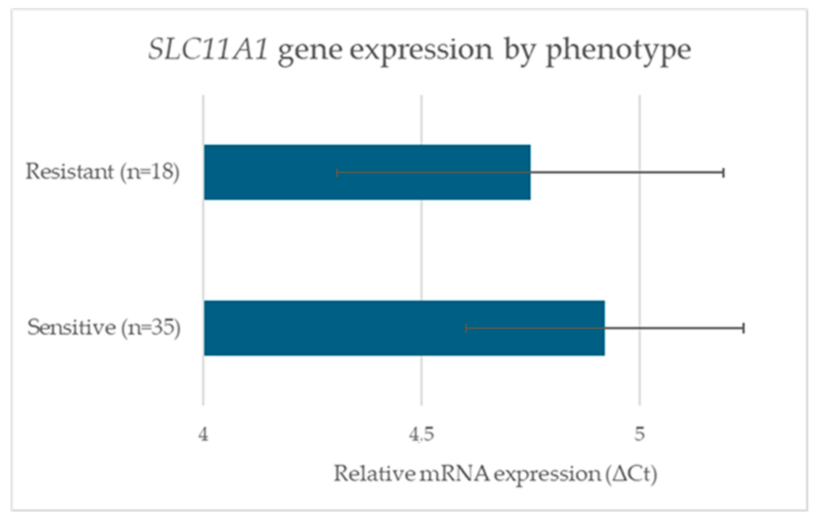
An independent-samples two-tailed t-test showed no significant difference between groups [mean ΔCt_S = 4.92 ± 0.84, mean ΔCt_R = 4.75 ± 0.91; t(51) = 0.63, p = 0.531; 95% CI for the difference: −0.39 to 0.73; Cohen’s d = 0.17] (Figure 2). A one-way ANOVA comparing expression across (GT)n genotype groups likewise found no significant effect [F(2,50) = 1.42, p = 0.25; η2 = 0.05]. Post hoc pairwise comparisons using Tukey’s HSD confirmed the absence of significant differences between genotypic categories.

4. Discussion

This study provides evidence that variation in the 3′UTR (GT)n microsatellite of the SLC11A1 gene is associated with resistance to natural MAP infection in sheep. Six allelic variants were identified within the 138 animals examined, with four alleles (GT)21, (GT)22, (GT)23, and (GT)24 being common in the study population. Among these, the (GT)21 and (GT)23 alleles appeared more frequently in sheep classified as resistant, whereas (GT)22 and (GT)24 were more frequent among sensitive animals. Although individual allele comparisons did not retain statistical significance after Bonferroni correction, the overall allelic distribution differed significantly between resistant and sensitive groups (p = 0.006), suggesting that the SLC11A1 3′UTR microsatellite may contribute to host resistance to MAP infection in sheep. The moderate effect size (Cramér’s V = 0.38) further supports the biological relevance of this association, despite the statistical limitations imposed by the sample size. These results are consistent with previous findings in goats and cattle, where variation in the SLC11A1 3′UTR microsatellite has been associated with altered resistance to bacterial infections, including paratuberculosis and brucellosis [21,23,24,25,43] (Table 2). The absence of significant differences in SLC11A1 mRNA expression between resistant and sensitive animals under basal conditions indicates that microsatellite variation does not strongly influence transcriptional levels in unstimulated PBMCs [22,23]. However, the possibility remains that such polymorphisms modulate expression upon immune stimulation, as demonstrated in goats and cattle following MAP challenge [22,23,49]. This dissociation between genotype and basal expression suggests a condition-dependent regulatory mechanism [21,22]. The (GT)n repeat may alter the secondary structure of the 3′UTR, affecting its interaction with microRNAs or RNA-binding proteins that regulate mRNA stability and translation specifically during immune responses [21,25]. Thus, constitutive SLC11A1 expression may not differ markedly between phenotypic groups, but the allelic context could still affect inducible expression during infection or cytokine stimulation, a hypothesis that merits further investigation using MAP-infected macrophages.
Importantly, this study provides one of the first characterizations of the SLC11A1 3′UTR microsatellite in sheep. Previous GWASs in this species have implicated regions on chromosomes 2, 3, and 20 containing immune-related genes, including SLC11A1 [33,34]. However, most GWASs rely on SNP-based analyses that may not capture functional variation in microsatellite loci [50]. The current findings therefore add complementary evidence by resolving a specific, functional polymorphism within a broader GWAS-implicated region, highlighting the potential importance of non-coding repetitive elements in host–pathogen interactions.
A limitation of the present work is the relatively small number of resistant animals (n = 18), which restricts statistical power and may increase the risk of Type II errors. Nevertheless, the study flock represents a closed population that has been maintained without external animal introductions since its establishment, under consistent management and monitored conditions. This closed design ensures uniform environmental exposure and infection pressure, minimizing confounding factors related to management or pathogen circulation [7,51,52]. Moreover, the flock has been monitored longitudinally for over a decade, enabling robust phenotypic classification based on repeated diagnostic testing. Both resistant and sensitive groups comprised exclusively Karagouniki sheep, as positive and negative diagnostic results were available only for this breed, precluding breed-stratified analyses. Future research including a broader range of breeds and larger sample sizes would enable assessment of potential breed-specific linkage disequilibrium patterns, as previously suggested by GWAS analyses [34,53].
Collectively, the present results provide evidence that variation in the SLC11A1 3′UTR microsatellite may contribute to differential resistance to paratuberculosis in sheep. Although individual allele associations did not remain significant after multiple testing correction, the observed trend aligns with prior functional studies in other ruminants, indicating a conserved regulatory mechanism influencing disease susceptibility. These findings position the SLC11A1 microsatellite as a candidate locus for marker-assisted selection. Expanding this research to larger, multi-breed cohorts and evaluating inducible SLC11A1 expression under infection conditions will be essential to establish the functional relevance of these microsatellite variants and assess their practical utility in breeding programs aimed at sustainable paratuberculosis control.

5. Conclusions

This study provides the first preliminary evidence that polymorphisms in the 3′UTR (GT)n microsatellite of the SLC11A1 gene are associated with resistance to natural MAP infection in sheep. A significant overall difference in allele distribution was observed between resistant and sensitive animals, with the (GT)21 and (GT)23 alleles emerging as potential resistance markers, while (GT)22 and (GT)24 were more frequent among sensitive individuals. Although individual allele associations did not remain significant after strict correction for multiple testing, the strength of the overall association and the moderate effect size indicate a biologically meaningful trend that merits further investigation. The absence of significant differences in basal SLC11A1 mRNA expression in PBMCs suggests that the microsatellite does not markedly influence constitutive transcription but may affect post-transcriptional regulation, potentially altering mRNA stability or translation efficiency upon immune activation.
While the relatively small number of resistant animals and the single-breed design limit broad generalization, this work establishes a foundation for further research into the regulatory role of SLC11A1 3′UTR variation in inducible immune responses. Future studies encompassing multiple breeds, larger cohorts, and integrated analyses of SNP and microsatellite variation together with gene expression under stimulation or infection will be essential to validate these associations and evaluate their potential utility in marker-assisted breeding strategies aimed at enhancing paratuberculosis resistance in sheep populations.

Author Contributions

Conceptualization, J.I.; methodology, A.M. and A.K.P.; software, A.M.; validation, A.M.; formal analysis, A.M. and A.K.P.; investigation, A.M.; resources, J.I.; data curation, A.M.; writing—original draft preparation, A.M., A.K.P. and J.I.; writing—review and editing, A.M. and J.I.; visualization, A.M.; supervision, J.I.; project administration, J.I. All authors have read and agreed to the published version of the manuscript.

Funding

This research received no external funding.

Institutional Review Board Statement

According to the Code of Research Ethics of the Agricultural University of Athens (available at: https://www.elke.aua.gr/wp-content/uploads/2019/01/kanonismos.pdf, accessed date 10 November 2025) and in compliance with Directive 2010/63/EU and its transposition into Greek legislation (Presidential Decree 56/2013, ΦΕΚ A’ 106/30.04.2013), the investigation reported in our manuscript does not fall under the scope of procedures requiring ethical review. All samples analyzed in the study were obtained within the framework of the routine veterinary health monitoring of the sheep flock maintained at the Agricultural University of Athens.

Informed Consent Statement

Not applicable.

Data Availability Statement

The original contributions presented in this study are included in the article. Further inquiries can be directed to the corresponding author(s).

Conflicts of Interest

The authors declare no conflicts of interest.

References

  1. Harris, N.B.; Barletta, R.G. Mycobacterium avium subsp. paratuberculosis in Veterinary Medicine. Clin. Microbiol. Rev. 2001, 14, 489–512. [Google Scholar] [CrossRef]
  2. Clarke, C.J. The pathology and pathogenesis of paratuberculosis in ruminants and other species. J. Comp. Pathol. 1997, 116, 217–261. [Google Scholar] [CrossRef] [PubMed]
  3. Bush, R.D.; Windsor, P.A.; Toribio, J.A.L.M.L. Losses of adult sheep due to ovine Johne’s disease in 12 infected flocks over a 3-year period. Aust. Vet. J. 2006, 84, 246–253. [Google Scholar] [CrossRef]
  4. Hasonova, L.; Pavlik, I. Economic impact of paratuberculosis in dairy cattle herds: A review. Vet. Med. 2006, 51, 193–211. [Google Scholar] [CrossRef]
  5. Raizman, E.A.; Fetrow, J.; Wells, S.J.; Godden, S.M.; Oakes, M.J.; Vazquez, G. The association between Mycobacterium avium subsp. paratuberculosis fecal shedding or clinical Johne’s disease and lactation performance on two Minnesota, USA dairy farms. Prev. Vet. Med. 2007, 78, 179–195. [Google Scholar] [CrossRef]
  6. Nielsen, S.S.; Toft, N. A review of prevalences of paratuberculosis in farmed animals in Europe. Prev. Vet. Med. 2009, 88, 1–14. [Google Scholar] [CrossRef]
  7. di Marco Lo Presti, V.; Ippolito, D.; Migliore, S.; Tolone, M.; Mignacca, S.A.; Marino, A.M.F.; Amato, B.; Calogero, R.; Vitale, M.; Vicari, D.; et al. Large-scale serological survey on Mycobacterium avium subsp. paratuberculosis infection in sheep and goat herds in Sicily, Southern Italy. Front. Vet. Sci. 2024, 11, 1334036. [Google Scholar] [CrossRef] [PubMed]
  8. Gautam, M.; Ridler, A.; Wilson, P.R.; Heuer, C. Control of clinical paratuberculosis in New Zealand pastoral livestock. N. Z. Vet. J. 2018, 66, 1–8. [Google Scholar] [CrossRef]
  9. Windsor, P.; Whittington, R. Ovine Paratuberculosis Control in Australia Revisited. Animals 2020, 10, 1623. [Google Scholar] [CrossRef] [PubMed]
  10. Koets, A.P.; Adugna, G.; Janss, L.L.G.; van Weering, H.J.; Kalis, C.H.J.; Wentink, G.H.; Rutten, V.P.M.G.; Schukken, Y.H. Genetic variation of susceptibility to Mycobacterium avium subsp. paratuberculosis infection in dairy cattle. J. Dairy Sci. 2000, 83, 2702–2708. [Google Scholar] [CrossRef]
  11. Reddacliff, L.A.; Beh, K.; McGregor, H.; Whittington, R.J. A preliminary study of possible genetic influences on the susceptibility of sheep to Johne’s disease. Aust. Vet. J. 2005, 83, 435–441. [Google Scholar] [CrossRef]
  12. Lugton, I.W. Cross-sectional study of risk factors for the clinical expression of ovine Johne’s disease on New South Wales farms. Aust. Vet. J. 2004, 82, 355–365. [Google Scholar] [CrossRef] [PubMed]
  13. Blackwell, J.M.; Searle, S. Genetic regulation of macrophage activation: Understanding the function of Nramp1 (=Ity/Lsh/Bcg). Immunol. Lett. 1999, 65, 73–80. [Google Scholar] [CrossRef] [PubMed]
  14. Buschman, E.; Vidal, S.; Skamene, E. Nonspecific resistance to Mycobacteria: The role of the Nramp1 gene. Behring Inst. Mitteilungen 1997, 99, 51–57. [Google Scholar]
  15. Vidal, S.; Gros, P.; Skamene, E. Natural resistance to infection with intracellular parasites: Molecular genetics identifies Nramp1 as the Bcg/Ity/Lsh locus. J. Leukoc. Biol. 1995, 58, 382–390. [Google Scholar] [CrossRef]
  16. Wyllie, S.; Seu, P.; Goss, J.A. The natural resistance-associated macrophage protein 1 Slc11a1 (formerly Nramp1) and iron metabolism in macrophages. Microbes Infect. 2002, 4, 351–359. [Google Scholar] [CrossRef] [PubMed]
  17. Thomas, N.; Joseph, S. Role of SLC11A1 gene in disease resistance. Biotechnol. Anim. Husb. 2012, 28, 99–106. [Google Scholar] [CrossRef]
  18. Awomoyi, A.A.; Marchant, A.; Howson, J.M.M.; McAdam, K.P.W.J.; Blackwell, J.M.; Newport, M.J. Interleukin-10, polymorphism in SLC11A1 (formerly NRAMP1), and susceptibility to tuberculosis. J. Infect. Dis. 2002, 186, 1808–1814. [Google Scholar] [CrossRef]
  19. Meilang, Q.; Zhang, Y.; Zhang, J.; Zhao, Y.; Tian, C.; Huang, J.; Fan, H. Polymorphisms in the SLC11A1 gene and tuberculosis risk: A meta-analysis update. Int. J. Tuberc. Lung Dis. 2012, 16, 437–446. [Google Scholar] [CrossRef]
  20. Ding, X.; Zhang, X.; Yang, Y.; Ding, Y.; Xue, W.; Meng, Y.; Zhu, W.; Yin, Z. Polymorphism, Expression of Natural Resistance-associated Macrophage Protein 1 Encoding Gene (NRAMP1) and Its Association with Immune Traits in Pigs. Asian-Australas. J. Anim. Sci. 2014, 27, 1189–1195. [Google Scholar] [CrossRef]
  21. Taka, S.; Gazouli, M.; Sotirakoglou, K.; Liandris, E.; Andreadou, M.; Triantaphyllopoulos, K.; Ikonomopoulos, J. Functional analysis of 3′UTR polymorphisms in the caprine SLC11A1 gene and its association with the Mycobacterium avium subsp. paratuberculosis infection. Vet. Immunol. Immunopathol. 2015, 167, 75–79. [Google Scholar] [CrossRef]
  22. Taka, S.; Liandris, E.; Gazouli, M.; Sotirakoglou, K.; Theodoropoulos, G.; Bountouri, M.; Andreadou, M.; Ikonomopoulos, J. In vitro expression of the SLC11A1 gene in goat monocyte-derived macrophages challenged with Mycobacterium avium subsp. paratuberculosis. Infect. Genet. Evol. 2013, 17, 8–15. [Google Scholar] [CrossRef]
  23. Korou, L.M.; Liandris, E.; Gazouli, M.; Ikonomopoulos, J. Investigation of the association of the SLC11A1 gene with resistance/sensitivity of goats (Capra hircus) to paratuberculosis. Vet. Microbiol. 2010, 144, 353–358. [Google Scholar] [CrossRef]
  24. Abraham, A.; Naicy, T.; Raghavan, K.C.; Siju, J.; Aravindakshan, T. Evaluation of the association of SLC11A1 gene polymorphism with incidence of paratuberculosis in goats. J. Genet. 2017, 96, 641–646. [Google Scholar] [CrossRef]
  25. Balasubramaniam, S.; Kumar, S.; Sharma, A.; Mitra, A. Microsatellite (GT)n polymorphism at 3′UTR of SLC11A1 influences the expression of brucella LPS induced MCP1 mRNA in buffalo peripheral blood mononuclear cells. Vet. Immunol. Immunopathol. 2013, 152, 295–302. [Google Scholar] [CrossRef] [PubMed]
  26. Ruiz-Larrañaga, O.; Garrido, J.M.; Manzano, C.; Iriondo, M.; Molina, E.; Gil, A.; Koets, A.P.; Rutten, V.P.M.G.; Juste, R.A.; Estonba, A. Identification of single nucleotide polymorphisms in the bovine solute carrier family 11 member 1 (SLC11A1) gene and their association with infection by Mycobacterium avium subspecies paratuberculosis. J. Dairy Sci. 2010, 93, 1713–1721. [Google Scholar] [CrossRef]
  27. Kravitz, A.; Liao, M.; Morota, G.; Tyler, R.; Cockrum, R.; Manohar, B.M.; Ronald, B.S.M.; Collins, M.T.; Sriranganathan, N. Retrospective Single Nucleotide Polymorphism Analysis of Host Resistance and Susceptibility to Ovine Johne’s Disease Using Restored FFPE DNA. Int. J. Mol. Sci. 2024, 25, 7748. [Google Scholar] [CrossRef]
  28. Bermingham, M.L.; Bishop, S.C.; Woolliams, J.A.; Pong-Wong, R.; Allen, A.R.; McBride, S.H.; Ryder, J.J.; Wright, D.M.; Skuce, R.A.; McDowell, S.W.; et al. Genome-wide association study identifies novel loci associated with resistance to bovine tuberculosis. Heredity 2014, 112, 543–551. [Google Scholar] [CrossRef]
  29. Gao, Y.; Jiang, J.; Yang, S.; Cao, J.; Han, B.; Wang, Y.; Zhang, Y.; Yu, Y.; Zhang, S.; Zhang, Q.; et al. Genome-wide association study of Mycobacterium avium subspecies Paratuberculosis infection in Chinese Holstein. BMC Genom. 2018, 19, 972. [Google Scholar] [CrossRef] [PubMed]
  30. Canive, M.; González-Recio, O.; Fernández, A.; Vázquez, P.; Badia-Bringué, G.; Lavín, J.L.; Garrido, J.M.; Juste, R.A.; Alonso-Hearn, M. Identification of loci associated with susceptibility to Mycobacterium avium subsp. paratuberculosis infection in Holstein cattle using combinations of diagnostic tests and imputed whole-genome sequence data. PLoS ONE 2021, 16, e0256091. [Google Scholar] [CrossRef] [PubMed]
  31. Canive, M.; Badia-Bringué, G.; Vázquez, P.; Garrido, J.M.; Juste, R.A.; Fernandez, A.; González-Recio, O.; Alonso-Hearn, M. A Genome-Wide Association Study for Tolerance to Paratuberculosis Identifies Candidate Genes Involved in DNA Packaging, DNA Damage Repair, Innate Immunity, and Pathogen Persistence. Front. Immunol. 2022, 13, 820965. [Google Scholar] [CrossRef]
  32. Alonso-Hearn, M.; Badia-Bringué, G.; Canive, M. Genome-wide association studies for the identification of cattle susceptible and resilient to paratuberculosis. Front. Vet. Sci. 2022, 9, 935133. [Google Scholar] [CrossRef] [PubMed]
  33. Usai, M.G.; Casu, S.; Sechi, T.; Salaris, S.L.; Miari, S.; Mulas, G.; Cancedda, M.G.; Ligios, C.; Carta, A. Advances in understanding the genetic architecture of antibody response to paratuberculosis in sheep by heritability estimate and LDLA mapping analyses and investigation of candidate regions using sequence-based data. Genet. Sel. Evol. 2024, 56, 5. [Google Scholar] [CrossRef]
  34. Moioli, B.; D’Andrea, S.; de Grossi, L.; Sezzi, E.; de Sanctis, B.; Catillo, G.; Steri, R.; Valentini, A.; Pilla, F. Genomic scan for identifying candidate genes for paratuberculosis resistance in sheep. Anim. Prod. Sci. 2015, 56, 1046–1055. [Google Scholar] [CrossRef]
  35. Purdie, A.C.; Plain, K.M.; Begg, D.J.; de Silva, K.; Whittington, R.J. Candidate gene and genome-wide association studies of Mycobacterium avium subsp. paratuberculosis infection in cattle and sheep: A review. Comp. Immunol. Microbiol. Infect. Dis. 2011, 34, 197–208. [Google Scholar] [CrossRef] [PubMed]
  36. Kravitz, A.; Pelzer, K.; Sriranganathan, N. The Paratuberculosis Paradigm Examined: A Review of Host Genetic Resistance and Innate Immune Fitness in Mycobacterium avium subsp. Paratuberculosis Infection. Front. Vet. Sci. 2021, 8, 721706. [Google Scholar] [CrossRef]
  37. Mataragka, A.; Sotirakoglou, K.; Gazouli, M.; Triantaphyllopoulos, K.A.; Ikonomopoulos, J. Parturition affects test-positivity in sheep with subclinical paratuberculosis; investigation following a preliminary analysis. J. King Saud Univ.-Sci. 2019, 31, 1399–1403. [Google Scholar] [CrossRef]
  38. National Research Council. Nutrient Requirements of Small Ruminants: Sheep, Goats, Cervids, and New World Camelids; The National Academies Press: Washington, DC, USA, 2007. [Google Scholar] [CrossRef]
  39. Kim, S.G.; Kim, E.H.; Lafferty, C.J.; Miller, L.J.; Koo, H.J.; Stehman, S.M.; Shin, S.J. Use of conventional and real-time polymerase chain reaction for confirmation of Mycobacterium avium subsp. paratuberculosis in a broth-based culture system ESP II. J. Vet. Diagn. Investig. 2004, 16, 448–453. [Google Scholar] [CrossRef]
  40. Dowling, R.J.O.; Bienzle, D. Gene-expression changes induced by Feline immunodeficiency virus infection differ in epithelial cells and lymphocytes. J. Gen. Virol. 2005, 86, 2239–2248. [Google Scholar] [CrossRef]
  41. Taylor, D.L.; Zhong, L.; Begg, D.J.; de Silva, K.; Whittington, R.J. Toll-like receptor genes are differentially expressed at the sites of infection during the progression of Johne’s disease in outbred sheep. Vet. Immunol. Immunopathol. 2008, 124, 132–151. [Google Scholar] [CrossRef]
  42. Livak, K.J.; Schmittgen, T.D. Analysis of Relative Gene Expression Data Using Real-Time Quantitative PCR and the 2−ΔΔCT Method. Methods 2001, 25, 402–408. [Google Scholar] [CrossRef]
  43. Pinedo, P.J.; Buergelt, C.D.; Donovan, G.A.; Melendez, P.; Morel, L.; Wu, R.; Langaee, T.Y.; Rae, D.O. Candidate gene polymorphisms (BoIFNG, TLR4, SLC11A1) as risk factors for paratuberculosis infection in cattle. Prev. Vet. Med. 2009, 91, 189–196. [Google Scholar] [CrossRef]
  44. Canive, M.; Casais, R.; Jimenez, J.A.; Blanco-Vazquez, C.; Amado, J.; Garrido, J.M.; Juste, R.A.; Alonso-Hearn, M. Correlations between single nucleotide polymorphisms in bovine CD209, SLC11A1, SP110 and TLR2 genes and estimated breeding values for several traits in Spanish Holstein cattle. Heliyon 2020, 6, e04254. [Google Scholar] [CrossRef]
  45. Okuni, J.B.; Afayoa, M.; Ojok, L. Survey of Candidate Single-Nucleotide Polymorphisms in SLC11A1, TLR4, NOD2, PGLYRP1, and IFNγ in Ankole Longhorn Cattle in Central Region of Uganda to Determine Their Role in Mycobacterium avium Subspecies paratuberculosis Infection Outcome. Front. Vet. Sci. 2021, 8, 614518. [Google Scholar] [CrossRef]
  46. Gopi, B.; Vir Singh, R.; Kumar, S.; Kumar, S.; Chauhan, A.; Sonwane, A.; Kumar, A.; Bharati, J.; Vir Singh, S. Effect of selected single nucleotide polymorphisms in SLC11A1, ANKRA2, IFNG and PGLYRP1 genes on host susceptibility to Mycobacterium avium subspecies paratuberculosis infection in Indian cattle. Vet. Res. Commun. 2022, 46, 209–221. [Google Scholar] [CrossRef]
  47. Wu, H.; Cheng, D.; Wang, L. Association of polymorphisms of Nramp1 gene with immune function and production performance of large white pig. J. Genet. Genom. 2008, 35, 91–95. [Google Scholar] [CrossRef] [PubMed]
  48. Sechi, L.A.; Dow, C.T. Mycobacterium avium ss. paratuberculosis Zoonosis—The Hundred Year War—Beyond Crohn’s Disease. Front. Immunol. 2015, 6, 96. [Google Scholar] [CrossRef] [PubMed]
  49. Dukkipati, V.S.R.; Blair, H.T.; Garrick, D.J.; Lopez-Villalobos, N.; Whittington, R.J.; Reddacliff, L.A.; Eppleston, J.; Windsor, P.; Murray, A. Association of microsatellite polymorphisms with immune responses to a killed Mycobacterium avium subsp. Paratuberculosis Vaccine Merino sheep. N. Z. Vet. J. 2010, 58, 237–245. [Google Scholar] [CrossRef] [PubMed]
  50. Tam, V.; Patel, N.; Turcotte, M.; Bossé, Y.; Paré, G.; Meyre, D. Benefits and limitations of genome-wide association studies. Nat. Rev. Genet. 2019, 20, 467–484. [Google Scholar] [CrossRef]
  51. Whittington, R.J.; Begg, D.J.; de Silva, K.; Plain, K.M.; Purdie, A.C. Comparative immunological and microbiological aspects of paratuberculosis as a model mycobacterial infection. Vet. Immunol. Immunopathol. 2012, 148, 29–47. [Google Scholar] [CrossRef]
  52. Begg, D.J.; Whittington, R.J. Experimental animal infection models for Johne’s disease, an infectious enteropathy caused by Mycobacterium avium subsp. paratuberculosis. Vet. J. 2008, 176, 129–145. [Google Scholar] [CrossRef] [PubMed]
  53. Ruiz-Larrañaga, O.; Langa, J.; Rendo, F.; Manzano, C.; Iriondo, M.; Estonba, A. Genomic selection signatures in sheep from the Western Pyrenees. Genet. Sel. Evol. 2018, 50, 9. [Google Scholar] [CrossRef] [PubMed]
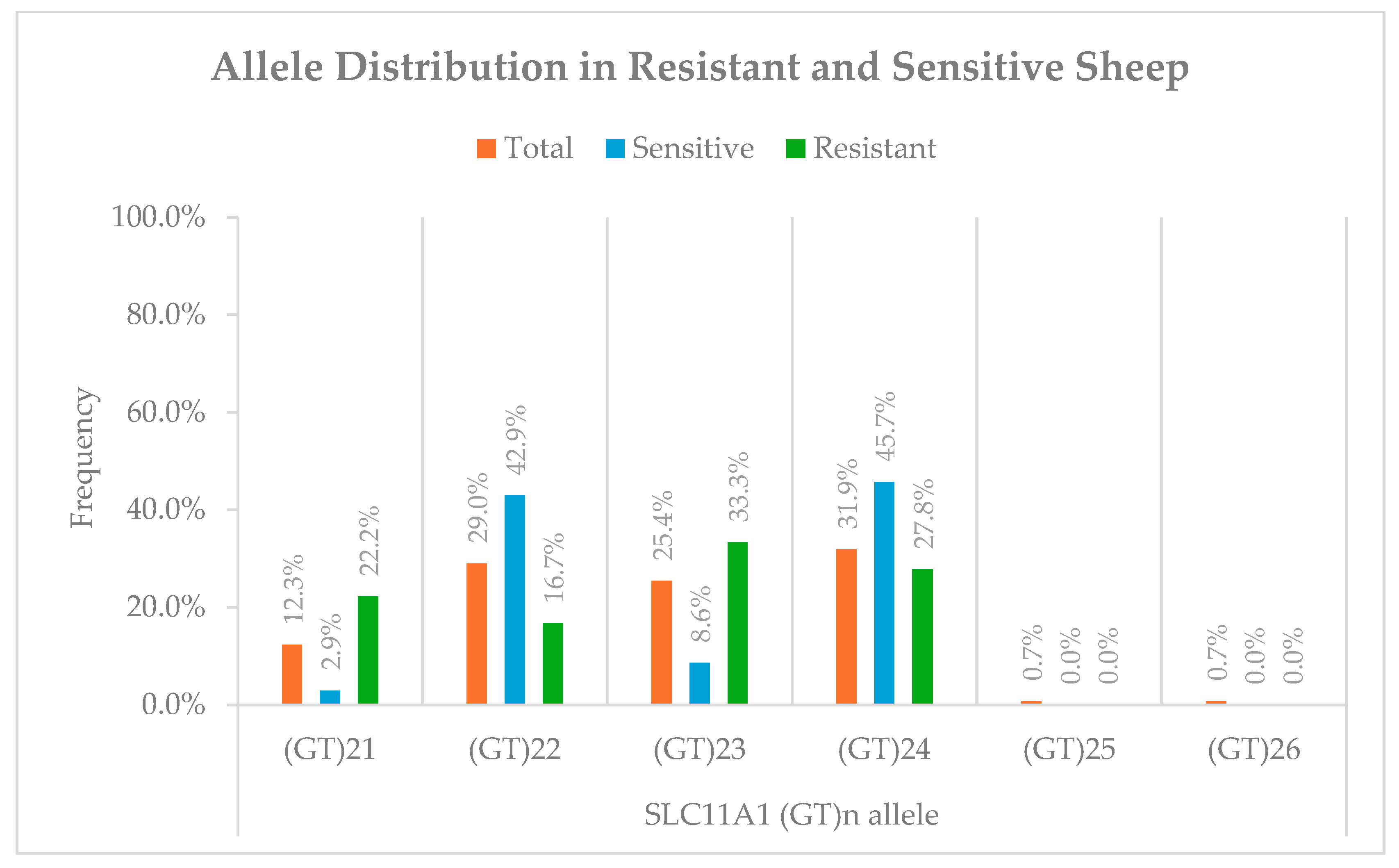
Figure 1. Distribution of ovine SLC11A1 (GT)n alleles in the study population and in MAP-resistant (R) and MAP-sensitive (S) phenotypic groups.
Pathogens 14 01150 g001
Figure 2. Comparison of SLC11A1 gene expression levels between MAP-resistant and MAP-sensitive sheep. Relative mRNA expression was quantified by RT-qPCR and reported as ΔCt values (normalized to GAPDH). Bars show the mean ΔCt for each group (Sensitive, n = 35; Resistant, n = 18). Error bars indicate the 95% confidence interval of the mean (Resistant: 4.29–5.21; Sensitive: 4.60–5.24). An independent samples t-test found no statistically significant difference between groups (t(51) = 0.63, p = 0.531, Cohen’s d = 0.17).
Pathogens 14 01150 g002
Table 1. The primer sequences, product sizes, cycling conditions and relevant references for the assays targeting IS900, SLC11A1 3′UTR, GAPDH, SLC11A1 mRNA, and β-actin used in this study.
TargetPrimers (5′-3′)SizeThermal ProfileReference
IS900F 1: AATGACGGTTACGGAGGTGGT
R 2: GCAGTAATGGTCGGCCTTACC
Pr 3: TCCACGCCCGCCCAGACAGG
76 bp95 °C for 3 min; 40 cycles of 95 °C for 3 s, 60 °C for 20 s, 72 °C for 1 s; 43 °C for 30 s[39]
3′UTR SLC11A1F: ACCTGGTCTGGACCTGTCTCATCA
R: CATTGCAAGGTAGGTGTCCCCAT
346 bp95 °C for 3 min; 35 cycles of 95 °C for 30 s, 59 °C for 30 s, 72 °C for 20 s; 72 °C for 3 min[23]
GAPDHF: TTCCAGTATGATTCCACCCATG
R: GCCTTTCCATTGATGACGAG
80 bp42 °C for 5 min; 95 °C for 15 s; 40 cycles of 95 °C for 5 s, 52 °C for 20 s, 72 °C for 1 s; 40 °C for 30 s[41]
SLC11A1 mRNAF: GGCTGTGGCTGGATTCAAAC
R: ATGGTCAGCCAGAGGAGAATG
168 bp42 °C for 5 min; 95 °C for 15 s; 40 cycles of 95 °C for 5 s, 57 °C for 20 s, 72 °C for 1 s; 40 °C for 30 s[23]
β-actinF: TGTCTCTGTACGCTTCTGG
R: GTGGTGGTGAAACTGTAGC
190 bp95 °C for 3 min; 40 cycles of 95 °C for 30 s, 55 °C for 30 s, 72 °C for 30 s; 72 °C for 3 min[40]
1 Forward; 2 Reverse; 3 Probe.
Table 2. Comparative summary of reported associations between SLC11A1 polymorphisms and disease-related phenotypes in livestock and humans.
SpeciesVariant/Region AnalyzedAssociation with Resistance/SusceptibilityNotesReferences
SheepGenetic influences (preliminary, candidate-based)Suggested possible genetic effect on Johne’s disease susceptibilityEarly evidence, not locus-specific[11]
SheepRetrospective SNP analysisIdentified associations near SLC11A1 with MAP resistanceBased on FFPE DNA, SNP focus[27]
SheepGWAS (antibody response to MAP)Regions linked to immune response; SLC11A1 implicatedHigh-resolution genomic mapping[33]
SheepGWAS (SNPs across genome)Regions associated with MAP resistance; included SLC11A1SNP-based, no microsatellite resolution[34]
Sheep3′UTR (GT)n microsatellite(GT)21 and (GT)23 associated with resistance; (GT)22 and (GT)24 with susceptibilityAssociation found despite no difference in basal expression[This study]
GoatsFunctional analysis, 3′UTR microsatelliteVariants affected inducible expression under MAP challengeDemonstrated functional mechanism[21]
Goats3′UTR (GT)n microsatelliteShorter alleles enriched in resistant goatsConsistent with ovine findings[23]
Goats3′UTR microsatelliteSpecific alleles associated with reduced paratuberculosis incidenceValidated earlier results[24]
CattleSNPs in SLC11A1Associated with MAP infection riskConsistent across populations[26]
CattleCandidate gene SNPs (SLC11A1, TLR4, IFNG)Associations with MAP susceptibilityPopulation-specific variation[43]
CattleSNPs in SLC11A1 and othersLinked with breeding values for MAP traitsLarge-scale genomic approach[44]
CattleSNPs in SLC11A1No association with MAP infectionSNPs polymorphic variants showed no allele/genotype differences between cattle[45]
CattleSLC11A1 SNP rs109453173Associated with resistance (GG genotype/G allele protective; CC/CG linked to susceptibility)Case–control study; suggests potential resistance marker[46]
Buffalo3′UTR microsatelliteAllelic variation influenced MCP1 mRNA after Brucella challengeFunctional immune effects[25]
PigsSLC11A1 polymorphismsAssociated with immune traitsCross-species evidence of functional role[47]
HumansSLC11A1 SNPs and promoter variantsAssociated with tuberculosis susceptibilityStrong parallels with livestock[18,48]
Disclaimer/Publisher’s Note: The statements, opinions and data contained in all publications are solely those of the individual author(s) and contributor(s) and not of MDPI and/or the editor(s). MDPI and/or the editor(s) disclaim responsibility for any injury to people or property resulting from any ideas, methods, instructions or products referred to in the content.