Article

Food Safety Concerns: Anisakis spp. in Ready-to-Eat Fish from the Greek Market

by
Evangelia N. Papapostolou
1,
Serafeim C. Chaintoutis
2,
Panagiota Gousia
1,
Aggeliki Karpouza
1,
Melania Kachrimanidou
3,* and
Anastasia Diakou
4,*
1
Department of Food Testing and Research Laboratories of Thessaloniki, Hellenic Food Authority (EFET), 57001 Thessaloniki, Greece
2
Diagnostic Laboratory, School of Veterinary Medicine, Faculty of Health Sciences, Aristotle University of Thessaloniki, 54627 Thessaloniki, Greece
3
Department of Microbiology, Medical School, Aristotle University of Thessaloniki, 54124 Thessaloniki, Greece
4
Laboratory of Parasitology and Parasitic Diseases, School of Veterinary Medicine, Faculty of Health Sciences, Aristotle University of Thessaloniki, 54124 Thessaloniki, Greece
*
Authors to whom correspondence should be addressed.
Pathogens 2025, 14(10), 981; https://doi.org/10.3390/pathogens14100981
Submission received: 28 August 2025 / Revised: 23 September 2025 / Accepted: 25 September 2025 / Published: 27 September 2025
(This article belongs to the Section Parasitic Pathogens)

Abstract

Nematode parasites of the family Anisakidae can cause human disease when ingested, at the third larval stage (L3), through infected fish or fish products. While human infection is often asymptomatic, in some cases it may present with symptoms ranging from mild to severe gastrointestinal disorders, skin rash, itching, or even anaphylaxis. Ingestion of destroyed L3 (e.g., following processing, sanitisation, or cooking) may still trigger allergic reactions. In the present study, the occurrence of Anisakidae L3 in ready-to-eat (RTE) fish products was investigated. A total of 108 RTE fish products, representing eight species and five processing methods, were examined using (a) macroscopic inspection, (b) artificial digestion (AD), and (c) molecular analysis of both the AD material (to detect non-visible parasite fragments) and the larvae collected by the first two methods. Nematodes morphologically identified as Anisakidae L3 were detected in 32 of the 108 samples (29.62%). Molecular analysis of the AD product did not reveal additional positive samples and confirmed the identification of the isolated larvae as Anisakis simplex sensu stricto and Anisakis pegreffii. Given the public health importance of anisakidosis, the findings of this study provide valuable insight into the prevalence of contamination in RTE fish products currently available on the Greek market.

1. Introduction

The nematodes of the family Anisakidae are among the primary zoonotic parasites that infect marine species, causing human anisakidosis. The genera of clinical interest include Anisakis (Anisakis simplex sensu stricto (s.s.) and Anisakis pegreffii), Pseudoterranova (Pseudoterranova azarazi, Pseudoterranova cattani, Pseudoterranova decipiens, and Pseudoterranova krabbei), and, more rarely, the genus Contracaecum (Contracaecum osculatum) [1]. Dolphins and whales, in the case of Anisakis, pinnipeds, in the case of Pseudoterranova, and fish-eating birds like pelicans and penguins, in the case of Contracaecum are the definitive hosts of these parasites. Small crustaceans serve as intermediate hosts, while cephalopods and fish act as paratenic hosts, carrying the infective third-stage larvae (L3). Humans can be become infected as paratenic and dead-end hosts (i.e., they do not contribute to the completion of the parasite’s life cycle) when consuming raw, undercooked, or inadequately frozen infected fish [1,2,3].
The global distribution and abundance of Anisakidae are attributed to their adaptability to a wide range of hosts and environmental conditions. In wild-caught fish, infection is common, although prevalence varies depending on the fish species and fishing area. Numerous studies have examined the occurrence of these parasites in commercially consumed fish [4,5,6]. In Greece, similar research has revealed a significant presence of Anisakidae in the Aegean Sea [7,8].
Humans are at high risk of infection when consuming raw, undercooked, or inadequately processed fishery products containing the L3 [9]. Anisakidosis may present with different clinical syndromes, depending on larval migration within the body (i.e., gastric, intestinal, or extra-gastrointestinal anisakidosis), as well as the potential onset of an allergic reaction (allergic anisakidosis) [10,11]. Clinical signs appear within a few hours after infection, including mild to severe gastrointestinal disorders and symptoms similar to those seen in gastric ulcers [2]. In certain cases, the larvae penetrate the intestinal wall, leading to extra-gastrointestinal inflammatory lesions that may cause severe abdominal pain resembling acute appendicitis and requiring surgical intervention. They may also induce granulomatous nodules in the peritoneum, which can be misdiagnosed as neoplastic lesions [10,12,13]. Allergic reactions, such as rashes, dermatitis, urticaria, rhinoconjunctivitis, and asthma, may occur after consuming either live or dead larvae [11,14,15,16]. In addition to consumers, fishermen and fishmongers who come into contact with infected fish and cephalopods are at risk of sensitisation and allergic reactions from ingesting or handling infected fish and cephalopods, or even asthma from inhaling antigens from these parasites [17]. Moreover, allergic reactions may occur after consuming meat of farmed poultry that has been fed contaminated seafood [18].
Anisakidosis is a serious emerging food-borne disease that is now spreading to all continents due to changing eating habits, international cuisine, population movements, and international trade, posing a risk to public health and economic challenges in fisheries and food safety [19]. Between 2001 and 2023, Anisakis spp. accounted for 73.6% of all parasitic infection notifications in the European Union’s Rapid Alert System for Food and Feed (RASFF) database, underscoring real-world implications of the problem [20]. The first report of anisakiasis due to Anisakis sp. in Greece, caused by the consumption of raw fish, serves as a critical example [21]. Furthermore, epidemiological data suggest that anisakidosis may be an underdiagnosed disease [2,12].
Food business operators (FBOs) producing fishery products must comply with legislation requirements to ensure food safety. In the European Union, Regulation (EC) 853/2004 supplemented by Regulation (EC) 2074/2005 and amended by Regulation (EC) 1276/2011 establishes health standards for producing safe fishery products including mandatory treatment to inactivate viable parasites and the visual inspection of catches to prevent contaminated fish from entering the market. This Regulation includes specific requirements such as the mandatory treatment of fish to eliminate viable parasites that pose health risks to consumers. Effective methods include freezing or heating at specific temperatures and time periods [22,23,24].
Despite these preventive measures, international literature indicates that Anisakidae L3 have been detected in processed foods widely distributed on the international market. Live larvae have been found in ready-to-eat (RTE) mackerel [25], marinated anchovies lacking further heat treatment [26], and smoked herring [27]. The efficacy of marination, smoking, and salting in inactivating Anisakidae larvae is influenced by parameters such as brine salinity, pH, and exposure duration [28,29,30,31]. Nevertheless, multiple studies have demonstrated that common marination and cold-smoking protocols are insufficient to ensure complete larval inactivation. As a result, regulatory authorities mandate preventive freezing for fishery products intended for raw or lightly processed consumption, as in the case of RTE products [32]. This study aimed to investigate the prevalence, species composition, and intensity of Anisakidae nematodes infections in RTE fish products available on the Greek market. Given that the risk of infection in farmed fish is considered negligible [33], products derived from such fish were excluded from the analysis. The findings contribute to mapping the contamination of RTE fish products in Greece and underscore a potential foodborne source of morbidity.

2. Materials and Methods

2.1. Sampling

A total of 108 samples of RTE fish products, labelled as originating from wild-caught fish, were randomly purchased between September and December 2024 from six supermarket chains in the city of Thessaloniki, all of which have branches across Greece. The production facilities were primarily located in central and northern Greece and had received official approval from the relevant authorities.
The sampled products included smoked, salted, and marinated fish of two types: one consisting of whole fish and the other consisting of fish fillets, preserved in oil or water. The fishery products included herring (Clupea harengus), mackerel (Scomber scombrus), Atlantic chub mackerel (Scomber colias), anchovy (Engraulis encrasicolus/Engraulis ringens), sardine (Sardina pilchardus), pollock (Pollachius virens), albacore (Thunnus alalunga), and Skipjack tuna (Katsuwonus pelamis). The products’ classification and the location of catches are provided in Table 1.
After collecting, the products were transported to the laboratory. Labelling information was recorded, and the samples were stored at a controlled temperature of 4 °C. Testing was initiated within a 48 h timeframe. Epidemiological indices were applied according to Bush et al. 1997 [34].

2.2. Artificial Digestion and Macroscopic Inspection

The methodology for artificial digestion (AD) adhered to the guidelines established by the European Union Reference Laboratory for Parasites (EURLP) [35], which is grounded in ISO 23036-2021 [36]. The method was applied as follows.
For sample preparation, fillets were drained of the preserving medium (oil or water). Salted fish were rinsed under tap water for 10 s to remove surface salt. The skin was then removed, the fish eviscerated, and the muscle tissue separated. Throughout the process, macroscopic inspection of both the discharged liquid (oil, brine or rinse water) and the fish was carried out to detect and collect parasites when present.
For each test, samples of either 50 or 100 g were used per product, depending on the product type. For larger fish species, such as herring and mackerel, 100 g of muscle tissue was sampled from the abdominal region, where larvae are more likely to be present according to ISO 23036-2021.
Visceral organs of non-eviscerated smoked herring were removed and examined separately. For smaller fish, the tests included 1 to 7 fish from the same batch, ensuring that the total size of each sample reached 100 g. The fillets were tested individually by package, in 50 g portions.
The following ingredient ratios were used to prepare the digestion liquid: In 2 L of tap water 10.8 mL of hydrochloric acid (HCl, 37%) was added. Ten grams of pepsin powder (1:10.000 NF; Opopharma, Handels GmbH, Hamburg, Germany) were incorporated into the mixture. The prepared sample was placed in a 2 L glass beaker containing the digestion liquid. A sample-to-digestive fluid ratio of 1:20 was maintained during the AD process. The beaker was covered with aluminium foil to maintain a stable temperature of 40–42 °C and to minimize evaporation. The temperature of the fluid was monitored regularly with a thermometer, and the stirring was adjusted to produce a deep vortex without splashing. The sample was digested for 30 min.
At the end of the procedure, the AD product was passed through a sieve, and the detected parasites were collected, counted and stored in saline solution at −20 °C for further examination.

2.3. Morphological Identification

The collected larvae were examined without further processing under the microscope (×100, ×400 magnification) for morphological identification at the family level [37,38,39].

2.4. Molecular Examination

Molecular analysis was performed on two types of material: (i) the sediments obtained from the AD (approximately 2 mL) to detect larvae potentially destroyed by production procedures or the AD process, and (ii) one larva per positive sample. The larvae were dissected, and DNA extraction was performed from their midsection. A previously described methodology for DNA extraction [40] was followed with minor modifications [41]. This process involved mixing and homogenising the sediment or larvae in DNA lysis buffer, followed by the application of a phenol/chloroform-based procedure, and subsequent binding to silica columns.
Molecular detection and identification involved PCR-based amplification of a 628 bp fragment of the mitochondrial cytochrome oxidase II (cox2) subunit gene, using the previously described primers 211F and 210R [42]. The QIAgen HotStarTaq DNA Polymerase was used in reaction mixtures, and amplification was performed in a SimpliAmp Thermal Cycler (Thermo Fisher Scientific, Waltham, MA, USA). A total of 10 μL from each PCR product were analysed by electrophoresis on a 1.5% agarose gel (110 V for 100 min), followed by staining with GelRed Nucleic Acid Gel Stain (Biotium Inc., Fremont, CA, USA) and visualisation under UV, to verify amplicon lengths. The PCR products were subsequently purified using the NucleoSpin Gel and PCR Clean-up kit (Macherey-Nagel, Düren, Germany). Sanger sequencing was performed bidirectionally, utilising the amplification primers. Forward and reverse sequencing reads were assembled using MEGA 11 software [43], and primer sequences were trimmed. Assembled sequences were analysed via BLAST for identification.

3. Results

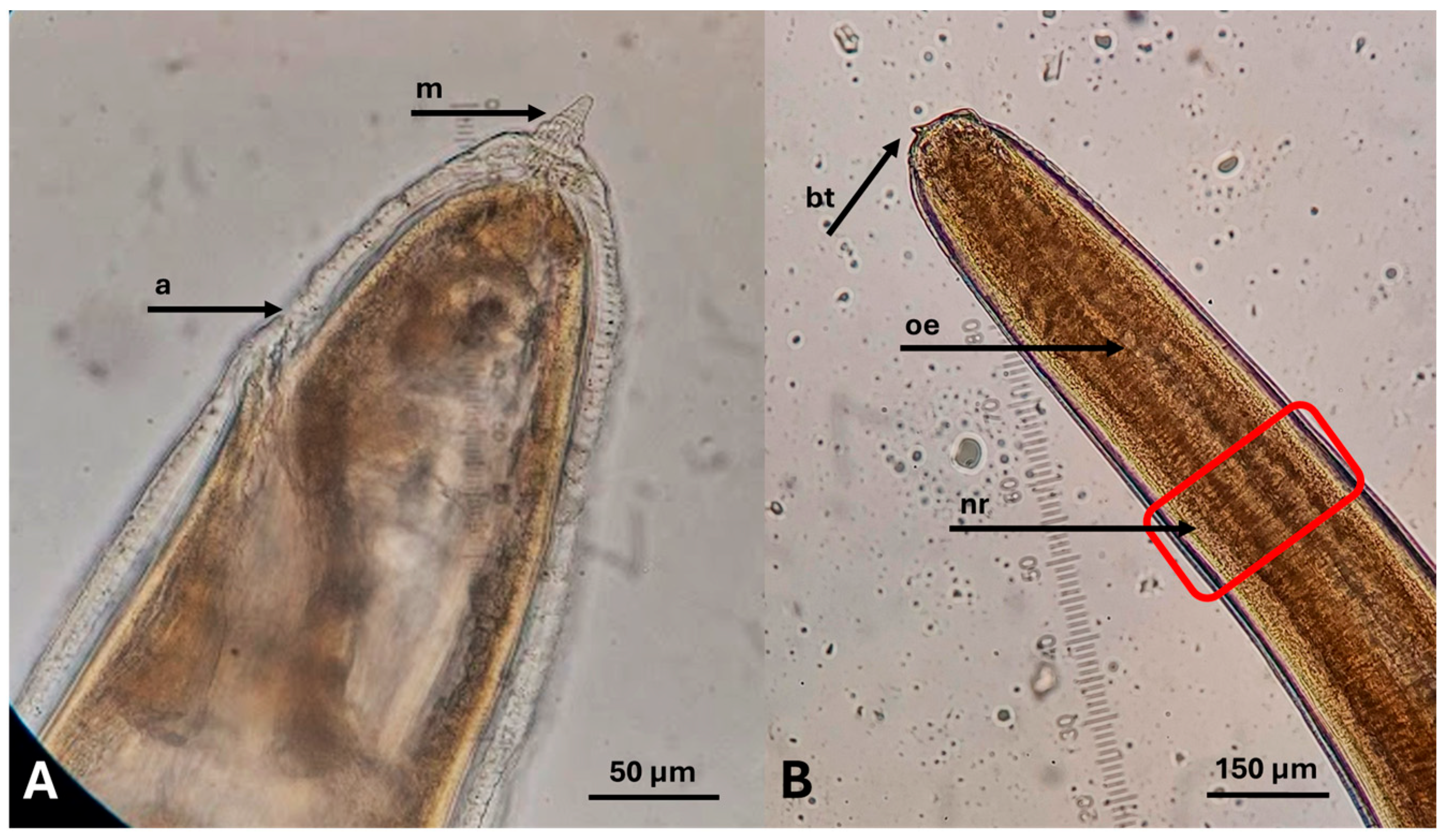
White to pale yellow coiled nematodes were detected in 32 of the 108 (29.62%) examined samples of RTE fish products (Figure 1). In all positive samples, parasites were detected only after AD, whereas none were observed in the fish or the discharged liquid during visual inspection during the preparation process. None of the parasites detected showed any sign of viability during observation and manipulation. Based on their morphological and morphometric characteristics (Figure 2), all isolated larvae were identified as belonging to the family Anisakidae.
The highest prevalence of infection was found in smoked whole herring (91.6%), followed by salted whole Atlantic chub mackerel (47.0%), smoked whole mackerel (46.6%), smoked mackerel fillet in oil (18.2%), marinated anchovy fillet in oil (12.5%), smoked herring fillet in oil (11.1%), and salted whole anchovies (10.0%). No larvae were found in salted whole sardines in oil, salted pollock fillet in oil, smoked albacore fillet in oil, or smoked Skipjack tuna fillet in water (Table 2).
The highest mean intensity of infection (number of larvae per sample) was found in salted whole Atlantic chub mackerel (7.5), followed by smoked whole herring (5.63), smoked whole mackerel (2.42), smoked mackerel fillet in oil (1.5), marinated anchovies fillet in oil (1.5), smoked herring fillet in oil, (1) and salted whole anchovies (1). No mobility of the larvae was observed during macroscopic examination after AD.
The molecular analysis of partial cox2 gene sequences obtained from the larvae confirmed the morphological identification and assigned all processed larvae as members of the Anisakis genus, showing high identity (≥99%) with Anisakis spp. reference sequences. The sequences from larvae recovered from Atlantic chub mackerel and anchovies displayed the highest nucleotide sequence identity (99.6–100%) to A. pegreffii reference sequences (e.g., acc. MF960821.1, KY418058). In contrast, the sequences from larvae recovered from herring and mackerel showed the highest nucleotide sequence identity (99.3–100%) to A. simplex s.s. reference sequences (e.g., acc. PQ126420.1, KT852492.1) (Table 3).

4. Discussion

This is the first survey to investigate the presence of Anisakidae in RTE fish products from the Greek market. The prevalence of infection ranged from 10.0% to 91.6%, depending on the product, demonstrating that these parasites represent a common challenge in food production. The significance of Anisakidae parasites mainly lies in their contribution to foodborne disease. Although these parasites have been recognised in fish since the 13th century, they were first documented in humans in Greenland in 1876 [44]. The first notable case of symptoms associated with the parasite was reported in 1950, when Dr. Straub in the Netherlands isolated a nematode from an eosinophilic granuloma in the small intestine of a patient suffering from acute abdominal pain [44].
Currently, thousands of cases of anisakidosis are reported worldwide each year, with Japan documenting over 19,000 cases annually and Spain reporting more than 7000 [45]. The first case in Greece was reported in 2024 in a young male with a history of regularly consuming raw fish [21]. It involved an invasive Anisakis sp. infection mimicking peritoneal malignancy, which led to hemicolectomy and omentectomy. The diagnosis of anisakiasis (infection by parasites of the genus Anisakis) was subsequently confirmed through histopathology and molecular identification [21].
The broad range of definitive, intermediate, and paratenic hosts of Anisakidae parasites, together with their adaptability to diverse environmental conditions, underlies their worldwide distribution. Fishing regions, including the Mediterranean, Japan, and the Atlantic Ocean, frequently report the occurrence of these parasites [1]. Commercially caught fish often contain Anisakidae larvae, which vary in prevalence depending on the fish species and catch area. Near the Canary Islands, five different species of Anisakis, including the zoonotic A. simplex (s.s.) and A. pegreffii, were found in frigate tunas (Auxis thazard), European hake (Merluccius merluccius), Atlantic chub mackerel and mackerel [4]. The infectious stage (L3) of A. pegreffii, Anisakis typica, and Anisakis ziphidarum was isolated from scorpionfish (Scorpaena scrofa) caught in the Aegean Sea [46] and from anchovies in the Western Mediterranean and Adriatic Sea [5,26,47]. The Atlantic herring is one of the marine fish species with a high prevalence and intensity of A. simplex infection, which has led to the parasite’s common name, the “herring worm” [6]. In the present study, molecular identification based on partial cox2 gene sequencing indicated that all isolated larvae belonged to the Anisakis genus, showing the highest sequence identity with reference sequences of either A. simplex (s.s.) or A. pegreffii. Recent research has also reported the occurrence of hybrid genotypes between A. simplex (s.s.) and A. pegreffii [48,49]. A limitation of the present study was that only a single genetic locus was analysed. The application of multilocus approaches is recommended for more robust species-level discrimination and for the detection of potential hybrid genotypes, similar to what has already been proposed [50].
Studies conducted in fresh fish in Greece have demonstrated significant parasite circulation in the Aegean Sea. Infected species include the European cod (Merlangius merlangus), Atlantic chub mackerel, and mackerel [7]. Research in the marine area around Lesvos Island reported an exceptionally high prevalence of infection (98.8%) in grey horse mackerel (Trachurus trachurus) [8].
Post-mortem migration of larvae into the edible flesh of fish represents a significant concern for food safety. Studies on A. pegreffii confirm significant post-mortem migration into fish musculature after 5 and 3 days at storage temperatures of 0 and 4 °C, respectively, triggered by biochemical factors such as pH and accumulation of biogenic amines [51]. Migration of A. pegreffii larvae from the viscera to the fillets was positively associated with both storage temperature (5 °C and 7 °C) and storage time (24, 48, and 72 h), with significantly higher worm burdens in the fillets and a simultaneous decrease in larval counts in the viscera as a result [5].
Conversely, no migration of A. simplex s.s. was observed in herring and mackerel stored < 2 °C, even after 48 h. However, this was not the case in blue whiting (Micromesistius poutassou), where at the same conditions, significant migration of A. simplex s.s. took place, showing that host species likely plays a crucial role in this phenomenon [52,53]. Overall, although maintaining storage temperatures below 2 °C prevents migration to an extend [52], early evisceration is strongly advocated in scientific literature [54] as a best practice to reduce migration risk, though it is not a legally enforceable requirement.
The influence of viscera on the occurrence and intensity of infection, depending also on the parasite species is evident in the present results; samples containing viscera were more frequently infected and exhibited higher infection intensities with A. simplex s.s. (Table 2 and Table 3). Specifically, smoked whole herring showed an infection prevalence of 91.6% and a mean intensity of 5.63, whereas smoked herring fillets showed markedly lower values of 11.1% and 1, respectively. A comparable pattern was observed for smoked whole mackerel and smoked mackerel fillets. In contrast, anchovies infected with A. pegreffii displayed no substantial differences in infection prevalence or intensity regardless of the presence of viscera. This observation may indicate that A. pegreffii has a greater propensity to migrate from the viscera to the muscles than A. simplex s.s., although further evidence is needed to confirm this.
Migration of the larvae from the viscera to the muscles may not always be the case in infected fillets; It has been shown in different species of salmon from Alaska returning from the open sea that A. simplex s.s. larvae are more abundant in the musculature than in the viscera immediately after catch, while interestingly, no migration of the larvae is observed from the viscera into the flesh during storage on ice or in refrigerated seawater for 24 h [53]. Intra vitam migration is a natural phenomenon that has been documented for both A. pegrefii and A. simplex, of which intensity depends on both host and parasite species and biology [5,52].
Studies examining the presence of Anisakidae in RTE fish products remain limited. In a survey conducted in Turkey by AD on 205 samples of various local RTE products from different fish species, no larvae were detected [55]. The examination by AD of 107 samples of RTE salted or marinated anchovies from Italian supermarkets revealed a high infection prevalence of 54.2% [56]. The authors emphasise that, although the risk of anisakidosis from dead parasites is negligible, the risk of allergic reactions remains significant. Furthermore, the presence of parasites in these products may pose a major market problem, as products with visible infection are rejected by consumers [56]. Notably, in the present study, no parasites were detected by naked eye before AD, despite the heavy infection in some cases, indicating that infection can remain unnoticed by the consumer. In a relatively recent survey, RTE anchovy (n = 45) and sardine (n = 45) fillets were collected from different markets in Italy and Spain [57]. In 17 anchovy and 13 sardine products, larvae were detected and identified as A. pegreffii (30%) and A. simplex (70%). The fillets were salted, in-oil preserved, and all the larvae were non-viable [57].
Interestingly, a study of RTE mackerel in Japan detected a notably high proportion of live larvae using tissue compression and parasite extraction [25]. These products consisted of fresh fish fillets with bones and viscera removed but had not undergone any processing, such as freezing, smoking, salting, or marinating. The lack of processing likely explains the high prevalence of live parasites in these RTE products. It has been demonstrated that AD may underrate the viability of L3 in frozen fish products [58]. The absence of live L3 in RTE products after AD should be interpreted with caution, as the procedure may deactivate the parasites [58]. In the present study, the larvae detected showed no signs of viability, which could be attributed either to the effect of processing methods (smoking, salting, or marinating) which may provide a safer alternative than freezing [32], particularly when freezing does not comply with established regulations [22], or to the effect of the AD per se.
Fishery products companies must comply with the legislation’s requirements for safe food production. Regulation (EC) 853/2004 supplemented by Regulation (EC) 2074/2005 [23] and amended by Regulation (EC)1276/2011 [24] enforce the treatment to kill viable parasites in fishery products for human consumption and mandates the visual inspection of catches to remove contaminated products from the market establishing health standards for producing safe fishery products [19]. It dictates the processing of catches to eliminate viable parasites that pose health risks to consumers. Specifically, catches must be frozen at −20 °C for at least 24 h or at −35 °C for at least 15 h, or the centre of mass must be heated above 60 °C for at least 1 min. Salting has been proposed as an alternative method of inactivating Anisakidae larvae [32]. The EFSA guidelines specify that ≥9% NaCl for ≥6 weeks, 10–20% NaCl for ≥4 weeks, or >20% NaCl for ≥3 weeks are effective for inactivating Anisakidae larvae in fish products [59]. Experimental data corroborate these findings [60]: 15% NaCl achieves complete larval inactivation within 7 days, 18.6% NaCl (via brining and dry salting) in baccalà achieves inactivation in 15 days, and 21% NaCl in anchovies also resulted in complete inactivation over 15 days [30]. Traditional marination (e.g., herring) with lower salt concentrations (≈9%) requires 5–6 weeks for complete inactivation, while reducing salt notably increases survival times [28]. These studies robustly support the assertion that low-salt or lightly salted fish (i.e., <10% NaCl) may not inactivate larvae, whereas higher concentrations (around 10% or above) combined with extended exposure are required.
Processing technologies can reduce the presence of larvae in products reaching the market but visual inspection remains essential and should be performed by well-trained people [57]. However, it is noteworthy that in the present study no parasites were detected during visual inspection of the products; in all cases, parasites were detected only after AD, highlighting the utility of this method when investigating the prevalence of infection in fish and fish products. The efficacy of alternative methods, such as irradiation, high hydrostatic pressure, and drying, has yet to be fully documented. Traditional methods such as marinating and cold smoking are not always sufficient to inactivate larvae. For example, it has been shown that although dry-salting is effective in destroying Anisakis spp. [31] the effect of marinating procedures depends on factors such as salt concentration, acidity and time [28,29,30]. Thus, further research and information to validate these and other techniques, including the use of essential oils, is warranted [33].
Nonetheless, it is important to consider the presence of even dead Anisakidae parasites in fish products, as they may trigger allergic reactions in sensitive individuals [14]. To minimise the presence of larvae in the edible parts of fish, operators employ various removal methods, including early gutting, filleting, and trimming, carried out manually or mechanically. These techniques are generally highly effective in ensuring parasite-free fish products [33]. The consumption of fish products prepared from aquaculture fish could be a safer option for Anisakis-sensitised patients, provided that the feed of these fish is free of Anisakis antigens [61].

5. Conclusions

The present survey demonstrates that A. simplex s.s. and A. pegreffii are present in RTE fish products available on the Greek market. The overall prevalence of infection is similar to that found in other EU markets of the Mediterranean [56,57], but greatly depends on the fish species and the presence of viscera in the product (whole fish or fillets only). The prevalence observed herein highlights the magnitude of the challenge in producing parasite-free RTE fish products. The presence of Anisakidae larvae in RTE products has important implications for the seafood industry. Contaminated products can result in recalls, financial losses, and reduced consumer confidence, potentially affecting market demand. Beyond compliance with existing EU regulations (e.g., freezing requirements and visual inspection), the industry can reduce risk by implementing additional preventive measures, such as rapid evisceration after catch, strict temperature control during storage and transport, and adoption of enhanced inspection or detection technologies. Regular staff training and HACCP plans that specifically address parasitic hazards can further improve safety. These steps not only mitigate the risk of infection but also help maintain consumer trust in seafood products, ultimately benefiting public health and market stability.

Author Contributions

Conceptualisation, A.D. and M.K.; methodology, A.D., E.N.P., P.G. and S.C.C.; validation, A.D.; formal analysis, S.C.C.; investigation, E.N.P. and A.D.; resources, E.N.P., S.C.C., P.G. and A.K.; data curation, E.N.P.; writing—original draft preparation, E.N.P.; writing—review and editing, S.C.C., P.G. and A.D.; supervision, A.D. and M.K.; funding acquisition, M.K. and E.N.P. All authors have read and agreed to the published version of the manuscript.

Funding

This research received no external funding.

Institutional Review Board Statement

Not applicable.

Informed Consent Statement

Not applicable.

Data Availability Statement

Data is contained within the article. No voucher specimens were deposited in a formal collection. The material has been preserved and is available upon request. Metadata linking each specimen to its host, product type, and collection site is maintained to allow verification of morphological and molecular identifications.

Conflicts of Interest

The authors declare no conflicts of interest.

Abbreviations

The following abbreviations are used in this manuscript:
ADartificial digestion
RTEready-to-eat
FBOsfood business operators
HACCPHazard Analysis and Critical Control Points

References

  1. Ángeles-Hernández, J.C.; Gómez-de Anda, F.R.; Reyes-Rodríguez, N.E.; Vega-Sánchez, V.; García-Reyna, P.B.; Campos-Montiel, R.G.; Calderón-Apodaca, N.L.; Salgado-Miranda, C.; Zepeda-Velázquez, A.P. Genera and species of the Anisakidae family and their geographical distribution. Animals 2020, 10, 2374. [Google Scholar] [CrossRef] [PubMed]
  2. Adroher-Auroux, F.J.; Benítez-Rodríguez, R. Anisakiasis and Anisakis: An underdiagnosed emerging disease and its main etiological agents. Res. Vet. Sci. 2020, 132, 535–545. [Google Scholar] [CrossRef]
  3. Mattiucci, S.; Palomba, M.; Cavallero, S.; D’Amelio, S. Anisakiasis. In Helminth Infections and Their Impact on Global Public Health; Springer International Publishing: Cham, Switzerland, 2022; pp. 451–495. [Google Scholar] [CrossRef]
  4. Martin-Carrillo, N.; García-Livia, K.; Baz-González, E.; Abreu-Acosta, N.; Dorta-Guerra, R.; Valladares, B.; Foronda, P. Morphological and molecular identification of Anisakis spp.(Nematoda: Anisakidae) in commercial fish from the Canary Islands coast (Spain): Epidemiological data. Animals 2022, 12, 2634. [Google Scholar] [CrossRef]
  5. Cipriani, P.; Acerra, V.; Bellisario, B.; Sbaraglia, G.L.; Cheleschi, R.; Nascetti, G.; Mattiucci, S. Larval migration of the zoonotic parasite Anisakis pegreffii (Nematoda: Anisakidae) in European anchovy, Engraulis encrasicolus: Implications to seafood safety. Food Control. 2016, 59, 148–157. [Google Scholar] [CrossRef]
  6. Kumas, K.; Al-Jubury, A.; Kania, P.W.; Abusharkh, T.; Buchmann, K. Location and elimination of Anisakis simplex third stage larvae in Atlantic herring Clupea harengus L. Int. J. Parasitol. Parasites Wildl. 2024, 24, 100937. [Google Scholar] [CrossRef]
  7. Chaligiannis, I.; Lalle, M.; Pozio, E.; Sotiraki, S. Anisakidae infection in fish of the Aegean Sea. Vet. Parasitol. 2012, 184, 362–366. [Google Scholar] [CrossRef] [PubMed]
  8. Tantanasi, J.; Diakou, A.; Tamvakis, A.; Batjakas, I.E. Anisakis spp. burden in Trachurus trachurus. Helminthologia 2012, 49, 16–20. [Google Scholar] [CrossRef]
  9. Shamsi, S.; Barton, D.P. A critical review of anisakidosis cases occurring globally. Parasitol. Res. 2023, 122, 1733–1745. [Google Scholar] [CrossRef]
  10. Hochberg, N.S.; Hamer, D.H. Anisakidosis: Perils of the deep. Clin. Infect. Dis. 2010, 51, 806–812. [Google Scholar] [CrossRef]
  11. Nieuwenhuizen, N.E. Anisakis–immunology of a foodborne parasitosis. Parasite Immunol. 2016, 38, 548–557. [Google Scholar] [CrossRef]
  12. Audicana, M.T.; Del Pozo, M.D.; Iglesias, R.; Ubeira, F.M. Anisakis simplex and Pseudoterranova decipiens. In International Handbook of Foodborne Pathogens; Miliotis, M.D., Bier, J.W., Eds.; CRC Press: Boca Raton, FL, USA, 2003; pp. 633–656. [Google Scholar] [CrossRef]
  13. Céspedes, M.; Saez, A.; Rodríguez, I.; Pinto, J.M.; Rodríguez, R. Chronic anisakiasis presenting as a mesenteric mass. Abdom. Imaging 2000, 25, 548–550. [Google Scholar] [CrossRef]
  14. Audicana, M.T.; Ansotegui, I.J.; de Corres, L.F.; Kennedy, M.W. Anisakis simplex: Dangerous—Dead and alive? Trends Parasitol. 2002, 18, 20–25. [Google Scholar] [CrossRef]
  15. Audicana, M.T.; Kennedy, M.W. Anisakis simplex: From obscure infectious worm to inducer of immune hypersensitivity. Clin. Microbiol. Rev. 2008, 21, 360–379. [Google Scholar] [CrossRef]
  16. Rahmati, A.R.; Kiani, B.; Afshari, A.; Moghaddas, E.; Williams, M.; Shamsi, S. World-wide prevalence of Anisakis larvae in fish and its relationship to human allergic anisakiasis: A systematic review. Parasitol. Res. 2020, 119, 3585–3594. [Google Scholar] [CrossRef]
  17. Purello-D’Ambrosio, F.; Pastorello, E.; Gangemi, S.; Lombardo, G.; Ricciardi, L.; Fogliani, O.; Merendino, R.A. Incidence of sensitivity to Anisakis simplex in a risk population of fishermen/fishmongers. Ann. Allergy Asthma Immunol. 2000, 84, 439–444. [Google Scholar] [CrossRef]
  18. Armentia, A.; Martin-Gil, F.J.; Pascual, C.; Martín-Esteban, M.; Callejo, A.; Martínez, C. Anisakis simplex allergy after eating chicken meat. J. Investig. Allergol. Clin. Immunol. 2006, 16, 258. [Google Scholar] [PubMed]
  19. Bao, M.; Pierce, G.J.; Pascual, S.; González-Muñoz, M.; Mattiucci, S.; Mladineo, I.; Cipriani, P.; Bušelić, I.; Strachan, N.J. Assessing the risk of an emerging zoonosis of worldwide concern: Anisakiasis. Sci. Rep. 2017, 7, 43699. [Google Scholar] [CrossRef] [PubMed]
  20. Pigłowski, M. Notifications on Anisakis spp. in the Rapid Alert System for Food and Feed (RASFF) Reported in 2001–2023. Sustainability 2024, 16, 5453. [Google Scholar] [CrossRef]
  21. Dinas, S.; Diakou, A.; Vasiliadis, K.; Chaintoutis, S.C.; Massa, E.; Konstantinou, G.N.; Totsi, A.; Xakis, A.; Papavasiliou, C. First case of human anisakiosis in Greece: Acute invasive infection mimicking peritoneal malignancy. Pathogens 2024, 13, 149. [Google Scholar] [CrossRef]
  22. European Parliament and Council of the European Union. Regulation (EC) No 853/2004 of 29 April 2004 laying down specific hygiene rules for food of animal origin. Off. J. Eur. Union 2004, L139, 55–205. [Google Scholar]
  23. European Commission. Commission Regulation (EC) No 2074/2005 of 5 December 2005 laying down implementing measures for certain products under Regulation (EC) No 853/2004 of the European Parliament and of the Council. Off. J. Eur. Union 2005, L338, 27–59. [Google Scholar]
  24. European Commission. Commission Regulation (EU) No 1276/2011 of 8 December 2011 amending Regulation (EC) No 1234/2007 as regards marketing standards for poultry meat. Off. J. Eur. Union 2011, L327, 39–40. [Google Scholar]
  25. Ohnishi, T.; Banzai, A.; Hara-Kudo, Y.; Sugiyama, H. Prevalence and abundance of Anisakis larvae in ready-to-eat mackerel products in Japan. Int. J. Food Microbiol. 2023, 395, 110181. [Google Scholar] [CrossRef]
  26. Mladineo, I.; Šimat, V.; Miletić, J.; Beck, R.; Poljak, V. Molecular identification and population dynamic of Anisakis pegreffii (Nematoda: Anisakidae Dujardin, 1845) isolated from the European anchovy (Engraulis encrasicolus L.) in the Adriatic Sea. Int. J. Food Microbiol. 2012, 157, 224–229. [Google Scholar] [CrossRef]
  27. Guardone, L.; Nucera, D.; Rosellini, N.; Tinacci, L.; Acutis, P.L.; Guidi, A.; Armani, A. Occurrence, distribution and viability of Anisakis spp. larvae in various kind of marketed herring products in Italy. Food Control 2019, 101, 126–133. [Google Scholar] [CrossRef]
  28. Karl, H.; Roepstorff, A.; Huss, H.H.; Bloemsma, B. Survival of Anisakis larvae in marinated herring fillets. Int. J. Food Sci. Technol. 1995, 29, 661–670. [Google Scholar] [CrossRef]
  29. Sánchez-Monsalvez, I.; de Armas-Serra, C.; Martínez, J.; Dorado, M.; Sánchez, A.; Rodríguez-Caabeiro, F. A new procedure for marinating fresh anchovies and ensuring the rapid destruction of Anisakis larvae. J. Food Prot. 2005, 68, 1066–1072. [Google Scholar] [CrossRef]
  30. Smaldone, G.; Marrone, R.; Palma, G.; Sarnelli, P.; Anastasio, A. Preliminary study on the inactivation of anisakid larvae in baccalà prepared according to traditional methods. Ital. J. Food Saf. 2017, 6, 6964. [Google Scholar] [CrossRef]
  31. Šimat, V.; Trumbić, Ž. Viability of Anisakis spp. Larvae After Direct Exposure to Different Processing Media and Non-Thermal Processing in Anchovy Fillets. Fishes 2019, 4, 19. [Google Scholar] [CrossRef]
  32. EFSA Panel on Biological Hazards (BIOHAZ). Scientific opinion on risk assessment of parasites in fishery products. EFSA J. 2010, 8, 1543. [Google Scholar] [CrossRef]
  33. Koutsoumanis, K.; Allende, A.; Alvarez-Ordóñez, A.; Bover-Cid, S.; Chemaly, M.; Bolton, D. Re-evaluation of certain aspects of the EFSA Scientific Opinion of April 2010 on risk assessment of parasites in fishery products, based on new scientific data. Part 1: ToRs1-3. EFSA J. Eur. Food Saf. 2024, 22, e8719. [Google Scholar] [CrossRef]
  34. Bush, A.O.; Lafferty, K.D.; Lotz, J.M.; Shostak, A.W. Parasitology meets ecology on its own terms: Margolis et al. revisited. J. Parasitol. 1997, 83, 575–583. [Google Scholar] [CrossRef]
  35. European Union Reference Laboratory for Parasites Department of Infectious Diseases Unit of Foodborne and Neglected Parasitic Diseases, Istituto Superiore di Sanità. Detection of Parasites in Fish Fillet by Artificial Digestion, Standard Operating Procedure (SOP). Available online: https://www.iss.it/documents/20126/0/POP-04+(rev+3)+Artificial+digestion+of+fish+fillet.pdf/cfa9a06a-dae9-e7b8-252f-434cc1c45d32?t=1712663409737 (accessed on 1 September 2024).
  36. ISO 23036-2:2021; Microbiology of the Food Chain—Methods for the Detection of Anisakidae L3 Larvae in Fish and Fishery Products Part 2: Artificial Digestion Method. ISO: Geneva, Switzerland, 2021. Available online: https://www.iso.org/standard/74373.html (accessed on 1 September 2024).
  37. Petter, A.J.; Maillard, C. Larves d’Ascarides parasites de Poissons en Méditerranée occidentale. Bull. Muséum Natl. Hist. Nat. 1988, 10, 347–369. [Google Scholar] [CrossRef]
  38. Smith, J.W. Anisakis simplex (Nematoda: Ascaridae): Morphology and morphometry of larvae from euphasiids and fish and a review of the life history and ecology. J. Helminthol. 1983, 57, 205–224. [Google Scholar] [CrossRef]
  39. Hartwich, G. Keys to genera of the Ascaridoidea. In CIH Keys to the Nematode Parasites of Vertebrates; Anderson, R.C., Chabaud, A.G., Wilmott, S., Eds.; Commonwealth Agricultural Bureaux, Farnham Royal: Slough, UK, 1974; pp. 1–15. [Google Scholar] [CrossRef]
  40. Chassalevris, T.; Chaintoutis, S.C.; Apostolidi, E.D.; Giadinis, N.D.; Vlemmas, I.; Brellou, G.D.; Dovas, C.I. A highly sensitive semi-nested real-time PCR utilizing oligospermine-conjugated degenerate primers for the detection of diverse strains of small ruminant lentiviruses. Mol. Cell. Probes 2020, 51, 101528. [Google Scholar] [CrossRef]
  41. Dimzas, D.; Rubiola, S.; Pacifico, L.; Veneziano, V.; Chiesa, F.; Chassalevris, T.; Diakou, A. Microscopic detection and molecular characterization of Sarcocystis miescheriana in wild boars (Sus scrofa): First report from Greece. Parasitol. Res. 2024, 123, 234. [Google Scholar] [CrossRef]
  42. Mattiucci, S.; Paoletti, M.; Borrini, F.; Palumbo, M.; Palmieri, R.M.; Gomes, V.; Casati, A.; Nascetti, G. First molecular identification of the zoonotic parasite Anisakis pegreffii (Nematoda: Anisakidae) in a paraffin-embedded granuloma taken from a case of human intestinal anisakiasis in Italy. BMC Infect. Dis. 2011, 11, 82. [Google Scholar] [CrossRef] [PubMed]
  43. Tamura, K.; Stecher, G.; Kumar, S. MEGA11: Molecular Evoluationary Genetics Analysis Version 11. Mol. Biol. Evol. 2021, 38, 3022–3027. [Google Scholar] [CrossRef] [PubMed]
  44. Myers, B.J. Research then and now on the Anisakidae nematodes. Trans. Am. Microsc. Soc. 1976, 95, 137–142. [Google Scholar] [CrossRef] [PubMed]
  45. Sugiyama, H.; Shiroyama, M.; Yamamoto, I.; Ishikawa, T.; Morishima, Y. Anisakiasis annual incidence and causative species, Japan, 2018–2019. Emerg. Infect. Dis. 2022, 28, 2105. [Google Scholar] [CrossRef]
  46. Aydin, C.; Pekmezci, G.Z. Molecular identification and infection levels of Anisakis species (Nematoda: Anisakidae) in the red scorpionfish Scorpaena scrofa (Scorpaenidae) from the Aegean Sea. Parasitol. Int. 2023, 92, 102691. [Google Scholar] [CrossRef]
  47. Roca-Geronès, X.; Segovia, M.; Godínez-González, C.; Fisa, R.; Montoliu, I. Anisakis and Hysterothylacium species in Mediterranean and North-East Atlantic fishes commonly consumed in Spain: Epidemiological, molecular and morphometric discriminant analysis. Int. J. Food Microbiol. 2020, 325, 108642. [Google Scholar] [CrossRef]
  48. Roca-Geronès, X.; Alcover, M.M.; Godínez-González, C.; Montoliu, I.; Fisa, R. Hybrid genotype of Anisakis simplex (ss) and A. pegreffii identified in third-and fourth-stage larvae from sympatric and allopatric Spanish marine waters. Animals 2021, 11, 2458. [Google Scholar] [CrossRef] [PubMed]
  49. Mattiucci, S.; Cipriani, P.; Levsen, A.; Paoletti, M.; Nascetti, G. Molecular epidemiology of Anisakis and anisakiasis: An ecological and evolutionary road map. Adv. Parasitol. 2018, 99, 93–263. [Google Scholar] [CrossRef]
  50. Gómez-Mateos, M.; Merino-Espinosa, G.; Corpas-López, V.; Valero-López, A.; Martín-Sánchez, J. A multi-restriction fragment length polymorphism genotyping approach including the beta-tubulin gene as a new differential nuclear marker for the recognition of the cryptic species Anisakis simplex ss and Anisakis pegreffii and their hybridization events. Vet. Parasitol. 2020, 283, 109162. [Google Scholar] [CrossRef] [PubMed]
  51. Šimat, V.; Miletić, J.; Bogdanović, T.; Poljak, V.; Mladineo, I. Role of biogenic amines in the post-mortem migration of Anisakis pegreffii (Nematoda: Anisakidae Dujardin, 1845) larvae into fish fillets. Int. J. Food Microbiol. 2015, 214, 179–186. [Google Scholar] [CrossRef] [PubMed]
  52. Cipriani, P.; Giulietti, L.; Bao, M.; Palomba, M.; Mattiucci, S.; Levsen, A. Post-mortem tissue migration of Anisakis simplex (ss) larvae (Nematoda: Anisakidae) in three commercially harvested fish species from the Northeast Atlantic: The role of storage time and temperature. Food Control 2024, 157, 110162. [Google Scholar] [CrossRef]
  53. Karl, H.; Baumann, F.; Ostermeyer, U.; Kuhn, T.; Klimpel, S. Anisakis simplex (ss) larvae in wild Alaska salmon: No indication of post-mortem migration from viscera into flesh. Dis. Aquat. Org. 2011, 94, 201–209. [Google Scholar] [CrossRef]
  54. Sciortino, C.; Giamporcaro, G.; Sgroi, F.; Costantino, S.; Giuffrida, A.; Virga, A.N.; Modica, F. Fish consumption and Anisakis Risk: An exploratory study of Sicilian consumer awareness. Food Humanit. 2025, 5, 100668. [Google Scholar] [CrossRef]
  55. Simsek, E.; Pekmezci, G.Z.; Yildirim, A.; Duzlu, O.; Onder, Z.; Ciloglu, A.; Sursal, N.; Yilmaz, E.; Gonulalan, Z.; Inci, A. Investigation of Anisakis larvae in different products of ready-to-eat fish meat and imported frozen fish in Turkey. Int. J. Food Microbiol. 2020, 333, 108829. [Google Scholar] [CrossRef]
  56. Guardone, L.; Nucera, D.; Lodola, L.B.; Tinacci, L.; Acutis, P.L.; Guidi, A.; Armani, A. Anisakis spp. larvae in different kinds of ready to eat products made of anchovies (Engraulis encrasicolus) sold in Italian supermarkets. Int. J. Food Microbiol. 2018, 268, 10–18. [Google Scholar] [CrossRef] [PubMed]
  57. Smaldone, G.; Ambrosio, R.L.; Marrone, R.; Ceruso, M.; Anastasio, A. Anisakis spp. Larvae in Deboned, in-Oil Fillets Made of Anchovies (Engraulis encrasicolus) and Sardines (Sardina pilchardus) Sold in EU Retailers. Animals 2020, 10, 1807. [Google Scholar] [CrossRef]
  58. Sánchez-Alonso, I.; Rodríguez, S.; Tejada, M.; Navas, A.; González-Muñoz, M.; Careche, M. The artificial digestion method underestimates the viability of Anisakis simplex (sl) L3 present in processed fish products. Food Waterborne Parasitol. 2021, 23, e00121. [Google Scholar] [CrossRef]
  59. Anastasio, A.; Smaldone, G.; Cacace, D.; Marrone, R.; Voi, A.L.; Santoro, M.; Pozio, E. Inactivation of Anisakis pegreffii larvae in anchovies (Engraulis encrasicolus) by salting and quality assessment of finished product. Food Control 2016, 64, 115–119. [Google Scholar] [CrossRef]
  60. Oh, S.R.; Zhang, C.Y.; Kim, T.I.; Hong, S.J.; Ju, I.S.; Lee, S.H.; Ha, S.D. Inactivation of Anisakis larvae in salt-fermented squid and pollock tripe by freezing, salting, and combined treatment with chlorine and ultrasound. Food Control. 2014, 40, 46–49. [Google Scholar] [CrossRef]
  61. Polimeno, L.; Lisanti, M.T.; Rossini, M.; Giacovazzo, E.; Polimeno, L.; Debellis, L.; Ballini, A.; Topi, S.; Santacroce, L. Anisakis Allergy: Is Aquacultured Fish a Safe and Alternative Food to Wild-Capture Fisheries for Anisakis simplex-Sensitized Patients? Biology 2021, 10, 106. [Google Scholar] [CrossRef] [PubMed]
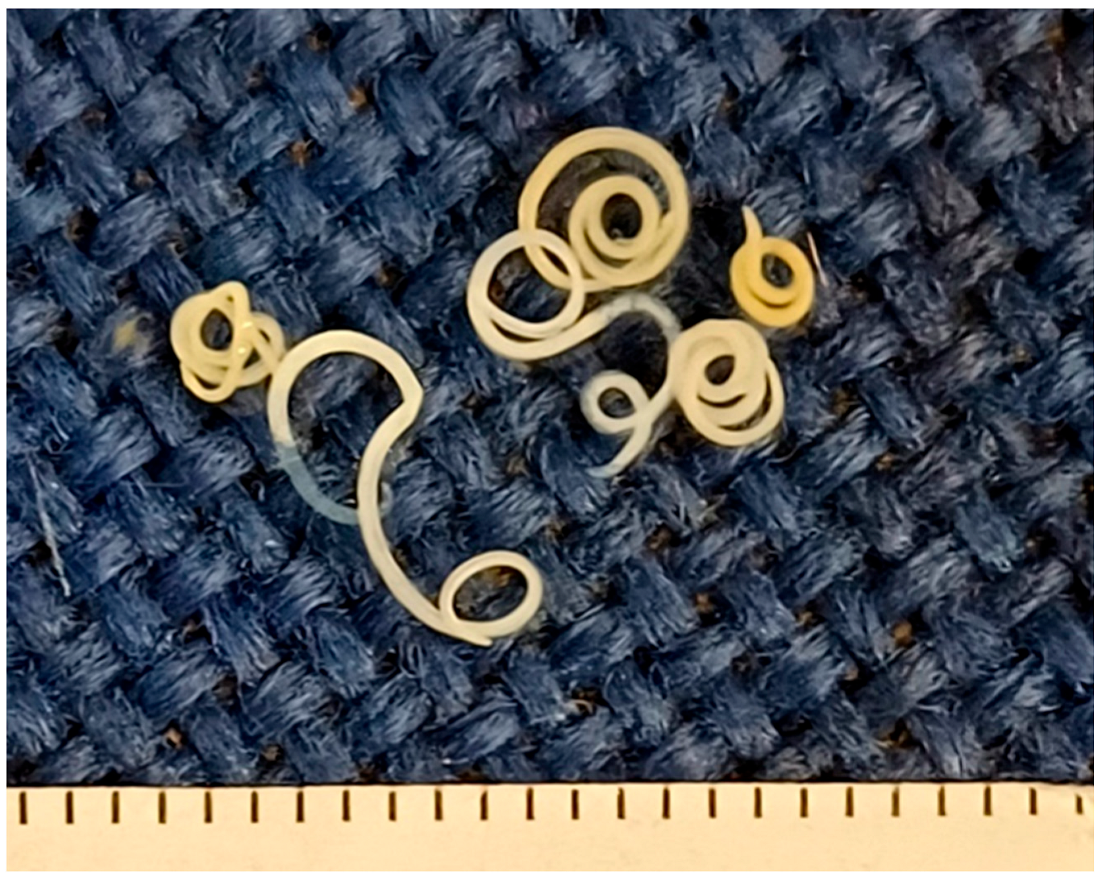
Figure 1. Nematodes isolated from ready-to-eat fish products from the Greek market. The scale (bottom) indicates millimetres.
Pathogens 14 00981 g001
Figure 2. Morphology of Anisakidae third-stage larvae, found in ready-to-eat fish products from the Greek market. (A) Posterior end. a: anus, m: mucron; (B) Anterior end. bt: boring tooth, oe: oesophagus, nr: nerve ring.
Pathogens 14 00981 g002
Table 1. Type of fish product, number of products examined, packaging, and fishing area.
Type of ProductNo. of ProductsPackaging Net Weight
(on Average)
Fishing Area
Smoked whole herring1212 × 200 gFAO 27
Smoked herring fillet in oil.99 × 110 gFAO 27
Smoked whole mackerel1515 × 300 gFAO 27
Smoked mackerel fillet in oil1111 × 100 gFAO 27
Salted whole Atlantic chub mackerel1717 × 100 gFAO 37
Salted whole anchovies1010 × 100 gFAO 37
Marinated anchovies fillet in oil1616 × 50 gFAO 37/FAO 87
Salted whole sardines in oil55 × 70 gFAO 37
Salted pollock fillet in oil55 × 80 gFAO 27
Smoked albacore fillet in oil33 × 100 gFAO 37/FAO 47
Smoked Skipjack tuna fillet in water55 × 80 gFAO 71
FAO 27: Atlantic, Northeast; FAO 37: Mediterranean & Black Sea; FAO 47: Atlantic, Southeast; FAO 71: Pacific, Western Central FAO 87: Pacific, Southeast.
Table 2. Ready-to-eat fish products found infected with Anisakidae larvae and infection intensity per sample.
Type of ProductNo. of SamplesPrevalence (%)No. of Retained LarvaeMean Intensity *No. of Larvae per Sample
Smoked whole herring1211 (91.6)625.639–15
Smoked herring fillet in oil91 (11.1)111
Smoked whole mackerel157 (46.6)172.421–6
Smoked mackerel fillet in oil112 (18.2)31.51–2
Salted whole Atlantic chub mackerel 178 (47.0)607.55–11
Salted whole anchovies101 (10.0)111
Marinated anchovies fillet in oil162 (12.5)31.51–2
Salted whole sardines in oil50 (0)0nana
Salted pollock fillet in oil50 (0)0nana
Smoked albacore fillet in oil30 (0)0nana
Smoked Skipjack tuna fillet in water 50 (0)0nana
Total10832 (29.62)147nana
* mean number of larvae per sample, na: non-applicable.
Table 3. Results of molecular analysis performed on the AD sediments and isolated larvae.
Type of ProductNo. of SamplesMolecular Analysis (AD Sediments)No. of Positive Samples by AD
(Presence of L3)
Molecular Analysis
(Larvae)
Smoked whole herring12Neg.11 + 2 *A. simplex s.s.
Smoked herring fillet in oil9Neg.1 A. simplex s.s.
Smoked whole mackerel15Neg.7A. simplex s.s.
Smoked mackerel fillet in oil11Neg.2A. simplex s.s.
Salted whole Atlantic chub mackerel 17Neg.8A. pegreffii
Salted whole anchovies10Neg.1A. pegreffii
Marinated anchovies fillet in oil16Neg.2A. pegreffii
Salted whole sardines in oil5Neg.0N/A **
Salted pollock fillet in oil5Neg.0N/A **
Smoked albacore fillet in oil3Neg.0N/A **
Smoked Skipjack tuna fillet in water5Neg.0N/A **
* Parasites detected in viscera of 11 samples; in 2 of these, also in muscle; ** N/A: not applied.
Disclaimer/Publisher’s Note: The statements, opinions and data contained in all publications are solely those of the individual author(s) and contributor(s) and not of MDPI and/or the editor(s). MDPI and/or the editor(s) disclaim responsibility for any injury to people or property resulting from any ideas, methods, instructions or products referred to in the content.