
Zoo-Sanitary Situation Assessment, an Initial Step in Country Disease Prioritization Process: Systematic Review and Meta-Analysis from 2000 to 2020 in Cameroon

 , , , ,
  and
and
Abstract
1. Introduction
2. Results
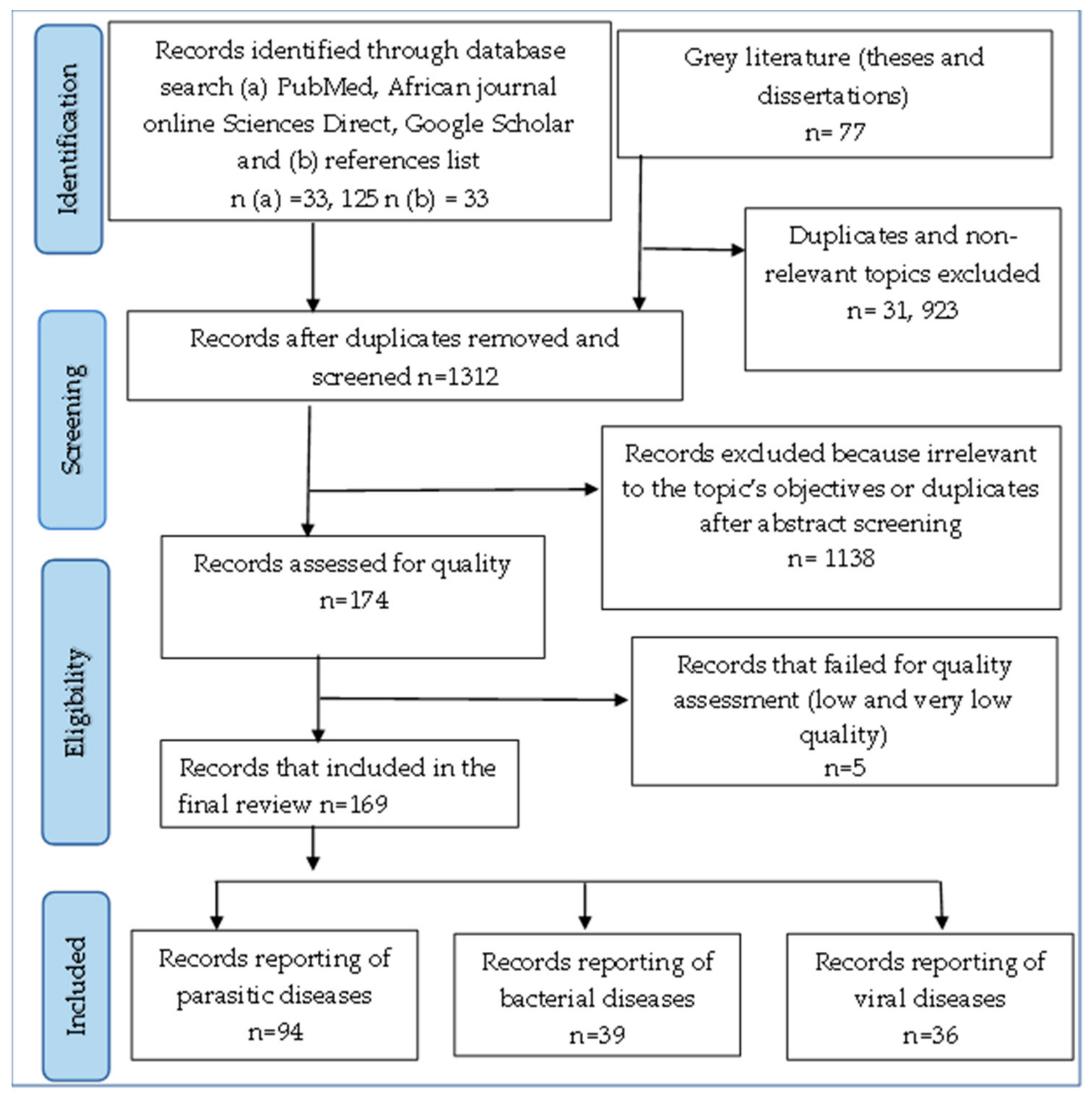
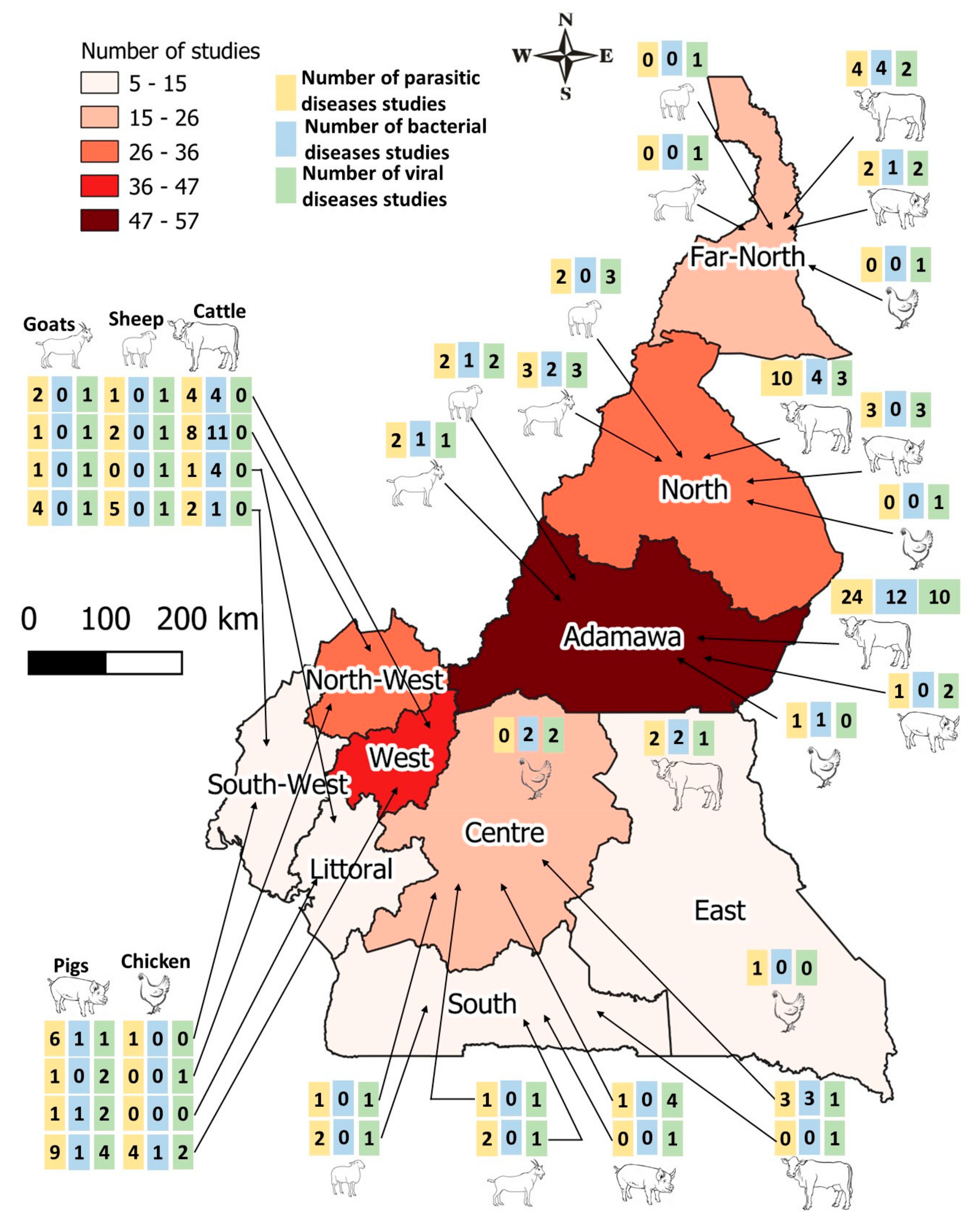
2.1. General Characteristics and Distribution of Studies Included in the Review
2.2. Parasitic Diseases Situation of Livestock in Cameroon
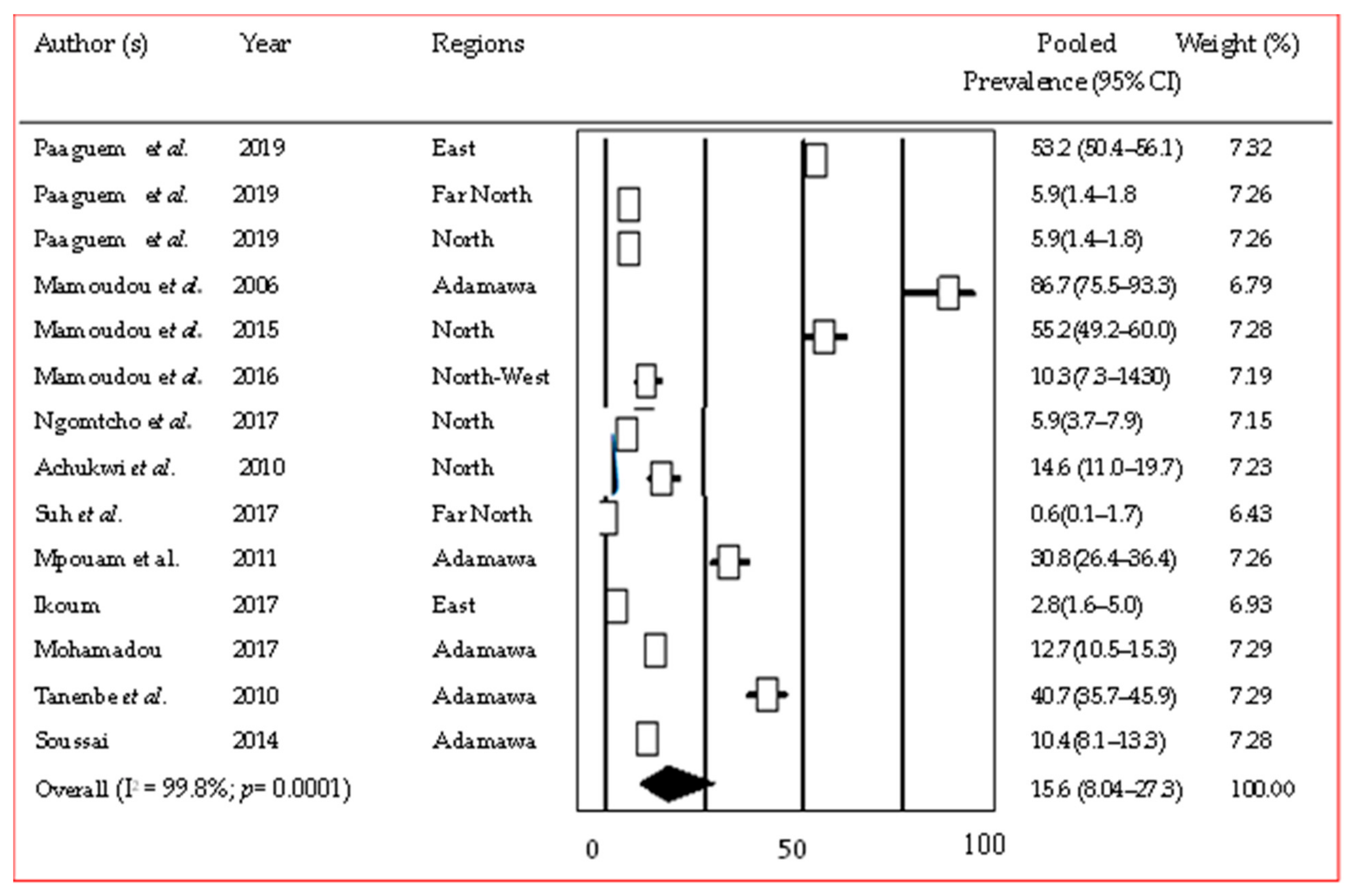
2.2.1. Trypanosomiasis
2.2.2. Tick-Borne and Other Blood Parasites
2.2.3. Gastrointestinal Parasites
2.2.4. Fascioliasis and Other Trematodes
2.2.5. Cysticercosis and Other Cestode Diseases
2.2.6. Nematodiasis
2.2.7. Coccidiosis and Other Protozoan Parasitoses
2.2.8. Ectoparasites
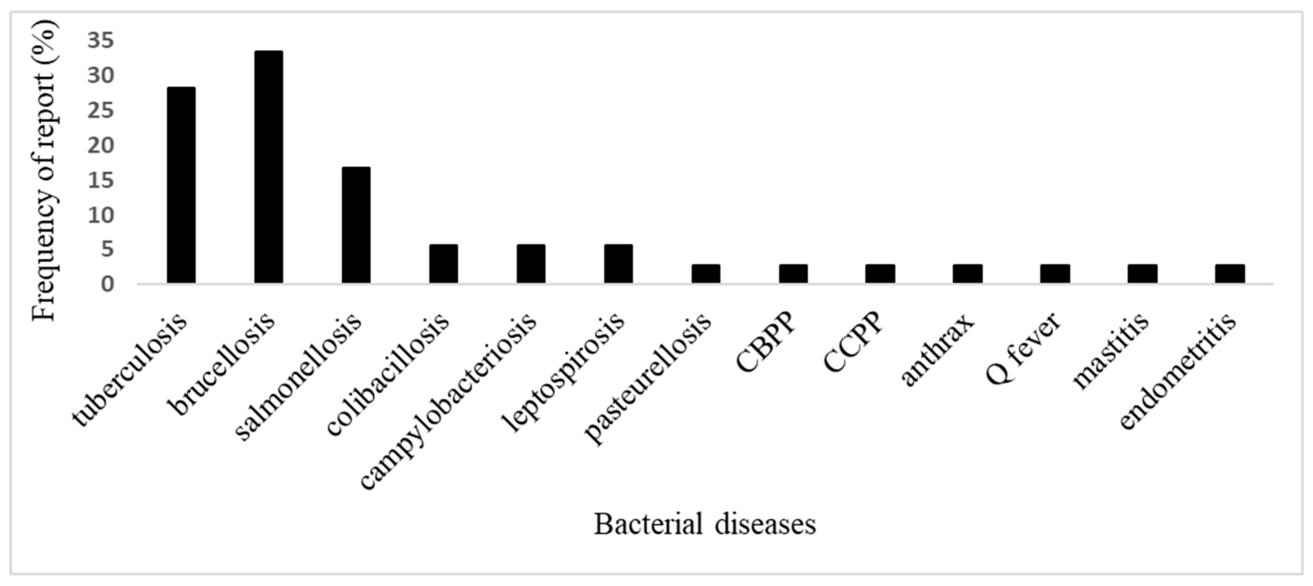
2.3. Bacterial Diseases
2.3.1. Brucellosis
2.3.2. Bovine Tuberculosis
2.3.3. Salmonellosis, Colibacillosis, and Campylobacteriosis
2.3.4. Other Bacterial Diseases
2.4. Viral Diseases
2.4.1. Foot-and-Mouth Disease (FMD)
2.4.2. Peste Des Petits Ruminants (PPR) Small Ruminant Plague
2.4.3. Rift Valley Fever (RVF)
2.4.4. African Swine Fever (ASF) and Other Viral Diseases of Pigs
2.4.5. Other Viral Diseases of Cattle, Pig, and Poultry
3. Discussion
4. Materials and Methods
4.1. Type, Period, and Area of Study
4.2. Search Strategy
4.3. Article Quality Assessment
4.4. Data Extraction and Analysis
5. Conclusions
Supplementary Materials
Author Contributions
Funding
Institutional Review Board Statement
Informed Consent Statement
Data Availability Statement
Acknowledgments
Conflicts of Interest
References
- Jones, K.E.; Patel, N.G.; Levy, M.A.; Storeygard, A.; Balk, D.; Gittleman, J.L.; Daszak, P. Global trends in emerging infectious diseases. Nature 2008, 21, 990–993. [Google Scholar] [CrossRef] [PubMed]
- Motta, P.; Porphyre, T.; Handel, I.; Hamman, S.M.; Ngu-Ngwa, V.; Tanya, V.; Morgan, K.; Christley, R.; Bronsvoort, B.M. Implications of the cattle trade network in Cameroon for regional disease prevention and control. Sci. Rep. 2017, 7, 43932. [Google Scholar] [CrossRef]
- Tambi, N.E.; Maina, W.O.; Ndi, C. An estimation of the economic impact of contagious bovine pleuropneumonia in Africa. Rev. Sci. Tech. Int. Off. Epizoot. 2006, 25, 999–1011. [Google Scholar] [CrossRef]
- Gebreyes, W.A.; Dupouy-Camet, J.; Newport, M.J.; Oliveira, C.J.; Schlesinger, L.S.; Saif, Y.M. The global One Health paradigm: Challenges and opportunities for tackling infectious diseases at the human, animal, and environment interface in low-resource settings. PLoS Neglected Trop. Dis. 2014, 8, e3257. [Google Scholar] [CrossRef]
- Mouiche, M.M.M.; Noumedem, G.N.R.; Namegni, P.R.; Kameni Feussom, J.M.; Moffo, F.; Okah-Nnane, N.H.; Munshili Njifon, L.H.; Wade, A.; Awah-Ndukum, J. African swine fever in the northern regions of Cameroon: Seroprevalence survey and spatiotemporal analysis of outbreaks from 2010 to 2017. Trop. Anim. Health Prod. 2021, 53, 214. [Google Scholar] [CrossRef]
- Kouam, K.M.; Biekop, H.M.F.; Katte, B.; Teguia, A. Risk factors of Salmonella infection in laying hens in Menoua Division, Western region of Cameroon (Central Africa). Comp. Immunol. Microbiol. Inf. Dis. 2019, 67, 101370. [Google Scholar] [CrossRef] [PubMed]
- MINEPIA (Ministry of Livestock, Fisheries and Animal Industries). Document de Stratégies du Sous-Secteur de L’élevage, des Peches et des Industries Animales, Rapport; MINEPIA (Ministry of Livestock, Fisheries and Animal Industries): Yaoundé, Cameroon, 2011.
- Killewo, J.; Bazeyo, W.; Mdegela, R. One Health Central and Eastern Africa: Historical and Future Perspectives. Int. Encycl. Public Health 2017, 2, 342–347. [Google Scholar] [CrossRef]
- Belay, E.D.; Kile, J.C.; Hall, A.J.; Barton-Behravesh, C.; Parsons, M.B.; Salyer, S.J.; Walke, H. Zoonotic Disease Programs for Enhancing Global Health Security. Emerg. Inf. Dis. 2017, 23, 65–70. [Google Scholar] [CrossRef]
- Wenholtt, M.T.A.; Cardoen, S.; Imberechts, H.; Van Huffel, X.; Ooms, B.W.; Frewer, L.J. Defining Europeans preparedness and research needs regarding emerging infectious animal diseases: Results from a Delphi expert consultation. Prev. Vet. Med. 2012, 103, 81–92. [Google Scholar] [CrossRef]
- Mpouam, S.E.; Mingoas, J.P.K.; Mouiche, M.M.M.; Kameni Feussom, J.M.; Saegerman, C. Critical Systematic Review of Zoonoses and Transboundary Animal Diseases’Prioritization in Africa. Pathogens 2021, 10, 976. [Google Scholar] [CrossRef]
- RESCAM/DSV (Reseau D’épidemiosurveillance du Cameroun/Direction des Service Vétérinaire). Rapport Annuel du Reseau d’Epidemiosurveillance du Cameroun; RESCAM: Yaoundé, Cameroon, 2020. [Google Scholar]
- Asante, J.; Norredin, A.; El Zowalaty, M.E. Systematic review of important bacterial zoonoses in Africa in the last Decade in Light of the ‘One Health’ concept. Pathogens 2019, 8, 50. [Google Scholar] [CrossRef]
- Paguem, A.; Abanda, B.; Ndjonka, D.; Weber, J.S.; Ngomtcho, S.C.H.; Manchang, T.K.; Mamoudou, A.; Eisenbarth, A.; Renz, A.; Kelm, S.; et al. Widespread co-endemicity of Trypanosoma species infecting cattle in the SudanoSahelian and Guinea Savannah zones of Cameroon. BMC Vet. Res. 2019, 15, 344. [Google Scholar] [CrossRef] [PubMed]
- Mamoudou, A.; Zoli, A.; Mbahin, N.; Tanenbe, C.; Bourdanne, A.; Clausen, P.H.; Marcotty, T.; den Bossche, P.V.; Geerts, S. Prevalence and incidence of bovine trypanosomosis on the Adamaoua plateau in Cameroon 10 years after the tsetse eradication campaign. Vet. Parasitol. 2006, 142, 16–22. [Google Scholar] [CrossRef] [PubMed]
- Mamoudou, A.; Ebene, N.J.; Suh, P.F.; Mfopit, M.Y. Prevalence and impact of bovine trypanosomiasis in Mayo Rey division, a Soudano-Sahelian zone of Cameroon. J. Parasitol. Vector Biol. 2015, 7, 80–88. [Google Scholar] [CrossRef]
- Mamoudou, A.; Mbakou, L.M.; Ngu Ngwa, V.; Sevidzem, S.L.; Zoli, A.P.; Achukwi, M.D. Preliminary assesment of bovine trypanosomiasis and its vectors in Santa, Bali and Bafut Sub-divisions of the North West Region, Cameroon. Int. J. Biol. Chem. Sci. 2016, 10, 1–12. [Google Scholar] [CrossRef]
- Ngomtcho, S.C.H.; Weber, J.S.; Bum, N.E.; Gbem, T.T.; Kelm, S.; Achukwi, M.D. Molecular screening of tsetse flies and cattle reveal different Trypanosoma species including T. grayi and T. theileri in northern Cameroon. Parasites Vectors 2017, 10, 631. [Google Scholar] [CrossRef]
- Achukwi, M.D.; Musongong, G.A. Trypanosomosis in the Doayo/Namchi (Bos taurus) and zebu White Fulani (Bos indicus) cattle in Faro Division, North Cameroon. J. Appl. Biosci. 2009, 15, 807–814. [Google Scholar]
- Suh, P.F.; Njiokou, F.; Mamoudou, A.; Ahmadou, T.M.; Mouhaman, A.; Garabed, R. Bovine trypanosomiasis in tsetse-free pastoral zone of the Far-North region, Cameroon. J. Vector Borne Dis. 2017, 54, 263–269. [Google Scholar] [CrossRef]
- Mpouam, S.E.; Achukwi, M.D.; Feussom, K.J.M.; Bengaly, Z.; Ouedraogo, G.A. Serological and parasitological prevalence of bovine trypanosomosis in small holder farms of the Vina division, Adamawa region of Cameroon. J. Parasitol. Vect. Biol. 2011, 3, 44–51. [Google Scholar]
- Ikoum, D. Infestation et Prévalence de la Trypanosomose Bovine dans le Ranch de la SODEPA de Ndokayo. Ph.D. Thesis, School of Veterinary Medicine and Sciences, Ngaoundéré, Cameroon, 2017, unpublished. [Google Scholar]
- Mohamadou, B. Etude de la Prévalence de la Trypanosomose chez les Bovins Abattus à L’abattoir Municipal de Ngaoundéré. Ph.D. Thesis, School of Veterinary Medicine and Sciences, University of Ngaoundere, Ngaoundéré, Cameroon, 2017, unpublished. [Google Scholar]
- Tanenbe, C.; Gambo, H.; Musongong, A.G.; Boris, O.; Achukwi, M.D. Prévalence de la trypanosomose bovine dans les départements du Faro et Déo, et de la Vina au Cameroun: Bilan de vingt années de lutte contre les glossines. Rev. Élev. Méd. Vét. Pays Trop. 2010, 63, 63–69. [Google Scholar] [CrossRef]
- Soussai, F.O. Prévalences et Facteurs de Risques des Hémoparasites des Bovins Abattus à L’abattoir Municipal de Ngaoundéré. Ph.D. Thesis, School of Veterinary Medicine and Sciences, University of Ngaoundere, Ngaoundéré, Cameroon, 2017, unpublished. [Google Scholar]
- Awah-Ndukum, J.; Kudi, C.; Bradley, G.; Ane-Anyangwe, I.N.; Fon-Tebug, S.; Tchoumboue, J. Prevalence of Bovine Tuberculosis in Abattoirs of the Littoral and Western Highland Regions of Cameroon: A Cause for Public Health Concern. Vet. Med. Int. 2010, 2010, 495015. [Google Scholar] [CrossRef]
- Awah-Ndukum, J.; Tchoumoube, J.; Niba, T.A. Prevalence of bovine tuberculosis at the SODEPA Doual Abattoir Cameroon, (1995–2003). Camer. J. Exp. Biol. 2005, 1, 116–120. [Google Scholar]
- Awah-Ndukum, J.; Kudi, A.C.; Bah, G.S.; Bradley, G.; Tebug, S.F.; Dickmu, P.L.; Njakoi, H.N.; Agharih, W.N. Bovine Tuberculosis in Cattle in the Highlands of Cameroon: Seroprevalence Estimates and Rates of Tuberculin Skin Test Reactors at Modified Cut-Offs. Vet. Med. Int. 2012, 2012, 798502. [Google Scholar] [CrossRef] [PubMed]
- Egbe, N.F.; Muwonge, A.; Ndip, L.; Kelly, R.F.; Sander, M.; Tanya, V.; Ngu Ngwa, V.; Handel, I.G.; Morgan, K.L.; Asuquo, A.; et al. Abattoir-based estimates of mycobacterial infections in Cameroon. Sci. Rep. 2016, 6, 24320. [Google Scholar] [CrossRef]
- Koro, F.K.; Bouba Foko, E.; Ngatchou, A.F.; Eyangoh, S.; Etoa, F.-X. First Insight into the Current Prevalence of Bovine Tuberculosis in Cattle Slaughtered in Cameroon: The Case of Main Abattoirs of Yaoundé and Douala. British Microbiol. Res. J. 2013, 3, 272–279. [Google Scholar] [CrossRef]
- Manchang, K.T.; Mfopit, Y.M.; Julius, A.N.; Joseph, N.A.; Bah, G.S.; Bayemi, H. Seasonal Evaluation of Some Disease Conditions of Cattle Slaughtered in Ngaoundere-Municipal Abattoir, Cameroon. Int. J. Livest. Res. 2016, 6, 46–53. [Google Scholar] [CrossRef]
- Ngoudjou, M.S.V. Prévalence des Lésions Tuberculeuses Viscérale à L’abattoir de Douala. Ph.D. Thesis, Institut Supérieur des Sciences de la Santé, Université, des Montagnes, Bangangté, Cameroon, 2018, unpublished. [Google Scholar]
- Kouengoua, K.A.P. Etude des Lésions de la Tuberculose Bovine à L’abattoir de Yaoundé. Ph.D. Thesis, Institut Supérieur des Sciences de la Santé, Université, des Montagnes, Bangangté, Cameroon, 2014, unpublished. [Google Scholar]
- Yemelong, D.W.S. Prévalence de la Co-Infection Fasciolose et Tuberculose Bovine à L’abattoir de Bangangte. Ph.D. Thesis, Institut des Sciences de la Santé, Université, des Montagnes, Bangangté, Cameroon, 2019, unpublished. [Google Scholar]
- Kelly, R.F.; Callaby, R.; Egbe, N.; Williams, D.J.L.; Victor, N.N.; Tanya, V.N.; Sander, M.; Ndip, L.; Ngandolo, R.; Morgan, K.L.; et al. Association of Fasciola gigantica Co-infection with Bovine Tuberculosis Infection and Diagnosis in a Naturally Infected Cattle Population in Africa. Front. Vet. Sci. 2018, 5, 214. [Google Scholar] [CrossRef]
- Bronsvoort, B.M.C.; Anderson, J.; Corteyn, A.; Hamblin, P.; Kitching, R.P.; Nfon, C.; Tanya, V.N.; Morgan, K.L. Geographical and age-stratified distributions of foot-and-mouth disease virus-seropositive and probang-positive cattle herds in the Adamawa province of Cameroon. Vet. Record. 2006, 159, 299–308. [Google Scholar] [CrossRef]
- Morgan, K.L.; Handel, I.G.; Tanya, V.N.; Hamman, S.M.; Nfon, C.; Bergman, I.E.; Malirat, V.; Sorensen, K.J.; Bronsvoort, B.M. Accuracy of Herdsmen Reporting versus Serologic Testing for Estimating Foot-and-Mouth Disease Prevalence. Emerg. Inf. Dis. 2014, 20, 2048–2054. [Google Scholar] [CrossRef]
- Sevidzem, S.L.; Mamoudou, A.; Mavoungou, J.F.; Ikoum, D.; Lebale, M.B.O.; Hiol, V.D.; Nguema, M.R.; Zinga-Koumba, C.R.; Acapovi-Yao, G.L.; Dickmu, S.; et al. Serological Epidemiology of Foot-and-mouth Disease among Sedentary Mixed-species Herds in Adamawa Region, Cameroon. J. Adv. Microbiol. 2019, 17, 1–14. [Google Scholar] [CrossRef]
- Ludi, A.; Ahmed, Z.; Pomeroy, L.W.; Pauszek, S.J.; Smoliga, G.R.; Moritz, M.; Dickmu, S.; Abdoulkadiri, S.; Arzt, J.; Garabed, R.; et al. Serotype Diversity of Foot-and-Mouth-Disease Virus in Livestock without History of Vaccination in the Far North Region of Cameroon. Trans. Emerg. Dis. 2014, 63, 27–38. [Google Scholar] [CrossRef] [PubMed]
- Kuate, W.B.V. Prévalence et Facteurs de Risque par les Eleveurs de la Fièvre Aphteuse chez les Bovins dans les Départements de la Vina, du Diamare et du Logone-et-Chari. Ph.D. Thesis, School of Veterinary Medicine and Sciences, University of Ngaoundere, Ngaoundéré, Cameroon, 2016, unpublished. [Google Scholar]
- Beloko, S.L. Prévalence et Facteurs de Risques de la Fièvre Aphteuse Bovine dans le Marché à Bétail de Garoua-Boulaï et dans la Zone de Transhumance du Département du Lom et Djerem à l’Est-Cameroun. Ph.D. Thesis, School of Veterinary Medicine and Sciences, University of Ngaoundere, Ngaoundéré, Cameroon, 2019, unpublished. [Google Scholar]
- Modjo, K.E.L. Séroprévalence de la Fièvre Aphteuse chez les Bovins dans la Région du Nord Cameroun. Ph.D. Thesis, Institut Supérieur des Sciences de la Santé, Université, des Montagnes, Bangangté, Cameroon, 2014, unpublished. [Google Scholar]
- Barman, N.N.; Patil, S.S.; Kurli, R.; Deka, P.; Bora, D.P.; Deka, G.; Bora, D.P.; Ranjitha, K.M.; Shivaranjini, C.; Roy, P.; et al. Meta-analysis of the prevalence of livestock diseases in North Eastern Region of India. Vet. World 2020, 13, 80–89. [Google Scholar] [CrossRef] [PubMed]
- Njayou, N.A. Chaine de Valeur des Petits Ruminants au Cameroun; Yaoundé, Cameroon, 2017; pp. 12–13. [Google Scholar]
- Asfaw, Y.; Ameni, G.; Medhin, G.; Alemayehu, G.; Wieland, B. Infectious and parasitic diseases of poultry in Ethiopia: A systematic review and meta-analysis. Poult. Sci. 2019, 98, 6452–6462. [Google Scholar] [CrossRef] [PubMed]
- Berkelman, R.; Cassell, G.; Specter, S.; Hamburg, M.; Klugman, K. The “Achilles Heel” of Global Efforts to Combat Infectious Diseases. Clin. Inf. Dis. 2006, 42, 1503–1504. [Google Scholar] [CrossRef] [PubMed]
- Peeling, R.; McNermey, R. Increasing Access to Diagnostics through Technology Transfer and Local Production; LSHTM Research Online: London, UK, 2011. [Google Scholar]
- Dhama, K.; Wani, M.Y.; Tiwari, R.; Kumar, D. Molecular diagnosis of animal diseases: The current trends and perspectives. Livest. Sphere 2012, 1, 6–10. [Google Scholar]
- Tesfaye, D.; Speybroeck, N.; De Deken, R.; Thys, E. Fardeau économique de la trypanosomose bovine dans trois villages de la zone de Metekel, dans le nord-ouest de l’Éthiopie. Trop. Anim. Health Prod. 2012, 44, 873–879. [Google Scholar] [CrossRef]
- Odeniran, P.O.; Ademola, I.O. A meta-analysis of the prevalence of African animal trypanosomiasis in Nigeria from 1960 to 2017. Parasites Vectors 2018, 11, 280. [Google Scholar] [CrossRef]
- Leta, S.; Alemayehu, G.; Seyoum, Z.; Bezie, M. Prevalence of bovine trypanosomosis in Ethiopia: A meta-analysis. Parasites Vectors 2016, 9, 139. [Google Scholar] [CrossRef]
- Mamoudou, A.; Suh, P.F.; Ebene, N.J. Trypanosomes and helminths infections in Mayo Rey Division of Cameroon and impact of concurrent infections on cattle. J. Vet. Med. Anim. Health 2015, 7, 215–220. [Google Scholar] [CrossRef][Green Version]
- Paramanandham, K.; Mohankumar, A.; Puttahonnappa Suresh, K.; Susan Jacob, S.; Roy, P. Prevalence of Anaplasma species in India and the World in dairy animals: A systematic review and meta-analysis. Res. Vet. Sci. 2019, 123, 159–170. [Google Scholar] [CrossRef]
- Haghi, M.M.; Etemadifar, F.; Fakhar, M.; Teshnizini, S.H.; Soosaraei, M.; Shokri, A.; Hajihasani, A.; Mashhadi, H. Status of babesiosis among domestic herbivores in Iran: A systematic review and meta-analysis. Parasitol. Res. 2017, 116, 1101–1109. [Google Scholar] [CrossRef]
- Awa, D.N.; Adakal, H.; Luogbou, N.D.D.; Wachong, K.H.; Leinyuy, I.; Achukwi, M.D. Cattle ticks in Cameroon: Is Rhipicephalus (Boophilus) microplus absent in Cameroon and the Central African region? Ticks Tick-Borne Dis. 2015, 6, 117–122. [Google Scholar] [CrossRef] [PubMed]
- Silatsa, B.A.; Simo, G.; Githaka, N.; Mwaura, S.; Kamga, R.M.; Oumarou, F.; Keambou, C.; Bishop, R.P.; Djikeng, A.; Kuiate, J.R.; et al. A comprehensive survey of the prevalence and spatial distribution of ticks infesting cattle in diferent agro-ecological zones of Cameroon. Parasites Vectors 2019, 12, 489. [Google Scholar] [CrossRef]
- Abebe, E.; Gugsa, G. A Review on Poultry Coccidiosis. Abyssinia J. Sci. Technol. 2018, 3, 1–12. [Google Scholar]
- Karshima, S.N.; Maikai, B.V.; Kwaga, J.K.P. Helminths of veterinary and zoonotic importance in Nigerian ruminants: A 46-year meta-analysis (1970–2016) of their prevalence and distribution. Inf. Dis. Poverty 2018, 7, 52. [Google Scholar] [CrossRef] [PubMed]
- Lamy, E.; Harten, S.V.; Sales-Baptista, E.; Guerra, M.M.M.; de Almeida, A.M. Environmental Stress and Amelioration on Livestock Production: Factors Influencing Livestock Productivity; Springer: Berlin/Heidelberg, Germany, 2012. [Google Scholar]
- Abakar, A.A. Prévalence et Impacts Economiques de la Fasciolose à L’abattoir Municipal de Ngaoundéré. Ph.D. Thesis, School of Veterinary Medicine and Sciences, University of Ngaoundere, Ngaoundéré, Cameroon, 2014, unpublished. [Google Scholar]
- Kouam, K.M.; Meningue, R.; Fon, D.E. Parasitic causes of organ condemnation in cattle slaughtered in Fako abattoirs, South-West region of Cameroon, and estimate of financial losses. J. Helminthol. 2018, 93, 367–371. [Google Scholar] [CrossRef]
- Awah-Ndukum, J.; Mouiche, M.M.M.; Kouonmo-Ngnoyum, L.; Bayang, H.N.; Manchang, T.K.; Poueme, R.S.N.; Kouamo, J.; Ngu Ngwa, V.; Assana, E.; Feusom, K.; et al. Seroprevalence and risk factors of brucellosis among slaughtered indigenous cattle, abattoir personnel and pregnant women in Ngaoundéré, Cameroon. BMC Inf. Dis. 2018, 18, 611. [Google Scholar] [CrossRef]
- Ngu-Ngwa, V.; Bessong-Takang, N.A.; Awah-Ndukum, J. Seroprevalence and Risk Factors of Leptospirosis among Slaughtered Cattle and Abattoir Workers in Ngaoundéré, Cameroon. Asian J. Res. Anim. Vet. Sci. 2020, 5, 8–19. [Google Scholar]
- MINEPIA (Ministry of Livestock, Fisheries and Animal Industries). Projet de Renforcement de la Surveillance Epidémiologique des Maladies Animales au Cameroun; MINEPIA (Ministry of Livestock, Fisheries and Animal Industries): Bamenda, Cameroon, 2014.
- MINEPIA (Ministry of Livestock, Fisheries and Animal Industries). Schéma Directeur Pour le Développement des Filières de L’élevage au Cameroun; MINEPIA (Ministry of Livestock, Fisheries and Animal Industries): Yaoundé, Cameroon, 2009.
- Douffissa, A. Recueil des Textes Régissant L’élevage, les Pêches et les Industries Animales, 2nd ed.; Réactualisée: Yaoundé, Cameroon, 2007. [Google Scholar]
- FAO (Food and Agricultural Organization of the United Nations). Economic Analysis of Animal Diseases; FAO: Rome, Italy, 2016. [Google Scholar]
- Bronsvoort, B.M.C.; Radford, A.D.; Tanya, V.N.; Nfon, C.; Kitching, R.P.; Morgan, K.L. Molecular Epidemiology of Foot-and-Mouth Disease Viruses in the Adamawa Province of Cameroon. J. Clin. Microbiol. 2004, 42, 2186–2196. [Google Scholar] [CrossRef]
- Page, M.J.; Moher, D.; Bossuyt, P.M.; Boutron, I.; Hoffmann, T.C.; Mulrow, C.D.; Shamseer, L.; Tetzlaff, J.M.; Akl, E.A.; Brennan, S.E.; et al. PRISMA 2021 explanation and elaboration: Updated guidance and exemplars for reporting systematic reviews. BMJ 2021, 372, n160. [Google Scholar] [CrossRef]
- Valentine, J.C.; Pigott, T.D.; Rothstein, H.R. How Many Studies Do You Need? A Primer on Statistical Power for Meta-Analysis. J. Edu. Behav. Stat. 2009, 35, 215–247. [Google Scholar] [CrossRef]
- Munn, Z.; Moola, S.; Riitano, D.; Lisy, K. The development of a critical appraisal tool for use in systematic reviews addressing questions of prevalence. Int. J. Health Policy Manag. 2014, 3, 123–128. [Google Scholar] [CrossRef] [PubMed]
- Crowther, M.; Lim, W.; Crowther, M.A. Systematic review and meta-analysis methodology. Blood 2010, 116, 3140–3160. [Google Scholar] [CrossRef] [PubMed]
- Begg, C.B.; Mazumdar, M. Operating characteristics of a rank correlation test for publication bias. Biometrics 1994, 50, 1088–1101. [Google Scholar] [CrossRef] [PubMed]
- Egger, M.; Davey Smith, G.; Schneider, M.; Minder, C. Bias in meta-analysis detected by a simple, graphical test. BMJ 1997, 315, 629–634. [Google Scholar] [CrossRef]
- Duval, S.; Tweedie, R. Trim and fill: A simple funnel-plot-based method of testing and adjusting for publication bias in meta-analysis. Biometrics 2000, 56, 455–463. [Google Scholar] [CrossRef]
- Boreisntein, M.; Hedges, V.L.; Higgins, P.T.J.; Rothstein, R.H. Introduction to Meta-Analysis, 2nd ed.; Willey: Hoboken, NJ, USA, 2021. [Google Scholar]


| Characteristics | Number of Studies on Parasitic Diseases N (%) | Number of Studies on Bacterial Diseases N (%) | Number of Studies on Viral Diseases N (%) | Total N (%) |
|---|---|---|---|---|
| Region | ||||
| Adamawa (AD) | 30 (31.91) | 14 (35.90) | 13 (36.11) | 57 (34.54) |
| Centre (C) | 5 (5.32) | 6 (15.38) | 8 (22.22) | 19 (11.51) |
| East (E) | 3 (3.19) | 2 (5.13) | 2 (5.55) | 7 (4.24) |
| Far North (FN) | 5 (5.32) | 5 (12.8) | 6 (16.67) | 16 (9.69) |
| Littoral (L) | 3 (3.19) | 4 (10.25) | 3 (8.33) | 10 (6.06) |
| North (N) | 15 (15.95) | 6 (15.38) | 10 (27.77) | 31 (18.78) |
| Northwest (NW) | 13 (13.83) | 12 (30.76) | 5 (13.88) | 30 (18.18) |
| South (S) | 2 (2.12) | 0 (0) | 3 (8.33) | 5 (3.03) |
| Southwest (SW) | 12 (12.76) | 1 (2.56) | 2 (5.55) | 15 (9.09) |
| West (W) | 20 (21.27) | 6 (15.38) | 8 (22.22) | 34 (20.60) |
| Study site not precise | 1 (1.06) | 0 (0) | 4 (11.11) | 5 (3.03) |
| Publication period | ||||
| 2000–2004 | 8 (8.51) | 1 (2.56) | 2 (5.55) | 11 (6.66) |
| 2005–2009 | 10 (10.64) | 4 (10.25) | 1 (2.77) | 15 (9.09) |
| 2010–2014 | 19 (20.21) | 13 (33.33) | 12 (33.33) | 44 (26.66) |
| 2015–March 2020 | 57 (60.64) | 21 (53.84) | 21 (58.33) | 95 (57.58) |
| Species | ||||
| Chicken | 7 (7.44) | 5 (12.82) | 5 (13.89) | 17 (10.30) |
| Pigs | 24 (25.53) | 2 (5.12) | 12 (33.33) | 37 (22.42) |
| Cattle | 50 (53.19) | 29 (74.35) | 14 (38.88) | 90 (54.54) |
| Sheep | 14 (14.89) | 1 (2.56) | 7 (19.44) | 22 (13.33) |
| Goats | 15 (15.95) | 3 (7.69) | 6 (16.66) | 24 (14.54) |
| Type of material | ||||
| Journal article | 56 (59.57) | 25 (64.10) | 22 (61.11) | 101 (61.21) |
| Dissertation (unpublished) | 38 (40.43) | 14 (35.90) | 14 (38.89) | 64 (38.79) |
| Language | ||||
| English | 53 (56.38) | 25 (64.10) | 21 (58.33) | 99 (60) |
| French | 41 (43.62) | 14 (35.90) | 15 (41.67) | 66 (40) |
| Study design | ||||
| Cross-sectional | 92 (97.87) | 37 (94.87) | 30 (83.33) | 155 (93.94) |
| Longitudinal | 2 (2.13) | 2 (5.13) | 6 (16.67) | 10 (6.06) |
| Samples | ||||
| Blood | 42 (44.68) | 17 (43.58) | 21 (58.33) | 80 (48.48) |
| Fecal or rectal swab | 29 (30.85) | 5 (12.82) | 1 (2.77) | 35 (21.21) |
| Meat or carcass | 16 (17.02) | 12 (30.76) | 4 (11.11) | 36 (21.81) |
| Skin | 8 (8.51) | 4 (10.25) | 1(2.77) | 13 (7.87) |
| Others (milk, uterine swabs…) | 0 (0) | 4 (10.25) | 4 (11.11) | 8 (4.84) |
| Diagnostic methods | ||||
| Serology (ELISA, RBPT, VN) | 9 (9.57) | 16 (41.02) | 16 (44.44) | 41 (24.26) |
| Molecular (PCR, sequencing) | 11 (11.70) | 1 (2.56) | 14 (38.88) | 26 (15.38) |
| PM/AM examination | 18 (19.15) | 11 (28.2) | 4 (11.11) | 33 (19.53) |
| Microscopy | 53 (56.38) | 8 (20.51) | / | 61 (36.10) |
| Others | 3 (3.19) | 3 (7.69) | 2 (5.55) | 8 (4.73) |
| Total | 94 (56.97) | 39 (23.64) | 36 (21.82) | 169 (100) |
| Disease Studied | Species | N° of Study Results | Total Number of Samples | N° of Positive Samples | Meta-Analysis Data | ||||||
|---|---|---|---|---|---|---|---|---|---|---|---|
| Model | Pooled Prevalence (%) | 95% CI | Tau2 | I2 (%) | p-Value | Relative Weight Range (%) | |||||
| Trypanosomiasis | Cattle | 18 | 7369 | 1641 | Random | 20.9 | 14.9–28.7 | 0.789 | 93.514 | 0.000 | 5.12–5.66 |
| Trypanosomiasis | Goats | 5 | 963 | 270 | Random | 28.4 | 12.7–51.9 | 1.233 | 95.934 | 0.000 | 18.74–20.84 |
| Trypanosomiasis | Pigs | 6 | 1491 | 551 | Random | 54.2 | 39.6–68.2 | 0.507 | 95.169 | 0.000 | 14.12–17.51 |
| Trypanosomiasis | Sheep | 5 | 657 | 185 | Random | 28.4 | 12.7–51.9 | 1.233 | 95.934 | 0.000 | 18.74–20.84 |
| Babesiosis | Small ruminants | 4 | 1081 | 35 | Random | 3.5 | 2.2–5.6 | 0.119 | 47.519 | 0.167 | 18.87–30.87 |
| Babesiosis | Cattle | 2 | 925 | 208 | Random | 21.6 | 4.9–59.7 | 1.455 | 98.956 | 0.000 | 49.85–50.15 |
| Theileriosis | Small ruminants | 5 | 1478 | 42 | Random | 4.9 | 1.9–11.8 | 1.079 | 96.164 | 0.000 | 18.49–21.45 |
| Anaplasmosis | small ruminants | 5 | 1478 | 189 | Random | 13.0 | 6.2–25.2 | 0.816 | 97.792 | 0.000 | 19.32–20.56 |
| Anaplasmosis | Cattle | 2 | 925 | 324 | Random | 35.1 | 4.7–85.5 | 2.951 | 99.546 | 0.000 | 49.96–50.04 |
| Gastrointestinal parasites | Cattle | 6 | 1690 | 989 | Random | 57.4 | 45.3–68.7 | 0.354 | 95.622 | 0.000 | 16.34–17.06 |
| Gastrointestinal parasites | Poultry | 3 | 1701 | 1076 | Random | 67.2 | 48.3–81.8 | 0.469 | 88.632 | 0.000 | 32.87–33.78 |
| Gastrointestinal parasites | Pigs | 3 | 741 | 638 | Random | 88.0 | 78.2–93.8 | 0.328 | 97.988 | 0.000 | 24.66–37.79 |
| Fascioliasis | Cattle | 16 | 18,510 | 3581 | Random | 17.3 | 11.0–26.2 | 1.114 | 99.39 | 0.000 | 5.46–6.47 |
| Fascioliasis | Small Ruminants | 3 | 1085 | 110 | Random | 8.8 | 2.1–30.2 | 1.710 | 97.727 | 0.000 | 19.38–58.21 |
| Paramphistomosis | Cattle | 4 | 1415 | 228 | Random | 12.6 | 4.5–30.8 | 1.251 | 97.834 | 0.000 | 21.98–26.11 |
| Paramphistomosis | Goats | 2 | 460 | 29 | Random | 19.7 | 3.2–64.7 | 2.982 | 98.463 | 0.000 | 49.48–50.52 |
| Paramphistomosis | Pigs | 2 | 495 | 171 | Random | 23.0 | 5.4–61.0 | 1.373 | 96.056 | 0.000 | 48.39–51.61 |
| Cysticercosis | Pigs | 10 | 7553 | 348 | Random | 6.2 | 3.3–11.3 | 0.940 | 96.092 | 0.000 | 3.8–11.74 |
| Coccidiosis | Poultry | 4 | 2581 | 1276 | Random | 49.2 | 28.0–70.7 | 0.859 | 99.107 | 0.000 | 24.86–25.12 |
| Coccidiosis | Cattle | 2 | 660 | 43 | Random | 4.5 | 1.1–17.0 | 1.003 | 87.628 | 0.004 | 45.05–54.96 |
| Coccidiosis | Goats | 2 | 349 | 122 | Random | 55.1 | 2.8–97.5 | 0.000 | 99.103 | 0.000 | 49.79–50.21 |
| Coccidiosis | Pigs | 4 | 1677 | 521 | Random | 30.2 | 10.3–61.9 | 2.157 | 98.983 | 0.000 | 24.92–25.10 |
| Monieziosis | Cattle | 3 | 1015 | 36 | Random | 3.6 | 2.4–5.4 | 0.044 | 30.120 | 0.239 | 12.29–46.27 |
| Strongylus spp. | Cattle | 5 | 1244 | 552 | Random | 34.5 | 17.2–57.2 | 1.097 | 98.019 | 0.000 | 19.71–20.34 |
| Strongylus spp. | Pigs | 4 | 1419 | 677 | Random | 48.6 | 28.6–69.1 | 0.750 | 98.182 | 0.000 | 24.23–25.35 |
| Trichuris spp. | Cattle | 2 | 555 | 44 | Random | 3.0 | 0.1–43.6 | 4.967 | 90.594 | 0.001 | 45.54–54.46 |
| Trichuris spp. | Pigs | 4 | 1281 | 81 | Random | 4.4 | 0.8–20.9 | 2.997 | 96.818 | 0.000 | 23.58–26.04 |
| Strongyloides spp. | Pigs | 5 | 1677 | 326 | Random | 22.0 | 14.1–32.6 | 0.358 | 94.502 | 0.000 | 19.41–20.55 |
| Strongyloides spp. | Cattle | 4 | 1104 | 121 | Random | 12.4 | 3.6–35.1 | 1.834 | 97.581 | 0.000 | 24.72–25.23 |
| Strongyloides spp. | Sheep | 2 | 303 | 30 | Random | 7.9 | 0.6–56.8 | 3.734 | 96.058 | 0.000 | 48.69–51.31 |
| Strongyloides spp. | Goats | 3 | 554 | 63 | Random | 10.0 | 1.0–54.3 | 4.283 | 97.949 | 0.000 | 32.82–33.79 |
| Strongyloides spp. | Poultry | 2 | 1272 | 108 | Random | 8.5 | 7.1–10.2 | 0.000 | 0.000 | 0.909 | 29.6–70.33 |
| Ascaridia spp. | Poultry | 3 | 1701 | 360 | Random | 22.9 | 11.8–39.7 | 0.488 | 97.722 | 0.000 | 33.14–33.49 |
| Heterakis spp. | Poultry | 3 | 1701 | 295 | Random | 18.4 | 7.3–39.5 | 0.865 | 98.442 | 0.000 | 33.18–33.44 |
| Capillaria spp. | Poultry | 2 | 1318 | 109 | Random | 8.9 | 3.7–19.7 | 0.425 | 95.423 | 0.000 | 49.95–50.05 |
| Disease Studied | Species | N° of Study Results | Total Number of Samples | N° of Positive Samples | Meta-Analysis Data | ||||||
|---|---|---|---|---|---|---|---|---|---|---|---|
| Model | Pooled Prevalence (%) | 95% CI | Tau2 | I2 (%) | p-Value | Relative Weight Range (%) | |||||
| Tuberculosis | Cattle | 15 | 121,612 | 1588 | random | 3.3 | 1.5–6.9 | 2.395 | 99.724 | 0.000 | 6.45–6.73 |
| Brucellosis | Cattle | 10 | 6776 | 436 | Random | 7.0 | 4.8–10.0 | 0.372 | 93.514 | 0.000 | 8.10–10.55 |
| Brucellosis | Pigs | 2 | 1081 | 20 | Random | 8.0 | 0.0–17.40 | 4.778 | 82.309 | 0.017 | 41.60–58.40 |
| Salmonellosis | Poultry | 3 | 484 | 343 | Random | 48.2 | 12.9–65.5 | 2.359 | 97.299 | 0.000 | 26.16–37.02 |
| Leptospirosis | Cattle | 2 | 1549 | 35 | Random | 2.5 | 0.0–64.2 | 9.202 | 98.448 | 0.000 | 49.43–50.57 |
| Pasteurellosis | Cattle | 3 | 362 | 198 | Random | 55.5 | 39.2–70.7 | 0.292 | 87.934 | 0.000 | 29.20–36.02 |
| Disease Studied | Species | N° of Study Results | Total Number of Samples | N° of Positive Samples | Meta-Analysis Data | ||||||
|---|---|---|---|---|---|---|---|---|---|---|---|
| Model | Pooled Prevalence (%) | 95% CI | Tau2 | I2 (%) | p-Value | Relative Weight Range (%) | |||||
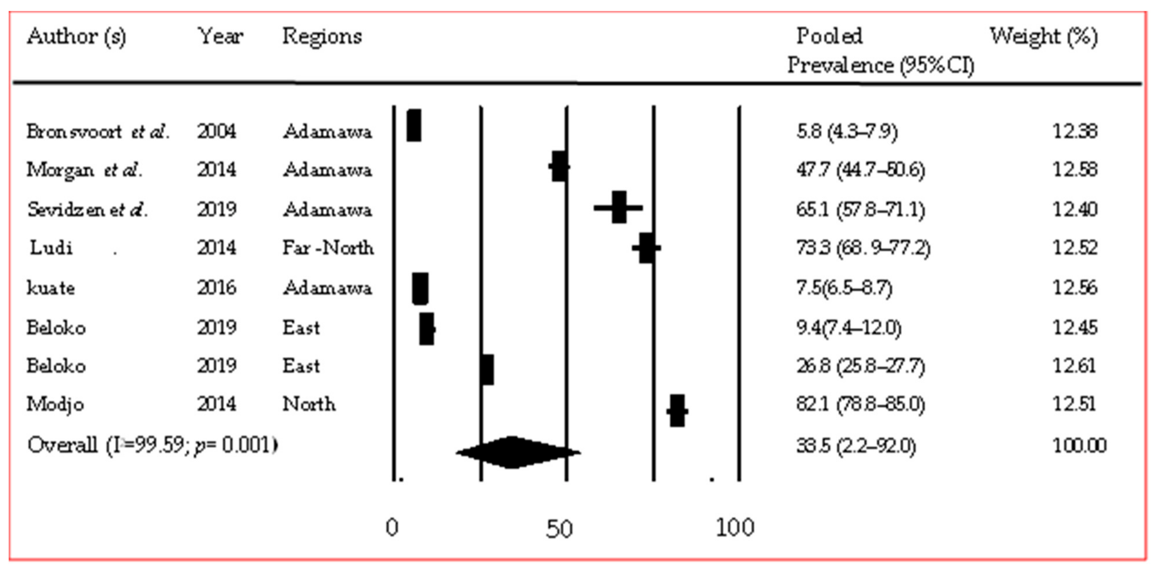
| Foot-and-Mouth Disease | Cattle | 9 | 19,778 | 4897 | Random | 39.4 | 20.4–62.4 | 2.000 | 99.679 | 0.000 | 11.03–11.16 |
| Sheep | 3 | 1251 | 128 | Random | 8.1 | 3.6–17.4 | 0.551 | 93.194 | 0.000 | 32.66–34.30 | |
| Pigs | 2 | 658 | 29 | Random | 4.4 | 3.1–6.3 | 0.000 | 0.000 | 0.710 | 6.97–93.03 | |
| Peste des Petits Ruminants | Small ruminants | 4 | 2090 | 791 | Random | 39.7 | 30.8–49.4 | 0.133 | 88.768 | 0.000 | 18.81–29.41 |
| Rift Valley Fever | Small ruminants | 10 | 1525 | 53 | Random | 3.7 | 2.9–4.9 | 0.000 | 0.000 | 0.566 | 1.82–28.12 |
| Cattle | 7 | 2532 | 256 | Random | 10.9 | 8.0–14.7 | 0.149 | 81.722 | 0.000 | 2.47–18.62 | |
| African Swine Fever | Pigs | 6 | 2472 | 643 | Random | 18.9 | 8.9–35.6 | 1.117 | 98.150 | 0.000 | 15.32–17.37 |
| Hepatitis E | Pigs | 5 | 507 | 73 | Random | 8.4 | 2.1–28.0 | 2.114 | 92.862 | 0.000 | 13.06–24.74 |
Disclaimer/Publisher’s Note: The statements, opinions and data contained in all publications are solely those of the individual author(s) and contributor(s) and not of MDPI and/or the editor(s). MDPI and/or the editor(s) disclaim responsibility for any injury to people or property resulting from any ideas, methods, instructions or products referred to in the content. |
© 2023 by the authors. Licensee MDPI, Basel, Switzerland. This article is an open access article distributed under the terms and conditions of the Creative Commons Attribution (CC BY) license (https://creativecommons.org/licenses/by/4.0/).
Share and Cite
Mouliom Mouiche, M.M.; Nguemou Wafo, E.E.; Mpouam, S.E.; Moffo, F.; Kameni Feussom, J.M.; Njayou Ngapagna, A.; Mfopit, Y.M.; Saegerman, C.; Abdoulmoumini, M. Zoo-Sanitary Situation Assessment, an Initial Step in Country Disease Prioritization Process: Systematic Review and Meta-Analysis from 2000 to 2020 in Cameroon. Pathogens 2023, 12, 1076. https://doi.org/10.3390/pathogens12091076
Mouliom Mouiche MM, Nguemou Wafo EE, Mpouam SE, Moffo F, Kameni Feussom JM, Njayou Ngapagna A, Mfopit YM, Saegerman C, Abdoulmoumini M. Zoo-Sanitary Situation Assessment, an Initial Step in Country Disease Prioritization Process: Systematic Review and Meta-Analysis from 2000 to 2020 in Cameroon. Pathogens. 2023; 12(9):1076. https://doi.org/10.3390/pathogens12091076
Chicago/Turabian StyleMouliom Mouiche, Mohamed Moctar, Eugenie Elvire Nguemou Wafo, Serge Eugene Mpouam, Frédéric Moffo, Jean Marc Kameni Feussom, Arouna Njayou Ngapagna, Youssouf Mouliom Mfopit, Claude Saegerman, and Mamoudou Abdoulmoumini. 2023. "Zoo-Sanitary Situation Assessment, an Initial Step in Country Disease Prioritization Process: Systematic Review and Meta-Analysis from 2000 to 2020 in Cameroon" Pathogens 12, no. 9: 1076. https://doi.org/10.3390/pathogens12091076
APA StyleMouliom Mouiche, M. M., Nguemou Wafo, E. E., Mpouam, S. E., Moffo, F., Kameni Feussom, J. M., Njayou Ngapagna, A., Mfopit, Y. M., Saegerman, C., & Abdoulmoumini, M. (2023). Zoo-Sanitary Situation Assessment, an Initial Step in Country Disease Prioritization Process: Systematic Review and Meta-Analysis from 2000 to 2020 in Cameroon. Pathogens, 12(9), 1076. https://doi.org/10.3390/pathogens12091076