Article

Epidemiological Surveillance of Hypodermosis in Cattle from Romania

Department of Parasitology and Dermatology, University of Life Sciences King Mihai I of Romania Timisoara, Calea Aradului 119, 300645 Timisoara, Romania
*
Author to whom correspondence should be addressed.
Pathogens 2023, 12(9), 1077; https://doi.org/10.3390/pathogens12091077
Submission received: 25 July 2023 / Revised: 18 August 2023 / Accepted: 18 August 2023 / Published: 24 August 2023
(This article belongs to the Special Issue Parasitic Infections and Diseases of Domestic Animals)

Abstract

:
Hypodermosis, or warble fly, is an endemic parasitic disease, common in countries from the northern hemisphere. The use of effective insecticides has decreased the frequency of this parasitic disease, with untreated cattle remaining to act as reservoirs. This study focused on assessing the status of hypodermosis in northwestern Romania by means of clinical examination (skin inspection and palpation performed in order to identify nodules) conducted on a number of 11.741 cattle. The study was carried out from March until June 2021. The identified larvae were subject to molecular assays for species identification and genotyping, followed by comparison with data available in the GenBank database. The average prevalence rate of parasitism caused by Hypoderma spp. was 0.31%, with values ranging from a minimum of 0.11% to a maximum of 1.32%. The dominant age group among positive animals was the 1–3 years old category and in terms of breed distribution, most positive cases were seen in cows belonging to an indigenous breed: Bruna de Maramures. April was the most prolific month in terms of nodule count/animal. The species identified in our study by means of molecular assays was H. bovis with two haplotypes: HB3 and HB8.

1. Introduction

Parasitic diseases are cosmopolite health issues, observed in any livestock farming area. The multitude of ectoparasitic species poses a serious threat to effective and productive livestock management due to the negative impact on animal health and productivity [1]. One of these parasitic infestations, especially in dairy cows from temperate areas, is hypodermosis.
The Hypoderma genus belongs to the Oestridae family which includes approximately 150 species of dipteran flies, their larvae being obligate parasites that develop in the tissues or organs of domestic and wild animals producing miasis. The Hypoderma genus includes seven dipteran species according to some authors [2] and six species according to others [3], of which three cause bovine hypodermosis, namely Hypoderma bovis, H. lineatum, and H. sinense. Hypoderma lineatum and Hypoderma bovis are the most common species.
Hypoderma parasites are encountered solely in the northern hemisphere. Diptera of the genus Hypoderma do not reproduce in Arctic, sub-Arctic, or tropical areas. The presence of cattle suffering from hypodermosis in the southern hemisphere has not resulted in the perpetuation of the species or the survival of the parasite population. The life cycle of warble flies is adapted to seasonal climatic changes in temperate areas. Additionally, warble flies are very well adapted, displaying a wide variability in their life cycle. In Europe, after fertilisation, H. bovis females fly and lay eggs on the hairs of cattle from May to September, while H. lineatum females fly from May to June. In Spain, the flight of females is encountered as early as April [4]. Adult Hypoderma parasites do not feed, living on reserves from their larval and summer life. During favourable periods (sunny weather), the warble flies only survive for 3–5 days, a period during which they can lay up to 800 eggs [5]. If the weather is cold, unfertilised females can live up to 28 days. Hypoderma bovis larvae hatch on about the fourth day after egg-laying and travel through the skin and subcutaneous connective tissue by means of undulating movements along the spinal nerves to the spinal canal. First-stage larvae of H. lineatum and H. sinense hatch and then migrate over several months through the somatic tissues of animals via the oesophagus [3]. Migration lasts for several months. Subcutaneous settlement in the dorsal and/or lumbar regions (less frequently in other regions) occurs in late winter and spring. First-stage larvae develop into second-stage larvae and then finally into third-stage larvae. This last stage, after slight dehydration, emerges through the skin orifice, falls to the ground, and pupates. Due to the very rich adipose layer formed during the larval stage, females can be fertilized within hours of hatching and lay eggs without feeding [6]. Eggs laid by females are ovoid in shape and have transverse striations, which can be used in differential diagnosis from eggs laid on hairs by lice, which have small alveoli on the eggshell.
In practical conditions, veterinarians cannot provide an early diagnosis and can only confirm the disease when skin nodules appear. However, immunological diagnosis can be used in the period from December to April, with maximum applicability from December to January. Passive haemagglutination and immunoelectrophoresis methods, like ELISA, can be employed. The antigen used is a protease (hypodermin C) secreted by first-stage larvae during migration. ELISA, having a very good specificity, is suitable for mass diagnosis because it can be performed on serum and whey samples [7].
The characteristics of bovine hypodermosis include chronicity, seasonality, and benignancy, with the main clinical signs consisting of subcutaneous nodules emerging in the dorso-lumbar region. The signs of ectoparasitic infections can be observed by the naked eye and do not require special equipment or diagnostic methods. Hair and skin appear affected, and the animals become restless due to intense pruritus.
The two species of warble flies common in Romania, namely H. bovis and H. lineatum, can be differentiated based on the morphological characteristics of third-stage larvae. Larvae of H. bovis have spines on the posterior that end only up to body segments four or five on the dorsal side and six on the ventral side, while the posterior stigmas are funnel shaped. In H. lineatum, spines are present up to the fourth body segment on the dorsal face and up to the seventh segment on the ventral face, and the posterior stigmas are flat [6].
Numerous studies have dealt with the comparative presentation and description of H. bovis, H. lineatum, H. diana, H. actaeon, and H. tarandi species, using both electron microscopy and molecular identification based on mitochondrial genes of cytochrome oxidase subunit I [8,9]. The stress caused by these clinical manifestations translates into decreased productivity due to the fact that animals cannot rest, eat or ruminate properly [2], especially if associated with internal parasitosis [10]. Grazing disturbance caused by flies during egg laying, especially in the case of H. bovis species, can lead to a behaviour known as gadding. Parasitism is associated with reduced reproductive indices, the possibility of inflicting self-injury, reduced weight gain, and reduced milk production. Even more important losses are related to the migration of Hypoderma larvae, which lead to damage of tissues, resulting in the seizure of affected meat in slaughtered cattle, and perforation of the skin, resulting in a reduction in its value [11]. Despite programs aimed to control hypodermosis being developed and implemented in the middle of the 20th century in many European countries, the disease is still present in most areas and causes significant economic losses for farmers facing this threat [10,12]. The study of hypodermosis is also important because the infection can be identified in humans as well. Cases of human infection with Hypoderma sp. are rare, especially in Europe. However, in one case report, a farmer who had never travelled outside Italy was presented with an H. lineatum infection, confirmed by molecular analysis [13]. Puente et al. (2010) [14] reported an H. sinense infection in a citizen who presented with abdominal pain and inflammation of the right inguinal and testicular area after travelling to northern India. The limited reports on the epidemiological situation of hypodermosis in Romania determined our engagement in the process of clarifying the lesser-known aspects regarding the prevalence of this disease and the species/strains of Hypoderma involved.

2. Materials and Methods
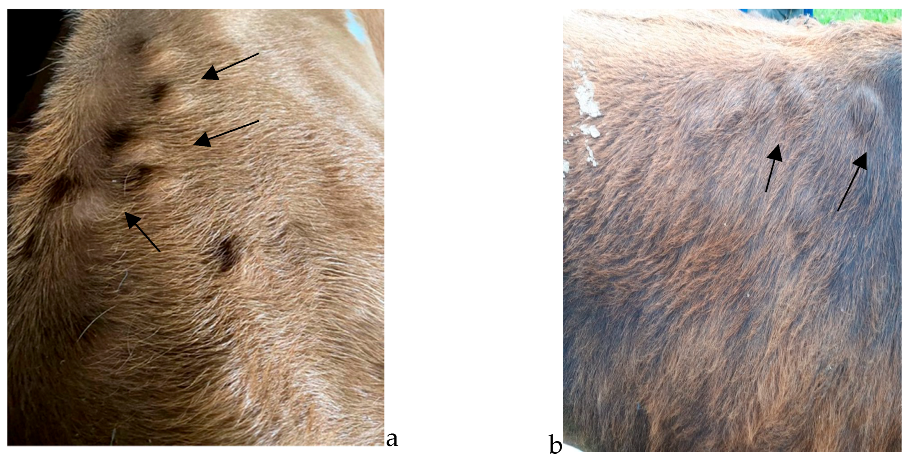
The study was carried out in five localities from the northwestern part of Romania during March, April, May, and June 2021.
The total number of studied animals was 11.741, including cattle belonging to two indigenous breeds (Brună de Maramureș and Bălțată Românească), as well as mixed-breed animals.
The nodules (Figure 1 and Figure 2) were identified visually, followed by a palpatory confirmation of their presence on the body of the examined animals. Data regarding age, breed, and monthly numeric evolution of nodules were recorded for the animals found clinically positive for the presence of Hypoderma spp.
Samples consisting of stage three Hypoderma spp. larvae were collected from the positive animals and prepared for further evaluation. Thus, they were fixed by immersion in 70% ethanol and examined with the aid of an optic microscope in order to properly observe the morphological characteristics unique to each species.
Subsequently, molecular analysis was employed as the method for species identification. The DNA extraction step was performed using the ISOLATE II Genomic DNA Kit (BIOLINE®), following protocol for tissues, in accordance with the manufacturer’s recommendations.
The PCR reaction was performed according to the technique described by Otranto et al., 2003 [15], with some minor changes. Amplification itself was carried out by classic PCR and was based on amplifying a partial DNA fragment of the COI gene with a size of ~579 bp for Hypoderma spp.
The primers used were UEA7, 5′TACAGTTGGAATAGACGTTGATAC-3 and the reverse primer UEA10, 5′ TCCAATGCACTAATCTGCCATATTA-3′ [15]. Amplification was carried out according to the protocol described in the article and modified according to the requirements of the mixture. MyTaqTM Red Mix (BIOLINE®) was used to carry out the reaction. The final volume of the PCR reaction was 25 µL.
The amplification program was carried out with the thermocycler My Cycler (BioRad®, Berkeley, CA, USA). This program included DNA denaturation steps at 95 °C for 1 min; 32 denaturation cycles at 95 °C, time 30 s, hybridization at 60 °C, time 30 s and extension at 72 °C, time 30 s; and incubation at 4 °C.
The analysis and control of the amplicons were carried out by horizontal electrophoresis in a system submerged in 1.5% agarose gel, with the addition of the fluorescent dye MidoriGreen (Nippon Genetics® Europe).
The 100 bp DNA Ladder (Bioline) marker was used in the first well of the gel.
For species confirmation and genotyping, PCR products were sequenced and compared with those available in the GenBank database using BLASTn alignment (http://blast.NCBI.NLM.nih.gov/blast.cgi, accessed on 24 July 2021).

3. Results

3.1. Descriptive Epidemiology

The overall prevalence rate that resulted from our study was 0.31% (n = 21), with values ranging from 0.11% (lowest point) to 1.32% (highest point) (Table 1, Figure 3 and Figure 4).
The age range of the positive animals was 2–6 years; however, a higher percentage of positive cases was recorded in animals aged 1–3 years (Table 2, Figure 5).
Positive cases originated from isolated geographic regions, with traditional farming methods and where previous prevention treatments were not properly implemented.
The very first positive cases were discovered in March however, the most prolific month in terms of case positivity and nodule count (n = 15, the maximum number of nodules counted on an individual) was April (Table 3, Figure 6). The animals were not examined concomitantly (some of them were examined in March, some in April, and others in May and June); thus, there is no way of telling whether the nodules found on the animals examined during April or May, for example, had been there prior to the examination or not. Certain breed predispositions (Table 4 and Table 5, Figure 4) proved statistically significant, namely the predisposition of Brună de Maramureș cattle compared with half-breeds (p < 0.001).

3.2. Molecular Epidemiology

All five samples were classified as H. bovis by molecular identification of Hypoderma spp. larvae 3 (performed by PCR). Following the BLASTn analysis of the sequences obtained from the amplification of the extracted DNA, it was observed that the identified sequences were identical to the isolates: GU984815.1, GU984814.1, KT600279.1. Based on mt-CO1 gene sequence analysis, haplotype HB3 and haplotype HB8 were identified. The results obtained by PCR confirm the results of the morphological identification of the H. bovis species.

4. Discussion

Prevalence rates for warbles vary widely according to region, and the rates have seen a significant drop in numbers in the past years, even up to eradication in some countries such as the UK, where the prevalence rate prior to eradication was reported around 40% in a population of 10 million cattle [15]. The prevalence rate of parasitism with H. bovis resulting from this study is fairly low compared with previously conducted studies, which reported prevalence rates between 32 and 43%, [16,17], 16.21% in Southern Romania [18] or with other countries: up to 85% in Italy [19,20], 67% in Turkey, 23% in Iraq [21], 43% in Belgium, 40% in France, 37.4% in Greece [22], 52.3% in Spain, 16% in Poland, 20% in Switzerland, up to 80% in China [23], and 31.1% in Albania [24]. The prevalence rate recorded during our study was 0.17%. This low rate may be due to easier access and, consequently, more frequent application of control measures (prevention treatments with insect repellents and seasonal treatments with Ivermectin for the treatment and prevention of other internal parasitic diseases). The results of the study show a significant decrease in rates recorded between 2005 (32–43%) [16,17] and 2023 (0.31%), which might outline the possibility of an emerging eradication of this disease. The difference in rates between our study conducted in the northern part of the country and the one conducted by Gorcea et al. [18] in the southern part may also be explained by high differences in temperatures between the two areas, conditions being clearly more favourable for the development of warble flies in the southern part of Romania.
At the end of the 20th century, a hypodermosis control programme was implemented in many European countries which significantly reduced the prevalence of hypodermosis [12]. However, in Poland, after the end of the programme, the prevalence again reached 20% up to 100% in cattle herds [25]. The fact that adults can fly up to 5 km in distance contributes to the spread of this parasite and to the relative importance of parasitological control measures in a given region [26]. In terms of age predisposition, the results of this study are similar to other reports that reveal a higher rate of infection among young animals (over the age of 1 year) [27] compared with older individuals, which develop immunity against warble larvae with ageing. Therefore, age can be a risk factor. Thus, young animals under 1 year of age, being usually kept in shelters and generally not allowed to graze freely, have a lower prevalence [24]. In contrast, animals aged 1 to 3 years are highly susceptible to both H. bovis and H. lineatum. Adult cattle may have partial or even total resistance. Jersey and Holstein breeds are very susceptible to H. bovis parasitism, while the Australian Sahival breed is resistant. Vitamin A deficiency may be a risk factor facilitating skin penetration by larvae I, which has been demonstrated for H. lineatum [16].
The increase in the number of nodules in April, May, and June is probably due to the period when the eggs were laid, i.e., the period when the females carried out their oviposition flight in the previous year. The oviposition periods can be influenced by many factors: the biology of the parasite, species of Hypoderma, climatic factors, age and species of the parasitized animals, possible treatments, insecticides used, etc. Species differentiation is an important tool when it comes to prevention, especially considering the behavioural variations among Hypoderma spp. (for example, H. bovis and H. lineatum differ in terms of animal approach, egg laying and size, migration pattern of larvae inside the affected host, pathogeny, severity of lesions, etc.). According to data presented by Hassan et al. [15], the most common species of Hypoderma are H. bovis and H. lineatum when it comes to domestic cattle. H. bovis is frequently reported in countries from the northern hemisphere, and its pathogeny is of greater importance to farmers, considering the severity of lesions that this infection might induce if treatment is not instituted promptly (when first-stage larvae are in the peri-rachidian channel, in the absence of treatment, they might induce paralysis of the hind quarters) [13]. The results of the morphological identification doubled by PCR assays based on mitochondrial COI sequencing for the identification of H. bovis [28] performed during our study revealed the presence of H. bovis with two haplotypes: HB3 and HB8. Another study based on molecular characterisation, conducted by Otranto et al. [29], revealed similar results. Other authors [30] have reported the presence of H. diana in deer found dead in forests in Romania.

5. Conclusions

The prevalence in northwestern Romania was 0.31%, with variations among the four studied areas ranging from 0.11% to a maximum rate of 1.32%. Warbles is a disease affecting mostly young individuals, especially animals from the 2–3 (p < 0.001) years of age category, while in terms of breed, statistical significance (p < 0.001) was noted between half breeds and Brună de Maramureș. The highest Hypoderma nodule count was noted in April. PCR assays and morphological identification methods revealed the presence of the H. bovis species, with two haplotypes: HB3 and HB8. For a more complete understanding of the epidemiological situation of this disease, we recommend future studies be conducted in other geographical parts of our country in order to update the status of this apparently disappearing disease in cattle from Romania.

Author Contributions

Concept: G.D., V.D.T. and M.S.I.; methodology: N.M., S.M. and M.I.; software: M.S.I.; validation: I.O. and S.M.; resources: V.D.T., T.F., G.D. and M.I.; data curation: G.D., S.M., M.S.I., M.I. and T.F.; writing—original draft preparation: G.D. and V.D.T.; review and editing: M.S.I., T.F., S.M., I.O. and M.I. All authors have read and agreed to the published version of the manuscript.

Funding

The present research was self-funded by the University of Life Sciences “King Mihai I of Romania” in Timisoara.

Institutional Review Board Statement

The study was conducted according to the guidelines of the Declaration of Helsinki and the proper procedures of the University Veterinary Clinics of the Faculty of Veterinary Medicine Timisoara, approved by the Ethics Committee protocol number 34/1.12.2012 of the Romanian Veterinary College.

Informed Consent Statement

Not applicable.

Conflicts of Interest

The authors declare no conflict of interest.

References

  1. Sanchez, J.; Dohoo, I.; Nødtvedt, A.; Keefe, G.; Markham, F.; Leslie, K.; Descôteaux, L.; Campbell, J. A longitudinal study of gastrointestinal parasites in Canadian dairy farms. The value of an indirect Ostertagia ostertagi ELISA as a monitoring tool. Vet. Parasitol. 2002, 107, 209–226. [Google Scholar] [CrossRef] [PubMed]
  2. Khan, M.N.; Iqbal, Z.; Sajid, M.S.; Anwar, M.; Needham, G.R.; Hassan, M. Bovine hypodermosis: Prevalence and economic significance in southern Punjab, Pakistan. Vet. Parasitol. 2006, 141, 386–390. [Google Scholar] [CrossRef]
  3. Otranto, D.; Johnson, G.; Syrvud, K.; Yoon, S.; Hunter, J.S.; Rehbein, S. Treatment and control of bovine hypodermosis with ivermectin long-acting injection (IVOMEC GOLD). Parasit. Vectors 2016, 9, 551. [Google Scholar] [CrossRef] [PubMed]
  4. Reina, D.; Hurtado, F.J.; Martinez-Moreno, F.J.; Hernandez-Rodriguez, S.; Navarrete, I. Method for collecting Hypoderma spp. (Diptera: Oestridae) larvae from cattle. J. Med. Entomol. 1998, 35, 327–329. [Google Scholar] [CrossRef] [PubMed]
  5. Hiepe, T.; Ribbeck, R.; Mieth, K.; Garz, I. Results of the systematic state-directed control of hypodermosis in the German Democratic Republic. Angew. Parasitol. 1974, 15, 57–67. [Google Scholar] [PubMed]
  6. Cosoroaba, I. Parazitologie Veterinara; Mirton: Timisoara, Romania, 2000. [Google Scholar]
  7. Darabus, G.; Oprescu, I.; Morariu, S.; Mederle, N.; Ilie, M.; Imre, M. Parazitologie si Boli Parazitare; Mirton: Timisoara, Romania, 2022. [Google Scholar]
  8. Colwell, D.D.; Martinez-Moreno, F.J.; Martinez-Moreno, A.; Hernandez-Rodriguez, S.; de la Fuente-Lopez, C.; Alunda, J.M.; Hall, M.J. Comparative scanning electron microscopy of third-instar Hypoderma spp. (Dipetra: Oestridae). Med. Vet. Entomol. 1998, 12, 181–186. [Google Scholar] [CrossRef]
  9. Otranto, D.; Traversa, D.; Guida, B.; Tarsitano, E.; Fiorente, P.; Stevens, J.R. Molecular characterization of the mitochondrial cytochrome oxidase I gene of oestridae larvae causing obligate myiasis. Med. Vet. Entomol. 2003, 17, 307–315. [Google Scholar] [CrossRef] [PubMed]
  10. Wiszniewska-Łaszczych, A.; Wysok, B.; Wojtacka, J.; Sołtysiuk, M. Effect of Mixed Invasions of Hypoderma bovis and Ostertagia ostertagi in Cattle on Milk Yield and Contents in Polish Dairy Farms. Animals 2021, 11, 464. [Google Scholar] [CrossRef]
  11. Rehbein, S.; Holste, J.E.; Smith, L.L.; Lloyd, J.L. The efficacy of eprinomectin extended-release injection against Hypoderma spp. (Diptera: Oestridae) in cattle. Vet. Parasitol. 2013, 192, 353–358. [Google Scholar] [CrossRef]
  12. Boulard, C. Durably controlling bovine hypodermosis. Vet. Res. 2002, 33, 455–464. [Google Scholar] [CrossRef]
  13. Rappelli, P.; Varcasia, A.; Vargiu, A.; Scala, A. Case report: First report of autochthonous Human Cutaneous Myasis caused by Hypoderma lineatum in Europe. Am. J. Trop Med. Hyg. 2018, 99, 618–619. [Google Scholar] [CrossRef] [PubMed]
  14. Puente, S.M.D.; Otranto, D.; Panadero, R.; Herrero, M.D.; Rivas, P.; Ramirez-Olivencia, G.; Mariscal, C.; Maria, J.R.; Perteguer, J.; Diez Banos, P.; et al. First diagnosis of an imported Human Myasis caused by Hypoderma sinense (Diptera: Oestridae), detected in a European Traveler returning from India. J. Trav. Med. 2010, 6, 419–423. [Google Scholar] [CrossRef] [PubMed]
  15. Hassan, M.U.; Khan, M.N.; Abubakar, M.; Waheed, H.M.; Iqbal, Z.; Hussain, M. Bovine hypodermosis—A global aspect. Trop. Anim. Health Prod. 2010, 42, 1615–1625. [Google Scholar] [CrossRef] [PubMed]
  16. Cozma, V.; Suteu, E. Epidemiology and Aetiology of Bovine Hypodermosis in Northwestern Romania. In Improvements in the Control Methods for Warble-Fly in Cattle and Goats; Tarry, D.W., Pithan, K., Webster, K., Eds.; Commission of the European Communities: Brussels, Belgium, 1995; pp. 65–68. [Google Scholar]
  17. Oprescu, I.; Dărăbuş, G.; Tântă, M. Evoluția hipodermozei bovine în județul Maramureş în perioada 2000–2003. Sci. Parasitol. 2005, 1–2, 132–135. [Google Scholar]
  18. Gorcea, F.C.; Calescu, N.; Gherman, C.M.; Mihalca, A.D.; Cozma, V. Diagnostic values of clinical, pathological and serologic findings in cattle hypodermosis in Pestisani, Gorj County, Romania. Sci. Parasitol. 2011, 12, 173–176. [Google Scholar]
  19. Puccini, V.; Giangaspero, A.; Fasanella, A. Incidence of bovine hypodermosis in Apulia region (Italy). In Improvements in the Control Methods for Warble-Fly in Cattle and Goats; Hernandez, S., Gasca, A., Martinez, J., Pithan, K., Eds.; Commission of the European Communities: Brussels, Belgium, 1991; pp. 9–20. [Google Scholar]
  20. Frangipane di Regalbono, A.; Capelli, G.; Otranto, D.; Pietrobelli, M. Assessment of cattle grub (Hypoderma spp.) prevalence in northeastern Italy: An immunoepidemiological survey on bulk milk samples using ELISA. Vet. Parasitol. 2003, 111, 343–350. [Google Scholar] [CrossRef]
  21. Abdul-Hak, J. Seasonal occurrence of Hypoderma spp. (Diptera: Oestridae) warble flies on cattle in Baghdad area. Iraq Bull. Endemic. Dis. 1973, 14, 73–81. [Google Scholar]
  22. Papadopoulos, E. Hypodermosis in Greece. Chin. J. Vet. Parasitol. 2004, 12, 20–23. [Google Scholar]
  23. Yin, H.; Miling, H.; Gailing, H.; Haung, S.; Zheijie, L.; Jiaxutn, L.; Guiquan, G. Hypodermosis in China. In Proceedings of the INCO Conference, Athens, Greece, 9–10 June 2010. [Google Scholar]
  24. Otranto, D.; Zalla, P.; Testini, G.; Zanaj, S. Cattle grub infestation by Hypoderm asp. in Albania and risks for European countries. Vet. Parasitol. 2005, 128, 157–162. [Google Scholar] [CrossRef]
  25. Cencek, T.; Ziomko, I. Development of the ELISA kit for the detection of Hypoderma bovis antibodies in cattle. II. Setting the best terms for collecting blood samples for the lavoratory diagnostic of hypodermyiasis in different regions of Poland. Wiad Parazytol. 2001, 47, 505–510. [Google Scholar]
  26. Beesley, W.N. Economics and progress of warble fly eradication in Britain. Vet. Med. Rev. 1974, 4, 334–347. [Google Scholar]
  27. Benakhla, A.; Lonneux, J.F.; Mekroud, A.; Losson, B.; Boulard, C. Hypodermose bovine dans le Nord-Est algérien: Prévalence et intensité d’infestation [Bovine hypodermosis in north-eastern Algeria: Prevalence and intensity of infestation]. Vet. Res. 1999, 30, 539–545. [Google Scholar]
  28. Fu, Y.; Li, W.; Duo, H.; Guo, Z.H.; Li, Y.; Zhang, Y.M. Genetic diversity and population genetics of the warble flies Hypoderma bovis and H. sinense in Qinghai Province, China. Parasit. Vectors 2016, 9, 145. [Google Scholar] [CrossRef]
  29. Otranto, D.; Traversa, D.; Colwell, D.D.; Guan, G.; Giangaspero, A.; Boulard, C.; Yin, H. A third species of Hypoderma (Diptera: Oestridae) affecting cattle and yaks in China: Molecular and morphological evidence. J. Parasitol. 2004, 90, 958–965. [Google Scholar] [CrossRef]
  30. Ilie, M.S.; Imre, M.I.; Hotea, I.O.; Imre, K.; Sorescu, I.D.; Andrei, S.; Onita, P.; Oprescu, I.; Morariu, S.; Mihali, C.; et al. Prevalence of Hypoderma infestation in deer in western Romania. Lucr. St. Med. Vet. 2012, XIV, 121–125. [Google Scholar]
Figure 1. (a,b)—Hypoderma spp. nodules in the dorsal area of indigenous-breed (Bălțată Românească) cattle (see arrows).
Pathogens 12 01077 g001
Figure 2. (a,b)—Multiple Hypoderma spp. nodules in the dorsal area of indigenous-breed (Brună de Maramureș) cattle (see arrows).
Pathogens 12 01077 g002
Figure 3. Graphical representation of positive cases according to the sampling location. BR = Bălțată Românescă; BM—Brună de Maramureș; MB—Mixed Breed.
Pathogens 12 01077 g003
Figure 4. Distribution of positive cases according to breed. BR = Bălțată Românescă; BM—Brună de Maramureș; MB—Mixed Breed.
Pathogens 12 01077 g004
Figure 5. Distribution of positive cases according to age.
Pathogens 12 01077 g005
Figure 6. Monthly variation of Hypoderma nodule count.
Pathogens 12 01077 g006
Table 1. Prevalence of parasitism with H. bovis in Northwestern Romania.
LocationNo. of Examined
Animals
No. of Positive
Animals (%)
Breeds
BRBMMB
Moisei26675 (0.18)140
Vișeu32158 (0.24)251
Borșa44205 (0.11)131
Săcel143919(1.32)2170
TOTAL11,74137 (0.31)6292
BR = Bălțată Românescă; BM—Brună de Maramureș; MB—Mixed Breed.
Table 2. Prevalence of parasitism with H. bovis according to the age group.
BreedNo. of Examined
Animals
No. of Positive
Animals (%)
Age Group (Positive/Total No. of Animals)
2–3 Years4–5 Years6–7 Years8–10 Years
BR28436 (0.21)5/3790/7931/6320/1039
BM632029 (0.45)18/84710/17601/11080/2605
MB25782 (0.07)1/3221/6370/7010/918
TOTAL11,74137 (0.31)24/154811/31902/24410/4562
Table 3. Identification of hypodermic nodules according to month.
LocationNo. of Positive AnimalsTotal No. of Hypodermic Nodules
March (Max–Min)April
(Max–Min)
May
(Max–Min)
June
(Max–Min)
Moisei517 (2–5)30 (4–8)19 (1–6)11 (1–4)
Vișeu821 (1–5)70 (4–15)47 (2–12)25 (2–7)
Borșa515 (2–4)48 (6–14)25 (3–7)20 (1–5)
Săcel199 (2–4)23 (5–10)18 (4–10)106 (1–11)
Total3762 (1–5)171 (4–15)109 (1–12)162 (1–11)
Table 4. Statistical significance of comparisons between age groups and breeds.
AGE
2–3vs.4–50.0001
2–36–70.0001
2–38–100.0001
4–56–70.0500
4–58–100.0001
6–78–100.1216
BREED
BR BM0.0979
BR HB0.2935
BM HB0.0044
Table 5. Statistical significance of comparisons between monthly nodule counts in the studied cases.
March (Max–Min)April (Max–Min)p value equals 0.0450
By conventional criteria, this difference is considered to be statistically significant.
17 (2–5)30 (4–8)
21 (1–5)70 (4–15)
15 (2–4)48 (6–14)
9 (2–4)23 (5–10)
March (Max–Min)May (Max–Min)p value equals 0.1543
By conventional criteria, this difference is considered to be not statistically significant.
17 (2–5)19 (1–6)
21 (1–5)47 (2–12)
15 (2–4)25 (3–7)
9 (2–4)18 (4–10)
March (Max–Min)June (Max–Min)p value equals 0.3025
By conventional criteria, this difference is considered to be not statistically significant.
17 (2–5)11 (1–4)
21 (1–5)25 (2–7)
15 (2–4)20 (1–5)
9 (2–4)106 (1–11)
April (Max–Min)May (Max–Min)p value equals 0.2609
By conventional criteria, this difference is considered to be not statistically significant.
30 (4–8)19 (1–6)
70 (4–15)47 (2–12)
48 (6–14)25 (3–7)
23 (5–10)18 (4–10)
April (Max–Min)June (Max–Min)p value equals 0.9295
By conventional criteria, this difference is considered to be not statistically significant.
30 (4–8)11 (1–4)
70 (4–15)25 (2–7)
48 (6–14)20 (1–5)
23 (5–10)106 (1–11)
May (Max–Min)June (Max–Min)p value equals 0.5861
By conventional criteria, this difference is considered to be not statistically significant.
19 (1–6)11 (1–4)
47 (2–12)25 (2–7)
25 (3–7)20 (1–5)
18 (4–10)106 (1–11)
Disclaimer/Publisher’s Note: The statements, opinions and data contained in all publications are solely those of the individual author(s) and contributor(s) and not of MDPI and/or the editor(s). MDPI and/or the editor(s) disclaim responsibility for any injury to people or property resulting from any ideas, methods, instructions or products referred to in the content.

